Microstructure: Moving Through Time
Oral
Diffusion
Wednesday, 20 June 2018
| N01 |
16:15 - 18:15 |
Moderators: Dmitry Novikov, Ileana Jelescu |
16:15
|
0883.
 |
 Including diffusion frequency dependence in the extra-axonal space to improve axonal diameter mapping using trapezoidal OGSE sequences Including diffusion frequency dependence in the extra-axonal space to improve axonal diameter mapping using trapezoidal OGSE sequences
Kevin GINSBURGER, Fabrice POUPON, Felix MATUSCHKE, Jean-François MANGIN, Markus AXER, Cyril POUPON
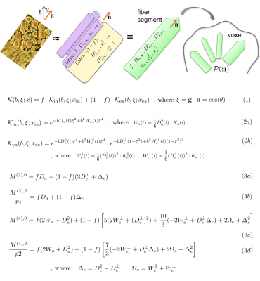
We performed Monte-Carlo simulations of the diffusion process in 3D biomimetic geometries accounting for the angular dispersion and tortuosity present in white matter. Diffusion MRI data synthesis using clinically plausible trapezoidal OGSE sequences was then performed and an extra-axonal linear-in-frequency dependence of the perpendicular diffusivity transverse to axons was observed over a wide range of axon diameter values. Simulated data was fed into an ActiveAx trapezoidal OGSE model accounting for this frequency-dependence. The estimation of axonal diameter is improved by this correction.
|
16:27
|
0884.
 |
LEMONADE(t): Exact relation of time-dependent diffusion signal moments to neuronal microstructure
Hong-Hsi Lee, Els Fieremans, Dmitry Novikov
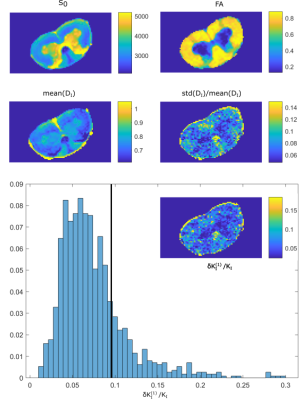
The purpose of this work is to solve the two complementary problems: (i) Estimate the time-dependent (non-Gaussian) signatures of intra- and extra-neurite tissue compartments separately, irrespective of neurite orientational dispersion; and (ii) cure the notorious degeneracy in parameter estimation of multi-compartment models. We derive exact relations between time-dependent 2nd- and 4th-order signal moments, and all relevant time-dependent diffusion parameters in the intra- and extra-neurite space. Remarkably, the temporal dimension lifts the degeneracy in parameter estimation: The relations are formally invertible, multiple "branches" of solutions do not appear, and time-dependent DKI is enough to estimate all parameters. Precision, though, remains a challenge.
|
16:39
|
0885.
 |
Diffusion Time Dependence of NODDI in in vivo Human White Matter
Masaaki Hori, Kouhei Kamiya, Katsutoshi Murata, Thorsten Feiweier, Issei Fukunaga, Akifumi Hagiwara, Ryusuke Irie, Christina Andica, Tomoko Maekawa, Saori Koshino, Koji Kamagata, Kanako Kumamaru, Michimasa Suzuki, Akihiko Wada, Shigeki Aoki
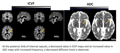
We investigated the diffusion time dependency of diffusion metrics, especially ICVF and INVF, compared with ADC using an oscillating gradient spin-echo sequence in in vivo human white matter at 3T. Our results show that the change ratio of both ICVF and INVF indicate similar tendency in the same white matter locations, with or without diffusivity calculation. Moreover, with higher oscillating frequency, ICVF decreased but ISOVF stayed almost unchanged, indicating that the rate of ICVF and the extra-cellular component may change with oscillating frequency beside the unchanged isotropic water component.
|
16:51
|
0886.
 |
Implications of nongaussian diffusion on the interpretation of multidimensional diffusion measurements
Sune Jespersen, Jonas Olesen, Andrada Ianus, Noam Shemesh
Multidimensional diffusion weighting, such as magic angle spinning of the q-vector (q-MAS), relies on the assumption of multiple Gaussian compartments (MGC). Then the kurtosis measured with q-MAS can be fully ascribed to ensemble variance of isotropic diffusivity. However, in compartments with nongaussian diffusion, anisotropic time dependence of the diffusion tensor imparts orientation dependence on the q-MAS measured mean diffusivity, which in the presence of orientation dispersion leads to additional contributions to kurtosis. Yet another contribution arises from intracompartmental kurtosis. Using simulations and experiments we demonstrate that q-MAS derived diffusion kurtosis conflates variance in isotropic diffusivity with dispersion and intracompartmental kurtosis.
|
17:03
|
0887.
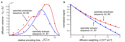 |
Spectral anisotropy in multidimensional diffusion encoding
Henrik Lundell, Markus Nilsson, Carl-Fredrik Westin, Daniel Topgaard, Samo Lasic
To account for time-dependent diffusion in multidimensional diffusion encoding (MDE), the relevant temporal characteristics of encoding waveforms need to be identified and controlled. Based on the frequency domain analysis, we suggest a framework for analyzing the spectral content in MDE, which is useful to experimentally disentangle the effects of time-dependent diffusion. We introduce a novel concept of spectral anisotropy and demonstrate how differentiated temporal characteristics along orthogonal encoding axes may be used to isolate time-dependent diffusion in anisotropic domains, which is not possible with existing approaches without a priori model assumption.
|
17:15
|
0888.
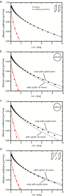 |
On the vanishing of the t-term in the short-time expansion of the diffusion coefficient for oscillating gradients in diffusion NMR
Frederik Laun, Kerstin Demberg, Michael Uder, Armin Nagel, Tristan Kuder
The short-time expansion of the diffusion coefficient in powers of t1/2 is universally connected to structural parameters of the boundaries restricting the diffusive motion. The t1/2-term is proportional to the surface-to-volume ratio. The t-term is related to permeability and curvature. The short-time expansion can be measured by application of oscillating gradients of long total duration. For oscillating gradients, the inverse of the oscillation frequency becomes the relevant time scale. The purpose of this work is to show that the oscillating gradient approach is blind to the t-term. Thus, the t-term does not bias the determination of the t1/2-term in experiments.
|
17:27
|
0889.
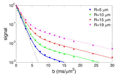 |
Towards new modalities for probing the microstructure at very high b-values
Denis Grebenkov
Modern clinical and animal scanners offer versatile options for diffusion MRI, including very high b-values, at which the conventional perturbative description and related model are not applicable. We demonstrate this failure for any nontrivial domain with a boundary, regardless its shape and permeability. Since the magnetization is localized near boundaries, the signal becomes more sensitive to the microstructure. Even the signal from the extracellular diffusion is shown to be non-Gaussian at high b-values. These non-Gaussian features offer new imaging modalities at ordinary gradients accessible on most MRI scanners but, if unnoticed, they may result in misleading biomedical interpretations.
|
17:39
|
0890.
 |
Imaging Microscopic Background Gradient Variations in the Mouse Brain
Video Permission Withheld
Choong Lee, Piotr Walczak, Lindsay Hill, Youssef Wadghiri, Jiangyang Zhang
Microstructures in biological tissues can produce susceptibility related microscopic background gradient (μBG). Mapping the spatial distributions of μBG can help us infer tissue microstructure. In this study, we reported an improved diffusion MRI based method to detect μBG and evaluated the method using a phantom as well as normal and dysmyelinated mouse brains. We found that 3D spatial variations of μBG were greater in white matter tracts than in gray matter structures. The contrast based on μBG spatial variations was able to distinguish white matter tracts in normal and dysmyelinated mouse brains. The method may be used to study myelin injury.
|
17:51
 |
0891.
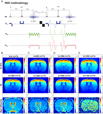 |
In-vivo diffusion-fMRI using Incomplete Initial Nutation Diffusion Imaging (INDI)
Daniel Nunes, Andrada Ianus, Noam Shemesh
Diffusion functional-MRI (dfMRI) aims to capture microstructural changes occurring with neural activity. One of the confounding factors for dfMRI is that zero and nonzero b-values need to be acquired to deliver accurate changes in diffusivity, and those images are typically separated by at least one repetition time. Recently, Incomplete initial Nutation Diffusion Imaging (IDNI) was proposed for mapping the two images with a separation of <50ms. Here, we performed the first INDI-fMRI experiments using forepaw-stimulated rats. Functional signals are captured well both in ROI and voxel-wise analyses, enabling a more direct comparison of each signal’s time course.
|
18:03
|
0892.
 |
Abundance of cell bodies can explain the stick model’s failure in grey matter at high b-value
Video Permission Withheld
Marco Palombo, Noam Shemesh, Andrada Ianus, Daniel Alexander, Hui Zhang
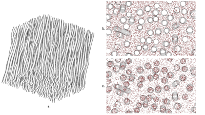
This work investigates the validity of the stick model used in diffusion-weighted MRI for modelling cellular projections in brain tissue. We hypothesize that the model will fail to describe the signals from grey matter due to an abundance of cell bodies. Using high b-value (≥3 ms/µm2) data from rat and human brain, we show that the assumption fails for grey matter. Using diffusion simulation in realistic digital models of neurons/glia, we demonstrate the breakdown of the assumption can be explained by the presence of cell bodies. Our findings suggest that high b-value data may be used to probe cell bodies.
|
|
