Cutting-Edge Multimodal fMRI Techniques
Oral
fMRI
Thursday, 21 June 2018
| N02 |
08:00 - 10:00 |
Moderators: Noam Shemesh, Alex Tze Lun Leong |
08:00
 |
1007.
 |
Simultaneous fMRI and multispectral fiber-photometry reveals neurovascular coupling during somatosensory, optogenetic, and chemogenetic stimulation of S1 principle neurons
Video Permission Withheld
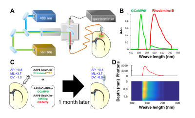
Tzu-Hao Chao, Weiting Zhang, Brittany Katz, Esteban Oyarzabal, Sung-Ho Lee, Guohong Cui, Yen-Yu Shih
In this study, we establish an experimental platform to simultaneously measure: a) genetically encoded calcium indicators (GCaMP) expressing on the excitatory neurons using fiber-photometry, b) cerebral blood volume (CBV) using fiber-photometry, and c) CBV using fMRI. By this platform, we assess neurovascular coupling (GCaMP and CBV comparisons) under chemogenetic stimulation of S1 excitatory neurons in a group of freely moving rats, and demonstrate this platform for concurrent GCaMP, photometry-CBV, and fMRI-CBV measurements with chemogenetic manipulation.
|
08:12
|
1008.
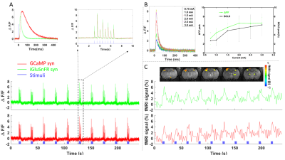 |
 Fiber optic mediated extracellular glutamate and intracellular calcium recording with simultaneous fMRI Fiber optic mediated extracellular glutamate and intracellular calcium recording with simultaneous fMRI
Yuanyuan Jiang, Xuming Chen, Xin Yu
Genetically encoded fluorescent reporter iGluSnFR for extracellular glutamate (Glu) sensing and genetically encoded calcium indicator GCaMP6f for calcium sensing are applied with two channel fiber optic recording system in combination with BOLD fMRI. The intracellular calcium signal from both neurons and astrocytes, as well as the extracellular glutamate signal, were recorded with concurrent BOLD fMRI signal from both hemispheres of anesthetized rats, showing unique temporal dynamic pattern. This multi-modal fMRI platform allows us to specify the neurovascular signaling through the neuro-glial-vascular network and provide better understanding on the cellular and molecular interaction underlying the BOLD fMRI signal.
|
08:24
 |
1009.
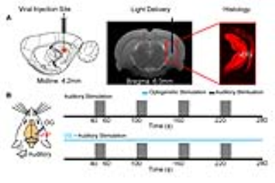 |
Does ventral hippocampus influence auditory processing? An optogenetic fMRI study
Eddie Wong, Russell Chan, Alex Leong, Celia Dong, Karim El Hallaoui, Anthea To, Ed Wu
Hippocampus has traditionally been associated to memory, navigation and emotional behaviors. However, little is known about its influence on processing auditory sensory information. In this study, we combine optogenetic stimulation and auditory fMRI to investigate the influence of the ventral hippocampus on auditory processing across the auditory pathway, including inferior colliculus, medial geniculate body and primary auditory cortex. Our results reveal for the first time the influence of the ventral hippocampus on auditory processing of behaviorally relevant sound at auditory midbrain, thalamus and cortex.
|
08:36
|
1010.
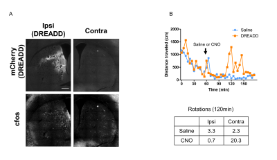 |
DREADD-fMRI combination detects effects of activation of the striatal D1 receptor-expressing neurons on the basal ganglia-thalamocortical network in mice
Yuki Nakamura, Assunta Pelosi, Boucif Djemai, Denis Herve, Jean-Antoine Girault, Tomokazu Tsurugizawa
The dorsal striatum is a key region in motor behavior and motor disorders such as Parkinson’s and Huntington's diseases. We expressed designer receptors exclusively activated by designer drugs (DREADD) in D1 dopamine receptor-expressing neurons in the dorsal striatum in mice and investigated the behavioral and BOLD signal changes during the neuronal activation by clozapine-N-oxide (CNO) treatment. The increased BOLD signals with no susceptibility artifact in the dorsal striatum and the correlated BOLD signal changes in several regions including the substantia nigra and the globus pallidus were observed after CNO injection.
|
08:48
|
1011.
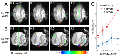 |
A novel method for mesoscale connectome mapping: focal infrared neural stimulation in high-field functional MRI
Augix Xu, Meizhen Qian, Zheming Li, Pei Li, Jianbao Wang, Yang Gao, Yi Sun, Peng Li, Xuemei Song, Xiaotong Zhang, Anna Roe
Establishing connection patterns between cortical columns is essential for understanding human brain networks. However, currently, there is no method to systematically map at this sub-millimeter scale. Here, we combined pulsed infrared neural stimulation (INS) with high field fMRI. Applying this method in cat and monkey brains, we found that single site INS stimulation produces reproducible, intensity-dependent activation. Our experiments revealed (1) connections between cortex and subcortical locations, (2) long-range cortico-cortical connections, and (3) local cortical connections. We suggest that INS-fMRI is a new in vivo functional tract tracing technique that can map networks with high spatial resolution.
|
09:00
|
1012.
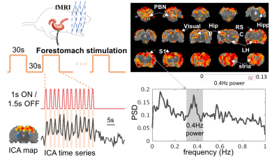 |
Vagus Nerve Stimulation Evokes Widespread BOLD Responses in the Rat Brain
Jiayue Cao, Kun-Han Lu, Terry Powley, Zhongming Liu
Vagus nerve stimulation (VNS) is an emerging treatment for brain disorders, such as depression and epilepsy. However, its efficacy varies, and its mechanism is unclear. Prior studies have used functional MRI (fMRI) to map brain activations with VNS in human brains but yielded inconsistent findings. The source of the inconsistency might be attributed to the complex temporal characteristics of VNS-evoked responses that cannot be fully explained by simplified response models. Using a rat model, we aimed to characterize the VNS evoked responses at the level of brain networks without assuming any priori response model. Our results suggest that the repetitive and block-wise stimulation to the vagus nerve induces activations at widespread brain regions. The responses are complex and variable across regions, much beyond what can be described with conventionally assumed HRF.
|
09:12
|
1013.
 |
Focused Ultrasound Induced Opening of the Blood-Brain Barrier Disrupts Inter-Hemispheric Resting State Functional Connectivity in the Rat Brain
Nick Todd, Yongzhi Zhang, Michael Arcaro, Lino Becerra, David Borsook, Margaret Livingstone, Nathan McDannold
Focused ultrasound can be used as a non-invasive method to disrupt the blood-brain barrier in a targeted, localized, and safe manner. This technology allows for targeted delivery of drugs into the brain for treatment and research applications. While FUS-induced BBB opening has been shown to be safe, there is evidence that it modulates neuronal activity and/or vascular hemodynamics. This study uses resting state fMRI data from rats to investigate these effects. We find that FUS BBB opening targeted to the primary somatosensory cortex reduces local functional connectivity in the ipsilateral sensorimotor cortical areas and in the contralateral primary somatosensory cortex.
|
09:24
|
1014.
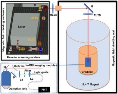 |
Development of Multimodal Neuroimaging Technology at Ultrahigh Field for Studying Brain Function from Cell to Network
Wei Zhu, Meng Cui, Bowen Wei, Yiyong Han, Xiao-Hong Zhu, Kamil Ugurbil, Wei Chen
Functional MRI based on blood oxygenation level dependent (BOLD) contrast has become an indispensable method for mapping effective and functional connectivity. However, subject to intrinsic resolution limit, fMRI cannot be used to directly explore cellular and neurovascular mechanisms underlying BOLD contrast at microscopic scale. To overcome this obstacle, a novel integrated two-photon fluorescence microscopy (TPM)-MRI system at ultrahigh field of 16.4 tesla is proposed and developed. This multimodal neuroimaging tool will enable noninvasive and simultaneous measurement of neurophysiological change and fMRI signal with unprecedented resolution and sensitivity, thus, bridging microscopic neural and vascular dynamics with macroscopic brain networks.
|
09:36
|
1015.
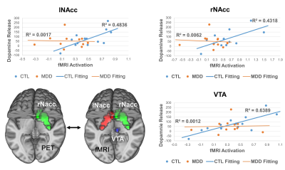 |
Dopamine Release and Neural Activation Differs between Control and MDD during Reward Anticipation: a Simultaneous [11C]Raclopride PET-fMRI Study
Xue Zhang, Fuyixue Wang, J. Paul Hamilton, Jingyuan Chen, Ian Gotlib, Mehdi Khalighi, Gary Glover
The interaction between the midbrain dopaminergic system and the striatum is implicated in reward processing; it is still unknown, however, how this interaction is altered in Major Depressive Disorder (MDD). In the current study, we compared coupling of dopamine release/binding and neural activity in adults diagnosed with MDD and healthy controls (CTLs) during a monetary incentive delay (MID) task using simultaneous $$$[^{11}C]$$$Raclopride PET and fMRI. We obtained significant correlations in CTLs but not in MDD patients, indicating that the decoupling of dopaminergic system and striatum, especially nucleus accumbens, may play a vital role in the pathophysiology of MDD.
|
09:48
|
1016.
 |
Validating the detection of slow BOLD changes with multi-echo fMRI denoised data using simultaneous EEG
Jennifer Evans, Silvina Horovitz, Peter Bandettini, Carlos Zarate, Prantik Kundu
In this study we use simultaneous electroencephalography (EEG) and multi-echo functional magnetic resonance imaging (ME-fMRI) to demonstrate the ability of ME-ICA denoising to resolve slow changes without need for baseline models. We use a visual flickering checkerboard with varying contrast to elicit a response measurable by fMRI and also EEG. We find that the ME-denoised data improves the fMRI timeseries correlation with the ideal task without removing the task signature that is shown to exist in the EEG data.
|
|
