fMRI: Physiology & Neurovascular Coupling
Oral
fMRI
Monday, 18 June 2018
| N03 |
08:15 - 10:15 |
Moderators: Kai-Hsiang Chuang, Ian Driver |
08:15
|
0046.
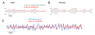 |
 Covariation of pulse oximetry amplitude and BOLD fMRI across vigilance states Covariation of pulse oximetry amplitude and BOLD fMRI across vigilance states
Catie Chang, Pinar Ozbay, Jacco de Zwart, Dante Picchioni, Miranda Chappel-Farley, Hendrik Mandelkow, Jeff Duyn
While pulse oximetry (PO) is often used with fMRI to provide the timing of heart beats, few studies have focused on fluctuations in the amplitude of PO waveform in the context of fMRI signals. Here, we examine correlations between spontaneous fMRI signals and PO amplitude (POA) variations, observing a strong dependence on vigilance state. During alertness, POA-fMRI correlations were weaker but encompassed regions comprising the default-mode network. During drowsiness and NREM sleep, POA co-varied extensively with BOLD signals across the brain.
|
08:27
 |
0047.
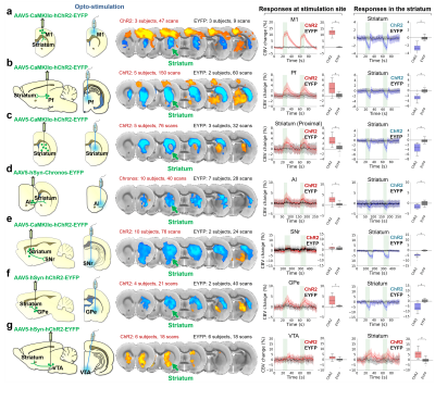 |
Mechanisms underlying negative fMRI response in the striatum
Domenic Cerri, Daniel Albaugh , Brittany Katz, SungHo Lee, Weiting Zhang, Lindsay Walton, Martin MacKinnon, Esteban Oyarzabal, Heather Decot, Nathalie Van Den Berge, Chunxiu Yu, Colleen Mills-Finnerty, Warren Grill, Amit Etkin, Guohong Cui, Garret Stuber, Yen-Yu Ian Shih
Optogenetic stimulation of striatal neurons and several afferents evoke robust negative fMRI responses in the striatum, while striatal electrophysiological recordings during the same stimulations show increases in neuronal activity. Pharmacological manipulations during D1MSN-induced negative striatal responses suggest responses are downstream of MSN activity, but not interneurons or local DA release. Fiber-photometry data from D2MSNs shows a similar pattern of neurovascular uncoupling/negative coupling. This negative fMRI response is also apparent in the human brain. Our results indicate that positive BOLD in the striatum is mediated through DA release, and that negative BOLD in the striatum is induced by local neuronal activations.
|
08:39
|
0048.
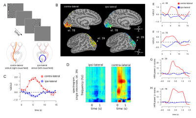 |
Visually evoked negative BOLD signal coupled with silenced neuronal activity; an fMRI and intra-cranial electrocorticography study in humans
Alessio Fracasso, Anna Gaglianese, Serge Dumoulin, Nick Ramsey, Natalia Petridou
Neuroimaging techniques provide a unique window on the study of human brain function in healthy as well as pathological conditions. Intra-cranial electrocorticography (ECoG) at high frequency broadband power is associated with positive blood-oxygenation-level-dependent signal, but the electrophysiological correlates of negative BOLD signals are are less well understood. Here we investigate the relationship between negative BOLD and neuronal population activity measured with ECoG, in humans, using a paradigm that excludes blood stealing as a source of negative BOLD signal.
|
08:51
|
0049.
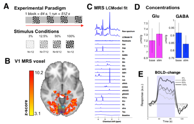 |
Visual contrast levels modulate excitation and inhibition in the human visual cortex – a combined fMRI-MR Spectroscopy study at 7 Tesla
Betina Ip, Uzay Emir, Andrew Parker, Holly Bridge
We used 7 Tesla combined fMRI-MRS to show that simultaneously measured hemodynamic and neurochemical responses in the human visual cortex changed as a function of visual contrast. BOLD change increased linearly to rise in visual contrast, glutamate increased and GABA decreased. Quantification of this change using an excitation:inhibition index demonstrated a switch from inhibitory-dominant to excitatory-dominant neurochemical response. Our results are a step towards disambiguating contributions of cortical excitation and inhibition to stimulus evoked hemodynamic response.
|
09:03
|
0050.
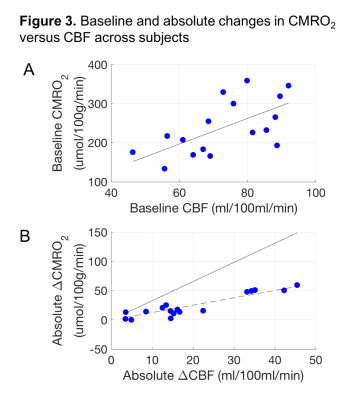 |
The potential for gas-free measurements of absolute oxygen metabolism during both baseline and activation states in the human brain
Eulanca Liu, Jia Guo, Aaron Simon, Frank Haist, David Dubowitz, Richard Buxton
We tested noninvasive methods to measure absolute oxygen metabolism (CMRO2) in both baseline and activation states without the use of special gases: VSEAN to measure baseline O2 extraction fraction (OEF), and FLAIR-GESSE to measure R2’ to estimate the scaling parameter M. Primary findings were: M derived from R2’ had less variation across subjects compared to hypercapnia-derived M; OEF values were in good agreement with previous PET findings; and, variation of baseline CBF/CMRO2 coupling across subjects does not follow activation coupling, suggesting different mechanisms may be involved. These results support the potential of gas-free methods for quantitative physiological measurements.
|
09:15
|
0051.
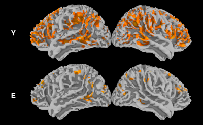 |
The dynamic BOLD-CBF coupling during resting-state in the aging brain: a dual-echo pCASL study
Piero Chiacchiaretta, Antonio Ferretti
Simultaneous acquisition of BOLD and CBF data using ASL allows the study of resting-state brain function from a different perspective with respect to functional connectivity. In particular, recent evidence showed that spontaneous BOLD and CBF fluctuations are strongly coupled in the cortex. Here we show that this coupling is markedly reduced in elderlies with normal cognitive scores, with a regional specific effect in the left supramarginal gyrus, an area known to be involved in different memory functions. These findings suggest that the study of dynamic BOLD-CBF coupling can potentially offer early biomarkers of functional changes in the aging brain.
|
09:27
|
0052.
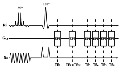 |
Dynamic measurement of oxygen extraction fraction using a Multiple-Offset-Spin-Echo Echo-Planar Imaging (MOSE-EPI) pulse sequence
Video Permission Withheld
Yayan Yin, Yaoyu Zhang, Yang Fan, Bing Wu, Jia-Hong Gao
The development of neuroimaging methods to detect functional oxygen extraction fraction (OEF) is crucial for understanding mechanisms of physiologic function and the underlying neuronal activities. However, dynamic measurement of the OEF is currently limited by the long acquisition time. In this study, a novel pulse sequence using a multiple-offset-spin-echo (MOSE) with an echo-planar imaging (EPI) acquisition scheme was developed to improve the temporal resolution of dynamic OEF measurements. A motor task experiment was performed for ten subjects. The OEF activation maps generated through the proposed technique and compared to traditional blood oxygenation level-dependent (BOLD) activation maps.
|
09:39
|
0053.
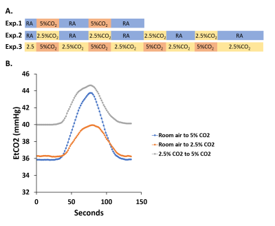 |
Influence of end-tidal CO2 on cerebrovascular reactivity mapping: within-subject and across-subject effects
Jill De Vis, Xirui Hou, Peiying Liu, Zheyu Wang, Siyuan Cheng, Yang Li, Harshan Ravi, Hanzhang Lu
The relationship between end-tidal (Et) CO2, and the output measure, BOLD signal, is highly non-linear, due to both physiological non-linearity between cerebral blood flow (CBF) and CO2 as well as biophysical non-linearity between CBF and BOLD. In this study, we found that, across subjects, baseline EtCO2 and ΔEtCO2 inversely influenced the measured CVR values. Therefore, these factors should be taken into account when comparing CVR between groups or patients. However, these inter-subject differences appear to have an independent physiological underpinning, as manipulations of these factors within the subject did not seem to have an effect on the measured CVR values.
|
09:51
|
0054.
 |
Hemodynamic response altered by focused ultrasound-mediated disruption of the blood-brain barrier
Nick Todd, Yongzhi Zhang, Margaret Livingstone, Lino Becerra, David Borsook, Nathan McDannold
Focused ultrasound (FUS) disruption of the blood-brain barrier (BBB) is a promising technology for achieving targeted delivery of pharmacological agents into the brain. While the method has been shown to be safe from the standpoint of not damaging tissue cells, it causes other changes to local physiology that are not fully understood. This study aims to characterize the effects on the hemodynamic response that FUS BBB opening causes. We present BOLD fMRI data showing the effect and preliminary ASL measurements of cerebral blood flow designed to better understand the effect.
|
10:03
 |
0055.
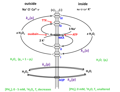 |
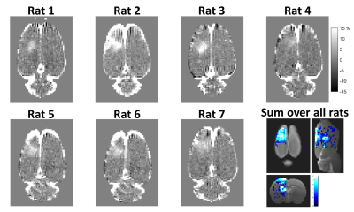
Active, neuronal-activity-dependent trans-membrane water cycling detected by NMR
Ruiliang Bai, Charles Springer, Dietmar Plenz, Peter Basser
Transmembrane water cycling has long been thought an entirely passive, diffusion-dominated process. Recent studies suggest that an energetically active water cycling (AWC) mechanism, driven by Na+-K+-ATPase (NKA) pump activity, also occurs in different cell types, including neurons and astrocytes. Here we hypothesize that monitoring AWC could provide a new, more direct physical indicator of neuronal activity, which involves much ion cycling and enhanced NKA activity. By investigating neuronal populations in vitro under resting conditions with spontanotanous activity, and perturbed with tetrodotoxin (TTX), we found TTX simultanously reduces neuronal spiking activity and AWC (by >63%) suggesting AWC directly reflects neuronal activity.
|
|
