fMRI: Spatiotemporal Dynamics
Oral
fMRI
Monday, 18 June 2018
| N04 |
13:45 - 15:45 |
Moderators: Catie Chang, Laurentius Huber |
13:45
 |
0147.
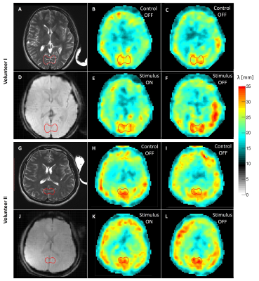 |
 Imaging Primary Neuronal Activity in the Human Optical Cortex at 1.35Hz Imaging Primary Neuronal Activity in the Human Optical Cortex at 1.35Hz
Jose de Arcos, Daniel Fovargue , Katharina Schregel, Radhouene Neji, Samuel Patz, Ralph Sinkus
In this work we have developed a novel functional MRE system for humans capable of probing stiffness changes in the brain driven by monocular visual stimulation. A continuous visual stimulus was applied at an ON/OFF frequency of 1.35 Hz during a segmented 2D multi-slice MRE sequence with 3D motion encoding operating at 50 Hz vibration frequency. Significant stiffness changes were recorded between ON/OFF during the stimulus experiment that also differed in baseline to control scans (OFF/OFF). Since the BOLD signal is entirely saturated at such high stimulation frequencies, we hypothesize that stiffness changes are due to direct neuronal activities. Data match similar results obtained in mice.
|
13:57
|
0148.
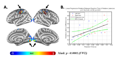 |
Inter-Regional BOLD Latency after Vascular Reactivity Calibration is Correlated to Reaction Time
Yi-Tien Li, Pu-Yeh Wu, Jacky Lu, Ying-Hua Chu, Yi-Cheng Hsu, Fa-Hsuan Lin
Inter-regional BOLD latency between visual and sensimotor cortices were first monitored with fast fMRI (TR=0.1 s) and then calibrated for vascular reactivity using a breath-holding task. Significant delayed response (left: t=4.0, p=0.0019; right: t=6.0, p<0.0001) in the sensorimotor cortex was observed than the visual cortex was detected after removing the vascular confound. Significant correlation between reaction time (428 ± 41ms) and the inter-regional BOLD timing difference (432 ± 149ms) was found within and across subjects.
|
14:09
|
0149.
 |
Global responses to microstimulation at 7T and comparison with vibrotactile stimulation
Ayan Sengupta, Rochelle Ackerley, Roger Watkins, Rosa Sanchez Panchuelo, Paul Glover, Johan Wessberg, Susan Francis
Single unit intra-neural microstimulation (INMS) allows the precise delivery of low-current electrical pulses into human peripheral nerves to stimulate individual afferent nerve fibres. We compare the global pattern of positive and negative BOLD response to INMS with that of perceptually matched vibrotactile stimulation of the skin. INMS and vibrotactile stimulation result in a similar pattern of positive BOLD response, but distinct differences in negative BOLD signals. INMS results in strong negative BOLD response of the DMN, whilst vibrotactile stimulation results in strong ipsilateral negative BOLD response likely to represent active inhibition which is not seen for INMS.
|
14:21
|
0150.
 |
Ultrahigh spatiotemporal-resolution fMRI reveals distinct brain-wide functional networks of different hippocampal subfields
Wei-Tang Chang, Kelly Giovanello, Weili Lin
The hippocampal formation consists of distinct subfields, which contribute to different aspects of memory function, and exhibit different brain network topologies. However, the brain-wide resting-state functional connectivity of hippocampal subfields in human remains poorly understood mainly due to technical limitations. Previous efforts to identify hippocampal subfield functional networks compromised either spatial resolution, spatial coverage or temporal resolution. We have developed a new approach, named Partition-encoded Simultaneous Multi-slab (PRISM), capable of acquiring ultrahigh isotropic resolution images while maintaining the acceleration capability. Our results of resting-state functional connectivity at 7T reveal distinct brain-wide functional networks associated with different hippocampal subfields.
|
14:33
|
0151.
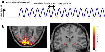 |
High-frequency BOLD responses in human thalamus detected through fast fMRI at 7 Tesla
Laura Lewis, Kawin Setsompop, Bruce Rosen, Jonathan Polimeni
No current technique can noninvasively localize neural activity in human subcortical structures at subsecond temporal resolution. Recent studies have demonstrated that fast (>0.2 Hz) fMRI responses can be detected in human cortex. We aimed to test whether fast fMRI signals can also be detected in the thalamus. We presented oscillating visual stimuli in order to induce oscillatory neural activity in visual thalamus, and observed large-amplitude fMRI oscillations at 0.5 Hz. We conclude that high-frequency fMRI responses can be detected in thalamus, suggesting fast fMRI has the potential to be used for whole-brain imaging.
|
14:45
|
0152.
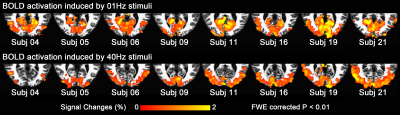 |
Functional organization of visual temporal frequency preference revealed by thalamo-visual correlation
Yuhui Chai, Daniel Handwerker, Sean Marrett, Andrew Hall, Javier Gonzalez-Castillo, Peter Molfese, Peter Bandettini
Thalamo-visual connections play an important role in the visual system. Little is known about the temporal frequency tuning properties of the thalamo-visual correlation in humans. Here we demonstrated that thalamo-visual correlation is significantly modulated by the temporal frequency of a stimulus. Using correlation with thalamus as an index, human visual cortex is organized along a temporal dimension, with the anterior calcarine preferring low temporal frequencies and posterior calcarine preferring higher temporal frequencies.
|
14:57
|
0153.
 |
Probing temporal information in fast-TR fMRI data during attention modulations
Luca Vizioli, Essa Yacoub
The introduction of fast-TRs has allowed for explorations of temporal features in fMRI data. Further, the ability to concurrently retain relatively high degrees of spatial precision as well as large volume coverage, while also maintaining high SNR efficiency, could provide unprecedented axis to the human brain. In this work we explore the possibility of exploiting the temporal specificity of fMRI using a temporal multi-voxel pattern analysis and high temporal resolution 7T fMRI data during attention modulations.
|
15:09
|
0154.
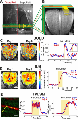 |
Mesoscopic and microscopic imaging of sensory responses in the same animal
Davide Boido, Ravi Rungta, Bruno-Felix Osmanski, Morgane Roche, Tomokazu Tsurugizawa, Denis LeBihan, Luisa Ciobanu, Serge Charpak
We developed a chronic olfactory bulb preparation compatible with repetitive imaging of the same mice with BOLD-fMRI (17.2 T), functional ultrasound imaging (fUS) and two-photon laser scanning microscopy. BOLD-fMRI and fUS mesoscopic signals are highly correlated with microscopic vascular and dendritic neuronal signals in response to odour concentrations. Furthermore, minimal odour stimulation reveals that there is no threshold of neuronal activation below which functional hyperemia is not triggered, warranting measurement of blood flow dynamics to detect the lowest levels of brain activation. These data establish the strengths and limits of mesoscopic imaging techniques to report neural activity.
|
15:21
|
0155.
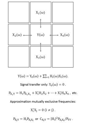 |
A transfer function model for local signal propagation in spatiotemporal MR data
Henning Voss, Jörg Stadler, Jonathan Dyke, Douglas Ballon
In order to understand the sources of dynamic EPI signals under complex stimuli or in the resting state, local signal transfer functions were computed from natural stimulation data sets, and the coherence vector was mapped and color coded analogously to conventional methods for structural connectivity maps. As expected, signal propagation is frequency dependent, whereas high frequencies are caused by cardiovascular pulsations, but low frequencies are less well understood. We conclude that the observed frequency dependence of this local signal transfer model might aid the understanding of the foundation of functional connectivity analysis and the meaning of observed complex patterns such as the resting states.
|
15:33
 |
0156.
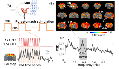 |
Visceral stimulation triggers high-frequency BOLD responses in the rat brain
Jiayue Cao, Kun-Han Lu, Robert Phillips, Terry Powley, Zhongming Liu
Blood oxygen level dependent (BOLD) fMRI reports brain activity by measuring the vascular response to neural activity mediated through neurovascular coupling. Theoretical modeling of neurovascular coupling suggests its effect as a low-pass filter that cuts off at <0.2Hz. However, recent evidence suggests that BOLD fluctuations may also contain high frequency components. From a different perspective to address the origin of high-frequency BOLD signals in the rat brain, we examined the BOLD and local field potential (LFP) responses to visceral stimulation (electrical stimulation of the stomach or the vagus nerve), in comparison with the corresponding responses to commonly used sensory stimulation, such as forepaw stimulation. We report herein that visceral (forestomach and vagal nerve) stimulation can induce high-frequency (up to 0.8Hz) BOLD responses. The neuronal origins of such responses are different from those underlying the responses to forepaw stimulation, and likely modulate hemodynamic fluctuations through a more rapid mechanism of vascular control.
|
|
