MR-Guided Intervention (Not Thermo nor HIFU)
Oral
Interventional MRI
Tuesday, 19 June 2018
| S04 |
16:15 - 18:15 |
Moderators: Rob Tijssen, Graham Wright |
16:15
 |
0598.
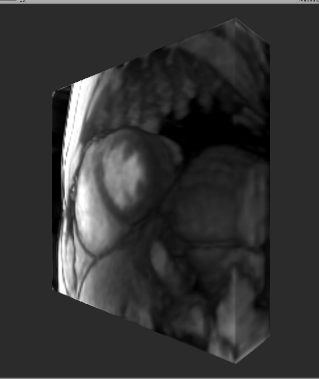 |
 Real-time acquisition, reconstruction, and mixed-reality display system for 2D and 3D cardiac MRI Real-time acquisition, reconstruction, and mixed-reality display system for 2D and 3D cardiac MRI
Dominique Franson, Andrew Dupuis, Vikas Gulani, Mark Griswold, Nicole Seiberlich
Cardiac images suitable for 3D visualization are acquired, reconstructed, and displayed in real-time using the Microsoft HoloLens. This system could be used for guiding cardiac (or other) interventions, or be used to view time-resolved 2D or 3D datasets to facilitate the visualization of anatomical changes through time.
|
16:27
|
0599.
 |
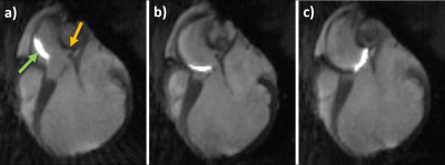
Initial results for MRI-guided catheterization in children and young adults with congenital heart disease using the partial saturation (pSAT) sequence
Mari Nieves Velasco Forte, Sébastien Roujol, Bram Ruijsink, Isra Valverde, Phuoc Duong, Sascha Krueger, Tobias Schaeffter, Steffen Weiss, Surendranath Reddy, Tarique Hussain, Kuberan Pushparajah, Reza Razavi
CMR is a promising alternative to x-ray fluoroscopy for the guidance of cardiac catheterization procedures. We have recently developed a partial saturation (pSAT) sequence which enables passive tracking of balloon-wedge catheters with positive contrast, using a dilution containing gadolinium. 23 patients from 2 different centres were recruited. MRI-guidance was performed using the pSAT sequence applied in the iSuite platform® or an interactive imaging mode. During real-time MRI catheterization the balloon was visualized during 64±19% of the scanning time. pSAT angle was 30-50° in all patients. Mean subjective image quality scores were 3.7 out of 5 for heart visualisation and 4.6/5 for balloon/blood contrast.
|
16:39
|
0600.
 |
MRI-guided right heart catheterization using a commercial nitinol guidewire: Experience in 6 patients
Adrienne Campbell-Washburn, Toby Rogers, Jaffar Khan, Rajiv Ramasawmy, Daniel Herzka, Elena Grant, Delaney McGuirt, Jonathan Mazal, William Schenke, Laurie Grant, Annette Stine, Robert Lederman
We have performed MRI-guided right heart catheterization in patients with a commercial nitinol guidewire for the first time. MRI-guided cardiovascular interventions have been limited by the unavailability of an guidewire that is safe and visible. Here, we use real-time spiral gradient echo imaging to reduce RF-induced heating on a commercial nitinol 150cm Glidewire. The Glidewire was found to generate <0.2°C in the ASTM-2182 phantom in all configurations. With IRB approval, the guidewire catheterization was performed on six patients. Blood biomarkers remained normal and the reported clinical benefits were increased shaft conspicuity, increased shaft stiffness, and ability to track to chambers.
|
16:51
|
0601.
 |
Fully MR-Guided Implantation of a Bioresorbable Scaffold in the Left Coronary Artery of a Pig at 3T
Simon Reiss, Timo Heidt, Thomas Lottner, Ali Özen, Axel Krafft, Klaus Düring, Constantin von zur Mühlen, Michael Bock
MR-guided cardiovascular interventions do not use ionizing radiation, provide excellent soft tissue contrast, and enable functional measurements. The feasibility of MR-guided percutaneous coronary intervention has been demonstrated in animal trials using metallic stents. Recently, bio-resorbable vascular scaffolds have been introduced for stenting of coronary arteries. These scaffolds enable artifact-free MR imaging of the coronary artery segment that the result of the intervention can be readily assessed via MR imaging. We show the first fully MR-guided PCI with a bioresorbable scaffold in the LCA of a pig at 3T using dedicated active guiding catheters, an MR-safe guidewire and BVS delivery system.
|
17:03
|
0602.
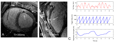 |
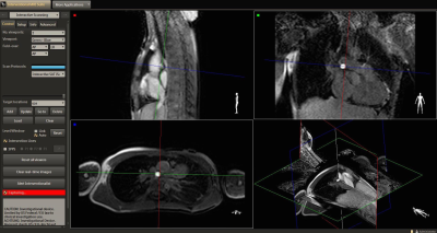
Assessing MR-guided catheter contact with the myocardium using device motion: A feasibility study
Philippa Krahn, Labonny Biswas, Venkat Ramanan, Sebastian Ferguson, Jennifer Barry, Mihaela Pop, Graham Wright
Assessing contact of the catheter tip with the endocardial wall remains challenging during MR-guided electroanatomic voltage mapping (EAVM) of the heart. In this study we investigated whether the synchronicity of catheter motion with the beating heart can serve as an indicator of catheter-endocardium contact. The results from this study show that that synchronicity could potentially provide an indirect measure of endocardial contact and could serve a metric by which quality of EAVM data can be evaluated.
|
17:15
|
0603.
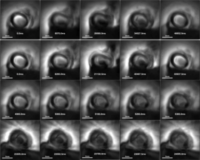 |
Real-time MRI endoscopy at up to 10 frames/sec
Xiaoyang Liu, Parag Karmarkar, Dirk Voit, Jens Frahm, Paul Bottomley
Mininally-invasive intravascular MRI at 3T and above is capable of providing high resolution imaging from within blood vessels and identifying atherosclerosis using miniaturized detectors ~2mm in diameter. Endoscopic MRI is a technique that employs the miniature probe itself to localize the MRI signal to a sensitive disk, and provide images from the view-point of the probe itself. At 3T, acquisition speed has been limited to 2 frames/sec at 300μm resolution, which although fast, is not truly real-time. Here we report a truly real-time MRI endoscope with fully integrated real-time continuous MRI visualization at up to 10 frames/sec.
|
17:27
|
0604.
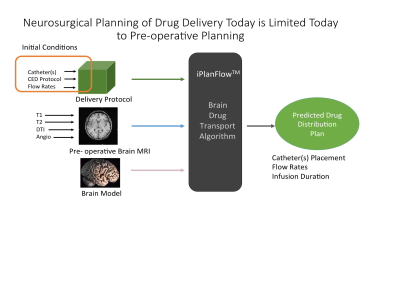 |
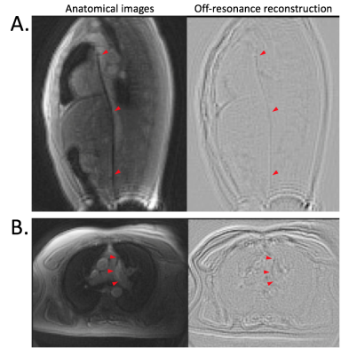
Real-time MR Brain Infusion Monitoring Enables Accurate Prediction of End Drug Distribution
Martin Brady, Raghu Raghavan, Peng Wang, Miles Olsen, Ethan Brodsky, Terrence Oakes, Andrew Alexander, Walter Block
The hetergeneity of the brain makes designing a desired end drug distribution through pressurized catheters difficult. We present a method to utilize real-time MR monitoring of a co-infused Gd tracer during initial stages of the infusion to derive a real-time 3D estimate of the velocity front. We demonstrate considerable improvement in predicting the actual drug distribution using the MR real-time data in four cases using a large animal surgical model.
|
17:39
|
0605.
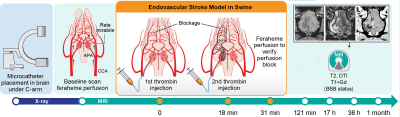 |
Real-time MRI-guided endovascular model of cerebral ischemia in swine
Dominika Golubczyk, Izabela Malysz-Cymborska, Lukasz Kalkowski, Michal Zawadzki, Piotr Holak, Joanna Glodek, Kamila Milewska, Marek Bogacki, Miroslaw Janowski, Zbigniew Adamiak, Wojciech Maksymowicz, Piotr Walczak
Animal models of stroke are essential for developing therapies. Rodent models of stroke are widely used but they lack clinical relevance. Endovascular models in large animals are most desired, but till now they were available in expensive and hard-to-access dogs and primates. Swine is preferred model but till now stroke modeling was through surgical craniotomy, a highly invasive procedure inflicting unrelated morbidity. Endovascular modeling was not possible due to vascular rete preventing catheter access to cerebral vessels. We circumvented this obstacle by intra-arterially injecting SPIO-labeled pro-coagulant thrombin under real-time MRI, which was instrumental to fine-tune injection to occlude cerebral arteries.
|
17:51
|
0606.
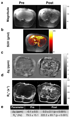 |
PET-MRI Quantification of 89Zr-Iron Oxide Nanoparticles for Targeted Magnetic Drug Therapy
Caroline Jordan, Misung Han, Sravani Kondapavulur, Denis Beckford Vera, Kiel Neumann, Carol Stillson, Teri Moore, Roland Krug, Spencer Behr, Youngho Seo, Henry VanBrocklin, Peder Larson, Mark Wilson, Alastair Martin, Steven Hetts
One recent application of magnetic nanoparticles is to deploy an endovascular magnetic device to selectively remove magnetically-linked chemotherapy during an intra-arterial treatment procedure. In order to measure the device’s efficacy, 89Zr-iron oxide nanoparticles could be imaged using PET-MR. We measured quantitative susceptibility values (Δχ), R2*, and 89Zr-PET uptake of increasing concentrations of 89Zr-IONP in vitro, and in liver in vivo by acquiring a multi-echo UTE GRE sequence and time-of-flight PET. Phantom evaluations demonstrated linear correlation between Δχ, R2*, and 89Zr-IONP uptake. In vivo, substantial increase was observed after 89Zr-IONP infusion. This approach shows promise tracking the biodistribution of radiolabelled magnetic nanoparticles.
|
18:03
|
0607.
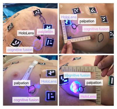 |
Perceptual Accuracy of a Mixed-Reality System for MR-Guided Breast Surgical Planning in the Operating Room
Stephanie Perkins, Michael Lin, Subashini Srinivasan, Amanda Wheeler, Brian Hargreaves, Bruce Daniel
One quarter of women who undergo lumpectomy to treat early-stage breast cancer in the United States undergo repeat surgery due to concerns that residual tumor was left behind. We have developed a supine breast MRI protocol and a system that projects a 3D “hologram” of the MR data onto a patient using the Microsoft HoloLens. The goal is to reduce the number of repeated surgeries by improving surgeons’ ability to determine tumor extent. We are conducting a pilot study in patients with palpable tumors that tests a surgeon’s ability to accurately identify tumor location via mixed-reality visualization during surgical planning.
|
|
