Poster: Neurodegeneration
Electronic Power Pitch Poster
Neuro
09:00 - 10:00
Thursday, 21 June 2018
Power Pitch Theater A - Exhibition Hall
| |
|
Plasma # |
|
0973.
 |
1 |
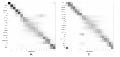
 Data-driven modelling of diffusion MRI changes in Amyotrophic Lateral Sclerosis (ALS) indicates evolution of distal prior to proximal corticospinal tract pathology Data-driven modelling of diffusion MRI changes in Amyotrophic Lateral Sclerosis (ALS) indicates evolution of distal prior to proximal corticospinal tract pathology
Matt Gabel, Stella Tsermentseli, Laura Goldstein, Ammar Al-Chalabi, Alexandra Young, Daniel Alexander, Nigel Leigh, Mara Cercignani
In order to investigate the likely progression of neurodegeneration in amyotrophic lateral sclerosis (ALS) patients, we applied a novel mathematical model to multi-site diffusion MRI data. Our results indicate directional evolution of disease in the corticospinal tracts (CSTs) of ALS patients, in a distal to proximal manner.
|
|
0974.
 |
2 |
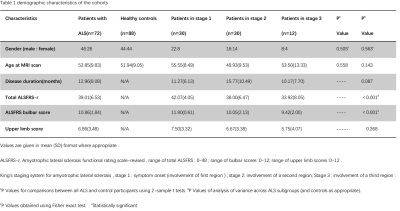
Progressive Cortical Thinning in Specific Motor Regions in Different Clinical Stages of Patients with Amyotrophic Lateral Sclerosis
Haining Li, Qiuli Zhang, Xiao Ling, Guirong Zhang, Ling Yang, Ming Zhang
Staging system for ALS is important for clinical practice. Howerer, the validation of the mechanism that underneath the proposed stages in ALS remains unclear. We used surface-based cortical morphology and more precise anatomical evaluation for 72 patients at different stages and 88 controls, confirmed the consecutive involvement of cortical thinning in PrG along disease progression. Moreover, the extensive but similar PrG involvement in patients at stage 2 and stage 3, highlighted a critical therapeutic window from stage 1 to stage 2, and also underlined the incorporation of cortical evaluation as additional features to King’s clinical stages for promising clinical management.
|
|
0975.
 |
3 |
Increased brain entropy in supplementary motor area and precuneus in amyotrophic lateral sclerosis
Liqin Yang, Yifang Bao, Yuxin Li, Daoying Geng
Amyotrophic Lateral Sclerosis (ALS) is a fatal disease, but no fully validated and clinically specific biomarkers have been identified yet. We studied the brain entropy (BEN) of ALS patients using resting state functional magnetic resonance imaging (rs-fMRI) on fifty-six ALS patients without cognitive impairments and forty-six age- and sex-matched healthy controls. We found increased low frequency entropy in SMA/SMF and increased whole frequency entropy in precuneur/PCC regions in ALS patients. The results may improved our understanding of ALS and provide new biomarkers for diagnosis of ALS.
|
 |
0976.
 |
4 |
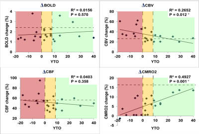
Impaired oxygen metabolism in the brain during visual stimulation in premanifest Huntington's Disease patients detected by 3D-TRIP MRI at 7T
Peter Klinkmueller, Martin Kronenbuerger, Xinyuan Miao, Russell Margolis, Peter van Zijl, Christopher Ross, Jun Hua
Huntington’s disease (HD) is a neurodegenerative disease caused by a single genetic mutation. Neurovascular abnormalities have been implicated in the pathophysiology of HD. Here, dynamic responses in BOLD, cerebral-blood-flow (CBF) and -volume (CBV) during visual stimulation were measured using 3D-TRiple-acquisition-after-Inversion-Preparation (3D-TRIP) MRI in premanifest HD patients and healthy controls, from which cerebral-metabolic-rate-of-oxygen (CMRO2) response was estimated. Decreased ΔCMRO2 and increased ΔCBV were observed in HD patients compared to controls, which correlated with genetic measures. The results suggested potential value of ΔCMRO2 as a biomarker for HD, and may shed light on the pathophysiology in HD in terms of mitochondrial deficiency.
|
|
0977.
|
5 |
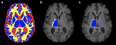
The Impact of Leukoencephalopathy on the White Matter Tracts of Long-Term Survivors of Childhood Acute Lymphoblastic Leukemia Treated with Chemotherapy Only
Noah Sabin, Yin Ting Cheung, Wilburn Reddick, Deepa Bhojwani, Wei Liu, John Glass, Tara Brinkman, Scott Hwang, Deokumar Srivastava, Ching-Hon Pui, Leslie Robison, Melissa Hudson, Kevin Krull
Survivors of acute lymphoblastic leukemia (ALL) can develop neurocognitive deficits and leukoencephalopathy. On-therapy and follow-up MRI examinations of the brain for 173 ALL survivors were reviewed for leukoencephalopathy. At follow-up, the survivors also underwent neurocognitive testing and brain diffusion tensor imaging (DTI). DTI parameters were associated with leukoencephalopathy in multiple regions of the brain. Although there were no associations between neurocognitive performance and leukoencephalopathy, increased mean diffusivity (MD) in certain fiber tracts was associated with neurocognitive impairment. DTI, in particular MD, may better detect loss of white matter integrity associated with neurocognitive deficits in ALL survivors than leukoencephalopathy.
|
|
0978.
 |
6 |
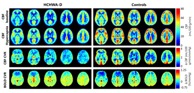
CO2-challenge measured with dual echo arterial spin labeling as a whole brain biomarker to assess the effect of amyloid deposition in HCHWA-D on the cerebrovascular reactivity.
Sophie Schmid, Jasper Verbree, Merlijn van der Plas, Ellis van Etten, Ingeborg Rasing, Pauline Croll, Madeline Redelijkheid, Gerda Labadie, Gisela Terwindt, Marieke Wermer, Mark van Buchem, Matthias van Osch
In this study a CO2-challenge was applied to test the effect of amyloid deposition in HCHWA-D, a hereditary form of cerebral amyloid angiopathy (CAA), on the cerebrovascular reactivity (CVR). With dual echo arterial spin labeling, providing cerebral blood flow (CBF) and BOLD images, the CVR was measured in grey and white matter and the CO2-challenge was evaluated as a whole brain biomarker. The effect of the CO2-challenge was best measured with CBF in the grey matter. Future research should prove whether it is sensitive enough to detect treatment induced changes in (pre-)symptomatic CAA patients.
|
|
0979.
 |
7 |
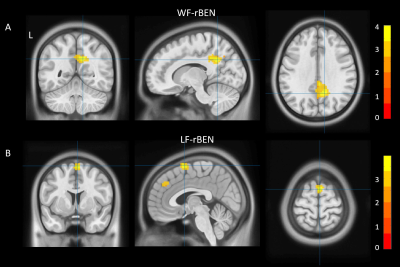
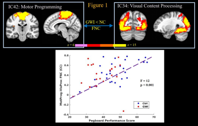
Connectomics Correlates of Neurocognitive Deficits in Gulf War Illness Patients: A Resting State fMRI Study
Kaundinya Gopinath, Unal Sakoglu, Bruce Crosson, Robert Haley
Around 200,000 veterans of the 1991 Gulf War (GW) suffer from GW illness (GWI), which is characterized by multiple deficits in cognitive, emotion, sensory and interoception domains. In this study we examined resting state fMRI data from 23 GWI patients and 30 age-matched controls with group independent components analysis (ICA). Deficits in neurocognitive assessment scores of different brain function domains in GWI veterans strongly correlated with impaired functional connectivity within and between specific brain function networks engaged during performance of the corresponding neuropsychological tests, thereby elucidating brain mechanisms underlying cognitive deficits in GWI.
|
|
0980.
 |
8 |
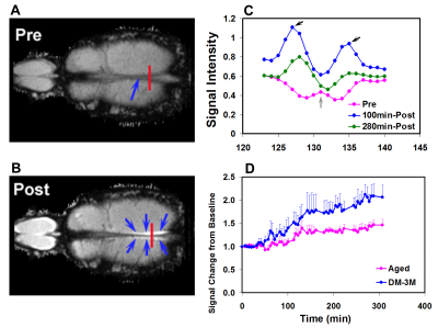
Interaction of vascular and glymphatic systems in brain waste clearance after diabetes
Quan Jiang, Hiani Hu, Guangliang Ding, Esmaeil Davoodi-Bojd, Yimin Shen, Li Zhang, Lian Li, Qingjiang Li, Michael Chopp, Zhenggang Zhang
The recently discovered glymphatic system has become an exciting area of research because of it plays an important role in neurological diseases. However, the interaction between the vascular and the glymphatic systems in terms of waste clearance from the brain is not clear. In addition to the glymphatic system, our preliminary MRI results suggest that the venous system involves in waste removal and waste clearance increases after diabetes. Current study provide the first investigation of the interaction between the vascular and the glymphatic systems in brain waste clearance after diabetes.
|
|
0981.
 |
9 |
Detection of Medication-Induced Changes in Thalamic GABA in Patients with Parkinson’s Disease Using J-Edited Spectroscopy
Paula Trujillo, Ya-Chen Lin, Nelleke van Wouwe, Kalen Petersen, Adam Stark, Nivedita Kukreti, Hakmook Kang, Manus Donahue, Daniel Claassen
A major neurochemical regulator of the thalamocortical network is GABA. We used J-edited MRS with compartment correction to evaluate the pharmacological effect of Dopamine agonist (DAA) therapy on thalamic GABA in patients with Parkinson’s disease (PD). Our findings suggest that DAA alters GABA release in PD patients, and that medication-induced changes may be associated to the presence of impulsive behaviors. These results provide evidence that J-edited MRS can be used to measure subcortical neurotransmitter concentration non-invasively, allowing the investigation of the pharmacological effects in GABAergic activity in humans.
|
|
0982.
 |
10 |
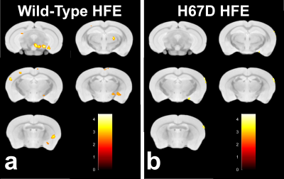
Environmental Paraquat and HFE genetics as factors in the development of Parkinson’s disease
Miranda Salvo, Carson Purnell, Qing Yang, James Connor, Mark Meadowcroft
The herbicide paraquat (PQ) is believed to have a neurotoxic role in the development of Parkinson’s disease (PD) as there is increased risk of exposure in rural areas. The HFEH63D polymorphism has been shown to cause brain abnormalities and disrupt iron homeostasis, key components of PD etiology. This work hypothesized that HFEH67D carrier status would augment PQ induced PD histological and imaging metrics. The R2 and histology data from PQ treated mice provide evidence that PQ had a marked effect in the WT but not HFEH67D mice, suggesting that the H67D polymorphism imparts a neuroprotective role in PD.
|
|
0983.
 |
11 |
Magnetic resonance spectroscopic imaging based biomarkers of Parkinson’s disease with mild cognitive impairment registered to MNI152 brain atlas after chemical shift correction
Sevim Cengiz, Dilek Arslan, Ani Kicik, Emel Erdogdu, Muhammed Yildirim, Zeynep Tufekcioglu, Basar Bilgic, Hasmet Hanagasi, Aziz Ulug, Hakan Gurvit, Tamer Demiralp, Esin Ozturk-Isik
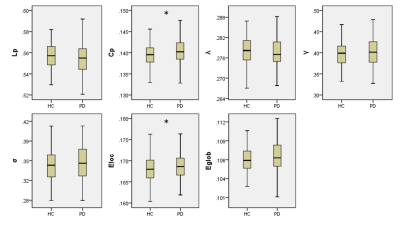
Mild cognitive impairment in Parkinson’s disease (PD-MCI) is one of the most significant risk factors for Parkinson’s disease dementia. In this study, we defined proton magnetic resonance spectroscopic imaging (1H-MRSI) based biomarkers of PD-MCI. After chemical shift misregistration correction and registration to MNI152 brain atlas of multi-voxel 1H-MRSI data, 101 regions defined in MNI structural and Harvard-Oxford cortical and subcortical structural atlases were analyzed for metabolic differences between PD-MCI, cognitively normal PD (PD-CN) and healthy controls. Temporal occipital fusiform cortex, and posterior divisions of parahippocampal gyrus and right temporal fusiform cortex were indicated as the main regions for metabolic differences.
|
|
0984.
 |
12 |
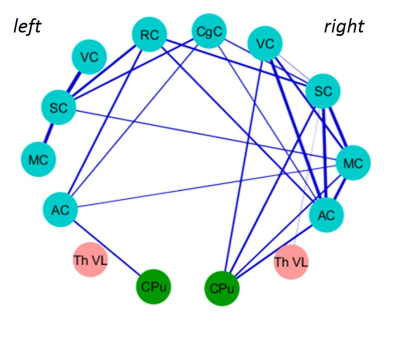
Disrupted Grey Matter Network Morphology in Parkinson's Disease
Xueling Suo, Du Lei, Nannan Li, Lan Cheng, Fuqin Chen, Running Niu, Rong Peng, Qiyong Gong
To use graph theory approaches and high resolution T1-weighted structural magnetic resonance imaging to explore the brain grey matter morphological network in patients with Parkinson's disease (PD). The individual morphological brain networks were constructed by estimating interregional similarity in the distribution of regional grey matter volume of 90 brain regions. The higher clustering coefficient and local efficiency in the PD patients relative to healthy controls were found, indicating that brain morphological networks are closer to regularization, which was different from previous functional connectome studies, suggesting relatively fixed structural network organization can produce diverse functional network patterns.
|
|
0985.
 |
13 |
Altered brain network structure during urethane-induced sleep states in a rat model of early-stage Parkinson’s disease
Ekaterina Zhurakovskaya, Jaakko Paasonen, Juuso Leikas, Aaro Jalkanen, Tiina Pirttimäki, Rubin Aliev, Heikki Tanila, Markus Forsberg, Olli Gröhn
6-hydroxydopamine (6-OHDA) striatal lesion is a well-established rat model of early-stage Parkinson’s disease. The aim of the study was to compare connectivity patterns between 20 6-OHDA lesion rats and 10 sham controls under urethane anesthesia, modelling natural sleep, by using functional magnetic resonance imaging (fMRI). We found that functional connectivity patterns were disturbed in lesion animals. The decrease in functional connectivity, however, occurred only in rapid eye movement (REM)-like state. Furthermore, thalamocortical functional connectivity was correlating with striatal dopamine depletion ratio, making these changes possible early diagnostic markers for Parkinson’s disease.
|
|
0986.
 |
14 |
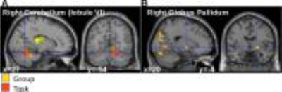
Effect of motor planning and dopaminergic medication on cerebellar network connectivity during dual motor tasking in Parkinson's disease
Silvina Horovitz, David Benninger, Traian Popa, Valerie Voon, Mark Hallett, Cecile Gallea
We investigated cerebellar deficits in dual-motor-tasking in Parkinson’s disease (PD) patients. Eighteen PD patients (scanned ON and OFF dopaminergic medication) and 18 matched controls performed simultaneous finger movements in a coupled or individuated fashion, and with different visual cues at 3T. We showed that cerebello-striatal network interactions play a role in symptomatic dual tasks in PD, and is influenced by dopaminergic medication. Our data suggest that cerebellar-striatal loop is involved in planning fine dexterous tasks without interacting with the cortical motor areas.
|
|
0987.
 |
15 |
Multimodal quantitative MRI biomarkers: identification of the specific damage in Progressive Supranuclear Palsy versus Parkinson’s disease
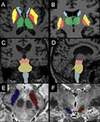
Nadya Pyatigorskaya, Rahul Gaurav, Lydia Yahia-cherif, Claire Ewenczyk, Cecile Gallea, Romain Valabregue, Fatma Gargouri, Eric Bardinet, Isabelle Arnulf, Cyril Poupon, Marie Vidailhet, Stephane Lehericy
We used quantitative multimodal MRI to investigate the region-specific damage in progressive supranuclear palsy (PSP) in order to generate a precise in-vivo model of neurodegeneration at various levels of the central nervous system. Additionally, we aimed to test the markers for differentiating the PSP from Parkinson disease (PD) patients and from healthy subjects. PSP patients showed extensive volume decrease and microstructural diffusion changes in the brainstem and the basal ganglia in agreement with previous pathological studies. These results suggest the possibility of direct non-invasive assessment of brain damage in PSP even in small brainstem nuclei.
|
|
