Poster: Neuro Acquisition: Seeing the CNS Better
Electronic Power Pitch Poster
Neuro
09:15 - 10:15
Tuesday, 19 June 2018
Power Pitch Theater A - Exhibition Hall
| |
|
Plasma # |
|
0312.
 |
1 |
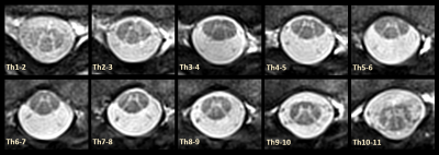
Imaging of the Thoracic Spinal Cord using Radially Sampled Averaged Magnetization Inversion Recovery Acquisitions (rAMIRA)
Video Permission Withheld
Matthias Weigel, Tanja Haas, Oliver Bieri
Particularly for improved imaging of the thoracic spinal cord, a radial sampling variant of the averaged magnetization inversion recovery acquisitions approach was developed (radial AMIRA). The radial AMIRA approach is less prone to fold over artifacts and it is demonstrated that simple pulse triggering removes potential streak artifacts that may occur near big vessels or near the heart. Just as for the conventional Cartesian AMIRA approach, a series of images with remarkable different tissue contrasts is acquired and some of these images can be combined to enhance and fine-tune the desired tissue contrast.
|
|
0313.
 |
2 |
 FLAWS imaging improves depiction of the thalamic subregions for DBS planning in epileptic patients FLAWS imaging improves depiction of the thalamic subregions for DBS planning in epileptic patients
Elise Bannier, Giulio Gambarota, Jean-Christophe Ferré, Tobias Kober, Anca Nica, Stephan Chabardes, Claire Haegelen
Accurate localization of the thalamic subregions is of paramount importance for Deep Brain Stimulation (DBS) planning. Current MRI protocols use T2 and Gadolinium-enhanced T1 images, to visualize both the basal ganglia and the vessels, in order to define the electrode trajectory and target. This study shows the usefulness of Fluid and White Matter Suppression, i.e. FLAWS imaging, in eleven drug-resistant epileptic patients for preoperative Deep Brain Stimulation planning and anterior thalamic nucleus targeting.
|
|
0314.
 |
3 |
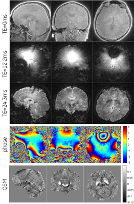
Silent T2* Encoding using ZTE Combined with Gradient-Echo Burst (BURZTE)
Rolf Schulte, Guido Buonincontri, Mauro Costagli, Anne Menini, Florian Wiesinger, Ana Beatriz Solana
ZTE image encoding was combined with burst imaging by reversing segments of 3D radial spokes both in time and amplitude. This recalls gradient echoes for the individual spokes. Multiple burst echoes can be acquired by repeating the trajectories. This “burzte” pulse sequence encodes T2* in a silent manner.
|
|
0315.
 |
4 |
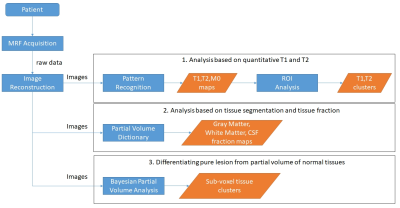
Using 3D high-resolution MR Fingerprinting (MRF) to assist detection and characterization of epileptic lesions
Dan Ma, Irene Wang, Imad Najm, Anagha Deshmane, Debra McGivney, Ken Sakaie, Mark Lowe, Vikas Gulani, Mark Griswold, Stephen Jones
The goal of this study is to develop 3D high resolution MRF scans and partial volume analysis methods to assist detection and characterization of epileptogenic foci in patients with drug refractory epilepsy undergoing presurgical evaluation. In addition to providing quantitative T1/T2 values for tissue characterization, we hypothesize that quantitative maps also provide better sensitivity in detecting subtle epileptic lesions. To this end, high resolution T1 and T2 maps, as well as gray matter, white matter fractions maps and tissue cluster maps, were used to detect and characterize epileptic lesions that were difficult to identify from the weighted images and from conventional voxel-based post-processing analysis based on T1-weighted images.
|
|
0316.
 |
5 |
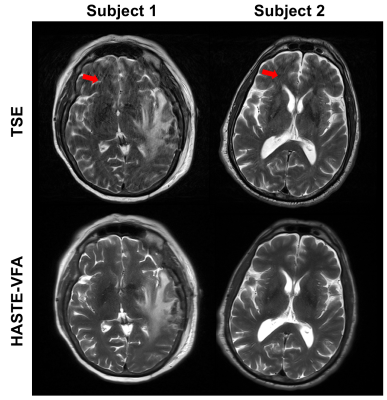
An Optimized Single-shot Sequence for Fast T2w Imaging of the Brain
Mahesh Bharath Keerthivasan, Blair Winegar, Unni Udayasankar, Ali Bilgin, Maria Altbach, Manojkumar Saranathan
T2-weighted imaging of the brain using single shot sequences such as HASTE suffer from reduced spatial resolution, blurring artifacts and decreased conspicuity of small lesions. We present an analytic framework to design the refocusing flip angles for the HASTE sequence tailored for brain imaging. The flip angles are optimized to minimize SAR and blurring, and maximize SNR. The utility of this sequence is demonstrated by incorporating it in a brain tumor protocol and comparing its performance to conventional T2w Turbo Spin Echo in 21 patients.
|
 |
0317.
 |
6 |
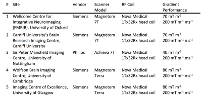
The UK7T Network – optimized design of a multi-site, multi-vendor travelling heads study.
William Clarke, Olivier Mougin, Ian Driver, Catarina Rua, Andrew Morgan, Stuart Clare, Susan Francis, Richard Wise, Adrian Carpenter, Keith Muir, Richard Bowtell
Research sites in the “UK7T Network”, a consortium of 7 tesla capable sites in the UK, have conducted a pilot multi-site, multi-vendor neuroimaging study. The Network aims to create a harmonized set of imaging protocols relevant for clinical research. This study evaluates the differences seen in a set of structural and functional sequences run on three different scanner models, manufactured by two different vendors, at four different sites. In this abstract we identify key differences found between systems and a provide a synopsis of the main findings. The results will inform a future ten subject “travelling heads” study.
|
|
0318.
 |
7 |
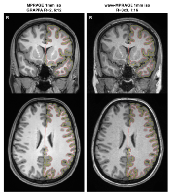
Evaluation of a wave-MPRAGE sequence for brain morphometery
Ross Mair, Jared Nielsen, Randy Buckner
A new readout method for 3D scans, wave-CAIPI, with low g-factor characteristics that enable high degrees of acceleration, can reduce the scan time for a 1.0 mm MPRAGE to under 90 seconds. We have validated the morphometrics from this rapid wave-MPRAGE with those from conventional MPRAGE scans. The wave-MPRAGE sequence produces a lower-SNR image of the brain, but surfaces created from the wave images match those from the conventional scans well. In morphometric data, a bias is seen toward the conventional MPRAGE scans in average cortical thickness and estimated total intra-cranial volume, however values for total brain volume are similar.
|
|
0319.
 |
8 |
Methods to accelerate STAGE: Toward 8 min for Twelve 3D images on 1.5T
Aiqi Sun, Feng Huang, Yu Wang, Wei Xu, Yiran Wang, Hongyu Guo, Yongsheng Chen, Ewart Haccke
A technique named Strategically Acquired Gradient Echo (STAGE) was recently published, which can acquire 10 images with sufficient resolution, good SNR, and co-registration in one 5-min scan on 3T. With an additional 4-min scan, MRAV and MRA can also be produced. However, to acquire a full set of 12 images on 1.5T takes over 20 minutes, which is still longer than clinical expectation. Novel acquisition and reconstruction schemes are developed in this work to further reduce acquisition time. Feasibility experiments demonstrate it is achievable to acquire 12 high quality clinically meaningful images with 0.67×1.33×2.7 mm3 in 8 minutes on 1.5T.
|
|
0321.
 |
9 |
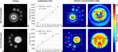
Myelin Lipid 1H Density Measurements by IR-UTE are Consistent Before and After D2O Exchange
Alan Seifert, Michael Wilhelm, Suzanne Wehrli, Felix Wehrli
In this work, the efficacy of long-T2 tissue water suppression by adiabatic inversion-recovery preparation is examined by IR-UTE imaging of an ovine spinal cord specimen in native hydration and deuterium oxide (D2O)-exchanged states. Myelin density in three white matter regions of interest (ROIs) was found to be 18.1% to 23.5% in the non-exchanged cord, and 19.9% to 22.5% in the D2O-exchanged cord, with the highest density in the dorsal columns. The agreement of myelin density measurements between non-exchanged and D2O-exchanged cords supports the efficacy of tissue water suppression in white matter by adiabatic inversion-recovery with an appropriately-chosen TI.
|
|
0320.
 |
10 |
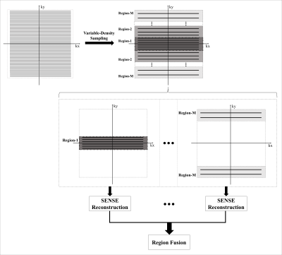
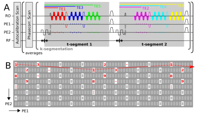
Accelerated quantitative susceptibility and R2* mapping with flexible k-t-segmented 3D-EPI
Rüdiger Stirnberg, Andreas Deistung, Jürgen Reichenbach, Tony Stöcker
We introduce and demonstrate a novel “k-t-segmented” gradient echo 3D-EPI variant for motion-robust, rapid quantitative susceptibility and R2* mapping at 3T. The combination of a versatile 2D-CAIPIRINHA EPI sampling (“k-segmentation”) and two complementing multi-echo options (“t-segmentation”) provides maximum time- and SNR-efficiency by acquiring exactly as many k-space lines per echo time as fit to the required R2* sampling without compromising spatial resolution. Offline averaging of multiple, rapid measurements (here: 6 averages, 6 echo times from 6.5-31.5ms, 52s/average) following optional, retrospective correction for motion and B0 drift, yields excellent quantitative whole-brain maps at 0.8mm isotropic resolution acquired in approximately 5 minutes.
|
|
0322.
 |
11 |
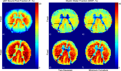
Formalin Tissue Fixation Biases Myelin Density Measurement by Quantitative Magnetization Transfer and Myelin Water Imaging
Alan Seifert, Melissa Umphlett, Mary Fowkes, Junqian Xu
Quantitative magnetization transfer (qMT) and multi-exponential T2-based myelin water imaging (MWI) are commonly-used MRI methods to quantify myelin content. Ex vivo MRI remains an essential step for validating these quantitative in vivo MRI biomarkers. However, ex vivo tissue is often preserved using formalin, which cross-links proteins and directly impacts these methods. We performed qMT and MWI on human spinal cord tissue before and after formalin fixation to quantify the effect of fixation on these biomarkers. QMT bound pool fraction (F) increased by 37.5% and MWI myelin water fraction (MWF) increased by 35.5-38.6%, but myelin-related image contrast was preserved.
|
|
0323.
 |
12 |
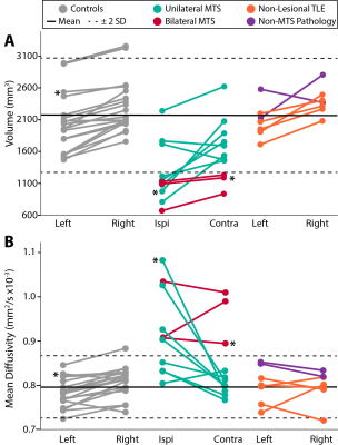
High Resolution Diffusion Tensor Imaging of the Hippocampus in Temporal Lobe Epilepsy
Sarah Treit, Trevor Steve, Tom Nowacki, Graham Little, Christian Beaulieu, Donald Gross
Diffusion tensor imaging of the hippocampus in temporal lobe epilepsy (TLE) has previously relied on very low spatial resolution acquisitions, limiting localization of hippocampal substructure and leading to partial volume effects in diffusion parameter quantification. This study uses a high resolution (1x1x1 mm3) single-shot diffusion protocol to yield excellent quality mean diffusion weighted images (DWIs) that allow for visualization of hippocampal substructure (e.g. presence/absence of the stratum lacunosum moleculare). Improved delineation of structure allows for segmentation of the hippocampus in native space (without co-registration to anatomical images), revealing elevated MD and loss of internal architecture in TLE subgroups.
|
|
0324.
 |
13 |
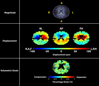
Repeatability of measuring pulsatile brain tissue motion and volumetric strain with retrospectively-gated DENSE at 7T
Ayodeji Adams, Jacob-Jan Sloots, Peter Luijten, Jaco Zwanenburg
Cardiac-induced brain tissue volumetric strain (an important metric reflecting microvascular blood volume pulsation) exhibits large inter-subject variability. We measured the intra-subject repeatability of brain tissue volumetric strain over the cardiac cycle in healthy subjects at 7T using a high resolution retrospectively-gated DENSE and found that the strain curve shape and peak value were very consistent between measurements. Furthermore, we validated the peak brain tissue volume change against measured spinal CSF stroke volume at C2-C3, and observed a strong correlation. The consistent findings strengthens the potential of volumetric strain as biomarker for microvascular function in the ageing brain.
|
|
0325.
 |
14 |
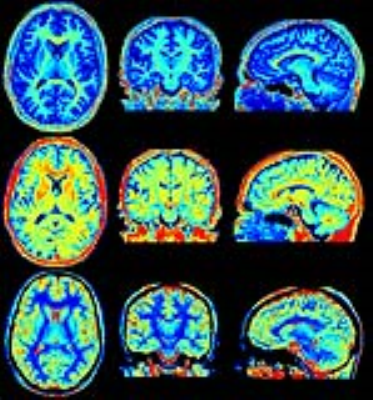
Isotropic 3D quantification of R1 and R2 relaxation and proton density in 6 minutes scan time
Marcel Warntjes, Peter Johansson, Anders Tisell, Peter Lundberg
Absolute quantification of R1 and R2 relaxation and proton density PD has been gaining considerable attention in the recent years. As yet, simultaneous quantification of R1, R2 and PD has been restricted to 2D methods, with high resolution in-plane but using relatively thick slices. A previously published method on cardiac quantification, QALAS, has been applied to the brain, providing a full quantification of R1, R2 and PD at 1.2 mm isotropic resolution in 3D using only 6 minutes scan time.
|
|
0326.
 |
15 |
Zero Time of Echo imaging with an Adiabatic Fat Suppression Pulse at 7T
Video Permission Withheld
Mark Symms, Mauro Costagli, Guido Buonincontri, Florian Wiesinger, Doug Kelley, Martin Janich, Giacomo Aringhieri, Massimo Marletta, Gareth Barker, Virna Zampa, Mirco Cosottini, Michela Tosetti
We present results using a Zero Time of Echo sequence ("Silent") with an Adiabatic Fat Suppression Pulse (ASPIR). As well as providing fat suppression that is relatively robust to field inhomogeneities at 7T, ASPIR-Silent also generates images with an additional off-resonant contrast. We show this contrast derives primarily from Magnetisation Transfer effects, and give an in vivo example.
|
|
