Poster: Techniques & Applications of Microcirculation Imaging
Electronic Power Pitch Poster
Neuro
17:15 - 18:15
Wednesday, 20 June 2018
Power Pitch Theater A - Exhibition Hall
| |
|
Plasma # |
|
0853.
 |
1 |
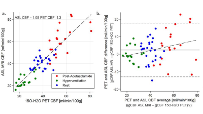
 Simultaneous measurements of global cerebral blood flow with 2D pseudo-continuous multi-TI arterial spin labeling and 15O-H2O PET in a hybrid PET/MR system Simultaneous measurements of global cerebral blood flow with 2D pseudo-continuous multi-TI arterial spin labeling and 15O-H2O PET in a hybrid PET/MR system
Oriol Puig Calvo, Ulrich Lindberg, Mark Vestergaard, Egill Rostrup, Adam Hansen, Henrik Larsson, Ian Law, Otto Henriksen
Arterial spin labeling (ASL) provides easy-access non-invasive quantification of regional cerebral blood (CBF) but its accuracy is not established. The aim of the study was to compare simultaneous measurements of absolute CBF obtained by ASL MRI and 15O-H2O PET CBF measurements in healthy subjects using a hybrid PET/MR system. Simultaneous 15O-H2O PET and ASL MRI were performed in resting state, hypoperfusion and hyperperfusion. Overall highly significant positive and linear correlation of ASL MRI and H2O PET in gCBF was observed across all perfusion states, confirming the ability of ASL MRI to quantify gCBF in good agreement with PET.
|
|
0854.
 |
2 |
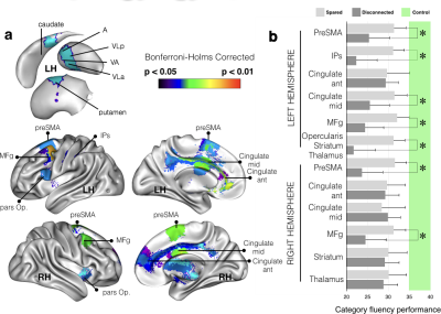
Advanced lesion symptom mapping analyses and implementation as BCBtoolkit
Chris Foulon, Leonardo Cerliani, Serge Kinkingnéhun, Richard Levy, Charlotte Rosso, Marika Urbanski, Emmanuelle Volle, Michel Thiebaut de Schotten
Even when focal, brain lesions have local and remote effects that impact functionally and structurally connected circuits. We developed a free open-source software (BCBtoolkit) that gathers different methods to estimate these effects on structural (using healthy controls tractography) and functional networks, using T1-images and fMRI data, and relate them to behavioral impairment. We applied these methods to 37 patients with a chronic focal brain lesion and 54 healthy controls in the context of category fluency. Our methods revealed a large set of directly and indirectly disconnected brain regions that had significantly impacted the performance.
|
|
0855.
 |
3 |
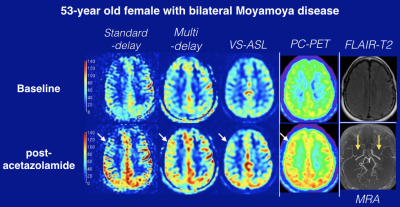
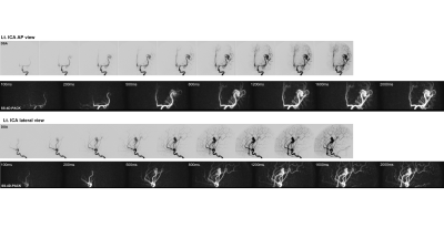
Validation of Cerebrovascular Reactivity by Arterial Spin Labeling MRI in Moyamoya Disease with Simultaneously Measured 15O-PET and Phase-contrast MRI
Yosuke Ishii, Thoralf Thamm, Jia Guo, Mohammad Khalighi, Mirwais Wardak, Dawn Holley, Harsh Gandhi, Jun Park, Bin Shen, Gary Steinberg, Fred Chin, Greg Zaharchuk, Audrey Fan
We validated cerebrovascular reactivity (CVR) in Moyamoya disease measured by arterial spin labeling (ASL) simultaneously with 15O-positron emission tomography (PET) using hybrid PET/MRI scanner. We compared the three types of ASL including standard-delay, multi-delay with extended post-label delay, and velocity selective ASL (VS-ASL). To quantify absolute PET cerebral blood flow (CBF) without arterial blood samples, we scaled the static PET maps using simultaneously collected phase-contrast (PC) MRI. Multi-delay ASL showed the strongest correlation with PC-PET on both baseline and post-ACZ CBF (R2 = 0.48 and 0.66 respectively). Multi-delay ASL and VS-ASL showed similar good correlations with PC-PET on CVR.
|
 |
0856.
 |
4 |
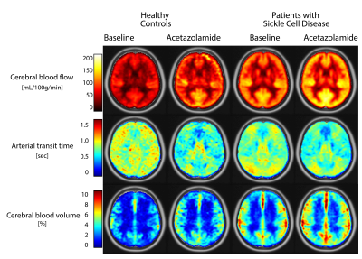
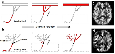
Adaptations in cerebral physiology due to chronic anaemia measured with Turbo-QUASAR ASL
Lena Vaclavu, Moss Zhao, Esben Petersen, John Wood, Henk Mutsaerts, Charles Majoie, Ed vanBavel, Bart Biemond, Michaell Chappell, Aart Nederveen
In this work, we applied a novel ASL method (Turbo-QUASAR) to evaluate the effects of life-long anaemia on cerebral physiology by comparing patients with Sickle Cell Disease to healthy controls. Turbo-QUASAR enables simultaneous assessment of cerebral blood flow (CBF), cerebral blood volume (aCBV) and arterial transit time (ATT) as well as tissue T1 and M0. We found normal ATT in the presence of elevated CBF in patients. In addition we found increased aCBV in patients. Acetazolamide administration shortened ATT with no change in aCBV suggesting maximal dilation and reserves being accessed by faster ATT. aCBV was inversely related to haemoglobin.
|
|
0857.
 |
5 |
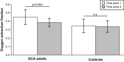
Noninvasive MRI measurements of oxygen extraction fraction reduction in response to blood transfusion in adults with sickle cell anemia
Meher Juttukonda, Lori Jordan, Larry Davis, Chelsea Lee, Niral Patel, Sumit Pruthi, Manus Donahue
Blood transfusions are often administered for secondary stroke prevention in adults with sickle cell anemia (SCA). We utilized noninvasive MRI methods to evaluate how oxygen extraction fraction (OEF) and cerebral blood flow (CBF) adjust after transfusion in adults with SCA. OEF reduced on average, while CBF did not change significantly. The OEF reduction paralleled increases in hematocrit but was unrelated to the reduction in hemoglobin-S. This implies that most patients receiving transfusions operate near autoregulatory reserve capacity even after transfusion, and improving oxygen delivery by increasing hematocrit can be visualized noninvasively with OEF-MRI.
|
|
0858.
 |
6 |
Relationship between the Degree of Unilateral Intracranial Artery Stenosis and Cerebral Perfusion: a High-resolution Intracranial Vessel Wall Imaging Study
Song Liu, Tianyi Qian, Jinxia Zhu, Wen Shen, Shuang Xia
High-resolution vessel wall imaging(HR-VWI) is an novel technique used to assess intracranial artery stenosis, and it has been useful in clinical practice. In addition, time-to-maximum (Tmax) maps, derived from PWI, are increasingly being used in studies of ischemic stroke, such as EPITHET and DEFUSE-2 . Our study aimed to investigate how the degree of stenosis of the MCA or ICA affects brain tissue perfusion. The results showed that the degree of MCA or ICA stenosis was positively correlated with the Tmax delay volume, and different degrees of ICA or MCA stenosis presented with different perfusion delay volumes.
|
|
0859.
 |
7 |
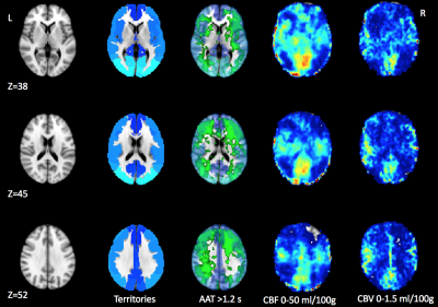
Identifying ischemic zone patterns and origins using multi-delay arterial spin labeling in vasospasm
Swati Rane, Daniel Hippe, Michael Levitt, Louis Kim, Jalal Andre
Our goal was to evaluate a multi-delay, pseudocontinuous arterial spin labeling (ASL) approach to detect focal delayed cerebral ischemia in patients with vasospasm. We show that inferences on vascular origins based on ASL and standardized perfusion territories correlated well with expert reader reads that rely on an invasive catheter-based digital subtraction angiography (DSA) examination. Importantly, ASL identified possible ischemic zones due to distal vasospasm, which are nearly undetectable on conventional DSA. Thus, use of ASL may prevent unnecessary DSA examinations and improve patient prognosis.
|
|
0860.
 |
8 |
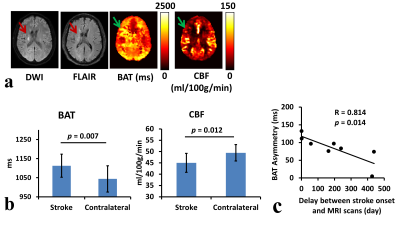
Non-contrast fingerprinting perfusion imaging reveals hemodynamic deficits in cerebrovascular diseases
Pan Su, Peiying Liu, Yang Li, Zixuan Lin, Lynsey Keator, Ye Qiao, Judy Huang, Argye Hillis, Hanzhang Lu
Perfusion imaging plays an important role in management decisions for a variety of cerebrovascular diseases. Most clinical perfusion MRI of stroke requires the use of contrast agent. However, contrast-agent perfusion cannot be used or fails to be used in 10-20% of patients. Therefore, an alternative technique to Gd-perfusion will benefit a substantial number of patients in clinical practice. Recently, a MR-Fingerprinting (MRF) ASL was developed for simultaneous estimations of CBF and bolus timing. In this study, we demonstrated the clinical utility of MRF-ASL in two types of cerebrovascular diseases, ischemic stroke and Moyamoya disease.
|
|
0861.
 |
9 |
High-Resolution, Non-contrast Pseudo-Continuous Arterial Spin-Labeling (PCASL) MR-Angiography compared to standard clinical MRI in the detection of intracranial vessel pathologies with emphasis on intracranial arteriovenous shunts.
Tilman Schubert, oliver Wieben, Patrick Turski, Huimin Wu, Kevin Johnson
The detection of intracranial AV-shunts may be difficult with non-invasive imaging. Due to the nature of spin-labeled protons, ASL-based MRA is likely to be highly specific for AV-shunting. We sought to determine the sensitivity and specificity of ASL-based MRA in a group of 32 patients, among those 14 with AV-shunts. Furthermore, the diagnostic performance for vascular pathology not associated with AV-shunting was assessed. We found ASL-based MRA to be more specific with an equivalent sensitivity compared to a clinical MRA-exam for intracranial AV-shunts. All vascular pathology not associated with AV-shunting were detected with ASL-based and clinical MRA.
|
|
0862.
 |
10 |
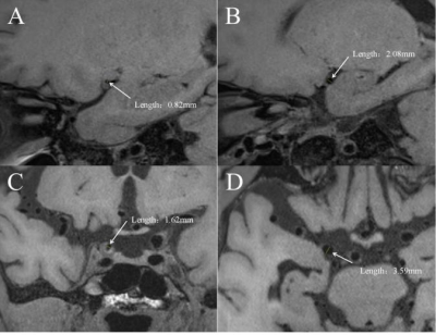
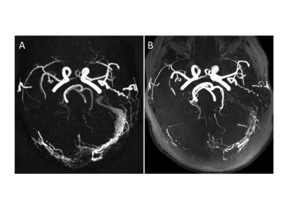
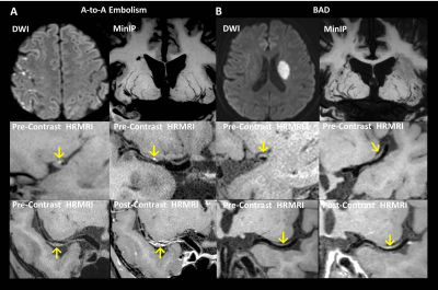
Characteristics of Plaques and Lenticulostriate Arteries in Stroke Patients by Whole-Brain Vessel Wall Magnetic Resonance Imaging
Fang Wu, Zhaoyang Fan, Tianyi Qian, Qi Yang, Debiao Li
The aim of our study was to investigate the plaque characteristics and the lenticulostriate artery (LSA) status in artery-to-artery (A-to-A) embolism and branch atheromatous disease (BAD) using whole-brain high-resolution MRI. The results showed that patients with A-to-A embolism presented more frequently hyperintense plaques (HIP), plaque surface irregularity, and severe stenosis than patients with BAD. A signi?cant reduction in the number and lengths of LSA branches in patients with BAD compared to A-to-A embolism was also found. Multivariate analysis further demonstrated that HIP was independently associated with A-to-A embolism, whereas the reduction in LSA branches was independently associated with BAD.
|
|
0863.
 |
11 |
Comparison of velocity-selective and pulsed ASL perfusion MRI in patients with suspected cerebral cortical ischemia
Divya Bolar, Bruce Rosen, Pamela Schaefer
Traditional pulsed ASL (PASL) suffers from arterial transit delay (ATD) effects, often resulting in inaccurate perfusion measurements in cerebral ischemia. Velocity selective ASL (VSASL), on the other hand, accurately measures perfusion independent of ATD. In this study, we compare VSASL and PASL perfusion in 27 patients with suspected cerebral cortical ischemia. Normal perfusion on VSASL and PASL had nearly perfect negative predictive value for ischemia, abnormal PASL perfusion had a high false positive rate for ischemia (25-35%), and abnormal VSASL perfusion had a ~0% false positive rate for ischemia.
|
|
0864.
 |
12 |
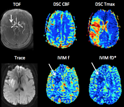
Intravoxel Incoherent Motion (IVIM) Perfusion Imaging in Hyperacute Stroke: Initial Results
Christian Federau, Max Wintermark, Soren Christensen, David Marcellus, Guangming Zhu, Maarten Lansberg, Gregory Albers, Jeremy Heit
Intravoxel Incoherent Motion (IVIM) MR perfusion is of particular interest in stroke because it derives intrinsic microvascular perfusion information, and might therefore include information regarding collateral blood flow without being influenced by time delay in blood arrival, which is a limitation of other perfusion techniques. In this preliminary work in 23 patients with hyperacute stroke, we found the IVIM microvascular perfusion paramteters were significantly reduced in the infarct core but not in the penumbra, compared to the contralateral side, suggesting that at time of imaging, microvascular perfusion is maintained through collateral blood flow in the penumbra.
|
|
0865.
 |
13 |
Superselective 4D MR Angiography with Pseudo-Continuous Arterial Spin Labelling Combined with CENTRA-Keyhole (SS-4D-PACK) Used to Visualize Brain Arteriovenous Malformations
Osamu Togao, Akio Hiwatashi, Makoto Obara, Michael Helle, Koji Yamashita, Daichi Momosaka, Tatsuhiro Wada, Hiroo Murazaki, Marc Van Cauteren, Hiroshi Honda
In the present study, we demonstrated the utility of superselective 4D-MR angiography with pCASL combined with CENTRA-keyhole (SS-4D-PACK) for the visualization of brain AVMs. This method enables a time-resolved and vessel-selective angiography within 5 minutes without a use of contrast agents. It was showed that almost perfect vessel selectivity was achieved with SS-4D-PACK. Although CNRs were slightly reduced in SS-4D-PACK due to a labeling loss during superselective label focusing, this was acceptable since visualization was well preserved. SS-4D-PACK can be a non-invasive clinical tool for assessing brain AVMs.
|
|
0866.
 |
14 |
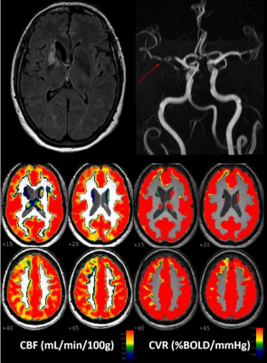
Impaired cerebrovascular reactivity assessed by BOLD hypercapnic fMRI is associated with increased risk of stroke in patients with symptomatic intracranial atherosclerotic stenosis
Jeremy Papassin, Olivier Heck, Naila BOUDIAF, Eric CONDAMINE, Johan PIETRAS, Florence TAHON, Olivier DETANTE, Alexandre KRAINIK
Intracranial atherosclerotic stenosis (IAS) remains at risk of recurrent ischemic events despite intensive medical management. Cerebrovascular reactivity (CVR) assessed by hypercapnic challenge using BOLD functional MRI (CVR BOLD fMRI) estimates cerebrovascular reserve and may allow to identify patients at higher risk of recurrent ischemic events. 19 patients referred for unilateral symptomatic IAS were studied to estimate the relationships between baseline characteristics, recurrence of ischemic events, and CVR. During follow-up, recurrent ischemic events were more frequent in patients with impaired CVR. CVR mapping may help to better select among IAS patients those at higher risk to discuss additional treatment.
|
|
0867.
 |
15 |
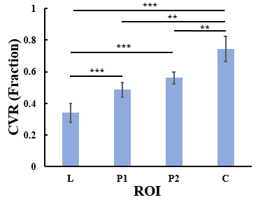
Resting-State BOLD MRI for Evaluating Cerebrovascular Reserve in Stroke Patients
Kamil Taneja, Hanzhang Lu, Argye Hillis, Peiying Liu
The early stage of hemodynamic failure in ischemic cerebrovascular diseases is characterized by diminished cerebrovascular reserve. Tissues at this stage are at high risk for stroke. Assessment of cerebrovascular reserve by cerebrovascular reactivity (CVR) measurement usually requires the administration of vasoactive challenges (e.g., acetazolamide or CO2), which is often difficult or impractical in stroke patients. In this study, we demonstrate that CVR can be mapped in stroke patients without a physiological challenge but using the natural fluctuations of the resting-state BOLD signal. Results indicate that this technique can assess CVR in lesion, peri-lesional, and healthy tissue in a reproducible manner.
|
|
