|
Poster: CV PowerBeat: Part 1
Electronic Power Pitch Poster
Cardiovascular
09:15 - 10:15
Monday, 18 June 2018
Power Pitch Theater A - Exhibition Hall
| |
|
Plasma # |
|
0001.
 |
1 |
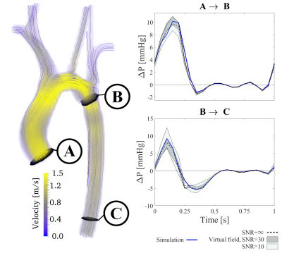
 Non-invasive pressure estimations by virtual fields – cardiovascular pressure drops from 4D flow MRI Non-invasive pressure estimations by virtual fields – cardiovascular pressure drops from 4D flow MRI
David Marlevi , Bram Ruijsink, Maximilian Balmus, Desmond Dillon-Murphy, Daniel Fovargue, Kuberan Pushparajah, Pablo Lamata, C. Alberto Figueroa, Massimiliano Colarieti-Tosti, Matilda Larsson, Reza Razavi, David Nordsletten
4D-flow-MRI enables the non-invasive assessment of cardiovascular pressure drops; however, estimation accuracy depends on vascular topology and acquisition noise. Here, we present a method that minimizes the impact of these by using virtual fields to isolate and probe hemodynamic pressure drops. We show that, in-silico, the method accurately assesses pressure drops over multiple segments of a patient-specific co-arcted aorta (average error below 22%), independent of anatomical bifurcations. Additionally, the method compares successfully against catheter measurements, using 4D-flow-MRI in-vivo (average error at peak systole below 15%). With this, the method represents a refined tool for hemodynamic analysis of cardiovascular flow.
|
|
0002.
 |
2 |
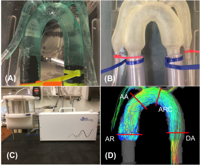
The Effect of Model Compliance and Pulsatile Flow for In-Vitro Simulation of the Aorta
Timothy Ruesink, Matthew Smith, Katrina Ruedinger, Christopher François, Alejandro Roldán-Alzate
In-vitro cardiovascular simulation permits quantification of hemodynamics that cannot be assessed in-vivo. However, simulation accuracy depends on anatomical and physiological realism of in-vitro models and flow. To determine the effect of model compliance and pulsatile flow, a rigid model of an aorta was compared with a geometrically identical compliant model. Models were perfused with pulsatile flow using a positive displacement pump. Flow dynamic parameters for simulations, obtained using 4D flow MRI, showed that model compliance plays a significant role in hemodynamics during pulsatile flow. Future development of realistic in-vitro simulation, paired with in-vivo validation, will aid in surgical planning.
|
|
0003.
 |
3 |
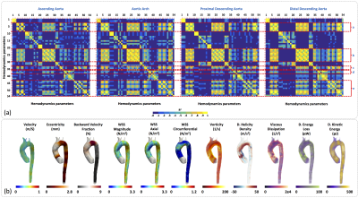
3D Hemodynamics Characterization in Patients with Hypercholesterolemia using 4D Flow data and a Finite Element Method.
Julio Sotelo, Animesh Tandon, Andrew Tran, Joaquín Mura, Daniel Hurtado, Tarique Hussain, Sergio Uribe
Familial hypercholesterolemia (FH) is an autosomal dominant disorder of lipoprotein metabolism, that are associated with premature atherosclerosis, early-onset of cardiovascular disease (CVD) with an elevated mortality. It would be prudent to develop and investigate imaging-based hemodynamics biomarkers that assist in cardiovascular risk assessment of FH patients. In this work, we obtain several hemodynamics parameters in HP patients using a single methodology, which is based on the analysis of 4D flow data using a finite element method. We found distinctive biomarkers as WSS (magnitude, axial, circumferential) and Kinetic Energy those present more significant differences along the entire aorta.
|
|
0004.
 |
4 |
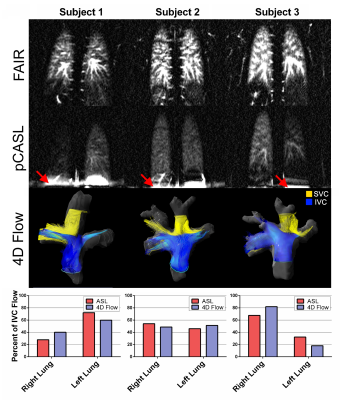
Caval Blood Flow Distribution in Fontan Circulation: Comparison between ASL-Measured Pulmonary Perfusion and 4D Flow
Joshua Greer, Jerry Michael, Barbara Burkhardt, Animesh Tandon, Gerald Greil, Tarique Hussain, Ananth Madhuranthakam
Caval flow contribution to each lung is of interest in Fontan circulation due to the increased risk of developing pulmonary arteriovenous malformations when flow is unevenly distributed. Existing methods to assess this risk are invasive and require ionizing radiation, posing additional risks to pediatric patients during longitudinal monitoring. In this study, we demonstrate a non-invasive, non-ionizing assessment of the origin of pulmonary blood flow, as well as quantitative pulmonary perfusion using arterial spin labeled MR, and compare with the previously proposed 4D-flow measurement of caval flow distribution.
|
|
0005.
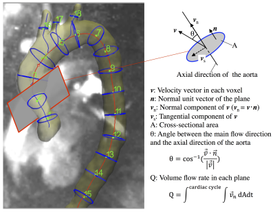 |
5 |
Postoperative changes in volume flow rate in the thoracic aorta and the aortic arch branches in patients with aortic valve stenosis: a prospective serial 4D flow MRI study
Video Permission Withheld
Hiroki Kamada, Hideki Ota, Masanori Nakamura, Yohsuke Imai, Wenyu Sun, Yoshiaki Komori, Ko Sakatsume, Ichiro Yoshioka, Yoshikatsu Saiki, Kei Takase
Postoperative hemodynamic changes in the aorta in patients with aortic valve stenosis (AS) remain unclear. Four-dimensional (4D) flow MRI was perfumed in 11 AS patients before and after aortic valve replacement (AVR). We evaluated volume flow rate and the main flow direction, setting 15 planes of the aorta and 3 planes of the arch branches. Volume flow rate significantly increased in the ascending aorta and the arch branches. The main flow direction came to match with the axial direction of the aorta. These results suggest that AVR results in more efficient blood transport to the upper body including the brain.
|
|
0006.
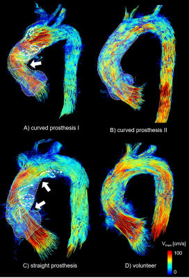 |
6 |
4D-Flow-MRI analysis of aortic flow patterns after replacement of the ascending aorta with a physiologically pre-shaped, 90° bent prosthesis
Thekla Oechtering, Jennifer Schlueter, Malte Sieren, Michael Scharfschwerdt, Christian Auer, Markus Huellebrand, Hans-Hinrich Sievers, Joerg Barkhausen, Alex Frydrychowicz
Altered aortic anatomy after prosthesis implantation has been shown to increase secondary flow patterns with potential long-term effects. Therefore, patients after valve-sparing aortic root and ascending aorta replacement with a physiologically pre-shaped prosthesis were examined with 4D-Flow-MRI and compared to patients with straight grafts and age-matched volunteers. A reduced angulation at the distal anastomosis as well as a slightly reduced intensity of secondary flow patterns was confirmed. However, there was no reduction of secondary flow patterns in comparison to straight prostheses potentially attributed to a residual angulation at the proximal anastomosis and a dilatation at the distal anastomosis.
|
|
0007.
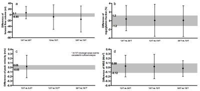 |
7 |
Impact of field strength (1.5, 3.0 and 7.0 Tesla) and sequence on quantification of aortic flow volumes, peak velocity and wall shear stress using 4D flow MRI
Stephanie Funk, Sebastian Schmitter, Marcel Prothmann, Carsten Schwenke, Florian von Knobelsdorff-Brenkenhoff, Andreas Greiser, Emilie Bollache, Michael Markl, Jeanette Schulz-Menger
For implementing 4D flow in clinical routine, standardization is important. We evaluated equivalence of 4D flow parameters in different sequences and at three different field strengths. Ten healthy volunteers were scanned at 1.5T, 3.0T and 7.0T. At 1.5T, three different sequences were applied. Ascending aorta, aortic arch and descending aorta of each scan were evaluated for diagnostic quality. After exclusion of non-diagnostic segments, equivalence testing for flow, wall shear stress and peak velocity was performed. Acceptable equivalence was determined by intra-rater analysis. Comparison of different field strengths as well as different sequences did not reach equivalence. 4D flow sequences are not interchangeable.
|
|
0008.
 |
8 |
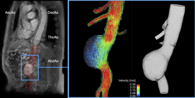
Quantifying the Impact of Velocity Field Distortions on Particle Tracking Techniques
Magnus Ziegler, Martin Welander, Marcus Lindenberger, Niclas Bjarnegård, Jonas Lantz, Matts Karlsson, Tino Ebbers, Toste Länne, Petter Dyverfeldt
Distortions in the velocity fields acquired by 4D Flow MRI affect particle tracking based visualization and quantification methods. Particle residence time was calculated in a subject with aortic aneurysm and was used to assess how particle tracking methods are impacted by noise and offset errors. To do so, a computationally derived ideal velocity field was created and distorted by adding noise and background phase-offsets. We found that particle tracking methods are severely impacted by offset errors, though more robust to random noise. Understanding the limits of these methods is crucial for their appropriate use.
|
|
0009.
 |
9 |
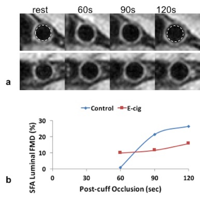
Quantitative MRI detects acute vascular effects of e-cig aerosol inhalation
Michael Langham, Alessandra Caporale, Felix Wehrli
The rapid rise in the popularity and use of the electronic cigarettes among adolescents are unsettling trends in spite of the limited science that exists on the health effects of e-cigarettes. The purpose of this project was to introduce a noninvasive method for the study of the systemic acute effects of e-cigarette aerosol inhalation on the cardiovascular system by means of quantitative MRI that targets various vascular territories. Preliminary data show reduced femoral vein SvO2 and impaired flow-mediated dilation in the femoral artery, along with elevated aortic arch pulse-wave velocity after an e-cigarette challenge equivalent to one conventional cigarette.
|
|
0010.
 |
10 |
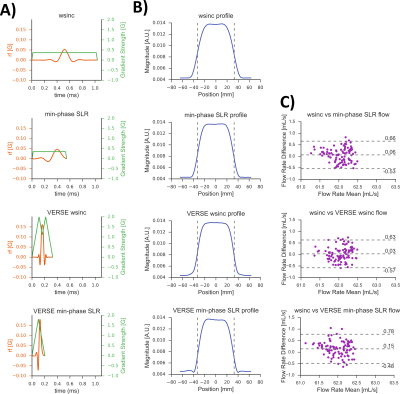
Accelerating 4D-Flow Acquisitions by Reducing TE and TR with Optimized RF and Gradient Waveforms
Michael Loecher, Patrick Magrath, Eric Aliotta, Daniel Ennis
4D-Flow MRI is a powerful technique for simultaneously imaging vascular anatomy and hemodynamics. However, its clinical utility is limited by long (10-20 minute) scan times. This work aims to shorten scan times by using fast RF pulses and convex optimized gradient waveforms. The waveforms are optimized with arbitrary shapes, and are designed to go as fast possible without causing peripheral nerve stimulation by including an additional PNS constraint. The optimized sequence is implemented and tested in flow phantoms and a volunteer. The data acquired with the optimized waveforms is up to 33% faster, with no significant difference in measured data compared to a reference sequence.
|
|
0011.
 |
11 |
A Dual Echo, Dual VENC (DEDV) phase contrast method for Simultaneous Measurement of Myocardial and Blood Flow Velocities
Afis Ajala, Jiming Zhang, Benjamin Cheong, Pei-Herng Hor, Raja Muthupillai
Dual VENC Velocity quantification has been previously carried out with the two different VENCs acquired in separate repetition cycles (TRs). We propose a magnetic resonance imaging sequence that acquires the phase matrix for two different VENC values in one TR, and use the developed sequence for slow motions and fast flows.
|
|
0012.
 |
12 |
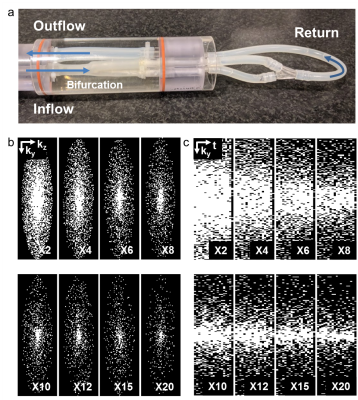
30 times accelerated 4D flow MRI in the carotids using a Pseudo Spiral Cartesian acquisition and a Total Variation constrained Compressed Sensing reconstruction
Eva Peper, Lukas Gottwald, Qinwei Zhang, Bram Coolen, Pim van Ooij, Gustav Strijkers, Aart Nederveen
4D flow MRI enables visualization and quantification of complex blood flow, and provides relevant biomarkers, such as wall shear stress. A 4D flow MRI acquisition, however, takes between 15-40 min, which complicates its use in clinical practice. In this work, we developed a technique to reduce scan time by prospectively undersampling k-space in a pseudo-spiral Cartesian fashion. Combined with a Compressed Sensing reconstruction using a total variation sparsifying transform in time, this technique makes 4D flow MRI in the carotid arteries 20-30 times faster, while preserving accuracy in flow and wall shear stress measurements.
|
|
0013.
 |
13 |
Alterations of Cardiac 4D Hemodynamics and Blood Energetics in Hypertrophic Cardiomyopathy
Aakash Gupta, Michael Markl, Bradley Allen, Lubna Choudhury, James Carr, Robert Bonow, Jeremy Collins
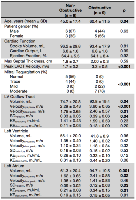
4D flow MRI supports the development of novel energetic biomarkers in hypertrophic cardiomyopathy (HCM) – a disease marked by dynamic left ventricular outflow tract obstruction with mitral regurgitation (MR). We compared kinetic energy and velocity metrics between obstructive and non-obstructive HCM subtypes to determine where in the left heart high kinetic energy and flows were generated. Left atrium showed significantly higher systolic kinetic energy and velocities throughout ventricular systole due to MR. Including these energetic parameters may be useful to detect significant MR that is underestimated by echo, identify patients at higher risk of atrial fibrillation, and inform treatment options.
|
|
0014.
 |
14 |
Dynamic flow imaging and quantification using cine FISS arterial spin labeling
Robert Edelman, Ali Serhal, Amit Pursnani, Jianing Pang, Ioannis Koktzoglou
We describe a new approach for flow imaging and quantification consisting of a prototype cine arterial spin labeling (ASL) pulse sequence using a highly-accelerated radial fast interrupted steady-state (FISS) readout. The technique was successfully applied in several vascular regions (coronary arteries, pulmonary arteries, renal arteries, circle of Willis). These preliminary results suggest that cine FISS ASL has the potential to provide an efficient and visually-appealing alternative to phase contrast for the depiction and quantification of blood flow.
|
|
0015.
 |
15 |
4D Flow Cardiac MRI Using Semi-Automated Retrospective Valve Tracking for Assessment of Severe Mitral Insufficiency
Carmen Blanken, Jos Westenberg, Pim van Ooij, Geertruida Bijvoet, Steven Chamuleau, Jean-Paul Aben, Stefan Boekholdt, Aart Nederveen, Tim Leiner, R Planken
Mitral insufficiency (MI) is difficult to quantify, due to cardiac motion and complex regurgitation patterns. This study evaluated the use of 4D flow MRI with semi-automated retrospective valve tracking for assessment of severe MI. Valve tracking of both the mitral and aortic valve allowed for direct measurement of retrograde flow and indirect measurement based on total left-ventricular inflow and aortic outflow. Conventional 2D MRI-based indirect quantification was used as a reference. Eccentric regurgitation patterns complicated direct quantification, necessitating manual plane angulation for accurate measurement. Indirect quantification corresponded well with 2D MRI and might be a more reproducible alternative.
|
|
