Poster: MRI in Cancer Therapy & Diagnostics
Electronic Power Pitch Poster
General Cancer Imaging
14:45 - 15:45
Monday, 18 June 2018
Power Pitch Theater B - Exhibition Hall
| |
|
Plasma # |
 |
0117.
 |
16 |
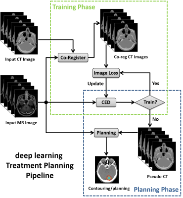
 MRI-only Treatment Planning using Pseudo CT Generation from Deep Learning Approach MRI-only Treatment Planning using Pseudo CT Generation from Deep Learning Approach
Fang Liu, Poonam Yadav, Andrew Baschnagel, Alan McMillan
A MRI-only treatment planning pipeline, deepMTP, was constructed using a deep learning approach to generate continuously-valued pseudo CT images from MR images. A deep convolutional neural network was designed to identify tissue features in volumetric head MR images training with co-registered kVCT images. A set of 40 retrospective 3D T1-weighted head images was utilized to train the model, and evaluated in 10 clinical cases with brain metastases. Statistical analysis was used to compare dosimetric parameters of plans made with pseudo CT images generated from deepMTP to those made with kVCT based clinical treatment plan, where no significant difference was found.
|
 |
0118.
 |
17 |
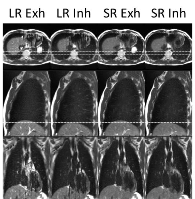
Online Super-resolution 4D T2-weighted MRI for MRI-guided Radiotherapy
Joshua Freedman, David Collins, Oliver Gurney-Champion, Simeon Nill, Uwe Oelfke, Martin Leach, Andreas Wetscherek
To assist treatment delivery to moving tissues on hybrid MRI-guided radiotherapy systems, high quality midposition (of the respiratory cycle) and 4D-T2w images are desirable. Current approaches to rapidly obtaining 4D-T2w MRI are often limited by thick slices, incomplete motion information and binning artefacts. Midposition and 4D-T2w images were calculated using motion-modelling and a super-resolution reconstruction, and verified by comparison with the initially acquired images. Calculated 4D-T2w images exhibited high spatiotemporal resolution (1.0x1.0x1.0 mm3, 8 respiratory phases), displayed reduced binning artefacts and no missing data. An acquisition time of 5.0-7.5 minutes was found sufficient to obtain representative midposition T2w images.
|
|
0119.
 |
18 |
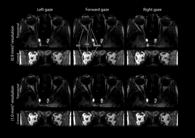
Rapid MR Imaging of Ocular Movement using Shared K-Space Data for Radiotherapy Planning
Luc van Vught, Kirsten Koolstra, Jan-Willem Beenakker
During ocular movement, the shape and location of the intra-orbital structures change. Outlining these changes will improve the accuracy of radiotherapy treatment planning for ocular tumors and the treatment of impaired ocular movement. The resolution of ocular MRI is, however, limited by acquisition time due to eye-motion. Therefore, an acquisition strategy in which outer k-space data is shared between gaze directions was developed and evaluated in 7 healthy and 2 myopic subjects. With this technique, the location of the orbital structures was determined for nine gaze directions in approximately one minute.
|
|
0120.
 |
19 |
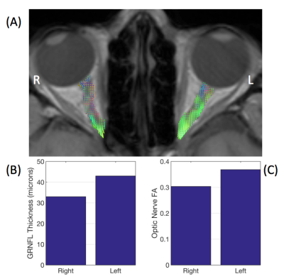
Visual pathway structure and localisation of tumour-induced disturbance in optic pathway glioma: correlations between diffusion-MRI, visual evoked potentials, and optical coherence tomography
Patrick Hales, Sian Handley, Alki Liasis, Darren Hargrave, Chris Clark
Optic pathway glioma (OPG) is a childhood tumour of the visual pathway. Clinical management of OPG remains challenging, as the extent of tumour infiltration of the visual pathway is not perceptible on conventional MRI. We used diffusion-MRI to delineate the visual pathway in OPG patients. Advanced ophthalmological assessment was also performed using visual evoked potentials (VEP) and optical coherence tomography. We demonstrate that fractional anisotropy (FA) in the optic pathway correlates with retinal nerve-fibre layer thickness and VEP response, and tumour invasion anterior to the optic chiasm can be detected via inter-ocular differences in optic nerve FA.
|
|
0121.
 |
20 |
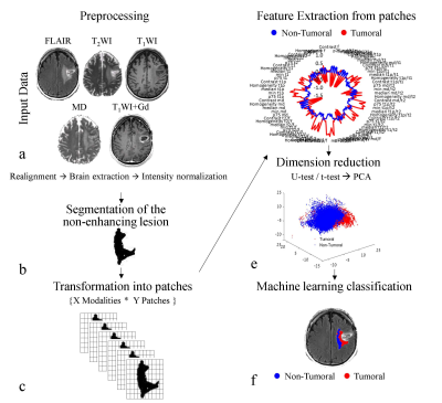
Differentiation between vasogenic edema and infiltrative tumor in patients with high grade gliomas using texture patch based analysis
Moran Artzi, Gilad Liberman, Deborah Blumenthal, Orna Aizenstein, Felix Bokstein, Dafna Ben Bashat
This study proposes a radiomics patch-based analysis, based on conventional MRI, for classification of the non-enhancing lesion area into vasogenic edema and infiltrative tumor in patients with high-grade-gliomas. 179 MRI scans obtained from 102 patients were included: 67 patients with high-grade-gliomas and 35 patients with brain-metastases. A total of 225 histogram and gray-level-co-occurrence-matrix based features were extracted from the non-enhancing lesion. Classification was performed using various machine-learning classifiers. The best results were obtained using Linear support-vector-machine, with accuracy=87%, sensitivity=86%, and specificity=89%. Preliminary results in patients treated with bevacizumab demonstrate the clinical potential of this method to improve therapy response assessment.
|
|
0122.
 |
21 |
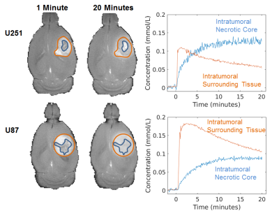
Evaluating intratumoral necrosis in gliomas: a multi-modal study of acidosis, cellularity, and vascularity
Maxime Parent, John Walsh, Lucas Adam, Daniel Coman, D.S. Fahmeed Hyder
The malignant form of human glioblastoma multiforme (GBM) is linked to intratumoral necrosis. Since novel immunotherapies are being sought to treat these patients, non-invasively characterizing intratumoral necrosis in gliomas is clinically important. Here, we describe a multi-modal MRI study of acidity, cellularity, and vascularity in two glioma models that feature comparable necrosis and proliferation, but differ in vascular markers. The intratumoral necrotic core (INC) and intratumoral surrounding tissue (IST) had distinct slow and fast Gd-enhanced profiles, respectively. Despite immunohistochemical/histopathological differences, these GBM models show similar profiles of INC-IST gradients for acidity and cellularity, but not for vascularity.
|
|
0123.
 |
22 |
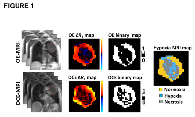
Oxygen enhanced-MRI detects radiotherapy-induced change in hypoxia in xenograft models and lung cancer patients
Video Permission Withheld
Ahmed Salem, Ross Little, Adam Featherstone, Muhammad Babur, Hitesh Mistry, Susan Cheung, Yvonne Watson, Victoria Tessyman, Marie-Claude Asselin, Alan Jackson, Kaye Williams, Geoffrey Parker, Corinne Faivre-Finn, James O'Connor
Oxygen-enhanced MRI (OE-MRI) has shown promise as a technique for quantifying and spatially mapping tumour hypoxia. Here, we report a world first-in-man study showing that OE-MRI signals in perfused tumour can non-invasively track changes in hypoxia induced by radiotherapy. We show that OE-MRI detects (1) reduction in hypoxia in Calu6 xenografts and that this change is due to hypoxia modification; and (2) reduction in hypoxia is also seen in 14 patients with non-small cell lung cancer. These data support first-in-man use of OE-MRI biomarkers in clinical trials of (chemo)-radiotherapy as single agent or in combination with hypoxia-modifying agents.
|
|
0124.
 |
23 |
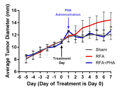
Assessment of Distant Tumor Stimulation from Liver Radiofrequency Ablation in a Rat Breast Carcinoma Model using Hyperpolarized 13C-Pyruvate MRI
Joseph Goodwin, David Mwin, Patricia de Souza, Svayam Dialani, John Moon, Aaron Grant, Muneeb Ahmed, Leo Tsai
Radiofrequency ablation (RFA) is commonly used to treat tumors such as hepatocellular carcinomas. However, there is evidence that liver RFA can stimulate growth in tumors at other sites, a process attributed to the HFG/c-Met pathway. Hyperpolarized 13C-pyruvate MRI showed increased intratumoral lactate production in subcutaneously-implanted R3230 tumors following RFA of normal liver in a rat model. This effect is suppressed when liver RFA is combined with a c-Met inhibitor. 13C MRI could potentially be used to identify tumors at risk for this off-target effect.
|
|
0125.
 |
24 |
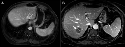
Late gadolinium enhancement of colorectal liver metastases post-chemotherapy is associated with tumour fibrosis and overall survival post-hepatectomy
Helen Cheung, Paul Karanicolas, Eugene Hsieh, Natalie Coburn, Tishan Maraj, Jin Kim, Howaida Elhakim, Masoom Haider, Calvin Law, Laurent Milot
Preoperative MRI is routinely used for diagnosis, staging, and operative planning of colorectal liver metastases (CRCLM), but still relatively unexplored for preoperative prognosis. Tumour fibrosis in post-hepatectomy CRCLM specimens is associated with long-term survival and late gadolinium enhancement is associated with tumour fibrosis in other disease processes (eg. cholangiocarcinoma). We performed a retrospective cohort study (n=121) in patients who received a clinical gadolinium-enhanced MRI prior to hepatectomy for CRCLM. We determined that strong enhancement on delayed phase MRI was associated with tumour fibrosis post-hepatectomy and overall survival.
|
|
0126.
 |
25 |
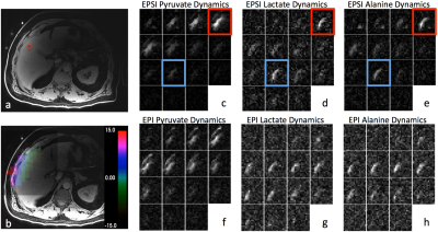
Human Hyperpolarized 13C MR of Liver and Bone Metastases using both EPSI and EPI Acquisitions
Zihan Zhu, Jeremy Gordon, Hsin-Yu Chen, Eugene Milshteyn, Daniele Mammoli, Lucas Carvajal, Peter Shin, Rahul Aggarwal, Robert Bok, John Kurhanewicz, Pamela Munster, Daniel Vigneron
Hyperpolarized 13C metabolic imaging is a powerful technique with proven safety and efficacy for human studies. The goal of this project was to investigate pyruvate metabolism in patients with prostate and breast cancer metastases with both echo-planar spectroscopic imaging (EPSI) and the echo-planar imaging (EPI) acquisitions in the same exam. The results revealed similarly valuable information of up-regulated LDH-catalyzed conversion of pyruvate to lactate with some advantages and differences between the two acquisition methods.
|
|
0127.
 |
26 |
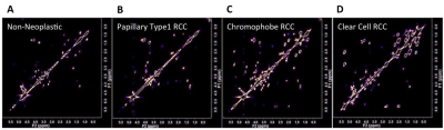
Two Dimensional COSY on Biopsy Distinguishes Indolent From Aggressive Kidney Masses
Aaron Urquhart, Sharon Del Vecchio, Lutz Krause, Robert Ellis, Keng Lim Ng, Hema Samaratunga, Sonja Gustafson, Graham Galloway, Glenda Gobe, Peter Malycha, Simon Wood, Carolyn Mountford
Kidney tumour metabolism may have chemical signatures that distinguish indolent from aggressive small renal masses (SRM). Indolent or benign SRM could be managed conservatively. Here we have used two dimensional (2D) COrrelation SpectroscopY (COSY) on nephrectomy samples to assign and measure metabolite and lipid resonances. High level bioinformatics analysis was then applied. We report metabolic chemical signatures that can be used to distinguish histologically-diagnosed kidney tumour subtypes.
|
|
0128.
 |
27 |
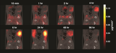
In vivo cancer detection and dynamics with magnetic particle imaging
Elaine Yu, Mindy Bishop, Bo Zheng, R Ferguson, Amit Khandhar, Scott Kemp, Kannan Krishnan, Patrick Goodwill, Steven Conolly
Magnetic Particle Imaging (MPI) is a novel, high-contrast, and quantitative imaging modality that directly detects superparamagnetic iron oxide nanoparticle (SPIO) tracers. These SPIOs have been previously used as a MRI contrast agent. However, with MRI SPIOs are limited by poor specificity and difficulty associated with quantifying the negative signal. MPI enables the direct detection of these SPIOs with both high sensitivity and positive contrast. MPI is well poised to support MRI in developing a clinically translatable cancer detection platform with SPIO. Here we demonstrate in vivo cancer detection and dynamic perfusion imaging with MPI.
|
|
0129.
 |
28 |
Ferumoxytol-enhanced MRI: Early Results in Solid Organ Masses.
Puja Shahrouki, Woo Kyoung Jeong, Steven Raman, Ely Felker, David Lu, J. Paul Finn
Whereas experience is growing rapidly with the use of ferumoxytol as an alternative to gadolinium based contrast agents (GBCA) for cardiovascular imaging, little has been reported about its potential for imaging of solid organs. The pharmacokinetics and relaxometry of ferumoxytol are very different to all of the available GBCA, and data are lacking about its behavior in extracranial solid organ lesions. We report early results of FE-MR imaging in a variety of solid organ mass lesions with focus on enhancement characteristics and clinical impact.
|
|
0130.
 |
29 |
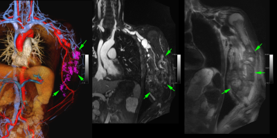
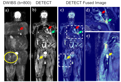
Whole-Body MRI for Metastatic Cancer Detection using T2-Weighted Imaging with Fat and Fluid Suppression
Xinzeng Wang, Ali Pirasteh, James Brugarolas, Neil Rofsky, Robert Lenkinski, Ivan Pedrosa, Ananth Madhuranthakam
Whole-body diffusion with background suppression (DWIBS) has increased sensitivity and specificity for metastatic cancer detection. However, DWIBS using echo-planar readout suffers from geometric distortions due to large B0 inhomogeneities associated with large FOV, used in whole-body MRI. Additionally, DWIBS suffers from low spatial resolution and long scan times due to low signal to noise ratio. In this work, we developed an alternative WB-MRI technique at 3T using fast, high-resolution and high-SNR T2-weighted imaging with simultaneous fat and fluid suppression, called DETECT. Patient studies show that, DETECT is time-efficient, robust, and generates distortion-free images with good lesion conspicuity, compared to DWIBS.
|
|
0131.
 |
30 |
Whole body functional and anatomical MRI: Accuracy in staging and interim response monitoring of Childhood and Adolescent Hodgkin’s Lymphoma compared to multimodality conventional imaging
Arash Latifoltojar, Shonit Punwani, Andre Lopes, Paul Humphries, Deena Neriman, Leon Menezes, Stephen Daw, Ananth Shankar, Bilyana Popova, K M Mak, Heather Fitzke, Paul Smith, Laura Clifton-Hadley, Stuart Taylor
Whole-body MRI (WB-MRI) offers an alternative non-ionising radiation technique to current gold-standard imaging, 18F-FDG PET-CT, for assessment of paediatric and adolescent Hodgkin's lymphoma (HL). In this work we prospectively evaluated WB-MRI, including diffusion-weighted-imaging (DWI), for initial staging and early interim response monitoring in 50 paediatric HL patients.
WB-MRI with DWI has reasonable intrinsic diagnostic accuracy for nodal and extra-nodal staging of paediatric HL but it fails to achieve full concordance with standard imaging for all disease sites in minority of patients.
WB-MRI has reasonable accuracy for interim response classification but tends to underestimate disease response, particularly in extra-nodal disease sites. |
|
