Poster: Molecular & Metabolic Imaging
Electronic Power Pitch Poster
Molecular Imaging
14:45 - 15:45
Monday, 18 June 2018
Power Pitch Theater A - Exhibition Hall
| |
|
Plasma # |
 |
0102.
 |
1 |
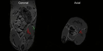
 A novel iterative sparse deconvolution method for multicolor 19F-MRI A novel iterative sparse deconvolution method for multicolor 19F-MRI
Jasper Schoormans, Claudia Calcagno, Mariah Daal, Christopher Faries, Brenda Sanchez-Gaytan, Aart Nederveen, Zahi Fayad, Willem Mulder, Bram Coolen, Gustav Strijkers
We introduce a new deconvolution approach for multicolor imaging of different 19F compounds with complex spectra. The method exploits the sparse nature of most 19F images by iterative sparse deconvolution, removes the chemical shift artifacts associated with multiple peaks in the 19F spectra, and efficiently separates multiple 19F compounds in the images. We performed numerical simulations and phantom experiments, which showed that the signal from two fluorinated compounds, i.e. PFOB and PFCE, can be separately detected without chemical shift artifacts. Moreover, we demonstrated chemical shift artifact-free multicolor detection of PFOB- and PFCE emulsion in a mouse.
|
|
0103.
 |
2 |
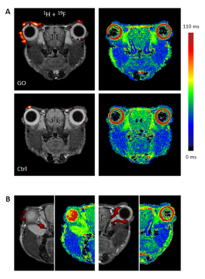
Multimodal Assessment of Orbital Immune Cell Infiltration and Tissue Remodeling During Development of Graves’ Disease by 1H/19F MRI
Ulrich Flögel, Anke Schlüter, Christoph Jacoby, Sebastian Temme, J Paul Benga, Anja Eckstein, Jürgen Schrader, Utta Berchner-Pfannschmidt
The purpose of the present study was to evaluate key molecular and cellular features of Graves’ orbitopathy by simultaneous monitoring of alterations in morphology, inflammatory patterns, and tissue remodeling. Beyond anatomical 1H MRI, we employed T2 mapping for visualization of edema, chemical exchange saturation transfer for detection of hyaluronan, and 19F MRI for tracking of in situ labeled immune cells after intravenous injection of perfluorcarbons. In particular, 19F MRI allowed a sensitive demarcation of inflammatory foci in the orbit, even when other markers indicated only weak or no signs of tissue alterations.
|
|
0104.
 |
3 |
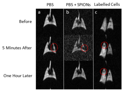
Hyperpolarized Xe-129 Imaging of Pluripotent Stem Cell-Derived Alveolar-Like Macrophages in the Lungs: Proof-of-Concept Study Using Superparamagnetic Iron-Oxide Nanoparticles
Vlora Riberdy, Michael Litvack, Elaine Stirrat, Marcus Couch, Martin Post, Giles Santyr
A promising approach to treatment of chronic lung diseases is the intratracheal delivery of pluripotent stem cell derived alveolar-like macrophages (PSC-ALMs) into the injured lung to facilitate repair of damaged tissue. This treatment is hindered by the inability to assess where in the lung these cells end up. Here, hyperpolarized Xe-129 MRI paired with iron-labeled cells was used to demonstrate a proof-of-concept visualization of cells introduced in the lungs of rats. Signal hypointensities were observed at least one hour after instillation of approximately two million labeled cells in one rat, compared to instillation of control solutions in separate rats.
|
|
0105.
 |
4 |
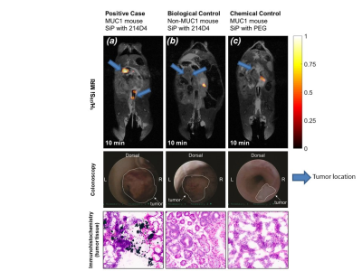
In Vivo Molecular Imaging of MUC1-Expressing Colorectal Tumors Using Targeted Hyperpolarized Silicon Particles
Did Not Present
Nicholas Whiting, Jingzhe Hu, Shivanand Pudakalakatti, Caitlin McCowan, Daniel Carson, Jennifer Davis, Niki Millward, David Menter, Pamela Constantinou, Pratip Bhattacharya
Silicon nano- and microparticles are well-suited for targeted molecular imaging, due to their biocompatibility, easily modifiable surface, and long-lasting 29Si MR signal (which can be enhanced by several orders of magnitude via dynamic nuclear polarization). We demonstrate targeted molecular imaging of human MUC1-expressing colon tumors in orthotopic mouse models using hyperpolarized 29Si MRI. The particles were able to selectively bind to MUC1-expressing tumors compared to controls, and the results were confirmed via histology of the excised tissue. The goal is to develop these targeted particles as a platform technology that will allow non-invasive screening of colorectal cancer using 29Si MRI.
|
 |
0106.
 |
5 |
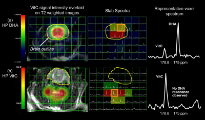
Imaging glutathione depletion in the rat brain using ascorbate-derived hyperpolarized MR and PET probes
Hecong Qin, Valerie Carroll, Renuka Sriram, Cornelius von Morze, Zhen Wang, Christopher Mutch, Kayvan Keshari, Robert Flavell, John Kurhanewciz, David Wilson
We studied brain redox capacity using ascorbate-derived hyperpolarized (HP) 13C MR and 11C PET probes. We first demonstrated the molecular transport and biodistribution features of the ascorbic acid (VitC)-dehydroascorbate (DHA) pair by leveraging both HP MR and PET modalities. We then showed that HP 13C DHA could detect redox modulation of the brain in a pharmacologic glutathione-depletion rat model. In conclusion, HP 13C DHA MR has the potential for non-invasive evaluation of brain redox capacity.
|
|
0107.
 |
6 |
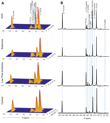
In-vivo metabolism of co-hyperpolarized [1-13C] pyruvate and [1,3-13C] acetoacetate identifies cytosolic and mitochondrial redox in ischemic perfused hearts
Gaurav Sharma, Craig Malloy, A. Dean Sherry, Chalermchai Khemtong
Cellular redox state is intricately linked with cardiac ischemia. Historically, tissue levels of lactate to pyruvate and β-hydroxybutyrate to acetoacetate have been used as indices of cytosolic and mitochondrial redox, respectively. The present study was designed to evaluate the potential of using co-hyperpolarized (HP) [1-13C] pyruvate and [1,3-13C] acetoacetate as reporters of cytosolic and mitochondrial redox, respectively. 13C NMR spectra of perfused rat hearts displayed increased production of both HP-lactate and HP-β-hydroxybutyrate in low flow ischemia, rotenone, and aminooxyacetate(AOA) treated hearts. The result suggests that HP-lactate and HP-β-hydroxybutyrate are sensitive metabolic markers of tissue redox in heart tissue.
|
|
0108.
 |
7 |
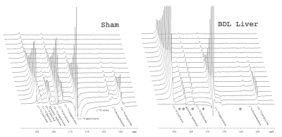
Probing perturbed hepatic metabolism in bile-duct-ligated rats with hyperpolarized 13C pyruvate and arginine
Hikari Yoshihara, Dmitri Firsov, Cristina Cudalbu, Rolf Gruetter
Detoxification of ammonia by the urea cycle and maintenance of glucose homeostasis by gluconeogenesis are two critical functions of the liver. The bile duct ligation (BDL) model of cirrhosis was used to test the ability of hyperpolarized [6-13C]arginine and [1-13C]pyruvate to detect changes in liver function. The conversion of hyperpolarized L-[6-13C]arginine to 13C-urea was observed in a sham-operated rat but not in BDL rats. Striking differences in pyruvate metabolism between the two groups were also noted, indicating that these probes can sense changes in hepatic mitochondrial and cytoplasmic metabolism associated with biliary cirrhosis.
|
|
0109.
 |
8 |
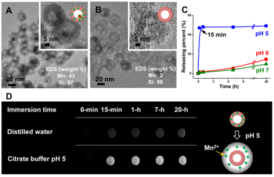
Hollow Manganese-Silicate (HMS) Nanoparticles as a Liver Specific MRI contrast agent
Video Permission Withheld
Moon-Sun Jang, Jin Goo Kim, Geun Ho Im, Jung Hee Lee, Won Jae Lee, In Su Lee
In this study, we demonstrate hollow manganese silicate nanoparticle (HMS) as a liver specific magnetic resonance imaging (MRI) contrast agent. HMS releases Mn2+ ions in the acidic physiological condition, which can be utilized for characterizing different tumor types.
|
|
0110.
 |
9 |
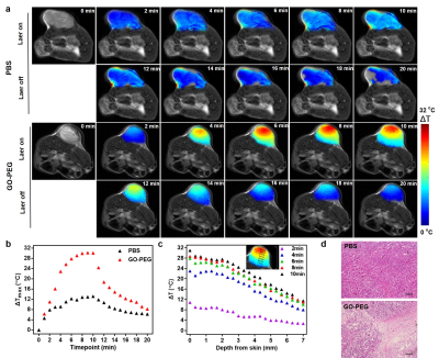
Magnetic Resonance Temperature Imaging for Nanoparticle-Mediated Tumor Photothermal Therapy
Did Not Present
Fu Guifeng, Guo Jianxin, Wei Xiaocheng, Zhang Fan, Yang Jian
Image-guided cancer therapy have the ability to integrate noninvasive imaging and minimally invasive interventions such as photothermal therapy (PTT) together to improve the precision of treatment. In the present study, we investigated magnetic resonance temperature imaging (MRTI) as a tool for non-invasive monitoring of tumor temperature distribution and changes during laser irradiation. To this end, we injected PEGylated graphene oxide (GO-PEG) to 4T1 tumor models and irradiated the tumors to induce PTT. Our studies demonstrate that MRTI, which is a feasible tool for the determination of temperature distribution, can be used to guide nanoparticle-mediated tumor photothermal therapy.
|
|
0111.
 |
10 |
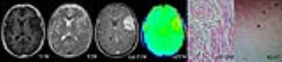
Amide proton transfer-weighted imaging in meningioma: Prediction of tumor grade, histologic subtype and association with Ki-67 proliferation status
Hao Yu, Xianlong Wang, Qihong Rui, Shanshan Jiang, Jinyuan Zhou, Zhibo Wen
A correct preoperatively predicting grade histologic subtype and tumor proliferation of meningioma is important in clinic treatment. APT imaging are designed to assess brain tumor on the level of cell and molecule. In this study we hoping to explore if this technique was useful in evaluating the meningioma comprehensively.
|
|
0112.
 |
11 |
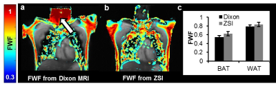
Brown Adipose Tissue Mass Measurement by Z-Spectrum Imaging
Alessandro Scotti, Rongwen Tain, Weiguo Li, Victoria Gil, Chong Liew, Kejia Cai
Brown adipose tissue (BAT) has a great relevance in metabolic diseases and has been shown to be reduced in obesity and insulin resistance patients. Currently, Dixon MRI is used to calculate fat-water fraction (FWF) and differentiate BAT from the less hydrated and more lipid-rich white fat. However, it may introduce fat-water swapping artifacts. Here, we showed that Z-Spectrum MRI can effectively measure FWF and BAT mass in vivo free from artifacts, due to the direct saturation of both water and fat protons.
|
|
0113.
 |
12 |
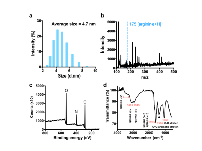
Carbon Nanodots as Diamagnetic CEST MRI Contrast Agents for Cell Labeling
Jia Zhang, Minling Gao, Yue Yuan, Yuguo Li, Peter van Zijl, Mingyao Ying, Guanshu Liu
Carbon nanodots (Cdots), are a relatively new type of eco-friendly nanoparticles that are being utilized extensively as drug carriers with inherent fluorescence imaging. In the present study, we explored their intrinsic chemical exchange saturation transfer (CEST) MRI contrast. We showed that Cdots can be directly detected by CEST MRI due to the abundant exchangeable hydrogen protons on their surfaces, which allows Cdots inherently bimodal contrast agents. Application to cell labeling was demonstrated in vitro. To the best of our knowledge, this is the first demonstration of diamagnetic nanoparticulate CEST MRI contrast agents.
|
|
0114.
 |
13 |
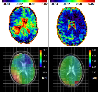
Correlation of tissue pH via 31P-MRSI with MTRasym derived from APT-CEST-MRI in glioblastoma and normal appearing white matter
Jan Schüre, Stella Breuer, Manoj Shrestha, Ralf Deichmann, Marlies Wagner, Ulrich Pilatus
The pH-value as physiological marker for clinical diagnosis can be measured with special hardware over 31P-MRSI. APT-CEST MRI offers an alternative method for such quantification but is challenging because of magnetization transfer from other tissue compartments. To evaluate the role of pH for APT-CEST, we investigated 12 patients with first diagnosis of glioblastoma and 11 healthy controls, based on asymmetric analysis of magnetization transfer (MTRasym) and calculated pH maps over 31P-MRSI.
Our results show a high correlation between both parameters and support the hypothesis that an enhancement of MTR asym at 3.5ppm is associated with increased pH.
|
|
0115.
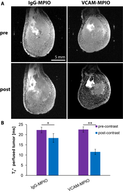 |
14 |
Imaging vascular inflammation as a marker for T-cell infiltration in preclinical tumor models.
Johannes Riegler, Vincent Javinal, Maj Hedehus, Jill Schartner, Richard Carano
Checkpoint inhibitors, adoptive T-cell transfer and tumor vaccination are different cancer immunotherapies which have demonstrated clinical efficacy in certain patients1-3. All of these therapies require sufficient infiltration of cytotoxic T-cells into the tumor and direct contact with cancer cells. However, the reasons why certain tumors present with high T-cell infiltration while others do not are poorly understood. We therefore set out to assess if imaging vascular adhesion molecule 1 (VCAM-1) expression in the tumor vasculature could explain some of the observed differences in T-cell infiltration.
|
|
0116.
 |
15 |
Translational Radiogenomics of Brain Tumors: From Lab-Invesigation to Clinical Application
Did Not Present
Dieter Heiland, Horst Urbach, Irina Mader
The interpretation of radiogenomic findings and its biological or clinical meaning is currently discussed and still unknown. Therefrom, a translational approach is aimed for integration of laboratory investigations and radiogenomic findings resulting in meaningful integration of radiogenomic discoveries. The presented study aimed to explore the molecular background of a MR-Spectroscopy imaging. In a stepwise approach findings were validated on tissue samples and finally taken into glioma cell models.
|
|
