Poster: Conductivity, Relaxation, Water-Fat & Beyond
Electronic Power Pitch Poster
Contrast Mechanisms
17:15 - 18:15
Tuesday, 19 June 2018
Power Pitch Theater A - Exhibition Hall
| |
|
Plasma # |
|
0533.
 |
1 |
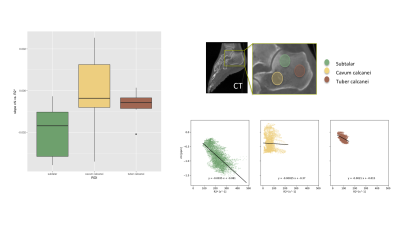
 On the Sensitivity of Bone Marrow Magnetic Susceptibility and R2* on Trabecular Bone Microstructure On the Sensitivity of Bone Marrow Magnetic Susceptibility and R2* on Trabecular Bone Microstructure
Maximilian Diefenbach, Anh Van, Jakob Meineke, Jan Kirschke, Benedikt Schwaiger, Thomas Baum, Alexandra Gersing, Dimitrios Karampinos
In numerical simulations and initial in vivo results of multi-parametric mapping in the calcaneus, the feasibility of measuring trabecular bone microstructure is explored. The combination of R2* for measuring intra-voxel dephasing and quantitative magnetic susceptibilty to detect trabecular bone density is investigated and indicates feasibility to differentiate trabecular bone networks with isotropic and anisotropic microstructure.
|
 |
0534.
 |
2 |
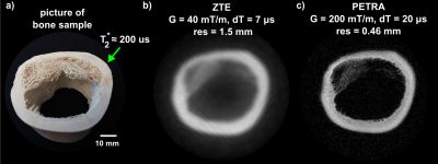
Pushing the limits of short-T2 MRI: 200 mT/m gradient strength and 2 MHz bandwidth
Romain Froidevaux, Markus Weiger, Manuela Rösler, David Brunner, Bertram Wilm, Benjamin Dietrich, Jonas Reber, Klaas Pruessmann
MRI of tissues with short transverse relaxation times below 1 millisecond such as bone or myelin raises both scientific and clinical interest. However, achieving high spatial resolution for short-T2 signals is challenging as large gradient strengths are required. Furthermore, large G implies high signal bandwidth, thus increasing the demands for short-T2 imaging techniques. Therefore, currently, short-T2 imaging faces significant restrictions with respect to spatial resolution and accessible T2s. In this work, all these challenges are tackled to expand the limits of short-T2 MRI using large G up to 200 mT/m and high BW up to 2 MHz.
|
|
0535.
 |
3 |
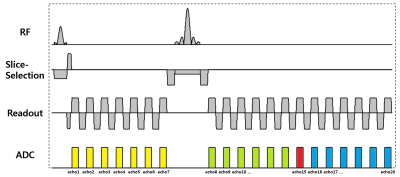
New post-processing methods for simultaneous measurement of R2, R2', R2*, QSM, positive and negative susceptibility maps using mGESFIDE acquisition
Dongmyung Shin, Se-Hong Oh, Doohee Lee, Jingu Lee, Jongho Lee
In this work, we demonstrated that a modified gradient-echo sampling of FID and echo (mGESFIDE) sequence was able to produce multiple contrast images (R2, R2', R2*, local field map, QSM, positive and negative susceptibility maps) in 5 minutes of scan time. We developed a new algorithm for improved R2 and R2' maps by considering RF slice profiles in the excitation and refocusing RF pulses. Additionally, we developed a new method that generated a high-quality local field map by utilizing all echoes of mGESFIDE.
|
|
0536.
 |
4 |
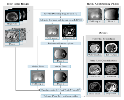
Repeatability and Reproducibility of a New Method for Quantifying Triglyceride Saturation Using Bipolar Multi-Echo MRI
Manuel Schneider, Felix Lugauer, Gemini Janas, Dominik Nickel, Brian M Dale, Berthold Kiefer, Andreas Maier, Mustafa R Bashir
Our purpose was to develop a robust method for joint quantification of water and fat fractions as well as fatty acid maps from bipolar multi-echo MR data. Its accuracy and reproducibility across field strengths and sequences was demonstrated using an oil phantom. Repeated in-vivo breath-hold acquisitions in n = 11 patients yielded median absolute differences of 4.8%, 1.0% and 8.2% for the saturated, mono-unsaturated and poly-unsaturated fat components in the liver, spleen and subcutaneous, perirenal and mesenteric fat depots.
|
|
0537.
 |
5 |
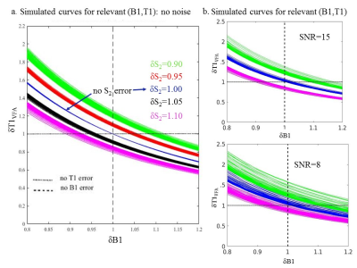
Calibrating variable flip angle (VFA)-based T1 maps: when and why a simple scaling factor is justified
Sofia Chavez
Variable Flip Angle (VFA)-based T1 maps are known to be prone to errors deriving from B1 errors (inaccurate knowledge of flip angles) and poor signal spoiling. In general, in vivo T1VFA values tend to overestimate T1 values obtained using a gold standard inversion recovery method :T1IR. Calibrating T1VFA with T1IR has been proposed but it requires knowledge of the exact relationship between these. This work models the contribution of B1 errors and poor spoiling to T1VFA errors and via simulations, the conditions for T1VFA/T1IR= constant (i.e. simple scaling) are derived. Experiments on phantoms and in vivo are used for validation.
|
|
0538.
 |
6 |
“In vivo” Field-Cycling relaxometry of tumours. Evidence for the role of the intracellular water lifetime as tumour biomarker.
Simonetta Geninatti Crich, Simona Baroni, Maria Rosaria Ruggiero, Stefania Pezzana, Gianni Ferrante, Silvio Aime
This work aims at developing an innovative diagnostic strategy, based on the "in vivo" measurements of longitudinal relaxation times at low and ultra-low magnetic fields with Fast Field Cycling FFC-NMR to obtain quantitative information on tumour metastatic potential, due to different water content and mobility, that is invisible to standard MRI. Preliminary results show that the endogenous contrast between normal and diseased tissue, due to differences in T1, is much greater at low field and the shape of the relaxation dispersion profiles may be used as a reporter of the molecular dynamical processes, biomarkers of the disease grade.
|
|
0539.
 |
7 |
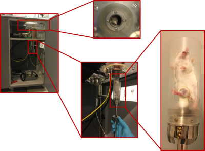
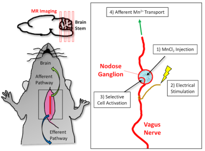
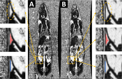
In-vivo Vagus Nerve to Central Nervous System Tracing using Manganese Enhanced Magnetic Resonance Imaging
Steven Oleson, Kun-Han Lu, Jiayue Cao, Zhongming Liu
Tracing neuronal connections of the peripheral and central nervous system has relied on invasive techniques that make it difficult to reconstruct information. We demonstrate the feasibility of utilizing manganese enhanced magnetic resonance imaging (MEMRI) and vagus nerve stimulation (VNS) to trace the vagus nerve to central nervous system (CNS) connections. This experimental approach shows the non-invasive visualization and quantified increased enhancement of manganese transport from the nodose ganglion to the left nucleus tractus solitaries (NTS).
|
|
0540.
 |
8 |
Fat Quantification Using A High-Resolution Bipolar Gradient Water-Fat Sequence
Alireza Akbari, Lanette Friesen-Waldner, Timothy Regnault, Charles McKenzie
In this work we demonstrate high-resolution bipolar water-fat imaging sequence produces same fat quantification as compared to conventional unipolar water-fat imaging sequence under the same scan time. Images of resolved boundaries in bipolar Proton Density Fat Fraction (PDFF) maps are presented. Fat quantifications of the same regions of interest drawn on bipolar and unipolar PDFF were compared and statistical analysis was performed to evaluate the similarity of the two methods.
|
|
0541.
 |
9 |
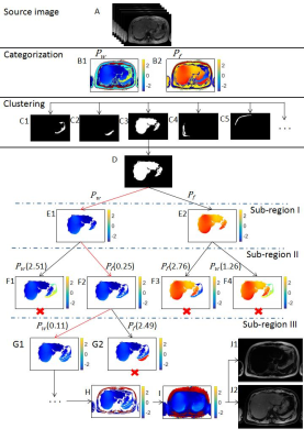
Robust Fat-water Separation using Binary Decision Tree Algorithm
Hao Peng, Chao Zou, Wenzhong Liu, Chuanli Cheng, Yangzi Qiao, Qian Wan, Changjun Tie, Xin Liu, Hairong Zheng
Purpose: To propose an robust fat water separation method using binary decision tree algorithm. Methods: In this paper, a novel fat-water separation algorithm using binary decision tree is proposed. Pixels are firstly clustered into sub-regions. Different from existing region growing algorithms, the proposed method solves the phasor ambiguity problem region by region. The method was tested on data sets from ISMRM 2012 Challenge.
Results:Fat-water separation were successfully achieved by the proposed method in the datasets.
Conclusion: A novel method using binary decision tree algorithm is proposed for robust and accurate water-fat separation.
|
|
0542.
 |
10 |
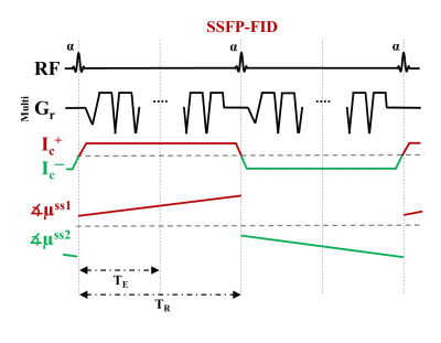
Human In-vivo Brain MR Current Density Imaging (MRCDI) based on Steady-state Free Precession Free Induction Decay (SSFP-FID)
Cihan Göksu, Lars Hanson, Hartwig Siebner, Philipp Ehses, Klaus Scheffler, Axel Thielscher
MRCDI is a novel technique for non-invasive measurement of weak currents in the human head, which is important in several neuroscience applications. Here, we present reliable in-vivo MRCDI measurements in the human brain based on SSFP-FID, yielding an unprecedented accuracy. We demonstrate the destructive influences of stray magnetic fields caused by the current passing through feeding cables, and propose a correction method. Also, we show inter-individual differences in MRCDI measurements for two different current profiles, and compare the measurements with simulations based on individualized head models. The simulations of the current-induced magnetic fields show good agreement with in-vivo brain measurements.
|
|
0543.
 |
11 |
Multi-receiver coil combination for breast phase-based Electrical Property Tomography Using B1- estimation
Jun-Hyeong Kim, Jaewook Shin, Soo-Yeon Kim, Dong-Hyun Kim
Phase-based EPT assumes the spatial homogeneity of both B1+ and B1- magnitude. However, when it comes to multi-receivers is used for improved SNR, the assumption becomes invalid especially for B1-. To overcome this problem, a subject-specific multi-Rx combination method was suggested [2]. However, this method can give a solution that is biased by the transmit field (B1+) when it is inhomogeneous. In this study, an alternative multi-Rx combination method is proposed. B1- is estimated from multi-receiver images by solving an inverse problem. Afterwards, optimal coil-coefficients for combination are calculated using the estimated B1- field. The method is applied to in-vivo breast conductivity imaging.
|
|
0544.
 |
12 |
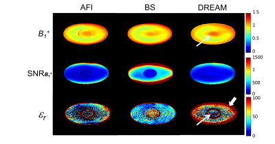
Electrical permittivity imaging at 3T: a precision and accuracy study of three $$$ \it{B_1^+}$$$ mapping techniques
Soraya Gavazzi, Cornelis van den Berg, Alessandro Sbrizzi, Mick Bennis, Lukas Stalpers, Jan Lagendijk, Hans Crezee, Astrid van Lier
The feasibility of permittivity imaging relies on high precision of the underlying $$$\it{B_1^+}$$$ amplitude maps. We tested AFI, Bloch-Siegert and DREAM $$$\it{B_1^+}$$$ mapping techniques on a pelvic-sized phantom at 3T, comparing their SNR in $$$\it{B_1^+}$$$ maps and (resulting) permittivity precision. Our results indicated that the DREAM-based permittivity map was the most sensitive to sequence-related systematic errors. The commonly-used AFI technique, instead, was the least precise method. We also found that Bloch-Siegert is generally best suited for permittivity mapping compared to the other two methods, due to its higher $$$\it{B_1^+}$$$ precision and accuracy.
|
|
0545.
 |
13 |
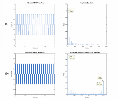
Multi-frequency MREIT Demonstrated using Semipermeable Membrane Models
Munish Chauhan, Andrew Xi, Neeta Ashok Kumar, Fanrui Fu, Rosalind Sadleir
Magnetic Resonance Electrical Impedance Tomography (MREIT) has been used to measure low frequency (ca. 10 Hz) electrical conductivity properties. Here, finite element simulations are used to show that it should be possible to measure electrical properties at frequencies in the range 10- ~5000 Hz using current waveform modulation. We also show imaging results that demonstrate differential signals can be measured in phantoms containing dialysis membranes with different thicknesses and permittivity characteristics.
|
|
0546.
 |
14 |
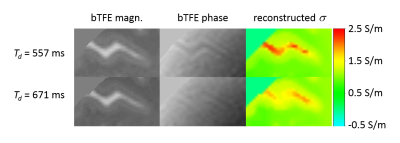
The impact of CSF pulsation on reconstructed brain conductivity
Ulrich Katscher, Christian Stehning, Khin Tha
It has recently been shown that the WHO grade of a glioma is correlated with its electric conductivity, which can be determined via post-processing the acquired MR transceive phase. However, the transceive phase and thus the derived conductivity might be corrupted by motion induced phase, particularly from cardiac pulsation transferred to the cerebrospinal fluid (CSF). In this study, the impact of CSF pulsation on the reconstructed conductivity was investigated by synchronizing the transceive phase acquisition to the cardiac cycle. It turned out that the pulsation significantly affects reconstructed conductivity of the CSF, but not of gray and white matter.
|
|
0547.
 |
15 |
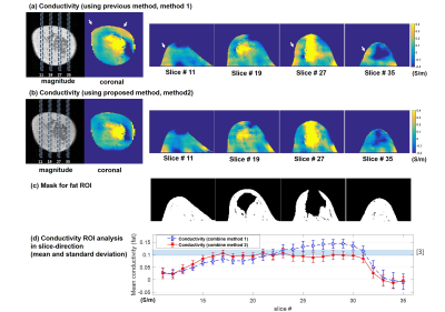
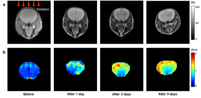
In Vivo Conductivity Imaging of Tissue Response after Radiation Therapy
In Ok Ko, Bup Kyung Choi, Nitish Katoch, Ji Ae Park, Jin Woong Kim, Hyung Joong Kim, Oh In Kwon, Eung Je Woo
Radiation therapy (RT) has been widely used as a powerful treatment to remove cancerous tissues because of its ability to control cell growth. Ionizing radiation works by damaging the DNA of cancerous tissue leading to cellular death. Medical imaging has limitations on credibility for evaluation of tissue response and prediction of therapeutic effect due to lacks of contrast information on gradual and minute tissue changes after RT. Conductivity mapping after RT may provide direct and high sensitive information on tissue response because its contrast mechanism originated from the concentration and mobility of ions in the extra- and intracellular environment.
|
|
