Poster: Advances in Contrast: MT, CEST & Perfusion
Electronic Power Pitch Poster
Contrast Mechanisms
17:15 - 18:15
Tuesday, 19 June 2018
Power Pitch Theater B - Exhibition Hall
| |
|
Plasma # |
|
0548.
 |
16 |
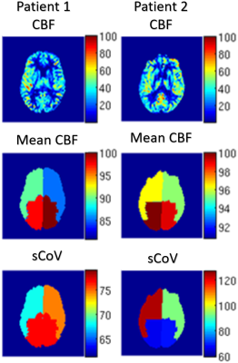
 Rethinking vascular artifacts: testing the sensitivity of ASL vascular signal as a biomarker of disease Rethinking vascular artifacts: testing the sensitivity of ASL vascular signal as a biomarker of disease
Zachary Mulhollan, Henk-Jan Mutsaerts, Jan Petr, Ronald Lazar, Randolph Marshall, Iris Asllani
Collateral perfusion has a major effect on clinical outcome in carotid steno-occlusive disease. Arterial spin labeling (ASL) may provide a biomarker of collateral perfusion by measuring arterial transit time (ATT) in addition to cerebral blood flow (CBF). However, the concomitant measurement of ATT and CBF is not feasible for clinical applications. As an alternative, ATT can be estimated from single post-labeling delay ASL image using the spatial coefficient of variance (CoV). In this study, we investigate whether the spatial CoV lateralization through collateral perfusion can predict the side of carotid occlusion as a proof-of-principle. In addition to spatial CoV, we also investigated whether temporal variance (tVAR) of the ASL signal could be used as a predictor of occlusion versus stenosis.
|
|
0549.
 |
17 |
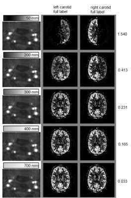
Exploiting small fluctuations in labeling efficiency in pseudo-continuous arterial spin labeling for combined flow-territory determination and CBF-mapping
Thijs van Harten, Matthias van Osch
Since the labeling efficiency in pCASL is dependent on velocity and off-resonance effects, one could imagine that small natural occurring fluctuations at the labeling plane would lead to similar fluctuations in the ASL-signal. Such fluctuations at the brain level would be similar for voxels belonging to the same flow territory and might enable detection of flow territories, e.g. by an ICA-type of resting-state ASL analysis that is normally used for identification of neuronal networks. By deliberately manipulating the labeling efficiency, we can report that with small fluctuations in labeling efficiency flow territories can be determined, which diminish in robustness for smaller fluctuations.
|
|
0550.
 |
18 |
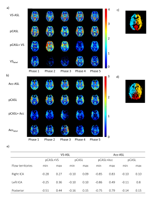
The Influence of the cardiac cycle on velocity-selective and acceleration-selective Arterial Spin Labeling
Suzanne Franklin, Sophie Schmid, Clemens Bos, Matthias van Osch
In this study we investigated whether velocity-selective arterial spin labeling (VS-ASL) and acceleration-selective spin labeling (Acc-ASL) can be used to measure pulsatility in the microvascular system of the brain, since the amount of generated label will depend on the velocity or acceleration of the spins at the moment the VS-ASL (Acc-ASL) module is applied. Results showed no significant variation in the amount of label when applying these modules at different cardiac phases. However, the generated label did show a pattern which coincides with the flow territories, suggesting a underlying physiological cause.
|
 |
0551.
 |
19 |
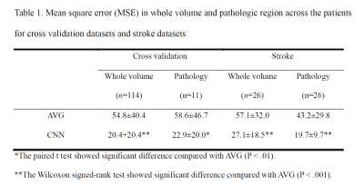
Improving Arterial Spin Labeling using Deep Learning
Ki Hwan Kim, Seung Hong Choi, Sung-Hong Park
We proposed a new convolutional neural network (CNN) framework to quantify cerebral blood flow (CBF) in Hadamard-encoded pseudo-continuous arterial spin labeling (HE-pCASL). Improving sensitivity and robustness in ASL signals allows CNNs to quantify CBF accurately with a smaller number of data acquisitions. The proposed methods outperformed the conventional averaging method in both normal and pathologic regions. Therefore, CNNs can be a good alternative to quantify CBF in ASL imaging.
|
|
0552.
 |
20 |
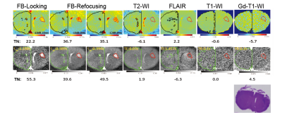
Contrast Enhancement for Early Tumor Detection by Active-Feedback Field Locking and Refocusing
Video Permission Withheld
Fang-Chu Lin, Chao-Hsiung Hsu, Yung-Ya Lin
Early detection of high-grade malignancy using novel contrast mechanisms and detection methods significantly increases the treatment options and the patients’ survival rate. For this purpose, the local magnetic-field-gradient variations due to irregular water contents and deoxyhemoglobin concentration in early glioblastoma multiforme (GBM) is sensitively detected by active-feedback electronic devices and nonlinear spin dynamics. The active-feedback phases and strength were tuned to manipulate the underlying spin dynamics and thus optimize the feedback-induced avalanching spin amplification effect. In vivo results from GBM mouse models show that up to 12 times of improved tumor-to-normal-tissue contrasts can be achieved to highlight early GBM.
|
|
0553.
 |
21 |
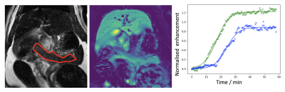
Measurement of artifact-free arterial input functions for T1-weighted dynamic contrast-enhanced MRI: Inter- and intra-patient variability
Leonidas Georgiou, Daniel Wilson, Nisha Sharma, David Buckley
In 2006, Parker et al published a population-average AIF that has subsequently been used by numerous investigators for both simulation and data analysis. Despite its utility, the published AIF suffers from a number of limitations. In this study we tried to address many of these limitations and made measurements in a population of patients with advanced breast cancer, resulting in a total of 74 individual AIFs. We fitted each AIF with a physiological model and assessed inter- and intra-patient variability.
|
|
0554.
 |
22 |
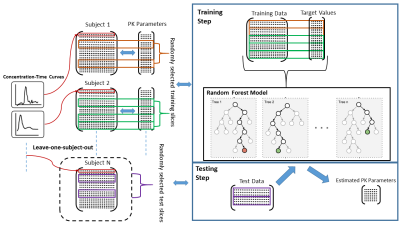
Estimation of Pharmacokinetic Parameters in Dynamic Contrast Enhanced MRI via Random Forest Regression
Cagdas Ulas, Michael Thrippleton, Ian Marshall, Mike Davies, Paul Armitage, Stephen Makin, Joanna Wardlaw, Bjoern Menze
We propose a novel alternative approach to estimate pharmacokinetic (PK) parameters of dynamic contrast enhanced (DCE)-MRI. Our approach leverages machine learning field and mainly targets to automatically learn temporal patterns of the voxel-wise concentration-time curves (CTCs) from a large amount of training samples in order to make accurate parameter estimations. We consider the estimation of parameters as a regression problem and specifically use Random Forest (RF) regression. We demonstrate its potential and utility to improve the conventional model-fitting based quantitative analysis of DCE-MRI especially in various noise conditions, and validate our method on clinical brain stroke datasets.
|
|
0555.
 |
23 |
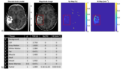
Influence of whole-brain DCE-MRI (k,t) sampling strategies on variance of pharmaco-kinetic parameter estimates
Yannick Bliesener, Sajan Lingala, Justin Haldar, Krishna Nayak
We investigate the influence of 2D $$$(k_y,k_z,t)$$$ sampling strategies on the minimum achievable variance without bias for pharmaco-kinetic parameter estimation in 3D whole-brain DCE-MRI (equivalent to the best possible precision without bias). Cramér-Rao analysis is combined with a pathologically- and anatomically-realistic digital reference object to objectively compare measurement procedures independent of any estimator. This study did not identify any significant difference between lattice and random undersampling, or between their uniform and variable density variants.
|
|
0556.
 |
24 |
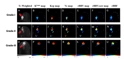
Characterization of Breast Lesion using T1-perfusion MRI: Semi- Quantitative Vs Quantitative Analysis
Video Permission Withheld
Snekha Thakran, Anup Singh, Pradeep Gupta, Vedant Kabra, Rakesh Gupta
In this study role of hemodynamic parameters (breast-blood-volume and breast blood-flow), obtained using T1-perfusion MRI data of breast, in the differentiation of benign from malignant breast lesions and classification of malignant lesions into different grades is evaluated. Hemodynamic parameters were also compared with the tracer kinetic parameters and semi-quantitative T1-perfusion analysis in term of grading. The high sensitivity and specificity of breast-blood-volume in differentiating between benign and malignant as well as in the grading of breast lesions (grade-I, grade-II and grade-III) were observed.
|
|
0557.
 |
25 |
Manganese-enhanced MRI: comparison of agents in the rat pancreas
Lucy Kershaw, David Lilburn, Maurits Jansen, Pilar Jimenez-Royo, Antonella Napolitano Rosen, Philip Murphy, Alexandra Morgan, Rob Janiczek, Shareen Forbes, Scott Semple
Type 1 diabetes mellitus results in autoiummune destruction of β-cells in the pancreas, which are responsible for insulin production. Paramagnetic Mn2+ ions are taken up by β-cells as a calcium analogue and could be used as an MR contrast agent to monitor β-cell function and therefore treatment or disease progression in these patients. Three manganese-based contrast agents (MnCl2, mangafodipir and Mn gluconate) were used to measure pancreas enhancement after saline and glucose challenge in healthy rats. All agents showed greater enhancement after glucose challenge, with no marked difference between the two agents that have been used clinically.
|
|
0558.
 |
26 |
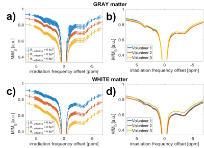
Novel contrasts at +2.7 ppm, +1.2 ppm, and -1.7 ppm investigated in vivo with high spectral resolution CEST MRI in the human brain at 9.4T
Mark Schuppert, Kai Herz, Anagha Deshmane, Moritz Zaiss
Using volumetric snapshot-GRE CEST MRI at 9.4T with high frequency sampling, we were able to separate novel CEST peaks at +2.7 ppm, +1.2 ppm and -1.7 ppm reliably in the CEST-spectrum and showed creation of maps of these CEST MRI contrasts in the healthy human brain to be feasible in vivo.
|
|
0559.
 |
27 |
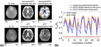
A Novel MR Fingerprinting Approach for Fast Quantitative Chemical Exchange Saturation Transfer MRI Analysis by Subgrouping Proton Exchange Models (CEST-SPEM)
Hye-Young Heo, Shanshan Jiang, Peter van Zijl, Jinyuan Zhou
Most current amide proton transfer (APT) imaging protocols, a variant of CEST-MRI, are essentially not quantitative and acquire qualitative APT-weighted images, limiting pH or concentration specificity. Herein, we propose a novel dictionary-free MRF concept to allow APT quantification by subgrouping proton exchange models (CEST-SPEM). The results of numerical phantom studies demonstrate that CEST-SPEM can enable absolute quantification of the APT exchange rate and concentration on 3T clinical scanners. The same model used for in vivo allows fast quantification of exchange rates and apparent concentrations for the combined solute components at the CEST frequency studied.
|
|
0560.
 |
28 |
Evaluating Feasibility of Creatine-weighted CEST MRI in Human Brain at 7T using Z-spectral Fitting Approach
Anup Singh, Mohammad Haris, Ayan Debnath, Kejia Cai, Hari Hariharan, Puneet Bagga, Ravinder Reddy
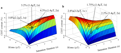
Creatine(Cr)-weighted chemical-exchange-saturation-transfer(CEST) MRI is being developed to detect alteration in Cr concentration during modulation as well as Cr-associated disorders. Cr-weighted CEST contrast show dependence on saturation parameters and overlap from other effects in brain. The conventional asymmetry approach results in a mixed contrast, which is less specific to Cr. By using appropriate saturation parameters, contamination from some of the metabolites/molecules can be reduced; however, it is difficult to suppress the contamination completely as shown by simulation studies. Proposed protocol and improved z-spectral fitting approach can be used for computing Cr-weighted CEST contrast with reduced contamination in human brain at 7T.
|
|
0561.
 |
29 |
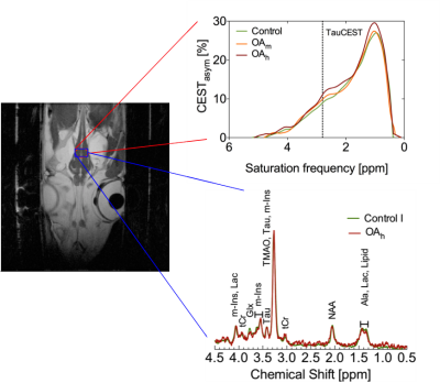
CO2 induced pHi changes in the brain of polar fish: a TauCEST application
Felizitas Wermter, Bastian Maus, Hans-Otto Pörtner, Wolfgang Dreher, Christian Bock
Chemical exchange saturation transfer from taurine to water (TauCEST) is primarily detectable in the low temperature range. Since, TauCEST asymmetry is bijective in the physiological pH-range (6.8-7.5), TauCEST is a potential candidate for in vivo studies on brain of polar fish. The specificity of TauCEST MRI on the brain of polar cod at 1.5°C shows a taurine contribution of 65%. TauCEST in brain of polar cod significantly increased under elevated CO2 concentrations by about 1.34%-3.17% in comparison to control, reflecting pHi changes since localized 1H NMR spectra show no significant changes in metabolite concentration for the different treatments.
|
|
0562.
 |
30 |
Characterizing the sensitivity of ihMT for various dipolar relaxation times (T1D) at high RF power using frequency-alternated and cosine-modulated RF pulses for dual frequency-offset saturation
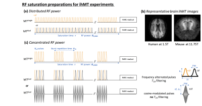
Guillaume Duhamel, Samira Mchinda, Valentin Prevost, Victor Carvalho, Gopal Varma, David Alsop, Olivier Girard
In the present study, we used both numerical simulations and experiments performed in a preclinical setup to characterize the mechanisms of the ihMT boost effect (signal enhancement achieved using concentrated bursts of high-power RF for saturation). We demonstrated that the ihMT boost effect depends on T1D and is more intense in short-T1D components. This feature allowed, using various strategies for dual frequency-offset RF saturation, different ihMT contrasts to be produced and revealed strong ihMT signal outside the brain.
|
|
