Poster: Motion & Image Analysis Highlights
Electronic Power Pitch Poster
Acquisition, Reconstruction & Analysis
09:15 - 10:15
Wednesday, 20 June 2018
Power Pitch Theater B - Exhibition Hall
| |
|
Plasma # |
 |
0653.
 |
16 |
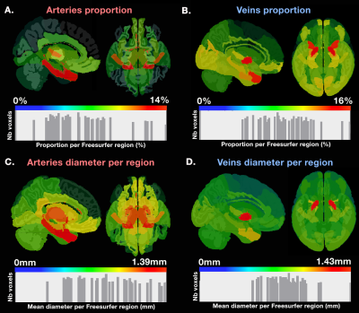
 The spatial distribution of arterial and venous vessels in the human brain The spatial distribution of arterial and venous vessels in the human brain
Michaël Bernier, Stephen Cunnane, Kevin Whittingstall
Although human cerebrovascular system is the basis of most non-invasive measures of neural activity, its structure is poorly understood owing to the difficulty in identifying, segmenting and separating venous and arterial vessels. To resolve this, we used Susceptibility Weighting Imaging (SWI) and Magnetic Resonance Angiography Time-of-Flight (MRA-TOF) to develop a probabilistic template of vascular architecture in the MNI space using an iterative back projection approach. This template is then paired with an anatomical atlas illustrates how some grey-matter areas are more vascularized than others. This could be the first steps toward a region-based vascular regression tool for the analysis hemodynamic-based measures of brain activity, such as fMRI.
|
|
0654.
 |
17 |
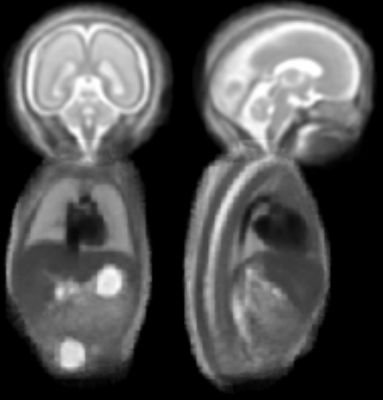
A spatiotemporal fetal MRI atlas for multi-organ segmentation and growth analysis
Video Permission Withheld
Tong Zhang, Maria Deprez, Paul Aljabar, Robert Wright, Alice Davidson, Mary Rutherford, Jo Hajnal, Julia Schnabel
We propose a comprehensive framework for spatiotemporal multi-organ fetal MR atlasing and segmentation. Volumetric fetal brain and trunk image data for 78 healthy fetuses acquired at different gestational age were reconstructed separately from motion corrupted MRI scans to 0.75mm isotropic resolution using slice-to-volume reconstruction. For each week, an affine template was generated from 15 subjects by averaging them in the atlas space. This template was further improved by non-linear registration of all the subjects using B-spline free-form deformations. For consistency, the template at each week was rigidly transformed. The annotated organs of the atlas include brain, lung, heart, and spine.
|
 |
0655.
 |
18 |
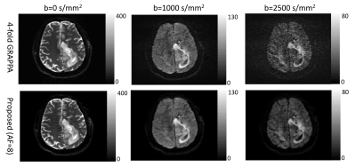
Accelerated high b-value diffusion-weighted MRI for higher-order diffusion analysis using a phase-constrained low-rank tensor model
Lianli Liu, Adam Johansson, James Balter, Jeffrey Fessler, Yue Cao
DWI acquired with b-values greater than 1000 s/mm2 and higher-order diffusion analyses based on such DWI series have the potential to improve tumor differentiation, while the extended sampling of b-values makes the acquisition time inconveniently long. We propose an acceleration scheme that sparsely samples k-space and reconstructs images using a new low-rank tensor model which exploits both global and local low-rank structure. Under an acceleration factor of 8, parameter mapping results on one simulated and 7 patient datasets show improved accuracy over another low-rank tensor model that exploits global correlation only, and comparable accuracy to clinically used four-fold GRAPPA reconstruction.
|
|
0656.
 |
19 |
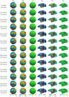
Construction of Spatiotemporal Infant Cortical Surface Atlas of Rhesus Macaque
Fan Wang, Chunfeng Lian, Jing Xia, Zhengwang Wu, Dingna Duan, Li Wang, Dinggang Shen, Gang Li
Rhesus macaque is a widely used animal model helping understand neural development in the human brain. Available adult macaque brain atlases are not suitable for infant studies, due to their dramatic brain difference. Building age-matched atlases is thus highly desirable yet still lacking. In this study, using MRI scans for 32 healthy rhesus monkeys, we constructed the first spatiotemporal cortical surface atlas for macaques aged from 2 weeks to 24 months, which were further equipped with developmental-trajectory-based parcellation maps. These surface atlases and parcellation maps will greatly facilitate the early brain development studies of macaques.
|
|
0657.
 |
20 |
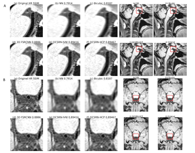
Efficient Super-Resolution in Intracranial Vessel Wall Magnetic Resonance Imaging using 3D Deep Densely Connected Neural Networks
Yuhua Chen, Zhaoyang Fan, Feng Shi, Zixiao Tian, Anthony Christodoulou, Yibin Xie, Debiao Li
Spatial resolution is of paramount importance for intracranial vessel wall (IVW) MR imaging because the vessel wall is submillimeter thin. However, high spatial resolution typically comes at the expense of long scan time, small spatial coverage and low signal-to-noise ratio (SNR). If we can reconstruct a high-resolution (HR) image from a low-resolution (LR) input, we can potentially achieve larger spatial coverage, higher SNR and better spatial resolution in a shorter scan. In this work, we propose a new Single Image Super-Resolution(SISR) technique which recovers an HR image from an LR image using 3D Densely Connected Super-Resolution Networks (DCSRN). We compared our network with state-of-art deep learning network in restoring 4x down-graded images and ours are faster and better.
|
|
0658.
 |
21 |
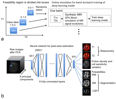
Simultaneous Relaxometry and Segmentation of Human Brain through a Deep Neural Network
Peng Cao, Jing Liu, Shuyu Tang, Andrew Leynes, Peder Larson
This study demonstrated a method for 3D simultaneous relaxometry and segmentation of human brain tissues through a deep neural network. Ranges of T1 and T2 values for gray matter, white matter and cerebrospinal fluid (CSF) were used as the prior knowledge. The proposed method can directly generate brain T1 and T2 maps in conjunction with segmentation of gray matter, white matter and CSF, and in particular was robust for relaxometry/segmentation in the challenging region of deep brain nuclei.
|
|
0659.
 |
22 |
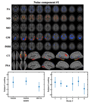
Combining Multi-Site/Study MRI Data: A Novel Linked-ICA Denoising Method for Removing Scanner and Site Variability from Multi-Modal MRI Data
Huanjie Li, Staci Gruber, Stephen Smith, Scott Lukas, Marisa Silveri, Kevin Hill, William Killgore, Lisa Nickerson
Large multi-site studies that pool magnetic resonance imaging (MRI) data across research sites present exceptional opportunities to advance neuroscience and enhance reproducibility of neuroimaging research. However, inconsistent MRI data collection platforms and scanning sequences both introduce systematic variability that can confound the true effect of interest and make the interpretation of results obtained from combined data difficult. Unfortunately, methods to address this problem are scant. In this study, we propose a novel denoising approach for multi-site, multi-modal MRI data that implements a data-driven linked independent component analysis to efficiently identify scanner/site-related effects for removal.
|
|
0660.
 |
23 |
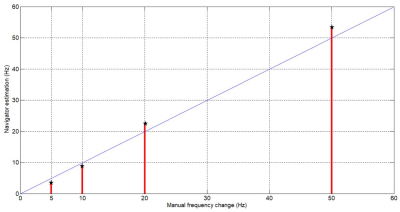
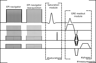
Real-time simultaneous shim and motion measurement and correction in CEST MRI using Double volumetric Navigators (DvNavs)
Gizeaddis Simegn, Andre Van der Kouwe, Borjan Gagoski, Frances Robertson, Ernesta Meintjes, Ali Alhamud
In Chemical Exchange Saturation Transfer (CEST) MRI, images are acquired by applying saturation RF pulses at multiple frequencies of small increments to generate the CEST-spectrum. This makes CEST sensitive to motion and field inhomogeneity. Several factors can also vary the shim prepared by the scanner. To date, no study has been conducted to evaluate and correct shim fluctuation during CEST acquisition. In this study, we implement CEST with Double volumetric Navigators (DvNavs) to evaluate and update in real-time motion, zero and first-order shim parameters. The results show the ability of the DvNavs to correct shim and motion during CEST acquisition.
|
|
0661.
 |
24 |
Real-time motion and dynamic transmit/receive B1 correction of CEST in the human brain at 7T
Sami Auno, Esau Poblador Rodriguez, Philipp Moser, Andre v.d.Kouwe, Stephan Gruber, Siegfried Trattnig, Wolfgang Bogner
Movement during MR imaging may cause significant motion artifacts that can impair the experiment. This is especially true in CEST imaging, which relies on comparison between images recorded at different time points during the experiment. We implemented a real-time motion correction method and a retrospective receiver sensitivity(B1-) correction based on volumetric EPI navigators interleaved with the CEST measurements. We tested these methods on phantoms and healthy volunteers. Together with adequate B0and B1+ corrections, motion and sensitivity corrections may completely restore CEST data fidelity in the presence of involuntary head motion, thereby facilitating CEST imaging of restless patients.
|
|
0662.
 |
25 |
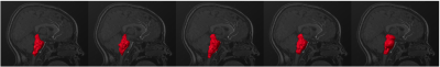
Brainstem abnormalities in structural MRI of young children with Autism Spectrum Disorder: evaluation of inter-method agreement.
Paolo Bosco, Alessia Giuliano, Jonathan Delafield-Butt, Filippo Muratori, Sara Calderoni, Alessandra Retico
Structural MRI studies have pointed out the potential role of the brainstem in the pathophysiology of ASD. However, the findings in volume alterations in subjects with ASD are controversial. In this study, structural MRI was used to measure brainstem volume in a group of 152 young children with and without ASD, with five different methods (FSL-FIRST, ANTs, FS 5.3, FS 6.0, FS 6.0 with substructures). One out of five (FSL-FIRST) showed poor agreement with the other segmentation methods, which, by contrasts, consistently showed Pearson correlations greater than 0.93 and average Dice indexes greater than 0.76 in comparison among each other.
|
|
0663.
 |
26 |
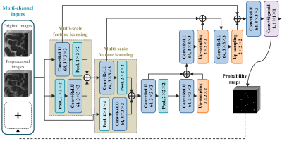
Automatic Segmentation of 3D Perivascular Spaces in 7T MR Images Using Multi-Channel Fully Convolutional Network
Chunfeng Lian, Mingxia Liu, Jun Zhang, Xiaopeng Zong, Weili Lin, Dinggang Shen
Advanced 7T MR imaging improves the visualization of perivascular spaces (PVSs) in human brains. However, accurate PVS segmentation for quantitative morphological studies is still challenging, since PVSs are very thin tubular structures with low contrast in noisy MR images. We proposed a new multi-channel fully convolutional network (mFCN) to automatically segment 3D PVSs. Our mFCN method adopts multi-channel inputs to complementarily provide enhanced tubular structural information and detailed image information. Multi-scale image features are automatically learned to delineate PVSs, without requirement of any pre-defined regions of interest (ROIs). The proposed method outperforms existing automatic/semi-automatic methods with a large margin.
|
|
0664.
 |
27 |
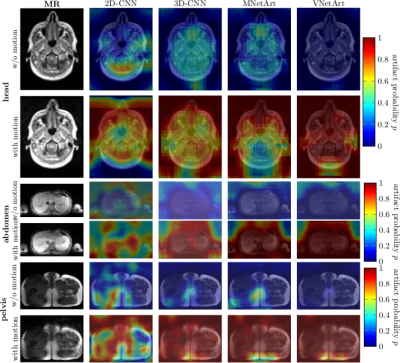
Motion artifact quantification and localization for whole-body MRI
Thomas Kuestner, Marvin Jandt, Annika Liebgott, Lukas Mauch, Petros Martirosian, Fabian Bamberg, Konstantin Nikolaou, Sergios Gatidis, Bin Yang, Fritz Schick
Motion is still one of the major extrinsic factors degrading image quality. Automated detection of these artifacts is of interest, (i) if suitable prospective or retrospective correction techniques are not available/applicable, (ii) if human experts who judge the achieved quality are not present, or (iii) if a manual quality analysis of large databases from epidemiological cohort studies is impracticable. A convolutional neural network assesses and localizes the motion artifacts. This work extends the previously published method by proposing a general architecture for a whole-body scenario with varying contrast weightings. High accuracies of >90% were achieved in a volunteer study.
|
|
0665.
 |
28 |
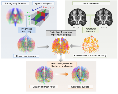
Tract-Based Cluster Analysis
Pedro Luque Laguna, Francisco de Santiago Requejo, Steven Williams, Laura Goldstein, Marco Catani, Flavio Dell'Acqua
The use of a-priori anatomical information can effectively improve statistical analysis of neuroimaging data. In this work, we introduce a new method called Tract-Based Cluster Analysis (TBCA) that exploits the rich anatomical information present in a whole-brain tractogram to inform the cluster-level inference analysis of voxel-based images. The method is based on the novel concept of hyper-voxel which incorporates local and global anatomical information derived from tractography data. When applied to real clinical data TBCA demonstrates clear benefits compared to previous cluster-level inference approaches.
|
|
0666.
 |
29 |
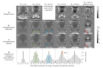
SEGUE: a Speedy rEgion-Growing algorithm for Unwrapping Estimated phase
Anita Karsa, Karin Shmueli
MRI phase images are increasingly used, for example for Susceptibility Mapping, and distortion correction in functional and diffusion MRI. However, measured phase images contain wraps, because the phase is defined only between 0 and 2π. PRELUDE is the current gold-standard method for robust, 3D, spatial phase unwrapping, but its computation time can become very long (e.g. 10 hours), especially at high field and outside the brain. Here, we developed a new method, SEGUE, that produced similar results to PRELUDE in multi-echo brain and head-and-neck images, successfully unwrapped some regions where PRELUDE failed and was between 1.6 and 83 times faster.
|
|
0667.
 |
30 |
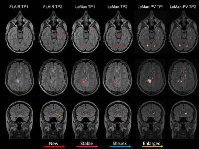
The Role of Partial Volume Modelling in Longitudinal Automated Multiple Sclerosis Lesion Segmentation
Mário João Fartaria, Tobias Kober, Cristina Granziera, Meritxell Bach Cuadra
Longitudinal analyses in Multiple Sclerosis are often performed to assess disease progression and evaluate treatment response. The number of new and enlarged lesions as well as total lesion volume variations over time are imaging biomarkers used in MS follow-up assessment. Here, we evaluate the performance of an in-house prototype algorithm for lesion detection and volume estimation in a longitudinal scenario. Our algorithm can be run with or without partial volume modelling. Both detection and volume estimation improved using the partial volume model with respect to manual delineations, especially in small lesions and at lesion borders.
|
|
