Poster: Tissue Microstructure
Electronic Power Pitch Poster
Diffusion
14:15 - 15:15
Thursday, 21 June 2018
Power Pitch Theater A - Exhibition Hall
| |
|
Plasma # |
|
1092.
 |
1 |
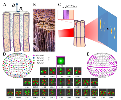
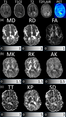
 Probing microstructure with different tomographic methods: Comparing dMRI and X-ray scattering-derived parameters in mouse and human brains Probing microstructure with different tomographic methods: Comparing dMRI and X-ray scattering-derived parameters in mouse and human brains
Marios Georgiadis, Dmitry Novikov, Manuel Guizar-Sicairos, Marianne Liebi, Vivianne Lutz-Bueno, Benjamin Ades-Aron, Timothy Shepherd, Aileen Schroeter, Markus Rudin, Els Fieremans
Despite MRI’s coarse resolution, diffusion MRI (dMRI) enables probing cellular microstructure. Advanced dMRI acquisition and biophysical modeling can provide microstructural metrics related to disease processes, and spherical harmonics (SH)-based orientation distribution function (ODF). Yet, these still need structural validation, and a gold standard for quantifying microstructure, particularly fiber dispersion, is missing. X-ray scattering directly probes tissue microstructure, exploiting the ~17nm myelin repeat distance, and can also represent ODF in a SH basis. Here, we show good correspondence between SH coefficients from dMRI and X-ray scattering on a mouse brain, with analysis on human samples and histological validation to follow.
|
|
1093.
 |
2 |
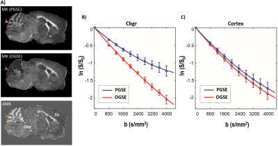
Diffusion-time dependence of diffusional kurtosis in the mouse brain using pulsed and oscillating gradients
Manisha Aggarwal, Kyle Martin, Matthew Smith, Peter Calabresi
Non-vanishing excess kurtosis and diffusion-time dependence are two key hallmarks of non-Gaussian or restricted water diffusion in complex brain tissue microenvironments. However, the relation between diffusional kurtosis and diffusion time in the brain remains elusive. In this work, we investigated the time-dependence of diffusional kurtosis in the mouse brain using pulsed- and oscillating-gradient (PGSE and OGSE) diffusion kurtosis imaging (DKI). The results of this work reveal unique tissue contrasts based on the time-dependence of diffusional kurtosis in both gray and white matter, with sensitivity to probe region-selective microstructural changes due to cuprizone-induced demyelination.
|
 |
1094.
 |
3 |
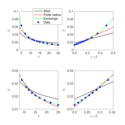
Biophysical modeling of the gray matter: does the “stick” model hold?
Jelle Veraart, Els Fieremans, Umesh Rudrapatna, Derek Jones, Dmitry Novikov
The use of term “neurites” implies that many biophysical models of diffusion in the white matter can be applied in the gray matter as well. However, the validity of representing dendrites as a collection of zero-radius impermeably sticks, a widely adopted representation of myelinated axons, has not been evaluated yet. By evaluating the diffusion-weighted signal decay as a function of the $$$b$$$-value up to $$$b=25000\,\mathrm{ms/\mu\,m^2}$$$ in the living human brain, we show that a more accurate representation of the diffusion in neuronal processes in the gray matter must account for a fast proton exchange between the intra- and extra-cellular compartments.
|
|
1095.
 |
4 |
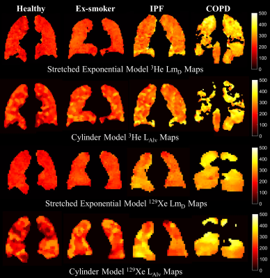
Mean lung alveolar dimension mapping with hyperpolarized 3He and 129Xe diffusion-weighted MRI
Ho-Fung Chan, Guilhem Collier, Jim Wild
Lung morphometry parameters can be derived from multiple b-value hyperpolarized gas diffusion-weighted (DW)-MRI using the cylinder (CM) and stretched exponential (SEM) models. Mean alveolar diameter (LAlv) and diffusive length scale (LmD) are calculated from the CM and SEM, respectively. This work compares the parameters LAlv and LmD derived from a range of subjects with both 3He and 129Xe DW-MRI. Excellent linear agreement is observed between the LAlv and LmD parameters for both gases. This indicates these parameters are equivalently representative indices of mean alveolar diameter dimension within the range of experimental conditions considered.
|
|
1096.
 |
5 |
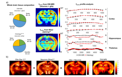
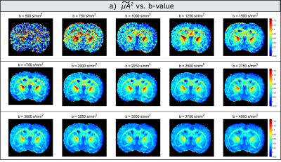
A compartment based model for non-invasive cell body imaging by diffusion MRI
Marco Palombo, Noam Shemesh, Andrada Ianus, Daniel Alexander, Hui Zhang
This study aims to open a new window onto brain tissue microstructure by proposing a new technique to estimate cell body (namely soma) size/density non-invasively. Using Monte-Carlo simulation and data from rat brain, we show that soma’s size and density have a specific signature on the direction-averaged DW-MRI signal at high b values. Simulation shows that, at reasonably short diffusion times, soma and neurites can be approximated as two non-exchanging compartments, modelled as “sphere” and “sticks” respectively. Fitting this simple compartment model to rat data produces maps with contrast consistent with published histological data.
|
|
1097.
 |
6 |
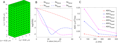
GPU-based Monte-Carlo simulation of diffusion in astrocytes reconstructed from confocal microscopy
Khieu NGUYEN, Edwin Hernandez Garzon, Julien Valette
Here we implement a GPU-based Monte-Carlo simulation of diffusion to efficiently simulate diffusion-weighting in realistic, complex cellular structures such as astrocytes as directly derived from confocal microscopy. This opens new possibilities to better understand intracellular diffusion, validate diffusion models, and create dictionaries of intracellular diffusion signatures.
|
|
1098.
 |
7 |
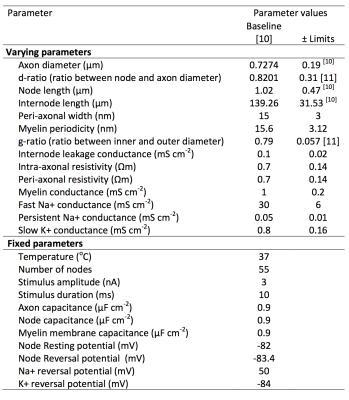
What is the feasibility of estimating axonal conduction velocity from in vivo microstructural MRI?
Mark Drakesmith, Derek Jones
Microstructural MRI provides non-invasive measures of the microstructure of white-matter axons, including diameter and g-ratio. These can theoretically be used to infer axonal conduction velocity (CV). However, several other morphological and physiological parameters also contribute to CV, which are not accessible through MRI. Using sensitivity analysis on a comprehensive model of axon electrophysiology, we test the feasibility of modelling CV and associated uncertainty using only MRI-accessible parameters. Results show 88.5% of the variance in CV is accounted for by axon diameter and g-ratio and uncertainty due to non-imageable parameters can be easily modelled by a simple linear function. When these measurements can be made reliably it is feasible to obtain estimates of axonal CVs from micro-structural parameters obtained from MRI.
|
|
1099.
 |
8 |
Measurement of Intra-Axonal Water Diffusivity in Normal Human White Matter
Bibek Dhital, Marco Reisert, Elias Kellner, Valerij Kiselev
How to measure the intra-axonal diffusion coefficient in the normal human brain? This contribution reports on such a measurement performed with minimum model assumptions. The result is about $$$2\,\rm\mu m^2/ms$$$, which constructively limits the scope of acceptable biophysical models for interpretation of diffusion MRI in terms of tissue microstructure. A side finding is the width of axonal orientation distribution in the most coherent fiber bundles, which is estimated as $$$19^\circ$$$ for the standard deviation. The core of the method is the suppression of signal from water that can move in the plane transverse to the principal fiber direction.
|
|
1100.
 |
9 |
Accurate Estimation of Microscopic Diffusion Anisotropy Using Multi-shell Double Diffusion Encoding
Andrada Ianus, Sune Jespersen, Ivana Drobnjak, Noam Shemesh
Advanced diffusion MRI acquisitions, such as double diffusion encoding (DDE), have been used to provide estimates of microscopic anisotropy, and the majority of DDE studies to date have acquired data at a single b-value. This study shows in simulations and ex-vivo experiments that the D(O)DE derived microscopic anisotropy metric strongly depends on the choice of b-value and proposes a multi shell estimation scheme which provides accurate measurements.
|
|
1101.
 |
10 |
From physical chemistry to human brain biology: unconstrained inversion of 5-dimensional diffusion-T2 correlation data
Chantal Tax, João de Almeida Martins, Filip Szczepankiewicz, Carl-Fredrik Westin, Maxime Chamberland, Daniel Topgaard, Derek Jones
Studying the diffusion MRI signal as a function of more experimental parameters allows to establish correlations between different chemical and physical properties and to disentangle different compartments. Such measurements are common in the field of physical chemistry to characterise heterogeneous media, but are rendered impractical on human scanners due to hardware limitations. Here, we leverage ultra-strong gradients to acquire a 5-dimensional in-vivo human brain correlation dataset, which allows the characterisation of microstructural features through unconstrained inversion.
|
|
1102.
 |
11 |
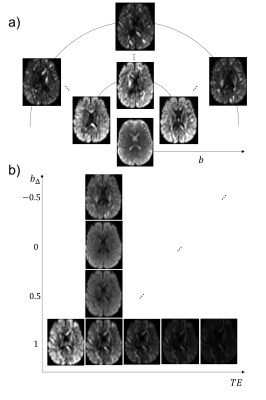
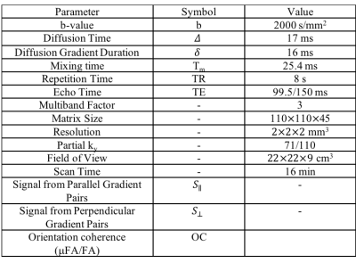
Echo Time Dependence of Double Diffusion Encoding Measurements of Microscopic Diffusion Anisotropy
Grant Yang, Jennifer McNab
In this study, double diffusion encoding (DDE) MRI measurements of microscopic fractional anisotropy (μFA) are compared at two different echo times (TE). At longer TE, higher mean μFA values were measured in single fiber regions but not in crossing fiber regions. Consistent with prior work on the TE dependence of fractional anisotropy (FA) the largest differences were found in regions known to be highly myelinated such as the genu of the corpus callosum.
|
|
1103.
 |
12 |
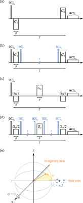
Validity extension of stimulated echoes to imaginary signals arising for double diffusion encoding of closed pores
Kerstin Demberg, Frederik Laun, Peter Bachert, Tristan Kuder
We extend diffusion-weighted acquisitions using the stimulated echo acquisition mode (STEAM) to imaginary signals. In classical diffusion encoding, only real signals arise; however, for special applications of double diffusion encoding (DDE), complex signals arise, as shown here for diffusion pore imaging, a method that allows determining the shape of arbitrary closed pores filled with an NMR-detectable medium. It is shown that the phase information of complex signals is preserved under application of stimulated echoes: We show the analytically derived signal for DDE with STEAM and use Monte Carlo simulations for validation. Consequently STEAM can be employed for diffusion pore imaging.
|
|
1104.
 |
13 |
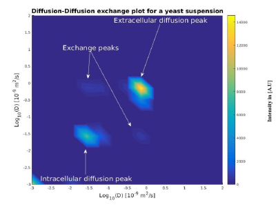
Measurement of diffusion exchange in yeast with Diffusion Exchange Spectroscopy (DEXSY)
James Breen-Norris, Bernard Siow, Ben Hipwell, Thomas Roberts, Mark Lythgoe, Andrada Ianus, Daniel Alexander, Simon Walker-Samuel
We demonstrate the feasibility of measuring diffusion exchange using Diffusion Exchange Spectroscopy (DEXSY), in yeast. We show data acquired from yeast suspensions with a 9.4 T Varian scanner using two different sets of DEXSY scan parameters in order to probe different diffusion lengths.
|
|
1105.
 |
14 |
Differences between treated glioblastoma and metastatic brain neoplasms revealed by non-Gaussian diffusion MRI and 18F-FET-PET
Farida Grinberg, Francesco D’Amore, Ganna Blazhenets, Ezequiel Farrher, Karl-Josef Langen, N. Jon Shah
Metastatic brain tumours constitute the majority of all brain tumours in adult population. Differentiation of metastases from primary brain tumours such as malignant gliomas may be a diagnostic problem and is important because of different treatment strategies. Recently, and advanced diffusion MRI method, diffusion kurtosis imaging, has been shown to provide information on tumour grade of cerebral gliomas. The purpose of this work was to study the potential of two non-Gaussian diffusion methods, including diffusion kurtosis imaging and gamma-distribution-function imaging, in differentiation of the brain metastases and high-grade tumours.
|
|
1106.
 |
15 |
Simulation of diffusion in axons with harmonic and stochastic trajectories
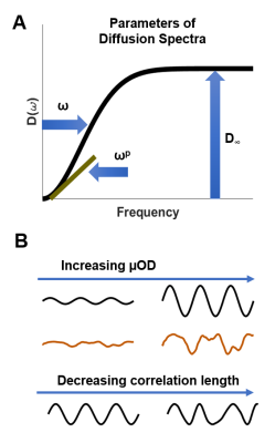
Jan Brabec, Samo Lasic, Markus Nilsson
We investigated effects on the diffusion MRI signal from harmonic and stochastic variations in the trajectories of thin axon-like structures. Trajectories with different types of variations exhibited characteristically different diffusion spectra (time-dependence) that, for low frequencies, bare similarities with those from restricted diffusion environments. Non-straight trajectories can thus bias axon diameter estimation. Results also indicated that observable effects of structural disorder may not be specific for extracellular diffusion but may also be found for intra-axonal spaces.
|
|
