Poster: Applications of Diffusion MRI
Electronic Power Pitch Poster
Diffusion
14:45 - 15:45
Wednesday, 20 June 2018
Power Pitch Theater A - Exhibition Hall
| |
|
Plasma # |
|
0743.
 |
1 |
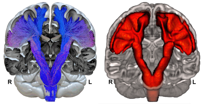
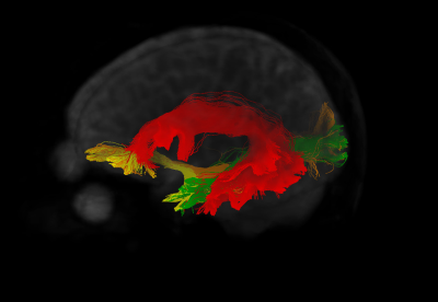
 Building a probabilistic atlas of the human corticospinal tract from 410 healthy participants by using enhanced bundle-specific tractography. Building a probabilistic atlas of the human corticospinal tract from 410 healthy participants by using enhanced bundle-specific tractography.
Chenot Quentin, Nathalie Tzourio-Mazoyer, François Rheault, Maxime Descoteaux, Laurent Petit
Current limitations of diffusion-weighted tractography algorithms face the complexity of white matter fiber crossings, especially for the cortico-lateral projections of the cortico-spinal tract (CST) in the human brain. In this work, to improve cortico-spinal tracking in crossing areas we combined accurate anatomical region positioning along the CST in each individual with a new bundle-specific tractography algorithm. We thus built a probabilistic atlas of the whole-fanning CST in 410 healthy participants.
|
|
0744.
 |
2 |
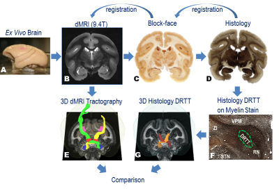
Validation of dentato-rubro-thalamic tract in squirrel monkey brain
Yurui Gao, Kurt Schilling, Iwona Stepniewska, Guozhen Luo, Bennett Landman, Hong Yu, Daniel Claassen, Benoit Dawant, Adam Anderson
The dentato-rubro-thalamic-tract (DRTT) has recently been suggested as a target for tremor control in deep brain stimulation and stereotactic radiosurgery, however, its efficacy has been challenged because different approaches to diffusion MRI (dMRI) tractography exhibited significantly different sensitivity in detecting the DRTT. We implemented a framework to quantitatively evaluate the performance of dMRI tractography by comparing the dMRI tracts to the histological DRTT identified from Nissl-stained sections in the same squirrel monkey brain. The Jaccard index between our dMRI tractography strategy and the histological DRTT is above 0.7. In the future, other tractography strategies can be tested using this framework.
|
 |
0745.
 |
3 |
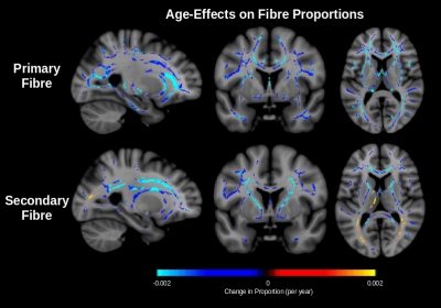
Assessing the asynchronous macrostructural changes in white matter tracts of the developing brain
Elinor Thompson, Matteo Bastiani, Matthew Brookes, Saad Jbabdi, Stamatios Sotiropoulos
We have developed an approach to elucidate the macrostructural changes of individual white matter tracts in neonatal brains, using deformation-based morphometry. Many studies have investigated how microstructure changes during development, however it is not clear how to disentangle changes in tissue microstructure from macrostructural volume changes. The latter is particularly challenging in the absence of longitudinal data from the same subject, which is the norm given the difficulty in scanning neonates. We propose an approach that is aimed towards this problem. Results are presented from analysis carried out on publicly available diffusion MRI data from the developing Human Connectome Project.
|
 |
0746.
 |
4 |
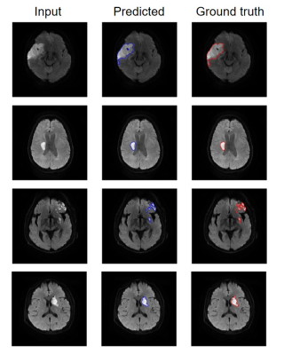
An accurate and efficient infarction segmentation method for diffusion weighted images using a deep convolutional neural network
Hanjing Kong, Fei Gao, Wenjian Huang, Weihai Xu, Yining Huang, Jue Zhang
Accurate identification of infarcted regions of the brain is critical in management of stroke patients. An efficient and accurate method based on a deep convolutional neural network for segmentation of infarcts in the diffusion-weighted images is proposed.
|
|
0747.
 |
5 |
Selective degeneration of crossing fibres and its relationship with fractional anisotropy
Jordan Chad, Ofer Pasternak, David Salat, J. Chen
Selective degeneration of crossing fibres has been reported in diffusion MRI studies of Alzheimer’s disease alongside increased fractional anisotropy (FA). Similar albeit more subtle selective degeneration has been suggested in healthy aging, but increased FA with age has not been reported on cross-sectional studies of aging. In this work, we use DTI tractography to measure selective degeneration of crossing fibres in healthy aging among 212 subjects. Increased FA with age is found in this cohort only after applying free water elimination (FWE), suggesting that increasing extracellular water in aging may introduce variability that obscure finer structural phenomena in cross-sectional designs.
|
|
0748.
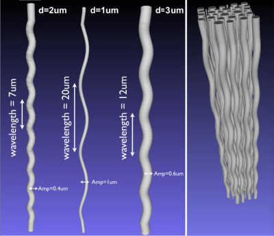 |
6 |
Quantifying diameter overestimation of undulating axons from synthetic DW-MRI
Alonso Ramirez-Manzanares, Mario Ocampo-Pineda, Jonathan Rafael-Patiño, Giorgio Innocenti, Jean-Philippe Thiran, Alessandro Daducci
This study aims to provide to the research community quantitative measurements about the axon diameter overestimation due to the straight-cylinder assumption usually made in state-of-the-art models for diameter mapping with DW-MRI. Our methodology uses a Monte Carlo diffusion simulator to compute the diffusion weighted magnetic resonance signals of undulating axons. We use in our experiments plausible tissue values, we also explore a broad parameter set that depicts undulation. The results of this study provide useful information to understand the differences between the estimators from histology vs. the estimated diameters when using the model assumption of simple shaped axons.
|
|
0749.
 |
7 |
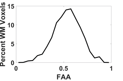
Diffusion Anisotropy of the Extra-Axonal Environment is Linked to Axon Alignment
Emilie McKinnon, Jens Jensen, Joseph Helpern
A better understanding of the complex water diffusion dynamics in the extra-axonal environment may aid in the mathematical modeling of the diffusion MRI signal and have application to pathologies that specifically impact glial cells and the surrounding extracellular space. Here we employ a novel method of combining diffusion MRI data for weak and strong diffusion weightings to show that extra-axonal water diffusion anisotropy strongly correlates with intra-axonal diffusion anisotropy and takes on large values in voxels with highly aligned axons. This connection suggests that the geometrical alignment of axonal fibers is important for both intra-axonal and extra-axonal water diffusion.
|
|
0750.
 |
8 |
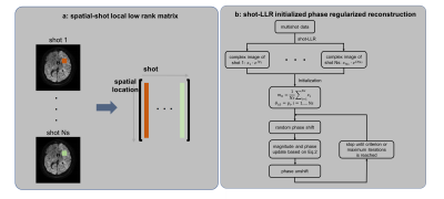
Multishot high-resolution brain diffusion-weighted imaging using phase regularized reconstruction
Yuxin Hu, Xiaole Wang, Evan Levine, Qiyuan Tian, Valentina Taviani, Frank Ong, Shreyas Vasanawala, Jennifer McNab, Bruce Daniel, Brian Hargreaves
Multishot imaging has been shown to provide high resolution diffusion-weighted images (DWIs) with reduced distortion, however, significant aliasing artifacts and signal cancellation still occur due to the mismatch of the motion-induced phase between different shots. The reconstruction becomes non-convex and intractable to solve when this phase is included into the forward model. The goal of this work is to solve this problem by circumventing the challenging phase estimation step. The brain and breast examples demonstrate that this can be efficiently achieved using a locally low-rank reconstruction approach.
|
|
0751.
 |
9 |
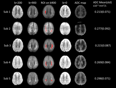
Reversible white matter restricted diffusion in patients with cerebral malaria via ADC measurement on a 0.35T MR scanner
Yuchuan Zhuang, Sarah Moghaddam , Samuel Kampondeni , Madalina Tivarus , Gretchen Birbeck , Michael Potchen , Jianhui Zhong
White matter abnormalities are commonly identified on cerebral malaria patients. We proposed a method to overcome technical limitations of low-field MRI, and quantitatively calculate the ADC and b0 maps, enabling discrimination of the true restricted diffusion from T2 shine-through. A unique pattern of reversible restricted diffusion was identified in some patients.
|
|
0752.
 |
10 |
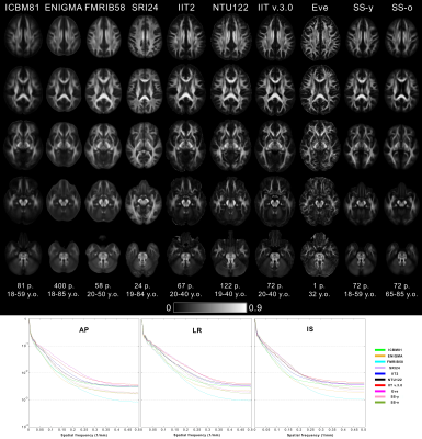
Evaluation of Standardized and Study-Specific Diffusion Tensor Imaging Templates of the Adult Human Brain
Shengwei Zhang, Konstantinos Arfanakis
DTI templates of the adult human brain are commonly used in neuroimaging, and their characteristics influence the accuracy of the application. The purpose of this work was to compare eight available standardized DTI templates to each other, as well as to study-specific templates, in terms of template characteristics and performance in spatial normalization and detection of small inter-group FA differences. The IIT v.3.0 template was shown to combine a number of desirable characteristics, and allows higher inter-subject spatial normalization accuracy and detection of smaller inter-group FA differences, compared to other templates, including study-specific templates, for both younger and older adults.
|
|
0753.
 |
11 |
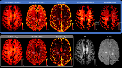
Characterization of Axonal Pathology Independent of Fiber Crossings in Multiple Sclerosis Using High-Gradient Diffusion MRI
Qiuyun Fan, Aapo Nummenmaa, Thomas Witzel, Ned Ohringer, Lawrence Wald, Eric Klawiter, Susie Huang
Axonal damage is thought to be the substrate of disability in multiple sclerosis. We have recently introduced a method based on the spherical mean framework that provides per-voxel axon diameter and volume fraction that is independent of fiber crossings/dispersion. We apply this approach to estimate whole brain axon diameter and density in a group of patients with MS to healthy controls. Widespread alterations in axon diameter and density were found throughout the NAWM and lesions of MS patients that may reflect diffuse axonal loss and swelling in the setting of chronic demyelination.
|
|
0754.
 |
12 |
Anatomy-constrained automated fiber tract reconstruction for surgery planning: a validation study in language-related white matter tracts
Matteo Mancini, Sjoerd Vos, Vejay Vakharia, Rachel Sparks, Karin Trimmel, Gavin Winston, John Duncan, Sebastian Ourselin
Diffusion MRI and tractography hold great potential for surgery planning, but fiber tract reconstruction requires an expert rater. In this work, we set up an automated reconstruction pipeline based on anatomical criteria that does not require manual intervention and we validated it on epilepsy patients with specific focus on language-related bundles. We first compared the results with the ones obtained from human raters and then further validated them using task fMRI. The fiber tracts reconstructed from the pipeline were in line with the agreement between different human raters and showed good overlap with function.
|
|
0755.
 |
13 |
Rapid Single Shot Whole Lung Acquisition of for Hyperpolarized Gas MRI Biomarkers of Airspace Enlargement.
Alexei Ouriadov, Dante Capaldi, David McCormack, Grace Parraga
Hyperpolarized gas pulmonary MRI provides physiologically relevant biomarkers of obstructive lung disease including emphysema, bronchopulmonary dysplasia, congenital lobar emphysema and alpha-1 antitrypsin deficiency. Recently, a stretched-exponential-model combined with under-sampling in the imaging and diffusion directions was proposed for the evaluation of hyperpolarized gas multiple b-value diffusion-weighted MRI. The major advantage of this method is the possibility to significantly speed up the data acquisition using acceleration factors between 7 and 10. We hypothesize that this method can be extended to provide whole lung hyperpolarized gas MRI-based emphysema biomarkers including static-ventilation, T2* ADC and morphometry maps with high spatial image resolution.
|
|
0756.
 |
14 |
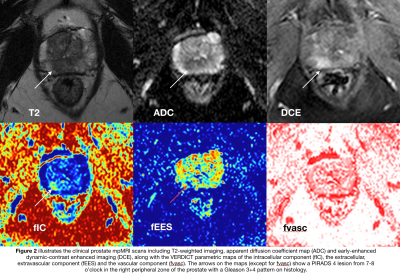
The intracellular component of VERDICT (Vascular, Extracellular, and Restricted Diffusion for Cytometry in Tumors) MRI distinguishes Gleason 4 pattern better than Apparent Diffusion Coefficient
Mrishta Brizmohun Appayya, Edward Johnston, Arash Latifoltojar, James O’Callaghan, Elisenda Bonnet-Carne, Hayley Pye, Dominic Patel, Susan Heavey, Alistair Grey, Sebastien Ourselin, David Hawkes, Caroline Moore, Hayley Whitaker, Alexander Freeman, David Atkinson, Daniel Alexander, Eleftheria Panagiotaki, Shonit Punwani
VERDICT (Vascular, Extracellular, and Restricted Diffusion for Cytometry in Tumours) MRI combines a diffusion-weighted MRI acquisition with a three-compartment mathematical model that describes signal from i) intracellular water (fIC), ii) water in extracellular-extravascular space (fEES) and iii) water in the microvasculature (fvasc). In contrast to VERDICT, clinical ADC is derived from a mono-exponential model. Upon comparison between VERDICT parameters and clinical ADC, we showed that fIC was better able to discriminate between Gleason ≥3+4 histology pattern and Gleason ≤3+3/benign histology. We also showed that image quality of VERDICT-MRI maps and clinical ADC was comparable.
|
|
0757.
 |
15 |
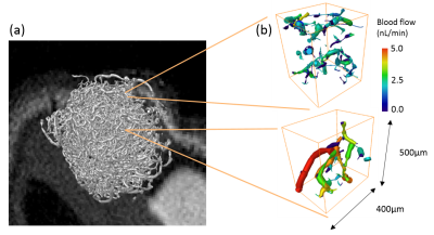
Validation of the VERDICT MRI Framework using a Novel Computational Model of Diffusion and Flow in Real-World Tumours.
Ben Hipwell, Tom Roberts, Paul Sweeney, Morium Ali, Angela D'Esposito, Eleftheria Panagiotaki, Mark Lythgoe, Daniel Alexander, Rebecca Shipley, Simon Walker-Samuel
Compartmental models are increasingly being used to quantify diffusion MRI signals from tumours. We have developed a complex, multiscale mathematical modelling platform for simulating tumour pathophysiology, using high-resolution optical imaging data from complete tumour samples. Diffusion MRI signals from these tumours were simulated, including vascular flow and intra- and extracellular diffusion. These data were fitted to the VERDICT compartmental model, and the resulting parameters compared against ground truth simulation values. Cell radius and intra/extracellular fractional volume parameters and respective ground truth values were strongly correlated. A more complex relationship was found in vascular volume fractions.
|
|
