Poster: Trending Topics: Flexible, Material, Portable, Wireless
Electronic Power Pitch Poster
Engineering
09:15 - 10:15
Monday, 18 June 2018
Power Pitch Theater B - Exhibition Hall
| |
|
Plasma # |
 |
0016.
 |
16 |
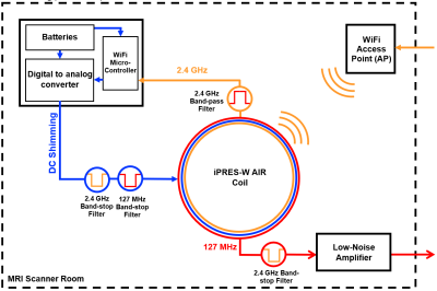
 The iPRES-W AIR Coil: A Flexible RF Coil for Simultaneous MR Image Acquisition, Wireless Communication, and Localized B0 Shimming The iPRES-W AIR Coil: A Flexible RF Coil for Simultaneous MR Image Acquisition, Wireless Communication, and Localized B0 Shimming
Jonathan Cuthbertson, Dean Darnell, Julia Bresticker, Robert Stormont, Fraser Robb, Allen Song, Trong-Kha Truong
The iPRES-W AIR coil is a highly novel RF coil design that can simultaneously perform image acquisition, wireless communication, and wirelessly controlled localized B0 shimming. In addition, the iPRES-W AIR coil benefits from all advantages of the recently unveiled AIR coil technology, which offers a flexible and ultra-lightweight coil for increased patient comfort and freedom in overlap positioning between coil elements without degrading the performance. This technology has enormous potential to improve image quality, spatial fidelity, diagnostic accuracy, and patient comfort in a wide range of MRI applications.
|
|
0017.
 |
17 |
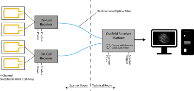
Towards wearable MR detection: A stretchable wrist array with on-body digitization
Video Permission Withheld
Andreas Port, Jonas Reber, Christian Vogt, Josip Marjanovic, Benjamin Sporrer, Lianbo Wu, Andreas Mehmann, David Brunner, Thomas Burger, Gerhard Troester, Qiuting Huang, Klaas Pruessmann
Today, MRI coils can be made flexible and stretchable. Signals can be sent out of bore by optical or wireless links. Nonetheless, wearable digital coil arrays have not yet found their way into MRI although enhanced patient comfort and workflow can be expected. In this work we explore the feasability of stretchable coil arrays with on-coil digitization and optical transmission, and present a first implementation for wrist imaging.
|
|
0018.
 |
18 |
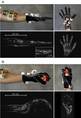
High Impedance Detector Arrays for Magnetic Resonance
Bei Zhang, Daniel Sodickson, Martijn Cloos
Resonant inductive coupling is commonly seen as an undesired fundamental phenomenon emergent in densely packed resonant structures, such as nuclear magnetic resonance phased array detectors. The need to mitigate coupling imposes rigid constraints on the detector design, impeding performance and limiting the scope of magnetic resonance experiments. Here we introduce a high impedance detector design, which can cloak itself from electrodynamic interactions with neighboring elements. We verify experimentally that the high impedance detectors do not suffer from signal-to-noise degradation mechanisms observed with traditional low impedance elements. Using this new-found robustness, we demonstrate an adaptive wearable detector array for magnetic resonance imaging of the hand. The unique properties of the detector glove reveal new pathways to study the biomechanics of soft tissues, and exemplify the enabling potential of high-impedance detectors for a wide range of demanding applications that are not well suited to traditional coil designs.
|
|
0019.
 |
19 |
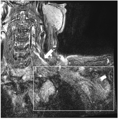
Highly Flexible, Light Weight 24 Channel 3T Bilateral Brachial Plexus Array Worn as a Close Fitting Variable Size Vest
Ed Boskamp, Victor Taracila, Scott Lindsay, Robert Stormont, Reni Biswas, Sheronda Statum, Shane Aldas, Cesar Barraza, Fraser Robb, Christine Chung, Won Bae
MR neurography of the Brachial Plexus (BP) is technically challenging, due in part to complex topographical anatomy that is sub-optimally accommodated with existing rigid receiver coil arrays. In this work we propose a wearable garment with integrated flexible loop receiver coils that conforms to the contour of the neck and shoulder, as well as variations in subject size. The loops can flex in multiple dimensions without seriously affecting noise correlation.
|
|
0020.
 |
20 |
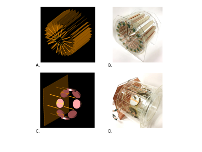
Custom, 3D Sprayed MRI receive coils
Alla Zamarayeva, Michael Liu, Joseph Corea, Karthik Gopalan, Kelvin Pang, Miki Lustig, Ana Claudia Arias
We developed process for fabricating patient-specific MRI receive coils via scalable and adaptable additive manufacturing approaches. Process relies on spray-depositing solution-processed electronic materials onto 3D printed custom substrates. We conducted careful materials selection, process optimization and fabricated fully functional coils. The coils spray deposited onto spherical substrate demonstrated higher SNR than control coil, when evaluated using phantom of the spherical shape, due to superior conformability to the phantom. Our approach is poised to enhance high quality clinical imaging by ensuring optimum fit of the MRI receive coils to the body parts with complex geometries, enabling reproducible placing on the patient and eliminating motion artifacts.
|
|
0021.
 |
21 |
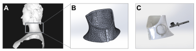
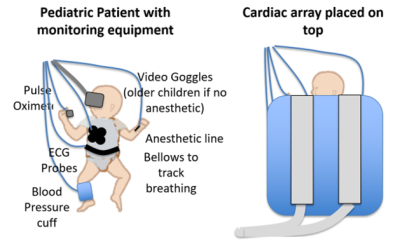
First clinical pilot study using screen-printed flexible MRI receive coils for pediatric applications
Simone Angela Winkler, Joseph Corea, Balthazar Lechene, Kendall O'Brien, John Bonanni, Fraser Robb, Greig Scott, John Pauly, Michael Lustig, Ana Arias, Shreyas Vasanawala
Pediatric MRI is often performed suboptimally by the use of heavy, large, and relatively inflexible coil arrays that are designed and built for adult MR imaging. For the child, these arrays can be intimidating and uncomfortable, restricting breathing. For parents, they contribute to the stress of the exam. For pediatric caregivers for smaller children, the coils complicate placing medical support equipment. Here, we assess the use of screen printed flexible coil arrays for pediatric applications, focusing on clinical image quality and caregiver acceptance. We conclude that a flexible screen-printed MRI receive coil is likely to yield diagnostic image quality and be preferred to a traditional coil by patients, parents, and caregivers.
|
|
0022.
 |
22 |
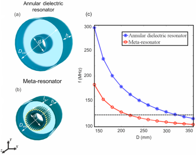
Volumetric resonators based on novel materials for 3 T MRI
Anna Mikhailovskaya, Alena Shchelokova, Dmitry Dobrykh, Ivan Sushkov, Alexey Slobozhanyuk, Andrew Webb
We propose and characterise a novel metamaterial-inspired approach which reduces substantially the required outer diameter of a dielectric resonator thus making possible to design compact structures for 3 T. When used in an inductively-coupled wireless mode, the sensitivity of the “meta-resonator” was measured to be slightly higher than that of a standard dielectric resonator operating in its degenerate circularly-polarized HEM11 modes. This study demonstrates the first application of a metamaterial-based approach to MR volume coil design.
|
|
0023.
 |
23 |
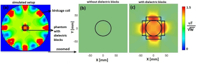
New ferroelectric ceramics for transmit efficiency enhancement at 1.5 Tesla
Irena Zivkovic, Alexey Slobozhanyuk, Elizaveta Nenasheva, Andrew Webb
The presence of medical implants often results in MR scans with low SAR being prescribed, which reduces the diagnostic quality of the images. This work shows that one can achieve an increase in local transmit efficiency and a corresponding decrease in SAR at 1.5 Tesla using new ferroelectric materials (based on BaTiO3 with ZrO2 and CeO2-additives) with relative permittivities higher than 4500. Simulations and phantom/in vivo experiments show an increased local transmit efficiency of ~50% from the body transmit coil, with a corresponding increase in SNR for a given value of SAR.
|
|
0024.
 |
24 |
Comparison of a 16-channel monopole/dipole hybrid array with a combined 8-channel monopole + 8-channel high dielectric constant (HDC) disk dipole array for head imaging at 10.5T
Myung Kyun Woo, Lance DelaBarre, Jerahmie Radder, Russell Lagore, Yigitcan Eryaman, Kamil Ugurbil, Gregor Adriany
We evaluate the performance both in simulation and experiment of two 10.5T head transceiver arrays. The first is a 16-channel monopole/dipole hybrid array (called the Mono-Dipole array for brevity). The second is a 16-channel monopole/HDC disk dipole array. (A combination of two 8-channel arrays: a monopole array and a high dielectric constant (HDC) disk dipole array). These novel coil designs were compared against a standard 16-channel stripline array to elucidate their relative benefits and drawbacks.
|
|
0025.
 |
25 |
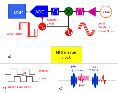
Pilot Tone Software Synchronization for Wireless MRI Receivers
Greig Scott, Shreyas Vasanawala, Fraser Robb, Pascal Stang, John Pauly
For wireless MRI to become a reality, methods must be developed to synchronize receivers that have no physical connection to the scanner. Here, we demonstrate that dual pilot tones are needed to correct for both timing skew and frequency offsets. Bench tests were performed with a master and free running slave receiver, in which the images were re-synchronized by frequency estimation methods on pilot tones.
|
|
0026.
 |
26 |
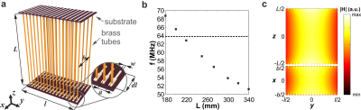
Demonstration of a new volumetric wireless coil for extremities imaging
Alena Shchelokova, Dmitry Dobrykh, Stanislav Glybovski, Mikhail Zubkov, Ekaterina Brui, Cornelis A.T. van den Berg, Irina Melchakova, Pavel Belov
We demonstrate experimentally a new design of self-resonant tunable volumetric wireless coil based on a periodic array of coupled split-loop resonators. The wireless coil operates via inductive coupling with a birdcage coil enhancing the sensitivity of the latter at 1.5T and can be used for extremities imaging. Phantom and in-vivo wrist imaging with the proposed coil demonstrate 8.6 times higher transmit power efficiency in comparison with the birdcage coil and up to 2 times higher SNR versus standard receive-only coil.
|
|
0027.
 |
27 |
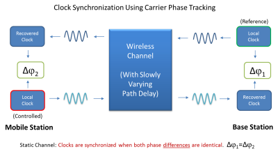
High Precision Wireless Clock Recovery for On-Coil MRI Receivers Using Round-Trip Carrier Phase Tracking.
Arne Reykowski, Paul Redder, Rodrigo Calderon Rico, Tracy Wynn, Tim Ortiz, Greg Dowling, Randy Duensing, Scott King
Wireless MRI coils require a local frequency reference that is very tightly synchronized with the main MRI system clock. In order to keep phase encoding errors to less than 1 degree of phase at 3T, the local reference clock cannot drift by more than +/-22ps. Unlike RF cables or optical fibers, a wireless communication channel is not constant in phase over time. Patient motion and multi-path fading cause the wireless signal to be modulated in amplitude and phase. When disciplining a local clock with the MRI system clock by wireless means, channel impairments have to be monitored and corrected.
|
|
0028.
 |
28 |
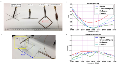
Antenna Design for Wireless Clock Syncing and Q-spoiling in MRI
Jonathan Lu, Thomas Grafendorfer, Shreyas Vasanawala, Fraser Robb, John Pauly, Greig Scott
This work explored the antenna design choices for wireless MRI, specifically for clock synchronization and scanner state detection. Antennas at 1.6GHz for the clock signal, and 3.5GHz for Q-spoil signal were tested for transmit and receive performance. Reflectors were constructed and improvements in antenna path loss were observed. A better understanding of these antennas is necessary to help satisfy link budget requirements.
|
 |
0029.
 |
29 |
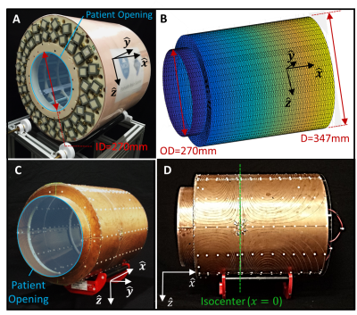
3D imaging with a portable MRI scanner using an optimized rotating magnet and 1D gradient coil
Patrick McDaniel, Clarissa Cooley, Jason Stockmann, Lawrence Wald
A low-cost portable brain MRI scanner could extend the reach of brain MRI to remote locations and point-of-care situations and extend the use of MRI into monitoring applications. We previously presented 3D imaging in a portable magnet without gradient coils using the natural field variation of a rotating permanent magnet for in-plane encoding and RF phase gradients for encoding the third direction. In this work, we show that an efficient gradient coil can phase-encode the third direction in a single shot CPMG train with minimal current (~2A) allowing low-cost amplifiers and negligible power needs, acoustic noise or heating.
|
|
0030.
 |
30 |
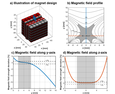
Portable single-sided MR: multicomponent T2 relaxometry and depth profiling with a Unilateral Linear Halbach sensor
Ashvin Bashyam, Chris Frangieh, Matthew Li, Jason Stockmann, Michael Cima
Widely accessible, fast, and portable MR relaxometry can provide highly valuable diagnostic information in many diseases including fluid overload, dehydration, and muscle degeneration. High cost, large size, and extended acquisition time limit the use of traditional MRI for relaxometry. We introduce a novel portable MR sensor with a large, remote, uniform magnetic field created by a Unilateral Linear Halbach array capable of performing spatially selective, multi-component relaxometry.
Strong agreement between simulation and experimental results indicates highly reliable magnet design methods. Spatial selectivity is achieved through variation of either RF pulse length or B1 frequency. Quantitative multi-component T2 relaxometry is demonstrated. |
|
