|
Traditional Poster Session
Musculoskeletal |
Monday, 18 June 2018
Traditional PosterMusculoskeletal
1369 -1393 Cartilage
1394 -1411 Muscle
1412 -1437 MSK: Other
1438 -1450 Bone |
| |
Cartilage
Traditional Poster
Musculoskeletal
Monday, 18 June 2018
| Exhibition Hall 1369-1393 |
08:15 - 10:15 |
|
1369.
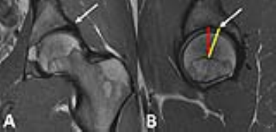 |
Ability of MRI to Predict the Severity and Location of Chondral and Labral Pathology at Arthroscopy
Alissa Burge, Stephen Lyman, Matthew Koff, Hollis Potter, Sydney Kersten, Bin Lin, Kara Fields, Bryan Kelly
Preoperative MRI and intraoperative arthroscopic images were independently reviewed in a cohort of 24 hips with femoroacetabular impingement with respect to severity and location of chondral, labral, and osseous pathology. Initial calculation of agreement between MRI and arthroscopic findings demonstrated fair to near perfect agreement for the severity of pathology; however, agreement for the location of pathology was highly variable. MR images were subsequently re-scored utilizing the indirect head of the rectus femoris as an anatomic landmark, in accordance with the system used by the operating surgeon, resulting in overall increased agreement across position-dependent variables.
|
|
1370.
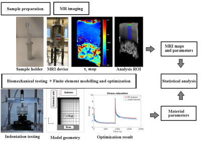 |
Correlation time mapping is associated with permeability of articular cartilage
Mikko Nissinen, Nina Hänninen, Petri Tanska, Olli Nykänen, Mithilesh Prakash, Matti Hanni, Juha Töyräs, Rami Korhonen, Mikko Nissi, Miika Nieminen
Correlation time τc is a parameter that describes the relaxation properties of soft tissues. In this study, articular cartilage from human cadaver patellae was studied using MR imaging and biomechanical testing and modeling. The statistical analysis revealed an association between the permeability, as revealed by mechanical modeling, and the correlation time measured for articular cartilage.
|
|
1371.
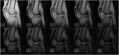 |
T2* Enhancement for Multi-Echo Data Image Combination -- Using least squares for echo prediction
Zhang Qiong, Chen Shi, Wei Binyan, Kang Yuanyuan
This work provides a virtual echo prediction method for Multi Echo Data Image Combination (Medic) based on least square estimation. The strong dependences between multi-echoes in Medic sequences are used to predict virtual echoes with assumed echo times, and then such predictions are combined with real acquired echoes for heavier T2* contrast enhancement.
|
|
1372.
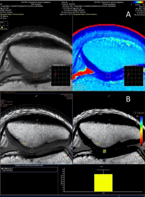 |
Comparison of Conventional and Synthetic MRI for Quantitative Cartilage T2 Mapping of the Patella
Le Roy Chong, Gideon Ooi, Jia Hui Ng, Hafiz Bin Abu Hassan
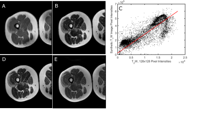
Synthetic MRI has been shown to be of comparable performance to conventional MRI in the assessment of intracranial abnormalities. This study compares synthetic MRI with conventional T2 mapping for quantitative assessment of cartilage T2 relaxation times. T2 values acquired via synthetic MRI are highly correlated with but not equivalent to conventional T2 mapping. Synthetic MRI could be a potential alternative in the quantitative assessment of chondral abnormalities, without the need for prolonged scan times and providing the benefit of dynamic tissue contrasts from a single acquisition.
|
|
1373.
 |
Associations between Osteoarthritis Molecular Biomarkers and MR-based cartilage composition and Knee Joint Morphology: Data from the Osteoarthritis Initiative
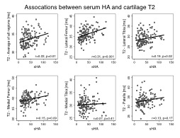
Gabby Joseph, Michael Nevitt, Charles McCulloch, Jan Neumann, John Lynch, Ursula Heilmeier, Nancy Lane, Thomas Link
This study assessed the relationships of serum/urine biomarkers for osteoarthritis with MR imaging measures of joint structure and composition, using data from the Osteoarthritis Initiative (OAI). Significant positive correlations between the serum/urine biomarkers (sHA, sMMP3) and MRI cartilage T2 relaxation time measurements, compositional markers of early cartilage degeneration were observed. However, no significant associations were found with cartilage morphology or Kellgren-Lawrence (KL) grade. Therefore, serum biomarkers and cartilage T2 composition may reflect similar features of the pathophysiology of cartilage matrix degenerative disease.
|
|
1374.
 |
Detailed T2-mapping analysis reveal disc characteristics that may be of significance for low back pain patients
Christian Waldenberg, Hanna Hebelka, Helena Brisby, Kerstin Lagerstrand
In this study, we address the lack of studies comparing intervertebral disc characteristics between symptomatic and asymptomatic individuals. Based on quantitative T2-mapping, small but relevant differences between low back pain patients and a control cohort were found on a global and regional level.
|
|
1375.
 |
Magnetization Transfer Ratio (MTRNOE) as a Biomarker of Hip Osteoarthritis
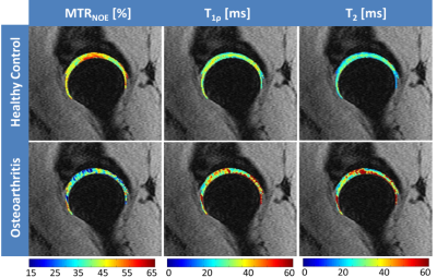
Hatef Mehrabian, Jasmine Rossi-Devries, Alan Zhang, Richard Souza, Sharmila Majumdar
Loss of cartilage collagen, proteoglycans (PG), glycosaminoglycans (GAG) are responsible for osteoarthritis (OA). MRI biomarkers T2 (sensitive to collagen), magnetization transfer (MT) and T1ρ, (sensitive to PG), and GAGCEST (sensitive to GAG) can detect OA at early stages. Similar to GAGCEST, CEST signal of Nuclear Overhauser Effect (NOECEST) at -1.6ppm also changes with OA. However, unlike GAGCEST, this NOECEST is measurable at 3T which is suitable for hip. MT ratio at this -1.6ppm (MTRNOE) represents the combination of MT, T2, NOECESTeffects. OA-related changes in these three parameters result in decreased MTRNOE making it a reliable biomarker for OA.
|
|
1376.
 |
T2 and T1rho mapping of ankle cartilage of female and male ballet dancers
Saya Horiuchi, Hon Yu, Alex Luk, Adam Rudd, Jimmy Ton, Edward Kuoy, Jeff Russell, Kelli Sharp, Hiroshi Yoshioka
This study demonstrated T2 and T1rho profiles of talar dome and tibial plafond cartilage from male and female ballet dancers using angular-segmentation methodology for quantitative assessment of cartilage in vivo. The results in this study showed both T2 and T1rho relaxation time indicated the lowest value over the central weight-bearing portion, while they indicated relatively higher values in the anterior and posterior portion. These findings can be due to the combination of the magic angle effect which has higher influence on T2 value and early cartilage degenerative changes which are more sharply detected by T1rho value.
|
|
1377.
 |
Analysis of the Local Associations between Morphology and Biochemical Composition of the Articular Cartilage after Anterior Cruciate Ligament Injury and Reconstructive Surgery using Voxel-Based Relaxometry
Onyekachi Nnabue, Hatef Mehrabian, Valentina Pedoia, Berk Norman, Benjamin Ma, Sharmila Majumdar
This study uncovered new insights on the local associations between cartilage thickness and T1ρ relaxation time (a marker of cartilage proteoglycan content). Using Voxel-based relaxometry, this study quantified the longitudinal and cross-sectional thickness changes that occur in both the ACL-injured knee and the healthy contralateral in the lateral femoral condyle, medial femoral condyle, trochlea, medial tibia, lateral tibia, and patella and examined compartment-specific associations with relaxometry at various time points.
|
|
1378.
 |
CS+SENSE for Fast UTE Knee Imaging: Technical Feasibility
Yongxian Qian, Li Feng, Tiejun Zhao, Richardo Otazo, Fernando Boada
Ultrashort echo time (UTE<1ms) imaging has advantages over traditional long TE (>10ms) imaging to detect asymptomatic (subclinical) cartilage damages in the knee joint, such as fissuring, fracturing and collagen fiber breakdown. To advance UTE imaging toward clinical use, its long scan time needs to be reduced to meet clinical requirement of short protocols. Compressed sensing (CS) and sensitivity encoding (SENSE) parallel imaging have the potential to do so. However, individual use of them has limitations. A combined use of both techniques has been shown in dynamic imaging to be able to achieve higher acceleration factor without SNR loss. This study explores the technical feasibility to extend CE+SENSE to static UTE imaging.
|
|
1379.
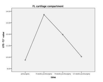 |
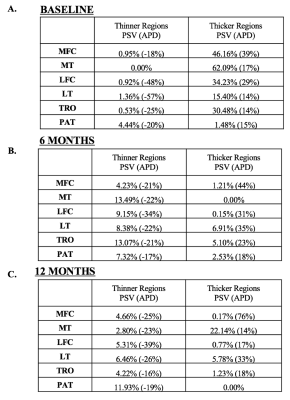
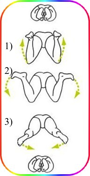
Quantitative evaluation of knee cartilage after anterior cruciate ligament reconstruction using UTE-T2* mapping in a rabbit model
Yiwen Hu, Jianxun Qu
Our study is a prospective longitudinal study conducted to find outcome of anterior cruciate ligament reconstruction in rabbit model. We evaluated degenerative changes of cartilage by UTE-T2* mapping. ACLR knees shows cartilage matrix degeneration at early stage of "ligamentization", though rabbit tibiofemoral cartilage is definitely thin.
|
|
1380.
 |
Comparison of T2 Relaxation Times in Knee Cartilage Between Breaststroke and Nonbreaststroke Swimmers
James Yoder, Feliks Kogan, Garry Gold
While MRI has been widely used to examine the effects of translational forces on cartilage matrix structure, studies looking at rotational forces are limited. Breaststroke swimmers are a population of interest since the repeated use of the breaststroke kick has been cited as a source of knee pain. However, the cartilage of breaststrokers has not been quantitatively measured to investigate possible differences and the potential increased risk of cartilage degeneration and osteoarthritis development. This study compares the T2 relaxation times of various compartments for patellar, femoral, and tibial cartilage at the superficial, deep, and aggregate levels between breaststrokers and nonbreaststrokers.
|
|
1381.
 |
Grey-Level Co-Occurrence Matrix Texture Analysis of T2, Adiabatic T1?, Adiabatic T2? and Dual-Echo Steady-State Magnetic Resonance Imaging Contrasts in Osteoarthritic Knee Articular Cartilage
Ines Barros, Arttu Peuna, Victor Casula, Marianne Haapea, Eveliina Lammentausta, Miika Nieminen
Grey-level co-occurrence matrix (GLCM) based texture analysis is a sensitive image processing tool for the evaluation of cartilage in knee osteoarthritis (OA). Texture analysis of T2, Adiabatic T1ρ (AdT1ρ), Adiabatic T2ρ (AdT2ρ) relaxation time maps as well as Dual-Echo Steady-State (DESS) images showed the ability to distinguish OA patients and asymptomatic volunteers. Moreover, texture analysis turned out to be more sensitive to cartilage degeneration than mean relaxation time values. Texture analysis can therefore supplement existing quantitative MRI techniques of articular cartilage.
|
|
1382.
 |
Simulated 1H-1H residual dipolar couplings of collagen-associated water
Jouni Karjalainen, Mikko Nissi, Miika Nieminen, Matti Hanni
Residual dipolar couplings have been suggested as the cause of the orientational dependence of relaxation times in anisotropic tissues, such as articular cartilage. We use molecular dynamics simulations to compute the residual dipolar couplings of water protons associated with a model collagen molecule. The results suggest that significant residual dipolar couplings appear without strong binding between the water and the collagen.
|
|
1383.
 |
Quantitative GagCEST MRI in Juvenile Bovine Articular Cartilage Exhibit Correlations between 3T and 7T
Lauren Watkins, Feliks Kogan, Marianne Black, Marc Levenston, Garry Gold
GagCEST is a quantitative MR technique that shows promise at 7T to specifically detect cartilage glycosaminoglycan content; however, its potential at 3T is still uncertain. This study utilizes a new optimized 3D GagCEST sequence to maximize SNR and GagCEST contrast at 3T. Comparison of GagCEST asymmetry maps obtained at 3T and 7T suggest that GagCEST can be used to distinguish zonal differences in cartilage composition at both 3T and 7T. This work demonstrates potential for whole joint GagCEST knee imaging at 3T with improved dynamic range.
|
|
1384.
 |
Automated segmentation of the cartilage from high-resolution isotropic T1rho MRI
Henry Rusinek, Rahman Baboli, Artem Mikheev, Azadeh Sharafi, Ravinder Regatte
We analyze the accuracy of atlas-based cartilage segmentation from isotropic T1ρ MRI and compare it to semi-automated "seed and blanket" method and manual segmentation (ground truth). Reference 3D cartilage masks were taken as the consensus of two human experts. For patella, our implementation of template matching yielded the root mean square volume measurement error RMSE of 0.66 cm3, with interclass correlation coefficient (ICC) = 0.765 and sufficient precision to detect the gender effect. Over two-fold improvement in accuracy, RMSE = 0.25 cm3 and ICC = 0.960 was achieved with a fast, semi-automated algorithm. Similar results hold for the accuracy of the average thickness of segmented masks.
|
|
1385.
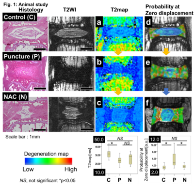 |
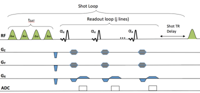
The novel and quantitative MRI technique: Q-space imaging for evaluating intervertebral disc degeneration: basic and clinical study.
Daisuke Nakashima, Nobuyuki Fujita, Junichi Hata, Takeo Nagura, Kanehiro Fujiyoshi, Hideyuki Okano, Masahiro Jinzaki, Morio Matsumoto, Masaya Nakamura
The conventional qualitative classification of intervertebral disc (IVD) degeneration: Pfirrmann classification on T2 weighted imaging does not have the enough sensitivity for the evaluation of IVD degeneration. In the present study, probability at zero displacement obtained from Q-space imaging (QSI) has a high sensitivity of IVD degeneration in both basic and clinical study compared with the conventional method: T2 mapping. In particular, probability at zero displacement made it possible to observe the effect of the regenerative drug: N-Acetyl Cystaine on IVD degeneration which could not be observed by using T2 mapping. Probability at zero displacement obtained from QSI has the possibility to be a novel biomarker of IVD degeneration.
|
|
1386.
 |
Effect of Fat-contamination and Fat-suppression on T2 Quantitation of Knee Articular Cartilage In Vivo
Petri Paakkari, Stefan Zbyn, Mikko Nissi, Eveliina Lammentausta, Miika Nieminen, Victor Casula
This study aims to investigate the effect of fat contamination and fat suppression (FS) on in vivo T2 mapping of knee cartilage. Four volunteers were imaged on a 3T MRI scanner and T2 values were calculated in several regions of tibiofemoral cartilage using a MSME sequence with and without FS. The use of FS improved repeatability of cartilage segmentation in several regions and reduced the chemical shift artifacts. However, the regional heterogeneity in FS sequence introduced further uncertainties in T2 measurements.
|
|
1387.
 |
T1 Relaxation Time Mapping of Articular Cartilage for Femoroacetabular Impingement (FAI) - A Clinical Pilot Study
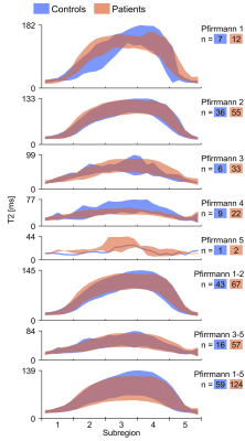
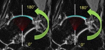
Jutta Ellermann, Douglas Martin, Casey Johnson, Robert Gao, Luning Wang, Patrick Morgan
In this pilot study we demonstrate the clinical utility of quantitative T1 relaxation time mapping to assess acetabular cartilage damage in patients with Femoroacetabular Impingement (FAI).
|
|
1388.
 |
Analysis of Knee Cartilage using Magnetization Transfer and Multi-exponential T2* Fitting
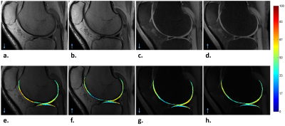
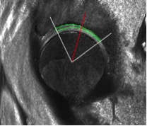
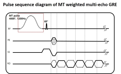
Sooyeon Ji, Se-Hong Oh, Young-Han Lee, Dongmyung Shin, Doohee Lee, Taehyun Hwang, Woojin Jung, Hyeong-Geol Shin, Jongho Lee
In this study, we explored the combined use of magnetization transfer (MT) weighting and bi-exponential T2* fitting as a potential tool to analyze the composition and microscopic geometry of the knee cartilage. The analysis results of deep cartilage areas showed that the MT ratio of the short T2* component had significantly larger values than that of the long T2* component. This observation may be explained by the geometry of collagen fibrils and proteoglycans.
|
|
1389.
 |
T1-T2 correlation of site-specific changes and zone-dependent anisotropy of osteoarthritic cartilage using multi-resolution MRI
Farid Badar, Yang Xia
Topographical and zonal based studies of healthy and OA canine tibial cartilage are shown to be essential for the early detection of osteoarthritis. A high-resolution T1-T2 correlation with the low-resolution imaging of depth-dependent T2 profiles shows a more detailed and sensitive method of measuring the early sign of cartilage degradation, beneficial to human OA MRI.
|
|
1390.
 |
Effect of spin-lock field direction on chemical exchange spin-lock (CESL) and evaluate its feasibility of glycosaminoglycan (GAG) detection at 3.0T
Baiyan Jiang, Weitian Chen
Chemical exchange spin-lock (CESL) is sensitive to fast exchange metabolites. CESL is performed across a range of resonance frequency offsets. At any frequency offset, either anti-parallel or parallel spin-lock directions can be used. However, different directions can affect the z-spectrum and the magnetization transfer ratio asymmetry analysis. We used simulations and in vivo experiments to demonstrate this effect and provided theoretical analysis. We also presented preliminary results of CESL for imaging of chemical exchange associated with glycosaminoglycan (GAG) in human knee at 3.0T.
|
|
1391.
 |
Macromolecular fraction from magnetization transfer ultrashort echo time (MT-UTE) modeling proportionally correlates with applied mechanical load on the cadaveric knee joint
Saeed Jerban, Yajun Ma , Wei Zhao, Michael Carl, Eric Chang, Jiang Du
Ultrashort echo time (UTE) MRI is able to assess long T2 tissues such as articular cartilage (AC) and short T2 tissues such as meniscus. Early stage of osteoarthritis is hypothesized to affect the mechanical properties of AC, sooner and quicker than its morphology. This study focused on the application of UTE imaging, including UTE magnetization transfer (UTE-MT) modelling, adiabatic T1r, T1 and T2* measurements in cadaveric human knee joints subject to sequential mechanical loading. Compression load application resulted in significant increases in macromolecular fraction estimated in AC and meniscus, obtained by two-pool MT modeling. T1, T1ρ and T2* biomarkers did not show consistent trends.
|
|
1392.
 |
Quantitative DCE-MRI perfusion imaging of the subchondral bone in knee osteoarthritis
Bas de Vries, Joost Verschueren, Dirk Poot, Gabriel Krestin, Edwin Oei
Changes in subchondral bone in knee osteoarthritis could be a marker of altered fluid dynamics. Perfusion can be visualized and quantified with MRI using dynamic contrast enhanced MRI (DCE-MRI). Using quantitative analysis of DCE-MRI, we compared perfusion in the affected compartment with the non-affected compartment in patients with unicompartmental knee osteoarthritis. We also evaluated the perfusion in subchondral bone marrow lesions (BMLs). Perfusion of the subchondral bone measured with DCE-MRI is not significantly different between the affected and non-affected compartment. Subchondral BMLs are significantly associated with increased perfusion parameters compared to subchondral bone regions without BMLs.
|
|
1393.
 |
Low-field MRI of osteoarthritis in humans: correlations between load-dependent cartilage properties and relaxation parameters
Erik Roessler, Carlos Mattea, Miika Nieminen, Sakari Karhula, Simo Saarakkala, Siegfried Stapf
At low magnetic fields, T1 variation within cartilage is a robust parameter that is employed to quantify the layered structure in the tissue and is sensitive to factors such as enzymatic degradation, external load, and diseases such as osteoarthritis. Variable-field relaxometry provides access to the content and local order of glycosaminoglycans and collagen via proton-nitrogen quadrupolar dips. In this study on 20 human cartilage samples, load-dependent low-field and variable-field techniques were combined for the first time to correlate NMR parameters with the severity of osteoarthritis.
|
|
Muscle
Traditional Poster
Musculoskeletal
Monday, 18 June 2018
| Exhibition Hall 1394-1411 |
08:15 - 10:15 |
|
1394.
 |
Impact of Rate of Cuff Inflation on the Post-Ischemia Hyperemic Response
Rajiv Deshpande, Erin Englund, Felix Wehrli
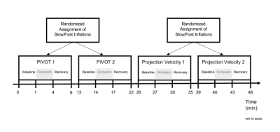
The ischemia-reperfusion paradigm can be used to evaluate skeletal muscle and peripheral vascular function. To induce ischemia, a cuff is inflated to a suprasystolic pressure, which leads to occlusion of the blood vessels, and reactive hyperemia results upon cuff deflation. This study was done to determine whether the rate at which the cuff inflates affects the hyperemic response. MRI data were acquired using the ischemia-reperfusion paradigm under slow and fast cuff inflation rates with PIVOT and projection velocity mapping in eight healthy subjects. The results suggest that there were no significant differences between hyperemic responses from slow and fast inflations.
|
|
1395.
 |
Simultaneous magnetic resonance elastography of the supraspinatus and the trapezius muscles
Daiki Ito, Tomokazu Numano, Koichi Takamoto, Kazuyuki Mizuhara, Hisao Nishijo
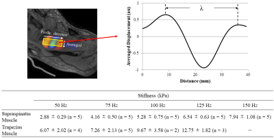
Palpation is difficult to distinguish stiffness of the supraspinatus and trapezius muscles. Magnetic resonance elastography (MRE) can measure stiffness of tissues quantitatively only if vibrations reach the tissues. We developed simultaneous MRE of the supraspinatus and trapezius muscles by adjusting the shape of a wave transducer and vibration frequency. MREs were performed using self-made wave transducer at 50-150 Hz, with a 25 Hz step. Both wave images of the supraspinatus and trapezius muscles showed clear wave propagation at 50 and 75 Hz. The results demonstrated that our techniques allow simultaneous MRE of the supraspinatus and trapezius muscles at 75 Hz.
|
|
1396.
 |
Multi-centric evaluation of stability of quantitative outcome measures in healthy calf muscles
Lara Schlaffke, Alberto De Luca, Louise Otto, Robert Rehmann, Marlena Rohm, Jedrzej Burakiewicz, Celine Baligand, Jithsa Monte, Chiel den Harder, Aart Nederveen, Hermien Kan, Martijn Froeling
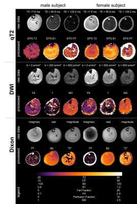
Clinical feasible, comparable muscle MR-techniques are crucial for monitoring disease progression and therapy in patients with neuromuscular diseases. We developed and evaluated a multi-modal quantitative MR protocol at 3T. Diffusion parameters, water T2 relaxation time and fat-fraction were measured and tested for temporal stability, multicenter reproducibility and covariate influence. Diffusion parameters stabilized after 15 minutes and were comparable between centers. Water T2 decreased 1ms within 1 hour. In dorsal muscles fat-fraction increased slightly, due to a decrease in muscle size. Temporal stability of quantitative parameters was shown and showed that T2 decrease needs to be considered when planning protocols.
|
|
1397.
 |
Exploring the Textural Differences between Diseased and Normal Muscle on T1 Weighted MRIs of the Mid-calf and Mid-thigh
Chang Tung Harold Yip, Phua Hwee Tang, Kein Meng Wendy Liew
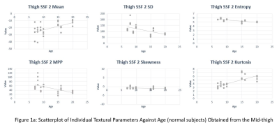
Textural analysis is a non-invasive objective method to characterize MRIs of subjects with muscular disorders. It has the potential to characterize muscle abnormalities that are not visible to the human eye. This allows the detection of muscle abnormalities earlier hence aiding early diagnosis and prognostication. This also allows textural analysis to be a potential quantitative outcome measure for clinical trials of drug treatments for muscular disorders. This study shows that the textural parameter entropy remains stable as age increases and can distinguish between diseased and normal muscle tissue.
|
|
1398.
 |
Fully automatic segmentation of all lower body muscles from high resolution MRI using a two-step DCNN model
Anudeep Konda, Katherine Crump, Daniel Podlisny, Craig Meyer, Silvia Blemker, Joe Hart, Xue Feng
Lower limb skeletal muscles play an essential role in athletic performance as wellas muscular health in patients with dystrophies. Quantitative mapping of all 35 lower body muscles from high resolution MRI has the potential to improve power and agility in athletes and assist the diagnosis and follow-up for certain musculardystrophies in medical applications. However, due to the weak contrast and insufficient boundary information, the accurate segmentation of each individual muscle is challenging. In this study we developed a fully automatic segmentation framework using a two-step DCNN model and showed accurate segmentation for all muscles.
|
|
1399.
 |
Robust multi-atlas MRI segmentation with corrective learning for quantification of local quadriceps muscles inflammation changes during a longitudinal study in athletes
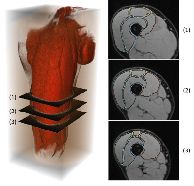
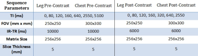
Hoai-Thu Nguyen, Pierre Croisille, Magalie Viallon , Charles de Bourguignon, Rémi Grange, Sylvain Grange, Thomas Grenier
This study propose an improved automatic segmentation of longitudinal MRI dataset of mountain ultra-marathon runners’ upper thighs acquired during the Tor des Géants 2014 by using a multi-atlas segmentation strategy with corrective learning with a small number of training set. Our highly accurate and robust segmentations allow us to locally study the inflammation of each quadriceps head induced by the extreme conditions of the race, a method that is of high interest to monitor the impact of eccentric efforts during the race, identify local physiopathology changes in patients, and benefits of eventual therapy or intervention.
|
|
1400.
 |
Using texture analysis based on T2WI, DWI and delayed T1-enhanced imaging to differentiate benign and malignant soft tissue tumors
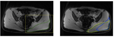
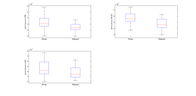
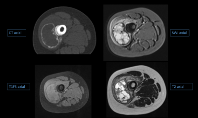
Nan Sun, Cuiping Ren, Ying Li, Jingliang Cheng, Zhizheng Zhuo
With the popularity of magnetic resonance technology in recent years, the detection rate of soft tissue tumors has been greatly improved. The soft tissue tumors in MR images show various signal intensity distribution in different modalities. This work investigated and evaluated the role of texture analysis on T2WI, DWI and delayed T1-enhanced images to characterize the soft tissue tumors, and then evaluate the textures by support vector machine classifiers (SVM) to differentiate benign and malignant soft tissue tumors. Results showed that the application of texture analysis in T2WI, DWI and T1-enhanced imaging is helpful to distinguish benign and malignant soft tissue tumors by SVM.
|
|
1401.
 |
Measurement of skeletal muscle extraceullar volume (ECV) in the healthy thigh: determination of the time to contrast equilibrium
Alex Goodall, Dr David Broadbent, Dr Raluca Dumitru, Prof David Buckley, Prof Maya Buch, Dr Ai Lyn Tan, Dr John Biglands
Five healthy volunteers were scanned at 3 T to determine the time to contrast equilibrium in skeletal leg muscle to establish whether extracellular volume (ECV) mapping is clinically feasible for skeletal muscle (as it has proved to be for myocardium). Time to contrast equilibrium was 13 minutes, and native T1 values were validated against the literature. It was also found that the difference in measurement of ECV using the aorta compared to the femoral artery was small. It is hoped that advancements in this technique could aid in the diagnosis and treatment of scleroderma patients with muscle involvement.
|
|
1402.
 |
Multi-parametric MRI-based classification for generating muscle percentage index in muscular dystrophy
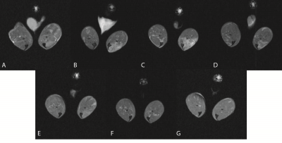
Aydin Eresen, Noor Hafsa, Lejla Alic, Sharla Birch, Jay Griffin, Joe Kornegay, Jim Ji
Imaging biomarker for muscular dystrophies, such as muscle percentage index (MPI), successfully differentiates between healthy and dystrophic muscles. However, the current methods to generate this biomarker are not well defined and therefore lack robustness and reproducibility. This study imaged ten Golden Retriever Muscular Dystrophy (GRMD) pectineus-muscle samples at a 4.7T MRI scanner. To facilitate estimation of MPI and to validate the results, we use trichrome-stained histology images. These images were registered accurately to multi-parametric quantitative MRI (qMRI). We use local gradient and texture information to classify qMRI into muscle and non-muscle with respective accuracies of 0.86 and 0.71.
|
|
1403.
 |
MRI characterization of skeletal muscles of two dystrophic mouse models
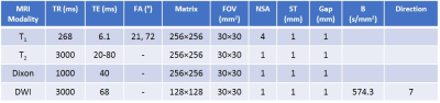
Ravneet Vohra, Joshua Park, Philip Kramer, David Marcinek, Jeffrey Chamberlain, Donghoon Lee
The mdx mouse model is one of the most commonly used animal model for Duchenne muscular dystrophy (DMD). However, it has a milder phenotype compared to patients with DMD. Evidence has demonstrated the presence of genetic modifiers that lead to phenotypic variability even with an identical gene mutation in both human and animal models of muscular dystrophy. We performed multi-parametric, high resolution MRI to demonstrate severity of disease progression in dystrophic mouse models on two different genetic backgrounds.
|
|
1404.
 |
Application of MR Elastography to Transvertebral Psoas Major Muscle
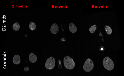
Tomokazu Numano, Daiki Ito, Koichi Takamoto, Kazuyuki Mizuhara, Hisao Nishijo
The aim of the present work was to develop the vibration techniques for the psoas major muscle (PM) MR elastography (MRE). The results indicated that the PM well vibrated, due to transmission of vibration from the lumbar spine. These findings suggest that placement of a narrow vibration pad under the supine body, along the lumbar spine, would allow PM MRE. The present techniques for the PM MRE provide a quantitative diagnostic tool for LBP-associated changes in the muscles, since increased stiffness of the muscle due to continuous contraction is suggested to be an important cause of LBP.
|
|
1405.
 |
Improved Spontaneous Activity Maps of Resting Skeletal Musculature by surface EMG-based Contraction Pattern Classification
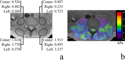
Martin Schwartz, Günter Steidle, Petros Martirosian, Michael Erb, Bin Yang, Klaus Scheffler, Fritz Schick
Reliable assessment and analysis of spontaneous mechanical activities in musculature (SMAM) visible in repetitive DWI is a relatively new technique for non-invasive characterization of skeletal musculature. To correct for data corrupted by intentional contractions, a surface electromyography-based contraction state analysis was investigated to reject undesired DWI data. It is demonstrated that the presented method enables a more reliable quantification of SMAMs and improved spontaneous activity maps.
|
|
1406.
 |
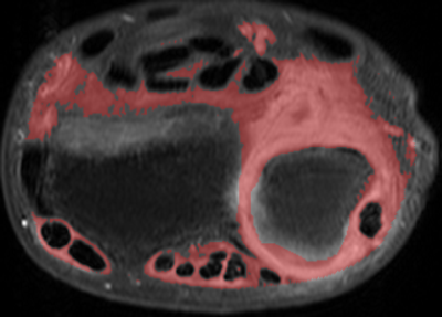
Validation of an Osirix Plugin for automatic fat infiltration measurements in Paraspinal muscles using T2 weighted images
Cristobal Arrieta, Julio Urrutia, Pablo Besa, Ignacio Osorio, Cristian Montalba, Daniel Hasson, Marcelo Andia, Sergio Uribe
Paraspinal muscle fat infiltration has been related with low back pain. This measurements are typically evaluated using T2w images, however, the accuracy of this method needs a proper validation, since inhomogeneities may produce severe signal changes. In this work, we developed and validated an OsiriX plugin which allows to segment infiltrated fat in T2w images. This tool also allowed us for validating the use of T2w images, considering Dixon fat images as gold-standard. To validate our plugin, we evaluated 5 cross sectional areas (L1-S1) of 4 paraspinal muscle groups for T2w images of 37 patients. To validate T2w images, we analyzed 10 healthy volunteers and 10 patients. We found that T2w segmentation with our OsiriX plugin is a reliable and an accurate method to evaluate the fat infiltration in paraspinal muscles.
|
|
1407.
 |
Ex vivo MRS evaluation of severe burn injury in mice shows metabolic changes in skeletal muscle
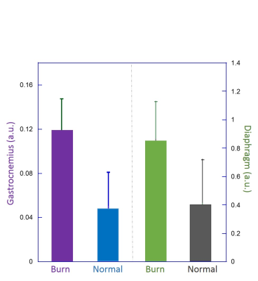
Leo Cheng, Bailing Li, Lindsey. A. Vandergrift, Jiake Chai, Zhongcong Xie
Patients of severe burn injury often suffer from sepsis, which results in multiple organ failure and prolonged metabolic derangement, leading to higher mortality. Accurate measurements of burn injury-associated metabolic changes may provide the burn clinic with quantitative tools to assess patient status. We tested the efficacy of High-Resolution Magic Angle Spinning (HRMAS) magnetic resonance spectroscopy (MRS) in evaluation of tissue metabolic changes with mouse skeletal diaphragm and gastrocnemius muscles after burn injury. HRMAS measurements indicated that IMTG and plasma FFA levels were increased after severe burn injury, with more pronounced differences detected in diaphragm muscle than in gastrocnemius muscle.
|
|
1408.
 |
Sensitivity of Quantitative Texture Metrics to Variations in Image Acquisition Parameters
Bruce Damon, Yuan Xie, Ke Li, Susan Kroop, Jane Park
The purpose of this study was to examine the dependence of a quantitative texture metric, the high gray level run length emphasis (HGRE) in T2-weighted images, on common variations in image acquisition parameters. We studied 13 muscle disease patients with quantitative fat/water MRI and contrast-based images. The ability of the HGRE was unaffected by image matrix size. We also measured the dependence of the regression parameters on TR and TE. The results support the use of quantitative texture analysis to study clinically acquired MR images in muscle disease patients.
|
|
1409.
 |
Assessment of perfusion-metabolism matching in exercising muscle from dynamic contrast-enhanced MRI and T2 mapping
Gwenael Layec, christopher Conlin, Jiawei Dong, Stephen Decker, Corey Hart, Nan Hu, Mariya Chadovich, Michelle Mueller, Lillian Khor, Christopher Hanrahan, Vivian Lee, Jeff Zhang
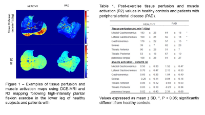
Using an MR approach combining DCE-MRI and T2 mapping, this study revealed that unlike PAD patients, muscle tissue perfusion was tightly correlated to exercise-induced changes in R2 in the lower leg muscles of healthy individuals. These findings suggest Q/Met mismatch following exercise in the skeletal muscle of PAD patients. The combination of DCE-MRI and T2 mapping opens a new avenue of research to investigate perfusion-metabolism heterogeneity in normal physiological conditions and muscle-related pathologies.
|
|
1410.
 |
Effects of PDE5A inhibition on skeletal muscle 1H2O T2 following an acute bout of downhill running and endurance training in dystrophic mice
Abhinandan Batra, Ravneet Vohra, Steve Chrzanowski, Donovan Lott, Glenn Walter, Krista Vandenborne, Sean Forbes
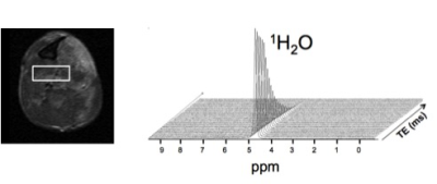
This study examined the effects of phosphodiesterase 5A inhibition with sildenafil citrate on skeletal muscle 1H2O T2 in dystrophic mice (mdx) following downhill running and during four weeks of low-intensity treadmill training. Skeletal muscle 1H2O T2 was measured from spectra acquired with a single voxel 1H-MRS STEAM sequence. Our findings showed less altered T2 after downhill running with sildenafil citrate treatment indicating less muscle damage and improved running performance during endurance training. Collectively, the results support the use of sildenafil citrate when combined with acute and chronic bouts of exercise as a potential therapeutic intervention in muscular dystrophies.
|
|
1411.
 |
Multi-Parametric MRI characterization for damaged dystrophic muscle
Joshua Park, Ravneet Vohra, Jeffrey Chamberlain, Donghoon Lee
Muscular dystrophy is a family of inherited diseases characterized by progressive muscle weakness that leads to muscle damage and wasting. Clinical measures of muscular dystrophy rely on surgical biopsy, which is invasive and limited. Magnetic resonance imaging (MRI) can provide valuable information pertaining to tissue characteristics of this disease noninvasively. We performed multi-parametric MRI to assess the changes due to muscle damage and subsequent recovery over 3 weeks starting at 12 weeks of age in disease affected mice. The differences observed through MRI measurements demonstrate MRI can be used effectively to track disease progression and responses to future therapy.
|
|
MSK: Other
Traditional Poster
Musculoskeletal
Monday, 18 June 2018
| Exhibition Hall 1412-1437 |
08:15 - 10:15 |
|
1412.
 |
A Prospective, Longitudinal Assessment of Adverse Local Tissue Reactions in Resurfacing Hip Arthroplasty Versus Primary Total Hip Arthroplasty
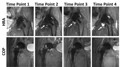
Jacqui Zhu, Matthew Koff, Bin Lin, Kara Fields, Danyal Nawabi, Edwin Su, Douglass Padgett, Hollis Potter
The purpose of this prospective study was to compare the prevalence of magnetic resonance imaging detected adverse local tissue reactions (ALTRs) in metal-on-metal hip resurfacing arthroplasty (HRA) and ceramic-on-poly (COP) total hip arthroplasty subjects. Images acquired at 4 time points with a 1-year interval showed a higher prevalence of ALTRs in the HRA than COP subjects. The self-assessed symptomatology scores did not significantly differ between the two groups at follow-up, indicating that ATLRs can be clinically silent. This study will permit better understanding of the natural history and follow up of ALTRs complicating hip arthroplasty.
|
|
1413.
 |
Dynamic contrast enhanced MR imaging in early stage knee osteoarthritis: A test-retest repeatability study
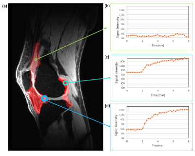
Faezeh Sanaei Nezhad, James MacKay, Josh Kaggie, Martin Graves, Fiona Gilbert, Andrew McCaskie, Rob Janiczek, Geoff Parker, Alexandra Morgan, Jose Ulloa
Dynamic contrast-enhanced MRI (DCE-MRI) has proven to be an effective method for qualitative and quantitative measurement of synovitis in the knee. Here we evaluate the test-retest repeatability of DCE-MRI measurements in the knee at 3 T. Eight patients with mild/moderate knee osteoarthritis (OA) were scanned twice, 4 weeks apart. DCE biomarkers from the extended Tofts model were measured. This is the first demonstration of the repeatability of DCE-MRI in knee OA. This evaluation provides data to enable sample size calculations for further longitudinal and interventional studies using DCE-MRI as a biomarker of inflammation in OA.
|
|
1414.
 |
Analysis of the Orientation-Dependent Frequency of Tendon via Ultrashort Echo Time (UTE) MRI
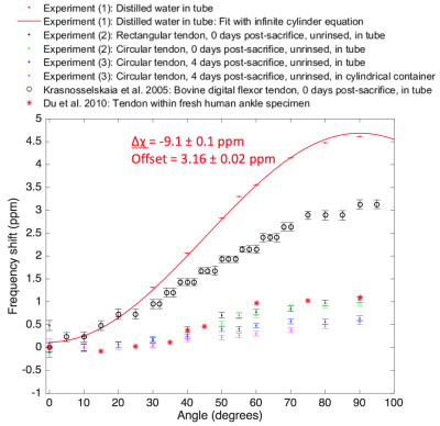
Adrienne Siu, Luca Biasiolli, Matthew Robson
Tendon exhibits changes in T2, T2*, and resonant frequency as a function of its orientation with respect to B0. An ultrashort echo time (UTE) sequence was employed to characterize the frequency of fresh bovine digital flexor tendon at angles of 0° to 90° relative to B0, causing a maximal frequency shift of 1.0 ppm. Factors that could influence the frequency of tendon were evaluated. It was found that the frequency of tendon was affected by the enclosing container, but not the geometry of the tendon.
|
|
1415.
 |
Cartilage and Meniscus T2 Relaxation Time in Subjects With and Without Meniscus Tears
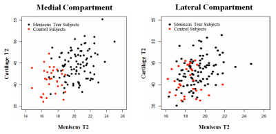
Richard Kijowski, Shivhumar Kambhampati, Joshua Bunting, Benjamin Beduhn, Kaitlin Woo, Fang Liu
This study was performed to compare cartilage T2 between subjects with and without meniscus tears. T2 mapping was performed on the knees of 30 control subjects without meniscus tears and 93 subjects with meniscus tears. Medial and lateral compartment cartilage T2 was measured. Radiographic osteoarthritis severity was assessed using the Kellgren-Lawrence (KL) grading scale. The 30 KL-0 control subjects without meniscus tears had significantly lower (p<0.001) medial compartment cartilage T2 than KL-0 (n=46), KL 1 (n=27), and KL-2 (n=20) subjects with meniscus tears and significantly lower (p<0.01) lateral compartment cartilage T2 than KL-1 and KL-2 subjects with meniscus tears.
|
|
1416.
 |
Accuracy of MRI-based measurements of aponeurosis dimensions
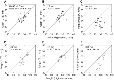
Lachlan Bird, Arkiev D'Souza, Iain Ball, Caroline Rae, Robert Herbert, Bart Bolsterlee
Aponeuroses are the thin, sheet-like tendons that cover substantial parts of muscles. We validated measurements of the dimensions of aponeuroses from T1, mDixon and ultrashort echo time (UTE) scans by comparing to direct measurements from dissection and digitisation. We used sequences that are feasible for human studies. Aponeurosis widths and lengths, measured on 20 lamb muscles, were substantially underestimated from mDixon scans. More accurate measurements were obtained from T1 and UTE scans, which had root mean square errors of 8-10% and 5-13% of the aponeurosis width and length, respectively, and did not systematically underestimate or overestimate aponeurosis width or length.
|
|
1417.
 |
Elevated conversion of hyperpolarized [1-13C]pyruvate to [1-13C]lactate is not associated with tissue acidosis, as measured with hyperpolarized [13C]bicarbonate, in a murine model of rheumatoid arthritis.
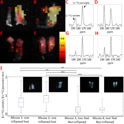
Alan Wright, Zoé Husson, De-en Hu, Gerard Callejo, Kevin Brindle, Ewan St. John Smith
Measurements of synovial fluid pH in patients with rheumatoid arthritis suggest acidosis can occur at inflamed joints. A widely used model of rheumatoid arthritis is produced by injecting complete Freund’s adjuvant into the hind paw of a mouse. We have investigated whether inflammation is associated with acidosis in this model using Magnetic Resonance Spectroscopic Imaging of injected hyperpolarised [1-13C]pyruvate, to detect the metabolic changes associated with inflammation, and hyperpolarised [13C]bicarbonate to measure extracellular pH. A significant increase in the [1-13C]lactate/[1-13C]pyruvate was observed throughout the inflamed tissue, but there is no apparent acidosis
|
|
1418.
 |
Is the anterolateral ligament affected by the rupture of anterior cruciate ligament? A tentative investigation based on magnetic resonance imaging
qian wang, Cuiping Ren, Jingliang Cheng, Zhizheng Zhuo
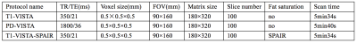
This study aimed to demonstrate the incidence of injured of ALL following ACL rupture, as well as observe the characteristics of thus injury based on MRI. In the study, we used the high resolution 3D TSE-based sequences including the optimized T1W-VISTA and T1W-VISTA-SPAIR to evaluate the 43 knees of patients who have ligament ruptured through clinical test. Chi-square test was performed to analyze the categorical variables. Binary logistic regression was performed to investigate the main cause. It indicated that ACL injuries has closer association with ACL injuries but less association with LM injuries, and the femoral portions of ALL were easily ruptered
|
|
1419.
 |
3D high resolution MR imaging of anterolateral ligament
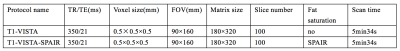
qian wang, Cuiping Ren, Jingliang Cheng, Zhizheng Zhuo
This study aimed to demonstrate the feasibility of optimized 3D high resolution MR imaging for scanning anterolaterl ligament, as well as provide more accurate imaging technique for patient with ACL and ALL injured. In the study, we used the high resolution 3D TSE-based sequences including the optimized T1W-VISTA, PDW-VISTA, and T1W-VISTA-SPAIR to evaluate the 60 knees of thirty healthy volunteers. There was significant difference between the three techniques for both the radiologists, and there was high consistency between the scores of two radiologists. 3D T1W-VISTA imaging technique has a high superiority in the three techniques, which may provide more information for clinical diagnosis.
|
|
1420.
 |
A machine learning method for tissue characterisation in the human thigh
Terence Jones, Sarah Wayte, Abhir Bhalerao, Nicola Gullick, Charles Hutchinson
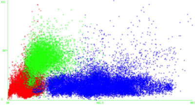
Inflammatory idiopathic myositis is a debilitating inflammatory muscle condition. Diagnosis relies on a battery of tests, but monitoring of disease severity can be challenging. We present a novel machine learning approach to classifying tissues using multi-parametric analysis of routine MRI sequences. A logistic regression model was trained to predict tissue type based on T1 and STIR signal intensity and 10-fold cross-validated. The system attained 93.8% sensitivity and 96.9% specificity overall (ROC area 0.991). Testing of this model showed a low level of ostensible muscle inflammation in 9/11 asymptomatic controls – likely due to misclassification of vessels.
|
|
1421.
 |
Usefulness of PETRA imaging for frozen shoulder patients
Ryuji Nojiri, Yasuaki Tsurushima, Hiroko Fukushima, Masaaki Hori, Murata Katsutoshi , Nobuhisa Shinozaki , Yasui Kenji , Kazuhiro Maeda, Ken Okazaki
Pointwise encoding time reduction with radial acquisition (PETRA) has made it possible to visualize those tissues which have a short T2* value such as ligaments and tendons as high signal images by using ultra-short echo time (TE). In this study, we evaluated the significant difference of the thickness of the joint capsule in the axillary pouch, depending on the stage or the symptom of patients with frozen shoulder.
|
|
1422.
 |
MRI Cytography: a biomarker of microstructural myofiber damage in Amyotrophic Lateral Sclerosis
Natenael Semmineh, Alberto Fuentes, David Medina, Rachael Sirianni, C Quarles
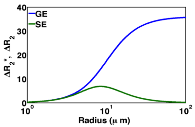
For patients diagnosed with Amyotrophic Lateral Sclerosis (ALS), the clinical heterogeneity of disease presentation and progression continues to confound the identification of robust outcome measures and biomarkers that can be used as surrogates of progression to provide faster and improved decision-making during clinical trials. To overcome this limitation we developed a non-invasive imaging strategy, termed MRI Cytography (MRC) that is uniquely sensitive to abnormal muscle cytoarchitecture. In a preclinical model of ALS, MRC was able to reliably differentiate between normal and degenerated muscle microstructure.
|
|
1423.
 |
Preliminary study of BOLD fMRI for the differentiation of musculoskeletal benign and malignant tumors
Nan Sun, Cuiping Ren, Ying Li, Jingliang Cheng, Zhizheng Zhuo
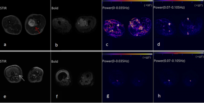
This work investigated and evaluated the role of Blood Oxygenation Level-Dependent (BOLD) based functional MRI in characterizing the musculoskeletal tumors, and furtherly evaluate the ability of the power calculated from the fMRI time series to differentiate benign and malignant tumors, which might be helpful for clinical diagnosis and studies.
|
|
1424.
 |
MRI findings in Early Rheumatoid Arthritis, their clinical correlate and method of assessment
Fan Xiao, Jacky Ka Ko, Jason Chi Shun Leung, Ryan Ka Lok Lee, David Ka Wai Yeung, Lai-Shan Tam, James Griffith
This study investigated the correlation between MRI parameters and clinical assessment in 106 treatment naïve patients presenting with early rheumatoid arthritis (ERA) i.e. symptoms < 24 months. The degree of synovial and tenosynovial proliferation, bone marrow oedema and bone erosions were semi-quantitatively and quantitatively measured on MR imaging. Quantitative MRI parameters showed better correlation with clinical assessment than semi-quantitative methods. Only quantitative MRI methods showed significant change after treatment for one year.
|
|
1425.
|
MR based changes in normal ACL hamstring graft over two years following reconstruction
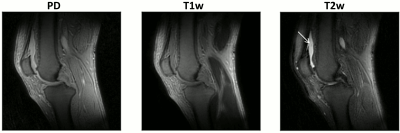
Fan Xiao, Jacky Ka Long Ko, Alex Wing Hung Ng, Jason Chi Shun Leung, David Ka Wai Yeung, Patrick, Shu Hang Yung, James Griffith
This study investigates that normal changes seen on MRI of the ACL graft over first two years after reconstruction. The graft and perigraft tissues were assessed on serial MRI examinations addressing features such as graft size, signal intensity and perfusion. MR changes were compatible with the histological process known as changes in the ACL graft, usually called ‘ligamentization of the graft’ seems to have stabilized by 24 months.
|
|
1426.
 |
Anisotropic analysis and decay characteristics of T2* relaxation of the human Achilles tendon studied with 7 T MR-microscopy
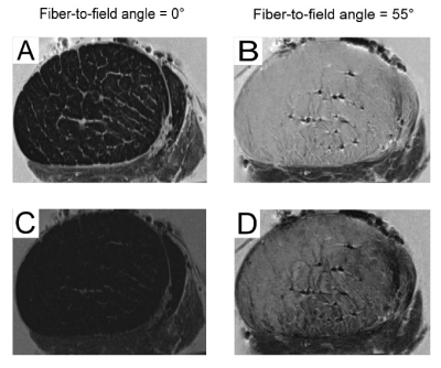
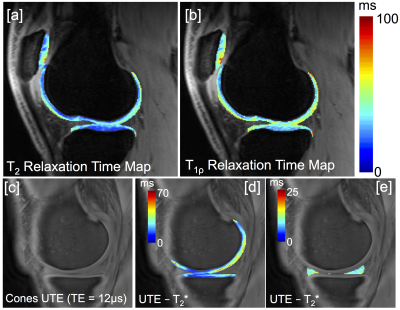
Benedikt Hager, Vladimir Juras, Martin Zalaudek, Joachim Friske, Xeni Deligianni, Oliver Bieri, Lena Hirtler, Andreas Berg, Markus Schreiner, Sonja Walzer, Siegfried Trattnig
The fiber-to-field angle dependence and the T2* characteristics of a human Achilles tendon were investigated. The results show an increase of approx. factor 20 in T2* values when the long axis of the tendon is change from 0° to 55°, which is much higher than previously reported. Moreover, in contrast to previous findings we found no homogenous biexponential decay behavior for the tendon on a small sized voxel basis. The results reported here are to our knowledge the first MR-microscopy evaluations of the orientational dependence of T2* relaxation in the Achilles tendon.
|
|
1427.
 |
MRI Methods for Exercise-based Perfusion Assessment of Diabetic Feet with Ulcers
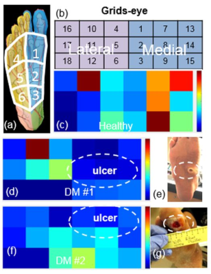
Masoud Edalati, Mary Hastings, Zayed Mohamed, David Muccigrosso, Ran Li, Michael Mueller, Jie Zheng
The purpose of this study was to develop MRI methods for comprehensive evaluation of foot muscle perfusion and perfusion reserve in patients with diabetes and foot ulcers. Healthy controls and patients with diabetic foot ulcers were scanned with a non-contrast MRI protocol at rest and during a standardized foot flexion exercise. Ischemic regions around foot ulcers were clearly identified with quantitative perfusion data during the exercise.
|
|
1428.
 |
T1?, T2, and RAFF are Sensitive to Acute Ischemic Injury to the Femoral Head in a Piglet Model of Legg-Calvé-Perthes Disease
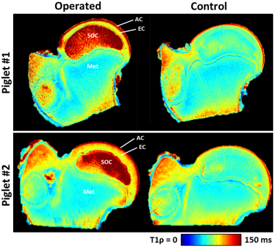
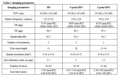
Casey Johnson, Cathy Carlson, Ferenc Toth, Harry Kim, Jutta Ellermann
We demonstrate that quantitative T1ρ, T2, and RAFF relaxation time maps are highly sensitive to bone/marrow and cartilage changes within 48 hours following ischemic injury to the growing femoral head. This work has important implications for the diagnosis and treatment of diseases associated with avascular necrosis of bone and cartilage.
|
|
1429.
 |
Impact of Respiratory Triggering in 3T Sub-Millimeter High Resolution Brachial Plexus MRI
Darryl Sneag, Jacqui Zhu, Susan Lee, Tina Jeon, Bin Lin, Maggie Fung
This study’s purpose was to compare non-respiratory and respiratory- triggered proton density and T2-weighted DIXON fat suppression sequences for high-resolution brachial plexus MRI. In a cohort of 5 volunteers and 20 patients, we were able to demonstrate that respiratory triggering substantially reduced ghosting artifact and improved delineation of nerve fascicular architecture with acceptable increased scan time.
|
|
1430.
 |
Advanced Knee Imaging Study in NCAA Division 1 Basketball: Protocol Development and Preliminary Results
Katherine Young, Feliks Kogan, Robert Peters, Matthew Koff, Valentina Pedoia, Marc Safran, Ben Ma, Riley Williams, Tom Wickiewicz, Marianne Black, John Sabol, Kimberly Amrami, Hollis Potter, Sharmila Majumdar, Garry Gold
Chronic knee injuries are especially common in jumping athletes, and in particular high-level basketball players. In this work, we developed an advanced quantitative MRI protocol to longitudinally study early degenerative changes in high-level basketball players across multiple sites. Studying these changes, between high and low impact athletes, within one season as well as over three seasons for a cumulative effect, will help provide better insight into these changes. In developing this protocol for a multi-center study, we use a common phantom to assess biases in quantitative measurements across study scanners.
|
|
1431.
 |
Ultra-short echo-time (UTE) imaging of the knee with curved surface reconstruction-based extraction of the patellar tendon
Martin Krämer, Marta Maggioni, Christoph von Tycowicz, Nick Brisson, Stefan Zachow, Georg Duda, Jürgen Reichenbach
Due to very short T2 relaxation times, imaging of tendons is typically performed using ultra-short echo-time (UTE) acquisition techniques. In this work, we combined an echo-train shifted multi-echo 3D UTE imaging sequence with a 3D curved surface reconstruction to virtually extract the patellar tendon from an acquired 3D UTE dataset. Based on the analysis of the acquired multi-echo data, a T2* relaxation time parameter map was calculated and interpolated to the curved surface of the patellar tendon.
|
|
1432.
 |
Analysis of collagen fibrillogenesis of a caprine patella tendon with magic angle imaging
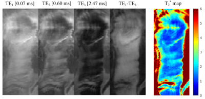
Karyn Chappell, Catherine Van Der Straeten, Donald McRobbie, Wladyslaw Gedroyc, Mihailo Ristic, Djordje Brujic
It is known that our collagen fiber alignment changes as we develop, reach maturity and then age: the crosslinking of collagen is considered one of the best biomarkers of aging. This study used magic angle imaging to visualise the collagen fiber changes between development and skeletal maturity in caprine knees. Immature tendons are less aligned during development, becoming more aligned as skeletal maturity is reached. This method has great potential to non-invasively improve our understanding of the development and degeneration of collagen rich structures.
|
|
1433.
 |
Feasibility of monosodium urate assessment using multi-echo gradient echo based quantitative imaging
Seung hee Han, Yoonho Nam, Joon-Yong Jung, Won-Hee Jees
Gout is a common disease caused by monosodium urate (MSU) accumulation in joints. Although conventional MR imaging well describes generic features of inflammation, sensitivity of MSU is relatively low compared to dual energy CT. Because MSU has diamagnetic susceptibility, high sensitivity can be expected in magnetic susceptibility related contrast imaging. However, calcium is another diamagnetic material existing in joints. Therefore, distinguishing MSU and calcium is an essential step for imaging MSU. In this context, we investigate the feasibility of multi-echo gradient echo based quantitative imaging for MSU assessment.
|
|
1434.
 |
The role of susceptibility weighted imaging (SWI) in musculoskeletal radiology as an alternative to computed tomography (CT).
Akshaykumar Kamble, Gaurav Gangavani
SWI has been used for detection of calcification and hemosiderin deposits in diagnosis of the neurological disorders, hemorrhagic disorders and neuroinfectious conditions. Our study tries to answer the question that whether the susceptibility weighted MR imaging can provide alternative to the CT scan and thus decreasing our dependency on the modality which has significant drawback of having radiation dose especially to our young patients. We compared SWi and CT for the characterization of lesion calcification and hemorrhage and we found there was no significant difference in detection rate of these characteristics between two modalities, thus proving SWI as equally sensitive.
|
|
1435.
 |
T1 and T2 Mapping of Delayed Gadolinium Enhancement in Osteoarthritis with MR Fingerprinting
Joshua Kaggie, James MacKay, Guido Buonincontri, Fiona Gilbert, Rolf Schulte, Alexandra Morgan, Robert Janiczek, Michela Tosetti, Andrew McCaskie, Martin Graves
Mapping of quantitative MRI relaxation values is promising for improving the assessment of MSK disease. Magnetic Resonance Fingerprinting (MRF) is a new method that enables fast quantitative MRI by exploiting the transient signals caused by the variation of pseudorandom sequence parameters.
This proof-of-concept work demonstrates the utility of MR Fingerprinting in the knee. Seven participants, four of which had Kellgren-Lawrence (KL) grade 2 or 3, were imaged eighty minutes after gadolinium injection with MRF on a 3.0T MRI. The mean T1 relaxation times were shorter in cartilage by 5-20% in KL=2,3 subjects when compared to normal subjects.
|
|
1436.
 |
Significant Metabolic Differences Between Benign Lipomatous Lesion and Liposarcoma Identified by High-Resolution 1H and 31P MRS: A Pilot Study
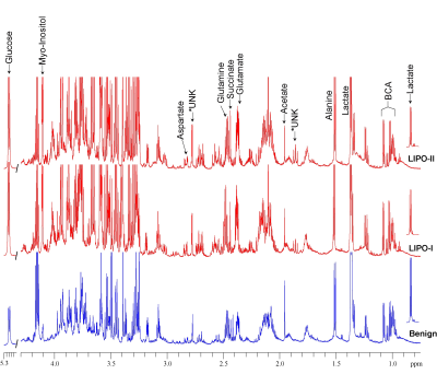
Santosh Bharti, Brett Shannon, Adam Levin, Carol Morris, Laura Fayad, Zaver Bhujwalla
Adipocytic tumors present a spectrum of neoplastic disease including benign lipomas and their variants, atypical lipomatous tumors, and malignant liposarcomas. Distinguishing areas of malignant dedifferentiation from benign and atypical lipomatous tumors is a diagnostic challenge due to overlapping magnetic resonance imaging characteristics, and pre-operative diagnostic accuracy is poor. Here we have identified dramatic differences in the metabolic profile of water-soluble and lipid extracts of adipocytic tumors, suggesting that magnetic resonsance spectroscopy may have the potential to improve diagnostic accuracy. Our data may also lead to potential metabolic targets for treatment.
|
|
1437.
 |
Automated Seed Points Selection Based Radial-Search Segmentation Method For Sagittal and Coronal View Knee MRI Imaging
Sandeep Jogi, Rafeek T., Sriram Rajan, Krithika Rangarajan, Anup Singh, Amit Mehndiratta
Knee disorders are generally marked in tibio-femoral bone junction. Most of available segmentation techniques use time consuming semi-automatic approach as radial search method, in sagittal view only. However, coronal view MRI Knee images are clinically equal important. Proposed approach automates seed points selection process for the radial search method, which work equally good on both sagittal and coronal view for identification of tibio-femoral junction.
|
|
Bone
Traditional Poster
Musculoskeletal
Monday, 18 June 2018
| Exhibition Hall 1438-1450 |
08:15 - 10:15 |
|
1438.
 |
Does chemical shift imaging offer a biomarker for the diagnosis and assessment of disease severity in multiple myeloma?
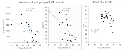
Miyuki Takasu, Takayuki Tamura, Yuji Akiyama, Chihiro Tani, Yoko Kaichi, Shota Kondo, Kazuo Awai
We investigated whether chemical shift imaging (CSI) is useful for differentiating multiple myeloma infiltration from hematopoietic bone marrow and for quantitatively assessing disease severity. For those myeloma patients with relatively high cellularity in the bone marrow, a lower signal drop on oppose phase images indicated a higher tumor burden. For bone marrow with relatively low cellularity, disease severity was not reflected on CSI. CSI did not prove useful for differentiating myeloma infiltration from hematopoietic bone marrow, which implies that differentiation between regrowth of hematopoietic bone marrow and minimal residual disease or relapse after chemotherapy might be difficult with CSI.
|
|
1439.
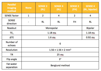 |
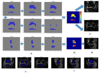
Towards Whole-Skeleton Fat Fraction Mapping: The Impact of Parallel Imaging
Vruti Dattani, Tim Bray, Alan Bainbridge, Margaret Hall-Craggs
Whole body MRI (WB-MRI) is increasingly used to image the skeleton in haematological diseases such as multiple myeloma (MM) and inflammatory disorders such as spondyloarthritis. WB-MRI can be used to acquire fat fraction (FF) maps, which can assess disease severity and treatment response. However, patients with bone pain find it difficult to lie in the scanner for long periods, necessitating the use of parallel imaging to accelerate the acquisition. The aim of this study was to determine the extent to which parallel imaging causes noise artifacts and fat-water swaps in FF maps, and to assess their impact on FF measurements.
|
|
1440.
 |
Fat Fraction Thresholds for Defining Bone Marrow Edema and Fat Metaplasia in Spondyloarthritis: More Objective than ‘A Tiny Bit of White’
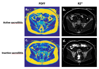
Timothy Bray, Alan Bainbridge, Corinne Fisher, Debajit Sen, Margaret Hall-Craggs
MRI is now widely used to diagnose spondyloarthritis, but existing methods for image analysis rely on qualitative visual analysis by radiologists, and suffer from poor reproducibility between observers. Here, we show that proton density fat fraction (PDFF) measurements can be used as an objective, quantitative alternative to visual analysis. Using receiver operating characteristic (ROC) analysis, we find that PDFF measurements enable accurate separation of bone marrow edema (active inflammation) and fat metaplasia (structural damage) from normal marrow. The described approach is more objective than looking for 'a tiny bit of white' on fat-suppressed images, which is the current clinical standard.
|
|
1441.
 |
Measure for Measure: Machine Learning Models for Osteoporosis MRI data
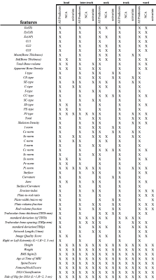
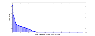
Uran Ferizi, Harrison Besser, Chamith Rajapakse, Punam Saha, Stephen Honig, Gregory Chang
We examine how Machine Learning can be used to identify novel risk factors of osteoporotic bone fracture. Using measurements from patient MRI scans at five anatomical sites, we sought to find which specific regions are best for stratifying the risk of osteoporotic fracture. Further studies on these models and other data will help improve clinicians’ ability to accurately diagnose Osteoporosis, so that patients at risk for bone fracture may be caught and treated earlier.
|
|
1442.
 |
Performance of different classifiers in the diagnosis of benign and malignant bone tumors based on MR diffusion kurtosis imaging
Zhizheng Zhuo, Ying Li, Cuiping Ren, Jingliang Cheng
Recently, the AI (Artificial Intelligence) is popular in the clinical diagnosis based on medical imaging. The major target is to identify or classify the disease condition through the features extracted from the clinical images. Different algorithms (or classifiers) can be applied to classify the disease and the performance might be different for a specific clinical issue. In this work, we tried to investigate the performance of different classifiers in the diagnosis of benign and malignant bone tumors based on MRI diffusion kurtosis imaging.
|
|
1443.
 |
Chemical Shift Quantitative Magnetic Susceptibility Study of Ex-vivo Human Cortical Bone Specimen with three-dimensional Cones ultra-short echo time (UTE) imaging
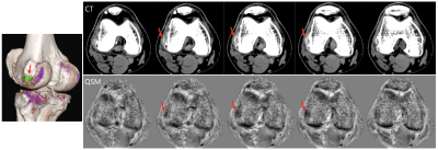
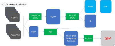
Xing Lu, Saeed Jerban, Michael Carl, Yajun Ma, Annette Drygalski, Eric Chang, Jiang Du
Bone mineral density (BMD) evaluation is crucial for the diagnosis of osteoporosis and related fractures. The purpose of this pilot study was to use a chemical-shift QSM method based on a 3D UTE-Cones sequence to assess the susceptibility values of human cortical bone specimens with consideration of gender and donor age, ranging over 5 decades. Significant differences between QSM values were observed for the different genders. A decaying trend between the minus QSM value and advancing age exists, which suggests a relationship between QSM values and BMD.
|
|
1444.
 |
Study of mono-exponential and intravoxel incoherent motion models in differentiation of metastasis from myeloma
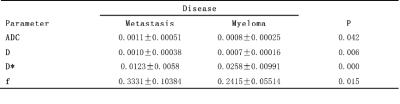
Xiaoying Xing, Ning Lang, Huishu Yuan
This study aimed to evaluate the diagnostic performance of diffusion weighted imaging (DWI) to differentiate metastasis from myeloma using the apparent diffusion coefficient (ADC) and parameters derived from the intravoxel incoherent motion (IVIM) theory. 40 patients with metastasis and 12 with myeloma underwent diffusion-weighted magnetic resonance (MR) imaging and dynamic contrast enhanced MRI (DCE-MRI). ADC, diffusion coefficient(D), pseudodiffusion coefficient(D*), and perfusion fraction (f) were calculated.Through our study it is feasible to d ifferentiate metastasis from myeloma by mono-exponential and IVIM models . IVIM-derived D and D* values showed significantly better diagnostic performance than ADC values in differentiating metastasis from myeloma.
|
|
1445.
 |
Clinical value of semi-quantitative and quantitative MR perfusion imaging in distinguishing malignant from benign bone tumors
Ying Li, Cuiping Ren, Jingliang Cheng, Zhizheng Zhuo
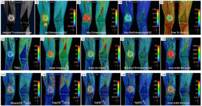
The dynamic contrast-enhanced magnetic resonance imaging(DCE-MRI) is a common scanning technology which contains semi-quantitative and quantitative perfusion information. This work investigated and evaluated the ability of semi-quantitative and quantitative perfusion information in characterizing the bone tumors, and furtherly evaluate the ability of semi-quantitative and quantitative parameters to differentiate benign and malignant tumors.
|
|
1446.
 |
Proton Density Zero Echo Time(ZTE) Imaging for Evaluating the Bone Involvement in the Femoral Tumor
Xin Lou, Jinfeng Li, Lin Xu, Xigang Zhao, Jianxun Qu, Lin Ma
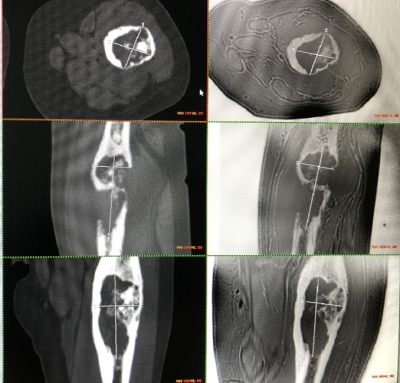
MRI can display the compositions of different tissues and adjacent involvements. In the patients of bone tumors, the integrity of cortical bone needed to be assessed for the preoperative planning. This study used proton density ZTE to display the bone involvement in patients of femoral tumors. Substantial agreement was found between CT and ZTE (r=0.98-0.99) and there was not statics significance between the measured diameters from CT and ZTE MRI (p=0.34-0.99). further development of ZTE may obviate the need of CT in evaluating the bone involvement of femoral tumors.
|
|
1447.
 |
Preliminary study of T1rho imaging technique in assessment of early intervertebral disc degeneration in asymptomatic pilots at 3.0T magnetic resonance
XiuLan Zhang, Yongmin Bi, Lizhi Xie
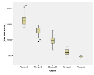
T1rho MRI in the lumbar spine may provide a tool for the diagnosis of early degenerative changes in the disc. In this study, the mean T1rho value of pilots was significantly lower than that of the control group. The degenerative grades of pilots mainly were grade III and IV, but control group were grade I and II. There were significant differences in T1rho values at each age group between pilots and control group. And overload on spine column of pilots may be the important reason in degeneration and accelerate the degeneration process.
|
|
1448.
 |
Utility of ZTE for the Characterization of Acute Ankle Fractures
Alissa Burge, Ryan Breighner, Megan Sahr, Matthew Koff, Ogonna Nwawka, Darryl Sneag, Gabrielle Konin, Bin Lin, David Helfet, Hollis Potter
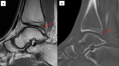
ZTE MRI provides CT-like tissue contrast, facilitating evaluation of mineralized bone. The utility of ZTE for evaluation of acute ankle fractures was evaluated in a series of 14 patients who underwent preoperative clinical MRI with an additional ZTE sequence, and subsequently underwent surgical fracture fixation. Fractures were characterized in a blinded fashion utilizing ZTE and CT, with subsequent operative confirmation. ZTE provided accurate characterization of fractures relative to both CT and surgery, with excellent inter- and intra-observer reliability.
|
|
1449.
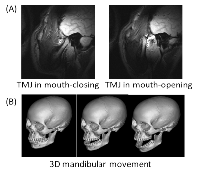 |
Analysis of the relationship between mandibular joint motion trajectory and masticatory muscle properties (volume, shape, T1&T2 value) with MR dynamic imaging
Ryusuke Nakai, Takashi Azuma, Toshihiro Togaya, Hiroo Iwata
For the diagnosis of temporomandibular joint disease, it is important to analyze with complete accuracy the range of mandibular motion and the tissue properties of the masticatory muscle in individual patients. In this study, we explored the parameters for accurate imaging of the mandibular motion trajectory using MR dynamic imaging, and then analyzed the relationship between the range of mandibular motion and the tissue properties of the masticatory muscle. As a result, we successfully identified the optimal imaging parameters and clarified that the range of side-to-side motion of the mandibular joint correlated with the tissue properties of the masticatory muscle.
|
|
1450.
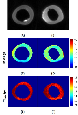 |
Macromolecular and water pools distribution maps in bovine cortical bone using ultrashort echo time (UTE) MRI combined with magnetization transfer (MT) modeling
Saeed Jerban, Yajun Ma , Wei Zhao, Xing Lu, Michael Carl, Eric Chang, Jiang Du
Collagenous matrix, bound and pore water pools are main responsible components for viscoelastic properties of the cortical bone. Quantitative ultrashort echo time MR imaging (UTE-MRI) has been shown to be able to assess bound and pore water components as indexes for bone microstructure. UTE magnetization transfer (UTE-MT) modelling can evaluate the macromolecular (MM) components of the bone (collagen). Pixel mapping of MR properties of collagen and water components in cortical bone helps to localize pathologic or traumatic bone defects. This study focused on deriving the pixel maps of MR properties of these key bone components on seven bovine bone specimens.
|
|
| Back |
| The International Society for Magnetic Resonance in Medicine is accredited by the Accreditation Council for Continuing Medical Education to provide continuing medical education for physicians. |
