Cancer Imaging
Traditional Poster
General Cancer Imaging
Monday, 18 June 2018
| Exhibition Hall 1509-1553 |
16:15 - 18:15 |
|
1509.
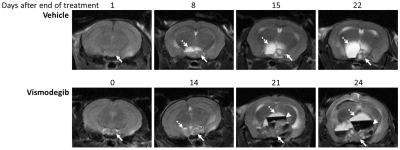 |
Using MRI to assess sonic hedgehog pathway inhibition in a genetically-engineered mouse model of adamantinomatous craniopharyngioma
Jessica Boult, Gabriela Carreno, John Apps, Laura Danielson, Laura Smith, Alexander Koers, Louis Chesler, Juan Pedro Martinez-Barbera, Simon Robinson
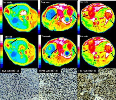
Expression of sonic hedgehog (SHH) pathway components is enriched in adamantinomatous craniopharyngiomas (ACPs) arising in Hesx1Cre/+;Ctnnb1lox(ex3)/+ mice compared to control pituitaries. An MRI-embedded trial of smoothened inhibitor vismodegib in this genetically-engineered mouse model was undertaken to assess SHH pathway inhibition in ACP. Longitudinal MRI identified accelerated solid tumour growth in response to 28 days vismodegib treatment, which was associated with increased tumour cell proliferation, and resulted in shorter survival. 7 days of treatment induced early tumoural lesions in Hesx1Cre/+;Ctnnb1lox(ex3)/+ pituitaries, resulted in a more undifferentiated and proliferative phenotype, and was associated with an elevated number of cells with clonogenic potential.
|
|
1510.
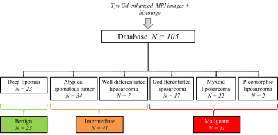 |
MRI-based radiomic to assess lipomatous soft tissue tumors malignancy: a pilot study
Benjamin Leporq, Amine Bouhamama, Fabrice Lame, Catherine Bihane, Michaël Sdika, Jean-Yves Blay, Olivier Beuf, Frank Pilleul
Aim of this study was to develop a MRI-based radiomic method to assess lipomatous soft tissue tumors malignancy. 105 subjects with lipomatous soft tissue tumors whose histology was known and with fat-suppressed T1w contrast enhanced MR images available were retrospectively enrolled to constitute a database. Based on histology, three groups were constituted according to malignancy from lipomas to high grade liposarcomas. A decisional algorithm based on 2 multivariate radiomic models was built to distinguish between these groups. Results demonstrate that the evaluation of lipomatous tumor malignancy is feasible using a routinely used MRI acquisition in clinical practice.
|
|
1511.
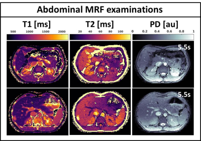 |
Magnetic resonance fingerprinting on a 1.5T MRI-Linac for tumor response monitoring
Tom Bruijnen, Bjorn Stemkens, Jan Lagendijk, Cornelis van den Berg, Rob Tijssen
Magnetic resonance fingerprinting (MRF) is the ideal tool for rapid daily tumor response monitoring on a MRI-Linac (MRL). The 1.5T MRL used in our institution has a modified gradient coil and magnet coil design that potentially complicates the parameter quantification in MRF. In this work we are the first to demonstrate the feasibility of 2D MRF in phantoms and in-vivo on a 1.5T MRL. Moreover, we investigate the accuracy and precision of the parametric maps.
|
|
1512.
 |
MRI-compatible intravital imaging window for longitudinal imaging of orthotopic mouse ovarian and pancreatic tumor stroma
Filip Bochner, Vishnu Mohan, Inbal Biton, Michal Neeman
Longitudinal multi-modal imaging of abdominal organs remains a challenge. In cancer research, where data acquired at multiple spatial and temporal scales is especially valuable, combination of powerful microscopic methods with MRI can yield complementary information about ECM and vascular components of the tumor stroma, both constituting a hallmark of pancreatic and ovarian tumors. Here we present the MRI compatible optical imaging window for longitudinal imaging of ovary and pancreas.
|
|
1513.
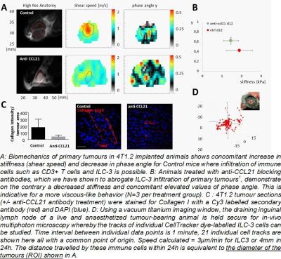 |
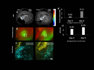
Exploring the use of MR Elastography to probe immune cell-stromal interaction in tumour microenvironment
Ralph Sinkus, Rachel Evans, Fabian Flores-Borja, Tony Ng
There is great, unmet need in understanding and monitoring non-invasively the immune cell changes within the tumour stromal microenvironment during cancer treatment. However there is as yet no reliable non-invasive method of identifying at very early time points patients who are most likely to benefit from this relatively expensive class of treatments which generally are only associated with a clinical response in 25-30% of patients1. We show here in a mouse model that changes 11 days after implantation in the liquid-to-solid ratio (phase angle y) of the tumour biomechanics are indicative for successful immune cell – stromal cell interactions.
|
|
1514.
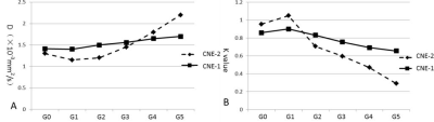 |
DKI can early detect radio-insensitive human nasopharyngeal carcinoma xenograft in nude mice
Xiang Zheng, Yunbin Chen, Youping Xiao, Dechun Zheng
In order to evaluate feasibility of DKI sequence in early differentiating radio-insensitivity of nasopharyngeal carcinoma xenografts, Seventy-two nude mice were implanted with CNE-1(low radiosensitivity) and CNE-2(high radiosensitivity) and the xenografts were obtained. MRI scanning was performed after fractional irradiation. There are differences of the changes of DKI parameters (both D and K) between CNE-1 and CNE-2 before tumor volumes changed. Therefore, Both D and K can early (before volumes changed) distinguish radio-insensitive NPC xenografts from others.
|
|
1515.
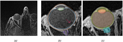 |
Adult eye segmentation in MRI using active shape model: towards a personalized eye model for radiation treatment of uveal melanoma
Huu-Giao Nguyen, PhD, Raphael Sznitman, Prof. , Marta Peroni, PhD, Jan Hrbacek, PhD, Damien C. Weber, Prof. MD, Alessia Pica, MD, Meritxell Bach Cuadra, PhD
We aim to construct a 3-dimensional patient-specific eye model from MRI data in order to later be integrated into proton radiation treatment planning. Our major challenge is the presence of motion, as subjects are awake and physiologically blink eyes. Additionally, fixing a point during acquisition might be challenging for some patients with ocular tumors. As such, in this study we evaluated an Active Shape Model (ASM) segmentation on a data set of 31 subjects, including 3 uveal melanoma (UM) patients. Quantitative evaluation in comparison with manual delineations shows good accuracy, even for images with the presence of UM and tantalum clips.
|
|
1516.
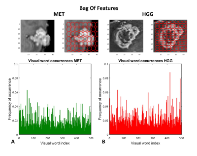 |
Automatic classification between high grade gliomas and brain metastasis using Bag-Of-Features in comparison to statistical and morphologic features
Moran Artzi, Gilad Liberman, Dafna Ben Bashat
This study suggests a clinical decision-support tool for automatic classification of brain tumors. Classification was performed on 179 MRI patients: 81 patients with high grade-gliomas (HGG) and 98 patients with brain metastases (MET, 55 breast, 43 lung, cancer origin). The input data were Bag-Of-Features (BoF) and statistical-&-morphologic features extracted from T1WI+Gd. Classification was performed using five ensemble classifiers and results were evaluated using five-fold cross-validation. Best classification results produced accuracy=83%, sensitivity=87%, and specificity=81% for discriminating between HGG and MET using Statistical-&-morphologic features, and accuracy=79%, sensitivity=76%, and specificity=80% for discriminating between breast and lung MET using BoF + Statistical-&-morphologic features.
|
|
1517.
 |
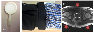
Dedicated 1.5T 16 channel array for MR-guided radiation treatment planning of head and neck tumors
Stefan Weick, Kathrin Breuer, Titus Lanz, Michael Sauer, Victor Lewitzki, Bülent Polat, Thorsten Bley, Michael Flentje
Precise target delineation and safety margin definitions are mandatory in radiation treatment of head and neck tumors. In this context, magnetic resonance imaging (MRI) is increasingly used in addition to computed tomography (CT) in the treatment planning system because of its superior soft tissue contrast. In this work, a novel 16 channel head and neck array coil is presented, which is adapted to the special requirements of radiotherapy planning. It allows for MR imaging of patients with brain and head and neck tumors in treatment planning position in individual immobilization masks.
|
|
1518.
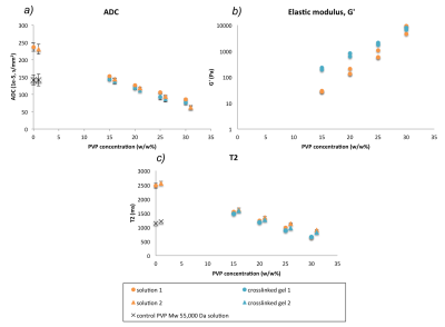 |
Investigating the effect of macromolecular cross-linking and increasing fiber density on the diffusion and viscoelastic properties of extracellular matrix materials using multiparametric MRI
Hannah Macdonald, Jeffrey Bamber, David Collins, Mihaela Rata, Maxim Ryadnov, Nandita deSouza
Synthetic polymer polyvinylpyrrolidone and fibrous protein collagen were used to investigate the effect of macromolecular cross-linking and increasing fiber density on the physicochemical properties of extracellular matrix models using clinical MRI parameters and torsional rheometry. T1 and T2 decreased with increasing viscoelastic moduli of both materials. Covalent cross-linking of macromolecules by irradiation affected stiffness, but had a smaller effect than polymer concentration on T1, T2 and ADC. Collagen at increasing concentrations sufficient to substantially affect tissue stiffness (reflecting increasing fiber density) affected the structure of water within tissue, (changes in T1 and T2), but did not hinder water diffusion.
|
|
1519.
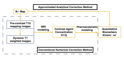 |
Assessment of Approximated Analytical B1+ Correction Method for prostate DCE-MRI with Multiple Noise Levels and in 3.0 T Systems
Xinran Zhong, Thomas Martin, Steve Raman, Holden Wu, Krishna Nayak, Kyunghyun Sung
B1+ correction is essential for quantitative prostate DCE-MRI. A simplified approximated analytical B1+ correction method was proposed previously, and we assess this method on a digital reference object (DRO) with various SNR levels and on 110 in-vivo cases from two 3.0 T systems. We find that the approximated analytical B1+ correction method achieves comparable performance to conventional correction method with substantially reduced computation. The approximated analytical correction method is simple and practical for application in the clinic.
|
|
1520.
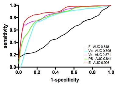 |
Characterization of endometrioid adenocarcinoma microcirculation using distributed parameter model in DCE MRI
Zhi Jun Ye, Gang Ning, Hui Zhu Chen, Yan Song
Objective: To clarify the features of vascular proliferation and permeability in endometrioid adenocarcinoma. Methods: The DCE-MRI was applied to 55 women who confirmed as endometrioid adenocarcinoma with postoperative pathology. The receiver operating characteristic (ROC) analysis was employed using parameters derived with the DP model to differentiate tumor and normal myometrium and assess the diagnostic efficiency of these parameters. Results: E and PS in tumor was lower. F in tumor was faster. Vp and Ve in tumor were lower. Areas under ROC curve (AUCs) for E and PS attained values of 0.906 and 0.844. AUCs for F attained value of 0.548. Vp and Ve in tumor with AUC values of 0.796 and 0.871. Conclusion: The permeability of vascular wall was significantly lower in endometrioid adenocarcinoma, and the vascularity was moderately lower, suggestive of very different cell growth environment in endometrioid adenocarcinoma in comparison with most solid tumours.
|
|
1521.
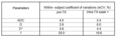 |
Repeatability of intravoxel incoherent motion diffusion-weighted MRI during chemoradiation therapy in head and neck cancers
Ramesh Paudyal, Nadeem Riaz, Vaios Hatzoglou, Xie Peng, Jonathan Leeman, David Aramburu Nunez, Yonggang Lu, Joseph Deasy, Nancy Lee, Amita Shukla-Dave
The aim of this study is to determine the repeatability of pre- treatment (TX) and intra- TX week 1 imaging metrics derived from intravoxel incoherent motion diffusion weighted imaging (IVIM-DWI) in head and neck (HN) cancer patients during chemoradiation therapy. ADC, D, and D* imaging metrics showed better repeatability measurement than f in the metastatic node of HN cancer patients.
|
|
1522.
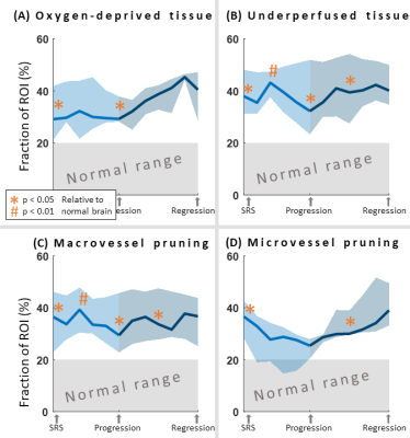 |
Brain metastases developing pseudoprogression have poor vascular function and supply
Ingrid Digernes, Endre Grøvik, Line Nilsen, Cathrine Saxhaug, Oliver Geier, Edmund Reitan, Dag Ottar Sætre, Birger Breivik, Kari Jacobsen, Åslaug Helland, Kyrre Emblem
Stereotactic radiosurgery of brain metastases can cause pseudoprogression. In this study, we use Vessel Architectural Imaging, based on dual echo DSC, to investigate the course of vascular function of brain metastases, both prior to and after pseudoprogression have occurred. Our results show that pseudoprogressing metastases were characterized by underperfused and oxygen-deprived tissue, and micro- and macrovessel pruning in the peritumoral regions. This was in contrast to peritumoral regions of responding metastases as well as normal-appearing brain tissue.
|
|
1523.
 |
Grading of gliomas using Neurite orientation dispersion and density imaging (NODDI) on a clinical scanner
Arush Honnedevasthana Arun, Aarthi Deepesh, Dhritiman Chakrabarti, Jitender Saini
Diffusion tensor imaging is sensitive to movement of water molecules but not specific as a biomarker in evaluating the highly complex microstructural environment of gliomas. Neurite orientation dispersion and density imaging (NODDI) uses different strengths of diffusion gradients to provide more specific indices of tissue microstructure than DTI. Patients with grade IV gliomas exhibited significant increase in both neurite density and orientation dispersion index as compared to grade III and II glioma cases. This study demonstrates clinical feasibility of using NODDI as a biomarker to grade tumors.
|
|
1524.
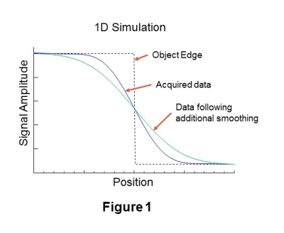 |
Convolution-Difference Method for Feature Segmentation of Low-Resolution Images
Andrew Maudsley
Automated lesion segmentation of clinical imaging studies is of potential value for treatment monitoring and radiation treatment planning. With low spatial resolution imaging systems, such as MR Spectroscopic Imaging, segmentation based on image intensity variations must take into consideration the broad spatial response function. In addition, the relative lesion-to-background intensity variation and the object size must be considered. In this report a new automated image segmentation method is presented that accounts for these factors, which is based on a subtraction of a smoothed version of the MRSI maps from the original data.
|
|
1525.
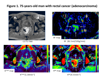 |
A comparison of pseudo continuous arterial spin labeling perfusion MRI (pCASL) and permeability imaging with dynamic contrast-enhanced MRI (DCE-MRI) in human rectal cancer
Yuichi Kumagae, Yoshihiko Fukukurra, Koji Takumi, Hiroto Hakamada, Tomoyuki Okuaki, Takashi Yoshiura
Our purpose was to investigate potential correlations between the blood flow (BF) measured by pCASL and dynamic contrast-enhanced (DCE) MRI-derived pharmacokinetic parameters in rectal cancer. There were significant positive correlations between BF and Ktrans (p = 0.006, r = 0.579) or Kep (p = 0.002, r = 0.644). These results suggested that pCASL may have the potential to be a noninvasive alternative to DCE MRI.
|
|
1526.
|
Surveillance in Germline TP53 Mutation Carriers Utilizing Whole-Body Magnetic Resonance Imaging
Kate Moodie, Nick Ferris, David Thomas, Mandy Ballinger, Emma Galligan, Marion Harris, Paul James, Gillian Mitchell, Eveline Niedermayr, Bimal Parameswaran, Deborah Schofield, Sue Shanley, Alison Trainer, Mary-Anne Young
Germline TP53 mutations are associated with Li-Fraumeni syndrome (LFS). Mutation carriers ascertained on family history have an extremely high lifetime risk of cancers arising from one or more of many possible sites. There is no established screening strategy for early detection and treatment of these cancers. Herein, we report preliminary data from a prospective study of a whole-body screening program that includes whole-body. Five new malignancies (3 de novo, 2 recurrent) have been identified in five of the first 30 participants, suggesting potentially significant benefits from screening in this population.
|
|
1527.
 |
Assessment of micronecrotic tumor tissue using dynamic contrast-enhanced magnetic resonance imaging
Olga Schimpf, Stefan Hindel, Lutz Lüdemann
Compartmental models for evaluation of dynamic contrast-enhanced magnetic resonance imaging (DCE-MRI) datasets assume a homogeneous interstitital volume distribution and homogeneous contrast agent (CA) distribution within each compartment, neglecting effects of CA diffusion within the compartments. When necrotic or micronecrotic tumor tissue is present, these assumptions may no longer be valid. Therefore, the present study investigates the validity of three compartmental models in assessing tumors with necrotic components.
|
|
1528.
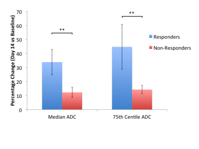 |
Early biomarkers of response to neoadjuvant chemotherapy in lung cancer: preliminary data from a multicenter international study
Dominic Carlin, Alexander Weller, Joost Kuijer, Gerbrand Kramer, Arturo Chiti, Mary O'Brien, Sanjay Popat, Yan Liu, Nandita deSouza
Whole tumor ADC histogram parameters were assessed as early response biomarkers to platinum-based neo-adjuvant chemotherapy in 14 patients with non small cell lung cancer. On completion of treatment, 3 of 11 patients with DW-MRI at baseline and day 14 were classed responders by RECIST criteria. At Day 14 of treatment, there was a significant reduction in ADC metrics in responders (2 of 3 beyond limits of agreement) compared to non-responders (2 of 11 beyond limits of agreement). An increase in ADC 75th centile (indicating more voxels with higher ADC values), was consistent with necrosis; non-responders did not show this change.
|
|
1529.
 |
Developing a Halbach Array for Brain Tumor Targeting
Areej Alghamdi, Munitta Munitta Muthana, Martyn Paley
Steering magnetic nanoparticles (MNPs) in a desired trajectory has been proposed for guiding magnetically labelled drugs to clinical targets1. In order to steer MNPs to a desired location, a strong magnetic field and field gradient is necessary and the deeper the location, the stronger the magnetic force required. External permanent magnets can provide a strong magnetic field and gradient. We hypothesise that external magnetic field/field gradient arrays of 1.1T can be designed to capture MNPs into tumors. Brain tumors are one of the most difficult cancers to treat due to the complex anatomy of the brain. Therefore, we are developing a 3D printed brain tumor model to investigate trapping of MNPs into a tumor using Halbach arrays.
|
|
1530.
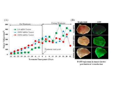 |
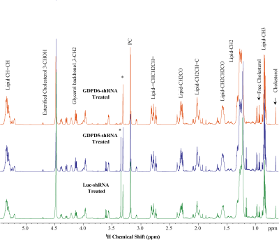
Lentiviral shRNA-mediated targeting of GDPD5 and GDPD6 in Orthotopic Human Breast Cancer Xenograft Models: A Metabolomics Study
Kanchan Sonkar, Marina Stukova, Caitlin Tressler, Balaji Krishnamachary, Zaver Bhujwalla, Kristine Glunde
Activated choline phospholipid metabolism is a hallmark of cancer. Aggressive breast cancers are characterized by high tumoral phosphocholine and glycerophosphocholine. In our ongoing efforts of evaluating the glycerophosphodiesterases GDPD5 and GDPD6 as cancer treatment targets, we have systemically injected mice growing orthotopic triple-negative MDA-MB-231 breast tumors with lentiviral vectors that silence the GDPD5 or GDPD6 genes as compared to mice injected with control viruses. We have analyzed extracted tumor tissue by means of high-resolution 1H MRS-based metabolomics. Differences in tumor growth and metabolic profiles were observed following silencing of GDPD5 and GDPD6 genes when compared to control mice.
|
|
1531.
 |
Development of a 3D radial MP2RAGE sequence for free-breathing T1 mapping of the mouse abdomen
Thibaut Faller, Aurélien Trotier, Sylvain Miraux, Emeline Ribot
T1 mapping could be useful to quantify the evolution of metastases over time and evaluate therapy efficiencies. The MP2RAGE sequence enables to obtain 3D T1 maps in reasonable scan time. Nevertheless, the standard sequence is too sensitive to respiratory motion, preventing its use at the abdominal level. Consequently, a 3D radial MP2RAGE sequence has been developed. The accuracy of the T1 measurements was evaluated in vitro and on the mouse brain. Then, abdominal 3D T1 maps were obtained without motion artifact while free breathing. Finally, the radial MP2RAGE sequence was used for the early detection and characterization of hepatic metastases.
|
|
1532.
 |
Co-registration of MRI and histological habitats in pre-clinical tumor models
Bruna Jardim-Perassi, Suning Huang, William Dominguez-Viqueira, Epifanio Ruiz, Mikalai Budzevich, Jan Poleszczuk, Marilyn Bui, Robert Gillies, Gary Martinez
Tumor heterogeneity, may give insight into natural selection through detection of tumor sub-regions, referred as imaging habitats. We used statistical clustering of multiple pixels based on multiple MRI parameter maps to identify tumor habitats in pre-clinical models of sarcoma and breast cancer using T2, T2*, ADC and three model free parameter maps determined from dynamic contrast enhanced images. MRI-derived habitat maps were determined by clustering multidimensional voxels using a Gaussian mixture model. 3D-printed tumor molds were used to successfully co-register MR imaging slices with their histological habitat-counterparts. Four distinct tumor habitats were detected by MRI and biologically corroborated by histology.
|
|
1533.
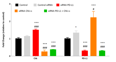 |
The Immune Checkpoint PD-L1 and Choline Kinase-a are inversely related in triple negative human breast cancer cells
Jesús Pacheco-Torres, Marie-France Penet, Yelena Mironchik, Balaji Krishnamachary, Zaver Bhujwalla
Immune checkpoint inhibition to activate the immune system has emerged as an exciting treatment option for several cancers. Programmed death-ligand 1 (PD-L1) plays a major role in immune suppression. We investigated the relationship between the aberrant choline metabolism observed in most cancers and PD-L1 expression in triple negative human MDA-MB-231 breast cancer cells. Using siRNA to downregulate choline kinase-α (Chk-α) or PD-L1 or both, we identified a close inverse interdependence between Chk-α, PD-L1 and phosphocholine. These results have significant implications for treatments that decrease Chk-α expression as these may drive up PD-L1 expression allowing escape of cancer cells from immune surveillance.
|
|
1534.
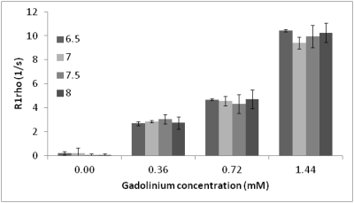 |
The relationship of R1rho to aqueous pH and macromolecular density
Petros Fessas, Syed Omar Ali, Joshua Kaggie, Martin Graves, Scott Reid, Gavin Houston, Ferdia Gallagher
We investigated the sensitivity of R1rho MRI to pH and macromolecular density in in vitro phantoms and in brains of volunteers to assess its suitability as an imaging modality for detecting and assessing the response of brain tumours. We find the dependence of R1rho signal on pH in the presence of macromolecules, but a lack of pH dependence in their absence. We confirm R1rho sensitivity to macromolecular density at constant pH.
|
|
1535.
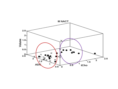 |
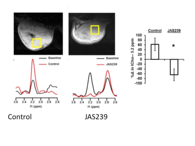
Multiparametric MR approach for monitoring the pathological response of breast cancer patients to neoadjuvant chemotherapy
Naranamangalam Jagannathan, Uma Sharma, Khusbhu Agarwal, Rani Sah, Sandeep Mathur, Vurthaluru Seenu, Siddhartha Gupta, Rajinder Parshad
A multiparametric MR approach using total choline (tCho), apparent diffusion coefficient (ADC) and tumor volume was undertaken for prediction of pathological response in 42 locally advanced breast cancer (LABC) patients undergoing neoadjuvant chemotherapy (NACT). 24 were pathologically responders (complete and partial) while 18 were non-responders. Percentage change in tCho, ADC and volume was higher in pathological responders than in non-responders after III NACT. Individually, all three parameters showed equal sensitivity (66.7%) with specificity in the range 64.7% to 70.6% for pathological response prediction. Combination of all three MR parameters yielded 66.7% sensitivity and a specificity of 64.7%.
|
|
1536.
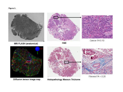 |
Functional MRI at ultra-high field strength (11.7 T) for evaluation of rectal cancer stromal heterogeneity ex vivo: correlation with histopathology
Trang Pham, Timothy Stait-Gardner, C. Soon Lee, Michael Barton , Gary Liney, Karen Wong, William Price
Diffusion Tensor Imaging (DTI) MRI at ultra-high field (11.7 T) was used to examine the stromal ultrastructure of malignant and normal rectal tissue ex vivo, and findings were correlated with histopathology. DTI was able to distinguish tumour from desmoplasia: tumour was found to have isotropic diffusion, whereas desmoplastic reaction or fibrous tissue had moderately anisotropic diffusion. DTI was useful in assessing depth of tumour infiltration into rectal wall: tumour was able to be distinguished from muscularis propria which was highly organised and anisotropic. This study showed that DTI-MRI can assist in more accurately defining tumour extent in rectal cancer.
|
|
1537.
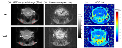 |
Assessment of treatment response of lymphoma in an animal model with in vivo MR elastography
Jing Guo, Animesh Bhattacharya, Gergely Bertalan, Jürgen Braun, Clemens Schmitt, Ingolf Sack
In this feasibility study, we have characterized the mechanical properties of lymphoma directly in the cervical lymph nodes with in vivo multifrequency MRE for the first time. Both MRE and diffusion weighted imaging were used to investigate the tumor's response to chemotherapy. We found that lymphomas stiffened 24 hours after chemotherapy which was accompanied by increased apparent diffusion coefficient (ADC) and reduced tumor volume. Wave speed obtained from MRE is sensitive in detecting the mechanical response of lymphoma to chemotherapy. Observed tumor stiffening post treatment needs to be validated by larger group size and should be explained by histological analysis.
|
|
1538.
 |
Assessment of Tumor Hypoxia Using Tissue Oxygen Level Dependent in a Rabbit VX2 Liver Tumor model
Xinming Li, Shuping Qin, Wen Liang, Yingjie Mei, Yangguang Yuan, Xianyue Quan
There is attractive focus in developing non-invasive methods that assess tumor hypoxia. We applied tissue oxygen level dependent (TOLD) MRI to explore tumor oxygenation using VX2 liver tumor xenografts in a rabbit model. In this study,we demonstrated alteration in tumor oxygen inhalation and correlation in different hypoxia levels.
|
|
1539.
 |
Dual-modality molecular imaging of choline kinase expression in lung cancer
Sofya Osharovich, Anatoliy Popov, David Holt, Sunil Singhal, E. Jim Delikatny
MR spectroscopy of tumors show elevated tCho resonances, reflecting increased levels of phosphocholine. This arises from overexpression of choline kinase (ChoK), which can be detected in breast tumor models using targeted near-infrared (NIR) probes and fluorescence optical imaging. This study translates these findings into lung cancer models, measuring elevated ChoK expression and activity in murine and human lung cancer cells and elevated ChoK levels in spontaneous canine adenocarcinomas. Dual modality molecular imaging could be employed using MRI and MRS for tumor staging, followed by NIR imaging for intraoperative surgical guidance, margin detection, and residual tumor removal, increasing patient survival.
|
|
1540.
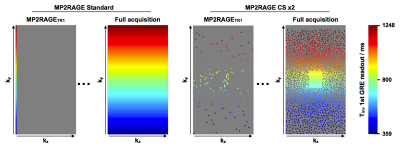 |
MP2RAGE-Compressed Sensing for fast metastasis detection and characterization in mice
Aurélien Trotier, Stanislas Rapacchi, Thibaut Faller, Sylvain Miraux, Emeline Ribot
In order to detect and characterize metastases in preclinical studies, 3D T1 maps can be obtained with the MP2RAGE sequence. As high spatial resolution is required, the acquisition duration becomes prohibitive for the monitoring of metastases. Thus, acceleration via Compressed Sensing technique was achieved, necessitating a new undersampling scheme. T1 maps of the mouse whole brain were obtained in <1min. The T1 of brain metastases was not affected by CS acceleration. Then, ultra-high spatially resolved maps (130x125x141μm) were acquired without lengthening scan time, to detect early-growing metastases and accurately measure their volumes.
|
|
1541.
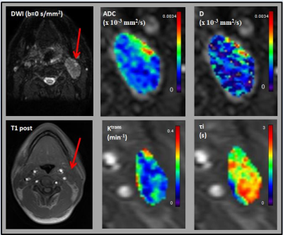 |
Tumor Metabolism, Diffusion, and Perfusion in Head and Neck Cancer: Pretreatment Multimodality Imaging with DCE-MRI, IVIM DW-MRI, 18F-FMISO PET/CT, and 18F-FDG PET/CT
David Aramburu Nunez, Milan Grkovski, Nancy Lee, Vaios Hatzoglou, Heiko Schoder, Ramesh Paudyal, Nadeem Riaz, Joseph Deasy, John Humm, Amita Shukla-Dave
The aim of this study is to understand the correlation of pretreatment quantitative imaging metrics obtained from multimodality imaging (MMI) techniques, such as DCE-MRI, IVIM DW-MRI, 18F-FMISO PET/CT, and 18F-FDG PET/CT giving us a comprehensive characterization of the tumor in head and neck cancer (HNC) patients. The results show complementary, rather than competitive, information about tumor metabolism, diffusion, and perfusion.
|
|
1542.
 |
MRI exploration of the subventricular region of the third ventricle and its association with neurofibromatosis type-1 and white matter integrity in children with optic pathway glioma
Natalie Boonzaier, Patrick Hales, Felice D’Arco, Kshitij Mankad, Darren Hargrave, Christopher Clark
The lateral subventricular zone has been explored in association with high-grade gliomas, both in-vivo and with MRI. The third ventricle subventricular zone (TVZ) has been explored in-vivo, using immunohistochemistry and microarray analysis, with regard to neurofibromatosis type-1-associated low-grade optic pathway gliomas. This remains unexplored with MRI. This study examined diffusion MRI features of the TVZ and its association with NF1-status and peri-tumour white matter integrity. TVZ features correlated with NF1-status, and peri-tumour white matter integrity. These results suggest that the state of the TVZ environment can potentially indicate whether a sporadic tumour might behave like its less disruptive NF1-associated counterpart.
|
|
1543.
 |
Creating patient-specific computational head models for the study of tissue-electric field interactions using deformable templates
Noa Urman, Shay Levi, Avital Frenkel, Ariel Naveh, Doron Manzur, Gitit Lavy-Shahaf, Hadas Hershkovich, Cornelia Wenger, Ofir Yesharim, Eilon Kirson, Ze'ev Bomzon
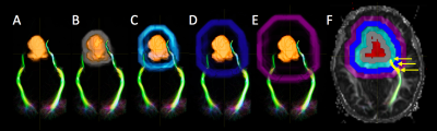
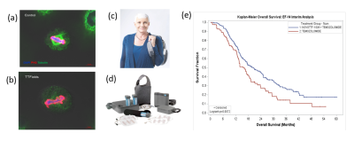
Tumor Treating Fields (TTFields) are electric fields at an intermediate frequency approved for treatment of Glioblastoma Multiforme. Understanding how TTFields distribution in the brain influences disease progression can be studied using numerical simulations. Creation of computational patient models involves accurate segmentation of patient MRIs, a task that cannot be performed automatically, and is therefore time-consuming. We present a method for rapidly creating patient head models using a healthy head model as a deformable template. The method is robust even when MRI data quality is low. It is enabling a study correlating the spatial distribution of TTFields and patient outcome.
|
|
1544.
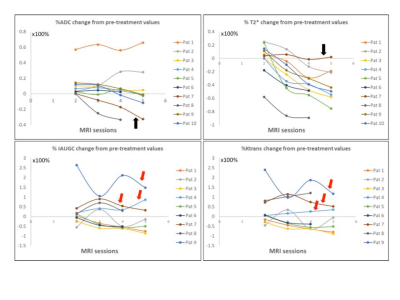 |
Dose reduction in myxoid liposarcomas: Initial descriptive results in the evaluation of response using multiparametric MRI.
Evanthia Kousi, Maria Schmidt, Shane Zaidi, Khin Thway , Cyril Fisher , Myles Smith, Dirk Strauss, Andrew Hayes, Eleanor Moskovic, Nicos Fotiadis, Elizabeth Barquin, Komal Amin, Rick Haas, Christina Messiou, Aisha Miah
Compared to other soft tissue sarcomas (STSs), myxoid liposarcomas (MLSs) are exquisitely radiosensitive. The clinicopathological response following pre-operative radiotherapy at 50 Gy/25# in MLS might be due to radiation induction vascular damage. Here we report initial results in using multiparametric MRI (diffusion-weighted imaging, pharmacokinetic modelling and T2* measurements) to evaluate MLS response during and after preoperative RT. Dynamic contrast-enhanced examinations demonstrated both heterogeneous and homogeneous enhancement patterns. The tissue enhancement curve was monotonically-increasing in all cases, suggesting a distinct vascular pattern. Permeability and perfusion decreases from baseline in responders show Ktrans and IAUGC60 can potentially predict response.
|
|
1545.
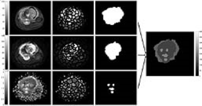 |
Superpixels-based Segmentation and Automated Identification of Active Tumour and Necrotic regions in Bone Tumor using T1 and Diffusion Weighted Imaging
Amit Mehndiratta, Esha Kayal, Sneha Patil Kulkarni, Raju Sharma, Devasenathipathy Kandasamy, Sameer Bakhshi
Proper Delineation of the tumour boundary and assessment of tumour size can take crucial part in treatment planning and monitoring treatment response. We investigate a fully automated Simple linear iterative clustering (SLIC) superpixel-based method for detection and segmentation of pathological tissues like oedema, tumour and necrosis associated with Osteosarcoma. Experimental results provide a close match to expert delineation and was able to estimate areas of active tumor and necrosis with good accuracy.
|
|
1546.
 |
Prostate MR Elastography: a comparison of image acquisitions strategies in healthy volunteers
Kay Pepin, Kevin Glaser, Yi Sui, Roger Grimm, Arvin Arani, Phillip Rossman, Richard Ehman, Michael Herman, Lance Mynderse
The purpose of this study was to compare image acquisition strategies for prostate MRE using external drivers. Additionally, to assess the normal heterogeneity of prostate mechanical properties in an age-matched cohort to the prostate cancer population. Improved resolution using higher MRE vibration frequencies, larger acquisition matrices, and distortion-reduction techniques, may help advance the clinical application of prostate MRE.
|
|
1547.
 |
Liver metabolomic investigation of lentiviral targeting of GDPD5 and GDPD6 for breast cancer treatment in a preclinical model
Kanchan Sonkar, Marina Stukova, Caitlin Tressler, Balaji Krishnamachary, Zaver Bhujwalla, Kristine Glunde
High-resolution 1H MRS is a powerful technique for metabolomics studies of tissues, cells, and body fluids. Here we have used this technique to explore metabolomic changes in the livers of mice that have been treated with lentiviral particles that silence either of the two glycerophosphodiesterase GDPD5 (GDPD5-shRNA) or GDPD6 (GDPD6-shRNA). We systemically administered lentiviral shRNA in mice with orthotopic breast tumor xenografts. We identified distinct increases in leucine, valine, glutathione, creatine, glucose, tyrosine, and histidine in the GDPD5-shRNA treated group, whereas cholesterol, isoleucine, beta-hydroxy butyrate, alanine, glutamate, glutamine, aspartate, fumerate, phenylalanine, and formate were elevated in the GDPD6-shRNA treated group.
|
|
1548.
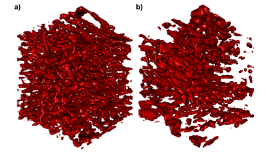 |
Vascular-induced spin dephasing in real vascular networks reveals useful decay characteristics to differentiate glioblastoma from healthy brain tissue
Artur Hahn, Thomas Kruewel, Julia Bode, Lukas Buschle, Björn Tews, Sabine Heiland, Martin Bendszus, Christian Ziener, Felix Kurz
The transverse relaxation attributed to spin dephasing, caused by microscopic field inhomogeneities throughout a single imaging voxel, induced by the BOLD-mechanism, is studied using realistic three-dimensional microvascular structures, attained with fluorescence ultramicroscopy from mouse brains, and custom-written simulations to uncover differences between glioblastoma and healthy brain tissue. The signal attenuation is weaker and more heterogeneous in tumor tissue. Relaxation rates scale differently with varying field strengths or blood properties and the relaxation processes exhibit strong deviations from Lorentzian decay. The results are important for the development of signal processing methods for tumor diagnosis without contrast agents.
|
|
1549.
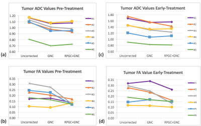 |
Effect of corrections for image distortion and gradient nonlinearity on longitudinal DTI tumor measurements in breast patients receiving neoadjuvant chemotherapy
Lisa Wilmes, Ek-Tsoon Tan, Evelyn Proctor, Wen Li, Jessica Gibbs, Nola Hylton, David Newitt
Diffusion weighted imaging has shown promise for assessing tumor response to treatment, but suffers from gradient nonlinearity and image distortion that may adversely affect quantitative accuracy. This work evaluates corrections for image distortion (susceptibility-induced and eddy current) and bias from gradient non-linearity (GN) on breast tumor DTI metrics prior to treatment (T0) and at an early-treatment time point (T1), in six breast cancer patients undergoing neaoadjuvnt chemotherapy. Both GN and distortion correction had significant effects on tumor ADC and FA values at T0 and T1. The addition of distortion correction also improved the alignment of DTI and DCE-MRI tumor ROIs.
|
|
1550.
|
18F-FDG PET/MRI in Children with Oncologic Diseases: Initial Experience
Hansel Otero, Carolina Maya, Sabah Servaes, Jeffrey Schmall, Lisa States
We describe our initial experience with integrated whole-body Fluor-18-Fluordesoxyglucose-PET/MR imaging in children in a retrospective study of all 18F-FDG-PET/MR at our institution. 51 studies were carried out in 41 children (34 girls, 17 boys) with a mean age of 10.16 years (10 months-24 years). Primary diagnosis included rhabdomyosarcoma (n=18) and Osteosarcoma (n=5). The majority of studies (n=29, 57.9%) were performed for treatment response/restaging. All studies were diagnostic (technical success rate 100%). The mean effective dose was 5.25 mSv (2.1-11.5 mSv). Mean total imaging time was 80 minutes (42-138 minutes). Thirty-eight (74.5%) cases had an average of 2.2 additional MR sequences. 18F-FDG PET/MR is technically feasible for the evaluation of oncologic processes in children at a fraction of the radiation dose.
|
|
1551.
 |
Integrating Magnetic Resonance Imaging with Live Lung Intravital Microscopy: A Novel Platform to Evaluate the Effect of Radiation on Lung Tumors
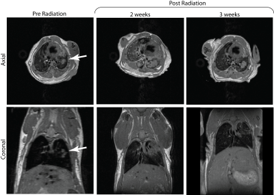
Shampa Chatterjee, Luis Loza, Mehrdad Pourfathi, Sarmad Siddiqui, Jian Tao, Harrilla Profka, Ian Duncan, Hooman Hamedani, Kai Ruppert, Diane Lim, Yan Liu, Jose Conejo-Garcia, Mary Spencer, Tahmina Achekzai, Stephen Kadlecek, Rahim Rizi
We propose that, when used in combination with MRI imaging, live lung intravital fluorescence microscopy can be a powerful tool for detecting the effects of radiotherapy on lung tumors. In this study, we monitored pulmonary nodules pre- and post-radiation in a novel murine model (Kras(G12D)/p53fl/fl/myr-p110) with tumor regulation by Cre-recombinase. Using the reporter gene EGFP fluorescence, a significant loss of the tumor was observed post-radiation, which correlated with reduced fluorescent signal from the same region of the lung.
|
|
1552.
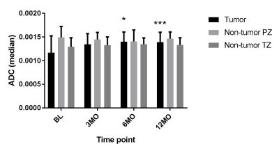 |
Effect of Stereotactic Body Radiotherapy on Perfusion and Diffusion in Prostate Tumor and Benign Tissue
Kristen Zakian, Hebert Vargas Alvarez, Andreas Wibmer, Aditi Iyer, Neelam Tyagi, Aditya Apte, Marissa Kollmeier, Boris Mychalczak, Karen Borofsky, Oren Cahlon, Yousef Mazaheri Tehrani, Margie Hunt, Michael Zelefsky
Multimodality MRI including DCE-MRI and DW-MRI were performed in patients prior to and following hypofractionated stereotactic body radiotherapy (SBRT). Diffusion and perfusion related parameters in both tumor and non-tumor benign tissue were calculated at 3, 6, and 12 months after SBRT. Radiation-induced changes were observed in perfusion and diffusion related parameters in tumors. In the non-tumor transition zone, SBRT induced changes in perfusion-related parameters. Multimodality MRI has potential for treatment effect monitoring in the prostate after SBRT.
|
|
1553.
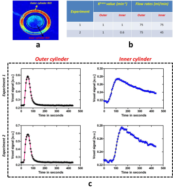 |
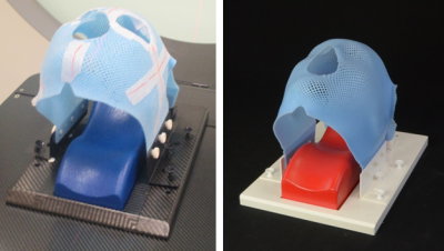
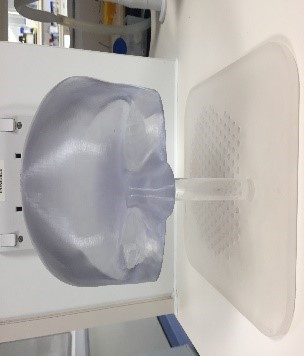
An integrated, semi-automated 3D printed Breast DCE-MRI phantom solution to generate diverse pharmacokinetic curves
Nithin Vajuvalli, Amaresha Konar, Shivaprasad Chikop, Ramesh Venkatesan, Sairam Geethanath
In vitro phantoms play a critical role in the assessment of novel Dynamic Contrast Enhanced MRI (DCE-MRI) methods related to acquisition and reconstruction, among other advantages such as repeatability and reproducibility. In this work, we demonstrate a 3D printed breast DCE-MRI phantom that is capable of producing diverse kinetic curves as those seen in human patients. The wash-in and wash-out characteristics were controlled through user controlled Ktrans values and the geometry of the phantom respectively. The phantom demonstrated in this work is 3D printed, cost effective, user interface controlled, and integrated with a peristaltic pump to obtain different kinetic curves.
|
|
