|
Traditional Poster Session
Neuro |
Tuesday, 19 June 2018
Traditional PosterNeuro
1765 -1802 Neonatal & Pediatric Neuroimaging
1803 -1845 Psychoradiology
1846 -1867 Myelin Imaging: From Mice to People
1868 -1902 Neurovascular Imaging Methods
1903 -1923 Neurovascular Clinical Studies
1924 -1947 Parkinson's Disease
1948 -1960 Epilepsy
1961 -1971 Head & Neck
1972 -1993 A Potpourri of Multiple Sclerosis
1994 -2034 Alzheimer's Disease & Other Dementias
2035 -2060 Brain Imaging Methodology
2061 -2085 Brain Pathology & Ageing Brain
2086 -2103 Novel Neuroimaging Methods
2104 -2131 Neuroimaging: Animal Models
2132 -2157 Brain Tumours |
| |
Neonatal & Pediatric Neuroimaging
Traditional Poster
Neuro
Tuesday, 19 June 2018
| Exhibition Hall 1765-1802 |
16:15 - 18:15 |
|
1765.
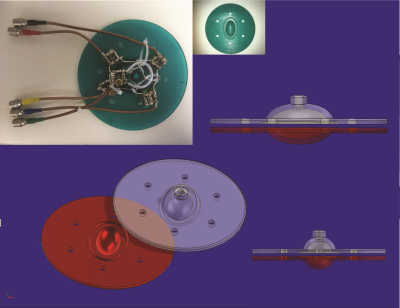 |
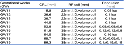
Towards a high-resolution MRI Atlas of the Human Foetus: a Post-Mortem Pilot Study of ex-vivo preserved Foetal specimens at 7 Tesla.
Sean Moen, Anthony Weinhaus, Joseph Metzger, Michael Garwood, Bharathi Jagadeesan, Pierre-François Van de Moortele
In an effort to expand the existing MRI reference material available to Medical professionals, including developmental anatomists, foetal specimens of gestational ages ranging from 7-26 weeks were scanned using ultra high field MRI systems ( 7 Tesla) and high resolution, multiplanar images of the whole body were obtained in each of these specimens. A unique set of processes, materials and equipment facilitated the execution of these MRI scans including custom built specimen holders, transmit and receive coils, protocol optimization and image reconstruction techniques. Using these techniques, a total of 21 preserved ex-vivo fetal specimens were successfully scanned.
|
|
1766.
 |
High-Resolution Radial Diffusivity Images Provide Insights of Fetal Brain Development
Akiko Uematsu, Keigo Hikishima, Junichi Hata, Hideyuki Okano
Investigating prenatal neural development provide depth knowledge of brain ontogeny. DTI-derived radial diffusivity (RD) imaging has advantage to provide information of microstructural tissue organization information without damaging the tissues. In this study, we investigate the changes of the radial diffusivity (RD) values during fetal development in non-human primate. The RD image contrast was enough to clearly depict the emergence of each brain regions as well as major white matter bundles during prenatal period. In addition, its whole brain intensity distribution histogram provided the information of critical period for the growth of myelination.
|
|
1767.
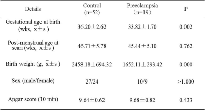 |
Preeclampsia related to delayed development of white matter and cortical infolding.
Ting Liu, Miaomiao Wang, Chao Jin, Xianjun Li, Jian Yang
Offspring born from preeclampsia exhibit deficits in cognitive impairment. But the pathogenesis is not clear. We assessed brain maturation and white matter development in neonatal period using total maturation score and tract-based spatial statistics. TMS showed the scores of TMS, B and C scores were lower in preeclampsia group. TBSS results displayed FA values decreased, while AD and RD values increased on anterior & posterior limb of internal capsule, external capsule, splenium of corpus callosum, optic radiation and centrum semiovale in preeclampsia group. The results indicated preeclampsia is associated with delayed development of white matter and cortical infolding.
|
|
1768.
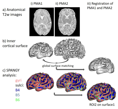 |
Is cortical microstructure related to folding during development? A longitudinal MRI study in preterms
Alexandra Hertz, Antonietta Pepe, Julien Lefevre, Marie Zomeno, Francois Leroy, Jessica Lebenberg, Linda de Vries, Floris Groenendaal, David Germanaud, Manon Benders, Jessica Dubois
The human brain cortex develops dramatically during the preterm period, in terms of both morphology, intra-cortical maturation and dendritic arborization. Here we aimed to investigate whether different stages of microstructural maturation are observed in cortical regions that fold successively. We studied preterm infants longitudinally at around 30 and 40 weeks of post-menstrual age, and combined measures from diffusion tensor imaging (DTI) and spectral analysis of gyrification (SPANGY). We highlighted that proxies of primary folds have an advanced microstructural maturation early on, and that the progression until term age is more intense in proxies of secundary folds than in gyri.
|
|
1769.
 |
Changes in neonatal regional brain volume associated with preterm birth and perinatal factors
Bonnie Alexander, Claire Kelly, Chris Adamson, Richard Beare, Diana Zannino, Jian Chen, Andrea Murray, Wai Yen Loh, Lillian Matthews, Simon Warfield, Peter Anderson, Lex Doyle, Marc Seal, Alicia Spittle, Jeanie Cheong, Deanne Thompson
In a cohort of 285 preterm and term infants at term equivalent age, associations were investigated between gestational age (GA) at birth, perinatal factors, and volumes of 100 regions of the M-CRIB neonatal brain atlas. Volumes increased with increasing GA in some regions, and decreased with increasing GA in other regions including primary visual, motor and somatosensory cortices. Robust increases in many regional volumes were found for birthweight standard deviation score, and male sex. These results provide increased insight into the complex array of correlates of preterm birth.
|
|
1770.
 |
T2 relaxometry MRI predicts cerebral palsy in preterm infants
Yi-Shan Tsai, Li-Wen Chen, Feng-Mao Chiu
T2 relaxometry brain MRI could be of prognostic value in preterm infants. The maturation patterns of periventricular white matter differed according to neurodevelopmental outcomes. T2 relaxation values over mid-body periventricular white matter at > 1 month old of corrected age could predict CP. T2 relaxometry brain MRI provides neuroimaging-outcome correlation among preterm infants, especially when interpreted with age-specific and area-selective considerations.
|
|
1771.
 |
Automatic Brain Segmentation in a Neonatal Population Using a Multi-Delay Multi-Echo Sequence
Maarten Naeyaert, Tim Vanderhasselt, Marcel Warntjes, Hubert Raeymaekers
Synthetic MRI using a multi-delay multi-echo sequence was applied to a pre-term neonatal and full term neonatal population. The brain was segmented into different tissue types using the relaxometric data and using an improved algorithm which suppresses CSF partial volume fractions in grey matter. The volumes and volume fractions were calculated. The relation between volumetric quantities and either gestational age (preterm patients only), or corrected age (whole population) was investigated. The Brain Parenchymal and grey matter fraction were found to be dependent on gestational age at birth, while grey matter, CSF, intracranial and brain parenchymal volume are dependent on age.
|
|
1772.
 |
Longitudinal Mapping of Local Relationship of Surface Area, Cortical Thickness and Cortical Folding in Infants
Dingna Duan, Shunren Xia, Zhengwang Wu, Fan Wang, Weili Lin, John H Gilmore, Dinggang Shen, Gang Li
A simple physical law on the global relationship of surface area, cortical thickness, and cortical folding is found across a full range of mammalian species’ brains, including adult human brains1,2. However, little is known about the local relationship of these cortical properties, especially in infant brains with rapid development in the first two years of life. To fill this knowledge gap, we explored the local relationship of surface area, cortical thickness and cortical folding on 73 normal infants, each of which was longitudinally scanned at 0, 1, and 2 years of age. We reveal that the relationship of these three cortical properties is age-specific and region-specific.
|
|
1773.
 |
Evaluation of cortical thickness estimation methods in neonates.
Martina Lucignani, Andrea Pittella, Maria Camilla Rossi Espagnet, Daniela Longo, Giulia Lucignani, Maurizio Schmid, Antonio Napolitano
Cortical thickness (CT) is a sensitive indicator of normal brain structural and functional development, aging, as well as a variety of neuropsychiatric disorders. The state of the art for cortical thickness estimation in children in not as good as the one for adults. We then compared two different algorithms and assess the agreement between these methods and their local variability.
|
|
1774.
 |
Asynchrony of the cortical maturation in the infant brain studied with MRI
Jessica Lebenberg, Jean-François Mangin, Cyril Poupon, Lucie Hertz-Pannier, François Leroy, Parvaneh Adibpour, Claire Kabdebon, Ghislaine Dehaene-Lambertz, Jessica Dubois
Intense changes in cortical microstructure occur during early infancy. Here, we aimed to study cortical maturation over this largely unexplored developmental period using quantitative MRI in 17 infants from 1 to 5 post-natal months. By taking benefit of robust intra- and inter-individual registrations of anatomical images and parametric maps, we measured T1, T2 relaxation times, and DTI longitudinal diffusivity over cortical surfaces and regions of interest. Results showed that each parameter relevantly but differently reflects the progressive maturation. This suggests that multi-parametric approaches might provide interpretable measures of the developing microstructure by accounting for the parameters complementarity.
|
|
1775.
 |
High resolution neonatal brain relaxometry in 10 minutes – A preliminary proof of concept
Rui Pedro A. G. Teixeira, Tomoki Arichi, Johannes Steinweg, Katy Vecchiato, Sophie Arulkumaran, Shaihan Malik, Mary A. Rutherford, Joseph V. Hajnal, Serena J. Counsell
Quantitative MRI promises to allow objective and reproducible tissue metrics which are of special interest in newborn brain maturation characterization. However, such methods require acquisition times above 20 minutes which hinders their clinical applicability. With an increasing trend towards examination without sedation during natural sleep, subject motion is an important issue for neonatal applications. With this in mind, this work builds on the previously described Joint System Relaxometry framework and presents a neonatal specific protocol which allows 1.25mm isotropic 3D maps of Proton Density, T1 and T2 relaxation times in a total of 10minutes examination time.
|
|
1776.
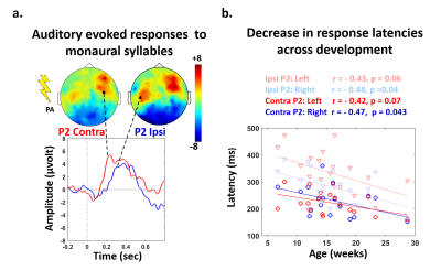 |
Anatomo-functional correlates of auditory development in infancy
Parvaneh Adibpour, Jessica Lebenberg, Claire Kabdebon , Francois Leroy, Ghislaine Dehaene-Lambertz, Jessica Dubois
Early infancy is a period of intense behavioral acquisitions and brain development. Nevertheless, how functional and structural maturations are inter-related has been little explored so far. Following studies of visual domain, we aimed to address this question for the auditory modality in 1 to 5-month-old infants, by combining EEG and quantitative MRI measures supposed to reflect fiber myelination and intra-cortical development of dendritic arborization. We investigated the relationships between the functional maturation of auditory-evoked responses in terms of latency and speed, and the maturation of microstructural properties for both white matter tracts and cortical regions of the auditory network.
|
|
1777.
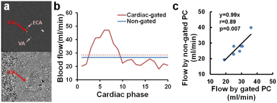 |
Optimization of phase-contrast MRI for cerebral blood flow quantification in neonates
Peiying Liu, Charlamaine Parkinson, Dengrong Jiang, Jill De Vis, Li Pan, Himanshu Bhat, Andrea Poretti, Frances Northington, Aylin Tekes, Thierry Huisman, W Golden
Knowledge of CBF in neonates may provide valuable information in many pathological conditions. When applied to very young children, CBF mapping using arterial-spin-labeling (ASL) MRI suffers from low signal-to-noise ratio and poor quantification, whereas phase-contrast (PC) MRI may provide reliable estimation of global CBF. This study aimed to optimize the PC-MRI protocol for future applications in neonates. By comparing the cardiac-gated and non-gated implementations, we found non-gated PC-MRI could provide accurate CBF measurement with shorter scan time. We also found lower imaging resolution would over-estimate CBF, and therefore recommend the use of 0.3mm resolution with 6 averages in neonates.
|
|
1778.
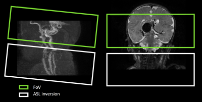 |
Clinical application of 4D ASL-MRA in neonatal Vein of Galen malformation
Magdalena Sokolska, Subhabrata Mitra, Yuriko Suzuki, Matthias van Osch, H Rolf Jäger, Adam Rennie, Fergus Robertson, Giles Kendall, Alan Bainbridge
This work investigates the feasibility of using time-resolved magnetic resonance angiography, based on arterial–spin-labelling (ASL), to investigate neonatal vein of Galen malformation for the purpose of aiding diagnosis and surgical treatment planning.
|
|
1779.
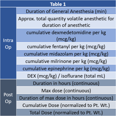 |
Intraoperative Volatile Anesthetic Exposure Predicts Reduced Frontal Lobe Connectivity Compared to Dexmedetomidine in Infants with Congenital Heart Disease
Vincent Lee, Phillip Adams, Benjamin Meyers, Lauren Dennis, Nancy Beluk, Tracy Baust, Lucas Saenz, Yulia Domnina, Joan Sanchez de Toledo, Vincent Schmithorst, Ashok Panigrahy
Anesthetic neurotoxicity in infants with repetitive exposure is a risk factors for adverse neurodevelopmental outcomes. Dexmedetomidine exposure is thought to have neuroprotective effects. We tested the hypothesis that intraoperative volatile anesthetic exposure is predictive of aberrant brain connectivity in the post-operative period in CHD infants, relative to dexmedetomidine exposure using DTI and BOLD imaging. Using both hypothesis driven and data driven approaches, as well as graph analysis we showed that Increased volatile anesthetic exposure in the intraoperative period is associated with reduced post-operative frontal brain connectivity in CHD infants, while DEX exposure was associated with metrics of improved brain connectivity.
|
|
1780.
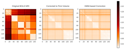 |
Application of Probabilistic Modeling to Motion Correction of Neonatal Brain Resting-State BOLD Data
Jenna Schabdach, Rafael Ceschin, Vince Lee, Vincent Schmithorst, Ashok Panigrahy
Functional connectivity studies commonly use resting-state BOLD MR images to study the neurodevelopment of healthy and at-risk neonates. BOLD images are highly sensitive to motion; post-acquisition motion correction techniques can be applied to BOLD data to compensate for motion. We compare the corrective performance of two motion correction techniques on a cohort of 17 healthy neonates: the traditional correction to the first volume technique and a novel, HMM-based motion correction technique. We evaluate the corrected images in terms of the Power et al. thresholds and show the HMM-based technique can be used to recover neonatal BOLD data corrupted by motion.
|
|
1781.
 |
Anisotropic similarity, a constrained affine transformation: application to brain development analysis
Antoine Legouhy, Olivier Commowick, François Rousseau, Christian Barillot
The study of brain development provides insights in the normal trend of brain evolution and enables early detection of abnormalities. We propose a method to quantify brain growth in three arbitrary orthogonal directions of the brain through linear registration. We introduce a 9 degrees of freedom transformation that gives the opportunity to extract scaling factors describing brain growth along those directions by registering a database of subjects in a common basis. We apply this framework to create a longitudinal curve of scaling ratios along fixed orthogonal directions from 0 to 16 years highlighting anisotropic brain development.
|
|
1782.
 |
New microstructural asymmetries in the brain
Junyu Guo, Yuanyuan Han, Yimei Li, Wilburn Reddick
Brain microstructural asymmetry can provide more direct causal explanations of functional lateralization than can macrostructural asymmetry. In this study, we discovered two new types of microstructural asymmetry that help to bridge the gap between macrostructural asymmetry and functional lateralization. Myelin-related asymmetry was prominent in the back brain, and axon-related asymmetry occurred in both the front brain and the back brain. These asymmetries early in development indicate that white matter is more mature and more myelinated in the left back brain, providing an explanation for the leftward lateralization of language and visual functions. The asymmetries continue to increase throughout childhood and adolescence.
|
|
1783.
 |
Comparison of Thalamus Segmentation Using Publicly Available Segmentation Methods in a Pediatric Population
Salem Hannoun, Rayyan Tutunji, Maria El Homsi, Roula Hourany
107 subjects were recruited between the ages of one month and 18 years. The study aimed to investigate the differences in the accuracy of five publicly available segmentation techniques on T1-enhanced and non-enhanced images compared to manual segmentation of the thalamus in a pediatric population. volBrain had the best outcomes in enhanced and non-enhanced images. Image segmentation using volBrain is the ideal methodology for thalamus segmentation. Gadolinium-enhancement negatively affects the outcomes of all the tested automated segmentation.
|
|
1784.
 |
Magnetization transfer ratio in cortical gray matter: a longitudinal study.
Yash Patel, Jean Shin, Penny Gowland, Tomas Paus
To assess the change in magnetization transfer ratio (MTR) in the human cerebral cortex during adolescence(14 to 19 years of age). We observe an age-related increase in average MTR in both sexes. Inter-regional profiles of MTR measured at a single time-point correlate with gene-expression profiles of CA1 pyramidal cells (membranes of dendritic arbor) but not of oligodendrocytes (myelin). On the other hand, profiles of the MTR change (from 14 to 19 years) correlate with gene-expression profiles of oligodendrocytes, suggesting that the change may be sensitive to intra-cortical myelination.
|
|
1785.
 |
Paediatric brain tissue properties measured with magnetic resonance elastography
Jade Yeung, Lauriane Jugé , Lynne Bilston
Magnetic resonance (MR) elastography is a technique to noninvasively measure the mechanical properties of soft tissues. While adult brain data obtained with MR elastography is readily available, there is little data for healthy paediatric brains throughout development. MR elastography was performed on 25 healthy paediatric subjects aged between 7-18 years at three frequencies, and the shear moduli of white and grey matter were calculated and compared to data obtained from 10 healthy adults. The shear modulus of paediatric brains was not found to be age dependent, with no significant differences between adult and paediatric brains.
|
|
1786.
 |
Clinical Equivalence Assessment of T2 Synthetic Pediatric Brain MRI
Basile Kerleroux, Tobias Kober, Tom Hilbert, Mohamed El Ouali, Dominique Sirinelli, Baptiste Morel
In a prospective randomized study, we compared the image quality of a synthetized T2 with conventional turbo spin echo T2 during pediatric brain MRI. According to several assessment criteria, synthetic T2 seemed to be an overall equivalent to standard TSE T2, with the advantage of new available T2 quantitative data with a similar acquisition time.
|
|
1787.
 |
Motor connectivity of the midbrain in healthy children defined using connectivity based parcellation
Sonja Soskic, Hannah Cooper, Alexandra Bonthrone, Chris Clark
Delineation of midbrain regions connected with the motor cortex may be useful in evaluating disruptions of motor pathways in paediatric patients. We used the established winner-takes-it-all method to parcellate the midbrain according to cortical connectivity in healthy children aged 6-12 years. The percentage of ipsilateral midbrain occupied by motor parcels was negatively associated with age on the right side only, producing an association between age and interhemispheric asymmetry. Our findings indicate that age and interhemispheric differences need to be taken into account if this method is to be utilised for quantitative comparisons of midbrain-motor connectivity in children.
|
|
1788.
 |
Assessing white matter development in peri-pubertal children using longitudinal fixel-based analysis
Sila Genc, Robert Smith, Charles Malpas, Vicki Anderson, Jan Nicholson, Daryl Efron, Timothy Silk, Marc Seal
Recent evidence suggests that the pubertal period corresponds with changes to white matter microstructure above and beyond age-related development. This study uses a longitudinal fixel-based analysis to investigate which regions of the brain correspond to changes in white matter fibre density and cross-section during pubertal development. We show that, over a 16-month follow-up period, increases in fibre density and cross-section are predominantly in the posterior white matter. These results add to evidence that white matter develops in a posterior-anterior fashion, and signifies the dynamic nature of brain development during puberty.
|
|
1789.
 |
Longitudinal myelin development in children born very preterm compared with typically developing peers
Deanne Thompson, Joseph Yang, Jian Chen, Claire Kelly, Bonnie Alexander, Lillian Matthews, Katherine Lee, Rod Hunt, Jeanie Cheong, Megan Spencer-Smith, Marc Seal, Jeffrey Neil, Terrie Inder, Lex Doyle, Peter Anderson
Myelin development over time in preterm children remains unclear. This study compared T1/T2 myelin maps for 81 very preterm (VP) and 29 full-term children between 7 and 13 years of age. On average, VP children had higher T1/T2 ratios than full-term children in most white matter tracts and deep gray matter structures at both time points. This may reflect compensation or developmental catch-up. T1/T2 ratios increased from childhood to adolescence in both VP and full-term children, shedding light on typical and atypical myelin maturation.
|
|
1790.
 |
Regional Brain Myelin Changes in Patients with Single Ventricle Heart Disease
Sadhana Singh, Bhaswati Roy, Xiaopeng Song, Nancy Halnon, Alan Lewis, Mary Woo, Nancy Pike, Rajesh Kumar
Single ventricle heart disease (SVHD) subjects show brain injury in multiple gray and white matter based on MRI procedures. However, the extent of regional myelin integrity in SVHD is unclear. We examined the regional brain myelin integrity in SVHD adolescents using the ratio of T1-weighted and T2-weighted MRI signal intensity, and found decreased values in critical autonomic, mood, and cognitive control sites, functions that are deficient in the condition, likely resulting from hypoxic/ischemic processes.
|
|
1791.
 |
Regional CBF differences underlie neurocognitive outcomes in older children with congenital heart disease: a voxelwise mediation analysis
Vincent Schmithorst, Ashok Panigrahy
We investigate in more detail the relationship between congenital heart disease (CHD), CBF, and neurocognitive outcome in older children by employing a novel voxelwise mediation analysis with CHD status the independent variable, NIH Toolbox scores the dependent variable, and voxelwise CBF the mediating variable. CHD patients display reduced CBF in the salience network (insula, medial prefrontal, caudate) which mediates lower performance on tests of memory and language function. However, the reduced CBF in the salience network mediates improved performance of executive function (flanker inhibitory control) likely due to less filtering out of presumed irrelevant but actually relevant information.
|
|
1792.
 |
Relationships between brain structure and behavior in children with specific learning disabilities revealed by diffusion spectrum imaging
Yi-Chun Liu, Hsiao-Lan Sharon Wang, Shan-Chih Lee, Jun-Cheng Weng
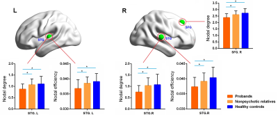
We used diffusion spectrum imaging (DSI) to investigate the relationships between brain structure and behavior in children with specific learning disabilities (SLD). The correlation between reading comprehension scores and the DSI indices was found in corpus callosum. The correlation between Chinese character recognition and the DSI indices was found in cingulate and corpus callosum. The correlation between tone awareness scores and the DSI indices was found in cingulate, superior frontal gyrus and corpus callosum. In summary, SLD not only had difficulty reading and spelling individual words but also more likely to have poorer phonological awareness.
|
|
1793.
 |
Altered regional brain activities and functional connectivities in children with nonsyndromic cleft and/or lip palate: a resting-state functional MRI study.
Hua CHENG, BO RAO, YANG FAN, YingZi Gao, WenJing Zhang, Yun Peng
Rs-fMRI has been widely used as an effective method to evaluate the brain functional changes in physiological and pathological process. Altered both regional brain activities and functional connectivities, especially in verbal and cognitive areas, were found in children with nonsyndromic CL/P using resting-state fMRI. It helps to understand the abnormality of functional architecture of CL/P which implies different structures and cognitive patterns in CL/P compared with normal development children.
|
|
1794.
 |
Alterations in brain connectivity during olfaction in impulsive children
Benito de Celis Alonso, Silvia Hidalgo Tobón, Eduardo Barragán Pérez, Pilar Dies Suarez
Impulsivity is a multi-dimensional construct of behaviors. Here we compared two cohorts of impulsive and control children. Both groups underwent a functional magnetic resonance imaging experiment which food related odor cues. Activations were larger for the impulsive group in: temporal lobe, cerebellum, supplementary motor area, frontal cortex, medial cingulate cortex, insula, precuneus, precentral, para-hippocampal & clacarine. Connectivity results showed that emotional reward based on the smell and processed in temporal lobes was the main cue driving impulsive children. This was followed by a focused attention and sensations of comfort and happiness modulated by precuneus and cingulum.
|
|
1795.
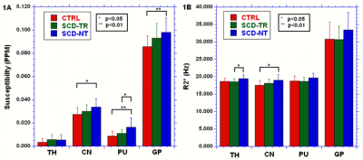 |
Investigation of sickle cell related changes in the basal ganglia of pediatric subjects using QSM and R2*.
Richard Jones, Binjian Sun, Deqiang Qiu, Susan Palasis, Thomas Burns, Clark Brown
In previous work on susceptibility differences between controls and subjects with sickle cell disease (SCD) receiving chronic transfusions we found no significant differences in the basal ganglia (BG). In this abstract we added a group of non-transfused SCD subjects and included an analysis of the R2* in order to better understand the nature of any observed changes. Significant differences between the groups were observed in the BG for both susceptibility and R2*, but the pattern of the changes was inconsistent, probably due to the multifactorial nature of R2* in tissues where iron is not the dominant contrast mechanism.
|
|
1796.
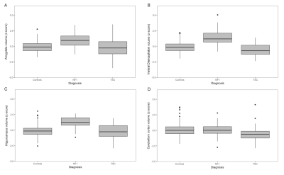 |
Quantitative subcortical morphometry in mTOR/AKT/PI3K pathway disorders: A novel clinical biomarker
Matthew Barkovich, Ryan Nillo, Chin Hong Tan, Leo Sugrue, Anthony Barkovich, Rahul Desikan
Subcortical volumes were quantitatively evaluated on clinical MRI exams of neurofibromatosis type 1 (NF1) and tuberous sclerosis complex (TSC) patients. Robustly larger volumes of several subcortical structures, including the thalamus, hippocampus and ventral diencephalon, were found in NF1; characteristic NF1 imaging abnormalities are found in these areas. In TSC, we found smaller cerebellar volumes; findings that have been associated with autistic phenotypes. Cluster analysis reveals three distinct clustering patterns, each corresponding to a patient class. These results show the feasibility of obtaining automatic quantitative measurements of anatomic structures from clinical MRI exams.
|
|
1797.
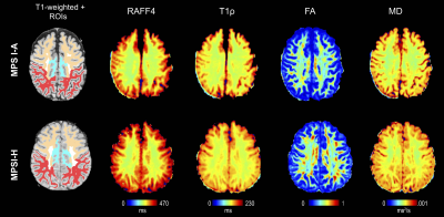 |
ROTATING FRAME MRI CONTRASTS FOR ASSESSMENT OF WHITE MATTER ALTERATION IN MUCOPOLYSACCHARIDOSIS TYPE I
Alena Svatkova, Bryon Mueller, Petr Bednarík, Carol Nguyen, Lubomír Vojtíšek, Silvia Mangia, Mikko Nissi, Shalom Michaeli, Igor Nestrasil
Mucopolysaccharidosis type I (MPSI) is an inherited metabolic disease with severe and attenuated disease subtypes. While both MPSI subtypes manifest pronounced morphological brain changes, little has been discovered about alterations of white matter (WM) microstructure. Here, we utilized rotating frame MRI contrasts along with DTI to detect WM alterations between in 11 severe and 9 attenuated MPSI patients at 3T. T1ρ and RAFF4 detected WM differences between MPS subtypes that were not depicted by DTI. Outcomes demonstrate an exceptional sensitivity of rotating frame methods to probe WM microstructure in MPSI.
|
|
1798.
 |
REDUCED INTRACRANIAL VOLUME IN FABRY DISEASE: A VOLUMETRIC MRI STUDY
Giuseppe Pontillo, Sirio Cocozza, Arturo Brunetti, Vincenzo Brescia Morra, Eleonora Riccio, Camilla Russo, Francesco Saccà, Enrico Tedeschi, Antonio Pisani, Mario Quarantelli
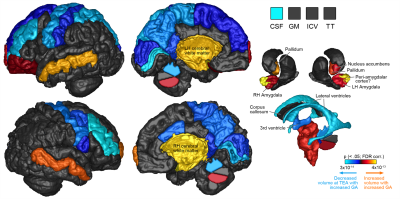
To investigate the possibility that in Fabry Disease (FD), similarly to other LSD, an abnormal brain development could occur, we performed a volumetric MRI analysis on 42 FD patients and 38 healthy controls (HC). MRI data were processed using SPM12 to obtain ICV values, as well as brain parenchymal (BPF) and gray matter (GMF) fractions. Mean ICV of FD patients was 8.1% smaller compared to HC (p < 5·10-5), without significant differences in terms of BPF or GMF, thus suggesting a harmonious volumetric reduction of intracranial structures, as a reflection of a possible abnormal brain development in this condition.
|
|
1799.
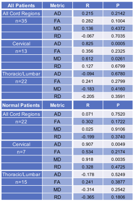 |
Quantification of Diffusion Tensor Imaging (DTI) in the Pediatric Spinal Cord: Application to Clinical Evaluation
Aashim Bhatia, Bryson Reynolds, Samantha By, Bhavesh Ramkorun, Quinn Weinberg, Mark Adams, John Wellons III, Seth Smith
The goal of the study was to apply optimized Diffusion Tensor Imaging (DTI) in the pediatric spinal cord and quantified to determine normative DTI-derived indices based on age. DTI was acquired in 35 patients, 22 being normal and AD, FA, MD, and RD were calculated.
DTI of the spinal cord in the pediatric population can be performed in the clinical setting to produce reliable DTI values. AD and MD demonstrated statistically significant changes based on age in both normal patients and the complete patient population.
|
|
1800.
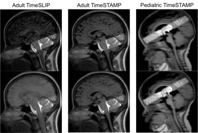 |
Tag-Based CSF Imaging Performance in Pediatric Patients and Adult Volunteers
Jieun Kwak, Tai-Wei Wu, Skorn Ponrartana, Benita Tamrazi, Wende Gibbs, Thomas Chavez, William Bradley, Marvin Nelson, J. Gordon McComb, Stefan Blüml, Matthew Borzage
We compared tag-based CSF imaging techniques (TimeSLIP and TimeSTAMP) in 10 healthy adults and 19 pediatric patients with cerebrospinal fluid (CSF) abnormalities. In adults, TimeSLIP and TimeSTAMP contrasts were quantitatively compared. TimeSTAMP sequences showed higher contrasts with decreased contrast variability versus TimeSLIP sequences. In pediatric patients, TimeSTAMP sequences were acquired to observe clinical utility and had similar contrast to the healthy adults. TimeSTAMP may be a superior imaging technique with clinical implications in adults and pediatric patients.
|
|
1801.
 |
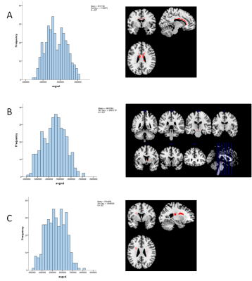
Factor analysis to determine white matter injury patterns following pediatric traumatic brain injury.
Brenda Bartnik Olson, Nirmalya Ghosh, Udo Oyoyo, Barbara Holshouser, Joy Nichols, Jamie Pivonka-Jones, Karen Tong, Stephen Ashwal
Several studies have shown regional disruptions in white matter integrity following TBI although conventional methods don't account for the relationship between regions. In this study we used factor analysis, a data reduction technique, to identify patterns of WM injury that are associated with neurocognitive outcome in pediatric TBI patients. Our findings identified 3 dominant patterns of WM injury in pediatric TBI patients, describing regional changes in: 1) subcortical + cortical diffusivity, 2) subcortical diffusivity, and 3) subcortical + cortical anisotropy. Factor analysis provides a unique statistical approach to analyze DTI data and potentially could be used to combine different data streams (DTI, MR spectroscopy, SWI) representing different elements of injury.
|
|
1802.
 |
Structural MRI derived connectivity in Paediatric Mild Traumatic Brain Injury: Acute Neuroimaging and its relationship with executive function outcomes
Daniel King, Stefano Seri, Vicki Anderson, Cathy Catroppa, Miriam Beauchamp, Amanda Wood
The aim of the current study was to identify acute differences in the topology of the structural covariance network of children after a mild traumatic brain injury (TBI). This was to assess the potential utility of this connectivity analysis applied to T1-weighted MR images, novel in the TBI literature. The main findings of this study were i) both patients and controls exhibited typical frequency distribution of few, highly connected nodes, ii) at a group level, patients exhibited connections between nodes a greater distance apart, iii) these differences were not associated with differences in executive function outcome. Future work will have to move to individual-level SCNS to allow for more complex analyses and to enable investigation of more subtle individual differences in structural covariance.
|
|
Psychoradiology
Traditional Poster
Neuro
Tuesday, 19 June 2018
| Exhibition Hall 1803-1845 |
16:15 - 18:15 |
|
1803.
 |
Morphological interrelationships in mid-line white-matter structures are altered in individuals carrying rare neuropsychiatric copy number variants.
Mark Drakesmith, Greg Parker, Jacqueline Smith , Elliot Rees, Michael Owen, Derek Jones, David Linden
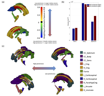
Neuropsychiatric copy number variants (CNVs) provide unique insights into the genetic basis of neuropsychiatric disorders. This study utilised a novel approach for characterising morphology of white-matter fibres and combines them with more traditional volumetric and microstructural indices of white-matter to study their relation to penetrance for psychopathology in a CNV cohort. Results show cingulum morphology is significantly affected by the presence of CNVs with high-penetrance for schizophrenia and developmental disorders. Additionally, volumetric interrelationships across several white-matter structures are also altered. In particular, the ratios of tract volumes across segments of the corpus callosum are altered. It is likely that both these effects stem from a single neurodevelopmental trajectory characteristic of neuropsychiatric CNVs.
|
|
1804.
 |
Quantitative magnetization transfer imaging in schizophrenia: a closer look at myelin dysfunction
Yu Sui, Pippa Storey, Hilary Bertisch, Matthew Lustberg, Taylor Coats, Donald Goff, Alexey Samsonov, Mariana Lazar
Myelin dysfunction has frequently been identified as one of the neural abnormalities in schizophrenia, yet systematic in vivo examination of myelin content in patients is lacking. The current study compared the degree of myelination in schizophrenia patients and comparison healthy controls. Myelin content was estimated by constructing quantitative whole-brain maps of macromolecular proton fraction, which is believed to be one of the biomarkers for myelination in neural tissues. Statistical analysis revealed that SZ patients were associated with a significant reduction in myelin content throughout white matter, as well as in several grey matter regions including cingulate cortex and hippocampus.
|
|
1805.
 |
Acutely treated antipsychotics haloperidol enhances BOLD responses to the somatosensory stimulation in anesthetized rats.
Yunbok Kim, Jeong Pyo Son, SoHyun Han, Seong-Gi Kim
The use of BOLD fMRI is rapidly increasing for probing the effects of antipsychotics in schizophrenia. Since fMRI BOLD is an indirect measurement of neural activities, it is critical to examine the effect of antipsychotics on neurovascular coupling to prevent misinterpretation of MR data. Acutely treated haloperidol (0.2mg/kg, i.v.) increased BOLD fMRI to the somatosensory stimulation in the 1.5% isoflurane-anesthetized rats (n=5). In parallel with the BOLD results, evoked CBF and LFP by somatosensory stimuli were increased after haloperidol administration (n=8). Our results indicate that acutely treated haloperidol could influence somatosensory responses and the increased BOLD signal is coupled with enhanced neural activities.
|
|
1806.
 |
Convolutional Neural Networks on Functional Connectivity Derived From r-fMRI: Explore the Effects of Thresholds
Xingjuan Li, Yu Li, Xue Li
In this study, we propose a novel CNN to predict autism from functional brain networks. Experimental results demonstrate that the predictive ability of CNN outperforms a logistic regression method by 8% and a five-layer fully-connected network (FCN) by approximately 7%. Network thresholding is often used to control false connections arising in the process of constructing functional brain networks. We also compare the influence of different thresholds on the performance of proposed CNN. Experimental results show that CNN is robust to false connections. Our study will contribute to predict reliable clinical outcomes in autism using deep learning on brain networks.
|
|
1807.
 |
Hippocampus and parietal lobe glutamate changes as a function of age in schizophrenia
Frank Gaston, S. Andrea Wijtenburg, Stephanie Korenic, Hongji Chen, Laura Rowland
MRS was used to examine the aging effects of glutamate in participants with schizophrenia versus healthy controls. The parietal lobe and hippocampus, regions associated with general aging and the pathophysiology of schizophrenia, were assessed. Results revealed that hippocampal glutamate was lower in older adults with schizophrenia versus older controls. In contrast, parietal glutamate was lower in schizophrenia versus controls, irrespective of age group. These results suggest that the hippocampus may be particularly vulnerable to aging in schizophrenia. Interventions that halt hippocampal glutamate decline may be beneficial for patients with schizophrenia.
|
|
1808.
 |
Amygdala dysfunction during negative emotional situation in Obsessive-Compulsive Disorder
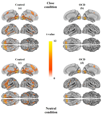
Hyunsil Cha, Sang Won Lee, Kyung Eun Jang, Hyejeong Choi, Eunji Kim, Moojin Yang, Jiung Yang, Moon Jung Hwang, Huijin Song, Seung Jae Lee, Yongmin Chang
We investigated brain activation in obsessive-compulsive disorder (OCD) patient using thought-action fusion (TAF) task to assess the influence of OCD symptom on amygdala response to the task. Within and between group analysis of close and neutral condition showed decreased amygdala activation in patients with OCD compared to healthy control.
|
|
1809.
 |
Assessment of brain volume and shape abnormalities in the major depressive disorders with and without suicidal ideation
Hui-Ming Tseng, Vincent Chin-Hung Chen, Yuan-Hsiung Tsai, Jun-Cheng Weng
There is very strong connection between patients with major depressive disorders (MDD) and suicide. We used voxel-based morphometry (VBM) and vertex-wise shape analyses to observe the difference between the MDD patients with and without suicidal ideation in their brain volume of gray and white matter as well as shape. We found the negative correlation between the brain volume of limbic system in MDD patients. We also found the significant difference in brain volume and shape of limbic system between suicidal ideation and non-suicidal ideation.
|
|
1810.
 |
Atypical associations between language comprehension network and attention pathways in autism spectrum disorders
Yu-Chun Lo, Susan Gau, Yu-Jen Chen, Yung-Chin Hsu, Wen-Yih Tseng
Impaired language comprehension has been consistently found in autism spectrum disorder (ASD). Development of language comprehension highly corresponds to joint attention and impulsivity. We used diffusion spectrum imaging to measure white matter integrity of the language comprehension network and the attention pathways in 60 ASD and 55 typically developing (TD) boys. ASD showed partially reduced white matter integrity in the targeted tracts as compared to TD. The tract covariance between the language comprehension network and the attention pathways showed different patterns in both groups which may shed light in the relationships of language and attention in ASD.
|
|
1811.
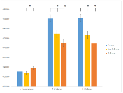 |
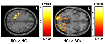
Connectome analysis of brain functional network alterations in depressed patients with and without self-harm
Yu-Syuan Chou, Vincent Chin-Hung Chen, Yuan-Hsiung Tsai, Shan-Chih Lee, Jun-Cheng Weng
We aimed to use resting-state fMRI (rs-fMRI) to investigate the functional connectivity difference between depressed patients with and without self-harm history as well as healthy participants. The graph theoretical analysis (GTA) and network-based statistic (NBS) analysis were also used to find the network difference between each group. In GTA and NBS analyses revealed different topological organization and poor global integration of the brain network in depressed participants compared with healthy participants. We suggested that depressed patients with or without self-harm history may affect their brain functional connectivity.
|
|
1812.
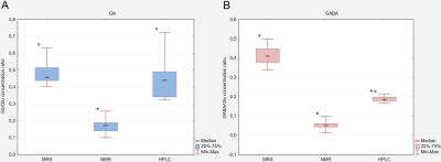 |
Measurements of rat hippocampus Glu, Gln and GABA using NMR, MRS and HPLC in animal models of autism
Pawel Senator, Elzbieta Zieminska, Wojciech Hilgier, Jaroslaw Orzel, Beata Toczylowska
The goal of our studies was to compare different measuring methods of glutamine, glutamate and GABA of rat hippocampus used for study of pathogenesis of autism. The methods under consideration were: in vivo MRS and two in vitro ones, NMR and HPLC. Univariate statistical analysis of ratios of tested amino acids with respect to glutamate concentration was performed using General Linear Model. This demonstrated statistically significant differences between the results from three methods for both, glutamine and GABA ratios. OPLS-DA analysis allowed build models for differentiation of two animal models of disease and control group in NMR and HPLC.
|
|
1813.
 |
Resting-state brain functional alteration in dorsal attention network associated with post-chemotherapy breast cancer
Chao-Yu Shen, Vincent Chin-Hung Chen, Xuan-Ru Zhang, Meng-Syuan Lin, Dah-Cherng Yeh, Yeu-Sheng Tyan, Ming-Chih Chou, Jun-Cheng Weng
The current study was to investigate post-chemotherapy breast cancer with rs-fMRI using mfALFF analysis and correlated with clinical cognitive testing. The results showed altered brain activity in the dorsal attention network in breast cancer patients compared to healthy controls and the affected areas were associated with MMSE, CAMS-R and IES-R scores.
|
|
1814.
 |
Principal Component Analysis of Schizophrenia Reveals Link Between Auditory Hallucination Severity and Fractional Anisotropy in the Corpus Callosum
Meighen Roes, Alexander Weber, Todd Woodward
A PCA analysis of fractional anistropy (FA) was conducted from a sample of schizophrenia patients (n=42) and healthy controls (n=40) resulted in three major components: “corpus callosum”, “internal capsule/temporal/brainstem”, and “corona radiata”. Average component scores did not differ as a function of group, but a correlation of PSYRATS scores and principal components revealed the frequency, amount of distress associated with voices, and disruption associated with voices correlated significantly with the corpus callosum component. Our findings suggest that reduced interhemispheric connectivity of the prefrontal cortex is related to hallucination severity in schizophrenia, perhaps mediated through top-down processes such as source monitoring.
|
|
1815.
 |
Diffusion kurtosis imaging and white matter model analysis of the brains of patients with major depressive disorder
Kouhei Kamiya, Naohiro Okada, Kingo Sawada, Yusuke Watanabe, Ryusuke Irie, Yuichi Suzuki, Shohei Hanaoka, Takeyuki Watadani, Shinsuke Koike, Harushi Mori, Akira Kunimatsu, Masaaki Hori, Shigeki Aoki, Kiyoto Kasai, Osamu Abe
We investigated the brain microstructural changes in major depressive disorder (MDD) using DKI and biophysical modelling. Twenty-six patients with MDD and 42 healthy control subjects were enrolled. TBSS whole brain analyses showed decrease of MK and RK in the patients as compared to the controls, predominantly in the frontal lobe, but widely distributed in the cerebral white matter. Model analysis revealed smaller intra-axonal volume fraction in the corpus callosum. The present results indicate the ability of DKI to demonstrate MDD pathology that are not fully depicted by DTI, and possibly to provide a new insights into the pathophysiology of MDD.
|
|
1816.
 |
Upregulation of hippocampal glutamatergic neurotransmission during acute episodes of major depression: Excitotoxic effects might be related to reduced hippocampal volumes
Jochen Bauer, Patricia Ohrmann, Bendix Labeit, Elke Scherbiski, Harald Kugel
Investigation of the glutamatergic metabolism with 1H-spectroscopy revealed a significant higher glutamate level in the hippocampus in patients with major depression. The excitotoxicity of increased glutamate levels on neural brain structures might be causally related to reduced volumes of hippocampi as found in patients with recurrend episodes.
|
|
1817.
 |
Histoarchitectonically distinct regions of anterior cingulate show altered glutamatergic metabolism in major depressive disorder
Louise Martens, Felicia von Düring, Lejla Colic, Shijia Li, Liliana Demenescu, Dominik Denzel, Inka Ristow, Matthias Vogel, Sarah Lison, Oliver Speck, Meng Li, Martin Walter
Increasing evidence suggests a hypoglutamatergic state in major depressive disorder (MDD), however spatial- and metabolite specific abnormalities have not been fully characterized. Using short TE/TM STEAM MRS, we evaluated Glu, Gln, Gln/Glu and GABA metabolism in two histoarchitectonically distinct subdivisions of the anterior cingulate cortex (ACC). The pregenual ACC, involved in emotion processing, showed altered glutamine-glutamine cycling but not altered GABAergic metabolism in MDD, whereas no differences between patients and controls were found in the anteromedial ACC. Increased Gln/Glu in MDD in pgACC but not aMCC confirms a regionally specific role of altered glutamatergic metabolism and neuronal-glial interaction.
|
|
1818.
 |
MR Spectroscopic evaluation of brain white matter metabolite abnormalities in Psychotic Spectrum Disorders
Ines Blockx, Matthew Lustberg, Taylor Coats, Hillary Bertisch, Oded Gonen, Donald Goff, Mariana Lazar
1H-MRS has been widely applied in studies with Psychotic Spectrum Disorders, however, findings are mixed and the exact cause of these disorders remains to be elucidated.The preliminary results of the present study show increased Gln/Cr levels in schizophrenia and schizoaffective patients in central WM reaching statistical significance in the bipolar group. The increase in Gln/Cr levels has been proposed to occur in the early stages of the disorder which is consistent with the population included here. The current study brings WM as a relevant area susceptible to damage into focus, which is likely to be involved in the early stages of PSD.
|
|
1819.
 |
Auditory system altered in auditory verbal hallucination studied using diffusion spectrum imaging, T1-weighted image and fMRI
Kayako Matsuo
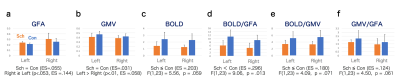
To understand the pathology of auditory verbal hallucination (AVH), we investigated 3 MRI indices: generalized fractional anisotropy (GFA) using diffusion spectrum imaging in the auditory radiation, gray matter volume (GMV) using T1-weighted images in Heschl’s gyrus (i.e., auditory cortex) and BOLD contrast estimates using task-fMRI in the auditory cortex. The BOLD relative to the GFA was significantly greater in controls than in patients with schizophrenia who had AVH. The GMV relative to the GFA also tended to show greater values in controls than in patients. An unregulated auditory sensation attributed to a dysfunction in the cortex might eventually encompass AVH.
|
|
1820.
 |
Grey abnormalities associate with suicide related behaviour in first episode non-affective psychosis patients
Manuel Canal-Rivero, Rosa Ayesa-Arriola, Esther Setien-Suero, Manuel Delgado-Alvarado, Benedicto Crespo-Facorro, Diana Tordesillas-Gutierrez
Little is known about brain abnormalities associated with suicide-related behaviours in first episode psychosis patients and controversial results have been reported. The main aim of the present study was to examine brain abnormalities related with suicidal behaviours in a large sample of first episode psychosis (FEP) patients. In particular, we found reduction grey matter volume in frontal area, middle temporal gyrus as well as posterior cingulate gyrus and precuneus. These areas appear to be associated with some of the greatest features related to suicidal behaviour such as impulsivity, emotional processing information, responses to pain and aggressiveness.
|
|
1821.
 |
The Differences of Amplitude of Low Frequency Fluctuation between Methamphetamine and Heroin use disorder: a resting-state functional magnetic resonance imaging study
Yan Liu, Wei Wang, Wei Li, Qiang Li, Yongbin Li, Jiajie Chen, Jing Chen, Shan Dang
These findings indicated different brain regions between MA users and heroin users in resting-state, as well as it’s function correlation with emotion.
|
|
1822.
 |
Myelin content and axonal size/density is reduced in early-course schizophrenia: Evidence from multi-echo T2 imaging study
Shivali Patel, Jennifer Losiowski, Muzamil Arshad, Naftali Raz, Vaibhav Diwadkar, Jeffrey Stanley
White matter aberrations have been well documented in schizophrenia using diffusion tensor or weighted imaging, but the differences in myelin macrostructure morphology have not been extensively explored. Here we used multi-echo T2 (ME-T2) imaging to examine myelin content and axonal size and packing density in schizophrenia in white matter regions, specifically association, commissural, and projection fiber tracts. We demonstrate reduced myelin content as well as increased axonal packing density in association and projection tracts, which may contribute to neural dysconnectivity mechanisms underlying the neuropathology of schizophrenia.
|
|
1823.
 |
Resting-state Network Evaluation of First-episode Schizophrenia Patients by fMRI
Kangkang Xue, Dandan Zheng, Jingliang Cheng
Schizophrenia is a chronic mental illness whose symptoms are thought to have a strong neurobiological basis. This work is to study the resting state networks changes in first-episode schizophrenia patients by resting-state functional magnetic resonance imaging. The current study explored that there were RSNs damages or multiple brain regions functional connectivity abnormalities in first-episode schizophrenia patients compared with healthy controls, which behave functional connectivity increase and decrease.
|
|
1824.
 |
A voxel-based diffusion kurtosis imaging study of whole-brain in chronic alcohol dependent patients
Hong-yan Nie, Jun Chen, Ya-qi Wang, Yang Fan
In the present study, diffusion kurtosis imaging (DKI), which is based on the method of voxel-based analysis(VBA), was used to investigate the alterations of microstructure of white matter and gray matter in chronic alcohol dependent patients. Thirty patients with chronic alcohol dependence and twenty healthy volunteers were scanned with DKI. Compared with the healthy control group, the brain regions associated with visual information processing, memory, movement coordination and emotional control capacity have been found to be abnormal in different degrees.
|
|
1825.
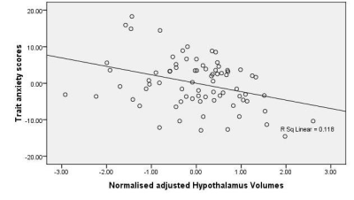 |
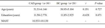
Structural correlates of trait anxiety: Volume reduction in hypothalamus
SHILPI MODI, DIVESH THAPLOO, PAWAN KUMAR, SUBASH KHUSHU
Trait anxiety affects brain functioning and cognition as suggested by various neuroimaging and behavioural studies. It is also a a prone phenotype for the development of psychiatric disorders. Therefore, in order to identify individuals that are at risk for the development of clinical anxiety disorders and depression, identifying hallmarks of trait anxiety becomes important, to fascilitate timely preventive interventions. We investigated the structural correlates of trait anxiety in healthy participants using high resolution structural MRI. Results suggest that a reduction in the gray matter volumes of the hypothalamus may be putative imaging marker for trait anxiety.
|
|
1826.
 |
Increased functional connectivity between medial prefrontal cortex and nucleus accumbens in morphine craving rats
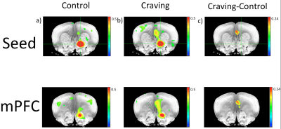
Hannes Wiesner, Shinho Cho, Yi Zhang, Erin Larson, Mark Thomas, Xiao-Hong Zhu, Wei Chen
Morphine is a potent analgesic with a high addictive potential. In this study we have shown a difference in brain connectivity related to drug-seeking behavior involving key neural decision and reward systems using rs-fMRI. The finding contributes to a better understanding of the neural underpinnings of opioid addiction and could help in a better assessment of relapse risk in individuals.
|
|
1827.
 |
Alterations in amplitude of low frequency fluctuation in drug-free major depressive disorder
Hu Xiaoxiao, Hu Xinyu, Li Hailong, Zhang Lianqing , Lu Lu, Bu Xuan, Tang Shi, Gong Qiyong, Huang Xiaoqi
The objective of this study was 1) to confirm whether the intrinsic brain activities (as evaluated by ALFF) in the anterior cingulate cortex (ACC) is associated with antidepressant treatment in a relative large sample of drug-free major depressive disorder (MDD) patients and 2) to determine whether the pretreatment ALFF activities predict the effect of the follow-up antidepressant treatment in MDD. Our findings demonstrate that intrinsic brain activities in the ACC was influenced by disease itself rather than antidepressant treatment and threw light on predictive value of the right thalamus as a marker of short term antidepressant treatment outcome in MDD.
|
|
1828.
 |
A pilot study of cerebral blood flow changes in patients undergoing electroconvulsive therapy
Karl Spuhler, Laura Kunkel, Adeeb Yacoub, Kenneth Wengler, Xiang He, Chuan Huang
Electroconvulsive therapy (ECT) is an effective choice for patients with untreatable depression. Although it is very effective, the mechanisms through which ECT works are poorly understood. We have previously collected PET/MRI data in patients receiving ECT which suggest that this treatment strongly affects the hippocampus. Herein, we supplement these preexisting data with arterial spin labeling data showing significantly reduced blood flow to the hippocampus following ECT in three responders.
|
|
1829.
 |
In search for a neuroimaging marker for neuroinflammation in neuropsychiatric systemic lupus erythematosus
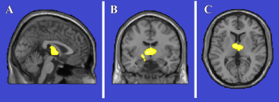
Marjolein Bulk, Ece Ercan, Cesar Magro-Checa, Louise van der Weerd, Itamar Ronen
We explored the link between neuroinflammation and related changes in tissue susceptibility by using quantitative susceptibility mapping (QSM) in a clinically well characterized cohort including inflammatory NP-SLE, ischemic NP-SLE and SLE patients. No significant differences were found after stratifying all patients for antibodies, SLE activity, cumulative SLE damage or complement components in subcortical structures. Subanalysis of inflammatory NP-SLE patients showed a residual correlation between QSM values in the globus palidus and low C1q levels, which need further investigation. Current work is underway to analyse QSM in a bigger sample size to further investigate its potential in identifying NP-SLE patients.
|
|
1830.
 |
Trait anxiety associated metabolic alterations in thalamus: An MRS study
SHILPI MODI, DIVESH THAPLOO, PRABHJOT KAUR, SUBASH KHUSHU
Trait anxiety is a prone phenotype for the development of anxiety disorders and depression. Therefore, in order to identify the individuals 'at risk', identifying the hallmarks of trait anxiety becomes important. Ones identified, timely preventive interventions may be given to such individuals. This study is an attempt to study the trait anxiety associated metabolic/ neurochemical alterations in the brain using proton magnetic resonance spectroscopy. We obtained an increase in the concentrations of Choline compounds in the thalamus as a function of trait anxiety of the subjects suggesting an altered cell membrane metabolism.
|
|
1831.
 |
Hippocampus Glutamate Concentrations in Schizophrenia and Bipolar Disorder
Nicolas Bolo, Olivia Lutz, Gautami Shashidhar, Li Yao, Yungxiang Tang, Brett Clementz, Godfrey Pearlson, Elliot Gershon, John Sweeney, Carol Tamminga, Matcheri Keshavan
Deficient hippocampus glutamatergic function could underlie cognitive deficits and positive-negative symptoms in schizophrenia (SZ) and bipolar disorder (BP). Using 1H MRS, we found that the glutamate concentration of left anterior hippocampus was significantly lower in SZ (6.3 ± 1.8 mM) vs. healthy controls (HC, 7.8 ± 1.2 mM, p=0.021) and BP (8.5 ± 1.3 mM, p=0.001) and trended higher in BP vs. HC (p=0.179). Decreased glutamate is consistent with deficient excitatory neurotransmission in the hippocampus of patients with SZ, which could alter synaptic plasticity underlying memory and cognition. Our findings are consistent with the glutamate hypothesis of SZ.
|
|
1832.
 |
Change of cortical thickness and hippocampal volume in adolescents with autism spectrum disorder
I-Ting Su, Tzu-chao Chuang, Ming-Ting Wu, Pinchen Yang
By using a surface-based method (Freesurfer), the cortical thickness, hippocampal volume, and amygdala volume measurement were performed on adolescents with autism spectrum disorder (n=17) and age-matched typically developing controls (n=10). ASD patients showed a thicker cortex in temporal and occipital regions, a thinner cortex in frontal regions, and larger right hippocampal volume compared to the controls.
|
|
1833.
 |
A meta-analysis of altered resting-state functional activity in medication-naive patients with first-episode major depression versus healthy controls
Xiaoyue Ma, Jia Liu, Taiyuan Liu, Yan Wang, Meiyun Wang, Tianyi Qian
This study aimed to use the voxel-based meta-analytic technique called anisotropic effect size-signed differential mapping (AES-SDM) to determine consistent regional brain activity alterations in medication-naive patients with first-episode unipolar major depression disorder (MDD) versus healthy controls (HCs). The pooled and subgroup meta-analyses found that MDD patients showed resting-state brain decreased activity in the left anterior lobe of the cerebellum and increased activity in the left amygdala and left hippocampus which have hitherto been neglected in previous studies and provide new implications for the pathophysiology of cognitive and emotional impairment in MDD patients.
|
|
1834.
|
Neurometabolic alterations in patients with major depression measured with short echo-time whole-brain MR spectroscopic imaging
Xiao-Qi Ding, Sirin Atalay , Andrew Maudsley, Sulaiman Sheriff , Anna Cummings, Birte Schmitz, Heinrich Lanfermann , Kai Kahl
Major depressive disorder (MDD) is a common mental disorder with unclear pathophysiology. Metabolite concentrations over brain lobes or cerebellum in patients with MDD were studied. The results revealed that brain metabolic alterations associated with MDD were related to brain region and metabolite, and were particularly present in right and left frontal lobes. The findings indicate neuronal dysfunction and altered glutamatergic neuronal activity in patients.
|
|
1835.
|
Longitudinal structural white matter alterations in adolescents at risk for psychopathology: a Randomised Controlled Trial.
Stijn Michielse, Jindra Bakker, Iris Lange, Liesbet Goossens, Koen Schruers, Ritsaert Lieverse, Therese Amelsvoort, Marieke Wichers, Jim Os, Machteld Marcelis
This project is an RCT in 51 individuals with mild psychopathology randomly assigned to Acceptance and Commitment Therapy (ACT) or topic discussion group conditions. Participants underwent Diffusion Weighted Imaging (DWI), Experience Sampling Method (ESM) and a Community Assessment of Psychic Experiences (CAPE) questionnaire before and after intervention. Results show no differences between conditions after the intervention in the white matter (DWI) or the amount of psychotic experiences (CAPE). The suspicious mood ESM item showed was significantly changed due to ACT-intervention. Therefore white matter changes do not seem to occur, while mood changes as a result after 12 week intervention.
|
|
1836.
 |
Investigation of resting-state fMRI and cognitive function changes in patients with late-onset depression after one year follow-up
Hongmin Xu, Hongmei Fu, Naying He, Fuhua Yan
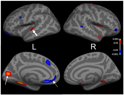
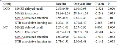
Late-onset depression is a common psychiatric disorder, depressed elderly often exhibit cognitive impairment that are substantial, prevalent, and disabling. The LOD patients with cognitive impairment has increased risk of conversion to dementia. The amplitude low-frequency fluctuation analysis based on resting state fMRI can directly reflect the intensity of spontaneous activity of neurons and provide information of local neurons in brain areas. In this study, we observed the changes of cognitive function and local brain functional activity in patients with LOD after one year follow-up, investigated the correlation between cognitive function and brain activity. And possibly provide an objective imaging basis for the early intervention in LOD patients with cognitive impairment before deteriorate into dementia.
|
|
1837.
|
Structural magnetic resonance imaging study on schizophrenic patients with violence risk
Yingna Li, Fengmei Fan, Zhiyuan Feng, Shuping Tan, Fude Yang
To explore the brain structual imaging differences between schizophrenic patients with or without violence risk. By structual MRI and Freesurfer software,the study founds that schizophrenic patients with violence risk show the brain cortex thickness and volum reduction and cortical meancurvature increase , especially the reduction of the cortex thickness in the postdorsal cingulate gyrus .
|
|
1838.
|
SUBCORTICAL VOLUMETRIC CHANGES IN PATIENTS WITH MAJOR DEPRESSIVE DISORDER: ROLE OF MRI
Mariia Rezakova, Elena Filimonova, Khurshed Ibrogimov, Olga Subbotina, Alexandr Shevchenko
We analyzed subcortical structures in patients with MDD (N=15) and control (N=15) using FreeSurfer. Patients with MDD had significantly lower left thalamus (p<0,01), left putamen (p<0,05), left hippocampus (p<0,05) and some hippocampal subfields volumes, relative to control. We found correlations (p<0,05) between patient’s age and putamen volume (r= -0,56), number of depressive episodes and molecular layer volume (r= -0,52). We didn’t reveal correlation between segmentation data and MDD severity.
|
|
1839.
 |
Voxel-based morphometry using silent T1-weighted sequence elucidates the brain volume difference between autism spectrum disorder and children with typical development
Yoshiyuki Watanabe, Masahiro Fujiwara, Takuya Fujiwara, Hiroto Takahashi, Hisashi Tanaka, Kuriko Shimono, Mariko Nakanishi, Ryuzo Hanaie, Ikuko Mohri, Noriyuki Tomiyama
Silent MR sequences are expected to be useful and promising in the evaluation of hyperacusia patients, especially autism spectrum disorder (ASD). The aim of this research was to apply silent T1W to evaluate the brain volume changes between ASD and children with typical development (TD). Results showed that the brain volume of ASD was significantly increased at the left inferior temporal lobe and the right cerebellar tonsils and decreased at the right insular cortex and the right medial frontal lobe compared to that of TD. Silent T1W sequence can detect brain volume difference between ASD and TD.
|
|
1840.
 |
White Matter Abnormalities in Never-Treated Patients with Long Term Schizophrenia
Yuan Xiao, Huaiqiang Sun, Bo Tao, Youjin Zhao, Wenjing Zhang, Qiyong Gong, John Sweeney, Su Lui
Do white matter abnormalities increase over the long-term course of schizophrenia, and is their trajectory influenced by antipsychotic treatment? In this cross-sectional study, more alteration of white matter microstructure were found in long-term but never-treated schizophrenia patients than duration-matched chronically treated patients. In the genu of the corpus callosum, there was an accelerated age-related reduction of fiber tract integrity in the never-treated patients. The more attenuated white matter changes in the treated patient group suggests that long-term antipsychotic treatment may have a neuroprotective effect on white matter tracts.
|
|
1841.
 |
Gray Matter Network Organization in Psychotic Disorders
Wenjing Zhang, Du Lei, Brett Clementz, Carol Tamminga, Matcheri Keshavan, Sarah Keedy, Godfrey Pearlson, Elliot Gershon, Jeffrey Bishop, Jieke Liu, Qiyong Gong, John Sweeney, Su Lui
Recently, new approaches have been developed using graph theory to identify deficits in gray matter networks at individual level. In the current study, by investigating single-subject graphs based on gray matter morphology to define neuroanatomic networks in a large group of individuals across psychotic disorders (n=330), we observed disrupted network organizations associated with superior temporal and prefrontal regions within the gray matter networks in patients, which were also negatively associated with severity of psychotic symptoms. These findings showed the utility of graph theory based measures of neuroanatomic network organization to extend our understanding of the neurobiology underlying psychotic disorders.
|
|
1842.
 |
Peripheral oxytocin and vasopressin modulates regional brain activity differently in men and women with schizophrenia
Siyi Li, Leah Rubin, Li Yao, Su Lui
Oxytocin (OT) and arginine vasopressin (AVP) exert sexually dimorphic effects on cognition and emotion processing in healthy individuals, and abnormalities in these neuroendocrine systems are observed in schizophrenia with a sex-dependent manner. Here we examined sex-dependent hormone associations with resting brain activity by applying resting-fMRI and their clinical associations in schizophrenia patients relative to healthy controls. We found that hormones differentially associate with brain networks, the sex-dependent alternation of hormone and brain activity are important for cognition and emotion processing in men and women with schizophrenia.
|
|
1843.
 |
Higher variability of individual functional brain networks in young children with autism
Chenying Zhao, Qinmu Peng, Minhui Ouyang, Hua Cheng, Yun Peng, Bo Hong, Hao Huang
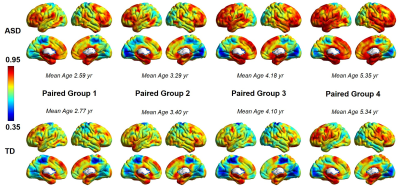
Individual’s functional brain networks are sensitive indicators of behaviors. Atypical functional connectivity have been observed in children with autistic spectrum disorder (ASD), manifesting characteristic and distinctive behavior at ages of 2- to 7-years. However, little is known about individual variability of the functional brain networks in children with ASD. In this study, using resting-state fMRI and variability analysis, we quantified distinguished variability pattern in children with ASD from typically developing (TD) children from 2- to 7-years of age, especially in higher-order functional networks. The higher inter-subject variability in children with ASD may be associated with their impaired behaviors.
|
|
1844.
 |
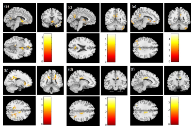
Brain Gray Matter Abnormalities in First-Episode, Treatment-Naïve Patients with Obsessive-Compulsive Disorder
Junhong Liu, Dandan Zheng, Jingliang Cheng
Examinations of 36 first-episode, treatment-naive pediatric OCD patients without any comorbidities and 37 matched healthy controls (HCs) were performed with 3.0T magnetic resonance imaging (MRI). Voxel-based morphometry (VBM) following Diffeomorphic Anatomical Registration using Exponentiated Lie algebra (DARTEL) was used to conduct voxel-wise tests for group differences in regional gray matter volume (GMV). Compared to HCs, the patient group exhibited significantly different GMV in bilateral anterior cingulate cortex (ACC), left fusiform gyrus and the left postcentral gyrus. It is believed that this noninvasive method might be useful for exploring the pathophysiology of OCD.
|
|
1845.
 |
Recuperative white matter integrity in long-term abstinent heroin addicts
wei Li, qiang Li, yan Liu, jing Chen, shan Dang, wei Wang
Heroin-induced white matter integrity disruption and the restorability during long-term abstinence have been reported. However, the characteristic of these recover during different stage of abstinence has not been well understood. Use the voxel-wised diffusion tensor method,we compared the white matter difference within 17 long-term abstinence heroin addicts (LA), 22 short-term abstainers (SA) and 20 healthy controls (HC). We found significantly decreased white matter integrity in SA and the time-dependent recover of white matter integrity, especially the restoration of myelin sheath, in LA,. These structural recover may contributed to the improvement of function in the duration of long-term abstinence.
|
|
Myelin Imaging: From Mice to People
Traditional Poster
Neuro
Tuesday, 19 June 2018
| Exhibition Hall 1846-1867 |
16:15 - 18:15 |
|
1846.
 |
The Observable Fraction of Myelin Lipid 1H Magnetization Imaged by IR-ZTE
Alan Seifert, Michael Wilhelm, Suzanne Wehrli, Felix Wehrli
Direct detection of myelin using solid-state imaging methods is challenging due to the extremely short lifetime of the myelin matrix 1H MR signal, which significantly limits its observability. In this work, the fraction of total myelin matrix 1H MR signal that is observable by an inversion-recovery (IR)-prepared zero echo-time (ZTE) imaging with pointwise encoding time reduction with radial acquisition (PETRA) sequence using various acquisition parameters is estimated by Bloch equation simulations. Only approximately 5% of total magnetization is observable under realistic experimental conditions. The adiabatic inversion-recovery pulse is mostly responsible for this low fractional observability.
|
|
1847.
 |
Magnetic Resonance Imaging (MRI) Assessment of Dimethyl Fumarate in Protecting Myelin in a Cuprizone Mouse Model
Peter Cheng-te Chou, Benxiu Ji, Jon Archbold, Ankur Thomas, Davide Gianni, Daniel Bradley, Haiying Liu, Brian Wipke
Multiple sclerosis (MS) is a debilitating disease that affects the central nervous system. Immune system destroys the myelin that protects the axon which leads to physical, neurocognitive, and psychiatric disorders. Symptoms may improve, but permanent neurological problems often remain. There is no known cure for MS but current treatments can improve symptoms and prevent relapse. MRI has a role in MS diagnosis and management. We demonstrated that advances in MRI techniques such as Magnetization Transfer Ratio Imaging and Diffusion Tensor Imaging can detect the protective effects of dimethyl fumarate, clinically approved MS treatment, in the corpus callosum of mice.
|
|
1848.
 |
Relevance of microglia receptor TREM2 for remyelination as revealed by multimodal MRI in the cuprizone mouse model
Anna Mechling, Eva Mracsko, Andreas Bruns, Thomas Mueggler, Irene Knuesel, Basil Künnecke
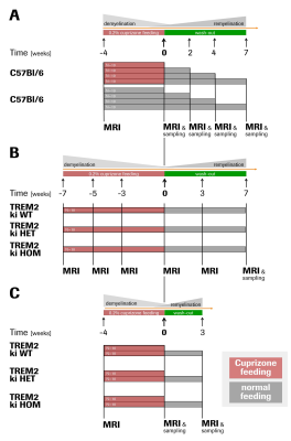
Demyelination and ensuing axonal damage are hallmarks of numerous neurodegenerative disorders. Novel treatment strategies seek to enhance remyelination and axonal recovery through acceleration of myelin debris clearance by phagocytic microglia. TREM2 is a receptor expressed by microglia that has been implicated in the regulation of phagocytosis, migration and anti-inflammatory activity. Here, we further elucidated the role of TREM2 in de- and remyelination processes by means of multiparametric in vivo MRI. We combined a TREM2 loss-of-function mouse model with cuprizone feeding as an accepted model for demyelination. Deficiency of TREM2 leads to progressive structural disintegration and absence of proper remyelination.
|
|
1849.
 |
Three-Dimensional Inversion Recovery Ultrashort Echo Time (3D IR-UTE) Magnetic Resonance Imaging of Myelin in Rats and Mice Subject to Cuprizone Treatment
Yajun Ma, Adam Searleman , Robert Bussell, Eric Chang, Srihari Sampath, Srinath Sampath, Lisa Deaton, Andrew Shumacher, Jiang Du
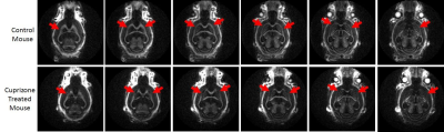
Ultrashort echo time (UTE) MRI is capable of directly imaging myelin protons. We present the first application of a UTE sequence to study an animal model of demyelination, using inversion recovery (IR) and 3D radial sampling. Mice treated with 0.2% cuprizone for 5 weeks show loss of the 3D IR-UTE signal in the lateral corpus callosum, which is expected to be maximally demyelinated at this time point. Future studies of histologically validated demyelination and remyelination in this model will further confirm the capability of 3D IR-UTE to selectively image myelin.
|
|
1850.
 |
Measurement of T1 and T2* Relaxation Times of Purified Animal Myelin by 3D UTE Cones Sequences at 3T
Adam Searleman, Yajun Ma, Eric Chang, Jiang Du
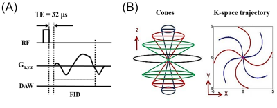
Determination of accurate T1 and T2* values of myelin protons is challenging because it is comprised of multiple lipid and protein components with an ultrashort T2*, but would be important for ultrashort echo time (UTE) sequence development. In this study, we present the first T1 and T2* measurements of intact myelin directly purified from white matter, with T1 measured using a 3D UTE Cones adaptation of actual flip-angle imaging (UTE-AFI) with variable TRs, and T2* measured using 3D UTE acquisitions with variable TEs. We find that myelin has a T1 of 367 ms and T2* of 225 ms at 3T.
|
|
1851.
 |
Dynamic Sensitivity of 3D Ultrashort Echo Time (UTE) Cones Imaging for Myelin Concentration Quantification
Adam Searleman, Shu-Juan Fan, Yajun Ma, Eric Chang, Jiang Du
Quantification of myelin has the potential to be used as a specific biomarker for demyelinating diseases of the nervous system such as multiple sclerosis. Ultrashort echo time (UTE) MRI has been shown to be able to directly detect signal from myelin protons, but the dynamic sensitivity of the 3D UTE Cones sequence remains unclear. This study examined the correlation between 3D UTE Cones signal intensities and different concentrations of myelin extract in D2O, and found a strong linear correlation up to a myelin concentration of 24% (w/v).
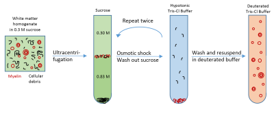
|
|
1852.
 |
Effect of aldehyde fixation on the myelin water fraction measurements in rat cervical spinal cord
Henry Chen, Jie Liu, Alex MacKay, Wolfram Tetzlaff, Piotr Kozlowski
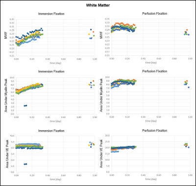
This study investigated the effect of tissue fixation on myelin water fraction (MWF), an MR derived measurement of myelin content. MWF was found to increase during aldehyde fixation due to an increase in myelin water. Differences in MWF between immersion fixation and perfusion fixation with immersion post-fixation were quantified. This study demonstrated that the measured MWF is sensitive to the changes induced by chemical fixation. The results bridge the interpretation of MWF in the in vivo situation to that of the ex vivo situation and provide a guideline for designing MWF studies with histological validation.
|
|
1853.
 |
Sequential Changes of Diffusion Anisotropy and Mean Kurtosis in Cuprizone-Induced Demyelination: A Rat Model
Ping-Huei Tsai, Hua-Shan Liu, Fei-Ting Hsu, Yu-Chieh Kao, Chia-Feng Lu, Hsiao-Wen Chung, Cheng-Yu Chen
The verification of cuprizone-induced demyelination in a rat model remains controversial. This study aims to develop a reliable cuprizone-induced demyelination rat model and to test the ability of DKI to monitor the sequential changes during brain demyelination. Our findings demonstrated that DKI could provide complementary information, associated with pathophysiological processes after demyelination in rat brain, which may have potential to detect microstructural changes at both acute and chronic stages and contribute to evaluations of further therapeutic strategies.
|
|
1854.
 |
Multicomponent relaxation analysis of myelin in the brains of rare progressive solitary sclerosis, compared to multiple sclerosis and healthy control subjects in vivo
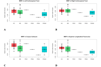
Lisa Eunyoung Lee, Jillian Chan, Irene Vavasour, Roger Tam, Anthony Traboulsee, Robert Carruthers, Shannon Kolind
Progressive solitary sclerosis (PSS) presents with an isolated demyelinating lesion along the corticospinal tract that results in progressive motor deficits. We used mcDESPOT-derived parameters to better understand the pathology in the normal-appearing white matter tracts (WMT) of PSS compared to relapsing-remitting multiple sclerosis (RRMS) and healthy control (HC) subjects. Overall, we found a trend of lower MWF (myelin content) and higher qT1 (inflammation/edema) in WMT in PSS, compared to RRMS and HC subjects. This suggested that there might be more extensive myelin damage in the normal-appearing brain, beyond the lesional site, that may be driving disease progression in PSS.
|
|
1855.
 |
A new rapid and high-resolution multi-slice inhomogeneous Magnetization Transfer protocol to evaluate diffuse and regional cervical cord myelination at 3T
Henitsoa Rasoanandrianina, Guillaume Duhamel, Aurélien Massire, Olivier Girard, Maxime Guye, Jean Pelletier, Bertrand Audoin, Virginie Callot
The inhomogeneous Magnetization Transfer (ihMT) technique has recently been proposed as a new method to probe the cervical spinal cord (CSC) myelin-content. Studies reported so far were limited to single-slice acquisition, hence precluding investigation of the whole CSC within a short acquisition time. To overcome this limitation, a 2D multi-slice single-shot Spin-Echo-Echo-Planar Imaging (SE-EPI) read-out approach was implemented at 3T along with strategies to correct for inherent susceptibility-induced image-distortions and post-saturation relaxation effect for each slice. Validated on phantom and applied to healthy subjects and a patient with multiple sclerosis, this preliminary study shows the promising value of SE-EPI ihMT in the clinical context.
|
|
1856.
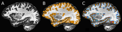 |
Assessing visual field integrity using gray matter myelination at 7T
Alessio Fracasso, Carlien Roelofzen, Giorgio Porro, Douwe Bergsma, Mies van Genderen, Serge Dumoulin, Natalia Petridou
High resolution 7T MRI allows to investigate the functional and structural organization of human cerebral cortex at an unprecedented level of detail, visualizing myelination patterns over the cortical surface and identifying a large number of cortical areas. In this study we hypothesize that myelin content co-varies with loss of visual input. We used a modified T1-w MPRAGE to enhance myelin visualization within gray matter and acquired data from patients with hemianopsia, a visual field defect consisting of an absolute scotoma limited to a single hemifield, and evaluate whether the clinical symptoms are reflected in gray matter myelination in the occipital cortex.
|
|
1857.
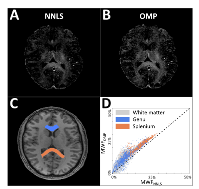 |
Myelin-Water Quantification: Orthogonal Matching Pursuit versus Non-Negative Least Squares
Gerhard Drenthen, Walter Backes, Albert Aldenkamp, Jacobus Jansen
Myelin-water quantification relies on modeling of multi-exponential T2-relaxation time decay. For this, we explore the greedy Orthogonal Matching Pursuit (OMP) method and compare it to the most commonly applied non-negative least squares (NNLS) method. The two methods are evaluated by means of simulations, phantom measurements and in vivo image data.
|
|
1858.
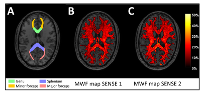 |
Reproducibility of Myelin Water Fraction for GRASE sequences with a varying SENSE factor
Gerhard Drenthen, Walter Backes, Albert Aldenkamp, Jacobus Jansen
For myelin-water quantification to become a feasible method in a clinical setting a rapid whole brain coverage acquisition is required, as well as reproducible results. Therefore, this study aims to measure the reproducibility of the Gradient-Spin Echo (GRASE) sequence with and without utilizing parallel imaging with sensitivity coding (SENSE) to investigate the impact of the acceleration (e.g. increased SENSE factor).
|
|
1859.
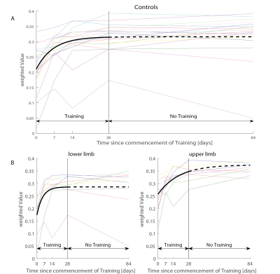 |
Training induced myelin and iron changes in healthy subjects using novel quantitative MRI techniques
Michela Azzarito, Eveline Huber, Maryam Seif, Gabriel Ziegler, Patrick Freund
Activity-dependent plasticity has significant implications for healthy development, learning, memory, and recovery from brain damage. However, the exact time course and the neural mechanisms behind brain plasticity are still not completely understood. In this study, longitudinal quantitative MRI protocols were used to assess training associated microstructural changes using markers sensitive to myelin and iron. We show that training improvements during a sensorimotor task performed over 4 weeks induces linear and non-linear increases in myelin and iron content in the primary motor cortex and cerebellum. This study provides new tools to assess training effects in healthy controls.
|
|
1860.
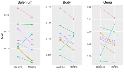 |
Decreased myelin water fraction in the corpus callosum at 6 months post mild traumatic brain injury
Bretta Russell-Schulz, Ivan Torres, Manraj Heran, Alex MacKay, William Panenka
Monitoring mild traumatic brain injury (TBI) presents challenges for conventional MRI and underlying myelin changes are not well understood. Myelin water fraction (MWF) presents an opportunity to examine myelin changes post injury. At 6 months post injury, corpus callosum genu and body MWF decreased from baseline (acute) in 8 out of 9 subjects. Splenium MWF decreased in 7 out of 9 subjects. When averaged across subjects, the average decrease in MWF was 2% for the genu and 5% for the splenium, not significantly different from baseline; the lack of significance was due to large MWF increases in one of the participants.
|
|
1861.
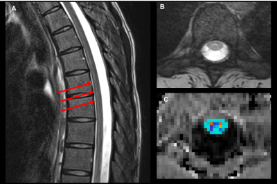 |
Spinal Cord (C1 to T12) Demyelination Measured by Magnetization Transfer Imaging: Characteristics of Acute, Sub-Acute and Chronic Disease Phases
Sze Nok Tam, Katie E Silva, David Zurakowski, Leslie Benson, Mark Gorman, David Borsook, Nadia Barakat
The diagnostic utility of Magnetization Transfer Imaging (MTI) was tested in a large cohort of patients with transverse myelitis – a demyelinating myelopathy affecting the spinal cord. We measured the reproducibility of MTI in a pediatric clinical model, at different disease stages. Our results showed that obtaining repeatable measures in the entire spinal cord (C1 to T12) is feasible. Our findings also showed significant differences in MTR values between patients and healthy controls, and between three sub-groups of patients (acute, sub-acute and chronic disease phases).
|
|
1862.
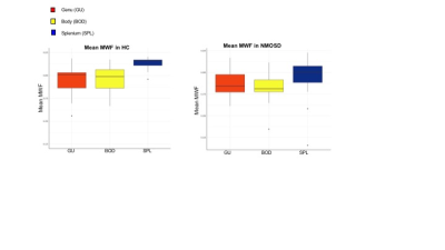 |
mcDESPOT-derived measurements are sensitive to differences in myelin content and thickness in the corpus callosum of neuromyelitis optica patients and healthy controls
Shawna Abel, Irene Vavasour, Lisa Lee, Roger Tam, Cornelia Laule, Robert Carruthers, Anthony Traboulsee, Anna Combes, Shannon Kolind
Histological studies suggest that white matter microstructure varies across different subregions of the corpus callosum (CC). We used mcDESPOT-derived measures to examine myelin content and thickness in vivo in 3 subregions of the CC in healthy controls (HC) and individuals with neuromyelitis optica spectrum disorder (NMOSD). Differences in both myelin content and thickness were observed in different subregions of the CC in HC. Myelin content was decreased in posterior CC in NMOSD relative to HC. mcDESPOT-derived myelin measurements are sensitive to differences in white matter microstructure and can be used to investigate the underlying pathology contributing to demyelinating diseases.
|
|
1863.
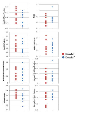 |
Quantitative MRI of diffusely abnormal white matter in multiple sclerosis at 3T
Irene Vavasour, Roger Tam, Shannon Kolind, Robert Carruthers, Anthony Traboulsee, David Li, Cornelia Laule
Diffusely abnormal white matter (DAWM) is found in the brain of some multiple sclerosis (MS) and clinically isolated syndrome (CIS) subjects. DAWM has poorly defined boundaries, with signal intensity higher than normal appearing white matter (NAWM) but not as high as lesions on FLAIR, proton density and T2-weighted MRI. We compared results from myelin water imaging, T1 and diffusion basis spectrum imaging in areas of DAWM and corresponding areas of NAWM in 20 MS/CIS participants. No significant differences in measures sensitive to myelin, axons, oedema and inflammation were found, although trends for increased T1 and reduced fibre fraction were observed.
|
|
1864.
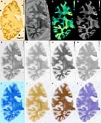 |
Rapid estimation of myelin for diagnostic imaging (REMyDI): A clinical and histopathological validation in multiple sclerosis
Russell Ouellette, Marcel Warntjes, Yngve Forslin, Michael Plattén, Martin Uppman, Åsa Bergendal, Fredrik Piehl, Sten Fredrikson, Maria Kristoffersen-Wiberg, Caterina Mainero, Tobias Granberg
Multiple sclerosis is a chronic inflammatory and neurodegenerative disease characterized by demyelination. To follow patients longitudinally and monitor treatment response, there is a need for robust and tissue-specific imaging biomarkers reflective of the heterogeneous disease course. Here, we aimed to validate REMyDI as an MRI-based measure of myelin ex vivo and in vivo. Histopathologically, REMyDI correlates well with all three of the studied myelin staining methods. In vivo, REMyDI revealed a strong sensitivity in differentiating white matter as compared to normal appearing white matter with associations to both cognitive (information processing speed) and physical disability (Expanded Disability Status Scale).
|
|
1865.
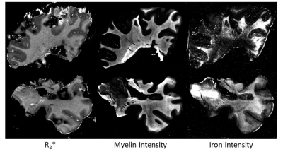 |
Pathological differentiation of multiple sclerosis lesions based on R2* at 3T: The influence of iron and myelin
Christoph Birkl, Vanessa Wiggermann, Verena Endmayr, Enedino Hernandez-Torres, Gregor Kasprian, Romana Hoeftberger, Stefan Ropele, Simon Hametner, Alexander Rauscher
Magnetic-susceptibility sensitive MRI as measure for tissue damage in multiple sclerosis (MS) lesions has been controversial, since the relationship between the MR signal and the underlying pathology is not fully understood. Here we assessed R2* of different white matter MS lesion types and normal appearing white matter (NAWM) in relation to the underlying iron and myelin densities. We observed lower R2* in all MS lesion types compared to NAWM, driven by lower iron and myelin densities. Shadow plaques showed significant higher R2* values than other MS lesions, in line with the hypothesis of remyelination and supported by myelin histology.
|
|
1866.
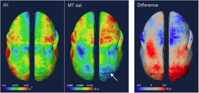 |
A Comparison of R1 and Magnetization Transfer Saturation for Mapping Intracortical Myelin
Cecil Yen, Kimberly Desmond, Afonso Silva, Nicholas Bock
Contrasts based on T1 and R1 (1/T1), including T1/T2-weighted hybrid contrast, have been proposed to map intracortical myelin in the mammalian brain. However, iron in the cortex may obscure changes in myelin investigated by T1-based contrast since T1 is also sensitive to myelin. Here we explore magnetization transfer contrast for mapping ICM, as it may be more specific for myelin. We compare R1 maps measured by MP2RAGE with MTsat measured by MT-FLASH in two marmosets, a species of small non-human primate. Although MTsat shows a similar pattern as R1 in some regions of the cortex, MTsat suffers from signal inhomogeneity issues and care is needed to correct these in future measurement protocols to properly compare R1 and MTsat contrasts.
|
|
1867.
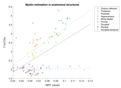 |
Comparison between quantitative magnetization transfer imaging and ratio of T1w/T2w approach in myelin mapping
Yu Sui, Pippa Storey, Alexey Samsonov, Mariana Lazar
Myelination is one of the essential indicators of brain maturation, and various abnormalities in myelin content have been found for different psychiatric disorders. However, reliable imaging techniques for human in vivo myelin measurement are still under intensive research, thus the degree and significance of myelin deficits for specific pathology remain indeterminate. The current study compared myelin mapping proposed as part of the Human Connectome Protocol using the ratio of T1 and T2 weighted image intensity to quantitative magnetization transfer mapping (qMT). The relationship between myelin content estimated by these two methodologies in various brain regions is discussed.
|
|
Neurovascular Imaging Methods
Traditional Poster
Neuro
Tuesday, 19 June 2018
| Exhibition Hall 1868-1902 |
16:15 - 18:15 |
|
1868.
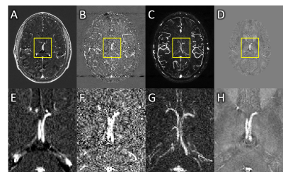 |
Magnetic resonance angiography and venography was not useful for correcting underestimated susceptibility measurements of sub-voxel objects on quantitative susceptibility maps
Natalie Wiseman, Sagar Buch, Yongsheng Chen, E Mark Haacke, Zhifeng Kou
We investigated two magnetic resonance angiography and venography (MRAV) methods for use in correcting quantitative susceptibility mapping (QSM) estimates in sub-voxel veins. An MRAV generated from an interleaved rephased/dephased gradient echo sequence (without contrast agent) suffered from low SNR in veins, whereas the contrast-enhanced T1-MRAV caused the vessels to appear larger than those in the pre-contrast images. Neither method offered a reliable correction of partial-volumed susceptibility measurements.
|
|
1869.
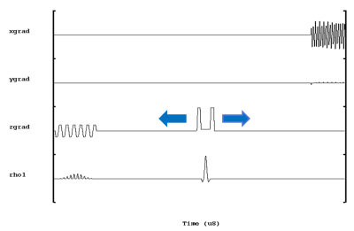 |
Increased cerebral oxygen extraction fraction measured in the ischemic stroke using an asymmetric spin echo EPI approach
Yong Zhang, Qiao Li, Jun Zhang, Bing Wu
The oxygen consumption by brain tissue can be measured with oxygen extraction fraction (OEF), a potential indicator of the occurrence of stroke. A single shot asymmetric spin echo (ASE) EPI sequence was implemented for OEF measurement in stroke patients. Increased OEF corresponded with the decrease blood flow in the ischemic brain region, as reported in the previous literature. ASE EPI showed the potential to provide quantitative OEF maps with good brain coverage and without the need of gas challenges. The measurement of OEF may provide a better assessment of viable brain stroke after a stroke attack for potential treatment.
|
|
1870.
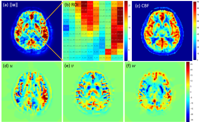 |
Vector Field Perfusion Imaging: A Validation Study by Using Multiphysics Model
Liangdong Zhou, Pascal Spincemaille, Qihao Zhang, Thanh Nguyen, Vincent Doyeux, Sylvie Lorthois, Yi Wang
A multiphysics model based on Navier-Stokes equation and continuity equation is built to simulate the arterial spin labeled (ASL) blood flow in the blood vessels. Blood velocity distribution is reconstructed by measuring the 4D time-resolved labeled blood concentration and doing inversion data fitting processing. The conventional lumped-element Kety’s equation provides a quantitative measurement of whole brain cerebral blood flow (CBF) suffering from the inaccurate estimation of arterial input function (AIF). The multiphysics model validates that the blood velocity involved vector field perfusion (VFP) with multiple post label delays does not rely on the AIF.
|
|
1871.
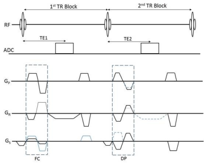 |
A rapid scan for simultaneous MRAV, MRA, tSWI, and QSM on 1.5T
Wei Xu, Yu Wang, Feng Huang, Tie Li, Hongyu Guo, Yongsheng Chen, Ewart Mark Haacke
Numerous diseases such as stroke, arteriovenous malformation (AVM), traumatic brain injury (TBI) and tumor evaluation require detailed vascular information for the best diagnostic interpretation1-4. Being able to collect both MR angiography and venography with sufficient SNR, CNR and co-registration in short time is critical for these diseases, especially for emergency patients. In this work, we developed a rapid 3D interleaved GRE sequence to acquire these vascular images simultaneously. Co-registered MRAV, MRA, QSM and tSWI for imaging arteries, veins and basal ganglia in 4 minutes and 24 seconds on a NMS 1.5T system covering the whole brain with 0.67×1.33 × 2.7 mm3 resolution
|
|
1872.
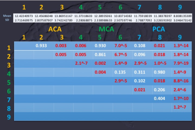 |
Brain Cloud of Carbogen-based Cerebrovascular Reserve : territorial and cortical specificity
Tzu-chen Yeh, Chou-ming Cheng, Chi-che Chou
To explore the spatial characters of carbogen-based cerebrovascular reserve (CO2-CVR), grouped analyses of CO2-CVR was obtained using BOLD-based fMRI for ninety normal subjects with the fully automatic delivery system of carbogens and parametric inhalation of 1-5% CO2. Distal territories of ACA, segment 3, showed the highest of CO2-CVR at v23ab (ventral portion of Brodmann area 23) as verified by territorial and cortical parcellation. Our findings supported the biological adapation of CVR for resting activity, e.g. default mode network.
|
|
1873.
 |
Simultaneous acquisition of T1- and T2-weighted images using Volumetric Isotropic Turbo spin echo Acquisition (VISTA): A feasibility study towards cerebral venous thrombus imaging
Yunduo Li, Shuo Chen, Zechen Zhou, Rui Li, Chun Yuan
This study demonstrated the feasibility of simultaneously acquiring T1 and T2-weighted images using dual-echo VISTA sequence. Phantom experiments showed that dual-echo VISTA can provide T1- and T2-weighted images as conventional T1/2 imaging sequences, and the performance of proposed sequence was further validated by in-vivo scan. By assembling flow-suppression, T1 and T2 contrast in one sequence, dual-echo VISTA has its potential to differentiate stages of thrombus more accurately.
|
|
1874.
 |
A comparative study of arterial spin labeling and CT perfusion on evaluation of cerebral perfusion changes after carotid endarterectomy
Ying Liu, Huimin Xu, Zheng Wang, Huishu Yuan
3D arterial spin labeling (3D ASL) and CT perfusion (CTP) can evaluate the changes of cerebral blood flow(CBF) after carotid endarterectomy(CEA). The aim of this study is to evaluate the changes of CBF after CEA using 3D ASL and CTP respectively, and to compare the consistency of the two methods. Compared with CTP, changes of CBF values obtained by ASL were similar. ASL has similar evaluation results with CTP. As ASL is a noninvasive imaging tool, it has potential to quantitative evaluate hemodynamic changes after CEA.
|
|
1875.
 |
Comparison of PET and MRI estimation of cerebral perfusion using multi parametric PET-MR in a non-human primate model of stroke
Justine DEBATISSE, Nikolaos MAKRIS, Nicolas COSTES, Michael VERSET, Océane WATEAU, Karine PORTIER, Mohamed AGGOUR, Jean-Baptiste LANGLOIS, Christian TOURVIEILLE, Didier LE BARS, Thomas TROALEN, Hugues CONTAMIN, Tae-Hee CHO, Emmanuelle CANET-SOULAS
Reliable estimation of cerebral blood flow (CBF) is crucial for a precise diagnosis of acute ischemia. PET using [15O]H2O remains the reference method to assess CBF but it can also be assessed using MRI. Several post-processing algorithms of perfusion MRI can be used to derive MRI-CBF values. CBF was simultaneously assessed with PET and MRI in a Macaca fascicularis model of stroke using a Siemens PET-MRI hybrid scanner. Four MRI post processing algorithms (sSVD, cSVD, oSVD and Bayesian) were compared against PET estimation of CBF. Bayesian algorithm seems to derive the most reliable estimation of CBF.
|
|
1876.
 |
Standard and Look-Locker FAIR-TrueFISP for arterial spin labelling on mouse at 9.4 T
Michael Gottschalk
The study investigates TrueFISP readout for FAIR either as standard inversion recovery (IR) or as Look-Locker (LL) inversion recovery. These two methods are compared to EPI readout as implemented by Bruker. The aim was to show the improved image quality using TrueFISP and to evaluate the alternatives standard IR and LL. For FAIR-TrueFISP an in-house written method was created. The method was tested on a group of C57BL/6 mice at the field strength of 9.4 T. The results show cerebral blood flow maps with less distortion than EPI and the values found are in agreement with the literature.
|
|
1877.
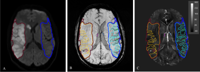 |
QSM in stroke: Veins, Tissue and Cerebral Microbleeds
Parisa Badihi Najafabadi, Ana Klahr, Hongfu Sun, Ahmed Elkady, Derek Emery, Kenneth Butcher, Alan Wilman
We assessed microbleed burden and cerebral oxygenation in veins and tissue in stroke patients by means of Quantitative Susceptibility Mapping (QSM). Results showed significant susceptibility difference for ipsi- and contralateral veins, and smaller area measurements for cerebral microbleeds compared to SWI and magnitude images due to elimination of blooming effects. Strong susceptibility difference of microbleeds compared to other brain tissue suggest the possibility of quantifying microbleeds by thresholding the images. QSM may be employed in stroke studies to study cerebral oxygenation in veins and microbleed assessment.
|
|
1878.
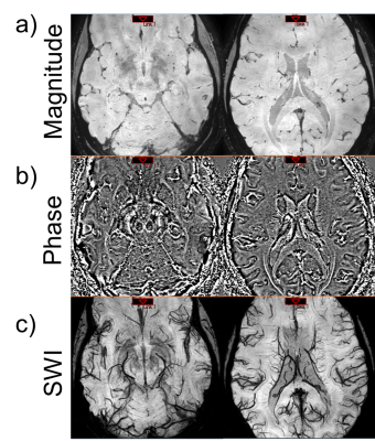 |
Ultra-High resolution SWI at 3T
Harshan Ravi, Wen-Tung Wang, Andrew Knutsen, Dzung Pham, John Butman
Susceptibility weighted imaging (SWI) uses phase and magnitude data to increase the conspicuity of sources such as blood vessels and hemorrhages. Typical resolution used in clinical SWI are approximately 0.5-1 mm in-plane and 1-2 mm through plane. Higher resolution has been achieved using 7.0 T MRI, but such units have limited availability. In this work, we generated ultra-high resolution (400 µm isotropic) SWI at 3.0 T using registration and averaging.
|
|
1879.
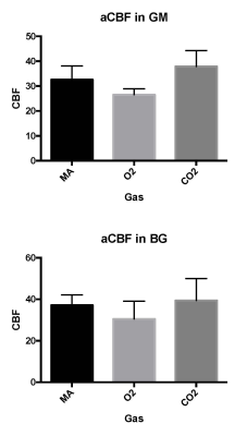 |
Vascular Change Assessed by Calibrated Multi-delay Arterial Spin Labeling Under Oxygen and Carbogen Gas Challenge
Michael Rohan, Clara Wellons, Megan Shevenell, Nicolette Schwarz, Xingfeng Shao, Daniel Wang, Blaise Frederick
Arterial Spin Labeling (ASL) measurements are employed here in a suite of hemodynamic assessments in our study of cerebrovascular reactivity. In this project we test a 3D Gradient and Spin Echo (GRASE) Multiple delay Pseudo-Continuous Arterial Spin Labeling (MPCASL) Magnetic Resonance (MR) acquisition in order to measure the change in these measures under gas challenge. Subjects were scanned with 3D GRASE MPCASL while breathing medical air, oxygen, or Carbogen (5% CO2 +95% O2) under controlled conditions. Changes in blood flow, volume, and arrival time that were observed will be used to calibrate novel delay assessment methods.
|
|
1880.
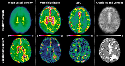 |
Template maps of vascular function and structure in the healthy brain
Endre Grøvik, Kyrre Emblem, Ingrid Digernes, Line Nilsen, Cornelius Eichner, Kourosh Jafari , Thomas Witzel, Behroze Vachha, Elizabeth Gerstner, Jayashree Kalpathy-Cramer, Kawin Setsompop , Steven Stufflebeam
In recent years, Vessel Architectural Imaging (VAI) has emerged as a promising tool in tumor diagnosis to reveal unique MRI-based information on vessel architecture, hemodynamic efficacy and metabolic activity. Healthy control data may further advance our knowledge on the VAI method and its underlying mechanisms, as well as serve as study controls. Here we propose a set of healthy-tissue template maps of all VAI derived parameters which may act as a toolbox to identify anomalies of various vascular brain diseases and ultimately help improve diagnostic and outcome assessment in clinical settings.
|
|
1881.
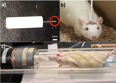 |
Simultaneous measures of brain oxygenation and perfusion using a 9.4T MRI in rats
Kevin Lee, Matthew Bouchard, Sara Bohnert, Jeff Dunn
We developed a novel method to simultaneously measure tissue oxygenation and cerebral blood flow. This technique combines chronically implanted fiber-optic oxygen sensors and continuous arterial spin labeling MRI. An added benefit is that one can measure oxygen while the animals are awake and freely moving.
|
|
1882.
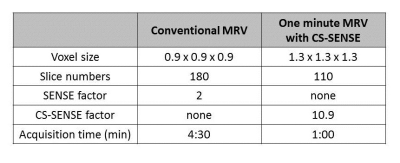 |
One minute Brain MR venography with Compressed SENSE at 3T.
Kayoko Abe, Kazufumi Suzuki, Shuji Sakai
Brain MR venograpy based on phase-contrast technique (MRV) contributes in the diagnosis of venous sinus thrombosis and helps to clarify venous anatomy before brain operations. However, MRV is not commonly taken in routine brain MRI examinations because it requires a longer acquisition time. Recently, Compressed SENSE, which is a combination of compressed sensing and parallel imaging technique: SENSE, has been developed, and can shorten acquisition times with minimum image quality deterioration. Therefore, we investigated the optimization of 1 minute MRV, which the acquisition time was 1 minute, using Compressed SENSE at 3T.
|
|
1883.
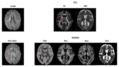 |
Microstructural Characterization of Post-Stroke Lesions in the Posterior Limb of the Internal Capsule in Subacute Patients using DTI and NODDI
Alfonso Mastropietro, Lucia Fontana, Maria Malosio, Laura Straffi, Simona Marcheselli, Marco Grimaldi, Giovanna Rizzo
The purpose of his work was to characterize the Posterior Limb of the Internal Capsule (PLIC) of subacute stroke patients using both DTI and NODDI approaches to investigate microstructural changes occurring in the lesioned with respect to the unlesioned hemisphere. Six patients having a brain damage involving the Corticospinal Tract (CST) were enrolled. MRI was carried out on a 3T scanner about 14 days after stroke occurrence. DTI and NODDI analysis showed CST alterations in subacute stroke patients. FA and ODI were the only parameters that underwent significant modifications in PLIC regions.
|
|
1884.
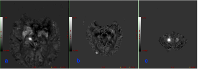 |
Clinical application of QSM sequence in cerebral microbleeds of patients with essential hypertension
Sainuchral Borjigin, Guang-ming Niu, Lizhi Xie
QSM sequence can quantitativeaccess tissue magnetic susceptibility, and applied to understand the distribution of iron content in the cerebral microbleeds. In the study, 3.0T MRI was applied to investigate intracerebral micro-hemorrhage in 33 patients with essential hypertension. QSM sequence was also adminstrated to quantitatively analyze the magnetic susceptibility of CMBs in hypertensive patients. The resultes indicated a significant difference between the magnetic susceptibility of the lesions in the basal ganglia and that of the lesions in the subcortical and infratentorial regions, respectively. Moreover, there was a positive correlation observed between the lesion area and the susceptibility value in each region.
|
|
1885.
 |
Automatic carotid vessel wall assessment based on a combined analysis of TOF-MR angiography and MSD T2-weighted MRI sequences
Lilli Kaufhold, Axel Krafft, Christoph Strecker, Markus Huellebrand, Ute Ludwig, Andreas Harloff, Anja Hennemuth
The quantitative analysis of vessel wall thickness in the carotid bifurcation region based on blackblood MR imaging is a difficult problem because of partial volume effects, strong variations in surrounding tissue contrast, and flow artifacts, which frequently appear in the carotid bulbus. The abstract presents an automatic vessel wall thickness quantification approach based on a segmentation that integrates the information from a TOF-MRA sequence and a MSD-T2-weighted variable flip angle 3D MRI sequence without changing the image data. The method is validated using a crossection-wise comparison with contours corrected by 6 different observers on 60 vessel crossections.
The results show a good overall agreement. Major deviations between observers and automatic segmentation occur in regions with strong artefact.
|
|
1886.
 |
Evaluation of cerebral perfusion changes using arterial spin labeling after carotid endarterectomy
Huimin Xu, Ying Liu, Huishu Yuan
Carotid endarterectomy (CEA) is a common surgical method for patients with significant carotid stenosis. The evaluation of perioperative cerebral perfusion is extremely important to evaluate the efficacy of CEA. It can provide information of both the etiology of stroke due to carotid stenosis and cerebral hemodynamic changes after CEA.[1] Arterial spin labeling (ASL) is a magnetic resonance imaging (MRI) technique that uses the protons of arterial blood water molecules as endogenous tracers to evaluate cerebral blood flow (CBF) noninvasively and repeatedly.[2] Territorial ASL (t-ASL), a modified ASL technique, allows independently labeling a single brain-feeding artery to visualize its cerebral blood perfusion territory. [3] Therefore, we supposed that ASL techniques have ability to provide more information for the evaluation of cerebral perfusion changes pre- and post- CEA.
|
|
1887.
 |
Feasibility of cerebral blood volume mapping by using velocity selective arterial spin labeling with 3D radial gradient echo acquisition
Mulan Jen, James Holmes, Patrick Turski, Kevin Johnson
Theoretically, velocity selective arterial spin labeling (VS-ASL) can be utilized for obtaining cerebral blood volume (CBV). However, challenges such as large vessel contamination and readout dependent artifacts make it difficult to obtain quantitative values. This work investigates the feasibility of VS-ASL CBV mapping by using three-dimensional gradient echo radial acquisition. Measured CBV’s were found to be comparable to previous literature, however results highlight the potential resolution dependence of low SNR ASL based CBV mapping.
|
|
1888.
 |
Quantitative assessment of USPIO uptake in cerebral small vessel disease
Michael Thrippleton, Gordon Blair, Maria Valdes-Hernandez, Andreas Glatz, Iona Hamilton, Fergus Doubal, Ian Marshall, Scott Semple, David Newby, Alex Vesey, Joanna Wardlaw
A method for assessing cerebral blood volume and inflammation in small vessel disease was piloted, employing T1 relaxometry and USPIO contrast agent. 12 stable patients with a history of minor stroke were recruited and scanned pre- and post-contrast, and at 24-30 hours. R1increased following USPIO administration and remained elevated at 24-30 hours; apparent cerebral blood volume did not change significantly in any tissue at 24-30 hours versus post-contrast (p > 0.20). Our work demonstrates the feasibility of T1 relaxometry for quantitative assessment of USPIO distribution but larger studies are required to determine whether detectable inflammatory uptake occurs.
|
|
1889.
 |
Simultaneous depiction of arterial and venous vasculature at high spatial resolution with 3D spoiled gradient multi-echo acquisition at 7T
Hana Hlavata, Mauro Costagli, Janine Lupo, Emiliano Perticaroli, Michela Tosetti, Mirco Cosottini
The simultaneous depiction of both arterial and venous vasculature has recently been demonstrated by using multi-echo sequences. We quantitatively and qualitatively assessed the simultaneous representation of intracranial arteries and veins at a higher resolution than previously reported using a customized 3D spoiled gradient multi-echo sequence at 7T. Such custom sequence had an overall better capability of depicting the arterial vasculature compared to conventional time-of-flight (TOF) arteriography. On the contrary, veins were in general better depicted by conventional susceptibility-weighted venography, however the custom multi-echo sequence provided superior quality images of the superficial veins.
|
|
1890.
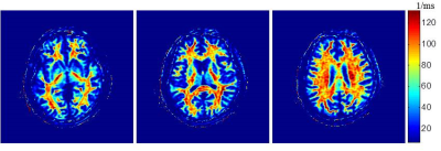 |
Quantitative Inhomogeneous Magnetization Transfer (ihMT) in Acute Stroke: A Preliminary Study
Chien-Yuan Lin, Xiaocheng Wei, Bing Wu, Yen-Chien Wu, Chi-Jen Chen
Inhomogeneous magnetization transfer (ihMT) has been recent developed and has shown promise for myelin-specific imaging. The abnormal lipid pattern in the myelin of the white matter has been observed and could play an important role on ischemic lesion after stroke. The aim of this study was to investigate the myelin change within ischemic lesions using ihMT. In our presentative case, the abnormal area on DWI appears larger than that on ihMT. The difference may result from heterogeneous tissue characteristic in acute ischemic brain, which might evolve with the time after symptom onset and indicate a different clinical outcome.
|
|
1891.
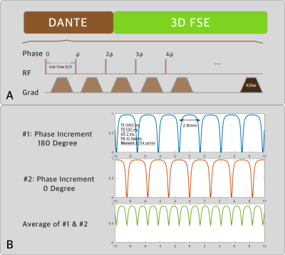 |
Banding free DANTE prepared vessel wall imaging incorporating multiple acquisition and phase cycling
Jianxun Qu, Tianye Lin, Xiaocheng Wei, Bing Wu, Feng Feng
Phase cycling was used to address the banding artefact in DANTE prepared black blood imaging. Simulation, phantom and in-vivo experiment were performed to illustrate and validate the effectiveness
|
|
1892.
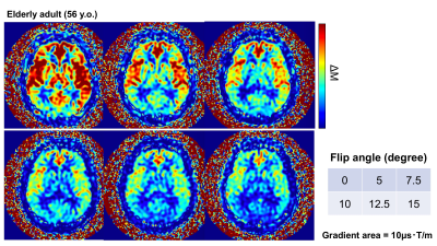 |
Intravascular Signal Suppression and Micro-Vascular Signal Mapping obtained from ASL Perfusion Imaging with DANTE Pulse
Yasuhiro Fujiwara, Hirohiko Kimura, Shota Ishida, Masayuki Kanamoto, Naoyuki Takei, Tsuyoshi Matsuda, R Lebel, Toshiki Adachi
In ASL perfusion imaging, the signal from the label that is still present in larger arteries at the time of imaging causes vascular artifact, which reduces the accuracy of quantification of cerebral blood flow. The purpose of this study is to eliminate the vascular artifacts in larger vessels using the delays alternating with nutation for tailored excitation (DANTE) pulse as vascular crushing gradients and to evaluate the efficiency of the DANTE pulse. The optimized DANTE pulse makes it possible to suppress the vascular signal depending on the flow velocity, which decreased the ASL signal of the arterial region. The relative vascular signal mapping may be helpful to reveal altered hemodynamic state, since the amount of suppressed signal directly associate with flow velocity.
|
|
1893.
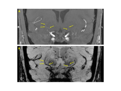 |
Visualizing the Lenticulostriate Arteries at 3T with a Dual-Echo White-Blood and Black-Blood Imaging Technique
M Louis Lauzon
A dual-echo white-blood (WB) and black-blood (BB) imaging technique was developed to visualize the lenticulostriate arteries at 3T. The WB echo, effectively a flow-compensated time-of-flight image, and the flow-sensitized BB echo complement each other such that using these two inherently co-registered echoes in unison helps to better depict and delineate the vessels.
|
|
1894.
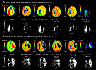 |
Correlation-based temporal similarity mapping of DSC-MRI data in patients with asymptomatic unilateral high-grade carotid stenosis
Mirja Wolf, Stephan Kaczmarz, Jens Göttler, Claus Zimmer, Christian Schwarzbauer, Christine Preibisch
High-grade internal carotid artery stenosis is a widespread cause of ischemic stroke. A recent study proposed an iterative correlation-based image analysis method allowing quick identification of regions with perfusion deficits in dynamic susceptibility contrast magnetic resonance imaging. Here, we evaluate whether correlation-based methods can successfully detect perfusion delay in brain tissue in patients with asymptomatic carotid artery stenosis. In addition, we employed a subtraction method to segment regions of delayed perfusion. Volumes segmented by the subtraction method showed good spatial correspondence with dynamic susceptibility contrast-based time-to-peak maps.
|
|
1895.
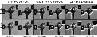 |
A phantom set-up to evaluate slow flow artefacts in vessel wall MRI of intracranial aneurysms
Eva Leemans, Bart Cornelissen, Rebecca Bot, Gerben te Rieg o/g Scholten, Charles Majoie, Bram Coolen, Henk Marquering, Cees Slump
To reliably assess the vessel wall, adequate blood suppression is of high importance. Most black-blood vessel wall MRI sequences rely on flow sensitive signal attenuation. Intra-aneurysmal flow is often chaotic with slower flows near the aneurysm wall. Therefore, certain regions within the aneurysm might be more difficult to suppress. In this study we developed a phantom set-up to evaluate slow flow artefacts in vessel wall MRI of intracranial aneurysms. This setup allows to study the sensitivity of different vessel wall MRI sequences (e.g. DANTE, MSDE, 3D TSE) in relation to specific aneurysm geometries and contrast agent concentrations.
|
|
1896.
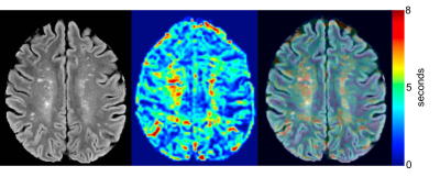 |
Mean Transit Time as a Marker of Vascular Change in Asymptomatic White Matter Disease
Blake Dewey, Xiang Xu, Linda Knutsson, Amod Jog, Jerry Prince, Peter Barker, Peter van Zijl, Paul Nyquist
White matter hyperintensity (WMH) has been associated with cognitive and motor decline. The condition is of presumed vascular origin and may involve decreased blood brain barrier (BBB) integrity. A double contrast injection scheme was used to access both dynamic contrast enhanced (DCE) and dynamic susceptibility contrast (DSC) perfusion-related parameters in an asymptomatic population with high prevalence of WMH. The mean transit time (MTT) was found to be significantly prolonged (5.87, p=0.002) in WMH when compared to normal appearing white matter and that there was no significant change in Ktrans (0.018, p=0.351) between the lesions and the white/gray matter.
|
|
1897.
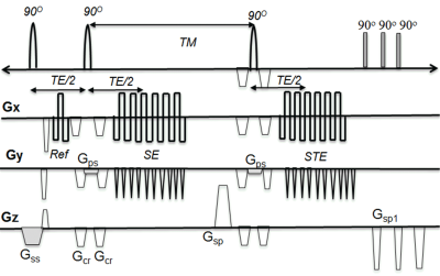 |
3D multi-shot(ms) Spin-Stimulated Echo(STE) EPI sequence Technique for accurate T1 quantification of contrast uptake within vulnerable large artery plaque
Seong-Eun Kim, J Scott McNalley, Adam de Havenon , Dennis Parker, Gerald Treiman
Large artery atherosclerotic disease is one of the most common causes of ischemic stroke. Post-contrast plaque enhancement (PPE), which may result from endothelial dysfunction or be secondary to intraplaque inflammation, is a vulnerable plaque feature that correlates with increased stroke risk independent of stenosis. Although PPE can be detected with vessel wall MRI better quantitative methods to measure PPE are needed. This work presents a new 3D high resolution T1 mapping technique for accurate T1 quantification of contrast uptake within vulnerable large artery plaque.
|
|
1898.
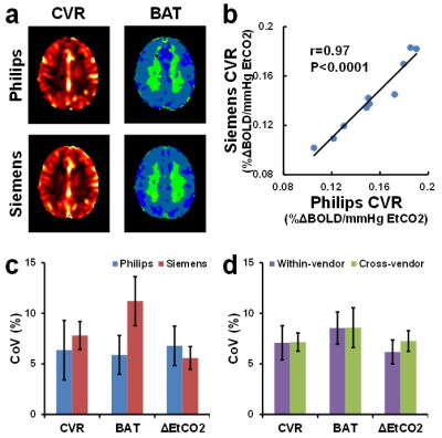 |
Cross-vendor comparison of cerebrovascular reactivity MRI using hypercapnia challenge
Peiying Liu, Dengrong Jiang, Yang Li, Xirui Hou, Jay Pillai, Hanzhang Lu
Cerebrovascular reactivity (CVR) is an important marker of the brain’s vascular health. BOLD MRI with hypercapnia challenge has been shown to be a promising method to measure CVR in various cerebrovascular conditions. To prepare this method for larger-scale multi-site studies, a cross-vendor comparison was performed to evaluate the variability of this CVR mapping method across different scanner platforms. CVR, bolus arrival time and functional connectivity networks were found to be measured reliably from both Philips and Siemens 3T scanners using this method. Although CVR was highly correlated between the two scanners, there was slight difference in CVR values between them.
|
|
1899.
 |
The diagnostic performance of DCE-MRI in glioma grading: A systematic review and meta-analysis
Zhe Liu, Xiang Li, Ting Liang, Tong Bian, Miao Wang, Li Sun, Gang Niu, Jian Yang
Different parameters of Dynamic contrast-enhanced magnetic resonance imaging (DCE-MRI) has been provided for noninvasive evluating gliomas pathology status. But the diagnostic performance of those parameters were variant among the recent reports during different type of gliomas. This study included 17 DCE-MRI studies regarding to differentiating different types of gliomas. The meta-analysis results demonstrated that Ve parameter of DCE-MRI has higher AUC in distinguishing HGGs from LGGs, grade? from grade ? and grade ? from grade?,respectively, Ktrans has higher AUC in distinguishing grade?from grade ?; Among all the pamameters from DCE, Ktrans,Ve,Vp showed higher diagnostic performance in distinguishing different grade of gliomas.
|
|
1900.
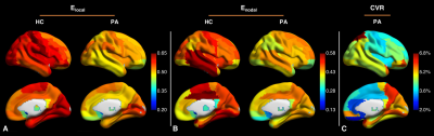 |
Remote effects of hemodynamic impairment on network efficiency in chronic steno-occlusive disease of the anterior circulation: A resting-state functional MRI study
Junjie Wu, Seena Dehkharghani, Fadi Nahab, Jason Allen, Ranliang Hu, Deqiang Qiu
In this abstract we explored remote effects of cerebrovascular hemodynamic impairment on the efficiency of functional connectivity in patients with chronic, anterior circulation steno-occlusive disease. We further evaluated the correlation between network efficiency and cerebrovascular reactivity (CVR), a measure of cerebral hemodynamics.
|
|
1901.
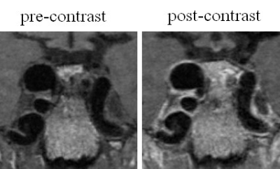 |
Visualizing Wall Enhancement Over Time in Unruptured Intracranial Aneurysms Using 3D Vessel Wall Imaging
Bing Tian, Shahed Toossi, Laura Eisenmenger, Christopher Hess, David Saloner
Advances in vessel wall imaging techniques using high-resolution MR sequences now allow for improved visualization of the walls of intracranial vessels. In this study, we present results obtained with a 3D SPACE to visualize the walls of intracranial aneurysms and to grade the extent of aneurysm wall enhancement in subjects whose aneurysms were monitored over time. Our studies showed that visualization of the aneurysm wall is significantly better on post-contrast images than on pre-contrast images, and the majority of unruptured aneurysms show wall enhancement. Furthermore, we found the wall enhancement scores to remain essentially unchanged on follow up studies.
|
|
1902.
 |
Comparison of Cerebral Blood Flow in a Rat Model of Hypertension and Age-Matched Controls
Abinand Rejimon, Diana Lee, Rebecca McPherson, Mustapha Bouhrara, Akshay Naraine, Kenneth Fishbein, Olga Fedorova, Richard Spencer
Continuous arterial spin labeling (CASL) was used to quantify and compare cerebral blood flow (CBF) in Dahl salt-sensitive (DSS) and Sprague-Dawley (SD) rats. CBF quantification was greatly facilitated through use of the recently-introduced NESMA non-local noise reduction filter. A blunted response to hypercapnia was observed in the DSS rats. These results demonstrate the dysregulation of cerebral vasodilatory responses in hypertension, and may have important implications in the understanding of the vascular basis for cognitive impairment in humans.
|
|
Neurovascular Clinical Studies
Traditional Poster
Neuro
Tuesday, 19 June 2018
| Exhibition Hall 1903-1923 |
16:15 - 18:15 |
|
1903.
 |
Quantitative Susceptibility Mapping analysis of cerebral microbleeds in hypertensive patients
Jinyu Song, Shengzhang Ji, Junjie Ren, Ling Li, Zhizheng ZHUO
Cerebral microbleeds (CMBs) was often found in hypertensive patients.Quantitative susceptibility mapping (QSM) could detect iron-containing lesions with high sensitivity and spatial accuracy in the presence of potentially confounding tissue abnormalities.The results of retrospective study showed that there was significant difference in CMBs between the hypertensive group and the control group.So the conclussion is MR quantitative susceptibility could directly explicate the evolution law of CMBs in hypertensive patients, timely intervention of hypertension could reduce the occurrence of CMBs.
|
|
1904.
|
Coupling of the regional cerebral blood flow and resting state functional connectivity in stroke patients with unilateral middle cerebral artery infarction
jiaxin zeng, yuan xiao, biqiu tang, lu liu, wenjing zhang, jieke liu, su lui
Coupling of rCBF and FC in stroke patients with unilateral middle cerebral artery infarction reveals positive correlation between rCBF and FC, especially in the ipsilateral hemisphere, which indicates improving the CBF in ipsilateral hemisphere in stroke patients.
|
|
1905.
 |
Language reorganization in pre-and post-operative drug refractory extra temporal lobe epilepsy patients: An fMRI based study
Kapil Chaudhary, Senthil Kumaran, Sarat Chandra, Ashima Wadhawan, Manjari Tripathi
Drug refractory epilepsy (DRE) patients have atypical language lateralization with ipsilateral and contra lateral hemispheric lesions and pathological abnormalities. Such kind of patients may have different language recovery after surgery. In this study, we have used a standardized Hindi-language paradigm using semantic, syntactic, judgement and comprehension components for testing in the North-Indian population. We observed greater improvement in language skills in ETLE-patients with correspondingly greater recruitment of the bilateral hemisphere.
|
|
1906.
 |
The value of diffusion tensor imaging (DTI) and tractography (DTT) in lumbar nerve roots display and lumbar disc herniation assessment
Qingwei Song, Shaowei Zheng, Yu Song, Qiang Wei, Bin Xu, Lizhi Xie
This is a prospective study on lumbar disc herniation patient and healthy control with diffusion tensor imaging (DTI) and tractography (DTT). We obtained a high success rate (>90%) of achieving the DTI with tractography of lumbar nerve roots was in this study, and revealed that DTI and DTT technique can both display intensity and morphology changes in the compressed areas of lumbar nerve roots. DTI with tractography provides an abundant diagnostic information with specificity on both qualitative- and quantitative-wise, which is great helpful to assess the disorders with lumbar nerve root compression.
|
|
1907.
 |
The effect of small vessel disease lesions on structural brain network
Xiaopei Xu, Kui Kai Lau, Yuen Kwun Wong, Henry Mak, Queenie Chan, Edward Hui
We aim to explore the influences of cerebral small vessel disease (SVD) lesion on the structural brain network of patients with transient ischemic attack or acute stroke. Our results demonstrated that the efficiency of both global and regional network of patients with SVD were lower compared to those without, and that higher total SVD burden was significantly associated with decreased network efficiency. These results suggested that both presence and severity of SVD related lesion load is associated with disrupted network organization, and brain network analysis is a sensitive method to monitor and assess SVD.
|
|
1908.
 |
7T TOF-MRA showed a decreased contrast-to-noise ratio of the lenticulostriate arteries in hemispheres with unilateral lacunar stroke
Qingle Kong, Haiqiang Qin, Jing An, Yan Zhuo, Zihao Zhang
7T TOF-MRA has demonstrated an exquisite capacity for imaging the lenticulostriate artery (LSA) due to its high spatial resolution and in-flow effect. However, due to the morphological variability of the LSA, a clinical application is needed to identify abnormalities of this vessel. In this study, we analyzed the LSA using 7T TOF-MRA on patients with unilateral lacunar stroke. Comparing the results with other morphological parameters, we found that a reduced contrast-to-noise ratio (CNR) was a more sensitive parameter for reflecting impairment of the LSA on the ipsilateral side of the lacunae.
|
|
1909.
 |
The usefulness of thick slice-basal ganglia rapid pCASL for acute ischemic stroke.
Daisuke Oura, Yoshimasa Niiya, Masahito Kawabori, Shinpei Sato, Kadoya Tomoka, Takumi Yokohama
In this study, we demonstrated the efficacy of the thick slice-basal ganglia pCASL (TB-pCASL) for acute ischemic stroke. The limited scan range and selection of thick slice retain signal noise to ratio (SNR) even in approximately 1min scan. TB-pCASL can rapidly estimate an ischemic region corresponding occlusion-stenosis region, and to combine with DWI can depict penumbra within 2min. TB-pCASL is reliable and useful tool for diagnosis of acute ischemic stroke in the emergency medical field.
|
|
1910.
 |
Imaging Patterns and Implications of Time-of-Flight Magnetic Resonance Angiography in Intracranial Atherosclerotic Stenosis
Jinhao Lyu, Ning Ma, Xiaoxiao Ma, Lin Ma, Xin Lou
The imaging pattern and implication of intracranial atherosclerotic stenosis on TOF MRA had not been fully understood. In patients with middle cerebral artery stenosis, we had used high-resolution vessel wall imaging to evaluate plaque morphology and conventional angiography to evaluate cerebral hemodynamics in groups with different TOF MRA pattern. We had found that the TOF MRA pattern was associated with stenosis percentage, the middle cerebral artery branch signal intensity distal to the site of stenosis on TOF MRA was associated with hemodynamic impairments and was determined by the status of antegrade flow.
|
|
1911.
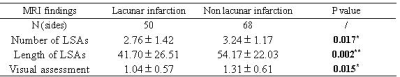 |
Visualization of lenticulostriate arteries by high-resolution vessel wall imaging on a 3T MRI system: a comparison study between subjects with and without lacunar infarction in the basal ganglia region
Weiwei Xie, Tianyi Qian, Jinxia Zhu, Wen Shen, Shuang Xia
The lenticulostriate artery may be associated with lacunar infarction. We aimed to visualize the lenticulostriate artery and explore the correlation between the number and length of lenticulostriate arteries and the number and volume of lacunar infarctions using High-Resolution Vessel Wall Imaging (HR-VWI) on a 3T MR scanner. The results indicated that the length of the lenticulostriate artery was associated with the number of lacunar infarctions. The lenticulostriate artery can be well visualized with HR-VWI, and the length of the artery may be associated with lacunar infarction.
|
|
1912.
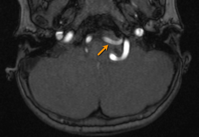 |
Ferumoxytol vascular imaging of the central nervous system in pediatric patients compared to noncontrast MRA: a single center’s initial experience
Josephine Ndolo, Aashim Bhatia
Ferumoxytol-enhanced MRA allows for improved visualization and characterization of vascular pathologies in the brain compared to noncontrast MRA.
|
|
1913.
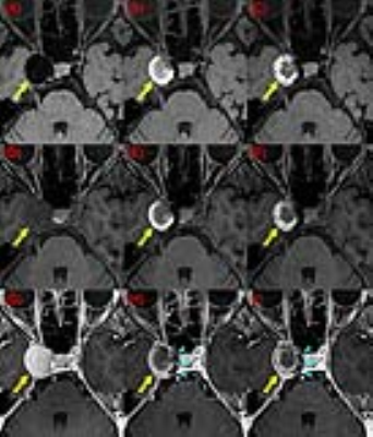 |
Evaluation of Treatment Effect for Saccular Aneurysm by DANTE T1-SPACE
Yasutaka Fushimi, Hidehisa Nishi, Akira Ishii, Tomohisa Okada, Akira Yamamoto, Tsutomu Okada, Takuya Hinoda, Takayuki Yamamoto, Hikaru Fukutomi, Yusuke Yokota, Sonoko Oshima, John Grinstead, Sinyeob Ahn, Kaori Togashi
The purpose of this study is to evaluate the therapeutic effect by FD stent on DANTE T1-SPACE imaging by comparing contrast enhanced 3D T1-weighted imaging. Patients underwent MR imaging for evaluation of pre-, post FD stent placement, and follow-up at 3T MR scanners were included. DANTE T1-SPACE of aneurysm showed dark intensity in pre-treatment study, and higher intensity in follow-up study, then darker intensity later. Enhancement ratio showed high value in pre-treatment study, and low value in follow-up study. DANTE T1-SPACE of aneurysm and enhancement ratio were negatively associated in all patients and exams.
|
|
1914.
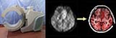 |
Comparison of Image Reconstruction Algorithms of “Flexible PET/MRI” with and without Non-Local Mean Regularization.
Yasutaka Fushimi, Tomohisa Okada, Mizue Suzuki, Takuya Hinoda, Ryusuke Nakamoto, Yuji Nakamoto, Kaori Togashi
Flexible PET (fxPET) is a prototype of MR-compatible mobile PET system. We have compared two different image reconstruction algorithms called as dynamic row-action maximum-likelihood algorithm (DRAMA) and DRAMA with non-local mean (DRAMA-NLM) by evaluating image quality and SUV. NLM filter can reduce artifacts and noise with keeping contrast. The image quality was almost similar between two algorithms and DRAMA-NLM shows significantly higher SUV than DRAMA.
|
|
1915.
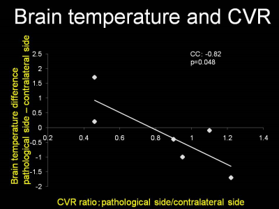 |
Noninvasive measurements of human brain temperature in patients with arteriovenous malformations using magnetic resonance spectroscopy
Takashi Inoue, Tomohisa Ishida, Shunsuke Omodaka, Miki Fujimura, Masayuki Ezura, Hiroshi Uenohara, Teiji Tominaga
The present study investigated whether brain temperature measured by proton magnetic resonance (MR) spectroscopy can detect cerebral hemodynamic impairment in patients with arteriovenous malformations (AVMs) as shown by single photon emission computed tomography (SPECT). Brain temperature, cerebral blood flow, and cerebrovascular reactivity were measured using proton MR spectroscopy and SPECT in five healthy volunteers and six patients with AVMs. A significant correlation was observed between brain temperature difference (affected side - contralateral side) and cerebrovascular reactivity ratio (affected side/contralateral side) (r=0.82, p=0.0480). Brain temperature measured by proton MR spectroscopy can detect cerebral hemodynamic impairment in patients with AVMs.
|
|
1916.
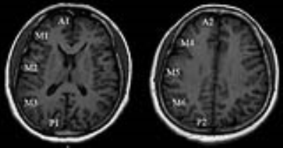 |
Cerebral blood flow in different severity degree moyamoya disease before and after artery bypass surgery
Chuanying Shi, Weidong Liu, Jianxun Qu, Jipeng Wang, Chuanchen Zhang
The goal of the present study was to assess the improvement of CBF after STA-MCA bypass surgery in the mid, moderate, and severe regions based on Tmax value in Moyamoya disease patients. For this purpose, 13 Moyamoya patients were scanned using 3D pc-ASL, and the different perfusion territories were separated based on ASPECT scoring system. The results indicate that mid regions did not get obvious CBF improvement after the surgery and the mid patients did not need to get the bypass surgery.
|
|
1917.
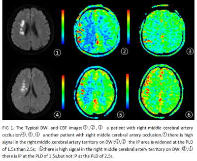 |
The ischemic penumbra assessment using 3D ASL at different post labeling delays in patients with unilateral middle cerebral artery severe stenosis or occlusion
Du Hui, Miao wei
It is necessary to consider the different PLDs to assess IP by 3D pCASL in ischemic cerebrovascular disease.
|
|
1918.
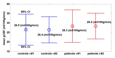 |
Pseudo Continuous ASL for Quantification of Regional Cerebral Hypoperfusion in Chronic Fatigue
Deirdre McGrath, Katija Khan, Annalena Venneri, Iain Wilkinson
In this study pseudo continuous arterial spin labelling (pCASL) was employed to measure cerebral blood flow (CBF) in chronic fatigue syndrome (CFS) patients and healthy volunteers, to determine if CBF was reduced in CFS and in post-exertional malaise. Normalised regional CBF was found to be reduced in CFS for 11 brain regions, predominantly in the left hemisphere, including 8 previously identified regions, along with the left paracentral lobe, and the left and right posterior cingulate. Patients were asked to return for a second scan during post-exertional malaise, in which rCBF was found to be reduced in the left temporal pole.
|
|
1919.
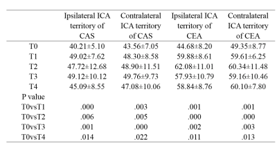 |
Longitudinal assessment of cerebral blood flow change following internal carotid artery revascularization for better prevention of Hyperperfusion syndrome
Yina Lan, Jinhao Lyu, Xiaoxiao Ma, Jianxun Qu, Lin Ma, Xin Lou
Hyperperfusion syndrome (HPS) is a rare but potentially fatal postoperative complication deriving from carotid artery stenting (CAS) and endarterectomy (CEA), while the pattern of post-operation cerebral blood flow (CBF) changes relating to HPS remained unclear. We had used pseudo continuous arterial spin labeling (pCASL) to monitore 4 consecutive time points at 24h, 48h, 72h, and 96h after CEA and CAS in patients with internal carotid artery (ICA) stenosis. We had found that attention should be focused on 72 hours after CAS and 48 hours after CEA to control blood pressure and prevent potential HPS.
|
|
1920.
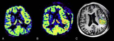 |
Territory Arterial Spin Labeling technique in evaluation of Superficial Temporal Artery to Middle Cerebral Artery Bypass Surgery in Moyamoya Disease
JING YUAN, JIANXUN QU, PEIYI GAO
The purpose of this study was to evaluate cerebral blood flow and territory through superficial temporal artery (STA) to middle cerebral artery (MCA) bypass in patients with Moyamoya disease after direct revascularization surgery using territory arterial spin labeling (tASL) technique. ASL and tASL scan were performed before and after bypassing surgery. our study demonstrated some bypasses can effectively supply blood flow into the brain and others cannot. tASL technique can selectively demonstrate perfusion territory through STA to MCA bypass. Thus, provide information about patency of STA to MCA bypass.
|
|
1921.
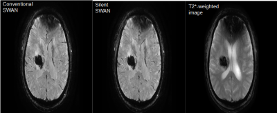 |
Silent Susceptibility Weighted MR Angiography; Clinical and Phantom Study
Takuya Fujiwara, Yoshiyuki Watanabe, Hisashi Tanaka, Hiroto Takahashi, Atsuko Arisawa, Chisato Matsuo, Masahiro Fujiwara, Tetsuya Wakayama, Pauline Worters, Christopher Hardy, Noriyuki Tomiyama
We compared silent susceptibility-weighted angiography (SWAN) with conventional SWAN (cSWAN) in the depiction of hemorrhagic lesions. We measured acoustic noise and performed phantom and clinical study using silent SWAN, cSWAN, and T2*-weighted images (T2*-WI). Acoustic noise of silent SWAN was significantly lower compared to cSWAN. In clinical and phantom study, the contrast-noise ratio (CNR) for silent SWAN and cSWAN were similar. The CNR for T2*-WI was lower than them. In clinical study, imaging quality was almost the same. T2*-WI had more artifact. Conventional SWAN may be replaced with silent SWAN which yields comparable imaging quality and lower acoustic noise.
|
|
1922.
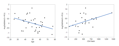 |
Decreased Cerebral Blood Volume among those with Chronic Brain Insult in HIV
Karen Chu, Ke Wei, Thao Tran, Timothy Yao, Kim Shriner, Kevin King
Despite advances in medications and modern practices of immediate antiretroviral therapy, chronic HIV infection remains associated with brain insults, cognitive decline, and related neurological disorders. Reduced N-acetyl-aspartete (NAA), a metabolic marker of neuronal injury, was associated with advanced age and lower CD4 nadir count in a chronic, asymptomatic HIV cohort. Using a novel, BOLD MR protocol incorporating hypercapnic and hyperoxic stimuli, NAA showed no relation to cerebrovascular reactivity (CVR) but was significantly correlated to cerebral blood volume (CBV). Our results may indicate future use of NAA and CBV as complementary non-invasive metrics to track brain health in HIV.
|
|
1923.
 |
Cerebral blood flow in a resuscitated septic shock population: an ASL study
Marie Anne Richard, Marie-Hélène Masse, Frédérick D'Aragon, Charles St-Arnaud, Michael Mayette, Steven Palanchuck, Etienne Croteau, Neil Adhikari, William Fraser, André Carpentier, David Gauthier, Luc Lanthier, Matthieu Touchette, Albert Lamontagne, Jean Chénard, Sangeeta Mehta, Yanick Sansoucy, François Lamontagne, Martin Lepage
Reduced cerebral blood flow (CBF) is often blamed for sepsis-associated encephalopathy. The present study compares the CBF and blood oxygen consumption (CMRO2) of healthy subjects and resuscitated septic patients under vasopressor (norepinephrine) treatment. Methods used are pseudo-continuous arterial spin labeling (PCASL) and T2-relaxation-under-spin-tagging (TRUST). We find that septic patients have elevated global and regional CBF, whereas CMRO2 seems reduced. Further studies are needed to elucidate the underlying mechanisms of this apparent uncoupling.
|
|
Parkinson's Disease
Traditional Poster
Neuro
Tuesday, 19 June 2018
| Exhibition Hall 1924-1947 |
16:15 - 18:15 |
|
1924.
 |
Determination of White Matter Tracts Implicated in Postural Gait Instability Disorder through Tract-Based Automated Analysis
Leon Ooi, Chu Ning Ann, Yun-Chin Hsu, Chen-Hsiang Weng, Ming-Ching Wen, HuiHua Li, Helmut Rumpel, Eng King Tan, Wen-Yih Isaac Teng, Ling Ling Chan
Tract-Based Automated Analysis (TBAA) in Diffusion Tensor Imaging allows for the study of microstructural properties along the tracts in white matter. Diffusivity measures extracted from TBAA for various tracts of the brain were correlated to Tinetti Balance Scale scores in Parkinson's Disease and Postural Gait Instability Disorder patients, allowing identification of tracts of interest in the pathological study of the diseases.
|
|
1925.
 |
The longitudinal changes in white matter of patients with Parkinson's disease as detected by using Fixel-Based Analysis
Shi-Ming Wang, Sung-han Lin, Chin-Song Lu, Yi-Hsin Weng, Yao-Liang Chen, Shu-Hang Ng, Yi-Ming Wu, Chih-Chien Tsai, Jacques-Donald Tournier, Jiun-Jie Wang
Parkinson's disease (PD) is a neurodegenerative disease as the result from the loss of cell in basal ganglia. Fixel-Based Analysis can qualified the fiber density and fibre-bundle cross-section in the white matter. The fiber density and fibre-bundle cross-section is feasible to interpret the microstructure changes in the brain of patients with PD. Therefore, the current study aimed to investigate the long-term white matter changes in Parkinson's disease by using Fixel-Based Analysis.
|
|
1926.
 |
Quantifying nigral degeneration indicates rapid eye movement sleep behavior disorder being a predictor of Parkinson's disease
hiroto takahashi, Yoshiyuki Watanabe, Masahito Mihara, Hideki Mochizuki, Hiroyoshi Adachi , Tian Liu, Yi Wang , Noriyuki Tomiyama
RBD is thought to be prodromal Parkinson's disease (PD), so we aimed to assess the utility of rapid eye movement sleep behavior disorder (RBD) as a predictor of PD using neuromelanin imaging and quantitative susceptibility mapping (QSM). Our results indicated that RBD-related dopamine cell loss and iron deposition in the substantia nigra pars compacta occur in the developmental process of PD. Thus, we conclude that RBD is prodromal PD and quantifying nigral degeneration in RBD is useful in predicting PD.
|
|
1927.
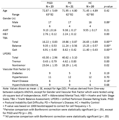 |
Characterizing Neuronal Loss To Differentiate Parkinsonian Subtypes Using Automated Deep Grey Nuclear Volumetry
Chu-Ning Ann*, Bénédicte Maréchal*, Eric Fang, Jie-Xie Lim, Celeste Chen, Julian Gan, Eng-King Tan, Ling-Ling Chan
Postural Instability Gait Disorder (PIGD), a Parkinson's Disease (PD) motor subtype, progresses rapidly with a higher prevalence of neurobehavioural changes. Using automated deep grey nuclear tissue classification combined with atlas-based segmentation, we investigated the performance of resulting estimated lesion load to aid differential diagnosis. Caudate lesion load in PIGD and idiopathic PD subtypes correlated with clinical balance and gait assessment. Combining caudate with abnormal white matter volumetric characterization further improved the discriminative power and could potentially support differential diagnosis of PD.
|
|
1928.
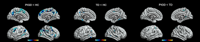 |
Focal Cortical thickness and Subcortical volume changes differ between Parkinson disease subtypes
Ming Huang, Hui Yu
Previous morphometric studies of Parkinson disease (PD) were mainly conducted by measuring gray matter volume and cortical thickness, and little attention has been paid to whether structure MRI improves PD diagnosis or helps differentiating between phenotypes, such as postural instability gait difficulty (PIGD) and tremor dominant (TD). From this study, compared with the control group, PIGD patients had significantly thinning cortical thickness in multiple brain regions, such as bilateral inferiorparietal, paracentral, postiocingulate, superiorfrontal, precuneus, caudalmiddlefrontal, superfrontal and right parsorbitals. TD patients had significantly thinning cortical thickness in left posteriocingulate, inferioparietal and right superiofrontal, superiortemporal, postcentral, precuneus, fusiform and parahippacampal . In addition, subcortical volume atrophy was identified in the bilateral hippocampus and bilateral amygdala of the patients with PIGD, only little bilateral hippocampus changes was found in the TD group.
|
|
1929.
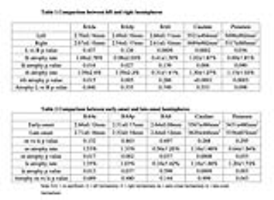 |
Brain morphological changes in early-stage Parkinson’s disease
Lanbo Wang, Xishan Ye, Thyagarajan Subramanian, Qing X Yang, Jianli Wang
At disease onset clinically, the motor symptoms and signs are usually asymmetric or unilateral in majority of Parkinson’s disease (PD) patients. When disease progresses to a later stage, the asymmetry becomes less significant. The cause of this asymmetry, and the relationship between functional deficits and the structural changes in the brain are not clear. In this study, we investigated the morphological changes in the brain hemispheres corresponding to the early-onset and late-onset body sides through a longitudinal study on 24 early-stage PD patients. Significant atrophy was observed in the motor cortex and basal ganglia nuclei.
|
|
1930.
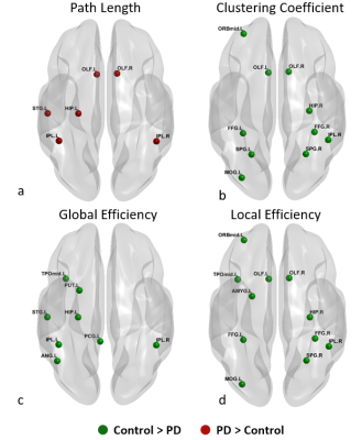 |
Disrupted Functional Connectivity and Network Topology in Early Parkinson’s Disease
Karthik Sreenivasan, Virendra Mishra, Zhengshi Yang, Christopher Bird, Xiaowei Zhuang, Dietmar Cordes, Ryan Walsh
Imaging biomarkers that reliably capture the impact of the spreading pathology of Parkinson’s disease (PD), including its impact on both white and graymatter, remain elusive. In this study, we applied graph-theoretical techniques to multi-site resting-state fMRI data from a cohort of unmedicated early PD-subjects in Parkinson’s Progressive Markers Initiative (PPMI) database. Altered functional connectivity and disrupted topological brain organization was seen in early PD-subjects. Our study opens new avenues to understanding disease progression and severity of PD from graph-theoretical approach.
|
|
1931.
|
Functional brain connectome architecture in a large cohort of Parkinson’s disease patients
Silvia Basaia, Federica Agosta, Homa Zahedmanesh, Tanja Stojkovic, Vladana Markovic, Iva Stankovic, Igor Petrovic, Elka Stefanova, Vladimir Kostic, Massimo Filippi
In this study, we investigated functional neural pathway organization in patients with Parkinson’s disease (PD) using advanced network-based techniques. At the regional network level, compared to controls, PD groups showed decreased functional connectivity within basal ganglia/sensorimotor network and parietal regions. Compared to early PD cases, mild-to-severe PD patients were characterized by a greater involvement of basal ganglia/sensorimotor networks. This study suggests that graph analysis and connectomics might represent a powerful approach to understand the pathophysiological process across different stages of the disease.
|
|
1932.
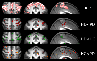 |
Sensorimotor resting-state functional connectivity at 7T: contrasting Huntington's and Parkinson's disease.
Sirius Boessenkool, Stefania Evangelisti, Patrick Pflanz, Stuart Clare, Campbell Le Heron, Johannes Klein, Richard Armstrong, Kinan Muhammed, Andrea Nemeth, Michele Hu, Gwenaelle Douaud
This preliminary study aims to explore high-resolution functional sensorimotor connectivity using resting-state fMRI in healthy controls (HC), Parkinson's (PD) and Huntington's (HD) disease patients. This 7T study therefore includes subjects showing all three states of the basal ganglia inhibitory function. Group ICA and dual regression analyses identified 2 sensorimotor networks: one in which PD and HD showed the same lower cortical connectivity pattern compared with HC in M1 (face area), but opposite pattern in the subthalamic nucleus; and another in which PD and HD showed opposite pattern in M1 and S1 (hand area). This demonstrates the capacity of 7T rs-fMRI to identify with remarkable detail meaningful differences between these two movement disorders.
|
|
1933.
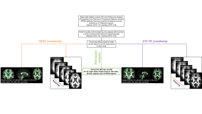 |
Evaluating the sensitivity of univariate and multivariate techniques on diffusion-derived metrics in classification of early Parkinson’s disease patients
Virendra Mishra, Zhengshi Yang, Karthik Sreenivasan, Xiaowei Zhuang, Dietmar Cordes
In this study, we utilized the diffusion MRI (dMRI) data of early Parkinson’s disease (PD) patients and healthy controls (HC) from the Parkinson’s Progressive Markers Initiative (PPMI) database and performed a plethora of multivariate and univariate statistical tests ranging from voxelwise measures, skeleton-wise measures from both TBSS and DTI-TK, and region of interest (ROI) analysis of major white matter tracts from JHU atlas at various smoothing levels. Our study revealed only voxelwise measures could classify HC from PD patients if a minimum smoothing level has reached, and skeleton-wise and ROI analysis (both univariate and multivariate) were associated with the disease.
|
|
1934.
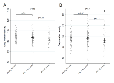 |
Baseline Symptoms and Basal Forebrain Volume Predict Future Psychosis in Early Parkinson Disease
Jamie Blair, Matthew Barrett, Scott Sperling, Mark Smolkin, T. Druzgal
Psychosis is a common neuropsychiatric symptom of Parkinson’s disease, and can serve as a clinical marker of advanced disease. Our study aimed to investigate the characteristics of psychosis in a longitudinal PD cohort, to verify baseline clinical risk factors for future psychotic symptoms in de novo PD patients, and to evaluate the relationship between baseline gray matter density in the nucleus basalis of Meynert and future psychotic symptoms in PD. We found lower NBM density at baseline to be associated with increased psychotic symptom burden compared to controls, suggesting utility for the NBM as a neuroimaging biomarker for advanced PD.
|
|
1935.
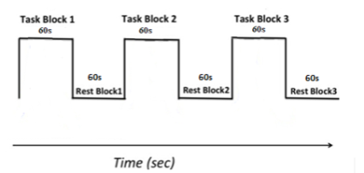 |
Studying the neural correlates of motor fatiguability in controls and people with Parkinson’s Disease
Yue Xing, Saadnah Naidu, Nin Bajaj, Dorothee Auer
Fatiguability, an objective decline in the amplitude of movements during sustained or fast repetitive motor tasks, is one of the primary clinical features demonstrated in Parkinson’s disease (PD). However, our understanding of its underlying pathophysiology is still limited. Here, we propose a fMRI protocol to study the neuronal correlates of fatiguability and present preliminary data in PD and control subjects while performing sustained finger tapping. There was significant reduction of tapping-related activation in the primary motor cortex, somatosensory cortex, premotor cortex and middle frontal gyrus in the fatiguing vs. no-or-less fatiguing subgroups, suggesting that those regions were involved in fatigue.
|
|
1936.
 |
Brain Motor Asymmetry in PD using Positron Emission Tomography and Diffusion Tensor Imaging
Dan Stein, Natalia Goldberg, Liran Domachevsky , Hanna Bernstine, Meital Nidam, David Groshar, Mordechai Lorberboym , Simon Israeli-Korn , Moshe Gomori, Yaniv Assaf, Sharon Hassin-Baer
The accuracy of clinical diagnosis of Parkinson disease is currently not satisfying, particularly in early Parkinson disease where clinical signs are not yet fully present. Imaging nigral structures has been proposed as a biomarker for PD but fails to provide effective differential diagnosis. In this study we compared motor brain regions between hemispheres in patients with asymmetrical motor symptoms using voxel based analysis and network analysis and have found significant regional differences between the more and less affected hemispheres as well as connectivity differences in frontal and cerebral regions as the main hubs.
|
|
1937.
 |
The fronto-parietal connectivity in freezing of gait: a left/right imbalance ?
Céline Tard, Caroline Moreau, Romain Viard, Christine Delmaire, David Devos, Pierre Lenfant, Kathy Dujardin, Luc Defebvre, Arnaud Delval, Renaud Lopes
The multimodal MRI assessment is here used to better understand the previous known parietoprefrontal networks' abnormalities in parkinsonian patients with freezing of gait. Anatomic disconnection was observed in the right prefrontal cortex in those patients and functional disconnection was major from the left one. The imbalance between left and right networks is discussed heyard the pathophysiology of freezing.
|
|
1938.
 |
Validation of a 1.5T FSE NM-sensitive MRI sequence
Joana Grilo, Sofia Reimão, Daisy Abreu, Joaquim Ferreira, Rita Nunes
Neuromelanin(NM)-sensitive MRI is a promising technique for enlightening pathological changes in NM-containing structures. Fast-Spin-Echo (FSE) based NM-MRI sequences have been applied at 3T for improved resolution and signal-to-noise ratio but scanner availability and safety concerns may prevent imaging at this field strength. A 1.5T NM-MRI FSE sequence was developed and compared to the standard 3T NM-MRI sequence. Semi-automatic segmentation of the Substantia Nigra (SN) was performed with good reliability at both fields. The Bland-Altman method was used to compare SN areas between field strengths showing good agreement, supporting the possibility for using NM-MRI at 1.5T, widening its scope of applicability.
|
|
1939.
 |
A Cycling Exercise Study of Parkinson’s Disease: The Effect of Exercises on Motor Cortex Functional Connectivity Revealed by Resting State FMRI
Jian Lin, Katherine Koenig, Erik Beall, Mark Lowe, Amy Jansen, Amanda Penko, Jay Alberts
Parkinson’s disease (PD) is a progressive neurological disorder which produces a general poverty of movement. Lower extremity forced exercise (FE) has been shown to provide therapeutic benefits for PD motor symptoms similar to that of antiparkinson medication1. In the current study, both voluntary exercise (VE) and FE were evaluated. Our results suggest that both modes of aerobic exercise have effects on motor functional connectivity similar to changes associated with antiparkinson medication.
|
|
1940.
 |
BOLD responses to light stimulus frequency in the rat visual pathway reveal profound effects of Parkinson’s disease in the Superior Colliculus
Emmanuelle Bellot, Arnaud Pautrat, Yassamine Rahmani Bouzina, Nora Collomb, Olivier Montigon, Véronique Coizet, Michel Dojat
Sensory disorders are associated with Parkinson Disease (PD) at an early stage. We explored with fMRI the visual pathway response to light stimulus frequency in PD rat models. Activation of the Superior Colliculus (SC) was exacerbated at low frequency (1-3%) and rapidly saturated compared to controls. These results confirm the possible role of SC as an early biomarker of the disease.
|
|
1941.
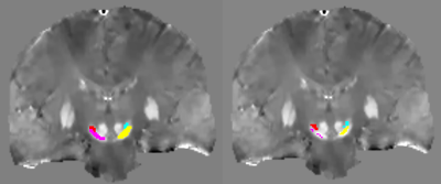 |
QSM versus R2* to study iron deposition in the substantia nigra and subthalamic nucleus in Parkinson’s disease and REM sleep behavior disorders
Mathieu Santin, Nadya Pyatigorskaya, Romain Valabregue, Rahul Gaurav, Lydia Cherif, Sara Fernandez-Vidal, Eric Bardinet, Graziella Mangone, Isabelle Arnulf, Marie Vidailhet, Jean-Christophe Corvol, Stéphane Lehéricy
Here, we compared R2* relaxation rate and QSM to study iron deposition in the substantia nigra (SN) and subthalamic nucleus (STN) in patients with early PD and idiopathic REM sleep behavior disorders with two different segmentation methods. PD patients showed increased iron deposition in the SN and STN as compared with healthy controls with QSM and R2*. iRBD only showed an increase tendency of QSM values compared to healthy controls. Obtained p-values were more systematically lower in QSM than in R2*.
|
|
1942.
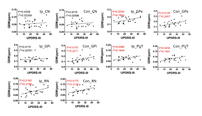 |
Iron Deposition Quantification in Patients with Parkinson’s Disease by Quantitative Susceptibility Mapping
Zhangxuan Hu, Yuhui Xiong, Xuesong Li, Rongsong Zhou, Suhua Miao, Le He, Yu Ma, Hua Guo
Parkinson’s disease (PD) is one of the most common neurodegenerative disorders worldwide. This study explores the relationships between iron accumulation in different nucleus, including red nucleus (RN), caudate nucleus (CN), global pallidus (GP), putamen (PUT), and the severity of PD, which is characterized by the Unified Parkinson’s Disease Rating Scale (UPDRS)-III. Significant bilateral difference was found in RN only. Significant correlations were found in bilateral GPe, PUT, RN, and contralateral GPi, which can serves as an evidence that iron deposition can be an important biomarker for the severity of PD.
|
|
1943.
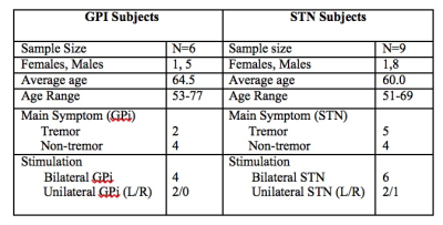 |
Use of Functional MRI to assess the differences of STN and GPI Deep Brain Stimulation in Parkinson Disease
Marisa DiMarzio, Ileana Hancu, Eric Fiveland, Julia Prusik, Radhika Madhavan, Suresh Joel, Michael Gillogly, Jeffery Ashe, Tanweer Rashid, Jennifer Durphy, Roy Hwang, Julie Pilitsis
Deep brain stimulation (DBS) of both the subthalamic nucleus (STN) and globus pallidus interna (GPi) are well-recognized effective treatments for Parkinson’s disease (PD). The mechanism of DBS and network responses produced by stimulation of these targets remains unknown. Conditional labeling of DBS now allows fMRI to be performed in the ON state. We examine whether GPI DBS and STN DBS affect blood oxygen level dependent (BOLD) brain activation/deactivation patterns similarly. Results show that both types of DBS activate the thalamus and deactivate the primary motor cortex; while the STN cohort showed activation in the cerebellum, an opposite effect was apparent in the GPi cohort.
|
|
1944.
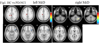 |
Altered marginal division connectivity in Parkinson disease with mild cognitive impairment revealed by resting-state fMRI
Li mingge, Chen yuanyuan, Feng jie, Zhang shiyu, Lou xin, Ma lin
The marginal division (MrD) functional connectivity is disrupted during mild cognitive impairment in Parkinson’s disease.
|
|
1945.
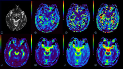 |
Microstructural Changes in Brain Gray Matter Nuclei of Patients with Parkinson's Disease:A Study Based on MR Diffusion Kurtosis Imaging
Qiyuan Sun, Heng Meng, Zhizheng Zhuo
Parkinson's disease is the most common extrapyramidal disease in the elderly people, and the overall prevalence rate is increasing year by year. Diffusion kurtosis imaging (DKI) which was an based on the extension of diffusion tensor imaging (DTI) to reflect the diffusion motion of water molecules in the non-Gaussian distribution between tissues have been proved reliable for the brain microstructural changes. Previous studies have shown that DKI could facilitate the detection of subtle structural changes in the gray matter nuclei of patients with PD, which may be related to the reduction of dopaminergic neurons, iron deposition and gliosis.
|
|
1946.
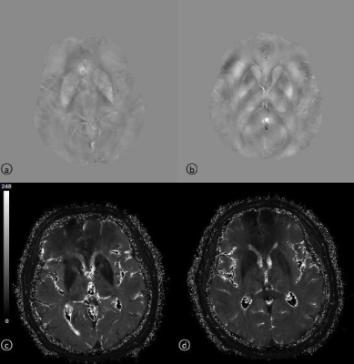 |
Iron Quantification in Brain Gray Matter Nuclei of Patients with Parkinson's Disease: A Study Based on MR Quantitative Susceptibility Mapping
Qiyuan Sun, Heng Meng, Zhizheng Zhuo
Parkinson's disease (PD) is the most common extrapyramidal disease in the elderly people, and the overall prevalence rate is increasing year by year. Quantitative susceptibility mapping (QSM) is based on the basis of susceptibility weighted imaging (SWI), and has more advantages in quantitative detection of brain iron content and display of microstructure. In this study, we tried to use QSM to analyze brain iron variations and microstructural changes in brain gray matter nuclei of patients with PD.
|
|
1947.
|
ROLE OF SUSCEPTIBILITY-WEIGHTED ANGIOGRAPHY (SWAN) QUANTITATIVE MAPPING IN PARKINSON DISEASE DIAGNOSIS
Mariia Rezakova, Khurshed Ibrogimov, Elena Filimonova, Olga Subbotina, Alexandr Shevchenko
We designed SWAN-based algorithm for assessment the pattern of ferromagnetic substances spatial distribution in brain tissue in patients with Parkinson disease. We achieved high diagnostic accuracy in identification of microhemorrhagic changes. In 27 of the 43 patients with PD were observed hemorrhagic lesions in the chronic phase. In the control group, such changes were not observed. In addition, patients with PD had specific localization of lesions (in the epiphysis and vascular plexus).
|
|
Epilepsy
Traditional Poster
Neuro
Tuesday, 19 June 2018
| Exhibition Hall 1948-1960 |
16:15 - 18:15 |
|
1948.
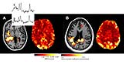 |
Resting state activity is depressed in regions of MRSI identified dysfunction in epilepsy
Jing Huei Lee, Arun Antony, Victor Yushmanov, R. Mark Richardson, Jullie Pan
This study describes co-registered rsfMRI and MRSI data in poorly localized epilepsy patients with the goal of identifying the aberrant epilepsy network. We used 3T rosette encoded spectroscopic image covering the fronto-parietal-temporal brain regions in conjunction with resting fMRI data. The MRSI defined masks of metabolic dysfunction which was then forward warped using Bo maps to define the equivalent regions in the rsfMRI data. The rsfMRI data was analyzed with a model-free evaluation of local connectivity (regional homogeneity). Regions identified by MRSI as metabolically abnormal exhibited lower local rsfMRI coherence in comparison to gray matter or temporal regions.
|
|
1949.
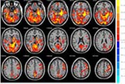 |
Resting-state functional connectivity of hippocampus in patients with drug-resistant idiopathic generalized epilepsy
Zhengge Wang, Lipei Cao, Bing Zhang, Bin Zhu
Previous studies have found altered resting-state functional connectivity in default mode network in drug-resistant patients with idiopathic generalized epilepsy (IGE). Recent studies showed that the volume of the hippocampus is decreased in IGE patients. Hippocampus abnormalities are often related to drug-resistant epilepsy. We investigated the alteration of resting-state functional connectivity of hippocampus in drug-resistant IGE patients by using seed-based functional connectivity and found divergent changes in drug-resistant and drug-sensitive IGE patients. Our findings indicate that the hippocampus and the related network may play an important role in drug-resistant IGE patients.
|
|
1950.
 |
Functional connectivity changes during epileptogenesis: a longitudinal rs-fMRI study
Emma Christiaen, Marie-Gabrielle Goossens, Benedicte Descamps, Paul Boon, Robrecht Raedt, Christian Vanhove
Abnormal functional brain networks could be involved in the development of temporal lobe epilepsy (TLE). In this longitudinal resting-state fMRI study, changes in functional networks during epileptogenesis in the intraperitoneal kainic acid (IPKA) rat model for TLE were mapped. Therefore, resting-state fMRI was acquired at several time points during epileptogenesis to identify functional networks that were analysed and compared with graph theory. Our results suggest that network connections in the functional brain network progressively become weaker during epileptogenesis. We also find a decreased segregation and integration of the network.
|
|
1951.
 |
Whole-Brain connectomics reveals network differences in patients with Non-Lesional Frontal Lobe Epilepsy
Maria Eugenia Caligiuri, Andrea Cherubini, Antonio Gambardella, Angelo Labate
In frontal lobe epilepsy (FLE) seizure onset is usually caused by the presence of lesions or cortical dysplasias of different location and size, challenging the identification of homogeneous samples for neuroimaging studies. However, there are patients in which, even if seizures start in the frontal lobe, no clearly identifiable abnormality can be seen on magnetic resonance imaging (MRI). Thus, it has been hypothesized that non-lesional FLE is indeed a network syndrome, rather the result of focal pathology. In the light of this, probabilistic tractography and graph analysis seem the ideal methodology to investigate the presence and extent of network alterations.
|
|
1952.
 |
MRI and CT derived 3D-printed patient specific brain model for localizing depth elecrodes for epilepsy surgery planning
Sarah Hurrell, Sean Lew, Wade Mueller, Peter LaViolette
We present a method for creating a patient specific, 3D printed model of depth electrode location in an epilepsy patient. We utilized a pre-surgery structural MRI scan and a post-electrode placement CT, which were aligned, and combined to visualize a cortical anatomy and electrode position. 3D models were then generated, edited, and 3D-printed to provide a visual and physical aid for surgical planning.
|
|
1953.
 |
Hemispheric Regional Based Analysis of Diffusion Tensor Imaging and Diffusion Tensor Tractography in Patients with Temporal Lobe Epilepsy
Mahdi Alizadeh, Lauren Kozlowski, Jennifer Muller, Benjamin Trieu, Jonathan Riley, Feroze Mohamed , Ashwini Sharan, Chengyuan Wu
Diffusion tensor imaging and diffusion tensor tractography help to better understand the pathological alterations in white matter structures, and in tracing axonal pathways involved in patients with temporal lobe epilepsy.
|
|
1954.
 |
Automated Hippocampal Subfield Segmentation using Ultrahigh Field MRI in Patients with Epilepsy
Judy Alper, Rebecca Feldman, Long Xie, Alexandru Rus, Lara Marcuse, Madeline Fields, Bradley Delman, Hung-Mo Lin, Patrick Hof, Priti Balchandani
Epilepsy is a widely prevalent, disabling condition, whose anatomical source is not clearly identifiable on clinical MRI scans. Identifying hippocampal subfields associated with epilepsy may elucidate mechanisms of epileptigenesis and assist treatment planning. We performed high-resolution 7T-MRI, enabling precise subfield measurements in thirty patients and matched controls. Greater CA1 and DG asymmetries were found in patients compared to controls. In a subset of mesial-temporal lobe epilepsy patients, we found reduced CA2 on the ipsilateral side in patients compared to controls. Identifying hippocampal subfield biomarkers in epilepsy can result in better treatment planning and monitoring in epilepsy.
|
|
1955.
 |
Comparison between two different post-processing techniques in the presurgical evaluation of Focal Cortical Displasya in a paediatric population.
Elena Bassanelli, Maria Camilla Rossi Espagnet, Nicola Pietrafusa, Luca De Palma, Nicola Specchio, Daniela Longo, Antonio Napolitano
The purpose of this study is to compare two different techniques for cortical dysplasia detection: Opti-MAP and the SUPR-FLAIR. The Opti-MAP is a children-optimized version of the Morphological analysis program (MAP), which is able to detect the “blurred-junction”, peculiar characteristics of focal cortical dysplasia in children, thanks to a voxel-based morphological analysis in which neuroanatomical differences are detected by comparison with a normal template. The SUPR-FLAIR analysis, instead, is a technique able to highlight hyperintensities in FLAIR images. These methods have been applied on paediatric subjects affected by pharmaco-resistant epilepsy.
|
|
1956.
 |
Imaging and involvement of visual pathways in children undergoing epilepsy surgery
Luis Lacerda, Martin Tisdall, Gavin Winston, Sian Handley, Alki Liasis, Chris Clark
Surgery is a key approach for achieving seizure control in children with epilepsy but it can affect or be in the vicinity of the optic radiations. Whilst tractography has shown that damage to optic radiations leads to postoperative visual field defects in adults it has not yet been properly explored in children. In this study we successfully performed tractography reconstructions in a paediatric cohort undergoing surgery. Furthermore, we showed that in cases with pre- and post-surgical visual function assessment, involvement of optic radiations corresponded to visual function disturbances. This highlights the importance of tractography to aid pre-surgical evaluation in children.
|
|
1957.
 |
Case Study: Evaluation of White Matter Disorganization in Temporal Lobe Epilepsy
Laura Barlow, Irene Vavasour, David Li, Martin Parent, Doris Doudet
Temporal lobe epilepsy (TLE) assessment on MRI is limited to qualitative analysis in the clinical environment. Diffusion Tensor Imaging has been used to interrogate white matter changes in TLE while Myelin Water Fraction has not. With this case study we compare diffusion tensor imaging with myelin water imaging in a non-human primate (NHP) with TLE and a healthy control to assess if the two methods are complementary in evaluating white matter disorganization.
|
|
1958.
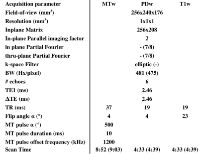 |
Setting up a Multi-centric Multiparametric hMRI Protocol for the Investigation of Temporal Lobe Epilepsy
Paul Summers, Fulvia Palesi, Francesco Padelli, Ileana Zucca, Marcella Malagoli, Carmelo Maccagnano, Stefano Meletti, Giuseppe Didato, Claudia Wheeler-Kingshott, Paolo Vitali
Quantitative characterization of MT, R1, R2*, and PD may aid in providing more consistent readings of alterations in temporal lobe epilepsy. As part of a multi-centric study we have set up a hMRI protocol for use at 3T across two manufactures of MR scanners. Because of differences in MT pulses and SAR calculations, near matching was achieved only through use of commercial or research options. Initial results from one scanner show excellent reproducibility within and between subjects for MT and R1. A cross-scanner evaluation is in course.
|
|
1959.
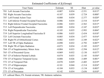 |
Comprehensive assessment of white matter microstructural integrity and its change across lifespan in patients with tuberous sclerosis complex
Tei-Wei Kao, Pi-Chuan Fan, Yung-Chin Hsu, Wen-Yih Tseng
In previous studies, white matter microstructural integrity and its lifetime change in patients with tuberous sclerosis complex (TSC) were not clearly identified. Therefore, we performed diffusion spectrum imaging using whole-brain tract-specific analysis to measure the generalized fractional anisotropy (GFA), and built an age-GFA quadratic linear model to investigate 76 major white matter tract bundles between TSC and healthy control groups. Twenty tract bundles showed a group effect with substantially lower GFA in childhood and older adulthood in patients with TSC. Our results suggest that TSC might pose detrimental effects on microstructural integrity in the developmental and aging periods of life.
|
|
1960.
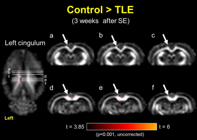 |
Progressive white matter changes in the pilocarpine-induced temporal lobe epilepsy with focal seizure rat model: A diffusion tensor imaging study
Yao-Chia Shih, Chih-Hsien Tseng, Fang-Chia Chang, Horng-Huei Liou, Wen-Yih Tseng
A more suitable pilocarpine rat model with microinjection into the left central nucleus of the amygdala and in-vivo diffusion tensor imaging acquisitions were used to investigate progressive changes in the white matter fibers at three different time points during epileptogenesis in temporal lobe epilepsy (TLE) with focal seizure. We found transient fractional anisotropy (FA) changes in the left fimbria of the hippocampus after status epilepticus and subsequent FA changes in the left cingulum after the presence of spontaneous recurrent seizure. The results demonstrate potential imaging markers for monitoring the progression and development of TLE with focal seizure.
|
|
Head & Neck
Traditional Poster
Neuro
Tuesday, 19 June 2018
| Exhibition Hall 1961-1971 |
16:15 - 18:15 |
|
1961.
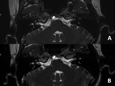 |
Compressed-Sensing Accelerated 3-Dimensional Magnetic Resonance Imaging of Inner Ear: A Feasibility Study of Volunteer
Yuan Jiang, Lina Zhu, Jing Liu, Xiaodong Zhang, Shuai Ma, Yi Liu, Zhiyong Lin, Ke Wang, Zhizheng Zhuo, Xiaoying Wang
Compressed-Sensing (CS) accelerated 3-dimensional magnetic resonance imaging (MRI) does not reduce image quality even with higher image quality scores compared to conventional MRI of inner ear, while significantly shortening the imaging time. It is a feasible protocol in inner ear imaging.
|
|
1962.
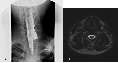 |
Diagnostic Performance of Short MR-neurography Protocol for Brachial Plexus Injuries
Siriwan Piyapittayanan, Natthawut Jarunnarumol, Panai Laohaprasitiporn, Orasa Chawalparit
The purposes of this study were to optimize the protocol of brachial plexus MRN for brachial plexus injuries, and to study the diagnostic performance of the protocol, using clinical contexts as the reference standard. Twenty-one patients with brachial plexus injury were performed brachial plexus MRN (T2-weighted image-high resolution, mDIXON and diffusion weighted image) before conventional myelography. The diagnostic yield of T2-weighted image-high resolution was comparable to conventional myelography. The combination of T2-weighted image-high resolution and mDIXON had the highest diagnostic yield and recommended for the evaluation of brachial plexus injuries.
|
|
1963.
 |
MRI Assessment of SPION Contrast in the Inner Ear
Wendy Oakden, Maya Kuroiwa Rivero, Lola Awofala, Greg Stanisz, Trung Le
A novel approach to diagnostic imaging and treatment of the inner ear disorders is magnetic targeting of therapy using superparamagnetic iron oxide nanoparticles (SPIONs). SPIONs were deposited onto the round window niche using a surgical approach, and then magnetic targeting was used, in the treatment group, to “pull” the SPIONs further into the inner ear. High resolution T2 weighted imaging was used to assess the treatment. Signal loss was observed in the vestibule and cochlea in both groups, while increased signal loss was observed at the apex of the cochlea in treated animals relative to the control group.
|
|
1964.
 |
The feasibility of ultrashort echo time imaging for visualization of sinonasal and skull base bony structures: preliminary study
Miran HAN, Jin Wook Choi, Sungmin Gho
We evaluate the feasibility of ultrashort echo time (UTE) imaging in the visualization of sinonasal and skull base bony structures. MRI with UTE imaging are feasible to assess not only the normal bony structures but also diverse anatomic variations of sinonasal cavity and skull base without radiation exposure. This technique may lead to a new application of diagnostic MRI in head and neck imaging and could be expected to prevent additional CT imaging and consequently reduce radiation exposure.
|
|
1965.
 |
Differentiating Neuromyelitis Optica (NMO)-related and Multiple Sclerosis-related Acute Optic Neuritis using Conventional Magnetic Resonance Imaging Combined with Readout-segmented Echo-planar Diffusion-weighted Imaging
Ping Lu, Yan Sha, Guohong Tian, Xilan Liu, Feng Wang, Zhongshuai Zhang, Yi Sun
In clinical practice, acute optic neuritis (ON) associated with the development of neuromyelitis optica (NMO) after the first attack is often indistinguishable from that associated with multiple sclerosis (MS)1-3; and different therapeutic strategies are required for the two diseases because of their immunopathogenic differences4. Therefore, we aimed to determine the optimal combination of features derived from conventional magnetic resonance imaging (MRI) and diffusion-weighted imaging using readout-segmented echo-planar imaging (RESOLVE-DWI) for the differentiation of the two types of acute ON.
|
|
1966.
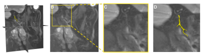 |
Diffusion-prepared magnetic resonance neurography for the visualization of the Facial nerve
Paula Bos, Bas Jasperse, Alfons Balm, Leon ter Beek, Fijs van Leeuwen, Michiel van den Brekel, Regina Beets-Tan, Tessa Buckle
The aim of the study was to investigate the feasibility of Diffusion-prepared MRI (D-prep MRI) to visualize the Facial nerve in head and neck cancer patients. Twenty-four patients (12 male, 60±11 year) received a D-prep MRI, where the main trunk and branches of the Facial nerve is reviewed by one neuro/head and neck radiologists. The main trunk was visible in fifteen patients and in four, six, six and one patients for the posterior auricular, zygomaticofacial, cervicofacial and temporal branches respectively. D-prep MRI is able to visualize the Facial nerve in most cases, but further improvement is required.
|
|
1967.
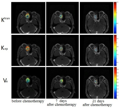 |
Quantitative Dynamic Contrast Enhancement MR Imaging Parameters in the Prediction and Evaluation of the Treatment Response of Malignant Sinonasal Tumors to Chemotherapy
Qing-Hua Chen, Xin-Yan Wang, Jun-Fang Xian, Lizhi Xie
This work assessed the feasibility of quantitative parameters derived from quantitative dynamic contrast enhancement MR imaging (DCE-MRI) parameters in the prediction and evaluation of the response to chemotherapy in patients with malignant sinonasal tumors.
|
|
1968.
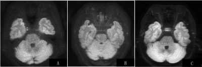 |
One-step high-resolution diffusion weighted imaging in ocular masses and optic nerve using a dedicated surface coil
Qinghua Chen, Zongrui Zhang, Xiaoqi Wang, Fei Yan, Junfang Xian
It is challenging for the routine clinical ocular MRI protocol to use a large FOV covering the whole orbits and sellar region with high spatial resolution relatively. The aim of this study was to evaluate custom-made ocular surface coil in diagnosing images for ocular masses and the optic nerve by comparing TSE DWI images. The dedicated ocular coil obtained large FOV and high spatial resolution images with higher SNR in TSE DW images as examples. The custom-made surface coil can demonstrate ocular masses and the optic nerve more clearly, and provide more details with high SNR in one-step.
|
|
1969.
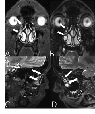 |
Three-dimensional fast spin echo with extended echo train acquisition (3D-FSE-Cube) integrate with two point water-fat separation Dixon methods (Flex): comparison with three-dimensional fast spin echo Cube in lachrymal drainage system imaging
ping liu, jing zhang
The normal membranous lacrimal passage and tear fluid play a very essential role in protecting and lubricating the ocular surface. An ideal lacrimal imaging is very for clinical therapy stratage. The MRI combine fluid is noninvasive and efficient. This study compared the image quality on 3D-FSE-Cube MRD and 3D-FSE-Cube-Flex MRD. The results demonstrated both of the technique has its own advantage. The mutual complementation of each other can fulfill the thorough application of MRI for qualitative images.
|
|
1970.
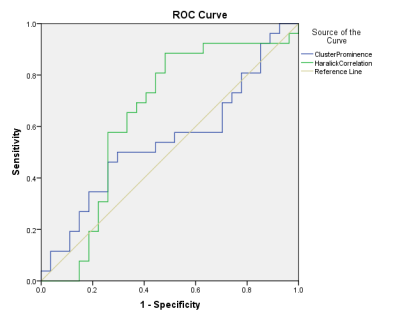 |
MAGNETIC RESONANCE IMAGING TEXTURE ANALYSIS (MRTA) OF NASOPHARYNGEAL CARCINOMA IN T2W AND CE-T1W IMAGES
NAFIR ABDUL JALEEL, LI JUN WANG, YAN WEI MIAO
Nasopharyngeal carcinoma is a common malignant tumour in Asian countries with nearly 80% of them being squamous cell carcinoma. The aim is to investigate the potential of MRI (T2W & CE-T1W) texture analysis to predict response in patients with advanced Nasopharyngeal carcinoma(squamous cell carcinoma).The patients were grouped into Residual/Non-Responders and Non-Residual/Responders based on the post-treatment MR images. Texture analysis was used to find significant parameters. On T2WI, significance were recorded with 2 parameters which showed potential to predict the response to treatment and can be further used in the future studies to predict and alter the treatment course and cycles
|
|
1971.
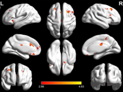 |
Regional cerebral blood flow alterations in patients with comitant exotropia: a pilot 3D-pCASL MRI study
Zhi Wen, Xuefang Lu, Xin Huang, Yang Fan, Yunfei Zha, Baojun Xie
Strabismus is a common eye disease characterized by abnormal eye position and ocular motor disorder. In this study, we compared the cerebral blood flow (CBF) in patients with comitant extropia (CE) relative to healthy controls using 3D-pCASL MRI. We found that CE patients had significantly increased CBF in the right parahippocampal region, bilateral medial FG/ACC, bilateral IFG, left SFG, bilateral MCC, right MFG (BA8), and right paracentral lobule. This study demonstrates the hypothesis that CE involves the dysfunction of visual pathway. Interestingly, the most significant CBF increase in the right parahippocampal region, suggests potential cognitive and mood compensation in CE.
|
|
A Potpourri of Multiple Sclerosis
Traditional Poster
Neuro
Tuesday, 19 June 2018
| Exhibition Hall 1972-1993 |
16:15 - 18:15 |
 |
1972.
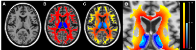 |
Periventricular innate immune cell activation drives tissue damage and clinical progression in multiple sclerosis
Emilie Poirion, Benedetta Bodini, Charline Benoit, Matteo Tonietto, Geraldine Bera, Bruno Stankoff
The objective of this study was to investigate the role of activated microglia in the periventricular damage of patients with MS, combining positron emission tomography with [18F]DPA714 and magnetisation transfert ratio (MTR). Using two-mm thick rings from the ventricular CSF surface to periventricular WM and thalamus, we describe the presence of a gradient of activated microglia together with a gradient of MTR, which correlate with the clinical worsening of patients. These results suggest that an increase of activated microglia and tissue damage might be triggered by the presence of CSF-derived factors, and could mediate the subsequent development of neuro-axonal irreversible damage in MS.
|
|
1973.
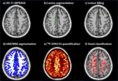 |
Microglia activation in multiple sclerosis lesions drives structural changes over time and correlates with clinical progression
Matteo Tonietto, Charline Benoit, Emilie Poirion, Geraldine Bera, Mattia Veronese, Federico Turkheimer, Marco Battaglini, Benedetta Bodini, Bruno Stankoff
In this study we develop a new method to generate individual maps of activated microglia from 18F-DPA-714 positron emission tomography images and we use it to reproduce in-vivo the histopathological classification of multiple sclerosis white matter lesions. This method allowed us to identify chronically active lesions which are not detectable with standard MRI. These lesions were found to be the most structurally dynamic over time, having a higher chance of enlarging or shrinking after one year. Furthermore, a higher number of active lesions was associated with a more severe clinical progression.
|
|
1974.
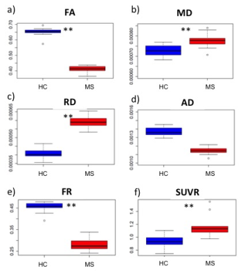 |
Microglial activation is accompanied by diffuse axonal loss in multiple sclerosis: in vivo evidence by multimodal 11C-PBR28 MR-PET and multi-shell diffusion imaging
Elena Herranz, Silvia De Santis , Constantina Treaba, Tobias Granberg, Russell Ouellette, Jacob Sloane, Eric Klawiter, Nicola Toschi, Caterina Mainero
Neuropathological studies of multiple sclerosis (MS) established that diffuse microglia activation with axonal loss in the normal appearing white matter (NAWM) is a main determinant of disease progression. The in vivo study of neuroinflammation and axonal integrity is still challenging. We combined 11C-PBR28 MR-PET with multi-shell diffusion imaging to investigate neuroinflammation and microstructural abnormalities in the NAWM of MS subjects. Results showed evidence of diffuse neuroinflammation accompanied by microstructural diffusion abnormalities with decreased axonal density. The axonal density estimate from the Composite Hindered and Restricted Model of Diffusion was more sensitive than diffusion tensor imaging measures in disclosing axonal damage.
|
|
1975.
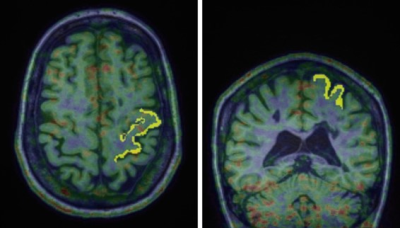 |
Cortical metabolic changes and glial cell activation in multiple sclerosis: An in vivo 11C-PBR28 MR-PET and magnetic resonance spectroscopy study.
Elena Herranz, Constantina Treaba, Eva Ratai, Valeria Barletta, Russell Ouellette, Marco Loggia, Jacob Sloane, Eric Klawiter, Caterina Mainero
We combined 11C-PBR28 imaging on a high resolution, integrated human MR-PET system with magnetic resonance spectroscopy to investigate brain metabolites abnormalities and microglia activation in the motor cortex of multiple sclerosis subjects relative to healthy controls. Our study provided increase of microglia activation and decrease of N-acetylaspartate, the latter indicating neuronal injury and/or loss, in multiple sclerosis compared to controls. None of the other metabolites (choline, myoinositol, glutamine, glutamate, phosphocholine) showed significant differences between the two groups. Also, we did not find a correlation between 11C-PBR28 binding and the metabolites concentration, suggesting that the two measures reflect distinct pathological aspects.
|
|
1976.
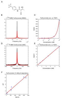 |
19F MR characterization of teriflunomide, a fluorinated drug indicated in Multiple Sclerosis
Christian Prinz, Jason Millward, João dos Santos Periquito, Ludger Starke, Paula Ramos Delgado, Stefanie Muenchberg, Andreas Pohlmann, Thoralf Niendorf, Sonia Waiczies
Teriflunomide is an anti-inflammatory drug indicated for the treatment of Multiple Sclerosis (MS). This disease presents with a wide spectrum of symptoms and available drugs have different effects, thereby posing a major treatment challenge. Due to its three fluorine atoms, teriflunomide can be detected non-invasively by fluorine-19 (19F) magnetic resonance. The objective of this work is to characterize the 19F MR properties of teriflunomide in order to adapt MR sequences for in vivo measurements. Here, we studied the relaxation times of teriflunomide and their modifications as a result of concentration, pH and temperature changes.
|
|
1977.
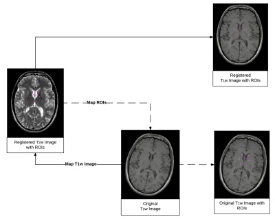 |
Comparison of Two Methods for the Measurement of T1 Hyperintensity in Multiple Sclerosis Patients with Repeated Exposure to Gadolinium-Based Contrast Agents
Megan Hii, Heejun Kang, Megan Le, Andrew Riddehough, Anthony Traboulsee, Shannon Kolind, David Li, Roger Tam
Exposure to gadolinium-based contrast agents is associated with long-term increase in T1 signal intensity in deep grey brain structures, but the measurement methodologies have not been well investigated. We propose marking regions of interest (ROIs) on registered serial T2w images, and compared two methods for measuring the signal changes in the corresponding T1w images: 1) Align the T1w to the T2w images (T2-space), and 2) Map the ROIs marked on the T2w images to the T1w images (T1-space). Applying these methods to frequent and infrequent scanning cohorts, we found signal increase to be associated with GBCA exposure, and T1-space is more sensitive.
|
|
1978.
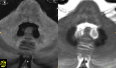 |
Gadolinium retention in the brain – an MRI relaxometry study comparing linear and macrocyclic types of gadolinium based contrast agents
Yngve Forslin, Juha Martola, Sara Shams, Åsa Bergendal, Maria Kristoffersen-Wiberg, Sten Fredrikson, Tobias Granberg
Gadolinium contrast agents (GBCAs) have been shown to be retained in the brain after multiple linear GBCA administrations. We aimed to quantitatively investigate T1 in relation to linear and macrocyclic GBCA-administrations in DN and GP by relaxometry. 80 MS patients who had received different types of GBCAs, were consecutively recruited. This study, in line with previous studies using semi-quantitatively methods, showed that exposure of GBCA leads to shorter T1 relaxation using linear GBCA in comparison to patients who had received macrocyclic types of GBCA, as well as patients without GBCA exposure and healthy controls.
|
|
1979.
 |
An individual radiomics nomogram for differential diagnosis between multiple sclerosis and neuromyelitis optica spectrum disorder
Yaou Liu, Di Dong, Liwen Zhang, Yunyun Duan, Jie Tian, Kuncheng Li
Clinically distinguishing the multiple sclerosis (MS) and neuromyelitis optica spectrum disorder (NMOSD) is critical, since the prognosis and treatment of these disorders differ. We extracted nine radiomics features from 485 radiomics features combining with clinical measurements to build the model for differentiating MS and NMOSD. The area under receiver operating characteristic curve (AUC) of the model was 0.8808 and 0.7115 in the primary and validation cohort. The model demonstrated good calibration. The current study revealed the different radiomics features between MS and NMOSD, and developed and validated an individual model to differentiate the two diseases.
|
|
1980.
 |
Sufficient Gradient Sampling for Diffusion Tensor Imaging in Clinical Trials
Ken Sakaie, Jian Lin, Josef Debbins, Mark Lowe, Robert Fox
Although many diffusion-weighting gradients are desirable for diffusion MRI, implementation may be difficult in a multicenter trial for practical reasons. This study retrospectively examines the adequacy of using as few as 6 directions, the minimum required for calculating the diffusion tensor, for tissue microstructure measurements.
|
|
1981.
 |
Biophysically meaningful MRI features for accurate classification of multiple sclerosis phenotypes
Antonio Ricciardi, Francesco Grussu, Wallace Brownlee, Baris Kanber, Ferran Prados, Sara Collorone, Enrico Kaden, Ahmed Toosy, Sebastien Ourselin, Olga Ciccarelli, Daniel Alexander, Claudia Gandini Wheeler-Kingshott
Quantitative MRI can provide maps of biophysically meaningful features (BMFs) that can be exploited using machine learning techniques to better correlate MR alterations with multiple sclerosis (MS) severity, and improve our understanding of the disease. In this study, a random forest classifier was trained over a rich multi-modal quantitative MRI dataset of healthy controls and MS patients with different phenotypes, to find the BMFs that best characterise disease course. Inflammation and atrophy were the most significant BMFs in distinguishing between controls and patients, with microstructural alterations arising particularly when comparing subjects who only experienced a clinically isolated syndrome with patients and controls.
|
|
1982.
 |
Evolution of functional and structural connectivity of motor network during 2 years of fingolimod therapy for multiple sclerosis
Pallab Bhattacharyya, Robert Fox, Hong Li, Jian Lin, Ken Sakaie, Mark Lowe
Evolution of resting state functional connectivity (fcMRI) between right and left primary motor cortices, and structural connectivity along corticospinal tract (CST) during 2 years of Filgolimod therapy of patients with multiple sclerosis were investigated. MS patients were scanned at baseline (just prior to start of treatment), 6, 12, 18 and 24 months after the start of treatment. Using echoplanar imaging for fcMRI and high angular resolution diffusion imaging for assessing normal appearing white matter integrity along CST, it was found that both functional and structural connectivity damage of motor network stabilized after one year of fingolimod treatment.
|
|
1983.
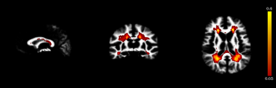 |
Integration of Probabilistic Atlas and Graph Cuts for Automated Segmentation of Multiple Sclerosis lesions
Francesca Galassi, Olivier Commowick, Christian Barillot
We propose a framework for automated segmentation of Multiple Sclerosis (MS) lesions from MR brain images. It integrates a priori tissues and MS lesions information into a Graph-Cuts algorithm for improved segmentation results. We formulate the energy terms to include a priori information as well as the information derived directly from the MR images. We validate our method on a dataset of 37 MS subjects with a broad range of lesion loads. Results indicate that integrating a priori information with the information derived from the images can improve the segmentation outcome.
|
|
1984.
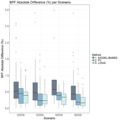 |
Reproducibility Study of a Longitudinal Pipeline for Brain Volumetry based on Partial Volume Estimation
Ricardo Corredor-Jerez, Mário Fartaria, Adrian Tsang, Robert Bermel, Stephen Jones, Izlem Izbudak, Ellen Mowry, Yvonne Lui, Lauren Krupp, Elizabeth Fisher, Tobias Kober, Bénédicte Maréchal
A reliable and accurate quantification of brain tissue loss is important to measure progressive atrophy caused by neurological diseases such as multiple sclerosis. However, accuracy and reproducibility of current methods are often limited by partial volume effects, especially at tissue interfaces where subtle atrophy patterns are likely to occur. We propose a longitudinal pipeline for brain tissue segmentation incorporating partial volume estimation to increase longitudinal robustness. Results show an increase in reproducibility of 44% compared to methods not including partial volume effects in volume estimation, suggesting that these effects should be taken into account for longitudinal atrophy measurements.
|
|
1985.
 |
Diagnostic Accuracy of Semiautomatic T2 Subtraction plus Quantitative Susceptibility Mapping in the Detection of New Multiple Sclerosis Lesions
Shun Zhang, Thanh Nguyen, Yize Zhao, Susan Gauthier, Yi Wang, Ajay Gupta
The ability to identify new MRI lesions in patients with multiple sclerosis (MS) on follow-up imaging is of great importance in monitoring disease activity and informing therapeutic decision-making. Gadolinium (Gd)-enhancing lesions tend to be isointense or slightly hyperintense on QSM images whereas non-enhancing tend to be hyperintense. However, characterization of QSM signal of MS lesions in isolation can be difficult without coregistered T2-weighted imaging. For this reason, we developed an algorithm of T2-subtraction based on two time points of FLAIR images, as well as an automatic lesion mask to help detect new MS lesions with the overall goal of combining this technique with QSM to predict the enhancement status of MS lesions. We found that T2 subtraction+QSM has a sensitivity of 90.9% to predict new enhancing lesions that had been previously identified by experienced neuroradiologists on T1w+Gd imaging. In discriminating between new enhancing versus new but nonenhancing lesions, our T2 subtraction+QSM protocol had a sensitivity of 87.5%, and specificity of 89.7%. Receiver operating characteristic (ROC) curve analysis using region-of-interest of susceptibility values on QSM showed an optimal cutoff susceptibility value of -4.92 ppb (referenced to CSF) in distinguishing new enhancing lesions from new but nonenhancing lesions (sensitivity 88.9%, specificity 80.0%). Our results suggest that T2 subtraction plus QSM no Gd protocol can be a useful tool in detecting the new enhancing MS lesions in clinical practice without Gd injection.
|
|
1986.
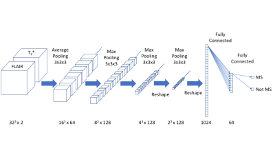 |
Automated Detection of Central Vessel Sign in Multiple Sclerosis using a 3D Deep Convolutional Neural Network
Richard Watts
A 3D deep convolutional neural network (dCNN) was trained to differentiate MS from non-MS lesions based on the orientation and location of a central vein ('central vein sign') relative to the lesion. Excellent performance was achieved using simulated FLAIR and T2*-weighted imaging, with realistic noise levels. The dCNN may be capable of identifying other discriminatory features from multimodal human imaging data.
|
|
1987.
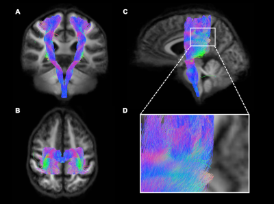 |
The corticospinal tract in relapsing-remitting multiple sclerosis: a preliminary tractography and fixel-based MRI analysis at ultra high-field
Myrte Strik, Camille Shanahan, Stacey Telianidis , Anneke Van der Walt , Rebecca Glarin, Roger Ordidge, Bradford Moffat, Fary Khan, Andisheh Bastani, Eduardo Cofré Lizama, Mary Galea, Trevor Kilpatrick, Jon Cleary, Scott Kolbe
Lower limb disability in multiple sclerosis (MS) is likely related to axonal damage in the corticospinal tract (CST), the main motor pathway. This study aimed to compare the degree of CST degeneration to clinical motor disability using high field (7T) diffusion weighted MRI and subsequent analyses methods like tractography and fixel-based analysis. Eleven minimally impaired relapsing-remitting MS patients (1m/10f, 42±12.4yrs) were tested. Results show loss of fiber density (FD) in the subcortical white matter of the CST was associated with increased pyramidal dysfunction (puncorrected<0.05). FD could provide a useful marker of disease progression leading to loss of mobility.
|
|
1988.
 |
Diffusivity and the neurocognitive domains of premorbid intelligence and visuospatial memory in Pediatric Multiple Sclerosis
Sindhuja Govindarajan, M. Andrea Parra, Tao Wang, Kenneth Wengler, Chuan Huang, Xiang He, Leigh Charvet, Lauren Krupp, Tim Duong
DTI has been commonly used to study multiple sclerosis (MS) patients1-3 and many studies have correlate DTI parameters with neurocognitive functions. However, only a handful of studies4, 5 have characterized such relationships in pediatric MS patients. The goal of this study was to investigate DTI characteristics in pediatric onset MS patients and to correlate them with neurocognitive functions (intelligence and visuospatial memory).
|
|
1989.
 |
Multi-shell diffusion imaging is a sensitive marker for longitudinal axonal degeneration in multiple sclerosis
Nicola Toschi, Silvia De Santis, Tobias Granberg, Russel Ouellette IV, Constantina Treaba, Elena Herranz, Qiuyun Fan, Caterina Mainero
Axonal loss, a crucial pathological process in multiple sclerosis (MS), can be disentangled non-invasively by the CHARMED diffusion model. 8 early MS subjects were scanned at baseline and after 1 year follow-up. At follow-up, TBSS analysis showed statistically significant changes (decrease in FR/FA, increase in MD) compared to baseline in widespread brain regions. The most extensive change was evident in FR, which also showed the greatest sensitivity, especially in areas of fiber-crossing. FR was the only index which detected longitudinal change in axonal density in lesions and therefore holds promise as a biomarker for early diagnosis and disease-monitoring purposes.
|
|
1990.
 |
A new texture-based method for assessing high angular diffusion MRI from patients with multiple sclerosis
Zahra Hosseinpour, Olayinka Oladosu, Wei-qiao Liu, Bruce Pike, Luanne Metz, Yunyan Zhang
The capacity of high angular resolution diffusion MRI to detect subtle pathology in multiple sclerosis (MS) patients can be enhanced when combined with image texture analysis techniques. This study proposes a new voxel-based analysis of diffusion image texture including entropy and angular second moment (ASM, homogeneity), and 45 direction-values per voxel. Results show that while all diffusion maps have differences between lesions and control tissue, both diffusion entropy and ASM maps have better contrast than the classical maps of fractional anisotropy. This new approach may enhance our ability in detecting subtle nerve fiber tract integrity.
|
|
1991.
 |
Multiple Sclerosis Gray Matter Shows Greater Abnormalities in Phosphate Metabolites than White Matter
Manoj Sammi, Yosef Berlow, Randy West, Katherine Powers, Vijayshree Yadav, Dennis Bourdette, Rebecca Spain, William Rooney
Phosphate metabolite distribution in gray matter and white matter in human brain is compared between healthy control (HC) and subjects with Multiple Sclerosis (MS) using 31P Magnetic Resonance Spectroscopic Imaging (MRSI) at 7T. Phosphate metabolites are decreased in GM in MS compared to healthy controls.
|
|
1992.
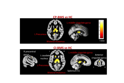 |
Gray Matter Atrophy and Microstructural White Matter Abnormalities Underlying Cognitive Impairment in Benign MS
Elisabetta Pagani, Gianna Riccitelli, Marta Radaelli, Paolo Preziosa, Giancarlo Comi, Andrea Falini, Massimo Filippi, Maria Rocca
The definition of benign multiple sclerosis (BMS) is based on long disease duration and low level of disability, without considering cognitive deficits. Aim of the study was to apply voxel-wise methods to investigate whether cognitive dysfunction in BMS patients is associated with specific patterns of regional damage in the brain gray matter (GM) and white matter (WM). High-resolution T1-weighted and diffusion tensor MRI scans were acquired from 50 healthy controls and 38 BMS patients, 42% of which were classified as cognitively impaired. Distinct regional patterns of abnormalities, functionally relevant for cognitive processing, were associated with cognitive impairment in BMS patients.
|
|
1993.
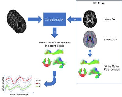 |
Clustering of Multiple Sclerosis Patient's Clinical Courses with White Matter Fiber-Bundle Profiles
Gabriel Kocevar, Claudio Stamile, François Cotton, Françoise Durand-Dubief, Dominique Sappey-Marinier
Predicting the individual multiple sclerosis (MS) patients evolution, based on markers available from disease onset, may help the neurologist in the patient care. However, such a prediction remains a challenge. In this study, we merged spatial information of fiber tracking with diffusivity metrics, measured in 68 patients presenting the three forms of MS, in order to classify patients using a white matter fiber-bundle profile analysis. The good performances of the clustering, reached with fractional anisotropy and mean diffusivity together, make our method a potential tool to better predict the disease evolution, especially the conversion of RR-MS to SP-MS.
|
|
Alzheimer's Disease & Other Dementias
Traditional Poster
Neuro
Tuesday, 19 June 2018
| Exhibition Hall 1994-2034 |
16:15 - 18:15 |
|
1994.
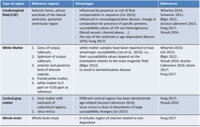 |
Test-retest reproducibility of quantitative susceptibility mapping in a multi-site study on Alzheimer disease: effect of reference region of interest choice
Anna Nigri, Giovanni Giulietti, Cristina Muscio, Giovanni Frisoni, Maria Bruzzone, Marco Bozzali, Daniela Perani, Pietro Tiraboschi, Claudia Ambrosi, Massimo Caulo, Pietro Chiarini, Elena Chipi, Stefano Chiti, Enrico Fainardi, Stefania Ferraro, Cristina Festari, Roberto Gasparotti, Ruben Gianeri, Andrea Ginestroni, Andrea Ginestroni, Lorella Mascaro, Riccardo Navarra, Lucilla Parnetti, Alberto Redolfi, Laura Serra, Roberto Tarducci, Fabrizio Tagliavini, Jorge Jovicich
Quantitative susceptibility mapping (QSM) is a neuroimaging marker of iron of interest as biomarker of Alzheimer disease (AD) progression. The purpose of this work was to assess the test-retest reproducibility of QSM in the Italian AD-NET project, a multi-site study on AD. We evaluated how QSM reproducibility is affected by acquisition (different clinical 3T MRI sites using vendor provided sequences) and analyses factors (choice of the reference region of interest used to compute within-subject relative QSM). The goal is to minimize reproducibility errors and thus maximize the sensitivity to detect longitudinal QSM changes related to the progression of AD.
|
|
1995.
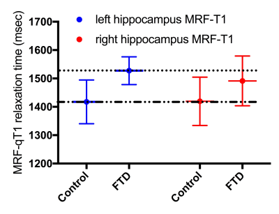 |
Quantitative T1 and T2 mapping with standard and MR fingerprinting techniques to assess frontotemporal dementia: A pilot study
Stilyana Bakoeva, Vera Keil, Alina Jurcoane, Mariya Doneva, Thomas Amthor, Peter Koken, Burkhard Mädler, Wolfgang Block, Klaus Fließbach, Elke Hattingen
This pilot study explored the utility of MR fingerprinting (MRF) and standard T1 and T2 relaxometry to discover focal anomalies in patients with the primary progressive aphasic form of frontotemporal dementia (FTD). MRF and standard techniques revealed longer T1 and T2 relaxation times of cortex and deep white matter as well as the hippocampus (left>right) in FTD compared to healthy controls. Relaxation times between MRF and standard differed and not all techniques revealed all structures as altered in relaxation time. In conclusion, MRF and standard relaxometry have the potential to quantify brain anomalies in FTD, which may be used for diagnosis and monitoring, but are not interchangeable.
|
|
1996.
 |
Detection of Alzheimer’s Disease Patients Based on Single Brain Region by Convolution Neural Networks
Yanwu Yang, Heather Ma, Chenfei Ye, Junjie Liu, Chushu Yang
Diagnosis plays an important role in preventing progress and treating the Alzheimer’s disease (AD). This paper proposed to predict the AD with a convolutional neural network (CNN), which can learn generic features capturing AD biomarkers. In particular, we extract some specific brain regions from structural MRI and apply MR features from the brain regions to detect AD patients in CNN framework, achieving accuracy up to 99% and outperforming some other classifiers from other studies.
|
|
1997.
 |
Structural and Functional Networks of Emotion Regulation Are Altered in Subjective Cognitive Decline
Pin-Yu Chen, Yung-Chin Hsu, Yu-Chen Wei, Yu-Ling Chang, Ming-Jang Chiu, Wen-Yih Tseng
Previous research suggested that subjective cognitive decline (SCD) may also present with other psychiatric diseases, personal traits, physical conditions and medication use. We hypothesized the depressive trait was the early factor and its neural correlates of function or structure changes may reflect such mental feature. We investigated the functional and structural connectivity of the emotion regulation network in SCD. We found that left amygdala to left IFG especially displayed both functional and structure changes. Our study suggests that the altered patterns of the emotion regulation network could serve as the neural basis of the emotion regulation function and display the depressive trait is the critical risk factor for SCD progression to memory disease.
|
|
1998.
 |
Riluzole improved Energy Metabolism in AßPP-PS1 Mouse Model of Alzheimer’s Disease
Anant Patel, Kamal Saba
Alzheimer’s disease (AD) is a neurodegenerative disorder, characterized by degeneration of neurons leading to memory loss, deterioration in cognitive function and behavior. Despite intensive research of several decades treatment of AD is still a major challenge. Riluzole is known to be neuro-protector and regulates the function of glutamatergic neurons by reducing glutamate release and helping astroglial uptake. In this study, we have evaluated the impacts of Riluzole on the neuronal activity in the AβPP-PS1 mouse model of the AD by 1H-[13C]-NMR spectroscopy together with infusion of [1,6-13C2]glucose. The finding of improved neurometabolism in AD mice suggests riluzole improved cognitive function in Alzheimer's disease.
|
|
1999.
 |
Influence of Different Anesthesia Protocols on Cerebrovascular Reactivity and Cerebral Blood Flow measured by Pseudo-continuous Arterial Spin Labeling
Marc Derieppe, Leon Munting, Ernst Suidgeest, Louise van der Weerd
Anesthesia protocols in animal studies greatly influence cerebral hemodynamics, so it is critical to devise standardized protocols in order to provide reproducible and comparable Cerebral Blood Flow (CBF) and Cerebral Vascular Reactivity (CVR) in different mouse strains or models. We compared strain-dependent sensitivity towards different anesthesia protocols for vascular reactivity experiments (high-dose isoflurane, medetomidine, low-induction dose isoflurane and high-dose isoflurane in intubated and mechanically ventilated mice), using pseudo-Continuous ASL (pCASL) and discuss the relative performance of these protocols.
|
|
2000.
 |
Quantification of Perfusion Asymmetries in MCI Subjects using Arterial Spin Labeling MR imaging
Li Liang, Heather Ma, ChenFei Ye, Susumu Mori
In this study, we recruited seven subjects with MCI and ten subjects as cognitive normal groups. All subjects underwent PASL and T1-weighted MR imaging. Multimodal images were upload to MRICloud for segmentation and quantification of regional CBF. T-test was used to detect significant changes in brain region volumes and perfusion asymmetries in contralateral regions. We observed tendencies of atrophy in right posterior cingulate cortex and dilation in bilateral fornix, significant perfusion asymmetries were found in regions of temporal lobe, basal ganglia and posterior cingulate cortex among MCI subjects.
|
|
2001.
 |
Changes in hippocampal and whole brain stiffness in 14-month old female mice with Alzheimer’s disease
Miklos Palotai, Katharina Schregel, Navid Nazari, Julie Merchant, Walter Taylor, Charles Guttmann, Ralph Sinkus, Tracy Young-Pearse, Samuel Patz
Alzheimer’s disease (AD) has been associated with human brain softening, but the underlying biomechanical mechanism is not fully elucidated. We used magnetic resonance elastography to investigate the effect of amyloid-beta accumulation on hippocampal and whole brain (WB) stiffness in transgenic AD and wild-type (WT) mice at 11 and 14 months of age. The only differences observed between AD and WT mice were that the longitudinal change in the loss modulus between 11 and 14 months for female AD mice was significantly different than that of either the WT or male AD mice.
|
|
2002.
 |
A combined dual-tracer PET/diffusion tractometry analysis of the posterior cingulum in a mild cognitive impairment ketogenic intervention
Maggie Roy, Stephen Cunnane, Étienne Croteau, Alexandre Castellano, Mélanie Fortier, Félix Morency, Jean-Christophe Houde, Maxime Descoteaux
In mild cognitive impairment (MCI), posterior cingulate cortex glucose hypometabolism may results from posterior cingulum (PCg) alterations. We suggest that raising ketone availability to the brain may overcome the brain energy deficit. We developed a dual-tracer PET/dMRI tractometry method to assess whether a ketogenic supplement has impact on fuel uptake in the PCg of MCI participants. Mean fuel uptake in the PCg was unchanged post-supplementation, but tract-profiling enabled the identification of sections with lower glucose uptake. Energy supply in white matter fascicles is crucial to sustain adequate axonal function and may be linked to the pathogenesis of MCI.
|
|
2003.
 |
Unbalanced large-scale brain networks during static and dynamic states in Alzheimer’s Disease
Xiaoqing Ji, Haiyang Geng, Rui Li, Le He, Chun Yuan
In this study, we applied Independent Component Analysis (ICA) and dynamic network approaches to explore the neural network mechanisms between Alzheimer’s disease (AD) patients and normal aging healthy controls (HC) from distinct brain states. We conducted rs-fMRI scanning on 12 ADs and 12 HCs. From ICA, we got three networks including DAN, VAN and DMN. From dynamic network analysis, we achieved three dynamic states. Two sample t-test results showed that, in AD, DAN had weaker connectivity, DMN had no difference both in static and dynamic states, VAN only had increased connectivity between IFG and other regions in static state.
|
|
2004.
 |
Prediction of Cognitive Impairment and Amyloid Deposition through Metabolic and Vascular Deficits in ADNI Cohorts
David Ma, Ai-Ling Lin
Recent research has been focused on developing diagnostics based on amyloid-β and tau. However, metabolic and vascular changes pre-date both by several decades. The aim of this study was to exploit the coupling between glucose uptake in aerobic glycolysis and cerebral blood flow to produce a biomarker for metabolic dysfunction and amyloid-β deposition. Here we found that a decrease in glucose uptake in the presence of stable blood flow is spatially correlated with an increase in amyloid-β deposition and that uncoupling between metabolic and vascular function could drive amyloid-β deposition.
|
|
2005.
 |
MRI and PET alterations in Alzheimer’s disease and cognitive normal HFEH63D polymorphism carriers
Carson Purnell, Qing Yang, James Connor, Mark Meadowcroft
Anatomical MRI and PET data from the genetic cohort of the ADNI database was analyzed for MRI volumetric, FDG-PET, and AV-45 differences between HFEH63D polymorphism and HFEWT carriers. A decrease in the amount of AV-45 amyloid binding was observed in the AD HFEH63D carriers as well as an increase in FDG metabolism and a decrease in regional brain volume. HFEH63D appears to be preservative in AD with respect to PET imaging biomarkers, but there was a negative interaction in the VBM analysis. This reinforces the hypothesis that HFEH63D has a preservative effect in AD.
|
|
2006.
 |
Regional brain iron accumulation in an Alzheimer’s mouse model fed lipophilic iron
Douglas Peters, Carson Purnell, Qing Yang, James Connor, Mark Meadowcroft
Alzheimer’s disease (AD) is a progressive neurodegenerative disorder characterized pathologically by amyloid beta (Aβ) deposition, microgliosis, and iron dyshomeostasis. The goal of this work was to observe how brain iron levels temporally influence Aβ plaque formation, plaque iron concentration, and microgliosis. Humanized APPNL-G-F knock-in and control mice were fed either lipophilic iron compound 3,5,5-trimethylhexanoyl ferrocene (TMHF), normal, or iron deficient diets for twelve months. Increased brain iron was observed in the olfactory, frontal and hippocampal regions and was associated with increased plaque-iron loading and microglial iron inclusions.
|
|
2007.
 |
Sex Differences in Behavior, Brain Structure and Functional Connectivity in the APOE Epsilon 4 Knock-In Rat Model of Alzheimer's Disease: Are Females the Stronger Sex?
Praveen Kulkarni, Dan Madularu, Thomas Morrison, Craig Ferris
APOE genotypes are a major focus for Alzheimer’s disease (AD) research following the localization of ApoE on neurofibrillary tangles and amyloids of senile plaques AD patients’ brain. The risk of developing AD increases with the frequency of the ε4 allele, with women outnumbering men. In this study we utilized multiple imaging modalities and behavioral assays to identify sex-specific anatomical biomarkers in a novel rat APOE-ε4 knock-in model. ε4+ males show greater variation in neural structure and function in terms of the proportion of brain areas affected; these results are reflected in sex-driven differences in behavior mirroring hippocampal function.
|
|
2008.
 |
Investigation of cerebral perfusion differences between the 2xTg Alzheimer’s disease mouse model and age-matched controls using FAIR ASL MRI
Diana Lee, Abinand Rejimon, Rebecca McPherson, Kenneth Fishbein, Mustapha Bouhrara, Simonetta Camandola, Mark Mattson, Edward Lakatta, Olga Fedorova, Richard Spencer
Cerebral blood flow (CBF) is an emerging biomarker of Alzheimer’s disease (AD). To correlate CBF to other known measures of AD, such as cortical thinning and volume loss, we assessed perfusion differences between a 2xTg-AD mouse model and age-matched wild-type mice using a FAIR RARE MRI sequence. Our results demonstrate greater systolic blood pressure (SBP) in AD mice as they age as well as hypoperfusion within the cerebral cortex at 12 months of age.
|
|
2009.
 |
Quantitative Susceptibility Mapping to evaluate the Iron deposition and Venous Blood Oxygenation in the brain for the differentiation of Mild Cognitive Impairment and Alzheimer’s disease: A pilot study
Sheelakumari R, Bejoy Thomas, Ramshekhar Menon, Ramesh Venkatesan, Chandrasekharan Kesavadas
Oxygen extraction fraction (OEF) in the brain can be obtained by Quantitative Susceptibility Mapping (QSM). QSM can also estimate iron which is implicated in the pathogenesis of Alzheimer’s disease and its pre-symptomatic antecedents. The differences of OEF and iron values among the controls, Mild Cognitive Impairment and AD were investigated by ROI based comparisons using a one-way analysis of variance. These values in the posterior brain regions were found to have a trend towards increment in patient groups compared with controls, in this pilot study. Future studies are required to validate the usefulness of this technique as potential biomarker.
|
|
2010.
 |
APOE e4 Allele Effect on White Matter Perfusion and Diffusion in Cognitively Normal and MCI Groups
Youngkyoo Jung, Jeongchul Kim, Megan Johnston, Christopher Whitlow, Laura Baker, Suzanne Craft
Hypo-perfusion was observed among APOE ε4 carriers in both white and gray matter from the previous study in cognitively normal and mild cognitive impairment groups. Diffusion tensor imaging metrics in the white matter was further examined in the hypo-perfusion region and compared with perfusion metrics. Multiple statistical trends match with the observations from the perfusion metrics, which may suggest evidence of that a perfusion abnormality among APOE ε4 carriers may precedes the disruption of white matter integrity in the group.
|
|
2011.
 |
Effect of Antiepileptic Treatment on Hippocampal Activity in Alzheimer’s Disease measured by ASL
Weiying Dai, Song Chen, Li Zhao, David Alsop, Daniel Press
Increased hippocampal perfusion in early AD has been reported, but the underlying mechanism is still not clear. We hypothesized that epileptiform activity occurs in the hippocampus with AD and causes increased perfusion. Here, we designed a placebo-controlled study using an antiepileptic drug, Levetiracetam to modulate epileptic activity of the hippocampus. Nine subjects with AD were scanned following drug or placebo. We observed decreased perfusion and increased perfusion fluctuation in entorhinal cortex with Levetiracetam. These findings support the potential epileptic activity effects of entorhinal cortex in AD. Due to neighboring locations of hippocampus and entorhinal cortex, further work will probe the effects of potential misregistration.
|
|
2012.
 |
MR Spectroscopy in a Transgenic Rat Model of Alzheimer’s Disease
Wendy Oakden, Christina Beckett, Bojana Stephanovic, Greg Stanisz, JoAnne McLaurin
The transgenic rat model of Alzheimer’s Disease (AD), TgF344-AD rats, manifests a more complete spectrum of age-dependent AD pathologies in conjunction with cognitive disturbance. Importantly, TgF344-AD rats exhibit amyloid and tau pathology as well as frank neuronal loss with aging. This study investigates brain metabolic changes, using magnetic resonance spectroscopy, in older TgF344-AD animals relative to younger and to non-transgenic littermate rats. Our data shows a statistically significant decrease in phosphocreatine, glutamate, and taurine, and a trend towards decreased NAA (p=0.053) in comparison to the combined younger and non-transgenic littermate rats.
|
|
2013.
 |
Understanding the Role of Gender in Progression and Severity of Alzheimer ’s Disease: A 1H-[13C]-NMR investigation
Narayan Soni, Sreemnatula Kumar, Dipak Roy, Anant Patel
The epidemiological data suggested more prevalence of AD in females than males. To understand the severity of AD in females, we have performed behavioral and neurometabolic analysis in female and male 3xTg-AD mice. Though, the learning and memory are impaired in both male and female AD mice, there is no neurometabolic impairment in female 3xTg-AD mice. In contrary, neurometabolism was severely compromised in male AD mice. The data from the current study suggest more severe AD in males as compared to females till their reproductive age.
|
|
2014.
 |
Quantitative vascular measurements in APOE-e4 knock-in female rats before the onset of AD
Codi Gharagouzloo, Praveen Kulkarni, Liam Timms, Ju Qiao, Srinivas Sridhar, Craig Ferris
There is an increasing body of evidence that suggests vascular dysfunction may play an important role in Alzheimer’s Disease (AD)1. Hyperperfusion has been shown to be associated with mild cognitive impairment (MCI) and hypoperfusion with the onset of AD, along with neurodegeneration2,3. In this study we utilized a novel imaging modality, QUTE-CE MRI4,5, to study the micro- and macro- vascular abnormalities in a APOE-ε4 knock-in model, since the APOE-ε4 allele is the single most important genetic risk factor for AD. While our 173-region characterization reveals both hyper- and hyop-vascularization, the changes in microvascularity are almost entirely hypervascular.
|
|
2015.
 |
Assessment of mild cognitive impairment detection in a community-dwelling population using quantitative, multiparametric MRI-based classification
Mark Bouts, Jeroen van der Grond, Meike Vernooij, Tijn Schouten, Frank de Vos, Lotte Cremers, Mark de Rooij, Wiro Niessen, M. Arfan Ikram, Serge Rombouts
Multiparametric MRI-based classification algorithms improve classification of dementia over single measure classifications. Yet, how accurate these algorithms are in identifying subjects with mild cognitive impairment (MCI) in a general population is unclear. We evaluated single and multiparametric algorithms that include structural and diffusion tensor MRI in their potential to accurately differentiate MCI from normal aging subjects in a community-dwelling population. While highest classification rates were observed for multiparametric algorithms, overall classification performance was low (AUC: 0.524-0.631). Our results suggest that accurate MRI-based single subject detection of MCI within a population-based setting may be difficult to achieve using MR imaging alone.
|
|
2016.
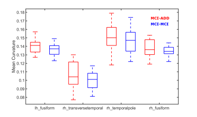 |
Higher temporal lobe curvature in early Alzheimer's indicative of subsequent cognitive decline
Christopher Bird, Sarah Banks, Dietmar Cordes, Karthik Sreenivasan, Xiaowei Zhang, Zhengshi Yang, Virendra Mishra
We selected ADNI patients with an initial diagnosis of mild cognitive impairment (MCI) due to early Alzheimer’s disease, a positive amyloid PET scan within 4 years, and comparable cognitive test scores during their initial visit. Patients were grouped according to their diagnosed outcome within 4 years of the initial visit, specifically, MCI subsequently diagnosed with dementia and stable MCI. We found that curvature within the temporal lobe was greater among patients subsequently diagnosed with dementia. Established measurements of atrophy, including hippocampal volume and temporal lobe thickness, did not differ between these groups.
|
|
2017.
 |
Functional and structural deficits in a novel transgenic rat model of Alzheimer’s Disease.
Cynthia Anckaerts, Ines Blockx, Christina Kreutzer, Hervé Boutin, Sébastien Couillard-Despres, Marleen Verhoye, Annemie Van der Linden
As improving our understanding of the underlying mechanisms of Alzheimer’s Disease (AD) pathology is of utmost importance, the development and characterization of innovative animal models is essential in AD-related research. Here, we further characterized a novel transgenic rat model of AD, the TgF344-AD rat, which manifests progressive AD pathology, much akin to human AD. Functional and structural deficits along the disease progression were assessed using resting state functional MRI (rsfMRI) and diffusion tensor imaging, respectively.
|
|
2018.
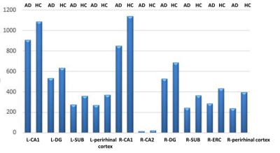 |
Follow up research of hippocampal subfield in patients with mild Alzheimer's disease
Ying Liu, Lizhi Xie
The aim of this study is to evaluate the atrophy pattern of hippocampal subfield and follow up the changes of hippocampal subfield by using automatic segmentation tool in patients with mild AD. The results indicate that volumes of hippocampal subfield decrease in patients with mild AD, and the declination are positive correlated with clinical scores. We conclude that substructures of hippocampal might serve as a good index to characterize subtle changes in AD patients.
|
|
2019.
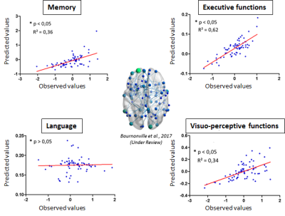 |
Prediction of long-term evolution of cognitive impairment following stroke using resting-state functional connectivity.
Clément Bournonville, Hilde Hénon, Christine Delmaire, Stéphanie Bombois, Jean-Pierre Pruvo, Xavier Leclerc, Régis Bordet, Renaud lopes
The mechanisms of chronic post-stroke cognitive impairments are currently poor understood. However, the study of functional connectivity gives new opportunities to better elucidate the physiopathology. Here, using resting functional connectivity and a machine learning approach, we tried to predict the evolution of cognitive functions up to 36 months after stroke. The results showed that the prediction capacity depends on the studied cognitive domain, and that a particular focus should be done on frontal and temporal cortices.
|
|
2020.
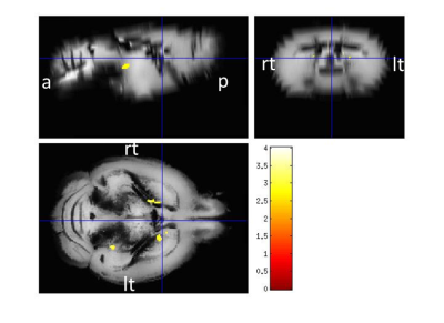 |
APPswe/PS1dE9 mice with cortical amyloid pathology show a reduced NAA/Cr ratio without apparent brain atrophy: A MRS and MRI study
Angela Kuhla, Fatemah Sakr, Claire Ruehlmann, Tobias Lindner, Stefan Polei, Stefan Hadlich, Bernd Krause, Brigitte Vollmar, Stefan Teipel
Amyloid-ß deposition is one of the hallmarks of Alzheimer’s disease (AD) that starts to progress decades before the onset of cognitive impairment. With the rise of the new diagnostic criteria of AD that considers the neuropathological changes as the main aspects for explaining the extent of the disease regardless the cognitive status of the patient & further highlighted the importance of finding reliable in-vivo biological markers to identify those in the preclinical stage of AD. Through the use of the transgenic mice models, particularly APPswe/PS1dE9 we could study the different pathomechanics contributing to the development of AD. So, in this study, we assumed an approach combining morphometry based on high-resolution MRI as a measure for the brain atrophy & proton magnetic resonance spectroscopy as a measure of neuronal functional viability. Then compare these data with a well known & standardized method as the histopathological assessments of neuron & amyloid plaques load. Using the quantitative neuroimaging allows us to translate these mechanistic findings in transgenic models to human phenotypes of brain morphology and function.
|
|
2021.
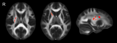 |
Increased Mode of Anisotropy in crossing-fibre areas predicts conversion from Mild Cognitive Impairment (MCI) to Alzheimer's disease (AD)
Matt Gabel, Meena Zaveri, Laura Serra, Marco Bozzali, Mara Cercignani
Diffusion MRI was used to examine whether any change in the white matter tracts of patients with mild cognitive impairment (MCI) can predict conversion to Alzheimer’s disease (AD) in a longitudinal study. Our data show increases in mode of anisotropy (MO) in a region of crossing fibres in the centrum semiovale for MCI patients who later converted to AD.
|
|
2022.
|
Investigating Glumphatic Function During Early Tau Pathology Using Dynamic Contrast-Enhanced MRI
Ozama Ismail, Ian Harrison, Jack Wells, Yolanda Ohene, Payam Nahavandi, Alexander Gourine, Zeshan Ahmed, Alice Fisher, Tracey Murray, Ross Johnson, Emily Collins, Michael O'Neill, Mark Lythgoe
Pathological accumulation of tau and amyloid in the brains of Alzheimer’s disease (AD) patients leads to a continuum of irreversible biochemical and pathological changes and pronounced neurodegeneration. Impaired ‘glymphatic’ clearance may be one of the earliest biological changes in AD, occurring many years prior to neurodegeneration, and therefore presents a unique opportunity for strategic therapeutic intervention. Here, we have mapped the extent of glymphatic inflow of an MRI contrast agent from cerebrospinal fluid, into the brain parenchyma. Leading on from previous studies, we have demonstrated that glymphatic inflow is impaired during the onset of pathology in an AD animal model.
|
|
2023.
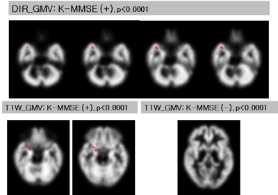 |
Correlation analysis between the gray matter volumes obtained with two different imaging sequences and the cognitive decline in Apolipoprotein E e4 carrier subjects
Na Young Choi, Hak Young Rhee, Soonchan Park, Chang-Woo Ryu, Geon-Ho Jahng, Wook Jin , Dal Mo Yang
To evaluate the association between GMV loss and cognitive decline in the APOE e4 carriers and to investigate alterations of GMV, MPRAGE and DIR images were acquired from 72 subjects (51 noncarriers, 21 carriers). Voxel- and ROI-based analyses were performed to evaluate the association between GMV loss and the MMSE score and to do the group differences of GMV for each sequence. GMV of carriers was positively correlated with the MMSE score for both sequences. DIR can be effective for identifying GMV loss in the carriers and may be useful to evaluate GMV changes in the early stage of dementia.
|
|
2024.
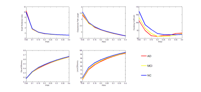 |
The brain functional network alterations of AD and MCI detected by DCCA
Zhizheng Zhuo, Haiyun Li
Pearson’s Correlation analysis has been applied to construct the connectivity network and describe the connectivity strength between different brain function areas. But the correlation coefficient was sensitive to the noise and just for stationary signals. In this study, a new functional connectivity network constructing method based on DCCA (Detrend Cross Correlation Analysis) for non-stationary signals was proposed and applied on AD and MCI.
|
|
2025.
|
Diffusion Kurtosis Imaging Study on Brain Deep Grey Matter in Alzheimer' s Disease
Zhou Yujing, Hu Rui, Miao Yanwei
We used Diffusion kurtosis imaging (DKI) to evaluate the microstructure changes of brain deep gray matter and to explore its relationship with cognitive function in AD.
|
|
2026.
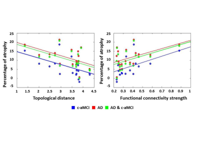 |
Changes in Functional and Structural Brain Connectome Along the Alzheimer’s Disease Continuum
Federica Agosta, Silvia Basaia, Elisa Canu, Francesca Imperiale, Giuseppe Magnani, Monica Falautano, Giancarlo Comi, Andrea Falini, Massimo Filippi
We investigated structural and functional brain network architecture in patients with Alzheimer’s disease (AD) and mild cognitive impairment (MCI); and assessed the relationship between healthy brain network functional connectivity and the topography of brain atrophy in patients along the AD continuum. Structural connectivity alterations distinguished MCI who converted to AD from those who did not. Brain regions most strongly connected with the disease-epicenter (left hippocampus) in the healthy functional connectome were also the most atrophic in both AD and converters MCI. Graph theoretical analysis provides insight on how neurodegeneration propagates across the human brain in the early phase of AD.
|
|
2027.
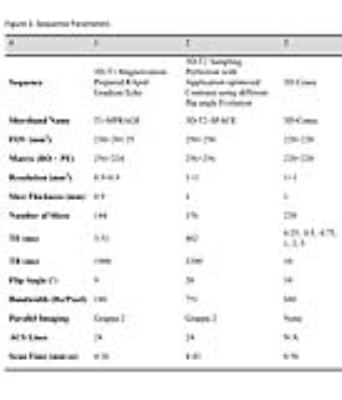 |
Iron Deposition in Alzheimer's Dementia Hippocampus is Associated with Increased R2* Values
Grayson Tarbox, Amin Nazaran, Neal Bangerter, Jonathan Wisco
We describe the utilization of UTE-3D Cones to create T2* maps of iron deposition in the hippocampus of an Alzheimer’s dementia subject, but not in a corticobasal degeneration subject. These results are consistent with histopathological studies involving post-mortem human brain tissue. UTE-3D Cones could be a promising imaging protocol for AD diagnostic imaging.
|
|
2028.
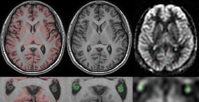 |
Characterizing Perfusion and Arterial Transit Time of the Choroid Plexus with Arterial Spin Labeling
LI Zhao, David Alsop
Choroid plexus signal is readily apparent on Arterial Spin Labeling images but its perfusion characteristics have not been systematically studied. Since the choroid plexus plays an important role in both cerebrospinal fluid production and composition, measuring its function may yield insights into cerebrospinal fluid physiology and disease. In this work, we report initial measurements of the choroid plexus blood flow with noninvasive arterial spin labeling methods using anatomically defined regional measurements.
|
|
2029.
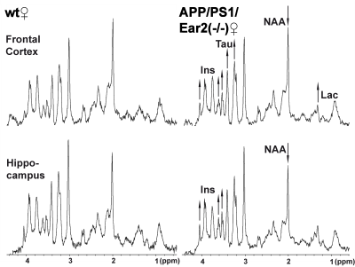 |
Noradrenaline shortage accelerates metabolic alterations in a transgenic model of Alzheimer´s disease
Takashi Watanabe, Ana Martinez-Hernandez, Jens Frahm, Thomas Michaelis
Cerebral MRS of APP/PS1/Ear2(-/-) mice in vivo reveals significant alterations of several metabolites suggesting (i) an impaired cellular respiration compensated for by accelerated anaerobic glycolysis (i.e., elevated lactate), (ii) a loss of neurons (reduced N-acetylaspartate, glutamate, total creatine, and γ-aminobutyric acid) possibly compensated for by osmoregulators (elevated myo-inositol and taurine), (iii) an accumulation of paramagnetic iron (shortened water proton T2) possibly associated with inflammation, and (iv) subsequent gliosis (elevated myo-inositol). More specifically, a 60-75% reduction of noradrenaline is shown to accelerate the reduction of N-acetylaspartate and glutamate in the hippocampus as well as the T2-shortening in the frontal cortex.
|
|
2030.
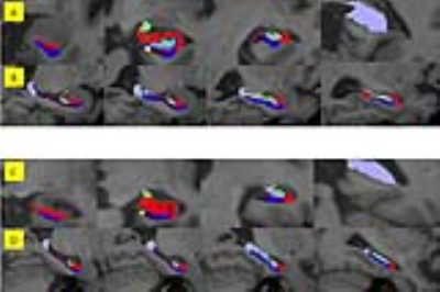 |
MRI Hippocampal subfield volume analysis: Comparison between Alzheimer's disease, mild cognitive impairment, and normal aging subjects in an amyloid PET project.
Natcha Wontaneeporn, Chanon Ngamsombat, Weerasak Muangpaisan, Panida Charnchaowanish, Orasa Chawalparit
Hippocampal atrophy evidenced by MRI is one of the most validated biomarkers of Alzheimer’s disease (AD). The previous neuropathological data showed a differential vulnerability of hippocampal subfields to AD processes. This study aims to use an automated analysis technique for subfield hippocampal volume measurement in order to differentiate early detection of AD. We demonstrated high diagnostic efficacy of using hippocampal subfield analysis for discriminating AD subjects from heathy control (HC) or mild cognitive impairment (MCI) than whole hippocampal volume and feasibility for discriminating MCI to HC as compared with amyloid PET result.
|
|
2031.
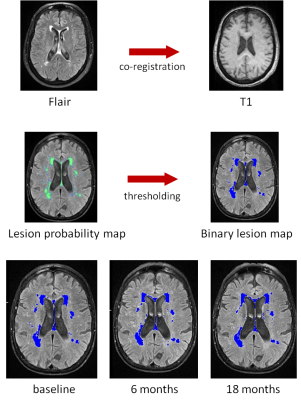 |
Nilvadipine slows progression of white matter hyperintensities in Alzheimer’s disease
Anne Rijpma, Brian Lawlor, Jurgen Claassen
Cerebrovascular disease, such as presence of white matter hyperintensities (WMH), contributes to Alzheimer’s disease (AD) pathology and progression. The antihypertensive nilvadipine may reduce WMH progression by reducing amyloid-induced vasoconstriction and improving cerebral perfusion. Here we show that in patients with mild to moderate AD, nilvadipine slows the increase of WMH after 6 months, but not after 18 months, when correcting for baseline WMH. This contradicts the view that reducing blood pressure in an elderly dementia population leads to progression of white matter damage and instead seems to have a beneficial effect on WMH.
|
|
2032.
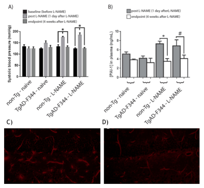 |
Effects of perivascular progenitor cells in combination with Abeta clearance on neurovascular function following transient hypertension in a transgenic rat model of Alzheimer’s Disease
Tina Beckett, Paolo Bazzigaluppi, Margaret Koletar, Conner Adams, Lynsie Thomason, Adrienne Dorr, Denis Gallagher, Clifford Librach, JoAnne McLaurin, Bojana Stefanovic
Examining the interplay between cerebrovascular compromise and AD in the development of therapies is complicated by long prodromal phases of both conditions, necessitating preclinical studies. Four-month-old TgAD-F344 rats, which by six months of age present amyloid deposits and hyperphosphorylated tau, were treated with a nitric oxide synthase inhibitor L-NAME for one month to induce transient hypertension. Human umbilical cord perivascular cells were then given in combination with scyllo-inositol, an inhibitor of Abeta peptide oligomerization and fibrillization to elicit cerebrovascular repair and clear amyloid. Following L-NAME, non-transgenic rats showed transient cerebrovascular changes, whereas TgAD-F344 animals exhibited sustained increase in cerebrovascular reactivity. The latter effect was ameliorated by the treatment.
|
|
2033.
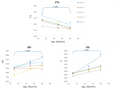 |
Diffusion MRI Changes in the Brain of the 3xTg Mouse Model of Alzheimer’s Disease
Xingju Nie, Maria Falangola, Emilie McKinnon, Joseph Helpern, Jens Jensen
The triple transgenic mouse model (3xTg) of Alzheimer’s disease (AD) exhibits both Aβ and tau pathology. Although diffusion MRI (dMRI) is an established tool for tracking changes in brain microstructure for aging and AD in humans, prior research using diffusion tensor imaging has called into question the sensitivity of dMRI for 3xTg mice. Here we investigated the sensitivity of an alternative dMRI method, diffusional kurtosis imaging, to detect brain changes associated with aging and disease progression in 3xTg mice. Our results indicate that dMRI is able to capture age and/or pathology related alterations in brain tissue for this mouse model.
|
|
2034.
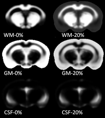 |
In vivo MR detection and automated quantification of amyloid plaques in a preclinical model of Alzheimer's disease
Steve Sawiak, Anne-Sophie Herard, Mathieu Santin, Thierry Delzescaux, Marc Dhenain
Amyloid plaque load is a key index of disease burden in Alzheimer’s disease, but methods for its quantification are slow and operator dependent. Recent advances in the use of contrast agents allow the plaques to be visualized in vivo, but as yet no direct quantification methods are available. Here we present a new technique for automatic segmentation of amyloid plaques and to evaluate age-related or therapy related changes on a voxel-based basis with minimal user intervention. We report localized age-related changes of amyloid load across the whole brain of APP/PS1 mouse model of amyloidosis.
|
|
Brain Imaging Methodology
Traditional Poster
Neuro
Tuesday, 19 June 2018
| Exhibition Hall 2035-2060 |
16:15 - 18:15 |
|
2035.
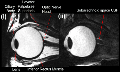 |
High Resolution Structural MRI of the of Eye: Initial Experience at Ultra High Field
Jon Cleary, Bao Nguyen, Rebecca Glarin, Scott Kolbe, Bradford Moffat, Rishma Vidyasagar, Bang Bui, Allison McKendrick, Roger Ordidge
While optical eye imaging techniques are available for examining anterior and retinal structures, they are limited in making 3 dimensional assessments of the whole eyeball. MRI is the preferred modality in these areas but fine eye structures are difficult to resolve on clinical systems. Ultra high field magnets offer increased signal-to-noise, providing higher resolution, but there have been only a limited number of studies so far. We performed an initial study to assess achievable resolution, the anatomy visible on differing image weightings and MR parameter measurements, in eyes of healthy subjects on a 7 Tesla system.
|
|
2036.
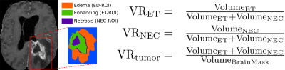 |
Investigating relevance of tumor shape features in overall survival prediction of glioblastoma multiforme patients using machine learning and multi-channel MR images
Parita Sanghani, Ang Ti, Nicolas King, Hongliang Ren
In this work, we study the impact of combining shape features with texture and volumetric features derived from glioblastoma multiforme (GBM) tumors for overall survival (OS) prediction. A comprehensive set of features were obtained from multichannel MR images of 163 GBM patients. Support Vector Machine-Recursive Feature Elimination (SVM-RFE) was used for feature selection, followed by SVM regression for survival prediction. The shape features used in this study have not yet been used for OS prediction in GBM patients and were found to improve the prediction accuracy.
|
|
2037.
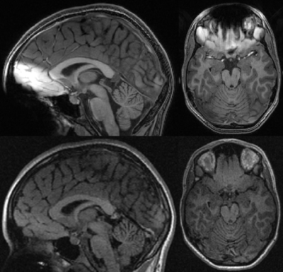 |
T1-weighted imaging of the orbitofrontal cortex in individuals with dental braces using 2D FLAIR
Sander Lamballais, Piotr Wielopolski, Aad van der Lugt, Vincent Jaddoe, Mohammad Ikram, Tonya White, Juan Hernández Tamames
Neuroimaging studies in (pre)adolescent populations have steadily increased in number over the last decade. However, dental braces are common amongst (pre)adolescents and introduce metal-related artifacts in the images. Excluding individuals with braces from epidemiological studies may lead to selection bias and missingness in repeated measures. To this end we configured a T1-weighted 2D FLAIR sequence and compared it to a T1-weighted 3D SPGR sequence. Compared to SPGR, the FLAIR sequence suffered less from metal artifacts and performed similarly in cortical reconstruction and volumetric segmentation. Thus, T1-weighted 2D FLAIR may be a useful alternative for neuroimaging in participants with dental braces.
|
|
2038.
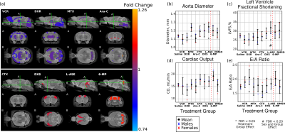 |
Combined MRI and Ultrasound Measurements to Assess the Impact of Systemic Chemotherapy on the Developing Brain and Heart
Leigh Spencer Noakes, Thomas Przybycien, Amanda Forwell, Yu-Qing Zhou, Ellen van der Plas, Brian Nieman
Combined multiple-mouse ex vivo MRI and high-frequency cardiac ultrasound were used to assess the impact of common chemotherapy agents on the developing brain and heart. Of the eight agents considered, vincristine had the most widespread impact on the brain. Doxorubicin, methotrexate, and L-asparaginase were also found to impact brain and/or heart development.
|
|
2039.
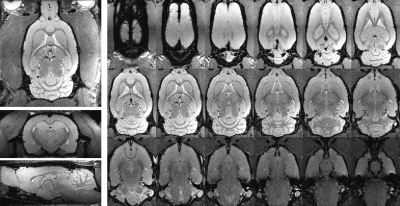 |
Variable flip angle RARE for High-Resolution Preclinical Brain and Spinal Cord Imaging
Matthew Budde
Variable flip angle RARE imaging has seen widespread utility in clinical brain and body imaging, but it has not been available for similar gains in preclinical MRI. This work demonstrates implementation and applications of vfaRARE in a the rat brain and spinal cord.
|
|
2040.
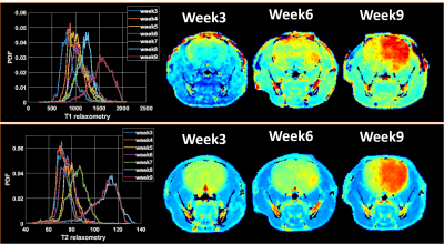 |
Improving sensitivity of infiltrative glioma detection by multi-parametric magnetic resonance imaging
Georgia Kanli, Anaïs Oudin, Simone P. Niclou, Rolf Bjerkvig, Olivier Keunen
Glioblastoma is characterized by poor prognosis and limited treatment efficacy. One main contributing factor is the presence of a large population of infiltrated tumor cells that are difficult to visualize and treat with resective surgery and radiochemotherapy. In the present study, we aim at establishing techniques that combine various contrast mechanisms available in MRI and PET to improve the sensitivity of the detection of infiltrated tumour cells. Such techniques are likely to improve prognosis by early tumor detection, better delineation of the target for radiotherapy, and better assessment of the full extent of the tumor and its response to therapy.
|
|
2041.
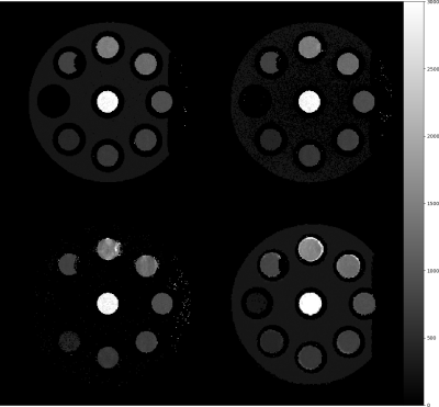 |
Spiral TAPIR with Compressed Sensing for Fast Sub-Millimetre T1 Mapping of Rapidly Relaxing Compartments at 3 Tesla
Robert Claeser, Markus Zimmermann, Nadim Shah
TAPIR is a highly accurate, precise and efficient method for T1 mapping of the brain. It combines an efficient slice-interleaving Look-Locker read-out to sample T1 relaxation by acquiring multiple k-space lines in one shot. However, mapping rapidly relaxing tissue requires the number of lines read in one shot to be small, thus increasing total measurement time. In this work we show how incorporating an interleaved spiral read-out into TAPIR enhances its T1 fitting abilities for rapidly relaxing tissue such as white matter myelin. Scanning time can be decreased by factors of up to 3.3 in comparison to classical Cartesian TAPIR.
|
|
2042.
 |
Quantitative assessment of automatic cortical surface reconstructions from Wave-CAIPI MPRAGE: A validation study
Yulin Chang, Stephen Cauley, Wei Liu, Daniel Polak, Borjan Gagoski, Berkin Bilgic, Kawin Setsompop, Jonathan Polimeni
Structural imaging of the brain using conventional MPRAGE at high resolution is vulnerable to motion artifacts due to prolonged scan times. MPRAGE acquired with wave-CAIPIRINHA technique (waveMPRAGE) and a multi-channel receive coil can significantly improve imaging speed with minimal noise penalty. We show that head motion can be observed from multiple waveMPRAGE scans in a time span similar to a single conventional MPRAGE, and that registering and averaging multiple short (approx. 1 min) waveMPRAGE repetitions produces reliable and reproducible cortical surfaces reconstructed automatically using FreeSurfer.
|
|
2043.
 |
USING MACHINE LEARNING TO CLASSIFY EARLY STAGES OF COGNITIVE DECLINE FROM TYPICAL AGEING - THE CEREBELLUM MORE THAN JUST A BYSTANDER
Muriel Bruchhage, Stephen Correla, Paul Malloy, Stephen Salloway, Sean Deoni
Alzheimer’s disease (AD) is one of the most common forms of dementia, marked by progressively degrading cognitive function. The cerebellum plays a role in AD development, but its predictive contribution to early stages of AD remains unclear. We used MRI machine learning based classification within myelin and grey matter of the whole, anterior and posterior cerebellum and the whole brain, between individuals within the first two early stages of dementia and typically ageing controls. Our findings suggest myelin and grey matter loss in early stages of AD, with distinct patterns of anterior and posterior cerebellar atrophy for each tissue property.
|
|
2044.
 |
Magnetic Resonance Electrical Impedance Tomography in Salamander Retina Neuronal Activity Imaging
Fanrui Fu, Munish Chauhan, Rosalind Sadleir
Magnetic resonance electrical impedance tomography (MREIT) is an innovate potential technique for imaging neural activity. To test its capability, we used salamander retina as a neuronal activity source. The neuronal activity activation was modulated by light, which enables us to compare the result between with and without neuronal activity. After obtained phase images, we expected that the standard deviation of phase in the region of interest (ROI) for the experiment group with neuronal activity is higher than that for the group without activity.
|
|
2045.
 |
Comparison Study between Quantitative Susceptibility Mapping and CT
Sonoko Oshima, Yasutaka Fushimi, Tomohisa Okada, Takuya Hinoda, Takayuki Yamamoto, Hikaru Fukutomi, Yusuke Yokota, Akira Yamamoto, Tsutomu Okada, Kaori Togashi
Quantitative susceptibility mapping (QSM) is a technique which can provide quantitative values of magnetic susceptibility. In this study, we compared magnetic susceptibility values with computed tomography (CT) numbers of brain structures and intracranial lesions in 30 subjects. QSM was able to differentiate between paramagnetic and diamagnetic substances. Susceptibility values showed positive correlations with CT numbers in globus pallidus and lesions with positive susceptibility, and negative correlations in choroid plexus and lesions with negative susceptibility.
|
|
2046.
 |
MP2RAGE, enhanced T1 contrast and beyond
Yishi Wang, Yajie Wang, Jie Shi, Wenjing Zhou, Xuesong Li, Chun Yuan, Hua Guo
MP2RAGE has been modified to acquire two image volumes that can be used to suppress the signals from white matter and CSF respectively. In this study, we show that based on the two intrinsically co-registered volumes, enhanced T1 contrast images compared to traditional MPRAGE images as well as other contrasts such as gray matter image and angiogram can be generated using simple post-processing.
|
|
2047.
 |
Application of support vector machines to multi-modal hemo-metabolic data for classification of disease severity in patients with extreme arterial steno-occlusive diseases
Spencer Waddle, Sarah Lants, Larry Davis, Meher Juttukonda, Matthew Fusco, Lori Jordan, Manus Donahue
Traditional hemodynamic imaging approaches such as arterial spin labeling (ASL) and hypercapnic blood oxygenation level-dependent (BOLD) reactivity provide contrasts that are frequently difficult to interpret using conventional analyses in arterial steno-occlusive disease patients with extreme blood arrival and vascular reactivity delay times. We investigated applying a supervised learning procedure to exploit endovascular and vascular compliance artifacts as potential indicators of disease severity; results show that less-conventional variables which report on endovascular blood signal and delayed vascular compliance outperform conventional variables, such as mean ASL signal and BOLD signal change.
|
|
2048.
 |
Silent Corrected Using Second Image (SCUSI) - Application of the MP2RAGE formalism to T1-weighted Zero Time Echo Imaging
Mark Symms, Florian Wiesinger, Mauro Costagli, Doug Kelley, Mirco Cosottini, Michela Tosetti
We applied the MP2RAGE formalism to a T1-weighted Zero Time Echo sequence. The complex ratio of ZTE images taken with and without inversion preparation showed a correction of the receive coil bias. Brain images of the head are presented showing improved contrast between grey and white matter.
|
|
2049.
 |
A Simplified Method to Estimate Perfusion Characteristics of Gliomas based on Diffusion-weighted Imaging
Mengqiu Cao, Shiteng Suo, Xu Han, Yawen Sun, Yao Wang, Weina Ding, Ke Jin, Xiaohua Zhang, Jianxun Qu, Yan Zhou
The purpose of the study was to evaluate the application of a simplified method to estimate the perfusion characteristics of glioma as an alternative less time-consuming approach. Fifty patients confirmed with glioma were assessed with multi-b-value DWI and DCE MR imaging. Results indicated that the simplified perfusion fraction (SPF) based on DWI acquired with three b-values showed strong correlation with IVIM-derived f and D*, and showed medium correlation with DCE MR imaging-derived Ktrans and vp. SPF achieved the highest accuracy for gliomas grading. SPF may serve as a valuable alternative to measure tumor perfusion in gliomas.
|
|
2050.
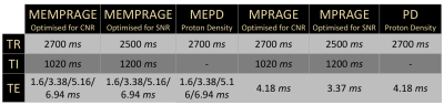 |
Quantitative comparison of image quality between averaged MPRAGE, averaged multi-echo MPRAGE, MP2RAGE and multi-echo MP2RAGE images at high field
Paolo Montagna, Domenico Zacà, Jorge Jovicich
T1 structural neuroimaging is challenged by spatial inhomogeneities of B1 and B0, especially at high fields (>= 3T). Different strategies have been proposed, MP2RAGE (less sensitive to ΔB1, giving also a T1 map) and multi-echo MPRAGE (MEMPRAGE, less sensitive to ΔB0, giving also a T2* map). Here we evaluate the combination of both approaches: MEMP2RAGE. We compare gray-white matter contrast (tissue_CNR) and intensity non-uniformity (INU), between MEMP2RAGE and 3 sets of images under comparable acquisition time: MP2RAGE, two averages MPRAGE, two averages MEMPRAGE. Both MP2RAGE images provide higher tissue_CNR and INU correction than standard MPRAGE images.
|
|
2051.
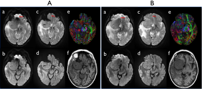 |
Distortion-Free Imaging: A Double Encoding Method (DIADEM), High-Resolution Diffusion Imaging of Brain Tumors on a Compact 3T Scanner
Myung-Ho In, Joshua D Trzasko, Yunhong Shu, Shengzhen Tao, Erin M Gray, Matt A Bernstein, John Huston
Recently, we developed a multi-shot method using spin-warp echo-planar encoding technique inspired by point-spread function mapping. Distortion-free imaging: a double encoding method (DIADEM) can achieve distortion-free, very high in-plane spatial resolution whole brain diffusion imaging in less than 10 minutes on a compact 3T scanner with high performance gradients. A clinical feasibility study of brain tumor diffusion imaging was performed to explore the efficacy of this approach compared to standard single-shot, echo-planar imaging commonly used in clinical practice. The results demonstrate that the proposed method allows considerable improvements in characterizing brain tumors especially at regions of the brain typically degraded by high susceptibility artifacts.
|
|
2052.
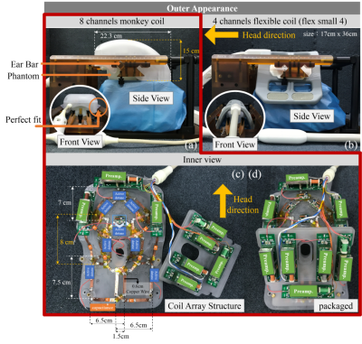 |
An 8 channel Rhesus Head coil for Neuroimaging on 3T
Jo Lee, Xing Yang, Qiaoyan Chen, Changjun Tie, Xiaoliang Zhang, Hairong Zheng, Ye Li
In this study, a custom-designed 8-channel monkey coil was made to match the specific stereotaxic instrument and also better fit the shape of rhesus monkey brain. In comparison with a commercially available coil array, monkey brain images acquired using the dedicated monkey coil array at 3T achieve better SNR, improved parallel imaging capability and higher spatial resolution.
|
|
2053.
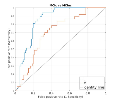 |
Improved Identification of MCI Converters and Non-Converters using Voxel-Based Morphometry and Low-Rank Plus Sparse Matrix Decomposition
Xiuyuan Wang, Steven Baete, Ying-Chia Lin, Ricardo Otazo, Fernando Boada
Early identification of mild cognitive impairment (MCI) patients presents significant challenges due to mild symptoms and low sensitivity of the algorithms proposed for MCI identification. In this study we employed low-rank plus sparse (L+S) matrix decomposition for identifying gray matter volume differences in bilateral hippocampi between MCI patients who converted to Alzheimer’s disease within 18 months and MCI patients who did not. The L+S decomposition identifies features that are common across subjects while minimizing the influence of individual variabilities and outliers. Sensitivity and accuracy are greatly improved and voxel-wise differences that couldn’t be assessed by previous analyses are also identified.
|
|
2054.
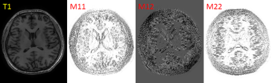 |
Eight fold acceleration for isotropic T2w and T2FLAIR imaging using Multi-Contrast Second-Order Directional Total Generalized Variation (dTGV).
Youngwook Kee, Junghun Cho, Thanh Nguyen, Pascal Spincemaille, Yi Wang
We propose a second-order directional total generalized variation (dTGV) that makes use of directional edge information in T1w to reconstruct highly undersampled T2w and T2FLAIR data. This allows a further doubling of the acquisition speed over the standard four fold accelerated protocol. The proposed dTGV regularizer promotes structural similarity between contrasts.
|
|
2055.
 |
Whole Tumor Histogram Analysis of T2-Weighted, Diffusion-weighted, and Postcontrast T1-Weighted Images in Medulloblatoma:Assessment Risk of Recurrence.
QINGQING LV
Retrospective analysis of 28 patients which were pathologically confirmed medulloblastoma.We find that MRI whole-tumor histogram analysis can be used as an important supplementary method to assess the risk of medulloblastoma recurrence.
|
|
2056.
 |
Regional Brain Iron Mapping in Patients with Heart Failure
Bhaswati Roy, Sadhana Singh, Xiaopeng Song, Ashish Sahib, Cristina Cabrera-Mino, Gregg Fonarow, Mary Woo, Rajesh Kumar
HF subjects show brain injury in multiple areas, which may contribute to altered iron concentration in those sites. However, regional brain iron load in HF subjects is unclear. We examined regional iron deposition using R2*-relaxometry procedures and found altered R2*-values in the amygdala, brainstem, thalamus, globus pallidus, hippocampus, cerebellum, insula, and frontal and temporal white matter regions. The altered iron concentration in HF subjects may result from neural and white matter injury, including myelin and glial dysfunction, with iron potentially accelerating tissue degeneration. These data suggest that interfering with the iron action may reduce the exacerbation of injury in HF.
|
|
2057.
 |
Identification of thalamic substructures in ultra-high b-value DWI
Nils Nuessle, Benjamin Bender, Uwe Klose
Precise implantation of deep brain stimulation devices in Parkinson, primary dystonia or epilepsy patients requires precise structural information about the thalamic region. Purpose of this study was to evaluate the capability of DWI in identifying thalamic substructures. Eight healthy volunteers underwent ultra-high b-value DWI (5000 s/mm2) at 3T. Images were denoised using total generalized variation and 7 substructures (Pulvinar and six nuclei) within the thalamus were drawn in and compared to histological atlases. In all volunteers, all seven structures could be identified due to signal intensities. High b-value diffusion weighted imaging therefore shows great potential in determining thalamic substructures.
|
|
2058.
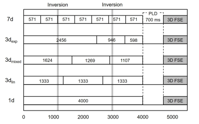 |
Practical parameter setting for simultaneous measurement of CBF and ATT with Hadamard-encoded ASL: Special reference for clinical practice
Shota Ishida, Hirohiko Kimura, Naoyuki Takei, Masayuki Kanamoto, Yasuhiro Fujiwara, Tsuyoshi Matsuda, R Lebel, Toshiki Adachi
Hadamard-encoded ASL (H-ASL) is a time-efficient method for measuring arterial transit time (ATT). The larger encoding matrix extends the scan time, but the accuracy of the ATT with a different encoding matrix was not clarified. This study aimed to propose a practical parameter selection in H-ASL for clinical use. The ATT was not significantly different between 3 and 7 delay encodings. Cerebral blood flow (CBF) obtained with 3 delay encodings with a linear division block design was equivalent to that obtained without encoding. Three delay encodings with a linear division block design provides accurate ATT and CBF within 4 minutes.
|
|
2059.
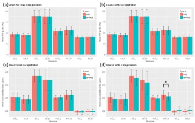 |
Impact of coregistration approaches on the reliability of R2* and Quantitative Susceptibility Maps (QSM) at 7 T
Seongjin Choi, Xu Li, Daniel Harrison
We assessed the unexplored reliabilities of QSM and R2* map acquired at 7 T by two different approaches that are 1) a direct coregistration of the quantitative maps from their native to a reference space and 2) processing the quantitative maps in a transformed space. R2* was reliable in both methods in the pixel value and the group-mean analyses. However, QSM was less reliable when it was processed in a transformed space in both analyses. Therefore, QSM is recommended to be calculated in its native space prior to any coregistration in a multi-modal study.
|
|
2060.
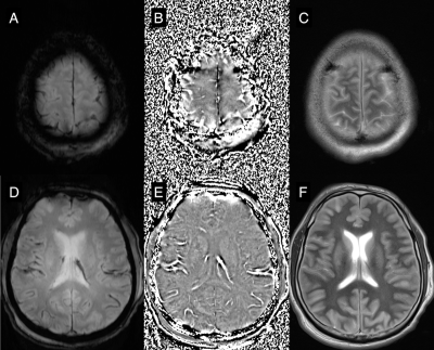 |
Prognostic value of phase images of 2D T2*-weighted GRE in cardiac arrest survivors: A pilot study
Jinhee Jang, Sang Hoon Oh, Yangsean Choi, Yoonho Nam, Kyu Nam Park, Kook-Jin Ahn
Because hypoxic ischemic injury of the brain occurs in cardiac arrest survivors, assessment of oxygen metabolism could be useful. In this work, we analyzed filtered phase images of 2D T2*-weighed gradient echo images in them. Three survivors with good neurologic outcome showed normal pattern of cortical and deep veins, as well as dural sinuses. However, patients with poor outcome showed two abnormal pattern of venous structures on filtered phase images; (1) attenuated contrast of venous structures and (2) strong and exaggerated venous contrast. Filtered phase images of 2D T2*-weighed gradient echo might useful to predict prognosis of cardiac arrest survivors.
|
|
Brain Pathology & Ageing Brain
Traditional Poster
Neuro
Tuesday, 19 June 2018
| Exhibition Hall 2061-2085 |
16:15 - 18:15 |
|
2061.
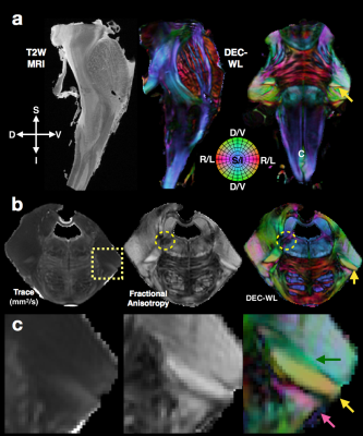 |
Revealing the three-dimensional intraparenchymal trajectory of the brainstem cranial nerve systems by diffusion MRI representation.
Elizabeth Hutchinson, Neda Sadeghi, Martin Lizak, Martha Quezado, Irini Manoli, Carlo Pierpaoli
The cranial nerve systems of the human brainstem are challenging to distinguish from their complex architectural surroundings, but anisotropy, orientation and tract-based diffusion MRI methods may address these challenges and enable mapping intraparenchymal trajectories of the cranial nerves. The objective of this study was to apply and evaluate DTI and tractography tools for segmentation and mapping of the cranial nerve systems at high spatial resolution in post-mortem human brainstems. Our findings demonstrate the salient features of scalar, directional and tract-based maps for distinguishing the cranial nerves and their nuclei with attention to their relative geometric complexity and architectural environment.
|
|
2062.
 |
Linking neurotransmitter concentration and functional connectivity of the hippocampus after stress: an in-vivo MRI study
Ricardo Magalhães, David Barriére, Ashley Novais, Fernanda Marques, João Sousa, João Cerqueira, Arnaud Cachia, Thérèse Jay, Nuno Sousa, Sébastien Mériaux, Fawzi Boumezbeur
Stress is a potent modulator of brain metabolism and function. Here we use a combined approach of blood corticosterone quantification, nuclear magnetic resonance spectroscopy and resting state functional magnetic resonance imaging to probe both metabolic and functional changes in the brain. We show correlations in the concentration of GABA/Glutamine and Glutamate/Glutamine in the hippocampus and how these two factors interact with the response to stress. Furthermore we explore how the changes in neurotransmitters correlate with functional networks, revealing several affected connections especially with the retrosplenial cortex, therefore suggesting a role of this relationship in the affected memory phenotype.
|
|
2063.
 |
White Matter Microvascular Changes in Healthy Aging
Ian Tagge, Valerie Anderson, James Obayashi, Xin Li, Joseph Quinn, Jeffrey Kaye, Dennis Bourdette, Rebecca Spain, Manoj Sammi, William Rooney
The extent to which changes in blood-brain-barrier permeability are associated with healthy aging is poorly understood. Pharmacokinetic modeling of dynamic-contrast-enhanced MRI yields quantitative estimates of BBB water permeability. DCE-MRI data were collected from 40 healthy controls (aged 34-80 yrs) at 7T. Declines in pharmacokinetic parameters were significant across the entire age range included in this study. Because changes in BBB permeability to water and other small molecules are likely to precede the leakage of CR and larger macromolecules, these estimates represent particularly important probes of the subtle BBB abnormalities that are likely to accompany healthy brain aging.
|
|
2064.
 |
Retinal Vascular Fractal Dimension and Cerebral Blood Flow, a pilot study
Jeremy Nadal, Jeremy Deverdun, Nicolas Menjot de Champfleur, Emmanuelle Le Bars, Vincent Daien
The retinal vascular fractal dimension (FD) is a marker of retinal vascular complexity of the vascular tree. It has been associated with systemic disorders but also with neurodegenerative and cerebrovascular diseases. The purpose of this study was to explore the relationship between cerebral blood flow (CBF), retinal vascular FD and other retinal vascular markers. CBF was estimated in vascular territories using 2D PASL sequence. CBF was positively associated to venular FD (R2=0.32, p=0.03). Non-invasive exploration of the retinal vasculature may be used as a proxy measure, with the condition of retinal vessels possibly reflecting the condition of the cerebral vasculature.
|
|
2065.
 |
Relationships among cerebrovascular reactivity, grey matter volume and markers of successful aging
Brittany Intzandt, Dalia Sabra, Laurence Desjardins-Crepeau, Said Mekary, Louis Bherer, Richard Hoge, Christopher Steele, Claudine Gauthier
Aging causes decline in brain health, which has a complex relationship with fitness and cognition. Here, we aimed to disentangle the interactions between these outcomes in healthy older adults. MRI was used to acquire anatomical and cerebrovascular reactivity (CVR) in all participants. VO2max and cognitive outcomes were also tested. Results revealed that increased CVR was associated with decreased fitness and cognitive performance, whereas increased grey matter volume was associated with increased fitness. It is apparent that the relationship between brain health and fitness and cognitive outcomes is intricate and other parameters, such as cerebral blood flow, are necessary to gain further understanding.
|
|
2066.
 |
Effect of autolysis, fixation, and storage in PBS on relaxation rates and macro-molecular tissue volume across fiber pathways of the human brain
Mohammad Ashtarayeh, Tobias Streubel, Klaus Püschel, Siawoosh Mohammadi
We evaluated the effect of autolysis, brain tissue fixation, and embedding into PBS on three potential quantitative myelin MRI markers across different white matter fiber pathways: longitudinal (R1) and effective transverse (R2*) relaxation rates, and macro-molecular tissue volume (MTV) using the quantitative multi-parameter mapping (MPM) protocol. We found that the effect of autolysis was most apparent in R2* and MTV, R1 drastically changed its contrast after fixation, and R1 and R2* values increased after storage in Phosphate-Buffered Saline (PBS) solution.
|
|
2067.
 |
Sleep Deprived and Well Rested Brains are Distinguishable by Machine Learning in T1w Imaging
Andrew Hall, Laurentius Huber, Daniel Handwerker, Emily Finn, Peter Bandettini
We investigated 166 T1-weighted datasets to identify neural biomarkers of sleep deprivation (3h sleep). We find that a linear classification algorithm is able to distinguish between sleep deprived and well-rested brains at 65% accuracy in T1-weighted images. The underlying hypothesis is that if glymphatic function is mediated by sleep, one should be able to tell the difference between sleepy and rested brains based on subtle shifts in T1 across brains.
|
|
2068.
 |
Nonlinear pattern of the emergence of white matter hyperintensity in healthy Han Chinese: an adult lifespan study
Chu-Chung Huang, Albert C. Yang, Kun-Hsien Chou, Mu-En Liu, Shih-Jen Tsai, Ching-Po Lin
WMH is one of the most obvious imaging traits in the aged brain. There is evidence that the WMH volume may have the potential to track with age and age-related cognitive decline. However, no study has investigated the trajectory of WMH progression and their impact on cognition during normal aging process. We show that increased age is nonlinearly correlated with increased PVWMH. In two-mediators mediation model, PVWMH is found to mediate the age-related decline of MMSE, but not DWMH. This study suggested that PVWMH could be a potential, and feasible biomarker in predicting age-related cognitive decline across the adult lifespan.
|
|
2069.
 |
MR Spectroscopy Study: Neural Effects of Induced Hypothermia Treatment after Myocardial Infarction & Anoxia
Lasya Sreepada, Jong Woo Lee, Huijun Liao, Benjamin Rowland, Alexander Lin
Coma after cardiac arrest is a common and debilitating incidence. This study aims to determine the neurochemical changes that occur in comatose cardiac arrest patients who underwent targeted temperature management. Single Voxel MRS was acquired in the posterior cingulate gyrus (PCG) and parietal white matter (PWM) of patients and age-matched controls with no history of neurological disease. Patients showed decreases in NAA, as well as increases in total Choline and Lactate in both PCG and PWM. Patients also showed decreases in glutamate in the PCG. These neurometabolic changes reflect neuronal, axonal and glial loss that would result in reduced neurotransmission.
|
|
2070.
 |
Cerebral Vascular Reactivity and Cognitive Decline in Healthy and in Early Stages of Pathological Aging.
Naila Boudiaf, Jan Warnking, Olivier Moreaud, Johan Pietras, Eric Condamine, Nathalie Fournet, Amandine Bossant, Monica Baciu, Alexandre Krainik
Cerebral blood flow (CBF) and cerebral vascular reactivity (CVR) decrease with age and might affect cognitive functions. In this study, we investigated their correlation with cognitive abilities during normal and pathological aging. We performed neuropsychological assessments on thirty-four participants: 13 healthy-young, 10 healthy-old and 11 impaired-old. We measured CBF and CVR using hypercapnia and Arterial Spin Labeling imaging.
Regarding the impaired-old, we found specific decrease in executive functions, short-term and working memory. Significant correlations were found between CVR and cognitive scores. Global CBF correlated only with age.
Unlike CBF, CVR reduction was specifically associated with cognitive decline during aging.
|
|
2071.
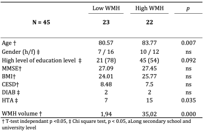 |
Cingulum tractography in old subjects presenting low or high white matter lesion burden
Manon Edde, Bixente Dilharreguy, Catherine Helmer, Jean-François Dartigues, Michèle Allard, Gwénaëlle Catheline
Tractography frequently fails in aging brain because diffusion parameters dramatically decrease in regions of WM hyperintensities (WMH) which one are very common in this population. We developed here a pipeline taking into account this pitfall to truly investigate the microstructural properties of the cingulum bundles in presence and absence of WMH.
|
|
2072.
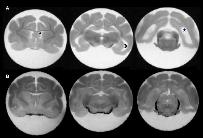 |
Differentiation and Quantification of White Matter Injury in Post-Hemorrhagic Hydrocephalus
Albert Isaacs, Harri Merisaari, Tsen-Hsuan Lin, James (Pat) McAllister, David Limbrick, Sheng-Kwei (Victor) Song
This study is the first of its kind, and uses diffusion basis spectrum imaging (DBSI) to quantify, as well as differentiate the complex pathologies that underlies the white matter injury in post-hemorrhagic hydrocephalus (PHH) in neonates, using a ferret model of PHH.
|
|
2073.
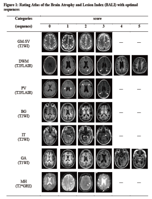 |
Improving MRI assessment of whole-brain structural health in aging: an approach involving multiple sequences
Hui Guo, Yunting Zhang, Ryan D’Arcy, Xiaowei Song
The process of brain aging is characterized by the accumulation of multiple structural changes, several of which can be visualized using clinical MRI. Brain Atrophy and Lesion Index (BALI) has been validated to collectively assess MRI-based whole-brain structural changes. This study aims to improve the BALI assessment of whole-brain structural changes in aging using multiple routine clinical MRI sequences (T1WI, T2WI, T2-FLAIR and T2*GRE).
|
|
2074.
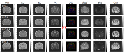 |
Nutritional intervention for developmental brain damage: neuroprotection with Lactoferrin following intrauterine growth restriction.
Yohan van de Looij, Camille Larpin, Petra Hüppi, Stéphane Sizonenko
Lactoferrin (Lf) is an iron-binding glycoprotein secreted in milk known as antioxidant, antimicrobial and anti-inflammatory. Infants exposed to adverse prenatal conditions of intrauterine growth restriction (IUGR), are at high risk for neurological morbidities. The aim of this work was to assess neuroprotective effect of Lf on brain microstructure by using diffusion imaging and NODDI model at 9.4T in a model of 50% gestational caloric restriction. Diffusion MRI derived parameters changes following IUGR were partially restored in the Lf supplemented group, providing evidence of a neuroprotective effect.
|
|
2075.
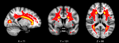 |
Differentiation of white matter hyperintensity severity using T2- and T1-weighted brain MRI.
Nina Reislev, Henrik Lundell, Hartwig Siebner, Christian Eriksen, Michael Kjær, Ellen Garde
This study presents a new method to differentiate brain white matter hyperintensity (WMH) severity using conventional T1-weighted and T2-weighted MRI. By combining normalized image intensity, heterogeneous tissue properties within lesions are revealed. Lesion severity is quantified through two distance measures of parallel and perpendicular deviation from normal appearing white matter. Correlations with diffusion imaging based measures suggest that multi-modal voxel-based lesion analysis provide comparable but high-resolution tissue information. Based on conventional MRI scans this method adds valuable insight into the differentiated impact of WMH lesions on brain structure and function.
|
|
2076.
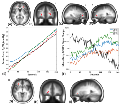 |
The Aging Brain: Cerebrovascular responses to CO2.
Larissa McKetton, Olivia Sobczyk, Julien Poublanc, Kevin Sam, Adrian Crawley, Lakshmikumar Venkat Raghavan, James Duffin, Joseph Fisher, David Mikulis
Measures of cerebrovascular reactivity (CVR) are used to judge the health of the brain vasculature. We report the use of several different analyses of BOLD responses to CO2 to provide a number of metrics for various aspects of CVR. To assess possible differences in these metrics with age, we compiled atlases reflecting voxel-wise means and standard deviations for different age ranges and compared them.
|
|
2077.
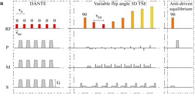 |
Vessel Wall Thickness Evolution Across the Lifespan Assessed using Non-invasive Intracranial Vessel Wall Imaging
Petrice Cogswell, Sarah Lants, L Taylor Davis, Spencer Waddle, Manus Donahue
Vessel wall imaging is becoming more widely applied, however normal, age-specific ranges for wall thickness have not been established. We applied a variable refocusing angle 3D-TSE acquisition with and without a DANTE flow suppression module to healthy subjects (ages=8-79 years; n=82). Vessel wall measurements revealed no significant change in wall thickness with age for the supraclinoid internal carotid arteries and basilar artery. The outer wall diameter and wall thickness were measured to be less for the acquisition with versus without DANTE. These data suggest that unlike tissue volume, vessel wall thickness is relatively constant across the lifespan for healthy subjects.
|
|
2078.
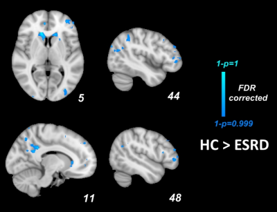 |
Altered Intrinsic Brain Activity and Memory Function Improvement in Patients with End-Stage Renal Disease During A Single Dialysis Session
Peng Li, Dun Ding, Xueying Ma, Huawen Zhang, Jixin Liu, Ming Zhang
The underlying neural mechanisms of the memory deficits in end-stage renal disease patients with dialysis treatment are poorly understood. Here we analyzed the resting-state brain activity changes and the related memory improvement by using mALFF and ReHo methods before dialysis (T1pre-dialysis) and after 24 hours (T2post-dialysis). The results indicated that regional spontaneous activity changes of the DLPFC were related with memory improvement after a single dialysis treatment, which may provide insight into the effect of hemodialysis on changes of brain function and cognitive impairments.
|
|
2079.
 |
Development of Individual Evaluation System for White Matter Hyperintesity Recognition Using Deep Convolutional Neural Network
Kyung Mi Lee, Hyug-Gi Kim, Jiwon Yoon, Mi-hyun Kim, Jang-Hoon Oh, In Young Lee, Soonchan Park, Chang-Woo Ryu, Eui Jong Kim, Woo Suk Choi, Na Rae Yang, Jihye Song
White matter hyperintensity (WMH) is one of the important characteristics of cerebral small vessel disease (cSVD). The objective of this study to investigate the feasibility of WMH recognition using deep convolutional neural networks (CNN). Furthermore, individual evaluation system was proposed to classify WMH groups.
|
|
2080.
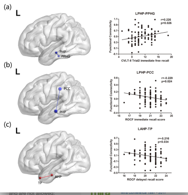 |
Integration and segregation of functional segmented anterior and posterior hippocampal networks in memory performance
Jingjing Xu, Xiaojun Guan, Xiaojun Xu, Minming Zhang
In this study, we used a novel functional segmentation method to subdivided the left and right hippocampus into anterior and posterior portions according to preferred functional connections with certain cortical regions. And we investigated the association between specific performance of verbal and visual memories and intra-hemispheric resting state FC across anterior and posterior hippocampal networks using resting functional MRI measures in healthy young volunteers. The present results demonstrated that, the anterior hippocampus was specifically involved in the visual memory processing, whereas the posterior hippocampus contributed to both the verbal and visual memories, which may have implications for a functionally synergetic and dissociable role of the hippocampus in different kinds of memory.
|
|
2081.
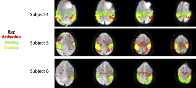 |
Presurgical planning: comparison between task activation and resting-state connectivity maps in the motor and language networks
Scott Peltier, Gaurang Shah
In this study, task and resting-state data from presurgical patients with brain tumors was analyzed. Task activation and data-driven resting-state connectivity maps for both motor and language networks were generated for each subject and compared for spatial overlap.
|
|
2082.
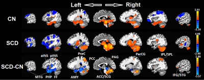 |
Daily Pain in Adults with Sickle Cell Disease is Associated with Alterations in Functional Connectivity of the Brain
Guangyu Chen, Arun Singavi, Nancy Wandersee, Collin Hubler, Amanda Brandow, Simpson Pippa, Shi-Jiang Li, Joshua Field
About half of Sickle Cell Disease (SCD) adults suffer from a chronic pain syndrome. What of the SCD brain contributes to the development and maintenance of the pain syndrome is unknown. We used resting state functional connectivity MRI (rfcMRI) technique, found significant differences between SCD and controls in areas known to contribute to the development and maintenance of a chronic pain syndrome, and the differences have significant associations with the pain phenotype measurements. The findings suggest that rfcMRI could be used as a biomarker to determine the efficacy of interventions targeted to chronic pain in SCD patients.
|
|
2083.
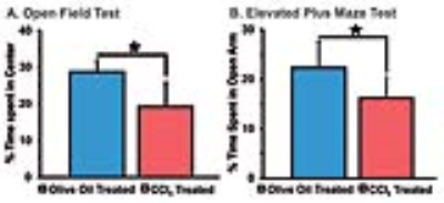 |
Impacts of Chronic Liver Injury on Brain Energy Metabolism: A 1H-[13C]-NMR Study on Hepatic Encephalopathy
TK Sampath Kumar , N Sairam, Anant Patel
It has been postulated that excess ammonia and neuroinflammation resulting from liver failure induces astrocytic swelling which can lead to increased BBB permeability and neuronal dysfunction. The impacts of high levels of blood ammonia on the brain energy metabolism is not clear. The objective of current study is to evaluate the neurotransmitter metabolism in CCl4 induced liver injury mouse model using using 1H-[13C]-NMR spectroscopy together with [1,6-13C2]glucose infusion. Our findings indicate reduction in the activity of glutamatergic and GABAergic neurons in the chronic liver damage condition.
|
|
2084.
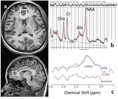 |
Increased Glutamate in Anterior Cingulate Cortex in Crohn’s Disease Patients with Abdominal Pain Revealed by Proton MR Spectroscopy
Kun Lv, Wenwen Song, Yihong Fan, Yong Zhang, Bin Lv, Maosheng Xu
Based on Brain-gut axis, the study used proton magnetic resonance (MR) spectroscopy, a noninvasive detection to reveal the alteration of metabolites in bilateral perigenual anterior cingulate cortex (pgACC) in patients with Crohn’s disease (CD) with abdominal pain. Twenty nine CD patients (cases with/without abdominal pain, 16/13) and 20 healthy controls were recruited for comparison. The pain CD group showed increased Glutamate (Glu) levels in bilateral pgACC, which might provide new insight into the neural mechanism of the disease in abdominal pain processing.
|
|
2085.
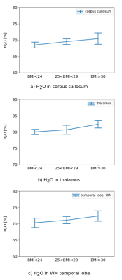 |
Changes in Quantitative Free Water Content with Increasing BMI in Elderly Subjects
Melissa Schall, Elene Iordanishvili, Svenja Caspers, N. Jon Shah, Ana-Maria Oros-Peusquens
A high body mass index is known to play a role in a variety of chronic diseases, which makes it an important biomarker. Using a 3D two-point quantitative mapping method, changes in several parameters including relaxation times, H2O and magnetisation transfer measures were investigated in lean and obese subjects. Preliminary results show a significant increase of H2O in corpus callosum (p<0.05), thalamus (p<0.005) and white matter of temporal lobe (p<0.05) with increasing BMI. Changes in the other parameters did not reach significance. These findings suggest the existence of regional low-grade brain inflammation in obesity.
|
|
Novel Neuroimaging Methods
Traditional Poster
Neuro
Tuesday, 19 June 2018
| Exhibition Hall 2086-2103 |
16:15 - 18:15 |
|
2086.
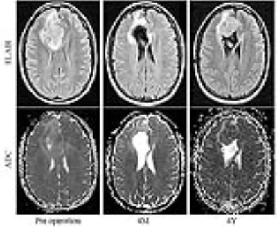 |
MRI based texture analysis on FLAIR and ADC to predict malignant transformation of Low Grade Gliomas
Shun Zhang, Gloria Chiang, Yihao Yao, Ramin Jafari, Rajiv Magge, Howard Fine, Rohan Ramakrishna, Yi Wang, Ilhami Kovanlikaya
Low grade gliomas (LGG) may undergo malignant transformation into high-grade gliomas, which generally occur within 5 years in about 50% of patients. Hence assessing whether or not a LGG will convert to high grade is of great importance in treatment. In this study, we use texture and histogram analyses on preoperative MRI FLAIR and ADC images to predict malignant transformation from low grade to higher grade glioma, as well as to discriminate between astrocytoma and oligodendroglioma. Based on the receiver operating characteristic (ROC) curves from training data, texture analysis had a higher area under the curve (AUC) value than histogram parameters, and it also more accurately predicted whether LGGs would convert and discriminated between astrocytoma and oligodendroglioma in the testing data. Texture analysis on conventional preoperative FLAIR and ADC images can accurately predict malignant transformation of low grade gliomas, as well as discriminate between astrocytoma and oligodendroglioma.
|
|
2087.
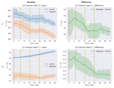 |
Observing the Hyaluronan Component of the Extracellular Matrix in the Brain with Quantitative MRI
Riccardo Metere, Carsten Jäger, Markus Morawski, Harald Möller
The tissue composition of the brain can be related to different contrast sources in quantitative MRI. Notably, T1 and T2* strongly correlate with myelin and iron. However, other components may play a role in contrast generation. In particular, the hyaluronan component of the extracellular matrix has been recently proposed as a possible important contributor to MRI contrast. Here, we quantify the bulk contribution of hyaluronan to quantitative relaxation maps. This is obtained by characterizing the evolution of the relevant MRI parameters over time during the enzymatic digestion of the hyaluronan.
|
|
2088.
 |
Noninvasive Analysis of Brain Shift Transformation in Closed Cranium using MR Images Acquired in Different Body Positions
Etsuko Kumamoto, Shigeto Hayashi, Kento Matsuda, Katsusuke Kyotani, Takashi Nishino, Tomoaki Nakai, Eiji Kohmura
Although the transformation of brain tissue during craniotomy is a well-known phenomenon, there has been a lack of methodological analysis related to physiological brain shift in the closed cranium. In this study, we analyzed brain shift and transformation using MRI volume data acquired in different body positions. The volume data were divided into voxels. Each voxel of the prone or the right volume was registered using voxels of the supine or the left volume as templates, and movement and rotation of each voxel were recorded. Experimental result shows that the displacement in the depth of the brain tended to be conspicuous and rigid compared to the displacement of the brain surface.
|
|
2089.
 |
Towards an optimized protocol for dynamic oxygen enhanced imaging of the brain.
William Lloyd, Adam Featherstone, Alan Jackson, Geoff Parker
T1w dynamic oxygen-enhanced MRI (OE-MRI) has been shown to be a promising method for the assessment and quantification of tumour hypoxia. This work presents a comparison between two possible methods; FFE and IR-TFE based sequences. IR-TFE is shown to give greater contrast for oxygen induced signal change as well as increased SNR. Further sequence optimisation demonstrates the possibility of scanning at high resolution while maintaining contrast and SNR.
|
|
2090.
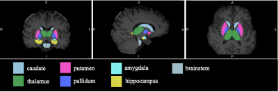 |
Differences in subcortical brain volumes between expert and novice chess players
Ethan Li, David Ouellette, Tim Duong
The goal of this study was to investigate the anatomical neural correlates underlying expertise acquisition between expert versus novice chess players using MRI. We found that the acquisition of expertise is accompanied by gray-matter volumetric changes in subcortical brain structures implicated in memory and reinforcement learning. By comparison, the anatomical circuits involved in acquired chess expertise differ from other expertise domains. Improving the understanding of the neural correlates underpinning expertise may prove useful in designing individualized training strategies.
|
|
2091.
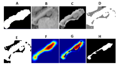 |
Pre-training and training of a Convolutional Neural Network for automatic and accurate hippocampus segmentation from T1-weighted MRI datasets
Samaneh Nobakht, Nils Forkert, Sean Nestor, Sandra Black, Phillip Barber
The hippocampus atrophy rate (volumetric loss per year) might be a good biomarker for predicting disease progression. However, hippocampus atrophy rate assessment requires accurate delineation of the structure from longitudinal scans. In this work, we propose an automatic approach based on convolutional neural network (CNN) for robust and reliable hippocampus segmentation. Therefore, the CNN was pre-trained using weakly annotated T1-weighted MRI datasets and fine-tuned using fully-annotated datasets. Leave-one-out cross validation revealed that the proposed method leads to robust and reproducible segmentation results with an average Dice coefficient of 0.89.
|
|
2092.
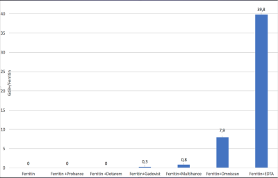 |
Concept of Gadolinium-Ferritin Interactions as Explanation of Signal Intensity Changes in Deep Brain Nuclei after Application of Gadolinium-Based Contrast Agents
Josef Vymazal, Jitka Neburkova, Martin Dracinsky, Mohan Pingle, Petr Cigler, Aaron Rulseh
Interaction between gadolinium-based contrast agents and metalloprotein ferritin may explain observed signal intensity changes in vivo due to T1 (and T2) shortening in the globus pallidus and dentate nucleus.
|
|
2093.
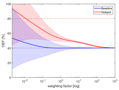 |
How does the weighting factor in a regularized quantitative BOLD approach affect the estimated oxygen extraction fraction?
Sebastian Thomas, Simon Hubertus, Sebastian Domsch, Lothar Schad
Applying the quantitative blood-oxygenation-level-dependent (qBOLD) method for measuring the oxygen extraction fraction (OEF) often suffers from bad contrast due to the low SNR typical at clinical scan times. In order to improve the evaluation, the choice of the weighting factors in a proposed regularization approach was analyzed. Using the regularization approach, simulations showed increasing precision but decreasing accuracy for increasing weighting factors. For a range of weighting factors a good trade-off between noise suppression and data-fidelity was achieved, which resulted in optimal contrast.
|
|
2094.
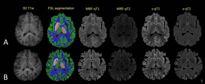 |
Visualizing healthy aging: A comparative study of quantitative T1 and T2 relaxometry with standard and MR fingerprinting techniques
Vera Keil, Stilyana Bakoeva, Alina Jurcoane, Thomas Amthor, Mariya Doneva, Peter Koken, Burkhard Mädler, Wolfgang Block, Elke Hattingen
Relaxometry aims at an absolute quantification of T1 and T2 times explained by physicochemical properties in the brain. MR Fingerprinting can be used for quantitative relaxometry, e.g. to explore the effect of age on brain structure. This study examined young and old age (n=26 each) volunteers with standard and MRF mapping techniques. We found that MRF and standard technique multiecho-derived T1 and T2 maps do not identify the same brain structures as affected by age-related relaxometric changes and show in part contradictory relationships between age and especially T1 relaxation. This limited comparability has strong clinical implications for the interpretation of relaxometric studies beyond the topic "aging".
|
|
2095.
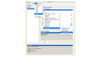 |
The hMRI analysis toolbox for quantitative MRI and in vivo histology using MRI (hMRI)
Evelyne Balteau, Tobias Leutritz, Antoine Lutti, Martina Callaghan, Bogdan Draganski, Christophe Phillips, Enrico Reimer, Lars Ruthotto, Maryam Seif, Nikolaus Weiskopf, Gabriel Ziegler, Siawoosh Mohammadi, Karsten Tabelow
Quantitative MRI finds increasing application in neuroscience and clinical research due to its greater specificity and its sensitivity to microstructural properties of brain tissue - myelin, iron and water concentration. We introduce the hMRI toolbox, an easy-to-use open-source tool for data handling and processing of quantitative MRI data. This toolbox, embedded in the SPM framework, allows the estimation of quantitative MRI maps (longitudinal and transverse relaxation rates R1 and R2*, proton density PD, and magnetization transfer MT), followed by spatial registration in common space for statistical analysis. It also offers flexibility for calculation of novel MRI biomarkers of tissue microstructure.
|
|
2096.
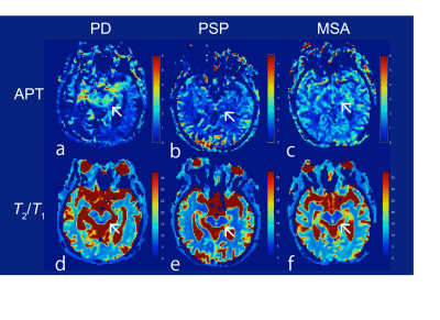 |
Chemical exchange saturation transfer imaging for neurodegenerative diseases
Yuki Matsumoto, Masafumi Harada, Yuki Kanazawa, Maki Otomo, Mitsuharu Miyoshi
The purpose of this study is to investigate the relationship between CEST imaging and several neurodegenerative diseases to verify the feasibility of an estimation parameter derived from the CEST approach. For this study, patients with Parkinson’s disease, progressive supranuclear palsy and multiple system atrophy as well as healthy volunteers were examined. Region-of-interest analysis was performed in the substantia nigra and red nucleus area. As the results, the CEST parameters were significantly different for each of the neurodegenerative diseases and healthy volunteers. CEST imaging might have the ability to obtain more detailed information concerning neurodegenerative disease.
|
|
2097.
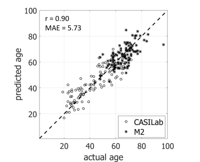 |
Predicting the age from time of flight MR angiography using 3D convolutional neural network
Yoonho Nam, Jaeho Lee, Dong-Hyun Kim, Jinhee Jang, Bumsoo Kim, Kook-Jin Ahn
The age-related changes involve the vasculatures of the brain because the brain has rich blood supply. Previous studies using time of flight (TOF) MR angiography suggested that the aging intracranial arteries were tortuous, irregular and heterogeneous in shape. However, the use of these hand-crafted features and qualitative visual assessments are limited in practical clinical use. Vascular aging could be used as an imaging biomarker for the brain if we could distinguish various age-related vascular changes automatically and quickly from MR angiography. In this study, we investigate the feasibility of deep learning based feature extraction as a tool for analysis of age-related change of brain vasculatures.
|
|
2098.
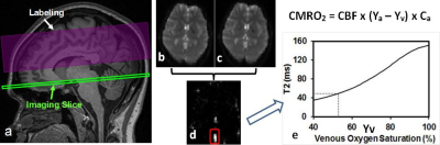 |
Simultaneous assessment of tDCS-induced neuronal responses with oxygen metabolic MRI
Yulin Ge, Abhishek Datta, Bryan Dobbs, Michael Shaw, Ashley Clayton, Oded Gonen, Leigh Charvet
Although transcranial direct current stimulation (tDCS) offers a therapeutic solution in many neurological diseases, it is still poorly understood how tDCS works underlying neuronal activity in real time. This work was to investigate the real-time tDCS (during stimulation) neuronal response measured with oxygen metabolic MRI. We found cerebral metabolic rate of oxygen (CMRO2) increased during tDCS as compared to sham and immediately reduced when tDCS was turned off but remained at slightly higher level than pre-tDCS. The results strongly support our hypothesis that electric current stimulation can induce neuronal activity and CMRO2 increase.
|
|
2099.
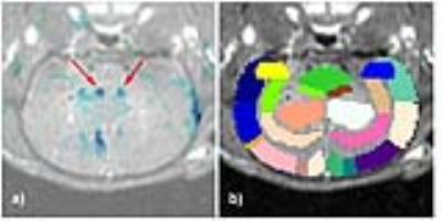 |
Visualizing the effects of ultrasound-based peripheral neuromodulation in the brain
Ileana Hancu, Vickie Cotero, Suresh Joel, Chitresh Bhushan, Jeanette Roberts, Ying Fan, Sireesha Kaanumalle, Jeffrey Ashe, Chris Puleo
We have demonstrated visualization of functional brain changes caused by non-invasive, ultrasound-based stimulation of specific axonal projections within the liver. Following lipopolysaccharide (LPS) injections in a rat animal model, the site-specific liver ultrasound (US) stimulation affected blood glucose levels. The glucose concentration changes were accompanied by increases in the apparent diffusion coefficients (ADC’s) in the paraventricular nucleus (PVN), a known center of afferent nerve pathway termination for integration of outgoing systemic signaling. The local sites of neuronal de-activation (as highlighted by diffusion fMRI) were confirmed by reduced hypothalamic cFOS staining (a marker of neuronal activation).
|
|
2100.
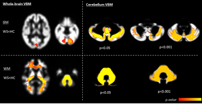 |
MR neuroimaging and proton spectroscopy in Wolfram syndrome
Stefania Evangelisti, Chiara La Morgia, Claudia Testa, David Manners, Claudio Bianchini, Michele Carbonelli, Giulia Amore, Alessandra Maresca, Leonardo Caporali, Raffaele Lodi, Valerio Carelli, Caterina Tonon
We characterized neurodegeneration in Wolfram syndrome by combining MR neuroimaging and proton MRS, and evaluated pathological accumulation of brain lactate as a. mitochondrial oxidative impairment marker. Cerebellar white matter loss was widespread, while grey matter loss was stronger within sensorimotor and cognitive cerebellar lobules. Infratentorial neurodegeneration was confirmed by biochemical signs of neuro-axonal degeneration in cerebellum and pons. The lack of abnormal ventricle lactate suggests an absence of dysfunction of mitochondrial metabolism. These morphological, microstructural and biochemical alterations were in line with neuropathological findings of loss of myelinated axons in the visual system, smaller brainstem and cerebellar white matter loss.
|
|
2101.
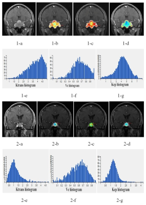 |
DCE-MRI texture analysis based on whole tumor volume for differentiating atypical from typical pituitary adenomas
Yangyingqiu Liu, Jin Shang, Yanwei Miao
The angiogenesis and microvascular permeability of atypical pituitary adenomas were quantitatively analyzed using DCE-MRI texture analysis based on whole tumor volume.
|
|
2102.
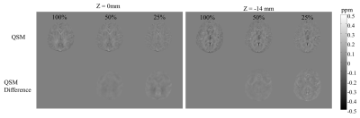 |
Thin Slab Cerebral Quantitative Susceptibility Mapping
Chia-Chen Tsai, Tzu-Cheng Chao, Ming-Hong Ho, Yi-Jui Liu, Ming-Long Wu
Quantitative susceptibility mapping has been a useful tool to monitor magnetic properties of the tissues. Conventional QSM uses thick slab volumetric scan to ensure accurate deconvolution of the dipole kernel for susceptibility estimation. The requirement of large volume coverage and appropriate resolution lead to very long scan time, which has limited QSM’s integration in a clinical protocol. After inspecting dipole kernel’s property, the present work hypothesized QSM should still be performed with a thinner slab to reduce scan time. The results suggest that the reconstructed susceptibility from a whole brain and a thin-slab scan is highly correlated with conventional QSM and the scan time can be reduced up to 4 times.
|
|
2103.
 |
Improving susceptibility mapping using multiple thresholding k-space division
Wen-Tung Wang, Harshan Ravi, Dzung Pham, John Butman
A major challenge in QSM is inverting the acquired phase measurement to estimate the underlying susceptibility. Thresholded K-space division (TKD) is a straightforward technique to calculate the magnetic susceptibility distribution from a single orientation phase images. In this work, we propose to obtain an optimal inverse dipole kernel by using multiple thresholding to minimize the RMSE of the resultant susceptibility map against a susceptibility map
|
|
Neuroimaging: Animal Models
Traditional Poster
Neuro
Tuesday, 19 June 2018
| Exhibition Hall 2104-2131 |
16:15 - 18:15 |
|
2104.
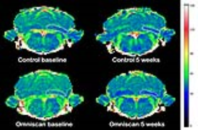 |
Comparison of intravenous and intraperitoneal routes of Omniscan administration with respect to its retention in the rat brain
Serguei Liachenko, Natalya Sadovova, Sherry Ferguson, Joseph Hanig, Merle Paule, Olayinka Dina, Anthony Fotenos, Adebayo Laniyonu, Ira Krefting
Preclinical investigation into the brain retention of gadolinium contrast agents after repeated dosing requires extensive animal handling, particularly for intravenous injections. To decrease the potential stress caused by surgical implantation of intravenous catheters and constant maintenance of those catheters for repeated dosing, we proposed to administer Omniscan for such studies via the intraperitoneal route to laboratory rodents. After 20 dosed over 5 weeks, Omniscan retention was similar in both routes of administration.
|
|
2105.
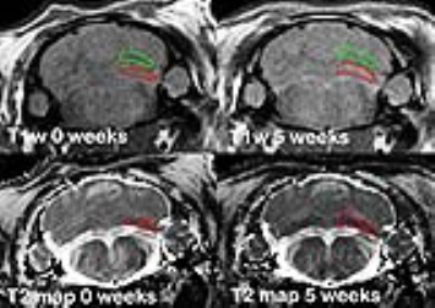 |
Quantitative T2 mapping can reliably detect the retention of Omniscan in the rat brain
Serguei Liachenko, Natalya Sadovova, Sherry Ferguson, Joseph Hanig, Zhen He, Merle Paule, Olayinka Dina, Anthony Fotenos, Adebayo Laniyonu, Ira Krefting
Current methods of investigating brain retention of gadolinium-based contrast agents use T1-weighted MRI, and rarely T1 quantitative mapping. The former does not provide easily quantifiable data and the latter require prolonged scanning time. We proposed the use of a simple ‘off-the-shelf’ T2 mapping technique to reliably quantify relaxation changes in the rat brain due to gadolinium accumulation. The sensitivity of this method is much better compared to the commonly used T1-weighted MRI.
|
|
2106.
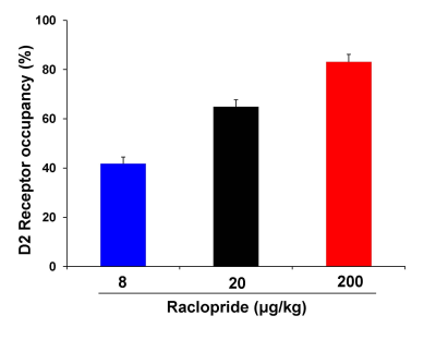 |
Pharmacological MRI response of raclopride in rat: relationship with D2 receptor occupancy or cataleptic behavior
Yukiko Masaki, Yuto Kashiwagi, Takemi Rokugawa, Kohji Abe
Pharmacological MRI allows the visualization of brain pharmacological effects of drugs using fMRI. In order to clarify the relationship between fMRI signal and receptor occupancy or behavioral response, we performed [11C]-raclopride PET, fMRI and the behavioral assessment with raclopride, dopamine D2 receptor antagonist. The positive fMRI response and cataleptic behavior were observed at the dose of raclopride showing 83% of D2 receptor occupancy, but not at the dose of raclopride showing 42% of D2 receptor occupancy. These results suggest that fMRI and behavioral response induced by raclopride will be needed the high D2 receptor occupancy.
|
|
2107.
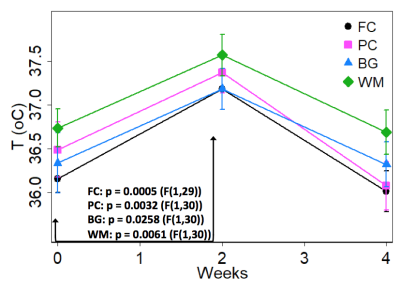 |
Simian immunodeficiency virus infection transiently increases brain temperature in rhesus macaques as detected with magnetic resonance spectroscopy thermometry
Dionyssios Mintzopoulos, Gilberto Gonzalez, Eva-Maria Ratai, Marc Kaufman
Our prior proton Magnetic Resonance Spectroscopy (MRS) studies in Simian Immunodeficiency Virus (SIV)-infected macaques reported higher brain choline and myo-inositol levels at 2 weeks post-infection, suggestive of ongoing inflammation. As brain inflammation has been associated with brain hyperthermia, we used Magnetic Resonance Spectroscopy Thermometry retrospectively to determine whether SIV infection increases brain temperature. At 2 weeks post-infection, we detected increased brain temperature in the frontal and parietal cortex, basal ganglia, and in white matter, relative to pre-infection temperatures. Brain temperatures were strongly correlated with choline levels, suggesting that SIV transiently increases brain temperature by increasing brain inflammation.
|
|
2108.
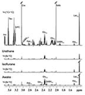 |
Understanding the Impact of Anesthetics on Neuronal and Astroglial Metabolic Activity using 1H-[13C]-NMR Spectroscopy
Anant Patel, Sreemantula Arun Kumar, Pooja Gautam
Neurometabolic rate is coupled with neurotransmitter cycling, which is perturbed during various neurological disorders. Though, anesthetics are widely used in neurometabolic studies, their impacts on neural function is unclear. In the present study, effects of isoflurane and urethane on brain energy metabolism were investigated using 1H-[13C]-NMR spectroscopy in tissue extract during an infusion of [1,6-13C2]glucose or [2-13C]acetate. The reduction in neuronal metabolic activity under isoflurane was higher than urethane in the cerebral cortex and striatum. The data from the study indicate that impacts of anesthetics on neuronal function is more compared to astroglia suggesting that astroglial function is less affected with increased brain activity.
|
|
2109.
 |
Cerebral Reflections of Conditioned Pain Modulation in the Rat: An fMRI study
Silke Kreitz, Tabea Klasfauseweh, Sandra Strobelt, Johannes Kaesser, Isabel Wank, Michael Uder, Andreas Hess
In this fMRI study we introduce an animal model to investigate the neural mechanisms of conditioned pain modulation (CPM) in rat brains. Here, a conditioning tonic cold (10°C water) stimulus at the right hindpaw was used to modulate nociceptive heat stimuli applied to the left hindpaw. Conditioned modulations in functional activation and related network connectivity could be observed in various brain structures involved in pain processing: brainstem and sensory input, lateral thalamus, sensorimotor cortex, frontal association cortex and limbic system. Additionally, over time decreasing resting state connectivity of brainstem and sensorimotor cortex due to cold water stimulation was found.
|
|
2110.
 |
Vitamin B12 Deficiency Perturbed Energy Metabolism in Pre Frontol Cortex: 1H-[13C] NMR Study
Anant Patel, Jitendra Sinha, Shampa Ghosh, TK Sampath Kumar , Manchala Raghunath
The consequences of severe deficiencies in micronutrients especially vitamin B12 on the developing brain during infancy and early post-natal period is not very clear. The current study aim to understand the effects of B12 deficiency on cognitive function using 1H-[13C]-NMR spectroscopy together with [1,6-13C2]glucose infusion in vitamin B12 deficient mice. Our findings indicate reduction in the metabolic activity of glutamatergic and GABAergic neurons in the prefrontal cortex of mice maintained with moderate and severe vitamin B12 defcient diet.
|
|
2111.
 |
MRI detects neural protective effects of DAPT treatment at the subacute stage following cerebral ischemia
Xiaozhu Hao, Yanmei Yang
Notch1 signaling plays time-dependent roles in the sequential process of neurogenesis after stroke. In this study, we aim to detect the appropriate therapeutic time frame of DAPT treatment based on the Notch1 signaling activation and NSCs responses after stroke. Combing the in vivo monitor of comprehensive microstructure changes with diffusion MRI and the in vitro analysis of neurogenesis and remyelination with immunohistology, we ultimately demonstrate the neurorestorative effects of DAPT treatment at the subacute stage after stroke. Our results suggested the appropriate therapeutic time window of inhibiting Notch1 signaling to maximally promote endogenous neurogenesis and axonal reorganization, which could enhance the efficacy of Notch-1 signaling related therapy and promote its application to clinical trials.
|
|
2112.
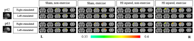 |
Physical exercise enhances adult cortical plasticity in neonatal hypoxic ischemic injured rats: Evidence by BOLD-fMRI and LFP electrophysiological recording
Sun Young Chae, Jun Ho Jang, Geun Ho Im, Moon-sun Jang, Won-Beom Jung, Seungsoo Chung, Jung Hee Lee
The developing brain has a powerful ability to modify its own structure and function for recovery from injury in efforts to compensate for loss of function1,2. In critical period, developing brain has maximal neuronal synaptic connections so it is most amenable to changes in response to external stimulus such as physical exercise3. However, after critical period, neuronal synaptic connections are reduced, and maintained at the reduced state3. Here, we demonstrate enhanced neuroplasticity with physical exercise performed beyond critical period for rats that are injured during critical period. We obtained the BOLD-fMRI response and the interneuron activity with LFP electrophysiological recording.
|
|
2113.
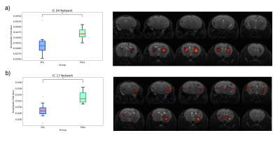 |
Differences in resting state functional networks during pregnancy in C57Bl6 mice
Guadalupe Soria, Raúl Tudela, Emma Muñoz-Moreno, Xavier López-Gil, Roberta Haddad-Tóvolli, Marc Claret
The purpose of this study was to investigate if resting state functional MRI is able to reveal brain network changes associated to pregnancy in C57Bl6 mice. 12 mice were scanned before and 3 weeks after pregnancy using a classical resting state fMRI proptocol. Dual regression was performed using these 20 components to find the subject-specific time-series and spatial maps for each network. Significant differences were observed in the striatal, the insula-amygdala and the hippocampal-brainstem networks. Our results reveal that in pregnant C57Bl6 female micethere is reorganization of brain connectivity in specific brain regions and networks.
|
|
2114.
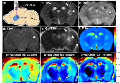 |
Monitoring LPS-induced gray matter inflammation through endogenous contrasts: MT, CEST and NOE
Chenwang Jin, Yanrong Chen, Chenyan Chu, Piotr walzcak, Xiaolei Song
Gray matter (GM) damage is a common phenomenon and clinically relevant in the onset and progression of many neuroinflammation diseases, including Multiple Sclerosis, Alzheimer’s Disease and Depression. However, conventional MRI techniques are insensitive to the detection of GM damage. Chemical exchange saturation transfer (CEST) is an innovative molecular MRI technique that bridges the tissue microstructure and cellular metabolic function, possibly allowing sensing metabolic changes. Our preliminary results suggest that NOE-MRI (Nuclear Overhauser Effect, NOE) may provide a novel biomarker in detection of slight inflammatory changes in cortex and deep GM and also potentially enable quantifing the diffusive GM damages.
|
|
2115.
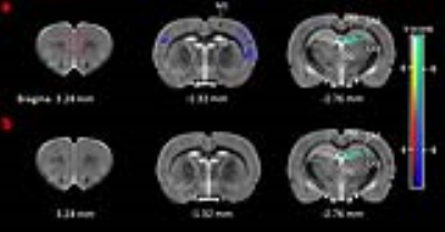 |
Differential Effects of (+)MK801 and (-)MK801 on Brain Structure and Metabolism in Adolescence Rats As Revealed by VBM Analysis and In Vivo 1H-MRS
Yijuan Zou, Hao Lei
N-methyl-Daspartate receptor (NMDAR) antagonists, such as phencyclidine (PCP), ketamine and dizocilpine (MK801), have been widely used for inducing schizophrenia animal models. As a noncompetitive selective NMDAR antagonist, MK801 has two stereoisomers, (+)MK801 and (-)MK801, which have been found to induce different behavioral phenotypes and histological changes in animals. In this study, we compared differential effects of (+)MK801 and (-)MK801 on brain structure and metabolism in adolescence rats with MRI/in vivo 1H-MRS. The results showed that (+)MK801 induced more severe gray matter (GM) atrophy and more evident metabolic changes than (-)MK801, and the different effects were related to their potency at NMDA receptors.
|
|
2116.
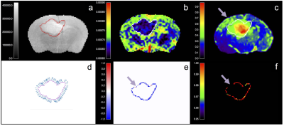 |
Apparent Diffusion Coefficient Correlates with Histological Tumor Burden at the Infiltrating Margins of a Preclinical Glioblastoma Model
Gerard Thompson, Antoine Vallatos, Haitham Al-Mubarak, Lesley Gilmour, Joanna Birch, Lindsay Gallagher, James Mullin, Adam Waldman, William Holmes, Anthony Chalmers
Assessing the imaging boundary of glioblastoma multiforme (GBM) has potential to characterize phenotypic invasiveness relevant to outcomes. Preoperative apparent diffusion coefficient (ADC) changes across this boundary predicts outcome in humans. The tissue specificity of this finding is unknown, hindering the interpretation, further development, and application of the technique. We selected and assessed a relevant preclinical murine infiltrating GBM orthotopic human xenograft model with a novel histological tissue tumor load assessment to investigate relationships between ADC on imaging and cellular infiltration. A robust and strong inverse correlation between the histological tumor infiltration measure and ADC transition is demonstrated, supporting the hypothesis that ADC changes across GBM boundaries represent tumor infiltration and therefore relate to the previously-proposed invasive phenotype imaging biomarker.
|
|
2117.
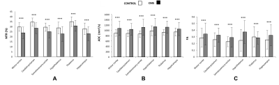 |
Multiparametric magnetic resonance and phenotypic characterization of a mild depression rat model
Teresa Navarro-Hernanz, David Alcázar, Fátima Sanchís, Pilar López-Larrubia
Depression is a common and serious medical illness with a direct impact both in the physical and mental health. It is a complex disorder of the mood with a high incidence in the world population and a tendency to continue increasing. The multifactorial and heterogeneous character of this disease hinders the understanding of the pathological mechanism. The use of an appropriate animal model of depression can contribute to improve the diagnosis and monitoring of the therapy outcome. In this work, we characterized with MRI and phenotyping studies a mild depression model developed in female rats.
|
|
2118.
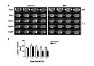 |
Chronic Oral Methylene Blue Treatment in a Rat Ischemic Stroke Model
Lei Huang, Yichu Liu, Zhao Jiang, Timothy Duong
Methylene blue (MB), an FDA-grandfathered drug, has been shown to reduce MRI-defined infarct volume in acute ischemic stroke. However, the efficacy of chronic MB treatment in stroke remains unknown. The goal of this study was to investigate the efficacy of chronic oral MB administration in ischemic stroke using MRI and behavioral tests. We found chronic MB treatment reduced MRI-defined total lesion volumes and improved functional behavioral outcomes, as well as reduced sub-acute hyperperfusion and white-matter damage. Our findings, for the first time, suggest that long-term MB oral administration is safe and has positive therapeutic effects in chronic stroke.
|
|
2119.
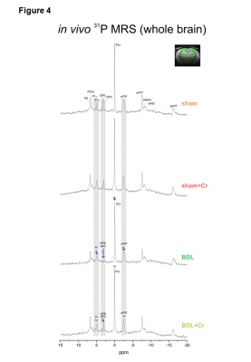 |
Protective effect of high creatine diet during chronic hepatic encephalopathy in young rats, an in vivo longitudinal 1H and 31P MRS study
Veronika Rackayova, Olivier Braissant, Dario Sessa, Stefanita Mitrea, Valerie McLin, Rolf Gruetter, Cristina Cudalbu
Chronic hepatic encephalopathy(CHE) is a serious neuropsychiatric disease with altered neurological status and changes in brain metabolites (among others, decrease in brain tCr). If CHE is acquired in childhood these conditions might perturb normal brain development. Our aim was to test whether oral Cr supplementation dampens the neurometabolic changes observed in CHE in a longitudinal model of chronic liver disease in young rats. Using in vivo longitudinal brain 1H and 31P-MRS, we showed rescued tCr levels, enhanced energy metabolism (restoration of ATP), improved antioxidant capacity (increased Asc), positive effect on phospholipid metabolism and smaller increase in Gln (marker of CHE).
|
|
2120.
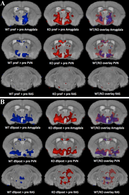 |
Neural activation imaged by MEMRI in mouse models of PTSD: Early Life Stress and Role of the Serotonergic System in Prolonged Response to Fear
Elaine Bearer, Daniel Barto, Alden Reviere, Russell Jacobs
PTSD results from life-threatening fear. We use mouse as an experimental model to investigate acute and persistent fear responses, imaging brain activity by MEMRI, coupled with behavioral responses and histologic confirmation of activity with c-Fos staining. We imaged neural activity at multiple time points in mouse lacking the serotonin transporter, SERT, and with/without early life stress. This approach represents an unbiased comprehensive method to look at the dynamics of the brain’s response to fear over time, not possible by other imaging methods. We find altered activity and circuits in mice after fear dependent on genotype and environment.
|
|
2121.
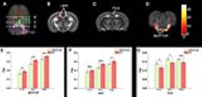 |
Diffusion tensor imaging reveals altered brain development of MECP2 overexpressing rat in cerebellar and limbic structures
Jian-kun Dai, Yu-yan Chen, Zhifeng Liang
In this study, we used diffusion tensor imaging to investigate the effect of MECP2 overexpressing (MECP2-OE) on the rat brain development. Our results showed the MECP2-OE mainly affected the cerebellar fiber tracts and limbic structures. Behavior tests showed the MECP2-OE rats presented significant defects of social interaction than the wild type (WT) rats.
|
|
2122.
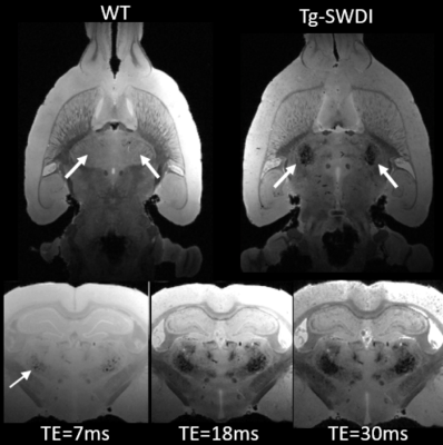 |
A novel transgenic rat model of evolving cerebral amyloid angiopathy (CAA)
Hedok Lee, Xiaodan Liu, Simon Sanggaard, Sunil Koundal, Feng Xu, William Van Nostrand, Helene Benveniste
Understanding the pathophysiology of cerebral amyloid angiopathy (CAA) has become increasingly important because there is evidence to suggest that vascular dysfunction plays an important role in early component in the development of Alzheimer’s disease (AD). To study CAA, Tg-SwDI transgenic mouse model was recently extended to rat (Tg-DI) and here we report the first MRI studies to characterize CAA in Tg-DI in both in vivo as well as in vitro using 3D-GRE sequence. Conspicuous lesions were detected in thalamus in Tg-DI at very early stage, consisting of multiple pathological changes including micro-bleeds, extravasation of blood products and/or occluded vessels.
|
|
2123.
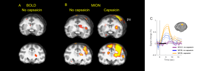 |
Comparison of BOLD and MION enhanced CBV fMRI to the noxious stimulus in anesthetized rhesus monkey
Eunha Baeg, Boo-Hee Choi, Chan-Ung Park, Choong-Wan Woo, Seong-Gi Kim
Using contrast agent in fMRI has the benefits of providing additional information of regional cerebral blood volume (CBV) and of enhancing sensitivity. Response comparisons of BOLD and MION enhanced CBV fMRI to the noxious stimulus in non-human primate showed signal increase for MION fMRI in the regions that are important in pain-processing network. Activities of some brain areas, including putamen, were captured with MION fMRI, not with BOLD. Capsaicin treatment augmented the responses of fMRI to the noxious stimulation for BOLD and MION fMRI. New insight can be obtained for the pain network through the comparison between BOLD and CBV fMRI.
|
|
2124.
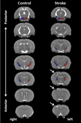 |
Changes in corticospinal tract integrity in relation to recovery after cortical stroke as measured with DTI-based tractography in rat brain
Geralda van Tilborg, Michel Sinke, Anu Meerwaldt, Annette van der Toorn, Caroline van Heijningen, Milou Straathof, Mohamed Ali, Khalid Al-Saad, Rick Dijkhuizen
Preserved or restored integrity of the corticospinal tract (CST) is critical for motor recovery after stroke. However, data on spatiotemporal alterations in CST integrity after stroke are largely lacking. Here we implemented diffusion tensor-based tractography to identify the CST in rat brain, which we applied to measure microstructural changes along the CST following experimental stroke to the sensorimotor cortex. Number of tractography streamlines, fractional anisotropy (FA) and axial diffusivity (AD) were reduced 1 week post-stroke, and recovered to control levels after 28 weeks. This temporal pattern, reflective of white matter remodeling, coincided with loss and recovery of sensorimotor function.
|
|
2125.
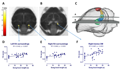 |
In vivo DTI to correlate in ‘real time’ testosterone-induced neural changes to song performance in a seasonal songbird
Jasmien Orije, Geert De Groof, Sofie Van Massenhoven, Elisabeth Jonckers, Veerle Darras, Annemie Van der Linden
The dynamic relationship between song performance and neuroplasticity induced by testosterone implantation was monitored longitudinally in a seasonal songbird (European starling) by using in vivo DTI. Voxel based analysis showed that the song bout length was positively correlated to the fractional anisotropy changes in different parts of the motor pathway. Meaning that the motor pathway strengthens as song performance advances under the influence of testosterone.
|
|
2126.
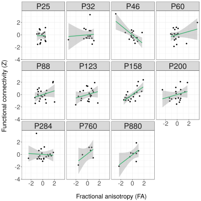 |
Dynamic Structural-Functional Relationship between Left and Right Somatosensory Cortex in Rats across the Lifespan
Michel Sinke, Milou Straathof, Paul Weerheim, Willem Otte, Rick Dijkhuizen
The relationship between structural and functional brain connectivity across the mammalian lifespan is largely unknown. To elucidate the temporal characteristics of this relationship we longitudinally acquired high-field resting-state fMRI and diffusion-MRI in rats, from early infancy to old age. We specifically examined the interhemispheric connectivity between homologous primary somatosensory cortices, a major part of the sensorimotor system. The structure-function correlation increased from about 0 during infancy to 0.4 around adulthood, followed by a further gradual increase towards old age. This reflects dynamic patterns of lifelong brain remodeling, which may underlie variations in brain disease etiology during development and ageing.
|
|
2127.
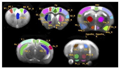 |
Kinase-inactive Met mice show altered forebrain functional connectivity: A resting state functional MRI study
Shiyu Tang, Elizabeth Powell, Reha Erzurumlu, Wenjun Zhu, Fu-Sun Lo, Su Xu
MET, the gene encoding tyrosine kinase receptor for hepatocyte growth factor, is a susceptibility gene for autism spectrum disorder (ASD). Genetically altered mice with a kinase-inactive Met offer a potential model for understanding neural circuit organization changes in autism. We employed resting-state functional MRI to a kinase-inactive Met mouse model to test our hypothesis that aberrant functioning of the somatosensory-thalamocortical system is at the core of the conspicuous somatosensory behavioral phenotypes observed in autism. Results showed impaired organization of large-scale network and increased somatosensory-thalamocortical connectivity with a sex dependent manner and differences between heterozygous and homozygous Met-Emx1 mice.
|
|
2128.
 |
Abnormal growth trajectories of white matter in spontaneously hypertensive rats when compared to non-hypertensive controls: Implications for small vessel disease progression
Sunil Koundal, Simon Sanggaard, Kristian Mortensen, Helene Benveniste, Maiken Nedergaard, Hedok Lee
The spontaneously hypertensive rat (SHR) is a clinically relevant animal model in studying small vessel disease. Whole brain morphological differences between SHR compare to normotensive Wistar-Kyoto (WKY) rats were evaluated in parallel with development of chronic hypertension. Voxel-wise deformation based morphometry indicated progressive enlargement of the cerebral ventricles in SHR compare to WKY, and a fraction of the body and splenium of corpus callosum in SHRs were significantly smaller in the middle-aged rats but not in young-aged rats.
|
|
2129.
 |
In-Vivo Analysis of the Superficial White Matter in the Macaque Brain Using High-resolution Diffusion MRI: preliminary results
Yann Bihan-Poudec, Slimane Tounekti, Nathalie Richard, Mathilda Froesel, Franck Lamberton, Thomas Troalen, Suliann Ben Hamed, Maxime Descoteaux, Bassem Hiba
This study was focused on the U-fibers located on the Superficial White Matter (SWM) of the Rhesus macaque brain. A diffusion MRI (dMRI) pulse-sequence with a 3D multi-shot-EPI module was used to achieve a 0.5 mm isotropic dMRI data in 4 macaques.
The organization of white matter in the region of arcuate sulcus (AS) was analyzed using diffusion tensor and fiber orientation distribution data, the U-fiber over all the AS was tracked and its water diffusion metrics were quantitatively assessed.
The results, obtained using high-resolution dMRI, pave the way for quantitative analyses of SWM for clinical and neuroscientific applications.
|
|
2130.
 |
An optimized DCE-technique detects weak contrast agent accumulation undetectable on post-contrast T2*-weighted acquisitions: application to a model of neuroinflammation
Teodora-Adriana Perles-Barbacaru, Corane Karoutchi, Isabelle Varlet, Monique Bernard, Angele Viola
No dynamic contrast-enhanced (DCE) study has been published so far in Experimental Allergic Encephalomyelitis (EAE), although DCE-MRI is used in human Multiple Sclerosis. This study reports a DCE protocol optimized for mouse brain imaging of subtle and delayed contrast agent accumulation and applies it to the study of EAE with moderate neurological signs. Two-fold signal increase with respect to the vascular volume fraction can be detected while even moderately enhancing lesions remain visually undetectable on pre and post-contrast T2w and T2*w acquisitions. Ventricles, midbrain and ventral olfactory bulb are first to be affected in moderate EAE.
|
|
2131.
 |
Advanced MR imaging characterization of a novel in vivo xenograft model mimicking recurrent glioblastoma
Mona Al-Gizawiy, Robert Wujek, Melissa Prah, Hisham Alhajala, Ninh Doan, Jeffrey Knipstein, Jennifer Connelly, Shama Mirza, Christopher Chitambar, Kathleen Schmainda
We have developed a robust and reproducible rat xenograft model of recurrent GBM by irradiating adult and pediatric GBM cell lines in vitro prior to brain inoculation. Both advanced MR imaging and histological analyses highlight the amplified aggressiveness of the resultant tumor compared to the conventional U-87MG xenograft, as evidenced by profound vascularization and increased cell proliferation. Moreover, our recurrent GBM model exhibited invasive lesions with areas of infiltrating neutrophils and necrosis, all features that are not associated with conventional U-87MG xenograft tumors. Shortened survival of animals bearing irradiated U87-10Gy or SJGBM2-10Gy tumors further reinforces the aggressive nature of the model.
|
|
Brain Tumours
Traditional Poster
Neuro
Tuesday, 19 June 2018
| Exhibition Hall 2132-2157 |
16:15 - 18:15 |
|
2132.
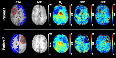 |
Quantifying tumour oxygenation using streamlined-qBOLD
Alan Stone, Esther Warnert, Puneet Plaha, Natalie Voets, Nicholas Blockley
Hypoxia and high metabolic demand are important identifying features of high-grade gliomas. Imaging methods capable of mapping tissue oxygenation therefore have the potential to provide non-invasive information about the metabolic environment of tumour tissue and may produce useful markers for stage grading and monitoring treatment efficacy. Here we demonstrate the use of streamlined-qBOLD for mapping tumour oxygenation.
|
|
2133.
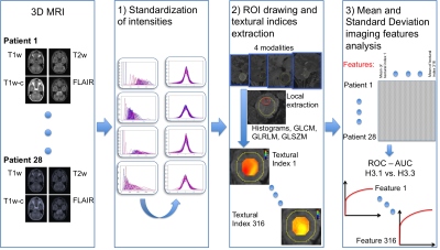 |
Volume-independent radiomic features from T2w-FLAIR MRI could reveal mutation of histones in diffuse intrinsic pontine glioma
Jessica Goya-Outi, Fanny Orlhac, Raphael Calmon, Cathy Philippe, Stéphanie Puget, Nathalie Boddaert, Irène Buvat, Jacques Grill, Vincent Frouin, Frédérique Frouin
In diffuse intrinsic pontine glioma, the mutations of histones (H3.1 versus H3.3) are correlated with patient survival. A new method to compute radiomic features free of tumor volume effect was applied to four structural MR modalities and patients were classified according to histone mutation. The tumor was scanned by a 5 mm radius sphere and textural indices were computed inside each position. A total of 37 features calculated from T2w-FLAIR yielded an area under the Receiver Operating Characteristics curve greater than 0.85. T2w-FLAIR appears to be the most informative modality to predict mutation type.
|
|
2134.
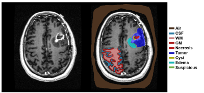 |
Quantifying individual and collective prediction accuracy of MR contrasts for glioma tissue compartment classification
Jason Parker, Emily Diller, Robert Lober
The purpose of this work was to evaluate the relative contributions of MR contrasts to tumor tissue classification. Seventeen (17) glioma patient datasets (WHO grade II-IV) containing T1, T1+gad, T2, FLAIR, and ADC were studied using multinomial logistic regression. T2 images had the highest individual classification accuracy (78.1%). Classification accuracy improved with each additional contrast, leading to an overall accuracy of 84.1% for all 5 contrasts. The multinomial logistic regression showed that together the 5 contrasts had greater tumor tissue classification accuracy than individually, but that the improvement in accuracy was not linear and decreased as more MR data was included. Lower grade gliomas and GBM could be predicted by the percentage of voxels classified as suspicious by the regression model, but not by any other class. These results may aid in clinical protocol development and optimization for neuro-oncologic imaging, especially in situations where overall scan time is limited.
|
|
2135.
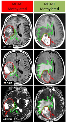 |
Multi-sequence and Habitat-based Radiomics Analysis to Predict MGMT Promoter Methylation Status in Grade II-IV Gliomas Using Magnetic Resonance Imaging
Jingwei Wei, Jie Tian, Dongsheng Gu, Xiaohan Hao, Guoqiang Yang, Yan Tan
In this study, we performed multi-habitat and multi-sequence MRI radiomics to make preoperative prediction on MGMT promoter methylation in grade II-IV gliomas. Quantitative imaging features were extracted on each habitat from CE-T1WI, T2FLAIR and ADC maps to reveal the genetic heterogeneity of the tumor and describe the subtle textural characteristics of different molecular subtypes. The habitat-integrated radiomics signature behaved more stable and had better predictive efficacy than one-region based radiomics signature. The final constructed predictive model incorporating the proposed radiomics signature and traditional clinical predictors achieved the optimal performance on the MGMT status.
|
|
2136.
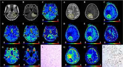 |
Comparative analysis of diffusion kurtosis imaging, diffusion tensor imaging and diffusion weighted imaging in grading and assessing cellular proliferation of meningioma
Lin LIN, Yunjing Xue , Qing Duan
An accurate evaluation of the WHO grade and cellular proliferation is particularly important in meningiomas, it may facilitate treatment decisions and improve clinical prognosis. But conventional magnetic resonance (MR) imaging were not sufficiently accurate in evaluating the meningioma grade and Ki-67 expression. This study prospectively evaluate and compare diffusion kurtosis imaging (DKI), diffusion tensor imaging(DTI) and diffusion weighted imaging (DWI) metrics in determining the grade and cellular proliferation of meningiomas. It was found that DKI is a better diffusion technique for assessing the grading and cellular proliferation of meningiomas compared to conventional diffusion imaging.
|
|
2137.
 |
Growth patterns of non-enhancing glioma assessed on DTI-derived isotropic and anisotropic maps are not associated with IDH and 1p19q codeletion status
Renske Gahrmann, J.K.H. Spoor, MMJ Wijnenga, S Leenstra, AJPE Vincent, M de Groot, PJ French, MJ van den Bent, M Smits
Previous reports show that IDH-mutation status can be determined in glioblastoma using DTI-derived isotropic (p) and anisotropic (q) maps to measure infiltrative growth along white matter tracts by determining the extent or pattern of p/q mismatch: abnormal p overlaps normal q-areas by >0.5cm. We use this method in presumed low-grade (i.e. non-enhancing) gliomas to see if infiltrative growth patterns correlate with IDH-mutation and 1p19q codeletion status, which in turn are correlated with prognosis.
|
|
2138.
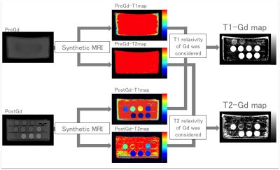 |
Gadolinium concentration map based on synthetic MRI and its application to brain metastases
Misaki Nakazawa, Akifumi Hagiwara, Christina Andica, Masaaki Hori, Moeko Horita, Koji Kamagata, Haruyoshi Houshito, Shigeki Aoki
Signal intensity measured on T1-weighted image is not proportional to the gadolinium concentration in vivo after administration of contrast agent. Thus, some calculations are required to estimate gadolinium concentration using quantitative values before and after gadolinium administration. We created gadolinium concentration maps that directly show the amount of contrast agent using quantitative maps calculated using synthetic MRI. The gadolinium concentration map we created using phantoms showed high accuracy and precision. The gadolinium concentration map could reliably measure the concentration of gadolinium in metastatic brain tumors.
|
|
2139.
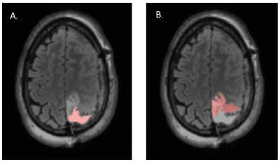 |
Quality of Life, Neurocognitive Function and T2 FLAIR Hyperintensity Volume in Stable Grade II and III Glioma Patients
Angela Jakary, Tracy Luks, Susan Chang, Jennifer Clarke, Nicholas Butowski, Nancy Ann Oberheim Bush, Jennie Taylor
Quality of life and neurocognitive function are important clinical outcome measures for patients with lower grade glioma. In this pilot study, we performed neurocognitive testing and quality of life assessments in radiologically and clinically stable grade II and III glioma patients who were not receiving active treatment. We found novel associations between standard clinical assessments and neuroimaging metrics at pre-surgical and follow-up timepoints. Further characterizing the longitudinal relationship between structural and functional neuroimaging, neurocognition and quality of life will better allow clinicians to proactively intervene to help patients in future.
|
|
2140.
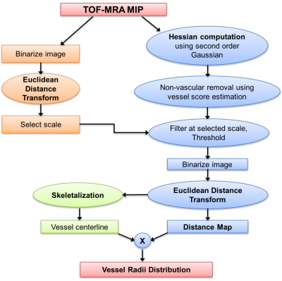 |
Robust Quantification of Changes in Arterial Cerebral Vasculature Post Radiation Therapy in Pediatric Brain Tumor Survivors
Sivakami Avadiappan, Sam Payabvash, Angela Jakary, Erin Felton, Melanie Morrison, Christopher Hess, Sabine Mueller, Janine Lupo
With the improved long-term survival of children with brain tumors, understanding the late effects of their therapy on small arterioles is of great importance. We developed a method for robust segmentation of arteries and quantification of their thickness using TOF-MRA at 7T and estimated the vessel radii distribution in irradiated patients compared to controls. Radiation-induced damage to the microvasculature resulted in a higher fraction of small vessels observed with time from radiation therapy, likely due to vessel thinning.
|
|
2141.
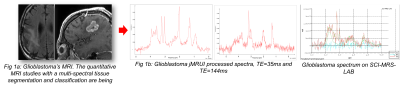 |
Importance of early spectral variations during 36 months of longitudinal follow MRI and MRS in 90 patients treated glioblastomas
J.-M. Constans, A. Heintz, O. Seloi , J.P. Chombar, N. Deleval, R. Hanafi, W. Dou, S. Ruan, J. Prades, D. Le Gars, O. Baledent, H. Deramond, A. Houessinon, A. Fichten, M. Lefranc, A. Coutte, P. Toussaint, C. Desenclos, B. Chauffert, M. Boone
MRS allows non-invasive follow-up of treated glioblastomas tumors. There is a large variability, but repetition and modelisation of spectroscopic measurements during longitudinal follow-up could allow us to diminish it and to improve prognostic evaluation especially in long survivors and patients with proliferation relapses. Studying the relationship between MRS measures, segmentation and perfusion parameters could lead to better understanding of tumoral processes and of therapeutic response, especially with regard to chemotherapy, radiotherapy and antiangiogenic molecules and in the future oxidative stress and hypoxia modulators.
|
|
2142.
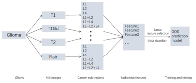 |
A Large Scale Radiomics Profiling Strategy for Glioma Overall Survival Prediction
Pan Sun, Defeng Wang, Queenie Chan, Lin Shi
Glioma is the most common brain intracranial malignancy, which accounts for about 80% of malignant brain tumors in adults and its median survival rate is 12 months. In clinical, how to accurately predict the glioma overall survival (GOS) is a crucial work and it will be beneficial to monitor tumor progression, execute surgery as well as plan radiotherapy and follow-up studies. However, the glioma generally has highly heterogeneity degrees in the histological tumor sub-regions. we propose a comprehensive multi-modality MRI radiomics way of predicting the GOS. Different features are proposed committing to different image modalities. A feature selection strategy is applied for the optimal features and then random forest is contributed to the classification of short-survivors and long-survivors. With the performance evaluation criteria, our model showed promising classification ability for the brain tumor.
|
|
2143.
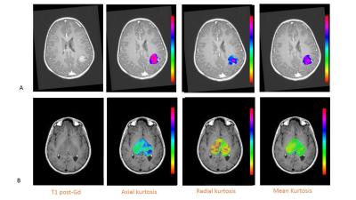 |
Diffusion kurtosis imaging (DKI) can help to differentiate low- and high-grade gliomas in pediatric patients: a prospective single centre experience with the simultaneous multislice (SMS) technique
Antonio Napolitano, Ioan Voicu, Lorenzo Lattavo, Maria Camilla Rossi Espagnet, Chiara Carducci, Angela Mastronuzzi, Paolo Tomà, Giovanna Colafati
Pediatric brain glioma is a very devastating brain tumour and the most frequent solid tumour in children. Differentiating low- from high-grade glioma without the use of invasive biopsy is important to optimize patient management strategies yet difficult with imaging alone. Diffusion kurtosis imaging is then an emerging technique that has shown the ability of discriminating grades in adults. We make use of multislice approach to acquire and evaluate kurtosis metrics in brain gliomas and show how estimation of the heterogeneity of the tumour might be indicative of its grade.
|
|
2144.
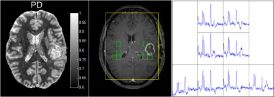 |
Quantitative proton density values compared to 1H MRSI in areas of contrast enhancement of glioma patients after surgical resection.
Felix Raschke, Tim Wesemann, Hannes Wahl, Steffen Appold, Mechthild Krause, Jennifer Linn, Esther Toost
In this study we measured mean proton density (PD) values in MR spectroscopic imaging (MRSI) voxels showing contrast enhancement of glioma patients 30 days ± 12 days after surgical resection. MRSI voxels with (partial) overlap with contrast enhancing areas were manually selected. Mean PD values showed a significant inverse correlation with NAA/Cho indicating that areas with higher PD are more likely to contain residual tumour tissue rather than surgery-related tissue damage. There was, however, no correlation of PD with Cho/Cr, which suggests that quantitative PD values are unable to determine tumour aggressiveness.
|
|
2145.
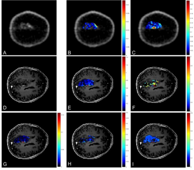 |
Association between pharmacokinetic parameters from DCE-MRI and metabolic parameters from dynamic 18F-fluoromethylcholine PET in human brain glioma
Marianna Inglese, Matthew Grech-Sollars, Katherine Ordidge , Vijaykumar Vaja , Lesley Honeyfield, Sameer Khan, Tara Barwick, Eric Aboagye, Adam D Waldman
Magnetic resonance imaging (MRI) is the standard imaging technique in the diagnosis of primary brain lesions. However, novel PET imaging techniques such as choline-PET are currently being investigated in the clinic to characterize tumour metabolism. In this study, we compared pharmacokinetic parameters resulting from the modelling of dynamic contrast enhanced (DCE) MRI data, using the Tofts model (TM) and shutter speed model (SSM), with metabolic macroparameters derived from the application of the spectral analysis (SA) to dynamic PET data. We observe a correlation between some pharmacokinetic parameters and the parameters obtained through spectral analysis of the dynamic choline-PET data.
|
|
2146.
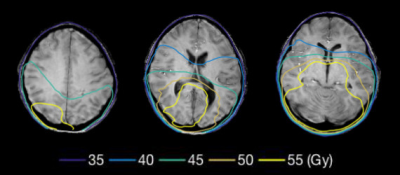 |
Quantitative susceptibility imaging for the assessment of early radiation-induced white matter injury in children with primary brain tumors
Junjie Wu, Susan Palasis, Natia Esiashvili, Richard Jones, Eduard Schreibmann, Deqiang Qiu
We examined radiation-induced white matter injury using quantitative susceptibility mapping (QSM) in children with primary brain tumors. Following radiation therapy, susceptibility changed with time and dose. QSM may be a useful biomarker for irradiation damage.
|
|
2147.
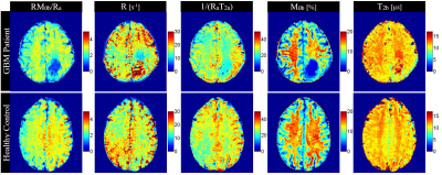 |
Effects of Glioblastoma (GBM) on quantitative MRI of Contralateral Normal Appearing White Matter
Hatef Mehrabian, Wilfred Lam, Sten Myrehaug, Arjun Sahgal, Greg Stanisz
Normal-appearing white matter on the contralateral hemisphere (cNAWM) of glioblastoma (GBM) has been shown with MRS and DTI to be abnormal which might be due to tumor cell infiltration into these distant normal appearing brain structures. Chemical exchange saturation transfer (CEST), quantitative magnetization transfer (qMT) and transverse relaxation time (T2) are sensitive to changes in tissue microstructure and metabolism. CEST, qMT and T2-mapping were used to investigate abnormalities in cNAWM. Results demonstrated differences in white mater cellular density (measured with T2 and qMT) as well as metabolism (measured with CEST) in cNAWM of GBM patients compared to healthy controls.
|
|
2148.
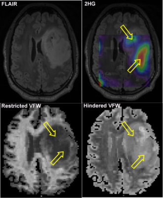 |
Relationship Between Tumor Cellularity and Metabolic Activity in IDH-Mutant Gliomas: A Correlative Study with 2-Hydroxyglutarate MRSI and a Novel Diffusion MRI Method
Ina Ly, Ovidiu Andronesi, Qiuyun Fan, Barbara Wichtmann, Aapo Nummenmaa, Brian Nahed, William Curry, Daniel Cahill, Tracy Batchelor, Jayashree Kalpathy-Cramer, Bruce Rosen, Elizabeth Gerstner
Anatomic T2/FLAIR sequences are the gold standard in the diagnostic and monitoring process of non-enhancing gliomas but do not provide accurate information about the underlying metabolic activity of the tumor. In this work, we investigated the combined use of 2-hydroxyglutarate (2HG) magnetic resonance spectroscopic imaging (MRSI) and a novel three-compartment diffusion MRI method (Linear Multi-Scale Model) to characterize isocitrate dehydrogenase-mutant gliomas, and found that high 2HG levels correlated with decreased restricted diffusion.
|
|
2149.
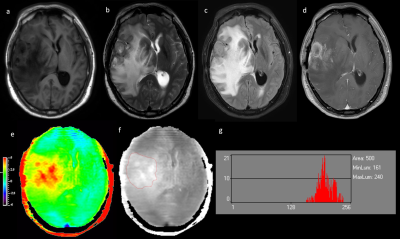 |
Differentiating glioma histologic grade using histogram analyses of Amide Proton Transfer MRI
Qihong Rui, Yingjie Mei, Hao Yu, Xianlong Wang, Shanshan Jiang, Jinyuan Zhou, Zhibo Wen
A correct preoperatively grading of glioma is always the important issue in clinic. APT imaging is designed to assess glioma on the level of cell and molecule. In this study we used the APT MRI histogram analyses ,to determine if it can help differentiate HGG from LGG.
|
|
2150.
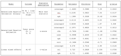 |
Multiparametric metabolic and physiologic MR-Imaging models for differentiating tumor from treatment effects in patients suspected of recurrent glioblastoma
Julia Cluceru, Sarah Nelson, Annette Molinaro, Joanna Phillips, Marram Olson, Marisa LaFontaine, Angela Jakary, Devika Nair, Soonmee Cha, Susan Chang, Janine Lupo
Despite previous research on physiological and metabolic MR imaging techniques with standard clinical anatomical MRI of patients with recurrent glioma, there is still no one parameter that can differentiate recurrent glioblastoma (rGBM) from treatment-induced effects (TxE) with high enough accuracy to be used clinically. We assessed the value of incorporating anatomical, perfusion-weighted, diffusion-weighted, and spectroscopic imaging parameters to identify TxE in patients suspected of rGBM. nPH from DSC perfusion-weighted imaging and Choline-to-NAA Index from MR spectroscopic imaging were found to be the most related to pathological markers of tumor and TxE.
|
|
2151.
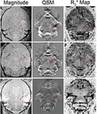 |
Comparison of R2* and quantitative susceptibility mapping in the characterizing tumor hypoxia in a mouse model of glioblastoma
Runze Yang, A. Hamilton, Hongfu Sun, Susobhan Sarkar, Reza Mirzaei, G. Pike, V. Wee Yong, Jeff Dunn
Hypoxia (low levels of oxygen) is an important biomarker in many solid tumors, as it is responsible for promoting tumor angiogenesis and resistance to radiotherapy. Hypoxia can be indirectly monitored by measuring levels of deoxyhemoglobin with MRI, using either R2* or quantitative susceptibility mapping (QSM). We compared the two methods for brain tumor and hypoxia imaging in a mouse model of glioblastoma. We found that both methods were sensitive at detecting a decrease in deoxyhemoglobin due to 100% oxygen. However, QSM provided better anatomical information and was better at detecting tumor heterogeneity. QSM is a promising tumor imaging method.
|
|
2152.
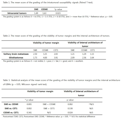 |
The diagnostic value of postcontrast susceptibility-weighted imaging in the assessment of intracranial brain neoplasm at 3T
Hyunkoo Kang
The aim of this study is to estimate the diagnostic value of postcontrast susceptibility-weighted imaging (CESWI) in the assessment of intracranial brain neoplasm at 3 T MRI. Our results showed that the SWI can be performed after gadolinium injection without information loss or signal change and the CESWI clearly visualized the characteristics and the architecture of brain neoplasm. The CESWI can be a match to the CET1 with regard to the visibility of tumor margin and internal architecture in intracranial tumors without information loss or signal change.
|
|
2153.
 |
The diagnostic value of the distribution pattern of intratumoral susceptibility sign of intracranial tumors on susceptibility-weighted imaging
Hyunkoo Kang, Seongwon Jang
The aim of this study is to determine whether the distribution pattern of intratumoral susceptibility sign (ITSS) derived from susceptibility-weighted imaging (SWI) could differentiate glioblastoma multiforme (GBM) and single brain metastasis. We compared the grade of the visibility of ITSS in the central portion of tumors (CITSS) and in the tumor capsular area (PITSS) on SWI. Our findings suggest that there were different characteristics of ITSS between GBM and brain metastasis on SWI due to the profound difference in histologic feature of capillary between the two tumor types.
|
|
2154.
 |
Comparison of Dynamic Susceptibility Contrast and Arterial Spin Labeling at the Target Locations of Image guided Tissue Samples for Patients with Glioma
Marisa Lafontaine, Janine Lupo, Marram Olson, Joanna Phillips, Susan Chang, Sarah Nelson
Arterial spin labeling and dynamic susceptibility contrast perfusion weighted imaging were both found to provide acceptable measures of blood vessel angiogenesis in brain tumors compared to pathological measures but dynamic susceptibility contrast may be better correlated with the underlying vascular morphology.
|
|
2155.
 |
Differentiation of grade II/III and Grade IV glioma by combining ‘T1 contrast enhanced brain perfusion imaging’ and susceptibility weighted quantitative imaging
Jitender Saini, Pradeep Kumar Gupta, Prativa Sahoo, Anup Singh, Rana Patir, Sunita Ahlawat, Manish Beniwal, K. Thennarasu, Vani Santosh, Rakesh Kumar Gupta
The purpose of this study is to evaluate the usefulness of T1-perfusion MRI and SWI in discriminating among grade II, III and IV gliomas. We found that combining T1-perfusion and SWI improves the diagnostic accuracy for discrimination of grade III from grade IV gliomas and T1-perfusion MRI derived rCBV alone appears to be an excellent measure for discriminating grade II from grade III glioma.
|
|
2156.
 |
ADC-map-based classification of glioma-subtypes in diffusion-weighted MR-Imaging
Nils Nuessle, Johann Hempel, Jens Schittenhelm, Uwe Klose
DWI showed great potential for estimation of histopathological and molecular profile of human glioma. 97 patients with suspected glioma underwent pre-operative MRI-scans, including high b-value DWI. ADC-maps from pairs of two b-values were calculated. Post-interventional histopathological tumor grading was realized on a molecular basis using the molecular markers IDH-mutation, 1p/19q- and ATRX-loss. Significant differences (p < 0.001) were found between oligodendroglioma, astrozytoma and GBM. Best discrimination was achieved when calculating the ADC-maps from b-values of 500 and 2500 s/mm2. Therefore, ADC-map based evaluation of glioma in DWI provides great potential in accurate pre-interventional diagnosing of glioma subtypes.
|
|
2157.
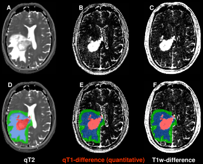 |
Quantitative T1-difference maps and T1-weighted difference images: which modality is better at identifying tumor infiltration in high grade gliomas?
Ulrike Nöth, Ralf Deichmann, Oliver Bähr, Julia Tichy, Stephanie Lescher, Elke Hattingen
In glioblastoma patients, differences of quantitative T1 (qT1) maps acquired before and after contrast agent (CA) administration are visually compared to the respective differences of conventional T1-weighted (T1w) images. Quantitative T1-differences are determined in the following regions-of-interest (ROIs): (1) enhancing tumor, (2) edema, (3) 5mm-rim around (1)+(2), (4) control tissue contralateral to the tumor. T1w- and qT1-differences clearly show the enhancing tumor, but only the qT1-difference maps show signal enhancement in the edema, which is in line with elevated qT1-difference values in this ROI. This indicates most likely CA leakage due to tumor infiltration.
|
|
| Back |
| The International Society for Magnetic Resonance in Medicine is accredited by the Accreditation Council for Continuing Medical Education to provide continuing medical education for physicians. |
