|
Traditional Poster Session
Contrast Mechanisms |
Wednesday, 20 June 2018
Traditional PosterContrast Mechanisms
2158 -2189 Perfusion Methods
2190 -2221 Quantitative Susceptibility Mapping
2222 -2249 CEST: Novel Methods & Applications
2250 -2265 Novel Contrast Mechanisms: Body
2266 -2297 Contrast Mechanisms |
| |
Perfusion Methods
Traditional Poster
Contrast Mechanisms
Wednesday, 20 June 2018
| Exhibition Hall 2158-2189 |
08:15 - 10:15 |
|
2158.
 |
Comparing pCASL CBF measurements between 3D-GraSE and 2D-EPI on 1.5T and 3T systems
Koen Baas, Henri Mutsaerts, Jan Petr, Joost Kuijer, Kim van de Ven
We have compared CBF value agreement in healthy subjects across two readouts, 3D-GraSE and 2D-EPI, and two field strengths, 1.5T and 3T, and investigated with which acquisition parameters we can reach the best agreement. Significantly higher GM CBF was observed with the 2D-EPI readout compared to the 3D-GraSE readout with equivalent acquisition voxel size (p < 0.005 for 1.5T and p < 0.05 for 3T). Better agreement was observed between 3D-GraSE and 2D-EPI on 3T systems when the resolution of the 3D-GraSE readout was increased to match the effective resolution to the 2D-EPI scan (ICC = 0.772 and ICC = 0.932 respectively).
|
|
2159.
 |
Reproducibility and repeatability of 3D-GraSE and 2D-EPI ASL on 1.5T and 3T systems in healthy elderly
Koen Baas, Henri Mutsaerts, Joost Kuijer, Kim van de Ven
We present the results of a reproducibility and repeatability study in 34 healthy elderly scanned on 1.5T and 3T systems employing pCASL with a 3D-GraSE and 2D-EPI read-out. Best repeatability and reproducibility were achieved when using 3D-GraSE readout on 3T systems leading to an average repeatability and reproducibility of GM CBF of 2.7% ± 1.8% and 2.9% ± 3.5% respectively. The repeatability and reproducibility of 2D read-out and of comparisons at 1.5T and 1.5T versus 3T were slightly lower. These results imply that 3D-GraSE pCASL at 3T should be preferred in multi-center trials as well as for clinical imaging.
|
|
2160.
 |
Background-suppression is more important for ASL at higher magnetic field strength
Lydiane Hirschler, Suzanne Franklin, Sophie Schmid, Wouter Teeuwisse, Matthias van Osch
Background suppression is a recommended and frequently employed strategy to improve the perfusion-temporal-SNR (tSNR) of ASL. Since physiological signal fluctuations are known to be a major source of data corruption in functional MRI at higher magnetic field-strengths, it might also be expected that the benefits of BGS are even stronger at higher field-strengths. In this study, we evaluated and compared the importance of the introduction of BGS-pulses at 3T and 7T and show that, at higher magnetic field, BGS is even more crucial.
|
|
2161.
 |
A novel hybrid of time-encoded and sequential multi-PLD PCASL for improved cerebral blood flow estimation
Joseph Woods, Michael Chappell, Thomas Okell
We present a novel hybrid combination of time-encoded and sequential multi-PLD pseudo-continuous ASL, which benefits from the advantages of both techniques, and demonstrate that the increased flexibility of this approach improves CBF precision compared to either method alone.
|
|
2162.
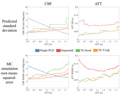 |
Comparison of optimized single-PLD, sequential multi-PLD and time-encoded PCASL for cerebral blood flow measurements
Joseph Woods, Michael Chappell, Thomas Okell
In this work, we use an objective approach to optimize sequential and time-encoded multi-PLD protocols, and compare them to the recommended single-PLD protocol using simulations, with the aim of determining which method is capable of producing the most accurate CBF estimates across a range of ATTs.
|
|
2163.
 |
Tracer kinetics of Velocity Selective Inversion pulses for Arterial Spin Labeling
Luis Hernandez-Garcia, Jon-Fredrik Nielsen, Douglas Noll
The tracer kinetic properties of velocity selective inversion pulses were characterized using a two compartment model. The properties of these pulses indicate that VSI pulses can produce large input functions and little or no transit time effects. These translate into speed and SNR gains for perfusion images of both grey and white matter without the use of contrast agents.
|
|
2164.
 |
Patch based low rank and sparse decomposition for arterial spin labeling perfusion MRI signal denoising
Hancan Zhu, Jian Zhang, Ze Wang
Arterial spin labeling (ASL) perfusion fMRI has much less neurovascular effects than BOLD fMRI, but its application in time-series analysis is still depreciated due to the low signal-to-noise-ratio (SNR). In this study, we propose a patch based low rank and sparse decomposition method to denoise ASL MRI. Our results showed that the proposed method can markedly increase the sensitivity of ASL MRI-based task activation detection.
|
|
2165.
 |
Blood-Brain Partition Coefficient Correction Improves Gray-White Matter Contrast in Blood Flow Measurement in Mice
Scott Thalman, David Powell, Ai-Ling Lin
The blood-brain partition coefficient (BBPC) is a tissue-specific parameter important in quantifying cerebral blood flow (CBF), but regional differences in BBPC are commonly ignored. Using an accelerated calibrated proton density imaging technique we measure BBPC directly, enabling a voxel-wise correction of CBF maps derived from arterial spin labeling acquisitions. We measure an elevated BBPC in the cortex (0.99mL/g) relative to the corpus callosum (0.93mL/g) and the hippocampus (0.95mL/g), and demonstrate that BBPC-correction improves gray-white matter contrast in CBF maps by 15% in the cortex and 7% in the hippocampus.
|
 |
2166.
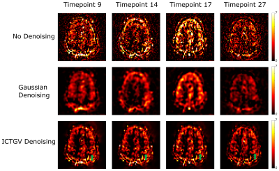 |
Improved functional Arterial Spin Labeling by spatio-temporal ICTGV denoising
Stefan Spann, Matthias Schloegl, Christoph Aigner, Karl Koschutnig, Martin Holler, Kristian Bredies, Rudolf Stollberger
Functional Arterial Spin Labeling (fASL) provides important information of perfusion changes over time and is therefore suitable for detecting neuronal activation due to cognitive functions or motor tasks. However, the low signal to noise ratio of ASL images restrains its application in clinical and research areas. In this study we propose a method for denoising fASL data using infimal convolution of total generalized variations (ICTGV). Compared to standard Gaussian denoising ICTGV denoising incorporates spatial and temporal information of the perfusion weighted time series. This leads to a substantial improvement in noise-suppression for fASL data.
|
|
2167.
 |
Measurement of Pulmonary Perfusion using PCASL True-FISP Imaging at 1.5 Tesla
Petros Martirosian, Ferdinand Seith, Rolf Pohmann, Martin Schwartz, Thomas Küstner, Klaus Scheffler, Konstantin Nikolaou, Fritz Schick
Pseudo-continuous-arterial-spin-labeling (PCASL) has been successfully applied in the liver and kidney providing high signal-to-noise-ratio. The goal of this work is to assess the potential of PCASL technique to measure the pulmonary perfusion at 1.5 T. Effective labeling of pulmonary blood flow was achieved by ECG triggering and an orientation of the labeling plane perpendicular to the pulmonary trunk. Fast True-FISP imaging with short TE of 0.9 ms was used to obtain high signal from lung parenchyma. The PCASL-True-FISP technique provides high quality perfusion images of the lung and allows quantitative measurements of pulmonary perfusion both in multiple breath-holds and under free breathing condition.
|
|
2168.
 |
Denoising arterial spin labeling cerebral blood flow images using deep learning-based methods
Danfeng Xie, Li Bai, Ze Wang
In this study, we use Deep Learning-based (DL) method to denoising ASL CBF images. Convolutional neural networks with a “wide” structure, residual learning and batch normalization are utilized as the core of our denoising model. Comparing to non-DL-based methods, the proposed method showed a significant SNR increase as well as partial volume effects improvement. Also, the DL-based method requires less CBF input images, which significantly shorten the acquisition time and reduce the chance of head motion.
|
|
2169.
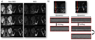 |
Introducing a fat-image guided registration technique for image-based retrospective motion compensation for free-breathing background suppressed renal pCASL
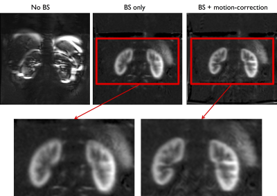
Isabell Bones, Anita Harteveld, Suzanne Franklin, Matthias van Osch, Jeroen Hendrikse, Chrit Moonen, Clemens Bos, Marijn van Stralen
Aiming for rapid and accurate perfusion measurement, background suppressed (BGS) ASL under free breathing is desired. Motion compensation on BGS ASL is challenging due to the lack of anatomical contrast. We investigated the benefit of BGS versus non-BGS ASL, guided by motion compensation based on the ASL-images themselves and additionally acquired fat-images. Registration effect on perfusion weighted signal (PWS) and temporal SNR (tSNR) was evaluated for ASL-image and fat-image based registration, proving increased tSNR and increased PWS robustness, without compromising signal intensity. We conclude that free-breathing BGS renal pCASL with image-based retrospective motion compensation yields better reproducibility than without BGS.
|
|
2170.
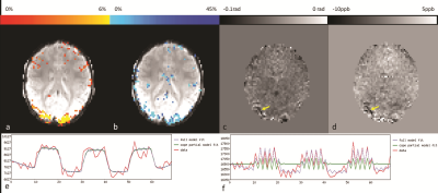 |
Simultaneous Acquisition of ASL, BOLD effect, Phase and QSM for Functional Multi-Parametric Brain Studies
Sagar Buch, Hacene Serrai, Ravi Menon
A 2D-GRE-EPI based sequence combined with the PICORE magnetization preparation technique was used to acquire functional Arterial Spin Labeling (ASL) perfusion data at high field (7T). BOLD and Cerebral Blood Flow (CBF) changes along with phase and susceptibility maps (QSM) are obtained and assessed from this scan. Using a pre-determined general linear model (GLM), a strong correlation between the change in these parameters in the activated region (visual cortex) has been found showing that this multi-parametric acquisition may help in resolving the multi-factorial BOLD signal for functional brain studies.
|
|
2171.
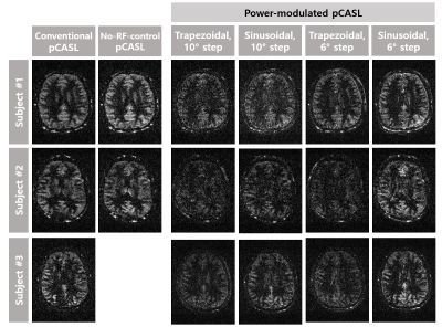 |
Reconstructing Pseudo-Continuous Arterial Spin Labeling Perfusion Signals through Modulation of Labeling RF Power and Fourier Analysis
Hyo-Im Heo, Paul Han, Seung Hong Choi, Sung-Hong Park
The conventional pCASL is vulnerable to data corruption and has high specific absorption rate. In this study, we propose a new pCASL approach using modulation of labeling RF pulse power and Fourier analysis. The proposed approach enabled us to acquire perfusion images comparable to those of the conventional pCASL. Under data corruption, the proposed approach maintained the perfusion signals well with no observable effect, while the conventional method showed almost no perfusion signal. The proposed approach has relatively low average SAR and instantaneous SAR, potentially advantageous at high fields. These advantages of the proposed method warrant further investigation.
|
|
2172.
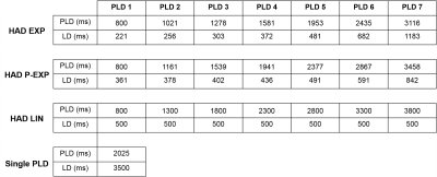 |
Evaluation of the Suitability of Hadamard Encoding Schemes for Pseudo-Continuous Arterial Spin Labelling
Jed Wingrove, Marc Lebel, Fernando Zelaya
Multi delay Arterial Spin Labelling offers the advantage of measuring neurophysiological properties such as arterial transit delay which can be used to hep improve cerebral blood flow estimation. Hadamard encoding pCASL is a method with improved temporal resolution and SNR compared to sequential multi delay methods. This work presents the findings of an evaluation of three different Hadamard encoded schemes for perfusion and transit delay estimation. All schemes were comparable with regards to perfusion estimation however showed some interesting regional differences in TD estimation.
|
|
2173.
 |
Brain connectivity assessment between rest condition and verbal fluency task through Arterial Spin Labeling
André Paschoal, Fernando Paiva, Renata Leoni
Arterial Spin Labeling (ASL) is a method designed to measure blood perfusion. In special, brain perfusion is measured as the cerebral blood flow (CBF), whose time-series fluctuations allow its use in functional analysis. This study aimed to run a dual-echo pseudo-continuous ASL acquisition and analyze its capacity to identify brain networks activated during a verbal fluency task and study the dynamic of brain areas during task and rest conditions. Results showed that it is possible to access language networks based on CBF-ASL, and reported differences in connectivity between both conditions analyzed.
|
|
2174.
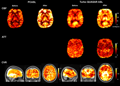 |
Investigating Cerebrovascular Reactivity Using Pseudo-continuous ASL and Turbo QUASAR ASL at Varying Blood Flow Conditions
Moss Zhao, Lena Vaclavu, Esben Petersen, Henk-Jan Mutsaerts, Bart Biemond, Ed van Bavel, Charles Majoie, Aart Nederveen, Michael Chappell
CVR has become an important biomarker to assess cerebrovascular health, and ASL is a non-invasive technique to quantify CVR. This work compared the CVR measurement from PCASL and Turbo QUASAR ASL at varying blood flow conditions induced by acetazolamide. Results showed that both ASL techniques were sensitive to CVR and that significant changes of ATT were detected by Turbo QUASAR ASL. The differences in CVR (higher in PCASL) may be due to the different sensitivity to ATT of the two ASL methods.
|
|
2175.
 |
Pushing the Limits of ASL Imaging for the Lifespan Human Connectome Projects
Xiufeng Li, Dingxin Wang, Steen Moeller, Danny Wang, Michael Chappell, Essa Yacoub, Kamil Ugurbil
Arterial spin labeling (ASL) imaging is included in the Lifespan Human Connectome Projects (HCPs) in order to investigate the evolution of cerebral blood flow (CBF) in children and elderly populations. To push the limits of ASL imaging for the Lifespan HCPs, we optimized and evaluated high-resolution 2D slice accelerated protocols for multi-delay PCASL imaging. The results suggest that high quality arterial transit time (ATT) and CBF maps with a 2.5 mm resolution can be reliably achieved in about 5.5 minutes.
|
|
2176.
 |
Regional Oxygen Extraction Fraction Measurements in the Middle Cerebral Artery Territory using Selective Localised T2-Relaxation-Under-Spin-Tagging (SL-TRUST)
Caitlin O'Brien, Thomas Okell, Peter Jezzard
Regional measurements of brain tissue oxygen extraction fraction (OEF) are an important indicator of tissue physiology and disease. We present an improved Selective Localised T2-relaxation-under-spin-tagging (SL-TRUST) sequence for regional venous blood T2 measurements, decoded in the superior sagittal sinus, from which cerebral tissue OEF can be calculated. A spatially selective WET saturation scheme is used to saturate signal outside the region of interest, enabling OEF measurements in a hemisphere and in the middle cerebral artery (MCA) territory. Using a multi-TI inversion recovery sequence we calculate subject specific blood hematocrit in the sagittal sinus and thus improve our OEF calibration.
|
|
2177.
 |
Influence of background suppression and retrospective realignment on free-breathing renal perfusion imaging using ASL
Manuel Taso, Arnaud Guidon, David Alsop
While a consensus exists on the benefits of background suppression for brain ASL to reduce physiological noise, conflicting results have been presented for renal applications. Furthermore, bulk motion management remains a challenge for clinical applications. In the current work, we investigate the effects and interactions between background suppression and retrospective motion-correction when used for single-slice free-breathing renal ASL. We emphasize the influence of BS and motion-correction on thermal and physiological noise levels and show that BS is critical for renal ASL using pCASL while retrospective motion-compensation helps in increasing image sharpness.
|
|
2178.
 |
Robust non-contrast perfusion imaging of whole-lungs using multi-slice FAIR at 3T
Joshua Greer, Xinzeng Wang, Ananth Madhuranthakam
2D Flow Alternating Inversion Recovery (FAIR) has been applied to measure non-contrast pulmonary perfusion in research environments, but its lack of coverage limits its applicability for clinical perfusion evaluation, where whole-lung coverage is often necessary. In this study, we optimized the multi-slice FAIR technique, including background suppression for robust image quality and inflow saturation to minimize the blood volume contribution, to measure pulmonary perfusion across the whole-lung at 3T.
|
|
2179.
 |
Automatic selection of local arterial input functions in perfusion MRI using cluster analysis and priority-flooding
Rami Tabbara, Alan Connelly, Fernando Calamante
We present a robust, multi-stage automated local arterial input function (AIF) method to quantify perfusion using dynamic-susceptibility contrast (DSC)-MRI. We show how this approach reduces potential AIF misclassifications observed in existing automated solutions that can lead to quantification errors and artefacts. Examples of our new approach eliminating such artefacts from scans of subjects who exhibit various cerebrovascular abnormalities are provided, with generated perfusion maps further showing regions of higher cerebral blood flow (CBF) relative to established global AIF methods, consistent with a reduction in quantification errors associated with bolus dispersion.
|
|
2180.
 |
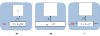
Evaluation of dynamic DCE-MRI of the temporomandibular joint
Ondrej Macicek, Erling Andersen, Oskar Angenete, Thomas Augdal, Karen Rosendahl, Radovan Jirik, Renate Grüner
The feasibility of DCE-MRI as a tool to investigate perfusion of temporomandibular joints (TMJs) in case of Juvenile Idiopathic Arthritis (JIA) in children is investigated. The hypothesis in this current study is that inflammation is associated with increased vascularity and is the origin of experienced pain. Contrary to previous studies, high temporal resolution (~4s) dynamic DCE-MRI using advanced pharmacokinetic models are for the first time applied when imaging the TMJ in JIA children aged 6-15. Results of deconvolution show that there is a difference in perfusion parameters between affected and unaffected patients, especially when permeability-surface area product (PS) and blood plasma flow (Fp) parameters are combined.
|
|
2181.
 |
Quantitative Modeling of Sequence Parameter Choices to Support Standardization for Quantitative DCE-MRI
Jakob Meineke, Jochen Keupp
Systematic and statistical errors in quantitative DCE-MRI measurements which adhere to standardization recommendations by the Quantitative Imaging Biomarker Alliance (QIBA) of the RSNA are assessed using EPG simulations. It is found that small sequence parameter changes, well within the bounds allowed by QIBA, can produce large changes in the estimated quantitative parameters.
|
|
2182.
 |
Incorporating Bolus Arrival Time Offset into Fast Linear Analysis Could Shorten Acquisition Times for DCE-MRI
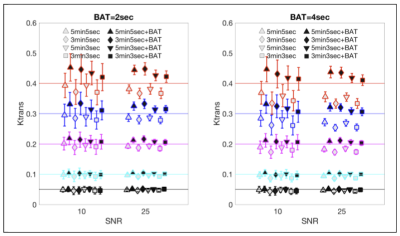
Sharon Peled, Ron Kikinis, Fiona Fennessy, Andrey Fedorov
Linearization of the Kety/Tofts model for DCE analysis drastically shortens computation time. We show that addition of bolus arrival time (BAT) compensation to the linearized analysis could also allow quicker acquisition times. With BAT inclusion, 3 minute sequences yield equivalent parameter estimation accuracy to 5 minute sequences without BAT compensation. The combination of shorter acquisition and real-time analysis would reduce the general time burden of DCE, which has potential implications for increased patient turnaround, and making DCE more acceptable as a tool, for example in evaluating response to therapy or in image guided therapy.
|
|
2183.
 |
RF Transmit Calibration for DCE-MRI
Yannick Bliesener, Yi Guo, Xinran Zhong, Ryan Bosca, Kyung Hyun Sung, Krishna Nayak
Spatial inhomogeneity in the transmitted RF introduces bias and increased variance in quantitative DCE-MRI metrics, which can dominate all other sources of error if uncorrected. The amount and pattern of inhomogeneity depends on the RF coil geometry, the driving circuits, and the vendor-specific pre-scan calibration. In this work, we (1) constructed human tissue-mimicking torso and brain phantoms, (2) measured and compared the spatial RF transmit inhomogeneity across different scanners, vendors, and sites, and (3) evaluated vendor-recommended methods for RF transmit measurement.
|
|
2184.
 |
Measuring transient T2* changes in vivo to validate Dynamic Distributed Spirals, a novel DSC-perfusion method
Dallas Turley, James Pipe
Validating new contrast-enhances sequences is problematic, as risks associated with gadolinium contrast agents generally preclude testing in healthy volunteers. The Dynamic Distributed Spirals trajectory (DDS) is a promising new dynamic susceptibility-contrast (DSC)-perfusion method. In this work, we show that DDS is capable of measuring the transient T2* changes induced by breathholding which are much lower in magnitude than the susceptibility changes induced by contrast agent transit in conventional DSC-perfusion experiments.
|
|
2185.
 |
Diffusion dependency of oxygenation measurements obtained with Vessel Architectural Imaging
Ingrid Digernes, Atle Bjørnerud, Grete Løvland, Einar Vik-Mo, Torstein Meling, Kyrre Emblem
With the dual echo DSC-based technique Vessel Architectural Imaging (VAI), measurement of oxygenation level (?SO2) can be obtained. However, how the ?SO2-parameter is influenced by diffusion have previously not been investigated. Based on simulations, we show that the measured ?SO2 obtained from VAI have a diffusion dependency proportional to 1/ √D. ADC-maps from 10 glioblastoma patients were used to display the range correction factors in white matter and tumor regions. In conclusion, the diffusion dependency should be corrected for to obtain more accurate measurements of ?SO2, and may be especially relevant for brain diseases with aberrant diffusion characteristics.
|
|
2186.
 |
Feasibility of measuring subtle Blood-Brain Barrier permeability change with reduced scan time using Dynamic Contrast-Enhanced Magnetic Resonance Imaging
Jonghyun Bae, Jin Zhang, Youssef Wadghiri, Atul Minhas, Harish Poptani, Yulin Ge, Sungheon Kim
The purpose of this study is to evaluate the feasibility of using a new contrast kinetic model to accurately measure changes in the low permeability of the blood-brain barrier due to the subtle vascular disruption in the development of neurodegenerative diseases. Our proposed kinetic model, named extended Patlak model (EPM), includes the plasma flow from the artery to capillary bed, which allows the accurate description of intake dynamics. We hypothesize that this extension allows EPM to estimate the permeability change more accurately than the conventional Patlak model (PM) with a reduced scan-time of around 10 min.
|
|
2187.
 |
Cerebral Perfusion Imaging: The Vascular Territory of Middle Cerebral Artery is Optimal for Automatic Arterial-Input-Function Selection
Irene Mikkelsen, Simon Eskildsen
A key issue in cerebral perfusion imaging is the selection of an arterial input function (AIF). AIF shape-properties have been used as criteria for automatic AIF selection. This study compares three brain regions for AIF target areas. The Middle Cerebral Artery (MCA) -M1 segment, the MCA-vascular territory and whole-brain. The prior displayed high noise levels, while the latter produced AIFs delayed compared to GM/WM tissue. The MCA-vascular territory is suggested as a region of interest for automatic AIF detection
|
|
2188.
 |
Systematic Assessment of Multi-Echo Dynamic Susceptibility Contrast (DSC) MRI using a Digital Reference Object (DRO)
Ashley Stokes, Natenael Semmineh, C. Quarles
Brain tumor dynamic susceptibility contrast (DSC) MRI is adversely impacted by contrast agent leakage that results in confounding T1 and T2* effects. While multi-echo acquisitions remove T1 leakage effects, there is no consensus on the optimal set of acquisition parameters. Using a validated DSC-MRI digital reference object (DRO), we assessed the influence of preload dosing, pulse sequence parameters (number of echoes, TEs, TR, FA), and leakage correction method on cerebral blood volume (CBV) accuracy. This computational approach permits the systematic evaluation of a wide range of acquisition strategies to determine the optimal multi-echo DSC-MRI perfusion protocol.
|
|
2189.
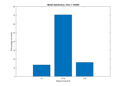 |
Incremental modeling in DCE-MRI in gliomas
Magne Kleppestø, Christopher Larsson, Atle Bjørnerud
This work compares three kinetic models for evaluation of DCE-MRI in high-grade gliomas: the Tofts-Kermode (TK) model, the extended Tofts model (ETM) and the two-compartment exchange (TCE) model. 25 patients underwent a combined 238 examinations, and kinetic analysis was performed using the three models. In tumor regions where the data was better fitted using TK or TCE, median Ktrans estimates obtained from this model was compared to that from using ETM. It was found that in tumor regions in which TCE provides the best fit, median Ktrans was significantly underestimated when applying ETM.
|
|
Quantitative Susceptibility Mapping
Traditional Poster
Contrast Mechanisms
Wednesday, 20 June 2018
| Exhibition Hall 2190-2221 |
08:15 - 10:15 |
|
2190.
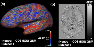 |
COSMOS for Estimating Variation in Single Orientation Quantitative Susceptibility Mapping of the Brain: An Ultra High Field Study
Jon Cleary, Hongfu Sun, Rebecca Glarin, Peter Yoo, Bradford Moffat, Roger Ordidge, Scott Kolbe
The purpose of this study was to use high resolution Calculation of susceptibility through Multiple Orientation Sampling (COSMOS) reconstructed QSM (QSMc) as a gold standard to estimate the variation, distribution and magnitude of a single orientation QSM reconstruction pipeline. QSMc processing is an emerging technique for overcoming artefacts characteristic of single orientation QSM (QSMs). However it requires at least 4 fold increases in image acquisition times or reductions in resolution and SNR. We sought to produce high resolution QSMc reference datasets from healthy subjects to quantify the differences from QSMs values across a variety of cortical and subcortical brain regions.
|
|
2191.
 |
Evaluating the Precision of Multi-Echo Combination Methods for Susceptibility Mapping by Analysing the Propagation of Single-Echo Phase Noise into Multi-Echo Field and Susceptibility Maps
Emma Biondetti, Anita Karsa, David Thomas, Karin Shmueli
In Susceptibility Mapping (SM) using multi-echo acquisitions, noise propagates from the single-echo phase images into the field map in a manner dependent on the method used for multi-echo combination. Field noise then propagates into the susceptibility map, determining the precision of the measured susceptibility. Here, we characterised the propagation of single-echo phase noise into both the combined field and susceptibility maps using three methods for multi-echo combination: fitting, averaging and echo time-weighted averaging. We calculated susceptibility noise maps for both simulated and acquired data, showing that, when choosing a pipeline for multi-echo SM, it is important to consider its precision.
|
|
2192.
 |
Effects of Motion in Quantitative Susceptibility Mapping of Brain
Ashmita De, Hongfu Sun, Ahmed Elkady, Peter Seres, Alan Wilman
Typical Quantitative Susceptibility Mapping (QSM) sequences have a long acquisition time which may yield motion artifacts that alter magnitude, phase and susceptibility values in the brain. Simulations and motion experiments were conducted on patients suspected of stroke and healthy volunteers to calculate the variations of susceptibility, magnitude and local field in brain. Variations between susceptibility and magnitude images were compared. In general, magnitude images were found to be more affected by motion than QSM in the brain areas studied.
|
|
2193.
 |
Fast Zoomed QSM of the Human Midbrain at 7T
Kyungmin Nam, Namgyun Lee, Anouk Marsman, Vincent Oltman Boer, Chulhyun Lee, Esben Thade Petersen
In this work, zoomed quantitative susceptibility mapping (QSM) is proposed as an alternative way of accelerating high resolution QSM data acquisition at 7T. Inner volume excitation is realized with 2D spatially selective excitation, targeting the midbrain, which is the primary region of investigation for Parkinson’s disease. The consequence of reducing the excited region on the reconstructed susceptibility maps was investigated via simulations, where the diameter of a brain mask was gradually decreased in the QSM processing pipeline. The susceptibility maps of a healthy volunteer at 7T acquired with inner volume excitation are compared to those derived from a whole brain.
|
|
2194.
 |
Phase Corrected Bipolar Acquisition for Simultaneous Water-Fat Separation and Quantitative Susceptibility Mapping of the Carotid Artery Wall
Pascal Ruetten, Andrew Priest, Jianmin Yuan, Ammara Usman, Jonathan Gillard, Martin Graves
In this work we investigated the feasibility and assessed the performance of a bipolar compared to a unipolar gradient echo readout for a combined method of water-fat separation and quantitative susceptibility mapping for application in the carotid artery wall.
|
|
2195.
 |
Simultaneous quantification of fat fraction, susceptibility and R2* from a single GRE acquisition: flip-angle effects
Junmin Liu, Spencer Christiansen, Maria Drangova
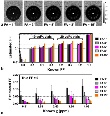
We report on a systematic investigation of the flip-angle (FA) effects on the quantification of fat fraction (FF), susceptibility, and R2* simultaneously from a single multi-echo GRE (mGRE) acquisition. Using a phantom with a range of oil-water emulsions and aqueous Gadolinium solutions we tested five different FAs (1°, 3°, 5°, 8° and 15°) with a bipolar mGRE protocol and were able to successfully generate the FF, susceptibility and R2* maps for all cases. Our results demonstrate that a single mGRE scan with optimized TEs has the potential to accurately measure quantitative FF, susceptibility, and R2* with a FA of 8°.
|
|
2196.
 |
Quantitative Susceptibility Mapping with Silent 3D Radial T2* Acquisition
Mauro Costagli, Ana Beatriz Solana, Guido Buonincontri, Florian Wiesinger, Michela Tosetti, Rolf Schulte
Recent implementations of radial Zero Echo Time (ZTE) techniques are capable of providing T2*-weighted signal. Quantitative Susceptibility Mapping (QSM) using such techniques might have several potential advantages, such as (i) robustness to head motion, flow artifacts and geometrical distortions, (ii) improved sampling efficiency, (iii) reduced acoustic noise, (iv) simultaneous acquisition of proton-density data. We assessed the QSMs obtained with two different silent radial techniques, and their accuracy was similar to that of QSM obtained with conventional scanning schemes, which encourages the development of ZTE-based techniques specifically tailored for efficient and silent QSM, to achieve important advantages in clinical applications.
|
|
2197.
 |
Machine Learning in QSM: Inversion Using Multi-Resolution Decomposition and Convolutional Neural Networks.
Kevin Koch, Tugan Muftuler, Robin Karr, Andrew Nencka
One of the remaining translational challenges in QSM is the need for post-processing algorithms that are rapid, robust, and accurate. Here, we present an alternative formulation of the QSM inversion problem. The field-to-source inversion is divided into a multi-resolution decomposition, whereby each resolution stage is divided into small independent processing regions. The basic premise of this concept is the isolate local susceptibility fields and sources at varying levels of resolution. When the susceptibility problem is divided in this fashion, field-to-source inversions can occur in regions of very volumetric matrix sizes (with varying voxel sizes per inversion). After inverting each of the sub-volumes, a combination procedure is implemented to combine the volumes and the resolution layers. Due to the small size of the inversion volumes, the dimensionality of the problem lends itself to the use of convolutional neural network modeling and application.
|
|
2198.
 |
DeepQSM - Solving the Quantitative Susceptibility Mapping Inverse Problem Using Deep Learning
Mads Kristensen, Kasper Bøtker Rasmussen, Rasmus Blendal, Lasse Østergaard, Maciej Plocharski, Andrew Janke, Christian Langkammer, Kieran O’Brien, Markus Barth, Steffen Bollmann
Quantitative susceptibility mapping (QSM) aims to extract the magnetic susceptibility of tissue by solving an ill-posed field-to-source-inversion. Current QSM algorithms require manual parameter choices to balance between smoothing, artifacts and quantitation accuracy. Deep neural networks have been shown to perform well on ill-posed problems and can find optimal parameter sets for a given problem based on real-world training data. We have developed a proof-of-concept fully convolutional deep network capable of solving QSM’s ill-posed field-to-source inversion that preserves fine spatial structures and delivers accurate quantitation results.
|
|
2199.
 |
Reconstruction of Quantitative Susceptibility Maps using Annihilating Filter-Based Low-Rank Hankel Matrix Approach
Hyun-Seo Ahn, Sung-Hong Park, Jong Chul Ye
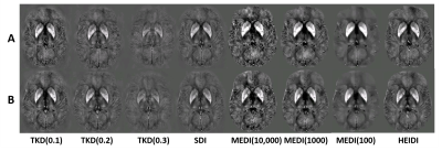
In this study, we proposed a novel QSM image reconstruction algorithm using annihilating filter-based low-rank hankel matrix (ALOHA) approach. Unlike the conventional algorithm that requires additional anatomical information, the proposed method estimates susceptibility map using direct 3-D k-space domain interpolation in the Fourier domain. The proposed method showed superior performance over the conventional methods (SWIM, TSVD, TKD, MEDI, and TVSB) in a numerical phantom and in-vivo human brains.
|
|
2200.
 |
Magnetic susceptibility source separation using multi-echo GRE data only
Taehyun Hwang, Jingu Lee, Hyeong-Geol Shin, Doohee Lee, Joon Choi, Hyunsung Eun, Yoonho Nam, Jongho Lee
In this work, we explored an alternative approach of using nominal $$$R_2^{\:'}$$$ instead of measured $$$R_2^{\:'}$$$ in separating the two susceptibility sources. The linear relationship between $$$R_2^{*}$$$ and $$$R_2^{\:'}$$$ was investigated and used to obtain the nominal $$$R_2^{\:'}$$$ values. The positive and negative magnetic susceptibility source maps using nominal $$$R_2^{\:'}$$$ showed similar susceptibility distribution to the map using measured $$$R_2^{\:'}$$$.
|
|
2201.
 |
Fast and accurate reconstruction for susceptibility source separation in QSM
Seyoon Ko, Jingu Lee, Joong-Ho Won, Jongho Lee
We investigate fast and accurate reconstruction methods for susceptibility source separation (S3) in quantitative susceptibility mapping (QSM). S3 separates positive and negative susceptibility sources within a voxel utilizing signal relaxation (R2') for dipole inversion. We propose new primal-dual (PD) methods for S3 and compare them with the alternating Gauss-Newton conjugate gradient (A-GNCG). A-GNCG alters the energy functional, and furthermore its convergence is not guaranteed. In contrast, the proposed PD methods are exact and have convergence guarantees. Validation on a simulated phantom and in-vivo data shows that the PD methods converge faster with better accuracies.
|
|
2202.
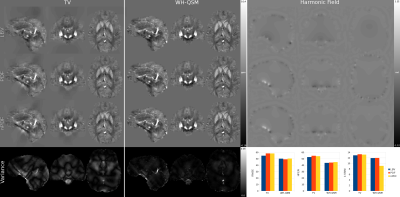 |
Weak-harmonic regularization for quantitative susceptibility mapping (WH-QSM)
Carlos Milovic, Berkin Bilgic, Bo Zhao, Christian Langkammer, Cristian Tejos, Julio Acosta-Cabronero
In the context of QSM, the background pre-filtering step often leaves remnants in the local field, particularly in the vicinity of trustable-region boundary. Since such remnant fields must satisfy Laplace's equation, i.e. they must be harmonic functions within the ROI, we propose a new regularization term based on a weak-harmonics formulation (WH-QSM) to remove spurious non-local components during inversion. The WH extension resulted in more accurate and reproducible results than conventional total-variation (TV) regularized QSM.
|
|
2203.
 |
Nonlinear projection onto dipole fields with preconditioning (nPDF)
Carlos Milovic, Berkin Bilgic, Bo Zhao, Christian Langkammer, Julio Acosta-Cabronero, Cristian Tejos
QSM requires to remove fields originated outside a region of interest prior to inversion. This is prone to generating artifacts due to noise and error propagation from previous processing steps such as coil combination or phase unwrapping. To address this, we reformulated the widely used projection onto dipole fields (PDF) method as a nonlinear problem with pre-conditioning. This new formalism is wrap-insensitive, results in improved noise/error management, and might enable a more straightforward implementation of multi-coil/-echo combination and background removal steps into a single optimizer.
|
|
2204.
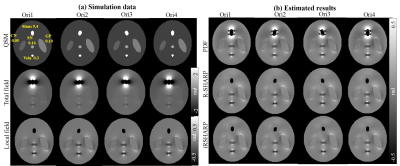 |
Background Field Removal for Large Susceptibility Anatomical Structures in Human Brain with Orientation Variations
Jinsheng Fang, Lijun Bao, Zhong Chen
We propose a novel background field removal method for large susceptibility anatomical structures, e.g., tissues around paranasal sinuses and interfaces of the tissue and skull, under various scanning orientations. The proposed method employs the gradient and magnitude of the phase map, combined with a normalized wrap count. Experimental results were both validated on four-orientation numerical simulation and in vivo human brain, which demonstrated the proposed method suppressed the residual phase error better than the other methods.
|
|
2205.
 |
Suitable image quality measures to evaluate quantitative susceptibility maps
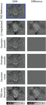
Janis Stiegeler, Sina Straub
The 2016 QSM Reconstruction Challenge urged the need for a suitable quality measure of susceptibility maps as classical image quality measures (root-mean-square error, high-frequency error-norm, structural similarity index) were no suitable indicators of the visual quality of susceptibility maps. Errors (noise, smoothing, streaking) were added to a reference susceptibility map and the sharpness-index-weighted structural similarity index was used to evaluate the degraded quantitative susceptibility maps and to compare the result with classical image quality measures. The sharpness-index-weighted structural similarity index was shown to be a suitable measure for QSM image quality with a strong devaluation of over-smoothed images.
|
|
2206.
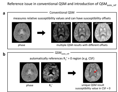 |
An automatically referenced quantitative susceptibility mapping algorithm: QSMauto_ref
Jingu Lee, Taehyun Hwang, Yoonho Nam, Se-Hong Oh, Jongho Lee
We proposed a new QSM algorithm that automatically sets CSF as a susceptibility reference. The algorithm utilizes susceptibility effects on R2’ as a regularization term. The proposed algorithm does not require either segmentation of CSF or a well-refined brain mask and, therefore, can be used reliably.
|
|
2207.
 |
Applications of magnetic susceptibility source separation: multiple sclerosis lesions and line of Gennari
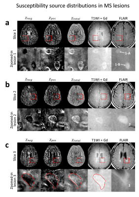
Jingu Lee, Taehyun Hwang, Yoonho Nam, Jinhee Jang, Woojun Kim, Se-Hong Oh, Masaki Fukunaga, Jongho Lee
Magnetic susceptibility source separation is a recently proposed technique that generates positive and negative susceptibility maps corresponding to iron and myelin distributions in the brain. In this study, iron accumulation and myelin degradation in a few typical types of multiple sclerosis lesions were visualized using the magnetic susceptibility source separation method. Additionally, the well-known co-localization of iron and myelin in the Gennari line was demonstrated in an ex-vivo brain sample.
|
|
2208.
 |
Structure tensor enhanced quantitative susceptibility mapping (ST-QSM)
Agnese Tamanti, Kristian Bredies, Marco Castellaro, Stefan Ropele, Berkin Bilgic, Christian Langkammer
Quantitative susceptibility mapping (QSM) is an MRI technique enabling the reconstruction of a basic physical property in vivo. However, retrieving susceptibility maps from the MRI phase data requires an ill-posed inverse problem to be solved, which is often achieved using regularization approaches. In this abstract, we extend an existing QSM algorithm by incorporating weights from the linear structure tensor (ST) of the magnitude images to stabilize the regularization. The new algorithm yields improvements regarding the visual appearance and the quantitative performance of the susceptibility maps obtained.
|
|
2209.
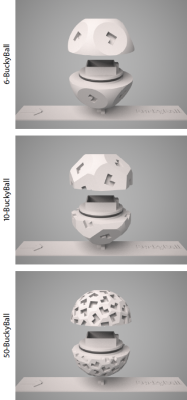 |
BuckyBall: Reproducible gradient-echo MRI measurements with variable magnetic field directions
Enrico Kaden, Irina Barskaya, Nathaniel Kelm, Mark Does, Daniel Alexander
The direction of the external magnetic field is typically fixed, although it is well-known that the signal of various MR modalities in brain white matter depends on the magnetic field direction. This work presents a general framework for analysing B0-direction dependent contrast. Specifically, we have developed a holder device, called BuckyBall, that enables the uniform orientation of the scanned object in a reproducible manner. Its feasibility and practicality are demonstrated in a multi-echo gradient-echo experiment with 50 unique magnetic field directions using a monkey brain sample.
|
|
2210.
 |
Measurement of Iron Concentration in Deep Gray Matter Nuclei over the Lifespan Using Quantitative Susceptibility Mapping
Gaiying Li, Rui Tong, Binshi Bo, Miao Zhang, Yu Zhao, Tian Liu, Yasong Du, Xu Yan, Yi Wang, Jianqi Li
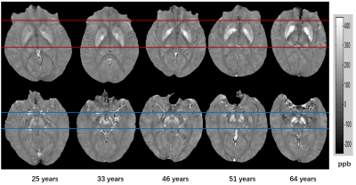
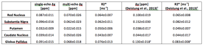
Histological in vitro analysis has demonstrated that iron accumulation rates in various gray matter nuclei are different throughout an individual’s lifetime. QSM provides excellent contrast of iron-rich deep nuclei to quantify iron in the brains. In this study, we investigated the linear and nonlinear correlation of magnetic susceptibility in the deep gray matter nuclei as a function of ageing using QSM. Compared with the published studies, the nonlinear analysis results showed the differential developmental trajectories of magnetic susceptibility in the deep gray matter nuclei over the lifespan.
|
|
2211.
 |
Improved depiction of subthalamic nucleus and globus pallidus internus with optimized high-resolution quantitative susceptibility mapping at 7 Tesla
Fei Cong, Yelong Shen, Bo Wang, Jing An, Zihao Zhang, Zhentao Zuo, Yan Zhuo, Lirong Yan
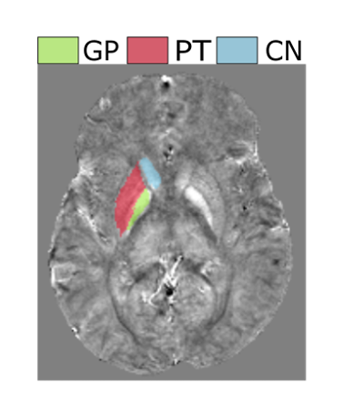
Quantitative susceptibility mapping (QSM) shows a potential to image subthalamic nucleus (STN) and globus pallidus internus (GPi). However, the image quality of QSM is dependent on the selection of regularization parameter during reconstruction. Here we proposed an approach to determine the optimal regularization parameter for imaging the sub-cortical nuclei at different spatial resolution and field strengths. Optimized QSM images were further compared with the other susceptibility weighted images for visualization of STN and GPi at 3T and 7T. Our results suggest that optimized 7T QSM with spatial resolution of 0.35x0.35x1.0mm3 provides better delineation of STN and GPi.
|
|
2212.
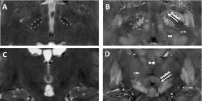 |
High resolution MRI for functional and structural depiction of subthalamic nuclei in DBS pre-surgical mapping: a comparison between QSM and T2w
Alexey Dimov, Ajay Gupta, Brian Kopell, Yi Wang
In this work, we investigate the use of a sub-millimeter quantitative susceptibility mapping (QSM) protocol for preoperative imaging of the suthalamic nucleui (STN) for planning of deep brain stimulation (DBS). Image scoring revealed superior performance of QSM compared to the conventional T2 weighted (T2W) protocol. In contrast to T2W, image scores further increased for QSM when resolution was increased.
|
|
2213.
 |
Magnetic susceptibility characterization of human habenula at 3T: comparison of QSM and R2*
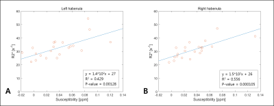
Seulki Yoo, Seung-Kyun Lee
To investigate the potential of magnetic susceptibility as a biomarker for habenula studies, we have obtained quantitative susceptibility maps (QSM) and R2* maps from 21 normal volunteers at high spatial resolution. Compared to R2* maps, QSM showed more conspicuous and localized contrast in habenula in about 75% of the volunteers. Measured susceptibility and R2* values exhibited clear positive correlation, indicating iron-dominance (as opposed to myelin) of the susceptibility contrast in habenula. Significant heterogeneity of the susceptibility contrast across the subjects and within the tissue appear to be a challenge for using QSM as a biomarker for human habenula research.
|
|
2214.
 |
QSM susceptibility patterns and their clinical implications
Kelly Gillen, Mayyan Mubarak, Shun Zhang, Somiah Dahlawi, Thanh Nguyen, David Pitt, Yi Wang
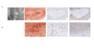
Multiple sclerosis is an autoimmune disorder characterized by focal inflammatory demyelination. We combined quantitative susceptibility mapping (QSM) with histopathology on MS autopsy tissue to identify chronic activation of iron-positive macrophages/microglia. We demonstrate that the QSM susceptibility pattern gives insight into the lesion inflammatory state. Only rim positive lesions indicate smoldering inflammation in the presence of iron, and therefore are of particular relevance in the clinic.
|
|
2215.
 |
Investigating the Effect of Prior Stroke on Regional Brain Iron Concentrations in Children with Sickle Cell Anaemia using MRI Susceptibility Mapping.
Russell Murdoch, Jamie Kawadler, Fenella Kirkham, Karin Shmueli
Regional iron concentrations in the brains of children with Sickle Cell Anaemia (SCA) were examined using susceptibility mapping (SM), in the first study to apply SM to an African cohort in Tanzania. Mean susceptibility values in three deep-brain regions were compared to age, blood ferritin levels and history of clinical stroke. Mean susceptibility values increased linearly with age, but there was no significant correlation between susceptibility values and blood ferritin levels. SCA patients who had suffered stroke prior to MRI had significantly lower susceptibility values than stroke-free patients. This may suggest a role for iron deficiency in stroke in SCA.
|
|
2216.
 |
Fast brain iron quantification using QSM with low spatial resolution
Xin Miao, Krishna Nayak, John Wood
This study investigates the impact of spatial resolution on QSM susceptibility mapping for brain iron quantification. We obtained 40 sub-millimeter resolution whole-brain QSM datasets, and simulated six levels of spatial resolution via k-space truncation. QSM-based iron quantification was performed at each spatial scale and compared against the reference. We found that estimation error was ≤ 5 ppb in the basal ganglia when the voxel dimension along all three axes was ≤ 2.0 mm. The finding suggests that scan time can be significantly shortened by reducing spatial resolution.
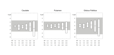
|
|
2217.
 |
QSM-MRI reveals increased brain iron deposition in anemia patients with blood transfusion
Xin Miao, Soyoung Choi, Krishna Nayak, John Wood
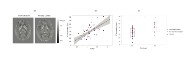
Sickle cell patients identified with high stroke risks and other genetically anemic patients are treated with chronic blood transfusions. Unfortunately, transfusions may cause iron overload. While transfusion-related iron overload has been shown in other major organs, less has been explored whether it impacts brain. This study compares brain iron content measured by quantitative susceptibility mapping (QSM) in 17 healthy controls and 33 patients with sickle cell or other types of anemia. We found significantly higher iron in the putamen of anemia patients receiving blood transfusion. The result of this study can provide insights on the neurological effects of blood transfusions.
|
|
2218.
 |
Are all susceptibility maps created equal? – An investigation of the impact of the field-to-source inversion step on the study outcome in patient-control group studies.
Poonam Choudhary, Niels Bergsland, Akshay Dhamankar, Michael Dwyer, Bianca Weinstock-Guttman, Robert Zivadinov, Ferdinand Schweser
Quantitative Susceptibility Mapping (QSM) is a relatively new post-processing technique for susceptibility-weighted gradient-recalled echo (GRE) phase images. The technique numerically solves an ill-posed inverse mathematical problem to reveal the tissue magnetic susceptibility distribution. Due to its uniquely high sensitivity on the tissue concentrations of myelin, calcium and iron, QSM is increasingly being applied in clinical studies of neurological diseases that are affected by demyelination and a disturbed iron homeostasis, such as multiple sclerosis (MS) and Parkinson’s disease. In the present work, to better understand the comparability and reproducibility of QSM studies, we evaluated several widely-used inversion algorithms concerning their ability to detect differences in susceptibility between two different groups of subjects, a typical scenario in clinical research.
|
|
2219.
 |
Reproducibility of Quantitative Susceptibility Mapping and R2* Mapping of the Human Brain at 7T: a Multi-Centre Pilot Study
Catarina Rua, William Clarke, Ian Driver, Olivier Mougin, Stuart Clare, Susan Francis, Keith Muir, Richard Wise, Guy Williams, Richard Bowtell, Adrian Carpenter
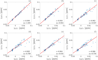
To perform cost-effective research with high-field imaging by increasing the size of the patient pool in investigations of brain diseases, it is important to guarantee cross-site reproducibility and consistency of the QSM and R2* results. This study is part of a pilot “travelling-heads” experiment from the UK7T network, in which we aim to develop harmonized approaches for T2*-weighted imaging in order to provide a framework for future multi-centre clinical studies at 7T.
|
|
2220.
 |
Quantitative Susceptibility Mapping at high and ultra-high field: a reproducibility study
Marta Lancione, Michela Tosetti, Paolo Cecchi, Graziella Donatelli, Mirco Cosottini, Mauro Costagli
The aim of this study is to assess the reproducibility of Quantitative Susceptibility Mapping (QSM), which is crucial to enable the application of this technique in clinical follow-up and multi-center studies. Five healthy subjects underwent multiple QSM acquisition sessions using two MRI systems at different field strength (3T and 7T). Both voxel-wise and automated atlas-based ROI analyses proved the goodness of intra-scanner repeatability and inter-scanner reproducibility, the latter being slightly weaker than the former.
|
|
2221.
 |
Validation of Quantitative Susceptibility Mapping of the Liver at 1.5T and 3.0T using SQUID-Based Liver Susceptometry as the Reference
Ruiyang Zhao, Valentina Taviani , Shreyas Vasanawala, Scott Reeder, Diego Hernando
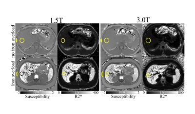
Accurate quantification of liver iron concentration (LIC) is needed for the assessment of iron overload. Quantification of magnetic susceptibility may enable accurate and reproducible estimation of LIC. SQUID-based biomagnetic liver susceptometry (BLS) is used clinically to measure magnetic susceptibility, but has very limited availability. MRI-based Quantitative Susceptibility Mapping (QSM) may enable liver susceptometry with much broader availability. However, the accuracy of QSM-BLS across field strengths remains unknown. In this abstract, we observed strong correlation (r2=0.90) between QSM-BLS (at both 1.5T and 3.0T) with SQUID-BLS in patients with known or suspected iron overload.
|
|
CEST: Novel Methods & Applications
Traditional Poster
Contrast Mechanisms
Wednesday, 20 June 2018
| Exhibition Hall 2222-2249 |
08:15 - 10:15 |
|
2222.
 |
A novel normalization to correct APT-CEST in the presence of fat
Ferdinand Zimmermann, Andreas Korzowski, Patrick Schuenke, Johannes Breitling, Mark Ladd, Peter Bachert, Steffen Goerke
Chemical Exchange Saturation Transfer (CEST) MRI in the human breast is affected by the fat content in the fibro glandular tissue. Although the spectral region of the amide proton transfer (APT) signal does not overlay with fat resonances, the fat signal leads to an incorrect normalization of the Z-spectrum and therefore to misleading CEST effects. We propose a novel method yielding a corrected normalization without the need for application of fat saturation schemes, thus enabling APT-CEST imaging corrected for fat signal contribution. Transfer of the gained insights to realize correct APT-CEST in the human breast at 7T is currently under investigation.
|
|
2223.
 |
Rapid and Quantitative Chemical Exchange Saturation Transfer (CEST) Imaging of In Vivo Rat Brain with Magnetic Resonance Fingerprinting (MRF)
Ouri Cohen, Shuning Huang, Michael McMahon, Matthew Rosen, Christian Farrar
CEST MRI suffers from several limitations including long image acquisition times and the qualitative nature of the CEST contrast. Clinical translation of CEST MRI would benefit greatly from the development of quantitative and rapid CEST methods. Here we build on the recently developed Magnetic Resonance Fingerprinting (MRF) technique and report the use of a fast CEST fingerprinting method for generating quantitative exchange rate and exchangeable proton concentration maps of L-Arginine phantoms and in vivo rat brain tissue.
|
|
2224.
 |
3D CEST MRI of human brain at 9.4T reveals vessel correlation of the effect at -1.7 ppm
Moritz Zaiss, Jonas Bause, Anagha Deshmane, Kai Herz, Klaus Scheffler
In vivo CEST imaging at 9.4T reveals that the peak at -1.7 ppm which was recently associated with red blood cells is spatially localized to blood vessels. A 3D CEST sequence with high resolution and dense sampling of the Z-spectrum shows that only the -1.7 ppm contrast is vascularly localized.
|
|
2225.
 |
Myocardial Creatine CEST in human heart using a segmented pseudo steady state acquisition over multiple short breathholds
Neil Wilson, Puneet Bagga, Kevin D'Aquilla, Hari Hariharan, Ravinder Reddy
A technique to acquire creatine CEST of the myocardial muscle is presented here. The technique uses a pseudo steady state saturation, segmented readout, and multiple, short breathholds.
|
|
2226.
 |
Pre- and post-contrast glucoCEST weighted MRI both detect hypometabolism following experimental TBI
Tsang-Wei Tu, Jaclyn Witko, Joseph Frank
This study compared the endogenous glucoCEST contrast to the glucoCEST with exogenous glucose delivered as contrast agent in experimental TBI. By giving relatively low concentration (0.3g/kg) of 2DG solution, the post-contrast glucoCEST weighted images could magnify the contrast changes in the brains before and after TBI. Meanwhile, the endogenous glucoCEST weighted images also detected the same pattern of decreased contrast in the TBI brains and that was validated by 2DG autoradiography. Our findings substantiate that the glucoCEST technique has potential to detect the hypometabolic syndrome following TBI, even without using exogenous contrast agent.
|
|
2227.
 |
Mapping elevated lactate levels after ischemic stroke using PROBE CEST/NOE: a feasibility study in patients at 3T
Tobias Lenich, André Pampel, Toralf Mildner, Ralf Mekle, Ramanan Ganeshan, Jochen Fiebach, Harald Möller
In ischemic stroke, anaerobic glycolysis leads to a local increase in lactate concentration. Such elevated levels of lactate can be detected via CEST/NOE. In vivo, several broad tissue contributions as well as metabolites lead to a complex intermingled baseline in Z-spectra. With PROBE, such effects are compensated based on healthy tissue, and a flat baseline is achieved. Stroke affected areas can hence be identified in direct contrast to healthy tissue. Lactate contributions to the Z-spectrum became distinctively observable. The feasibility was demonstrated in-vivo for thalamic stroke in a clinical setting at 3T.
|
|
2228.
 |
Dependence of rNOE-CEST signals on molecular weight
Steffen Goerke, Johannes Breitling, Karel Klika, Mark Ladd, Peter Bachert
In this study, rNOE-CEST signals of proteins have been demonstrated to depend on molecular weight. This finding can explain the observed intensity decrease of aliphatic rNOE-CEST signals in tumors in comparison to healthy tissue.
|
|
2229.
 |
Optimization and acceleration of Selective Inversion Recovery imaging for practical whole-brain quantitative Magnetization Transfer measurements
Matthew Cronin, Junzhong Zhu, Daniel Gochberg, Richard Dortch
Selective inversion recovery quantitative magnetization transfer (SIR-qMT) imaging offers increased efficiency relative to conventional pulsed-saturation qMT due to its ability to quantify MT parameters without the need for independent estimates of B0, B1+, and T1. Despite this, qMT acquisition at a reasonable resolution over a large field of view remains prohibitively time consuming. Here, we employ an optimised acquisition strategy and accelerated readouts to acquire whole-brain SIR-qMT data at 2 x 2 x 3 mm3 resolution in ~10 minutes; opening the door to qMT imaging on a time scale practical for clinical application.
|
|
2230.
 |
Multiple Interleaved Mode Saturation (MIMOSA) for B1+ inhomogeneity mitigation in chemical exchange saturation transfer.
Andrzej Liebert, Moritz Zaiss, Rene Gumbrecht, Benjamin Schmitt, Peter Linz, Frederik Laun, Michael Uder, Armin Nagel
Due to high sensitivity to B1+-inhomogeneities, Chemical Exchange Saturation Transfer MRI requires a correction or mitigation of the B1+-inhomogeneity at ultra-high magnetic field strengths (B0 ≥ 7 Tesla). A novel approach for mitigation of B1+-inhomogeneity effects that affects the saturation process is presented. The method employs two interleaved excitation modes during the saturation pulse train. Simulations show a decrease of the relative difference of the MTRRex metric caused by B1+ inhomogeneity. This “Multiple Interleaved Mode Saturation” scheme leads to improved homogeneity in both, phantom and in vivo measurements at 7 Tesla.
|
|
2231.
 |
Single-shot whole-brain CEST imaging using centric-reordered 3D-EPI
Suzan Akbey, Philipp Ehses, Rüdiger Stirnberg, Moritz Zaiss, Tony Stöcker
We present a 3D CEST sequence that allows 2mm isotropic whole-brain acquisition within 6s per frequency offset. The 4.5s CEST preparation is followed by a 1.5s centric-reordered 3D-EPI readout with water excitation. The single-shot readout improves robustness against physiological noise and provides complete freedom in the design of the saturation block. We acquired whole-brain CEST data at 7T and show metabolite maps obtained from a Lorentzian fit to the Z-spectra.
|
|
2232.
 |
CEST Feasibility in Rectal Cancer Patients at 7T for Detection of Residual Tumor
Catalina Arteaga de Castro, Quincy van Houtum, Sieske Hoendervangers, Alice Couwenberg, Martijn Intven, Helena Verkooijen, Dennis Klomp, Marielle Philippens
Five patients were scanned at a 7T MR scanner, 9 weeks after chemoradiation treatment. Three patients showed extreme artifacts on the calculated CEST maps due to B0 artifacts from air contained in the rectum or poor B0 shimming. Two successful CEST measurements showed matching amide-CEST maps to the residual tumor observed in the MRI. CEST applied to rectum patients after chemoradiation might be the appropriate technique to avoid surgical resection in some patients without residual tumor after treatment.
|
|
2233.
 |
Optimization of OH-CEST contrast at 3T for clinical application of glucoCEST MRI
Chirayu Gandhi, Dario Longo, Annasofia Anemone, Kai Herz, Anagha Deshmane, Tobias Lindig, Benjamin Bender, Silvio Aime, Klaus Scheffler, Moritz Zaiss
A 3D snapshot CEST sequence is optimized for contrast originating from hydroxyl groups of glucose molecules. Multi-B1-multi-pH measurements allow fitting of exchange rates of four glucose hydroxyl groups, which are then used to optimize pre-saturation parameters in simulation. The optimal protocol gave highly reproducible signals in 6 healthy volunteers, and showed no contrast when tested in a brain tumor patient. This protocol provides a robust baseline for glucose injection studies.
|
|
2234.
 |
Concentration and relaxation rate independent clinical pH-weighted metabolic imaging at 3T using pulsed radiofrequency chemical exchange saturation transfer spin-and-gradient-echo echoplanar imaging (CEST-SAGE-EPI)
Jingwen Yao, Benjamin Ellingson
Noninvasive pH measurement with chemical exchange saturation transfer (CEST) MRI often suffers from various confounding factors. In this study, we investigate the feasibility of using a “ratiometric method” to obtain tissue relaxation rates and concentration independent pH-weighted MR image contrast at clinical field strengths using short RF saturation pulse trains and a multi-echo echoplanar readout. Results from numerical simulation and phantom experiments indicate that the new metric R(Δω1,Δω2) has an approximately linear relationship with pH, and is not sensitive to water relaxation rates or amino acid concentration. This approach will be highly valuable for investigating metabolic changes in many diseases.
|
|
2235.
 |
Feasibility of ACIDOCEST using Iodixanol in a Rat Glioma Model
Dushyant Kumar, Ravi Nanga, Puneet Bagga, Kavindra Nath, Ranjit Ittyerah, Damodara Reddy, Hari Hariharan, Ravinder Reddy
In vivo pH mapping within tumor and kidney have been successfully demonstrated in both preclinical and clinical settings using MRI based imaging modality, known as AcidoCEST. This method uses iodinated contrast agents (ICAs) as exogenous contrast agent which are normally used in CT scans. So far, these methods have mainly utilized CEST contrast from to the amide peaks (~4.2, 5.6 ppm) ICAs. We demonstrate the feasibility of detecting the CEST contrast from both hydroxyl groups (~0.8 ppm) and amide groups (~4.2 ppm) from Iodixanol in the glioma model.
|
|
2236.
 |
A Parallel Scheme of RF Irradiation and Data Acquisition for Chemical Exchange Saturation Transfer (CEST) MRI
Byungjai Kim, Jaejin Cho, Kinam Kwon, Seohee So, Wonil Lee, Hyunwook Park
The chemical exchange saturation transfer (CEST) MRI usually requires long RF irradiation before every data acquisition to achieve the steady-state CEST mechanism. To eliminate the repeatedly required RF irradiations and to increase the scan efficiency, a new CEST MRI technique that performs the RF irradiation in parallel with data acquisition is developed. The results of MR experiments demonstrate the feasibility of the proposed technique in amide proton CEST.
|
|
2237.
 |
Ammonia-weighted imaging by chemical exchange saturation transfer – MRI at 3 T
Helge Zöllner, Markus Butz, Gerald Kircheis, Stefan Klinker, Dieter Häussinger, Benjamin Schmitt, Alfons Schnitzler, Hans-Jörg Wittsack
Chemical exchange saturation transfer (CEST) is an advanced MR contrast, which is sensitive to metabolic parameters as pH or protein content. The present study shows the ammonia-sensitivity of amide proton CEST imaging at a fixed pH value. The in vivo applicability is tested in a population of patients suffering from hepatic encephalopathy (HE), which is linked to ammonia accumulation within the brain. In HE, the CEST signal is especially reduced within occipital and cerebral regions. This reduction may be related to increased ammonia levels in HE patients.
|
|
2238.
 |
In vivo imaging of Nucleus of the solitary tract at ultra-high field: a preliminary study
Nikos Priovoulos, Benedikt Poser, Roberta Sclocco, Vitaly Napadow, Frans Verhey, Heidi Jacobs
The nucleus of the solitary tract consists of a set of nuclei in medulla oblongata involved in several homeostatic systems. No method has been proposed so far to image it in vivo, due to its low contrast with standard T1 and T2-weighted methods, its small size and its position deep in the medulla. In this study we present preliminary results that indicate that NTS may be sensitive to magnetization transfer effects.
|
|
2239.
 |
How Valuable is T1 and T2 Information for Model-based Analysis of CEST MRI in Disease?
Paula Croal, Kevin Ray, James Larkin, Manon Simard, Brad Sutherland, James Kennedy, Nicola Sibson, Michael Chappell
T1 and T2 are often altered by pathology, and while this may have significant impact on quantification of CEST MRI, acquisition of T1 and T2 maps may not be feasible within a clinical setting. However, Bayesian model-based analysis of CEST MRI can incorporate estimation of T1and T2, with or without quantitative maps. Here we explore how valuable T1 and T2 knowledge is for the detection of pathological alterations in the CEST effect using APT MRI,in both ischaemic stroke and tumours, demonstrating acquisition and analysis of should in part be tailored to the pathology in question.
|
|
2240.
 |
Steady-state CEST-MRI using a reduced saturation period
Johannes Breitling, Steffen Goerke, Jan-Eric Meissner, Andreas Korzowski, Patrick Schuenke, Mark Ladd, Peter Bachert
In this study, we propose a novel approach to determine the steady-state of CEST experiments without the application of prolonged saturation periods. This is achieved by numerically calculating the steady-state from a measurement with a reduced saturation period (in the order of the water proton T1). This may allow quantitative CEST measurements, capable of providing information about pH and metabolite concentrations, in a reasonable and clinical relevant time frame.
|
|
2241.
 |
Improved estimation of amide proton exchange rate and concentration using Bayesian model fitting of Z-spectra acquired with multiple saturation powers
Kevin Ray, Peter Jezzard, Michael Chappell
A Bayesian model-based approach to analysis of CEST MRI quantifies CEST effects more accurately than alternative approaches by fitting the Bloch-McConnell equations to measured Z-spectra and estimating the exchange rate and concentration of each labile proton population. However, estimates of exchange rate and concentration using this approach are correlated, making accurate estimation of either parameter in isolation difficult. In this study, we demonstrate using simulation and in vivo data that separation of this correlation may be possible by analysing data acquired with different B1 powers simultaneously.
|
|
2242.
 |
Implications of tissue compartmentalisation on APT MRI
Kevin Ray, Michael Chappell, Nicola Sibson
Amide proton transfer (APT) MRI is assumed to report on the intracellular environment. However, no attempt has been made to verify this assumption, or examine the extent to which tissue compartmentalisation contaminates measurements of biophysical parameters (e.g. protein concentration, pH) from APT MRI data. In this simulation study, we show that measurement of biophysical parameters by APT MRI is influenced by tissue compartmentalisation when the transcytolemmal exchange rate is slow (<2Hz). Since recent studies have reported transcytolemmal exchange to be in such a regime, it may be possible to separate intra- and extracellular APT signals.
|
|
2243.
 |
A Phantom Investigation into the Biosynthesis Pathway of Serotonin Using CEST
Ryan Oglesby, Wilfred Lam, Greg Stanisz
The four metabolites involved in the biosynthesis pathway of serotonin were scanned at 7T using CEST MRI in order to characterize the Z-spectrum of each. It was found that each metabolite was distinguishable from one another according to their peak location and amplitude at physiological temperature and pH within experimental uncertainty. Using a Bloch-McConnell exchange model, each metabolite was fitted for T1, T2, peak location Δ0C, exchange rate RC, and pool size M0. The in vitro CEST MRI data acquired during this investigation may increase the specificity of in vivo Z-spectrum interpretation during an investigation focused on detecting serotonin.
|
|
2244.
 |
Presaturation power adjusted pulsed (PPAP)-CEST: A method to increase the independence of target CEST signals
Kazufumi Kikuchi, Keisuke Ishimatsu, Shanrong Zhang, Ivan Dimitrov, Hiroshi Honda, A. Dean Sherry, Masaya Takahashi
We previously demonstrated in the phantoms that the chemical exchange saturation transfer (CEST) peaks identified during 0 to 3.5 ppm are often quite broad and overlap with each other, which caused in obvious interference between the CEST signals. We attempted a presaturation power adjusted pulsed (PPAP)-CEST method which aimed to increase the independence of glutamate CEST signal by eliminating an interference from a neighboring CEST signal in the kidney in mice. The CEST signal of glutamate was less impacted by concentration changes in other exchanging species by subtracting CEST signals at two different power levels.
|
|
2245.
 |
Investigating the Effect of Rapid Exchange Rate on the Accuracy of the Bayesian CEST Model at 7T
Alex Smith, Kevin Ray
The Bayesian model-based approach has shown a remarkable ability to accurately characterize the APT-CEST effect in vivo. However, no studies have been performed to ensure this performance is maintained when examining labile protons exchanging at faster exchange rates. Here, we examine how exchange rates in the intermediate-to-fast exchange regime affect the characterization of the CEST effect by a model-based approach, and compare it with the MTRasym measurement. The results suggest that the Bayesian model accurately characterizes the CEST effect in question at exchange rates up to 2000 Hz, and outperforms the MTRasym when faced with multiple confounding pools.
|
|
2246.
 |
Comparison between static and dynamic B0-mapping methods for accurate frequency correction of CEST in the presence of temporarily fluctuating B0 inhomogeneities at 7T
Esau Poblador Rodriguez, Philipp Moser, Sami Auno, Siegfried Trattnig, Wolfgang Bogner
Chemical Exchange Saturation Transfer is prone to inhomogeneities of the static magnetic field (B0). Hence, accurate frequency correction is mandatory for reliable quantification. Currently established B0 correction approaches assume B0 inhomogeneities to be static during CEST experiments, but this is questionable in the presence of subject motion and scanner instabilities. Thus, we propose three different dynamic B0 correction methods for CEST that can compensate for B0 instability for each Z-spectral point separately and compare them to three established static B0 correction approaches that apply the same frequency shift to all Z-spectral points in phantom and in vivo experiments.
|
|
2247.
 |
gagCEST on patients with focal knee cartilage defects
Markus Schreiner, Vladimir Mlynarik, Stefan Zbyn, Vladimir Juras, Pavol Szomolanyi, Didier Laurent, Celeste Scotti, Harry Haber, Siegfried Trattnig
The gagCEST technique is a promising tool for determining concentration of glycosaminoglycans in articular cartilage. In this study, the performance of gagCEST in a group of patients with ICRS grade I-II knee cartilage defects was investigated. It was found that the method gives significantly different mean MTRasym values for cartilage defects, normal weight-bearing and normal non-weight-bearing femoral cartilage. The clinical use of the gagCEST technique is currently limited by its long measurement time and sensitivity to patient motion.
|
|
2248.
 |
High-resolution total Creatine mapping of the mouse brain at 11.7T using CEST
Lin Chen, Zhiliang Wei, Xiang Xu, Yuguo Li, Shuhui Cai, Guanshu Liu, Hanzhang Lu, Peter Barker, Robert Weiss, Peter van Zijl, Jiadi Xu
A combined polynomial and Lorentzian Fitting (PLOF) scheme was developed to map total creatine (tCr) signal using a CW-CEST sequence under short saturation time situation. At 11.7T, the guanidinium proton signals of tCr and tissue proteins are not coalesced with the water signal and the line-shape fitting procedure can correct the direct saturation and magnetization transfer contrast introduced spill-over effects, allowing the guanidinium CEST signal to be extracted and subsequently quantified. A series of Cr phantom and mouse brain studies with different saturation times and powers were carried out to determine the optimal parameters for protein-signal corrected creatine CEST quantification.
|
|
2249.
 |
Low power Z-spectrum analysis for isolated NOE and amide CEST-MRI at 3T with comparison to 9.4T
Anagha Deshmane, Moritz Zaiss, Benjamin Bender, Tobias Lindig, Johannes Windschuh, Kai Herz, Klaus Scheffler
A snapCEST sequence was optimized for imaging of protein CEST effects at 3T with low saturation power. Full Z-spectrum sampling allows Lorentzian fitting of amide, NOE, semisolid MT, and water pools. Validation against data acquired at 9.4T demonstrates effective labeling of selective amide and NOE CEST effects at 3T. Data acquired in a brain tumor patients demonstrates clinical feasibility.
|
|
Novel Contrast Mechanisms: Body
Traditional Poster
Contrast Mechanisms
Wednesday, 20 June 2018
| Exhibition Hall 2250-2265 |
08:15 - 10:15 |
|
2250.
 |
A Novel Phase-unwrapping Method by Using Phase Jumps Detection and Local Polynomial Surface Fitting: Application to Dixon Water-Fat MRI
Cheng Junying, Mei Yingjie, Chen Maodong, Wang Changqing, Liu Xiaoyun, Chen Wufan, Feng Yanqiu
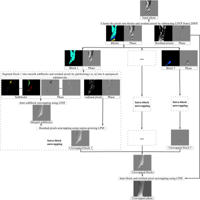
Current phase-unwrapping algorithms are generally challenged by severe noise, rapid-varying phase or disconnected regions. We present a novel phase-unwrapping method by using phase jumps detection and local polynomial surface fitting. The proposed method first segments the whole phase map into blocks by exploiting the phase jumps that are automatically identified. Intra-block wrapping may still exist if the true phase difference between adjacent pixels exceeds π inside a block. To address potential intra-block wraps, we further segment each block into subblocks using the phase partition method, and perform inter-subblock unwrapping using the block-growing method. Simulation and in vivo Dixon water-fat separation experiments were implemented to evaluate the performance of the proposed method, with comparisons to PRELUDE and CLOSE. This method has great potential in phase-related MRI applications in practice.
|
|
2251.
 |
Quantitative estimation of sub-voxel fat and water fractions based on two T2-component fitting in calf muscle.
Jannette Nassar, Dvir Radunsky, Noam Omer, Yann Le Fur, David Bendahan, Noam Ben-Eliezer
T2 relaxation is an effective biomarker for muscle pathology including inflammation, necrosis, or fatty infiltration. Accurate quantification of T2 values, however, is hampered due to the inherent bias of rapid multi-SE protocols by stimulated echoes. Recently, we introduced the echo modulation curve (EMC) algorithm, which successfully overcomes this problem and provide accurate T2 values that are stable across scanners and scan-settings. In this work, we present extension of the EMC algorithm for two component fitting, water and fat, allowing to quantify the sub-voxel infiltration of fat into the muscle, along with the corresponding T2 value of each component.
|
|
2252.
 |
A dual-step iterative temperature estimation method for accurate and precise fat referenced PRFS temperature mapping
Chuanli Cheng, Chao Zou, Yangzi Qiao, Changjun Tie, Qian Wan, Xin Liu, Hairong Zheng
Temperature imaging based on proton resonance frequency shift (PRFS) fails in fat containing tissues as the proton frequency of fat does not change with temperature. A dual-step iterative temperature estimation of fat referenced PRFS method is proposed to improve both the accuracy and precision of fat-referenced PRFS method. The method is evaluated with fat-water phantom and ex vivo BAT tissue excised from rats. Compared to the existing methods, the proposed method has least bias to the fluorescent optical fiber thermometer while maintaining the best noise performance.
|
|
2253.
 |
Longitudinal Stability of a Quantitative Fat-Water Phantom
Benjamin Ratliff, Samir Sharma, Jean Brittain, Scott Reeder, Diego Hernando
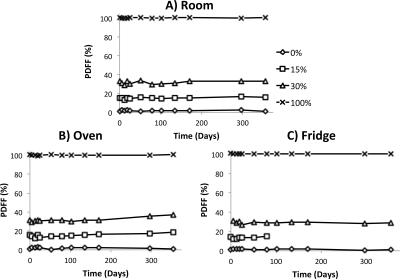
The purpose of this work was to evaluate the long-term stability of a previously validated fat-water phantom under a range of environmental conditions. Two separate phantoms were constructed, each with a range of fat concentrations. The first phantom was subjected to three different temperature conditions over one year. The second fat phantom was kept at room temperature and studied over three years. Our results show that the fat-water phantom has excellent long-term stability at room temperature and is robust to different temperature conditions.
|
|
2254.
 |
Evaluation of A Method for Simultaneous in vivo Measurements of Blood T1 and T2
Jialu Zhang, Dingxin Wang, Xiaotong Zhang, Lynn Eberly , Gregory Metzger, Donald Dengel, David Tupper, Anne Murray, Xiufeng Li
The longitudinal and transverse relaxation time constants of blood are important parameters for MRI methods and biomedical research studies. However, these parameters can vary largely across subjects, and change significantly across developmental stages, with physiological states, and due to specific diseases, which has motivated in vivo measurements of these parameters. We implemented a fast method for simultaneous in vivo measurements of blood T1 and T2. The study results suggest that the in vivo measurements of blood T1 and T2 can be achieved in about 25 s using the implemented method.
|
|
2255.
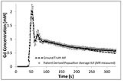 |
Quantification of the absolute accuracy and precision of DCE-MRI measurements of the arterial input function
Silvin Knight, James Meaney, Andrew Fagan
A novel anthropomorphic phantom test device was used to investigate the effects of temporal resolution (Tres), B1+-field non-uniformities, and pharmacokinetic (PK) model fitting methods on the absolute accuracy and precision of DCE-MRI measurements of the arterial input function (AIF), and resulting PK parameter estimates. Optimizing the Tres was found to reduce the maximum errors in PK parameter estimation from ~47% to ~20%. By correcting for B1+-field non-uniformities these errors were further reduced to ~7%. Using a linear rather than non-linear version of the standard Tofts model further increased the accuracy and precision of PK parameter estimations.
|
|
2256.
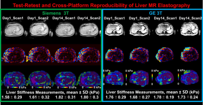 |
Baseline System Variability of Test-Retest and Cross-Platform Liver MR Elastography
Kevin Glaser, Roger Grimm, Brad Jr. Bolster, Richard Ehman, Jun Chen
Liver MR Elastography (MRE) has become an important noninvasive liver fibrosis imaging modality. It is essential to know the system variability of the stiffness measurements as only liver stiffness changes greater than the system variability are meaningful and reflect the true liver changes with 95% confidence. Our study was to perform a single-institution, single-reader study of fasting, normal subjects to assess the baseline (minimum possibly) MRE system variability within and between days, within and between platforms.
|
|
2257.
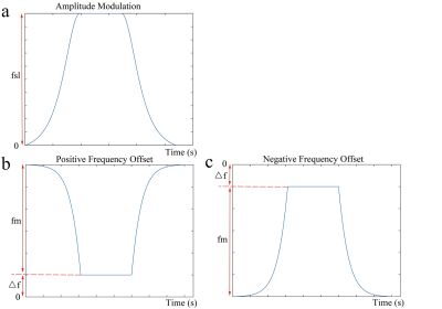 |
Robust chemical exchange spin-lock (CESL) using adiabatic pulses
Baiyan Jiang, Jing Yuan, Queenie Chan, Yi-Xiang Wang, Weitian Chen
Chemical exchange spin-lock (CESL) is a recently reported technology for probing metabolites which have intermediate to fast chemical exchange with bulk water. However, the conventional CESL is susceptible to B1 radiofrequency (RF) and B0 field inhomogeneity. The presence of these system imperfections leads to signal distortions and errors in contrast map. In this work, we report an approach to address this problem. We used simulation and in vivo experiments to demonstrate our proposed method.
|
|
2258.
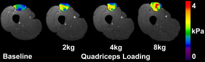 |
Morphometric Adaptions of Rectus Femoris to Muscle Strain Revealed Through ‘Dynamic Magnetic Resonance Elastography’ (DMRE)
Michael Perrins, Michiel Simons, Andre Attard, Colin Brown, Leela Biant, Edwin van Beek, Neil Roberts
Magnetic Resonance Elastography (MRE) allows for the quantification of tissue stiffness. When MRE is applied in muscle it allows for the measurement of muscle strain, with strain having an impact on structure morphology. This research investigated whether the bi-articular design of the Rectus Femoris gives an anatomical advantage in adapting to muscle strain and avoiding injury, as this is a vital muscle for movement. It was found that the mono-articular Quadriceps muscles showed significant muscle strain from loading, whereas the Rectus Femoris showed significant changes in cylindrical shape, and as expected, adapted to increased loading.
|
|
2259.
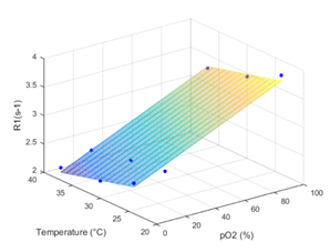 |
Lipid nanocapsules for tissue oxygenation determination using MRI
Janske Nel, Florence FRANCONI, Nicolas JOUDIOU , Bernard GALLEZ, Laurent LEMAIRE
To determine tissue pO2, lipid nanocapsules (LNCs) were used in conjunction with the rapid mapping of changes in tissue oxygenation, based on the higher solubility of O2 in lipids than in water, (MOBILE) MR sequence. LNCs were injected into the femoral muscle (n = 5) of C3H mice and T1 relaxation was measured whilst the animal was breathing air or carbogen (95 % O2, 5 % CO2) gas . In all explored mice a shortening in T1 relaxation was observed following the carbogen challenge, and T1 relaxation maps were produced indicating a response of the LNCs to the tissue O2 environment.
|
|
2260.
 |
Simultaneous Multi-slice Rapid MR Elastography of the Liver
Waqas Majeed, Arunark Kolipaka
We demonstrate the feasibility of combining simultaneous multi-slice (SMS) excitation with in-plane acceleration to achieve highly accelerated MR elastography data acquisition. The proposed approach enables the acquisition of diagnostic liver MRE data in a single breath-hold, which was not possible using the previous approaches. Our results indicate excellent agreement between the data acquired with and without SMS.
|
|
2261.
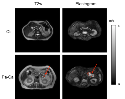 |
Tomoelastography of pancreatic tumors: Preliminary results
Stephan Marticorena Garcia, Christian Burkhardt, Rosa Schmuck, Guo Jing, Bernd Hamm, Jürgen Braun, Ingolf Sack
High-resolution stiffness maps of the pancreas were generated using multifrequency magnetic resonance elastography (MRE) and tomoelastography data processing in healthy controls (Ctr) and patients with pancreatic carcinoma (Pa-Ca). Pa-Ca have higher stiffness than control tissue and non-tumorous pancreatic parenchyma in patients without overlap to normal values. Subregional analysis for pancreatic head, corpus and tail revealed no difference between these anatomical regions. Tomoelastogaphy is sensitive to pathological changes in viscoelastic properties of Pa-Ca and offers a quantitative measure of stiffness of pancreatic tissue.
|
|
2262.
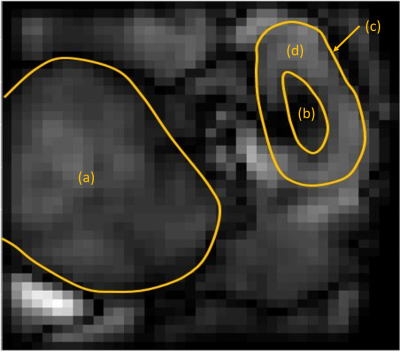 |
In vivo measurements of T1-dispersion maps in a kidney tumor mouse model using FFC-MRI around 1.5 T
Nicolas Chanet, Geneviève Guillot, Ingrid Leguerney, Rose-Marie Dubuisson, Catherine Sebrié, Alexandre Ingels, Noémie Assoun, Estelle Daudigeos-Dubus, Birgit Geoerger, Nathalie Lassau, Lionel Broche, Ludovic de Rochefort
Fast Field Cycling MRI offers the possibility to explore new contrasts generated from NMR dispersion (NMRD) profiles of tissue. Exploiting the dispersion properties of tissues may provide an additional biomarker of diseases through a deeper understanding of molecular mobility. Kidney tumors and healthy kidneys were analyzed among a cohort of twenty-seven mice to give insight into the potential of FFC-MRI for clinical applications. Here, we present R1-dispersion maps performed around 1.5 T to show that the intrinsic dispersion of tumors measured in vivo differs from the one of healthy kidneys.
|
|
2263.
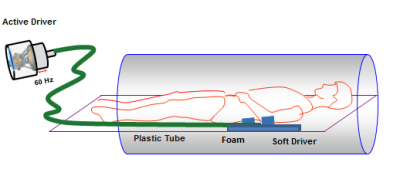 |
MR Elastography of Kidneys Using SE-EPI: A Reproducibility Study and Comparison to ADC and FA Measurements
Deep Gandhi, Prateek Kalra, Huiming Dong, Brian Raterman, Arunark Kolipaka
Stiffness change has been associated with progress of disease. Magnetic resonance elastography(MRE) is an imaging-based alternative that can measure stiffness. Diffusion Tensor Imaging(DTI) provides apparent diffusion coefficient(ADC) and fractional anisotropy(FA) of the tissue. Previous studies have investigated stiffness and diffusion in kidneys individually. However, none of the studies have investigated the two together. Aim of this study is to show reproducibility of spin-echo echo-planar imaging(SE-EPI) MRE and correlate it with ADC and FA measurements obtained from DTI. Preliminary results showed good reproducibility in stiffness measurements and moderate correlation between MRE stiffness and ADC and FA values from DTI.
|
|
2264.
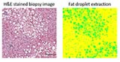 |
Monte Carlo Modeling of Liver MR Signal in the Presence of Fat
Changqing Wang, Benjamin Ratliff, Claude Sirlin, Scott Reeder, Diego Hernando
Recent studies using chemical shift-encoded MRI in patients with elevated liver fat content, but no iron overload, have shown a positive correlation between proton density fat fraction (PDFF) and R2*. In this work, we investigate the underlying biophysical mechanism of this observation using Monte Carlo simulations. Results from this Monte Carlo study show a positive correlation between PDFF and R2* consistent with previous in vivo observations. Based on the PDFF-R2* relationship, the Monte Carlo simulations may provide a new means to correct for the effect of fat on R2* quantification.
|
|
2265.
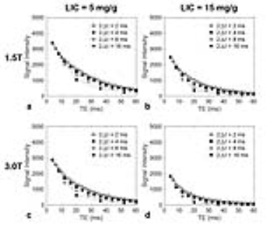 |
Monte Carlo Modeling of Multiple Spin Echo Signals in the Presence of Liver Iron Overload
Changqing Wang, Scott Reeder, Diego Hernando
Multiple spin echo (MSE) imaging may enable improved quantification and characterization of tissue iron deposition, with application for assessment of liver iron overload. However, iron deposition generally results in non-exponential signal decay in MSE imaging, and MSE-based R2 (1/T2) relaxometry can depend on the inter-echo time. Additionally, it is cumbersome and expensive to empirically calibrate the R2 relaxometry-iron concentration relationship. In this work, we investigate the effect of inter-echo time on MSE signal in the presence of liver iron overload using Monte Carlo simulations. This Monte Carlo approach may enable improved calibration of MSE-based measurements of iron concentration.
|
|
Contrast Mechanisms
Traditional Poster
Contrast Mechanisms
Wednesday, 20 June 2018
| Exhibition Hall 2266-2297 |
08:15 - 10:15 |
|
2266.
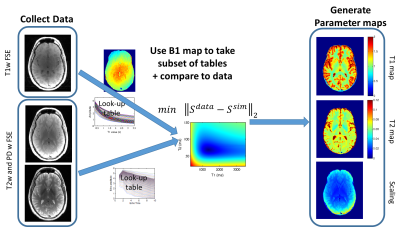 |
Quantification of T1 and T2 from Standard MR Images
Kelly McPhee, Alan Wilman
Exact sequence modelling using the Bloch equations is employed to directly extract quantitative T1 and T2 relaxation maps from standard MRI sequences. The need for excess specialized sequences was eliminated by measuring relaxation directly from T1, T2, and PD-weighted images, and a rapid B1 map. This approach enables wider use of quantitative MRI.
|
|
2267.
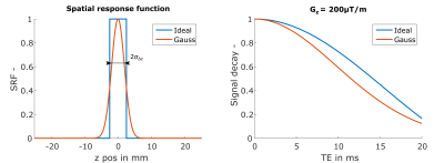 |
$$$R_2^*$$$ Correction for Gradient Echo with a Gaussian Excitation Pulse
Martin Soellradl, Lukas Pirpamer, Jan Sedlacik, Franz Fazekas, Stefan Ropele, Christian Langkammer
Macroscopic field inhomogeneities increase the effective transverse relaxation rate R2*. In contrast to conventional models assuming ideal rectangular pulses, we developed an R2* correction model for Gaussian excitation pulses. After demonstrating the validity of the model in phantom measurements we measured 10 volunteers with 2mm and 4mm slice thickness, respectively. Uncorrected and corrected R2* values were assessed regionally and a significant effect of the correction was observed. An advantage of the proposed method is that it only requires two echoes, rendering it useful in clinical MRI.
|
|
2268.
 |
Accuracy and precision of measured T1 in hepatic portal vein blood using a variety of Look- Locker and Modified Look-Locker Inversion Recovery sequences.
Svein Are Vatnehol, Atle Bjørnerud, Camilla Haglerød, Per Kristian Hol, Mahmood Amiry-Moghaddam, Tryggve Storås
The longitudinal variation and averaged T1 measured in the hepatic portal vein (HPV) obtained with 12 variations of Look-Locker (LL) and Modified Look-Locker Inversion Recovery (MOLLI) sequences were compared to identify the sequence with least variation. Among the sequences studied, LL sequence with 5 beat readout and 45o flip angle and MOLLI with an acquisition scheme 10 beats readout, 5 beats recovery followed by 5 beat readout (10(5)5) were shown to be the most stable. Method of image analysis and the use of simulated versus real-time EKG did not significantly affect the stability of the T1-estimates.
|
|
2269.
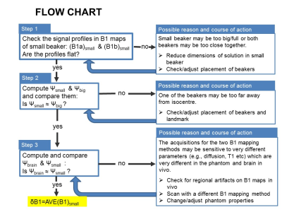 |
Assessing B1 map errors in vivo: measuring stability and absolute accuracy despite the lack of gold standard
Sofia Chavez
B1+ field inhomogeneity is a major source of errors in quantitative mapping. The accuracy of B1 maps, depicting the effects of B1+ inhomogeneity on the flip angle, is thus critical. However, there is no gold standard B1 mapping method in vivo so absolute accuracy is difficult to determine. In this work, we propose steps that exploit known B1 effects in a small phantom to obtain absolute accuracy estimates in vivo. Two B1 mapping methods are required, but neither need be accurate. We demonstrate the proposed assessment by obtaining stability and absolute accuracy measurements of the Method of Slopes B1 maps.
|
|
2270.
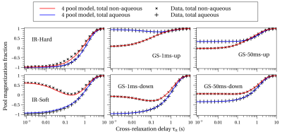 |
Aqueous and Non-aqueous T1 relaxation in brain under six diverse initial conditions
Alan Manning, Carl Michal, Alex MacKay
A consistent view of T1 relaxation in white matter remains elusive. We use an NMR spectrometer to observe white matter T1 relaxation behavior in both aqueous and non-aqueous protons following six diverse initial magnetizations. The data is analyzed in the context of both an unrestricted and restricted four pool model. We show how the observed multi-component T1 relaxation behavior depends sensitively on the initial conditions of the different pools, suggesting that great care must be taken in interpreting T1 relaxation measurements.
|
|
2271.
 |
Towards Quantifying pO2 via 1H Longitudinal Relaxation of Water: Quantifying the Confounds
Kelsey Meinerz, Scott Beeman, James Quirk, Joel Garbow, Joseph Ackerman
Crosslinked bovine serum albumin phantoms are used as tissue surrogates/mimics to investigate the physiologic confounds to R1-based tissue-O2 quantification under precise, laboratory controlled conditions. The relaxation-rate constants for both the rapidly relaxing and the slowly relaxing populations are affected by changes in temperature, pH, and protein concentration.
|
|
2272.
 |
Increased CEST specificity for amide and fast exchanging amine protons using exchange-dependent relaxation rate
Xiao-Yong Zhang, Feng Wang, Junzhong Xu, Daniel Gochberg, John Gore, Zhongliang Zu
It is challenging to remove overlapping chemical exchange saturation transfer (CEST) signals from nearby exchanging sites. Our previous study showed that the contributions of fast exchanging amines to CEST signals at 3 ppm induce a broad spectral region that overlaps with the amide proton transfer (APT) spectrum centered around 3.5 ppm. In this work, we apply an exchange-dependent relaxation rate (Rex) for quantifying CEST effects to increased CEST specificity for amide and fast exchanging amine protons. Our results demonstrate that Rexreduces the influences of overlapping CEST signals for APT imaging, and thus can significantly enhances the CEST detection specificity.
|
|
2273.
 |
MR Elastography of the brain: Comparison between anisotropic and isotropic stiffness with age
Prateek Kalra, Brian Raterman, Xiaokui Mo, Arunark Kolipaka
Noninvasive measurement of mechanical properties of brain tissue using Magnetic Resonance Elastography has been a promising method for investigating neurological disorders such as multiple sclerosis, hydrocephalus and Alzheimer’s. However, due to regional and directional dependency of brain stiffness, estimating anisotropic stiffness is important. Previous studies have investigated anisotropic and isotropic stiffness separately but none of them investigated the two together. Objective of this study is to investigate both isotropic and anisotropic stiffness together and independently compare with age and with each other. Results demonstrated a significant decrease in isotropic and anisotropic stiffness with age in some regions of the brain.
|
|
2274.
 |
Using healthy volunteers to optimize amide proton transfer CEST sequences.
Robert Brand, Nicholas Blockley, Michael Chappell , Peter Jezzard
Optimising CEST sequences for clinical use is difficult due to the lack of representative phantoms. Healthy volunteers do not show the variation in pH or concentration that these sequences seek to detect. However, in this work we show how the inherent T1 sensitivity of CEST sequences [1] can be exploited to optimise them in healthy volunteers. We demonstrate that the sequence conditions that maximise the grey/white matter contrast in exchange maps are also the parameter conditions that maximise the exchange sensitivity. This method provides an effective way to optimise in vivo CEST sequences without the need for phantoms or simulations.
|
|
2275.
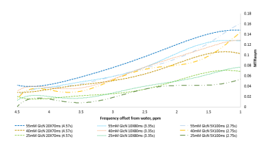 |
Chemical Exchange Saturation Transfer (CEST) MRI of glucosamine at 3T
Michal Rivlin, Daniel Barazany, Gil Navon
In our previous work using preclinical 7T MRI scanner we have shown that tumors in mice can be imaged using CEST-MRI of glucosamine. Moving toward clinical application, considering the excellent safety profile of glucosamine, we tested the CEST-MRI of glucosamine on a 3T clinical scanner. Here we report significant CEST MRI signal up to ~3.5 ppm from the water signal corresponding to the exchangeable protons of the glucosamine hydroxyls and amine residues. Thus, CEST MRI using glucosamine has the potential to report on the activity of tumor metabolism, noninvasively by using MRI.
|
|
2276.
 |
Live monitoring of red and yellow bone marrow in long bones of the mouse at 9.4T
Nicolas Kunz, Josefine Tratwal, Olaia Naveiras
When hematopoiesis is compromised, as after lethal irradiation, the red BM is rapidly infiltrated by fat, then slowly recovers hematopoietic function following BM transplantation. Monitoring this red-to-yellow-to-red BM transition non-invasively using a tree point Dixon technique would provide important information on the reconstitution of the hematopoietic system that precedes blood formation as measured by bleeding, and thus be extremely useful in experimental hematology. In this preliminary study we investigate the feasibility to track differences in bone marrow adiposity in the C57B6 mouse femur and tibia post-irradiation by monitoring the fat content.
|
|
2277.
 |
SafeNet: Artificial Neural Network for Real-Time T2 Mapping with Quality Assurance
Doohee Lee, Woojin Jung, Jingu Lee, Jingyu Ko, Hyeong-Geol Shin, Hyunsung Eun, Yoonho Nam, Jongho Lee
Accurate T2 mapping using multi-echo spin-echo data is a time-consuming process due to stimulated echo correction. In this study, we developed an artificial neural network for real-time T2 mapping. The training dataset using both in-vivo data and model-based synthetic data demonstrated the best performance. The resulting T2 map shows mean T2 errors of less than 0.3 ms with minimal computation time (less than 1 sec as opposed to 8.3 hours for conventional method). An additional algorithm was developed to ensure the fidelity of the T2 map at the cost of slightly increased computation time.
|
|
2278.
 |
Validation of intrinsic actuation MR Elastography through a 1Hz experimental phantom system
Scott Gordon-Wylie, Matthew McGarry, Ligin Solamen, Elijah Van Houten, John Weaver, Keith Paulsen
A 1Hz MR elastography (MRE) phantom system is presented to validate the spatial accuracy of mechanical property images in intrinsic actuation MRE. A custom hydraulically driven actuator generated 1Hz shearing motions in gelatin phantoms with stiff inclusions which were measured using a retrospectively gated QFLOW sequence on a 3T Philips Achieva MRI. Maps of the octahedral shear strain showed low strain in stiff inclusions, and high strains in areas of stress concentrations, as expected from theory. Shear modulus maps computed by a viscoelastic nonlinear inversion MRE algorithm were spatially accurate, and identified the correct stiffness contrast of phantom components.
|
|
2279.
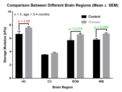 |
MR Elastography in a mouse model of Alzheimer’s disease: 5XFAD Mice
Shreyan Majumdar, Rachana Mishra, Orly Lazarov, Dieter Klatt
In vivo magnetic resonance elastography (MRE) experiments on the 5XFAD Alzheimer’s disease (AD) mouse model were conducted. The AD and Control mice were in the age group of ~1 month (n = 2 for both) and 3~4 months (n = 5 for both). Median stiffness values were measured over different regions of the brain. The overall brain tissue was stiffer in the disease model when compared to the control, with results being significant at the 3~4-month time point. Further experiments are underway at the 1-month time-point for conclusive age-based comparisons.
|
|
2280.
 |
Accuracy and precision of Synthetic MRI
Gabriel Mangeat, Russell Ouellette, Marcel Warntjes, Michael Plattén, Love Nordin, Nikola Stikov, Tobias Granberg, Julien Cohen-Adad
Synthetic MRI (SyMRI) provides quantitative PD, T1 and T2 maps via a rapid single-volume acquisition. Here, we aim to validate the precision and accuracy of SyMRI quantification by performing five scan re-scans, at two field strengths, of the quantitative NIST phantom and one control subject. Results show a good accuracy of T1 and T2 quantification at 3T, and a very good precision of all the phantom and subject measurements at 1.5T and 3T (95% confidence intervals width are respectively lower than 6% and 4%, of the measured value). This study brings confidence in comparing SyMRI quantitative measurements across subjects or time.
|
|
2281.
 |
Dynamic Contrast Enhanced DWI in a split dynamic framework
Tryggve Storås, Endre Grøvik, Kjell-Inge Gjesdal, Sebastian Meltzer, Kathrine Redalen
Most MRI contrast agents distributes extracellular only. If the extracellular water signal is suppressed by diffusion weighting the short ranged T1-relaxation effect of a the contrast agent will be solely through water exchange through the cell membrane. In this paper we describe the implementation of a dynamic contrast enhanced diffusion weighted acquisition facilitating the study of diffusion weighting on the relaxivity of contrast agents. This is all done within a Split Dynamic framework allowing for this study to be performed without sacrificing assessment of the established pharmacokinetic parameters.
|
|
2282.
 |
Low frequency excited MR elastography of the brain using displacement encoding with stimulated echoes and multi phase offset readouts
Johannes Strasser, Franz Fazekas, Stefan Ropele
In Magnetic Resonance Elastography (MRE), mechanical tissue parameters are assessed by sampling shear wave propagation via a set of motion encoded phase offset images. We here investigate a multiple phase offset image acquisition strategy based on displacement encoding with stimulated echoes (DENSE) for multi-slice human brain MRE together with a low frequency mechanical excitation and short echo times. Clear wave images could be acquired using the proposed imaging approach and estimates of the magnitudes of the complex shear modulus could be calculated from the derived wave image data set.
|
|
2283.
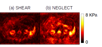 |
Can the slow compression wave in MRE data be inverted? An exploratory analysis
Eric Barnhill, Jürgen Braun, Ingolf Sack
Magnetic Resonance Elastography (MRE) data show high-amplitude, low frequency artifact which does not accord with the viscoelastic model in near-incompressible tissue. This exploratory study investigates whether the low frequency behavior is poroelastic, and if so whether slow compression wavelengths can be estimated. A cohort of abdominal MRE acquisitions at four frequencies were convolved with a fine-grained Gabor filter bank, and the frequency response of the acquisitions were pooled across subjects for liver and spleen regions. The pooled frequency responses for both liver and spleen showed a shifting peak in the response function mass that tracked with the increase in frequency, with wavespeeds in the shear regime. A second peak identified a lower frequency regime. This regime produced values similar to those observed in tissue poroelastic behaviors.
|
|
2284.
 |
High Resolution Low Field MR Elastography
Muhammad Waqas, Huihui Xu, Shadi Othman
In this study, we extend MRE to a low field strength of 0.5T that offers in-plane resolution of 150 micron x 150 micron. To verify the method, shear wave images through gel phantoms were obtained at a mechanical excitation frequency of 370Hz. Preliminary studies on rat brains demonstrate the feasibility of the using low filed MRE in determining mechanical properties.
|
|
2285.
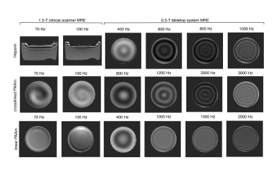 |
Wideband mechanical tests of the viscoelastic powerlaw behavior of phantom materials for Magnetic resonance elastography
Felix Schrank, Heiko Tzschätzsch, Angela Ariza de Schellenberger, Paul Janmey, Jürgen Braun, Ingolf Sack
Shear rheometry was combined with magnetic resonance elastography (MRE) in a 1.5-T clinical system and a 0.5-T tabletop MRE system to investigate the viscoelastic powerlaw behavior of heparin and polyacrylamide (PAAm) over more than three orders of magnitude dynamic range. While heparin has softer properties than encountered in soft in-vivo tissues, crosslinked PAAm has similar stiffness as measured for in-vivo tissues, however, with lower dispersive properties. Overall both materials are good candidates for the use as standard phantom materials in MRE due to their well predictable springpot properties across the full frequency range relevant for MRE investigations.
|
|
2286.
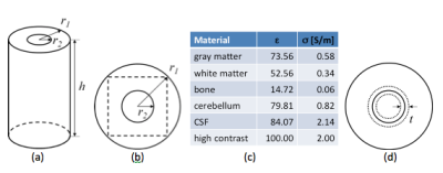 |
Region-specific regularization of convection-reaction Magnetic Resonance Electrical Property Tomography (MREPT) for improving the accuracy and noise-tolerance of EP reconstruction
Adan Garcia, Shaoying Huang , Wenwei Yu
Magnetic resonance electrical property tomography (MREPT) is a technology for noninvasively reconstructing electrical properties (EPs) (permittivity, e, and electrical conductivity, s) of the human body from B1-map from MRI. Boundary inaccuracy and noise sensitivity are two problems of most MREPT methods. Previous studies showed that regularization can be one solution for both of the problems. However, there have been few reports on how to set up regularization terms. In this study, we show how region-specific regularization can achieve higher accuracy and noise tolerance.
|
|
2287.
 |
Low Frequency Magnetic Resonance Conductivity Imaging By Means of Oscillating Gradient Fields
Hasan H. Eroglu, Mehdi Sadighi, B. Murat Eyuboglu
Recently, low frequency (LF) magnetic resonance electrical conductivity imaging by means of oscillating gradient fields is reported to be infeasible. In these studies, LF phase measurements are modeled with radio frequency (RF) leakage due to geometric shifts in MR images. Although RF leakage is related with conductivity, we have not come across a conductivity image reconstructed using this model. In this study, LF conductivity imaging is evaluated for an MRI pulse sequence including multiple gradient pulses. Geometric shifts are evaluated by focusing on the MR magnitude. A procedure is proposed for the reconstruction of conductivity, based on LF phase measurements.
|
|
2288.
 |
Quantitative MRI made easy with qMRLab
Tanguy DUVAL, Ilana Leppert, Jean-François Cabana, Mathieu Boudreau, Ian Gagnon, Gabriel Berestovoy , Julien Cohen-Adad, Nikola Stikov
Quantitative MR (qMR) methods exist for most MRI sequences (e.g. diffusion, magnetization transfer, inversion recovery). All these methods have a similar methodology: a biophysical model (i.e. an analytical equation), that relates the MRI contrast to some microstructural and physical features, is used to fit experimental data. Although open-source software packages are available online for certain qMR techniques, there does not exist a single stand-alone platform that can implement and compare a wide range of quantitative MRI methods. With qMRLab, we propose an open-source, MATLAB-based, object-oriented software with separate modules for each technique. We envision qMRLab as a standard platform with a growing list of contributors, where the qMR community can replicate and cross-validate a wide range of qMR methods. qMRLab includes a user-friendly graphical user interface (GUI), batch scripts examples, and qMR datasets. The software can be used to fit and check the quality of qMR data, to optimize protocols, compare fitting models, and simulate the effects of various model assumptions.
|
|
2289.
 |
Implementation and validation of delta relaxation enhanced MRI at 3T: A system for quadrupole enhanced relaxation imaging
Markus Bödenler, Martina Basini, Maria Casula, Evrim Umut, Danuta Kruk, Hermann Scharfetter
The frequency-selective nature of quadrupolar relaxation enhancement offers a high potential for designing smart molecular probes for the usage as novel MRI contrast agents by cycling the main magnetic field. Their validation and application requires a fast field-cycling MRI system. In this work, we present the first implementation and validation of such a system at the clinical field strength of 3T. The complete FFC-MRI setup was successfully validated by R1 dispersion imaging with dispersive iron oxide magnetic nanoparticles, thus providing a ready-to-use hardware setup for the future investigation of new compounds.
|
|
2290.
 |
Quadrupolar relaxation enhancement in selected Bismuth-Aryl compounds: Promising precursors for novel T1 MRI contrast agents
Hermann Scharfetter, Christian Gösweiner, Evrim Umut, Carina Sampl, Roland Fischer, Stefan Spirk, Andreas Petrovic, Danuta Kruk
209Bi-aryl compounds have the potential for designing novel class of smart MRI T1 contrast agents which are sensitive to the chemical environment and the B0 field. We have confirmed quadrupolar relaxation enhancement (QRE) of protons as the underlying mechanism in two solid organobismuth-compounds in the B0 range 0.5 – 3T. We also show first QRE peaks of solvent protons in a solution of Tris-(2-orthomethoxy-Phenyl)Bismuthane in tetrahydrofurane. This very important first step yields two promising candidates for the development of QRE-based CAs and opens the way for the second step, i.e. grafting them onto water-soluble nanoparticles for optimizing the relaxivity.
|
|
2291.
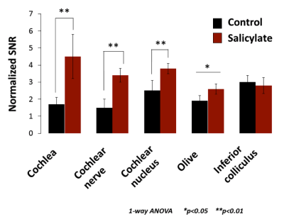 |
MANGANESE ENHANCED MRI: A METHOD IN ORDER TO VALIDATE PHYSILOGICAL MARKERS OF TINNITUS IN RODENTS
Amandine Laboulais, Maïda Cardoso, Sergio Gonzalez, Gaëlle Naert, Yves Cazals, Arnaud J. Noreña, Sylvie Cosnier-Pucheu, Celia Belline, Christophe Goze-Bac
The present study is designed to show physiological markers of tinnitus in rodents. The tinnitus is an auditory phantom sensation experienced in absence of an external stimulus. The prevalence of tinnitus shows a worrying growth curve with the development of new lifestyles (exposure to noise, urbanization, …). One promising tool is used, called Manganese Enhanced MRI (MEMRI). The use of manganese chloride as an MRI contrast agent enables to follow brain neuronal activity. T1-weighted MRI images are collected in order to investigate the specific areas activated in presence of tinnitus or not. Two analysis methods are used: Statistical analysis by Signal to Noise Ratio (SNR) and T2 rate cartography. Results enable to shown a complementary between the two analysis methods and allows us to discriminate between healthy and tinnitus rats.
|
|
2292.
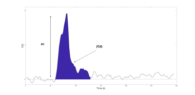 |
Relative perfusion mapping using BOLD imaging with induced hypoxia
Chau Vu, Julie Coloigner, Soyoung Choi, John Wood
DSC MRI is a popular perfusion technique that requires the use of an invasive exogenous contrast. This study proposes an alternative technique which uses BOLD imaging and 100% nitrogen inhalation to map relative perfusion values (rCBV, rCBF and rMTT) without the use of contrast. We evaluated its performance on a cohort of healthy controls and sickle cell disease patients with a large range of global cerebral blood flow.
|
|
2293.
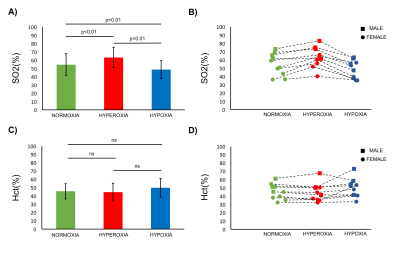 |
MR Measurements of Blood Oxygenation and Hematocrit during Gas Challenges
Thomas Christen, Jia Guo, Wendi Ni, Audrey Fan, Michael Moseley, Greg Zaharchuk
A new MR approach has been proposed to obtain simultaneous measurements of blood oxygen saturation (SO2) and hematocrit (Hct) by measuring and combining blood MR relaxation times. Although the first results were encouraging, the method has not been properly validated. In this study, we tested this approach in 10 volunteers subjected to gas challenges with the intent to modify SO2 while keeping Hct constant. The method was also tested in 10 Moyamoya patients and compared to photometric analysis. Results suggest that reliable MR estimates of both SO2 and Hct can be obtained in vivo.
|
|
2294.
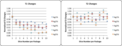 |
Incidental magnetization transfer in qMRI: effects of multi-slice imaging with mixed-TSE.
Ning HUA, Mitchell Horn, Adam Aakil, Stephan Anderson, Hernan Jara
Purpose: To evaluate the effect of inherent and incidental magnetization transfer (MT) on T1 and T2 measurements when using the mixed turbo spin echo sequence (mixed-TSE). Methods: mixed-TSE was applied to a phantom of 1-5% agarose gel. The levels of the MT effects were induced and controlled by varying the number of slices per acquisition package. Results: T1 values were underestimated in multi-slice mixed-TSE. No obvious trend was observed for T2 measurements. Conclusion: mixed-TSE is powerful and efficient tool for qMRI, yet caution should be taken when interpreting the derived T1 values because of MT effects.
|
|
2295.
 |
The Hematocrit Dependence of Blood T2 Relaxometry Parameters in the Weak Field Approximation
Avery Berman, Jonathan Polimeni, G Bruce Pike
The weak field approximation (WFA) is a theory that relates T2 relaxation from tissue to the underlying tissue properties and is commonly applied to the analysis of relaxation from red blood cells (RBCs) in blood. This study examines the hematocrit-dependence of the different parameters of the WFA using simulated populations of RBCs and published experimental relaxometry results from two studies. Both the simulations and the experimental results show an unexpected result that the characteristic perturber size estimate is not constant with hematocrit but is negatively correlated with it. This has important implications for the implementation and interpretation of the WFA theory on blood relaxometry data.
|
|
2296.
 |
Use of Entwined Magnitude and Phase-sensitive Inversion REcovery (EMPIRE) Pulse Sequences to Study the Brain and Knee
Yajun Ma, Wei Zhao, Adam Searleman, Jiang Du, Nikolaus Szeverenyi, Graeme Bydder
The combination (addition/subtraction) of magnitude and phase-sensitive IR images (termed EMPIRE technique) when appropriate TIs were used was found to provide increased tissue contrast over specific ranges of tissue T1. This behavior was explored numerically and summarized in signal intensity vs. T1 plots. Clinically relevant applications were demonstrated in brain and knee cartilage using FSE and UTE data collections. In addition to increased contrast, this approach allowed detection of short T2 tissue signals while suppressing unwanted signal from longer T1 tissue fluids.
|
|
2297.
 |
Age-associated changes in skeletal muscle morphology assessed by intramuscular adipose and connective tissue
Bharath Ambale Venkatesh, Yoko Kato, Jaclyn Sesso, Jason Ortman, John Pitts, Michio Ozaki, Yoshimori Kassai, Joao Lima
In human skeletal muscles, the aging process causes a decrease of contractile function likely associated with an increase in intramuscular adipose and connective tissues. The accumulation of non-contractile tissues and loss of muscle tissue may contribute to sarcopenia and frailty observed at older age but their quantification is challenging1. The purpose of this study was to establish MR imaging-based methods to quantify the relative amounts of fat and connective tissue in healthy human subjects, and investigate their association with age.
|
|
| Back |
| The International Society for Magnetic Resonance in Medicine is accredited by the Accreditation Council for Continuing Medical Education to provide continuing medical education for physicians. |
