|
Traditional Poster Session
Body: Breast, Chest, Abdomen, Pelvis |
Wednesday, 20 June 2018
Traditional PosterBody: Breast, Chest, Abdomen, Pelvis
2418 -2432 Breast Imaging
2433 -2443 129Xe & 3He Imaging
2444 -2450 Body Imaging: Fetal/Placenta & Pelvis
2451 -2480 Thoracic MRI
2481 -2489 Pancreas/GI
2490 -2495 Body Imaging: Renal
2496 -2511 Body: Fat Imaging
2512 -2517 Body: Animal Models
2518 -2535 Body: Technical Advances
2536 -2552 Body: Liver
2553 -2577 Prostate
2578 -2587 Body: Liver Fat & NASH
2588 -2596 Body: MRE
2597 -2609 Body: Liver Iron
2610 -2626 Body: Liver Imaging Using Perfusion, Diffusion, T1, & T1rho |
| |
Breast Imaging
Traditional Poster
Body: Breast, Chest, Abdomen, Pelvis
Wednesday, 20 June 2018
| Exhibition Hall 2418-2432 |
16:15 - 18:15 |
|
2418.
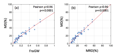 |
A MRI-based breast density measure which is directly comparable to mammographic density
Jie Ding, Alison Stopeck, Yi Gao, Marilyn Marron, Betsy Wertheim, Maria Altbach, Jean-Philippe Galons, Denise Roe, Fang Wang, Gertraud Maskarinec, Cynthia Thomson, Patricia Thompson, Chuan Huang
High breast density is an independent risk factor for breast cancer. Mammography, the most widely used method for breast density determination, is limited by ionizing radiation exposure and its relatively low reliability for density assessment. We propose an automated, safe, and highly reproducible breast density measurement based on fat-water decomposition MRI. The technique yields a measure directly comparable to mammographic density which is easy for clinicians to use and for patients to understand.
|
|
2419.
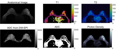 |
Rapid and Simultaneous T1, T2 and Diffusion Quantification using MR Fingerprinting in the Breast
Yun Jiang, Katherine Wright, Jesse Hamilton, Wei-Ching Lo, Ananya Panda, Gregor Körzdörfer, Shota Hodono, Michael Boss, Nicole Seiberlich, Vikas Gulani, Mark Griswold
High quality, distortion-free T1, T2 and diffusivity maps in breast imaging are simultaneously generated using MRF framework. A good agreement of T1, T2 and ADC between the proposed MRF method and the traditional spin echo methods is demonstrated in a phantom and in vivo in breast imaging. This method enables the simultaneous collection of T1, T2 and diffusion maps for tissue characterization without the need to co-register separately acquired maps as in conventional MRI.
|
|
2420.
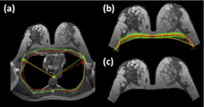 |
Automatic Breast and Fibroglandular Tissue Segmentation Using Deep Learning by A Fully-Convolutional Residual Neural Network
Yang Zhang, Vivian Park, Min Kim, Peter Chang, Melissa Khy, Daniel Chow, Jeon-Hor Chen, Alex Luk, Min-Ying Su
A deep learning method using the fully-convolutional residual neural network (FCR-NN) was applied to segment the whole breast and fibroglandular tissue in 289 patients. The Dice similarity coefficient (DSC) value and accuracy were calculated as evaluation metrics. For breast segmentation, the mean DSC was 0.85 with an accuracy of 0.93; for fibroglandular tissue segmentation, the mean DSC was 0.67 with an accuracy of 0.75. The percent density calculated from ground truth and network segmentations were correlated, and showed a high coefficient of r=0.9. The initial results are promising, suggesting deep learning has a potential to provide an efficient and reliable breast density segmentation tool.
|
|
2421.
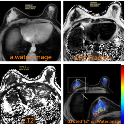 |
T2 star for breast invasive ductal carcinoma histopathological grade
Meiying Yan, Xiaoqi Wang, Rengen Xu
Chemical shift encoded MRI (CSE-MRI) utilizes the water-fat signal model method, and its corresponding T2*mapping has less artifacts from water-fat shift. We extracted the fat-influence-free T2* to investigate the correlation between T2 * mapping and histological grading of breast invasive ductal carcinoma, and found T2 * value for IDC-3 significantly higher than in IDC-2. This finds may provide more understanding of invasive ductal carcinoma microstructure and metabolism.
|
|
2422.
 |
Radiomic analysis of breast can distinguish benign phyllodes tumors from fibroadenomas
Lina Zhang, Gang Yuan, Qingwei Song, Yanwei Miao, Ailian Liu, Yan Guo, Dandan Zheng
The distinction between phyllodes tumor of breast (PTB) and fibroadenoma(FA) is clinically important, as approximately 20-30% of resected PTBs are malignant. Only limited information on the MRI characteristics of PTB is available. This study was performed to compare the MRI features (radiomics) of PTBs and FAs, which may resemble each other on conventional MRI.
|
|
2423.
 |
Application of multiple b-value diffusion weighted imaging in diagnosing ductal carcinoma in situ
Lina Zhang, Kai Zhang, Qingwei Song, Ailian Liu, Lizhi Xie
Multiple b-value diffusion weighted imaging (DWI) provides quantitative measurement of ADCslow for cellularity and ADCfast and ffast for vascularity. It is helpful for the differentiation between benign and malignant breast lesions. This study concerned perfusion as well as diffusion information in normal breast tissues and breast lesions from intravoxel incoherent motion (IVIM) imaging based on the biexponential analysis of multiple b-value DWI and then compared these parameters to ADC obtained with monoexponential analysis on the diagnosis of different grades of ductal carcinoma in situ (DCIS).
|
|
2424.
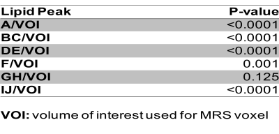 |
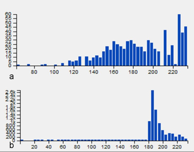
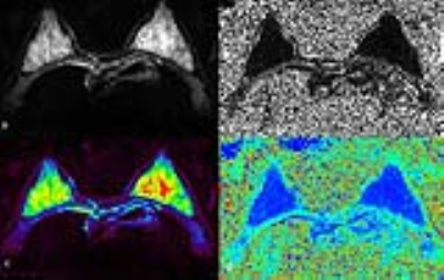
Proton MR Spectroscopy in Breast: Lipid Metabolite Concentrations as Valuable Quantitative Imaging Biomarkers for Cancer Diagnosis
Sunitha Thakur, Sandra Brennan, Ileana Hancu, Blanca Bernard-Davila, Michael Weber, Elizabeth Manderski, Elizabeth Morris, Katja Pinker
Differential expression of lipid metabolism-related proteins was recently reported in breast cancer patients. In this retrospective MR spectroscopy (MRS) study, the spectral lipid profile was assessed in breast cancer patients with malignant and benign lesions. Single-voxel MRS data from 176 breast lesions was analyzed to quantify multiple lipid metabolite concentrations using LCModel. Lipid peak analysis highlighted significant differences in lipid metabolite concentrations with significantly low concentrations in malignant compared to benign lesions and in luminal cancers compared to other molecular subtypes. MRS-based lipid metabolite profile may provide a valuable tool for breast cancer diagnosis.
|
|
2425.
 |
Diffusion tensor and Intravoxel incoherent motion magnetic resonance imaging of the normal breast in young premenopausal women during menstrual cycle
Qiuju Fan, Hui Tan, Nan Yu, Qi Yang, Shaoyu Wang, Yong Yu
DTI and IVIM can provide valuable information on tissue microstructure, microcirculation and pathophysiology that has been extensively used on the breast cancer [1,2].However, the breast is a hormonally responsive organ and undergoes periodic variations according to the menstrual cycle. Thus, the periodic variations of DTI and IVIM-derived measurements need to be considered.
|
|
2426.
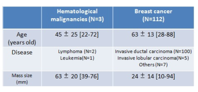 |
Kurtosis as a potential tool to differentiate breast hematological malignancies from breast cancer
Mizue Suzuki, Masako Kataoka, Mami Iima, Shotaro Kanao, Kanae Miyake, Rena Sakaguchi, Ayami Kishimoto, Maya Honda, Tadakazu Kondo, Tatsuki Kataoka, Takaki Sakurai, Masakazu Toi, Kaori Togashi
Since breast hematological malignancies show various image findings, it is not easy to differentiate them from breast cancer using conventional MRI. Non-Gaussian diffusion MRI is a relatively new method using multi b values from low to high, reflecting the interaction of water molecules with tissue features. We compared non-Gaussian parameters of breast hematological malignancies and breast cancer to investigate the advantage of non-Gaussian diffusion imaging. Our preliminary results suggest potential advantage of kurtosis as a marker of cellular structure and usefulness in differential diagnosis between breast hematological malignancies and breast cancer.
|
|
2427.
 |
Ultra-high field Dynamic Contrast Enhanced Magnetic Resonance Imaging of the Breast with pharmacokinetic (PK) modeling: Value for the Differentiation of Benign and Malignant Breast Tumors and Molecular Breast Cancer Subtypes
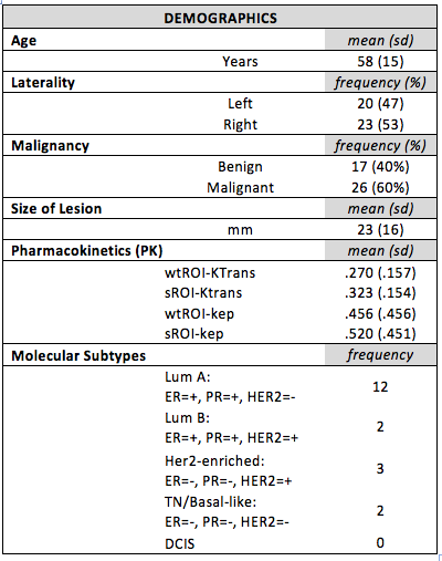
Rosa Elena Ochoa Albiztegui, Joao Machado Horvat, Sunitha Thakur, Blanca Bernard-Davila, Siegfried Trattnig, Thomas Helbich, Elizabeth Morris, Katja Pinker-Domenig
To investigate ultra-high field DCE-MRI of the breast at 7T with pharmacokinetic modeling for differentiation of benign and malignant breast tumors and molecular breast cancer subtypes. 37 patients with 43 breast lesion were included and underwent a 7T DCE-MRI of the breast. Quantitative pharmacokinetic imaging biomarkers ktrans and kep aid in the differentiation of benign and malignant breast tumors. Selection of ROI- using a whole tumor and a 10mm2 ROI- does not influence diagnostic accuracy. Quantitative pharmacokinetic imaging biomarkers ktrans and kep are not able to differentiate molecular breast cancer subtypes.
|
|
2428.
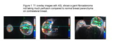 |
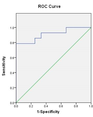
New frontiers: the role of Arterial Spin Labeling (ASL) and Diffusion Tensor Imaging (DTI) to differentiate between malignant and benign breast lesions.
Akshaykumar Kamble, Manju Popli
There was the time when contrast enhancement was critical to identify and differentiate malignant from benign tumor, but as the field of MR has made strides towards advanced imaging, we now can use the methods which doesn't require contrast. It is especially helpful in end stage renal patients. As the world demographic is slowly tilting towards geriatric population it will soon become essential to come up with alternative ways to detect the malignant pathologies independent of exogenous contrast. In our study we have demonstrated by plotting the ROC curve that ASL and DTI are promising methods to detect breast cancer.
|
|
2429.
 |
Breast phyllodes tumor: histogram analysis of the apparent diffusion coefficient for assessment of tumor grading
Wenrui Tang, Yan Zhang, Dandan Zheng, Jingliang Cheng
Phyllodes tumors are uncommon, biphasic, fibroepithelial lesions of the breast, characterized by leafy stromal fronds capped by benign bilayered epithelium. Grading of breast phyllodes tumors is critical for diagnosis, treatment options and preoperative evaluation. This study is to assess the feasibility of diffusion weighted image (DWI) for determining phyllodes tumors grades in the femoral breast. Our results reveal that histogram analysis of apparent diffusion coefficient (ADC) parameters derived from DWI can be used to classify the benign and malignant breast phyllodes tumors patients. This can be applied for clinical diagnose and treatment.
|
|
2430.
|
Correlation of MR Imaging Features with PIK3CA Mutation Status in Patients with Invasive Breast Cancer: A Preliminary Study
Min Sun Bae, Mary Hughes, Maxine Jochelson, Elizabeth Morris, Katja Pinker-Domenig
PIK3CA mutation frequency ranges from 8% to 40% in breast cancer. PIK3CA mutations have been shown to be associated with favorable clinicopathologic features including estrogen receptor positive status. In this study, we investigated whether MRI features are correlated with PIK3CA mutation status in patients with invasive breast cancer. Of the 54 patients, 20 (37%) had a PIK3CA mutation. PIK3CA mutated tumors were significantly less likely to show intratumoral T2 high signal intensity compared to wild type (P = .004). In conclusion, intratumoral signal intensity on T2-weighted MR images is significantly associated with PIK3CA mutation status.
|
|
2431.
|
Preoperative diagnostic value of DKI combined with quantitative dynamic contrast - enhanced MRI in breast lesions
Ting Li, Siying Wang, Yun Xiong, Kangan Li
The aim of this study is to evaluate the diagnostic efficacy of 3.0T MRI diffusion kurtosis imaging and quantitative dynamic contrast enhancement in benign and malignant breast lesions, and to explore the differential diagnosis ability of different pathological types and molecular subtype lesions.
|
|
2432.
 |
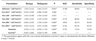
Apparent Diffusion Coefficient as a Quantitive Imaging Biomarker for Prediction of Immunohistochemical Receptor Status, Proliferation Rate and Molecular Subtypes of Breast Cancer
Joao Horvat, Michelle Zhang, Blanca Bernard-Davila, Elizabeth Morris, Sunitha Thakur, Thomas Helbich, Zsuzsanna Bago-Horvath, Katja Pinker
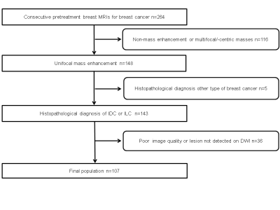
Molecular subtype classification of breast tumor is of paramount importance in determining aggressiveness and prognosis. The ability to use diffusion weighted imaging (DWI) for the prediction of molecular subtypes may improve management in breast cancer. In this study, two radiologists retrospectively evaluated different metrics on apparent diffusion coefficient maps of 107 patients with invasive breast cancer. ER and PR positive lesions had lower ADC values while HER2 positive and high-proliferating had higher values. Luminal cancers had lower ADC values than other subtypes, thus DWI may be used to predict tumor subtype in breast cancer.
|
|
129Xe & 3He Imaging
Traditional Poster
Body: Breast, Chest, Abdomen, Pelvis
Wednesday, 20 June 2018
| Exhibition Hall 2433-2443 |
16:15 - 18:15 |
|
2433.
 |
Regional Lung Function Quantification by Combining Gas-Phase Saturation with Hyperpolarized Xenon-129 Dissolved-Phase MRI
Kai Ruppert, Hooman Hamedani, Faraz Amzajerdian, Luis Loza, Yi Xin, Ian Duncan, Harilla Profka, Sarmad Siddiqui, Mehrdad Pourfathi, Stephen Kadlecek, Rahim Rizi
Hyperpolarized xenon-129 MRI has previously been used to assess pulmonary gas exchange between the alveolar volume and lung tissue. In this work, we quantified changes in the downstream xenon dissolved-phase signal in the left ventricle in response to a regional saturation of the pulmonary gas-phase signal. This approach permitted us to extract the relative gas-exchange efficiency of the lung volume unaffected by the GP signal saturation, demonstrating increased gas exchange efficiency in the posterior regions of the lung in supine rabbits. The proposed technique might be especially valuable in lung transplantation, during pharmaceutical interventions, or for lung-volume reduction surgeries.
|
|
2434.
 |
Observing Pulmonary Gas-Transport Dynamics Using Rapid 1D Hyperpolarized Xenon-129 Dissolved-Phase Measurements
Kai Ruppert, Hooman Hamedani, Faraz Amzajerdian, Luis Loza, Yi Xin, Ian Duncan, Harilla Profka, Sarmad Siddiqui, Mehrdad Pourfathi, Stephen Kadlecek, Rahim Rizi
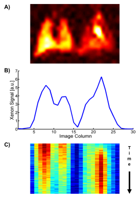
Monitoring the dissolved xenon-129 signal in a central downstream location such as the left ventricle of the heart provides a convenient measure of the lung’s gas transport dynamics, and thereby of total lung function. To demonstrate the feasibility of this approach, we combined a rapid simultaneous gas-phase / dissolved-phase 1D-projection acquisition with regional gas-phase saturation to monitor the gas-transport dynamics of the lung as signal variations in the heart of a rat model of radiation-induced lung injury. Our measurements indicate that this method can identify the reductions in regional lung function associated with partial lung irradiation.
|
|
2435.
 |
Measuring the Impact of PEEP on Pulmonary Gas Transport Using Hyperpolarized Xenon-129 Dissolved-Phase MRI
Kai Ruppert, Hooman Hamedani, Faraz Amzajerdian, Luis Loza, Yi Xin, Ian Duncan, Harilla Profka, Sarmad Siddiqui, Mehrdad Pourfathi, Maurizio Cereda, Stephen Kadlecek, Rahim Rizi
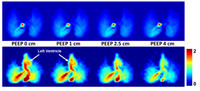
Higher positive end-expiratory pressure (PEEP) during mechanical ventilation can result in improved oxygenation, but it can also give rise to ventilator-induced lung injury. In this work, we used a rabbit model to evaluate the sensitivity of a hyperpolarized xenon-129 MRI technique that allows a comprehensive assessment of the pulmonary gas-transport by the entire lung for monitoring the impact of PEEP on lung function. We observed that increased PEEP resulted in a large decrease in pulmonary gas transport that is most likely linked to a lengthened pulmonary transit time.
|
|
2436.
 |
Hyperpolarized 129Xe MR functional imaging to monitor the response of the human lungs after segmental lipopolysaccharide challenge
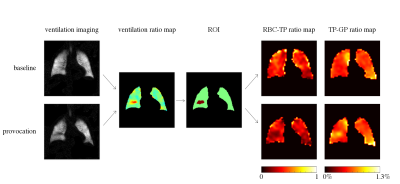
Agilo Kern, Heike Biller, Filip Klimes, Andreas Voskrebenzev, Marcel Gutberlet, Alexander Rotärmel, Christian Schönfeld, Julius Renne, Olaf Holz, Kun Qing, Kai Ruppert, Frank Wacker, Jens Hohlfeld, Jens Vogel-Claussen
Hyperpolarized 129Xe MRI has been shown to be sensitive to inflammatory changes after lung provocation by lipopolysaccharide (LPS) in an animal model. The purpose of this work was to investigate feasibility of monitoring the response of the human lungs after segmental LPS challenge using 129Xe MRI. Dissolved-phase imaging and chemical shift saturation recovery were employed to assess inflammatory changes and to compare MRI results with inflammatory cell counts from bronchoalveolar lavage. Both MRI methods show a significant reduction of the 129Xe in red blood cells and lung tissue ratio in the affected region but no significant correlations with inflammatory cell counts.
|
|
2437.
 |
Revealing Pulmonary Gas Transport Dynamics using a 3D Radial Hyperpolarized Xenon MRI Acquisition with Variable Flip Angles
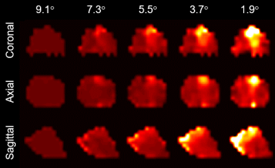
Faraz Amzajerdian, Kai Ruppert, Hooman Hamedani, Yi Xin, Ian Duncan, Harrilla Profka, Mehrdad Pourfathi, Sarmad Siddiqui, Luis Loza, Stephen Kadlecek, Rahim Rizi
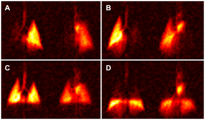
We demonstrated that reducing the flip angle drives the distribution of acquired dissolved-phase xenon downstream towards the heart. By exploiting this principle, the dynamics of pulmonary gas transport were captured through a single 3D double golden means radial acquisition with linearly decreasing flip angles. Reconstruction with a sliding window generated a series of consecutive images with declining average flip angles, depicting the gradual uptake and accumulation of xenon by the heart and lungs.
|
|
2438.
 |
129Xe signal dynamics and chemical shift in the cardio-pulmonary circuit using cardiac-gated hyperpolarized 129Xe NMR
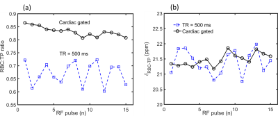
Graham Norquay, Jim Wild
The sensitivity of the 129Xe chemical shift to red blood cell oxygenation makes hyperpolarized 129Xe MR spectroscopy a promising technique for measurement of blood oxygenation in vivo. In addition, dissolved phase 129Xe MRS is of interest as a biomarker of gas exchange and interstitial lung disease. Both the signal dynamics and chemical shift of 129Xe have been shown to be modulated by the cardiac cycle, potentially adding confounding effects to interpretation of the 129Xe MRS chemical shift. In this study, we demonstrate that cardiac-gating in129Xe MRS reduces the variability in the measured dissolved 129Xe signal and chemical shift in the cardio-pulmonary circuit.
|
|
2439.
 |
Using a hybrid multibreath hyperpolarized (HP) 129Xe imaging technique for simultaneous assessment of lung function and structure in a two-hit radiation induced lung injury (RILI) model.
Sarmad Siddiqui, Hooman Hamedani, Yi Xin, Luis Loza, Faraz Amzajerdian, Mehrdad Pourfathi, Stephen Kadlecek, Kai Ruppert, Harrilla Profka, Rahim Rizi, Shampa Chatterjee, Ian Duncan
In this study we developed a two-hit hemi-thorax radiation-induced lung injury (RILI) model that better simulates the etiology of the disease in humans, and characterized it via a multibreath hyperpolarized (HP) 129Xe imaging technique to assess lung function and structure one month post-radiation. We observed an increased PAO2 of 145±41 Torr in the radiated lung compared to 124±40 Torr in the contralateral lung. We also observed a corresponding decrease in oxygen uptake in the radiated lung. The preliminary findings suggest that HP 129Xe-derived functional parameters, particularly changes in the alveolar oxygen tension and oxygen uptake can serve as biomarkers during the early fibrotic stage of RILI.
|
|
2440.
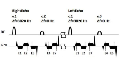 |
Fast Imaging of Hyperpolarized Xe-129 in the Airspace, Barrier and Red Blood Cells in the Human Lung
Junshuai Xie, Haidong Li, Huiting Zhang, Xiuchao Zhao, Xianping Sun, Chaohui Ye, Xin Zhou
Xe-129 in the barrier and red blood cells could be separated by the dissolved-phase (DP) Xe-129 MRI with radial sampling strategy. However, the number of the RF pulse was usually large and thus resulted in long acquisition time. An MRI strategy in the Cartesian coordinate has been used to for high-resolution rodent lung imaging of He-3 in the airspace. The concept was introduced into fast acquisition of the DP Xe-129 in the human lung with the multi-point Dixon method. The number of the RF pulse reduced and the results of TP/Gas and RBC/Gas agreed with the previous study.
|
|
2441.
 |
Next-Generation Automated Clinical-Scale Batch-Mode Xe-129 Hyperpolarizer
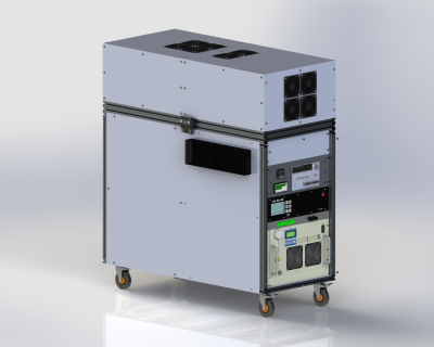
Panayiotis Nikolaou, Aaron Coffey, Bryce Kidd, Megan Murphy, Boyd Goodson, Michael Barlow, Eduard Chekmenev
Over the last two decades there have been many advances in the field of hyperpolarized (HP) noble gas production and imaging, largely enabled by the development of low-cost, high-power frequency-narrowed laser diode arrays (LDAs) and the improvement of 129Xe polarizer technology in general. Here we present the development and features of the new 3rd-generation Batch-Mode 129Xe hyperpolarizer. As with most previous 129Xe polarizers, the new device utilizes Spin Exchange Optical Pumping (SEOP), a process in which resonant, circularly polarized photons optically pump Rb electrons, which in turn hyperpolarize the 129Xe nuclear spins via hyperfine interactions (the “spin-exchange” process).
|
|
2442.
 |
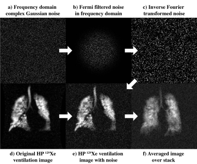
A paired approach to the segmentation of proton and hyperpolarized gas MR images of the lungs
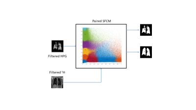
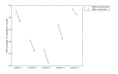
Alberto Biancardi, Laure Acunzo, Helen Marshall, Bilal Tahir, Paul Hughes, Laurie Smith, Nicholas Weatherley, Guilhelm Collier, Jim Wild
Quantitative analyses of hyperpolarized gas and 1H lung MRI together provide quantitative information on lung obstruction. Quantification requires segmentation of the ventilated and non-ventilated regions of the hyperpolarized gas MRI and definition of the lung cavity from the paired 1H MRI. Spatial fuzzy c-means segmentation was developed to segment these image pairs simultaneously. Error measures with respect to manual reference segmentations and qualitative grading showed significant improvements when compared to an established method. This work may help towards standardisation and automation of lung ventilation image analysis, and help improve accuracy and reproducibility.
|
|
2443.
 |
A Study of Lung Function Variability in Chronic Obstructive Pulmonary Disease Using Hybrid Hyperpolarized 3He Imaging
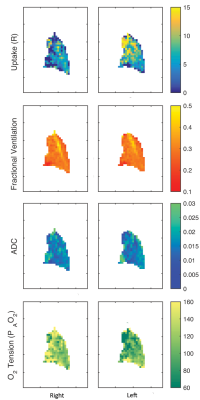
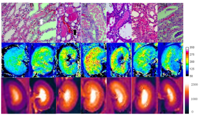
Hooman Hamedani, Ryan Baron, Sarmad Siddiqui, Yi Xin, Mary Spencer, Faraz Amzajerdian, Stephen Kadlecek, Kai Ruppert, Mehrdad Pourfathi, Luis Loza, Ian Duncan, Tahmina Achekzai, Maurizio Cereda, Rahim Rizi
To better understand variable lung function in COPD, we imaged a subset of COPDGene subjects at baseline, one week post-baseline and one month post-baseline using a multifaceted hyperpolarized (HP) 3He scheme to measure apparent diffusion coefficient (ADC), fractional ventilation (FV), alveolar oxygen tension (PAO2) and oxygen uptake (R) variability.
|
|
Body Imaging: Fetal/Placenta & Pelvis
Traditional Poster
Body: Breast, Chest, Abdomen, Pelvis
Wednesday, 20 June 2018
| Exhibition Hall 2444-2450 |
16:15 - 18:15 |
|
2444.
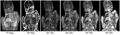 |
MT contrast in the Post-mortem Neonate: A pilot study
Amy McDowell, Susan Shelmerdine, Sara Lorio, Owen Arthurs, David Carmichael
Post-mortem MRI imaging (PMMR) is rapidly becoming a useful tool in the minimally invasive autopsy of fetal and perinatal death allowing clinical diagnosis and assessment of major congenital abnormalities. A recent study suggested that magnetisation transfer values may be a more specific measure of post-mortem heart abnormalities, but there has little application of MT imaging in this area. We performed a preliminary exploration of MT contrast and MT pulse optimisation in whole body PMMR in neonates as part of a multi-parameter mapping protocol.
|
|
2445.
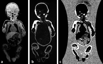 |
High Resolution Rapid Neonatal Whole Body Composition Using 3.0 Tesla Chemical Shift Magnetic Resonance Imaging
Jonathan Dyke, Amanda Garfinkel, Alan Groves, Arzu Kovanlikaya
To evaluate a whole body rapid imaging technique to calculate neonatal lean body mass and percentage adiposity using 3.0 Tesla chemical shift Magnetic Resonance Imaging (MRI). A rapid 2-Point Dixon MRI technique was used to calculate whole body fat and water images at 3.0 Tesla in term (n=10) and preterm (n=15) infants in 42 seconds/scan. MRI calculated whole body mass correlated closely with measured body weight (R2=0.87;p<0.001). Scan-rescan analysis demonstrated a 95% limit of agreement of 1.3% adiposity. At term corrected age, former preterm infants had significantly reduced lean body mass compared to term born controls 1935g versus 2416g (p=0.002).
|
|
2446.
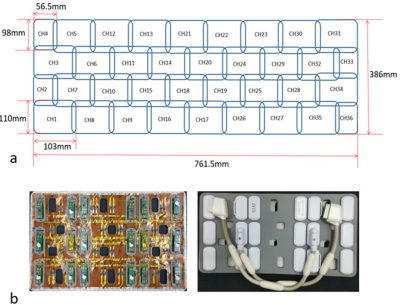 |
Design of a 36-channel receive coil array for fetal MRI at 3T
Qiaoyan Chen, Guoxi Xie, Chao Luo, Xing Yang, Jin Zhu, Jo Lee, Xiaoliang Zhang, Xin Liu, Ye Li
Due to lack of dedicated fetal imaging coils, the standard commercial abdominal coil is often used for fetal imaging, of which the performance is limited by its insufficient coverage and element number. In this work, a dedicated 36-channel coil array for fetal imaging was designed, capable of covering a range of pregnancy from 20 to 37+ weeks. Compared to a commercial abdominal coil array, the proposed 36-channel fetal coil provides improved performance in SNR, parallel imaging capability, and image quality.
|
|
2447.
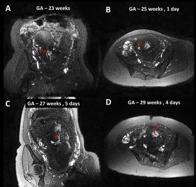 |
Fetal non-contrast MR angiography in second and early third trimester
Uday Krishnamurthy, Swati Mody, Brijesh Yadav, Pavan Jella, Edgar Edgar Hernandez-Andrade, Anabela Trifan, Ewart Haacke, Roberto Romero, Jaladhar Neelavalli
To evaluate the robustness and utility of non-contrast MRA as a means to visualize fetal vasculature, particularly in fetuses younger than 30 weeks gestation.
|
|
2448.
 |
Non-rigid motion correction for arterial spin labeled (ASL) perfusion imaging of the placenta using ANTs
Zhengjun Li, Eileen Hwuang, Jeffrey Duda, Marta Vidorretta, Nadav Schwartz, John Detre, Walter Witschey, Dylan Tisdall
Non-rigid motion of the placenta due to maternal breathing and fetal movement is one of the main challenges in placental MRI. In this study, we evaluated non-rigid motion correction of the placenta during arterial spin labeled (ASL) perfusion imaging, using Advanced Normalization Tools (ANTs). The results showed that non-rigid motion correction with ANTs improved the resulting perfusion images as evidenced by reduced the residual power of control-label regression, increased the tSNR, and reduced the power of respiration in the signal.
|
|
2449.
 |
Diffusion Tensor Imaging for Differentiating Borderline From Malignant Epithelial Ovarian Tumors
XU HAN, MEI-YU SUN, MENG-YAO WANG, LI-ZHI XIE, RUI FAN
To assess the fitted parameters of DTI in ovarian tumors and to investigate their potential in distinguishing borderline from malignant epithelial ovarian tumors, which can provide detailed information for clinical treatment. DC avg, Exat, FA and VRA in DTI were valuable information in distinguishing borderline from malignant epithelial ovarian tumors and can be used as non-enhancement quantitative indexes, which has a good application prospect.
|
|
2450.
 |
A Subspace Approach to Accelerated HASTE Acquisition for Fetal Brain MRI
Bo Zhao, Borjan Gagoski, Justin Haldar, Elfar Adalsteinsson, Ellen Grant, Lawrence Wald
HAlf-fourier Single-shot Turbo spin Echo (HASTE) acquisition is widely used in fetal MR imaging due to its T2 contrast and motion robustness, but speed and T2-blurring remain a problem for fully sampled acquisitions. In the work, we describe a new reconstruction approach based on low-rank and subspace modeling of local k-space neighborhood to accelerate HASTE acquisition. The proposed approach decreases the echo-train length with improved image quality and noise robustness compared to conventional reconstruction. It is compatible with the vendor-provided acquisition. The effectiveness and utility of the proposed approach is evaluated with both retrospectively and prospectively undersampled fetal imaging data.
|
|
Thoracic MRI
Traditional Poster
Body: Breast, Chest, Abdomen, Pelvis
Wednesday, 20 June 2018
| Exhibition Hall 2451-2480 |
16:15 - 18:15 |
|
2451.
 |
Usefulness of morphological characteristics for the differentiation of benign from malignant peripheral solitary pulmonary lesions using MR T1-weighted 3D Star VIBE
Shan Dang, Haifeng Duan, Dong Han, QI Yang, Xin Tian, Nan Yu, Yuxin Lei, Shaoyu Wang, Sujue Lu, Guangming Ma
Can MR T1-weighted 3D Star VIBE alternate the MSCT in morphological features of the peripheral solid pulmonary lesions?
|
|
2452.
 |
Free-breathing T1-weighted 3D STAR VIBE: versus Thin-Section Computed Tomography for the Assessment of Pulmonary Parenchyma Diseases
Zhanli Ren, Shan Dang, Yuxin Lei, Nan Yu, Yong Yu, Taiping He
Free-breathing T1-weighted 3D star vibe is useful for lung and mediastinum assessment and evaluation of radiological findings for patients with various pulmonary parenchyma diseases.
|
|
2453.
 |
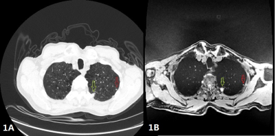
MRI Ventilation Texture Features Discriminate Severe Asthmatics with and without Eosinophilic Airway Inflammation
Sarah Svenningsen, Nanxi Zha, Rachel Eddy, Dante Capaldi, Melanie Kjarsgaard, Katherine Radford, Parameswaran Nair, Grace Parraga
Previous work suggests that inhaled gas MRI conceals minable features that are distinctly different between severe asthma inflammatory endotypes and these may be used to predict inflammatory endotype. We evaluated the performance of inhaled gas MRI ventilation defect percent, ventilation coefficient of variation and texture features to discriminate severe asthmatics with and without the eosinophilic inflammatory endotype. MRI measurements of ventilation significantly discriminated asthmatics with eosinophilic inflammation from those without eosinophilic inflammation. Non-invasive MRI-based biomarkers and signatures of asthma inflammatory endotype may serve to guide treatment selection in individual asthmatics or evaluate the effectiveness of anti-inflammatory treatments in clinical trials.
|
|
2454.
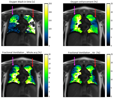 |
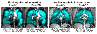
Extraction of fractional ventilation from dynamic oxygen enhanced MRI experiments: preliminary results
Marta Tibiletti, Jose Ulloa, Geoff Parker
Fractional ventilation (FV) weighted maps were extracted from free-breathing dynamic O2 enhanced (dynOE) experiment in cystic fibrosis patients. FV is related to the local expansion of the tissue due to gas arrival in inspiration, while dynOE maps the local rate of the arrival of O2 and the maximum enhancement obtained. These parameters can be extracted from the same acquisition, providing complementary information regarding local lung function.
|
|
2455.
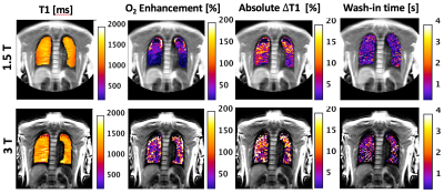 |
Comparative study of 3D inversion recovery centric ordered fast field echo in lung dynamic oxygen enhanced MRI at 1.5 T and 3 T
Marta Tibiletti, Jose Ulloa, Alexandra Morgan, Geoff Parker
Dynamic oxygen–enhanced MRI (dOE-MRI) techniques have previously been apply to study the rate and level of O2 enhancement in the lung. Lung MRI investigations are mostly conducted at 1.5T, because signal loss due to stronger susceptibility artefacts in lung tissue is expected at higher field strength. In this work, we demonstrate the feasibility of dOE-MRI at 3T on healthy volunteers. The observed signal enhancement is comparable between 1.5T and 3T, but translates in a lower relative T1 change due to higher baseline T1 at 3T. Fitting performance of O2 wash-in curve may be reduced by the lower SNR at 3T.
|
|
2456.
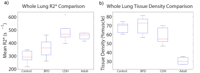 |
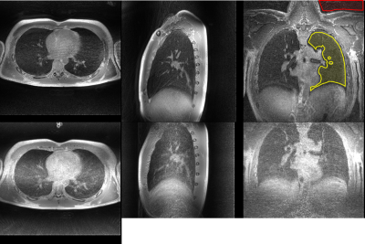
Effects of Neonatal Lung Abnormalities on Parenchymal R2* Estimates
Andrew Hahn, Nara Higano, Jean Tkach, Laura Walkup, Robert Thomen, Xuefeng Cao, Stephanie Merhar, Paul Kingma, Jason Woods, Sean Fain
We estimate pulmonary tissue densities (TD) and R2* in neonatal intensive care unit patients with and without diagnoses of lung disease as well as in healthy adults using multi-echo 3D ultrashort echo time MRI. As anticipated, a clear negative relationship between TD and R2* is evident. However, after correcting for TD variation, we find significant differences in R2* between diseased and non-diseased neonates, suggesting that MRI can probe differences in susceptibility and/or sub-voxel tissue geometry which may increase understanding of neonatal lung tissue pathologies.
|
|
2457.
 |
Implementation of the FLORET Ultrashort Echo-Time Sequence for Lung Imaging
Matthew Willmering, Ryan Robison, Hui Wang, James Pipe, Jason Woods
MRI of lungs is inherently challenging due to the short T2* and intrinsic motion from the respiratory and cardiac cycles. Ultrashort echo-time (UTE) sequences are often implemented for their shorter echo times and relative insensitivity to motion. Spiral UTE sequences have been touted recently as having greater k-space sampling efficiencies than radial UTE, but few are designed well for the shorter T2* of lung. In this study, FLORET (Fermat looped, orthogonally encoded trajectories), a recently-developed spiral 3D UTE sequence, was implemented in human lungs for the first time and outperformed traditional radial UTE for imaging of lung tissue.
|
|
2458.
 |
The Impact of Inspiration Levels on the Repeatability of Quantitative Pulmonary Perfusion DCE-MRI in Patients with Chronic Obstructive Pulmonary Disease and Cystic Fibrosis
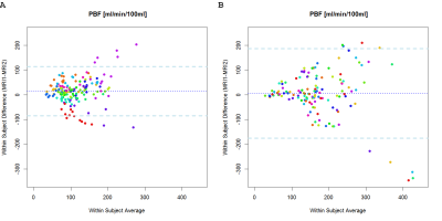
Marilisa Schiwek, Frank Risse, Simon Triphan, Monika Eichinger, Sabine Wege, Mirjam Stahl, Olaf Sommerburg, Marcus Mall, Hans-Ulrich Kauczor, Michael Puderbach, Ralf Eberhardt, Claus Heussel, Gudula Heussel, Mark Wielpütz
The objective of this study was to investigate the 4-week repeatability of contrast-agent based pulmonary perfusion quantification in clinically stable patients with COPD and CF. Software including fully automated lung segmentation was used to determine pulmonary blood flow (PBF). While a good agreement of PBF was found in the majority of patients, high variabilities were found. Several influence factors were considered as explanations. Differences in SNR due to different inspiratory levels are likely to influence whether quantification in each voxel succeeds. Thus, it may be necessary to modify voxel-based quantification to compensate for differences in inspiratory levels and low SNR.
|
|
2459.
|
Magnetic Resonance Imaging of Pulmonary Nodules
Chi Wan Koo, Aiming Lu, Edwin Takahashi, Jessica Magnuson, Peter Kollasch, Jennifer Geske, Julie An, Dennis Wigle, Tobias Peikert
Magnetic resonance imaging had been explored as a potential alternative to computed tomography but the majority of prior MRI nodule studies was performed with 1.5-T scanners and not with the most up to date sequences. Our study demonstrated that biomarkers derived from state of the art 3T MRI sequences can distinguish benign from malignant pulmonary nodules and correlate with morphologic and physiologic values derived from commonly used noninvasive imaging modalities.
|
|
2460.
 |
Pulmonary Perfusion MR Imaging with Ultra-Short TE: Comparison of Capability for Regional Perfusion Assessment and Postoperative Lung Function Prediction with Perfusion SPECT and/ or Conventional CT Methods
Yoshiharu Ohno, Masao Yui, Yu Chen, Yuji Kishida, Shinichiro Seki, Katsusuke Kyotani, Takeshi Yoshikawa
Gadolinium-based blood volume (Gd-based BV) map generated between unenhanced and contrast-enhanced UTE-MRIs may have a potential for regional perfusion assessment like lung perfused BV map on dual-energy CT in patients with pulmonary diseases. We hypothesized that Gd-based BV map has a potential to regional perfusion assessment and postoperative lung function prediction as well as perfusion SPECT and/ or conventional CT methods in NSCLC patients. The purpose of this study was to directly compare the capability of Gd-based BV map for regional perfusion assessment and/ or postoperative lung function prediction in NSCLC patients with perfusion SPECT and conventional CT methods.
|
|
2461.
 |
Differentiation of Malignant and Benign Pulmonary Lesions with DCE-MR imaging
Xin Sui, Xiaoli Xu, Lan Song, Tianyi Qian, Yi Sun, Wei Song, Zhengyu Jin
The aim of this study was to estimate the diagnostic accuracy of DCE-MR in the differential diagnosis between malignant and benign pulmonary lesions. Thirty patients with suspected lung cancer were recruited. 13 malignancies were proved by pathology. The DCE-MR data was acquired with the TWIST-VIBE technique, and quantitative parameters (Ktrans, Kep, and Ve) were calculated by the Tofts model. Our results demonstrated that malignant lesions had significant higher Ktrans and kep values than benign lesions. The Ktrans and Kep derived from DCE-MR are promising quantification parameters for differentiating lung lesions.
|
|
2462.
 |
Pre-treatment DCE MRI predicts overall survival in patients with primary lung cancer
Wei Wu, Daniel Hippe, Nina Mayr, William Yuh, Liming Xia, Stephen Bowen
We tested whether pre-treatment standard DCE MRI imaging and clinical features can predict overall survival (OS) of 37 patients with primary lung cancer. Primary tumor volume (hazard ratio [HR] = 3.19 per 1-SD increase, P=0.001) and minimum intensity of the peak enhancement phase on DCE MRI (HR = 0.45, P=0.012) were significant predictors of OS on univariate Cox regression analysis. Univariate primary tumor volume model (c-index = 0.76, P=0.002) and multivariate LASSO Cox models based on DCE MRI features (c-index = 0.69, P=0.046) were positive predictors for OS with no statistically significant difference in performance (P=0.36).
|
|
2463.
 |
Machine learning of DCE MRI intensity histogram radiomic features for pulmonary lesion classification
Wei Wu, Chunyan Duan, Nina Mayr, William Yuh, Liming Xia, Daniel Hippe, Stephen Bowen
To classify malignant/benign lesions can be challenging and non-invasive means to further improve the diagnostic accuracy would have major impact on management in patients with pulmonary lesions. 62 patients with histologically confirmed pulmonary lesions were retrospectively reviewed. Intensity voxel histogram (IH) features were extracted from DCE-MRI. The efficacy of IH features to classify pulmonary lesions were assessed by correlation with pathology. Under cross-validation, a support vector machine algorithm achieved a diagnostic accuracy, sensitivity and specificity of 95%, 99 and 86%. Our results demonstrate that machine learning of DCE-MRI IH features has potential for accurately classifying pulmonary lesions for clinical translation.
|
|
2464.
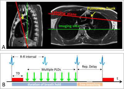 |
Temporal and spatial evaluation of pulmonary blood flow using multiple delay PCASL at 1.5 Tesla
Ferdinand Seith, Rolf Pohmann, Martin Schwartz, Thomas Küstner, Klaus Scheffler, Konstantin Nikolaou, Fritz Schick, Petros Martirosian
Pseudo-continuous-arterial-spin-labeling (PCASL) has been successfully applied in abdominal organs to image organ perfusion. The aim of this work was to evaluate the pulmonary blood flow in dependence on the cardiac cycle using PCASL at 1.5T. Labeling of pulmonary blood flow was achieved by ECG triggering and an labeling plane perpendicular to the pulmonary trunk (tagging duration 300ms). In five volunteers, eight measurements were acquired with fast True-FISP imaging (in-plane-resolution, 2.5×2.5mm2, coronal view) with post-labeling delays between 100 and 1500ms. The PCASL-True-FISP technique was able to precisely assess blood flow of pulmonary arteries, as well as perfusion of the lung parenchyma.
|
|
2465.
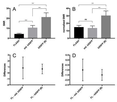 |
GRE bSSFP vs. FLASH based Fourier Decomposition lung MRI at 1.5T: evaluation of image quality, fractional ventilation and lung perfusion in healthy volunteers
Alexander Rotärmel, Andreas Voskrebenzev, Filip Klimes, Marcel Gutberlet, Frank Wacker, Jens Vogel-Claussen
The comparison between different MRI sequences for assessment of lung ventilation and perfusion using phase-resolved functional lung MRI post-processing (PREFUL) needs further evaluation to support clinical translation. Our study compares two gradient echo (GRE) balanced steady state free precession (bSSFP) sequences (one commercially available and one modified by Bauman et al.) and one GRE Fast Low Angle Shot (FLASH) sequence regarding signal-to-noise ratio, fractional ventilation and lung perfusion. In summary, the bSSFP sequence modified by Bauman provides significantly higher SNR values and better perfusion values in the lung parenchyma compared to the commercially available bSSFP and FLASH sequences using PREFUL.
|
|
2466.
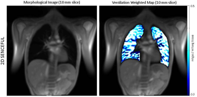 |
UTE-SENCEFUL: high resolution 3D ventilation weighted maps
Lenon Mendes Pereira, Andreas Weng, Tobias Wech, Manuel Stich, Christian Kestler, Simon Veldhoen, Andreas Kunz, Thorsten Bley, Herbert Köstler
In this work we present a method to assess lung ventilation in 3D by combining Self-gated Non-Contrast-enhanced Functional Lung MRI (SENCEFUL) with an ultra-short echo time (UTE) acquisition and a 3D image registration technique. Ventilation weighted maps were generated and the quantitative ventilation value for a healthy volunteer was assessed. Lung ventilation and image quality were compared between the new UTE-SENCEFUL and the standard 2D-SENCEFUL methods. UTE-SENCEFUL was able to present a 3D reconstruction of the breathing cycle, 3D ventilation weighted maps with high resolution and quantitative ventilation values in agreement with the literature.
|
|
2467.
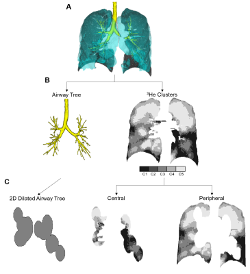 |
Contributions of Large Versus Small Airways to MRI Ventilation Heterogeneity in Asthmatics
Rachel Eddy, Heather Young, Andrea Kassay, Dante Capaldi, Sarah Svenningsen, David McCormack, Grace Parraga
Pulmonary functional MRI identifies the exact location of functional abnormalities within the asthmatic lung, however the relative contributions of large and small airways to ventilation heterogeneity in a given patient are unknown. Here, we differentiated hyperpolarized noble gas MRI ventilation into regions corresponding to the large and small airways using patient-specific airway trees and calculated the ventilation defect percent (VDP) related to large and small airways independently. The classification of small and large airway VDP may help with clinical treatment decisions for individualized therapies.
|
|
2468.
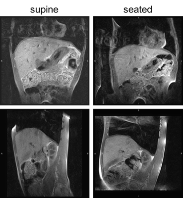 |
Assessment of the diaphragm morphology in upright seated and supine position
Christoph Arthofer, Charlotte Bolton, Zhenghao Wang, Andrew Cooper, Andrew Peters, Michael Barlow, Dorothee Auer, Richard Bowtell, Ian Hall, Penny Gowland
The morphology of the diaphragm is an important factor in the consideration of dyspnoea and treatment of respiratory diseases. The acquisition of images with commonly used methods is limited by the patient position or duration of the procedure. We present the first images of the diaphragm acquired in an upright MR scanner, and estimate repeatability and differences in morphology depending on posture.
|
|
2469.
 |
Dynamic contrast-enhanced MRI in the lung – evaluation of measures of pulmonary oedema and pulmonary endothelial permeability in healthy subjects and patients with chronic heart failure
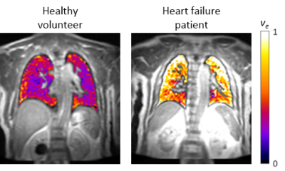
Alexandra Morgan, Joseph Cheriyan, Caleb Roberts, Martin Graves, Ilse Patterson, Rhys Slough, Rosemary Schroyer, Disala Fernando, Linda Henderson, Subramanya Kumar, Geoffrey Parker, Dennis Sprecher, Robert Janiczek
MRI has previously demonstrated increased lung water content in patients with heart failure (HF), but has not yet been used to distinguish between intravascular and extravascular water in these patients. This study evaluated dynamic contrast-enhanced MRI (DCE-MRI) for measuring pulmonary oedema and endothelial permeability in healthy volunteers (HV) and chronic HF patients at rest and post-exercise. DCE-MRI showed a redistribution of lung water towards the interstitial space in chronic HF, as compared to HV, suggesting this method may have value as a novel endpoint for dose-ranging and proof-of-mechanism studies in chronic HF. No exercise-induced change was seen in either group.
|
|
2470.
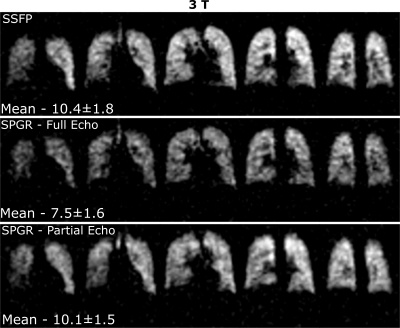 |
Optimization of Steady-state Free Precession with 19F Perfluoropropane for Increased Signal-to-Noise for Human Lung Ventilation Imaging at 3 T
Adam Maunder, Madhwesha Rao, Jim Wild
Fluorinated gas MRI is an alternative modality to hyperpolarized gas MR for imaging lung ventilation, but is constrained by lower SNR. Improvement of the signal-to-noise ratio of human lung ventilation images with 19F the steady-state free precession (SSFP) sequence was previously explored at 1.5T. Here, we present optimization of SSFP for imaging lung ventilation at 3T. The achievable improvement of in-vivo imaging quality with realistic relaxation parameters is demonstrated with comparison against the spoiled gradient echo sequence. Limits in applying the SSFP sequence due to specific absorption ratio at 3T and the dependence on T2* within the lungs are detailed.
|
|
2471.
 |
Probing changes in lung physiology in COPD using CT, perfusion MRI and hyperpolarized xenon-129 MRI
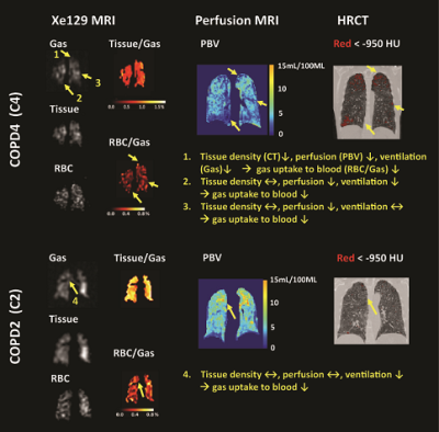
Kun Qing, Nicholas Tustison, John Mugler, III, Jaime Mata, Zixuan Lin, Li Zhao, Da Wang, Xue Feng, Kai Ruppert, Talissa Altes, Joanne Cassani, Y. Shim
In this study, by using chest CT, Gadolinium-enhanced perfusion MRI, and hyperpolarized xenon-129 ventilation and gas uptake MRI, we assessed the quantitative changes in tissue density, pulmonary perfusion and gas uptake in patients with COPD compared to normal subjects. We found evidence for compensatory pulmonary vasoconstriction to match impairment of ventilation, and also pulmonary shunt and dead space. By incorporating a new lobar segmentation method for proton MRI, we performed statistical analysis to evaluate the regional interrelationships among different measures. We demonstrated that xenon-129 MRI has high potential to identify changes of multiple aspects of lung physiology in one acquisition.
|
|
2472.
 |
Combination of Perfluoropropane and oxygen-enhanced MRI-derived washout kinetics for detection of ischemic injury to lungs in a porcine ex-vivo perfusion system
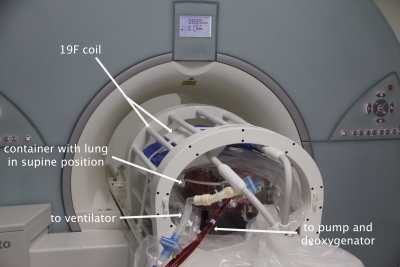
Julius Renne, Marcel Gutberlet, Andreas Voskrebenzev, Agilo Kern, Till Kaireit, Jan Hinrichs, Peter Braubach, Christiane Falk, Klaus Höffler, Gregor Warnecke, Axel Haverich, Frank Wacker, Jens Vogel-Claussen, Norman Zinne
Ex-vivo lung perfusion and ventilation systems are a promising new tool for conditioning marginal lung allografts. However, reliable biomarkers for evaluating graft function are missing. In this study MRI-derived fluorine and oxygen washout times are to be evaluated as lung function parameters in a porcine model of ischemia. Washout time for oxygen is prolonged while fluorine washout is not in lungs after warm ischemia compared to normal controls, which might reflect pulmonary edema limiting oxygen diffusion. Determination of fluorine and oxygen washout is feasible in an ex-vivo lung perfusion system and seems to be promising tools for evaluating graft function.
|
|
2473.
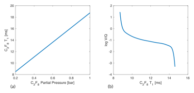 |
Mapping of Ventilation/Perfusion Ratios in the human lung using 19F MRI of Perfluoropropane
Arnd Obert, Marcel Gutberlet, Alexander Rotärmel, Frank Wacker, Jens Vogel-Claussen
In this work, the correlation between longitudinal relaxation time (T1), alveolar partial pressure and ventilation-perfusion ratio (V/Q) of an inhaled fluorinated gas is used to compute quantitative V/Q maps of the human lung. The trapping of inert Perfluoropropane (C3F8) in poorly ventilated regions of the lung (low V/Q) leads to an increase of its alveolar partial pressure which is detectable as an increase of T1 in 19F MR Imaging. Here, V/Q maps of three patients with Chronic Obstructed Pulmonary Disease (COPD) were calculated and compared to a V/Q map of a healthy volunteer.
|
|
2474.
 |
Accelerated 19F-MR Imaging of Inhaled Perfluoropropane for Assessment of Pulmonary Ventilation
Mary Neal, Ben Pippard, Kieren Hollingsworth, Pete Thelwall
MRI of inhaled perfluoropropane offers a safely repeatable modality for mapping pulmonary ventilation. However, as a thermally polarised gas, signal is scarce and acquisitions are limited to breath hold durations or require respiratory gating. Improving the temporal resolution would present the opportunity to implement dynamic imaging or improve image quality in breath hold acquisitions. In this study, the acquisition time was reduced by partially sampling k-space using a compressed sensing technique. A 3-fold decrease in acquisition time was achieved whilst maintaining visually similar image quality. An average SNR of 25:1 was measured in a 6s 3D acquisition in healthy volunteers.
|
|
2475.
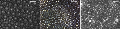 |
Microporous Lung Phantoms for 19F-MRI of Inhaled Imaging Agents with Physiologically Representative Relaxation Times
Mary Neal, Helena Sexton, Eric Hughes, Pete Thelwall
A primary characteristic of 19F-MRI of pulmonary ventilation is the short in vivo T2* of the inhaled imaging agent caused by the inhomogeneous magnetic environment proximal to the alveolar walls. This study describes two novel methods for fabrication of phantoms that mimic the physical and magnetic properties of alveolar tissue. In both cases the perfluorinated gas phase imaging agent is suspended in a stable microporous foam medium. The fabrication techniques permitted precise control of either bubble size or gas/liquid ratio. Highly monodisperse stable foams were formed with a perfluoropropane T2* of 2ms, comparable to that measured in the human lung.
|
|
2476.
 |
Assessment of ventilation heterogeneity using hyperpolarized gas MRI histogram analysis
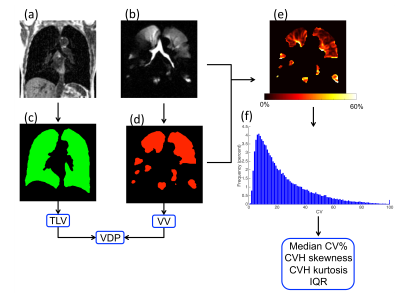
Paul Hughes, Laurie Smith, Felix Horn, Alberto Biancardi, Neil Stewart, Graham Norquay, Madhwesha Rao, Ina Aldag, Chris Taylor, Helen Marshall, Guilhem Collier, Jim Wild
Development of sensitive imaging biomarkers to differentiate health from disease is an important research topic in pulmonary MRI. This work aimed to make use of the rich spatial and signal intensity information in hyperpolarized gas MR ventilation images to determine metrics of ventilation heterogeneity. Retrospective analysis was performed on 3He ventilation images acquired from healthy volunteers and patients with cystic fibrosis, asthma and chronic obstructive pulmonary disease.
|
|
2477.
 |
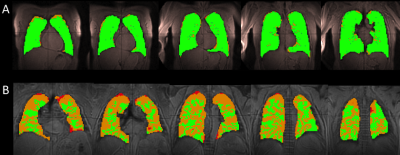
SNR and Dose Requirements for Quantitative 6-Zone Analysis of Hyperpolarized (129)Xe Ventilation MRI
Fei Tan, Mu He, Leith Rankine, Rohan Virgincar, John Nouls, Steven Shipes, Bastiaan Driehuys
Hyperpolarized (HP) 129Xe ventilation MRI can be used for non-invasive assessment of lung obstruction. However, the minimum 129Xe dose to obtain HP 129Xe ventilation MRI with sufficient signal-to-noise ratio (SNR) for reliable quantitative analysis has not yet been established. In this work, we introduced the reader-based six-zone analysis, which is used with 133Xe and 99mTc ventilation and perfusion scintigraphy, and applied to Rician noise degraded 129Xe ventilation MRI of COPD patients. We found that the minimum required SNR for 6-zone quantification of ventilation is 4.4±5.8 (mean±SD), which suggests a minimum required 129Xe dose equivalent of 89.2 ml for this resolution.
|
|
2478.
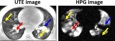 |
Hyperpolarized 129Xe gas and ultra-short echo MRI for evaluation of structure-function correlates in cystic fibrosis lung disease: a comparison of analysis methods
Robert Thomen, Laura Walkup, David Roach, Nara Higano, Zackary Cleveland, Andrew Schapiro, Alan Brody, John Clancy, Jason Woods
A number of techniques for analysis of hyperpolarized gas (HPG) images have emerged and demonstrated sensitivity to lung disease severity. However, the precise extent of lung function decline due to specific pathologies associated with obstructive lung disease has not been established. Here we have performed HPG 129Xe analysis using 3 common methods from the literature (mean-anchored, percentile-anchored, and k-means methods) in order to evaluate correlations with structural pathologies identified in ultra-short echo-time (UTE) images. The presence of bronchiectasis and mucus plugging correlated best with whole-lung ventilation defect percentage (VDP). Consolidation and air-trapping demonstrated weaker (though still significant) correlation with VDP.
|
|
2479.
 |
Absolute Reference for Dissolved-Phase 129Xe Spectroscopy Leads to Peak Reassignment
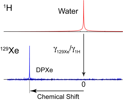
Michael Antonacci, Le Zhang, Alex Burant, Drew McCallister, Rosa Branca
Dissolved-phase 129Xe (DPXe) chemical shift (CS) measurements could benefit from a robust reference system that can provide consistent CS values independently of gas partial pressures, lung inflation, subject position, and shimming conditions. We demonstrate that, by referencing the DPXe frequency to that of nearby protons, consistent CS values can be obtained, both in vitro and in vivo, enabling correct assignment of some of the spectral lines observed in vivo.
|
|
2480.
 |
Quantifying Regional Lung Function in Interstitial Lung Disease with Hyperpolarized Xenon-129 3D SB-CSI
Mackenzie Carlson, Borna Mehrad, Yun Shim, Nicholas Tustison, John Mugler, Talissa Altes, Lucia Flors, Grady Miller, Jaime Mata
In this study, lung ventilation and gas uptake/exchange was assessed in healthy and interstitial lung disease (ILD) subject populations using 3D Single-Breath Chemical Shift Imaging, a combination of MR spectroscopic imaging and hyperpolarized xenon-129 gas imaging. By probing metrics such as Tissue/RBC, Tissue/Gas, RBC/Gas, T2* and chemical shifts in lung parenchyma and red blood cells, we find statistically significant distinctions in the lung physiology between healthy and ILD subjects.
|
|
Pancreas/GI
Traditional Poster
Body: Breast, Chest, Abdomen, Pelvis
Wednesday, 20 June 2018
| Exhibition Hall 2481-2489 |
16:15 - 18:15 |
|
2481.
 |
Tumor necrosis factor (TNF) antagonist therapy in small bowel Crohn’s disease (CD): association of the apparent diffusion coefficient (ADC) with treatment response.
Bradley Spieler, Hector De Jesus, Christopher Rouse, Catherine Hudson, Scott Kleinpeter, Catherine Batte, Raman Danrad, Kara De Felice
Diffusion weighted imaging (DWI) has proven beneficial in the assessment of disease activity and therapeutic response in a myriad of pathology. Studies have shown an inversely proportional correlation between bowel inflammation in Crohn’s disease (CD) and apparent diffusion coefficient (ADC) values of involved bowel wall. This beckons an intriguing opportunity for gauging treatment response, particularly with respect to some of the most commonly used agents, tumor necrosis factor (TNF) antagonists. This study retrospectively measured the ADC value of affected small bowel segments before and after anti-TNF infusion therapy and compares it to the clinical response in patients with active CD.
|
|
2482.
 |
Semi-automatic method for generating multiplanar reformatting views of MR post-contrast T1-weighted images for visualizing and assessing pediatric Crohn’s disease
Yechiel Lamash, Sila Kurugol, Moti Freiman, Simon K Warfield
In this proposed study, we aim to develop a semi-automated method for generating multiplanar reformatting images (MPR) of pediatric Crohn’s disease (pCD) segments from T1-weighted post-contrast MR image data. We demonstrate that this method can efficiently visualize and assess this disease. Importantly, the centerline length can be used as a reliable measure of the extent of disease. Moreover, the MPR image can be used as a platform for intestinal wall segmentation and for more accurate depiction of luminal narrowing. We also expect such MPR views to be used as a unified parametric platform for evaluating disease progression in follow-up scans.
|
|
2483.
 |
MRI assessed small bowel dysmotility and its relationship with patient reported symptoms: An exploration of automated vs subjective assessment techniques
Ruaridh Gollifer, Alex Menys, Andrew Plumb, Frans Vos, Jaap Stoker, Stuart Taylor, David Atkinson
The pathophysiology of chronic abdominal symptoms in Crohn’s disease (CD) is complex. Recent pilot data using automated quantification of motility MRI suggests reduced variation in apparently normal bowel may underpin symptoms, including pain and diarrhoea. This two-centre validation study tests this association and compares automated measurements with subjective radiologist bowel motility assessment. We confirmed that reduced spatial variation of motility is significantly associated with the severity of abdominal symptoms, although the correlation was not strong. Automated measurement had superior inter-reader variability than subjective radiologist assessment, and showed a stronger association with patient symptoms.
|
|
2484.
 |
The workflow for the validation of USPIO-enhanced MRI for the detection of lymph node metastases in rectal cancer
Rutger Stijns, Bart Philips, Chella van der Post, Iris Nagtegaal, Carla Wauters, Luc Strobbe, Fatih Polat, Johannes de Wilt, Stefan Rietsch, Sascha Brunheim, Stephan Orzada, Harald Quick, Jurgen Fütterer, Tom Scheenen
For patients with rectal cancer, the presence of lymph node metastases is an important risk factor for determining prognosis and stratifying for treatment. Clinically, lymph node staging is very challenging, especially when lymph nodes are small (<5mm). By using ultrasmall superparamagnetic iron oxide (USPIO) particles combined with (ultra) high magnetic field imaging (combidex-enhanced MRI), the detection rate of these metastatic lymph nodes may improve significantly. In this abstract we present the workflow for validating combidex-enhanced MRI by performing a node to node comparison of in vivo combidex-enhanced MRI findings with histopathological examination.
|
|
2485.
|
Quantitative assessment of pancreatic proton density fat fraction (PDFF) and R2* with preoperative T2* corrected multi-echo chemical-shift-encoded MRI in patients undergoing pancreatic resection: comparison with single-voxel 1H-MRS
Yali Qu, Mou Li, Zhen Zhang, Zixing Huang, Chunchao Xia, Bin Song
Many studies have shown multi-echo chemical-shift-encoded magnetic resonance imaging (CSE-MRI) has good performance for the evaluation of fat and iron in liver. However, the relevant studies in pancreas are fewer. We found that pancreatic PDFF and R2* estimated by T2* corrected multi-echo CSE-MRI showed a moderate correlation with 1H-MRS results in patients undergoing pancreatic resection. In addition, our study showed that pancreatic PDFF was not to be significantly associated with clinically relevant postoperative pancreatic fistula.
|
|
2486.
 |
The value of IDEAL-IQ in evaluating pancreatic fat quantification in patients with non-alcoholic fatty liver disease(NAFLD)
Qinhe Zhang, Ailian Liu, Lizhi Xie
The study aims to assess the pancreatic fatty quantitation in NAFLD by use of IDEAL-IQ. It was concluded that IDEAL-IQ is a new way to evaluate the pancreatic fat quantification in patients with NAFLD. The fat fraction of the pancreas in patients with NAFLD is significantly higher than that in normal subjects, and the distribution of pancreatic fat in various regions of the pancreas in the NAFLD patients is well.
|
|
2487.
 |
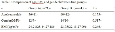
Quantitation of metabolites in human tumour (paraganglioma and GIST) tissues with mitochondrial mutations (SDH and IDH1) by HRMAS 1H NMR spectroscopy
Basetti Madhu, Ruth Casey, Benjamin Challis, Graeme Clark, Alison Marker, Olivier Giger, Venkata Bulusu, Mary McLean, Ferdia Gallagher, Eamonn Maher
In this study we report, for the first time, the detection of 2HG in IDH1 mutated human GIST tumour tissues by HRMAS 1H NMR spectroscopy. We quantified the levels of Succinate and 2HG in human paraganglioma and GIST tissues. The lactate, glutamate and glycero-phosphocholine (GPC) concentrations were significantly lower in SDHx mutated tumours compared to wild type (WT) tumour tissues, Detection of higher levels of Succinate in SDH mutated tumour tissue and 2HG in IDH1 mutated tissue and their quantitation will be helpful in the stratification of patient treatment in the clinics.
|
|
2488.
 |
Assessment of Colonic Motility Using Magnetic Resonance Imaging: Reproducibility of a Macrogol Challenge
Victoria Wilkinson-Smith, Alex Menys, Christopher Bradley, Maura Corsetti, Luca Marciani, David Atkinson, Carol Coupland, Stuart Taylor, Penny Gowland, Robin Spiller, Caroline Hoad
This study assessed the reproducibility of a previously developed diagnostic test using a macrogol stimulus and MRI measures to assess colonic motility. This test was performed twice on healthy volunteers and the results were compared. The data showed some variability across visits representing both variability in baseline data and the physiological response of the colon to the stimulus. Correlation data suggested that although intra-subject variability existed the maximum measured MRI parameters all increased post stimulus. This colonic stimulus test allows us greater insight into potential pathologies behind GI disorders and as such may be of value here.
|
|
2489.
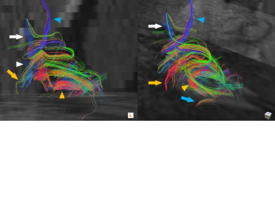 |
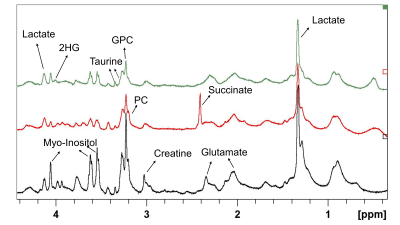
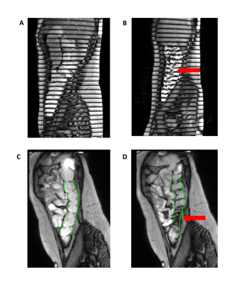
Case report: Three-dimensional visualization of the normal human perirectal muscle with diffusion tensor imaging (DTI)
Koji Tokunaga, Shigeki Arizono, Koji Fujimoto, Tomoaki Okada, Katsutoshi Murata, Hiroyoshi Isoda, Kaori Togashi
Diffusion tensor imaging (DTI) can provide the directionality of water diffusion in tissues, informing on its underlying microstructures and microdynamics. There has been no previous report on the visualization of anterior portion of the longitudinal anal muscle (aLAM). In this case study, we present the 3D visualization of the aLAM in normal male subjects with DTI. By adjusted parameters for DTI sequence, we could successfully visualize thin smooth muscle layer of the rectum. This technique could be useful when planning operation for rectal and anal diseases.
|
|
Body Imaging: Renal
Traditional Poster
Body: Breast, Chest, Abdomen, Pelvis
Wednesday, 20 June 2018
| Exhibition Hall 2490-2495 |
16:15 - 18:15 |
|
2490.
 |
Characterization of Renal Solid Masses Using Multiparametric Diffusion-Weighted Imaging
Jianjian Zhang, Guangyu Wu, Yongming Dai
Preoperative characterization of the renal lesions has clinical significance in determining the appropriate treatment strategy and evaluating prognosis. The current study aims to investigate the potential of multiparametric DWI models, including monoexponential, biexponential, stretched-exponential, and kurtosis models in distinguishing between benign and malignant renal lesions, different tumor types as well as different grading of RCC. Compared with monoexpontial model, these highly parameterized non-Gaussian diffusion models may provide more information in the characterization of renal lesions, which would be helpful in improving therapy strategies and prognoses in the future, and further evaluation are required.
|
|
2491.
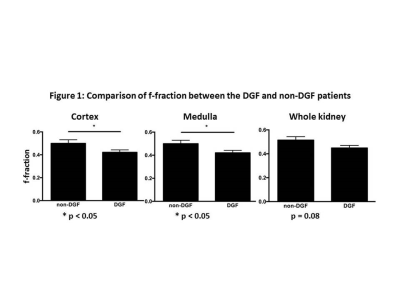 |
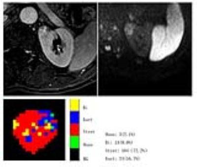
Intravoxel incoherent motion-diffusion weighted imaging (IVIM-DWI) parameters distinguish kidney allografts with delayed graft function
Eyesha Hashim, Darren Yuen, General Leung, Anish Kirpalani
Delayed graft function (DGF) complicates 21-36% of all deceased donor kidney transplants, and leads to early inpatient post-transplant dialysis, higher risk of graft failure and death. In this abstract, we show that IVIM-derived flow (f)-fraction, is significantly different in kidney allografts exhibiting DGF compared to those that do not develop DGF. Furthermore, f fraction shows a significant negative correlation with time to recovery and a positive trend with renal function at 3 months post-transplant as measured with eGFR.
|
|
2492.
 |
Compensating for Bulk Motion in Feed and Wrap Renal Dynamic Radial VIBE DCE-MRI using Bulk Motion Removal and Non-Rigid Registration
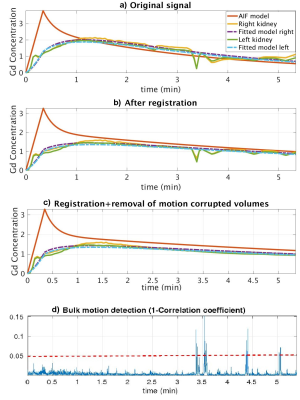
Sila Kurugol, Onur Afacan, Catherine Seager, Richard Lee, Jeanne Chow, Simon Warfield
Dynamic Radial VIBE DCE-MRI enables motion-robust imaging with high spatiotemporal resolution for accurate estimation of kidney function. However, in feed and wrap DCE-MRI, bulk motion during infant’s sleep reduces the quality of images affected by motion and limits clinical utility of this method for imaging without sedation. This work evaluated the ability of detecting bulk motion using the center-of-k-space line, removing corrupted volumes, and compensating for motion using non-rigid registration for improved parameter estimation accuracy. Results showed that volumes affected by motion were successfully detected and removed in all patients, and the goodness-of-fit to the tracer kinetic model was improved.
|
|
2493.
 |
A Preliminary Study of the Longitudinal Changes in a Reversible Unilateral Ureteral Obstruction Rat Model using Intravoxel Incoherent Motion and Arterial Spin Labeling Imaging
Genwen Hu, Xianyue Quan, Jianmin Xu, Liangping Luo, Yingjie Mei, Siying Wang
The longitudinal changes of intravoxel incoherent motion (IVIM) and arterial spin labeling (ASL) imaging in a RUUO model
|
|
2494.
 |
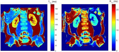
Application of T1rho and T1 mapping MRI in Tracking Renal Ischemia Reperfusion Injury Process in Rats
Yangguang Yuan, Jingjing Huang, Yingjie Mei, Siying Wang, Wen Liang
Previous studies using T1rho and T1 mapping in the liver and heart demonstrated that T1rho value and T1 relaxation time can be used to assess acute injury and long-term tissue fibrosis1. However, to the best of our knowledge, these techniques have not been explored to evaluate acute kidney ischemia damage. In our study, we found that T1rho value and T1 relaxation time showed high specificity and sensitivity in a rat renal ischemia reperfusion injury (IRI) model.
|
|
2495.
 |
R1rho dispersion in human kidney
Ping Wang, John Gore
R1ρ (=1/T1ρ) imaging has been applied in many human organs to characterize tissue biochemical changes. However, R1ρ imaging in human kidney has been rarely reported partly due to the challenges associated with field inhomogeneities and respiratory motion. We developed an R1ρ imaging protocol for human kidney which used adiabatic half passage pulse and volume shimming to overcome field inhomogeneities. In addition, R1ρ dispersion was evaluated via a simple method with a fixed locking time but different locking frequencies. The volunteer scans exhibited characterized R1ρ maps in kidney, also there was greater R1ρ dispersion between locking frequencies of 100Hz and 300Hz.
|
|
Body: Fat Imaging
Traditional Poster
Body: Breast, Chest, Abdomen, Pelvis
Wednesday, 20 June 2018
| Exhibition Hall 2496-2511 |
16:15 - 18:15 |
|
2496.
 |
A Dedicated Protocol for Fat Fraction Mapping in Obese Patients: Preliminary Findings in Skeletal Muscle
Naomi Sakai, Timothy Bray, Alan Bainbridge, Rachel Batterham, Stuart Taylor, Margaret Hall-Craggs
Obesity is associated with ectopic fat deposition and chronic inflammation in skeletal muscle (SM), which contributes to insulin resistance. Novel treatments for obesity such as bariatric surgery can reduce insulin resistance by reducing ectopic fat deposition, but this effect is inconsistent and poorly understood. Therefore, we need a fast, non-invasive method that can help to study the link between ectopic fat deposition and insulin resistance. Here, we describe a protocol for scanning obese patients, which is fast, tolerable and accurate, and reveals significant changes in SM proton density fat fraction (PDFF) in obese patients.
|
|
2497.
 |
In Vivo Proton Magnetic Resonance Spectroscopy of Hepatic Fatty Acid Change: Identification of Lipid Contents with Correct and Incorrect Terminal Methyl Group in Hepatic Steatosis at Ultra High Field
Kyu-Ho Song, Min-Young Lee, Chi-Hyeon Yoo, Song-I Lim, Bo-Young Choe
Magnetic resonance spectroscopy (MRS) with optimized relaxation time provides an effective means for quantifying lipid content and characterizing hepatic steatosis. The aim of this study was to quantify the difference in hepatic lipid content with metabolic changes and determine effect of diet on high-fat diet (HFD)-fed mice by measuring the main localized MRS sequence with relaxation times.
|
|
2498.
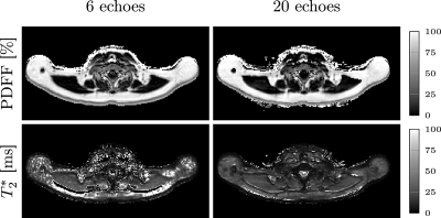 |
Differentiating supraclavicular from gluteal adipose tissue based on simultaneous PDFF and T2* mapping using a twenty-echo gradient echo acquisition
Daniela Franz, Maximilian Diefenbach, Jan Syväri, Dominik Weidlich, Ernst Rummeny, Hans Hauner, Stefan Ruschke, Dimitrios Karampinos
PDFF and T2* have been previously proposed as two important parameters in quantitative MRI of adipose tissue. This study investigates the difference between gluteal and supraclavicular adipose tissue T2* and the relationship between adipose tissue T2* and PDFF using a twenty-echo multi-echo gradient echo acquisition. A highly significant difference between the PDFF in different fat regions was detected in water-fat separation results when using either the first 6 echoes or the full 20 echoes. However, T2* values were only significantly different between fat regions, when using the full 20 echoes and not when using the first 6 echoes. PDFF also correlated with T2* when using the full 20 echoes.
|
|
2499.
 |
Magnetic Resonance Imaging and Spectroscopic Investigation of interscapular BAT and Skeletal Muscle IMCL in High Intensity Exercise Trained Rats
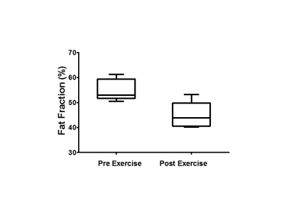
Venkatesh Gopalan, Rengaraj Anantharaj, Le Giang, Sanjay Verma, Jadegoud Yaligar, Anna Ulyanova, Karthik Mallilankaraman, S Sendhil Velan
There is a large interest in developing non-pharmacological approaches such as exercise and nutritional compounds for activating BAT to improve metabolic health. In this study, we have investigated the effect of high intensity exercise on interscapular BAT and Intramyocellular lipids (IMCL) from skeletal muscle of rats. Exercise-induced adrenergic receptor stimulation improves quality of iBAT by remodeling of WAT into beige fat and improved mitochondrial fatty acid oxidation. Skeletal muscle IMCL also reduced with exercise along with increased PGC-1α expression due to energy expenditure.
|
|
2500.
 |
Abdominal and organ fat content quantification in PROFAST trial (Probiotics and intermittent fasting to improve pre-diabetes)
Dech Dokpuang, Rinki Murphy, Lindsay Plank, Reza Nemati, Jun Lu
The primary objective of this study was to test quantification protocols on human abdominal and organ fat data acquired using magnetic resonance (MR) imaging or spectroscopy. Liver, pancreatic, visceral and subcutaneous fat in 10 obese patients with prediabetes were measured before and after a 12-week intermittent fasting programme with daily probiotic or placebo supplementation. All participants were scanned by a Siemens 3.0T MR scanner. The quantification of fat contents was performed using ImageJ (for MRI data) and SIVIC software (for MRS data). Two methods of quantifying pancreas fat were compared.
|
|
2501.
 |
Metabolic Imaging and Characterization of Browning Adipose Tissue by DCE-MRI and Dixon Imaging
Jadegoud Yaligar, Sanjay Kumar Verma, Venkatesh Gopalan, Rengaraj Anantharaj, Giang Le Thi Thu, S. Sendhil Velan
Browning of white adipose tissues is emerging as a promising strategy to increase whole body energy expenditure and to reduce obesity. At a whole body level, increasing the beige or BAT volume and enhancing its functional activity is a promising strategy for management of obesity. There is a lack of non-invasive methods for imaging the browning process. For the first time we demonstrated the feasibility of non-invasive imaging of browning adipose tissue by fat fraction imaging and DCE-MRI. The browning adipose tissues show significant reduction in fat fraction and increase in tissue perfusion parameters including Ktrans and ve
|
|
2502.
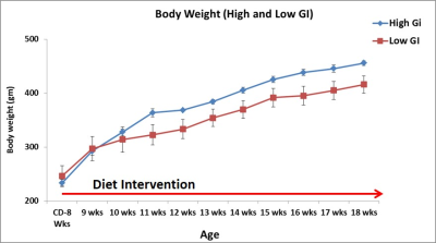 |
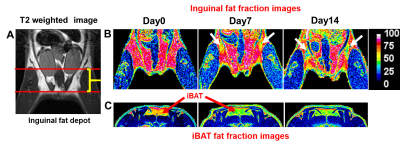
Metabolic Imaging of Brown Adipose Tissue in Response to High Glycaemic Diet and Systemic Metabolic Effects on Whole Body Fat Metabolism
Jadegoud Yaligar, Rengaraj Anantharaj, Le Thi Thu Giang, Sanjay Kumar Verma, Venkatesh Gopalan, Bhanu Prakash K N, Karthik Mallilankaraman, S. Sendhil Velan
High-GI diet has been linked with insulin resistance, type 2 diabetes and cardiovascular risk factors. Brown fat activity positively correlates with increased energy expenditure during β3-agonist/cold induced BAT activation, suggesting regulatory link between BAT and energy metabolism. In this study we evaluated long term metabolic effects of high and low-GI diets on brown adipose tissue metabolism and ectopic fat accumulation in liver and abdomen by MRI and MRS. Low-GI diet fed animals were responsive to prolonged BAT activation for metabolizing the fat. Weight and volumes of iBAT increased with β3-agonist treatment, implying potential remodeling of WAT into Beige.
|
|
2503.
 |
Metabolic Imaging of brown adipose tissue in leucine deficient diet fed mice.
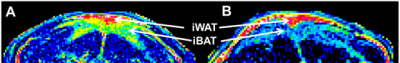
Anna Ulyanova, Jadegoud Yaligar, Anantharaj Rengaraj, Giang Le Thi Thu , Sanjay K Verma, Venkatesh Gopalan, S Sendhil Velan
Brown adipose tissue plays an important role in energy expenditure. The deficiency of the essential amino acid leucine has been linked with CREB/TRH pathway and regulation of energy expenditure and food intake. Here we investigated the effect of leucine deficient diet on interscapular brown adipose tissue (iBAT) in mice. Dixon imaging was performed to assess fat fraction changes within iBAT followed by RNA analysis. There was a decrease in fat fraction for leucine deficient diet fed mice together with increased UCP1 expression indicating that short term leucine deprivation leads to iBAT activation.
|
|
2504.
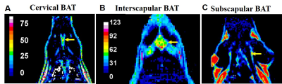 |
Identification and Characterization of Brown and White Adipose Tissue Depots in Rats by 3D Whole Body Imaging
Rengaraj Anantharaj, Sanjay Kumar Verma, Jadegoud Yaligar, Julian Gan, Giang Le Thi Thu, Kavita Kaur, Venkatesh Gopalan, Kuan Jin Lee, S. Sendhil Velan
Excess body adiposity results in obesity and metabolic dysfunction. Identification and characterization of various white, brown and browning adipose tissues and the possibility of reversing pre-diabetic pathology is of current clinical interest for combating obesity and diabetes. In this study, we have identified and characterized various brown and white fat depots by whole body imaging in rats using a Siemens 3T Skyra system.
|
|
2505.
 |
Evaluation of Simultaneous MRI/PET of Supraclavicular BAT for Detecting Adaptive Thermogenesis after Sympathetic Nervous System Activation
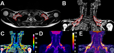
Sanjay K Verma, Lijuan Sun, Suresh Anand Sadananthan, Navin Michael, Hui Jen Goh, Priya Govindharajulu, John Totman, David Townsend, Houchun H Hu, Melvin Khee-Shing Leow, S Sendhil Velan
There is a large interest in detecting and quantifying brown adipose tissue (BAT) in humans for evaluating its potential to design therapeutic strategies to combat obesity-related metabolic dysfunction. In the current study, we evaluated the use of simultaneous PET/MRI of supraclavicular BAT (sBAT) for distinguishing subjects with high or low adaptive thermogenesis after sympathetic nervous system activation by cold exposure and capsinoids ingestion. As a sub-study, We also evaluated the duration of cold-exposure for changes in 18F-FDG uptake and Dixon-based fat-fraction. We found that adaptive thermogenesis after capsinoids ingestion was too low to be detected by either modality, while PET was successful in identifying high responders to cold stimulation.
|
|
2506.
 |
In Vivo Diffusion Magnetic Resonance Spectroscopy of Brown and White Adipose tissues
Sanjay K Verma, Kavita Kaur, Jadegoud Yaligar, Navin Michael, Anantharaj Rengaraj, Le Giang, Venkatesh Gopalan, Suresh Anand Sadananthan, Melvin Khee-Shing Leow, S Sendhil Velan
There is a large interest in understanding the biophysical properties of BAT, WAT, and beige adipose tissues for evaluating its potential to improve whole body metabolism. Diffusion properties of tissues provide information on microstructure, anisotropy, and pathology. In the presence of cellular and sub-cellular barriers, and heterogeneity the lipid diffusion is restricted. Water diffusion has been well characterized in several organs. Fat diffusion has not been studied due to the hardware limitations. In this study, we have implemented diffusion-weighted spectroscopy for investigating in-vivo diffusion properties of BAT and WAT.
|
|
2507.
 |
The Associations between Water-Fat MRI Measurements of Brown Adipose Tissue and Abdominal Adiposity and Glucose Metabolism in Children and Adolescents
Elin Lundström, Joy Ljungberg, Jonathan Andersson, Robin Strand, Anders Forslund, Peter Bergsten, Daniel Weghuber, Katharina Paulmichl, Kurt Widhalm, Matthias Meissnitzer, Håkan Ahlström, Joel Kullberg
Investigating the role of brown fat (BAT) in child/adolescent metabolism and obesity is important for elucidating its potential as an antiobesity/antidiabetes therapeutic target. This study presents associations between MRI estimates of BAT (by cervical-supraclavicular adipose tissue fat fraction and T2*) and abdominal adiposity and glucose metabolism parameters in children/adolescents. Associations between the BAT estimates and adiposity were observed, supporting previous indications of decreasing BAT amounts with increasing adiposity. Additional associations between the BAT estimates and important glucose metabolism parameters may reflect a role for BAT in glucose and energy metabolism and potentially a link to development of type 2 diabetes.
|
|
2508.
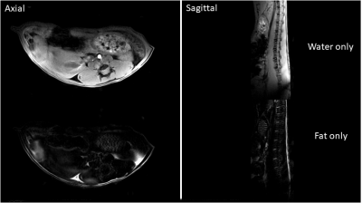 |
2-phase Dixon technique to assay dermal white adipose tissue loss as potential early diagnostic biomarker of scleroderma using genetic Fra-2 mice
Nicola Bertolino, Roberta Goncalves Marangoni, Daniele Procissi, Cynthia Yang, Sol Misener, Warren Tourtellotte, John Varga
Scleroderma is an autoimmune disease leading to fibrosis resulting in stiff skin, formation of ulcers, joint contractures and ultimately functional incapacity. Loss of dermal white adipose tissue (dWAT) was observed ex-vivo prior to the development of fibrosis. In this study we demostrated the feasibility of employing CSE MRI Dixon technique to detect and quantify in-vivo dWAT thickness using a genetic Fra-2 fibrosis mice model. The proposed non-invasive diagnostic method to evaluate or predict skin fibrosis would greatly improve clinicians’ ability to track progression and response to treatment and also provide a tool to investigate pathogenesis in animal models.
|
|
2509.
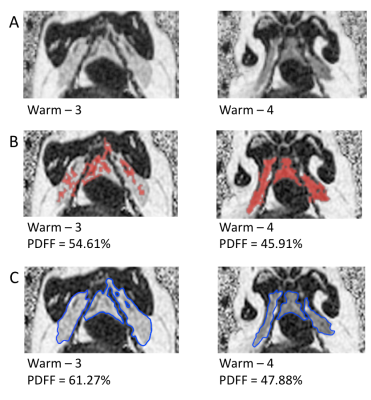 |
Using water-fat MRI to detect remodeling of adipose tissue
Amanda MacCannell, Kevin Sinclair , James Staples, Charles McKenzie
Hibernating mammals use brown adipose tissue (BAT) as a primary source of heat production for arousal from torpor. In hibernators, both white adipose tissue (WAT) and BAT volumes increase in autumn even when temperatures are warm, unlike non-hibernators which require cold exposure for BAT growth. Differentiation of WAT from BAT between depots in close proximity can be achieved using IDEAL water-fat MRI. Hibernating mammals exposed to constant warm environments showed drastic molecular changes to their BAT depots that could ultimately be detected my MRI, proving IDEAL’s versatility and specificity.
|
|
2510.
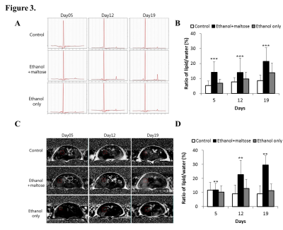 |
Novel model of alcoholic hepatitis and alcoholic steatohepatitis using C57BL/6N mice and magnetic resonance imaging/spectroscopy
Jeeheon Kang, Su Jung Ham, Yoonseok Choi, Seul-I Lee, Jinil Kim, Jae Im Kwon, Ho-jin Kim, Do-Wan Lee, YongJun Lee, Chul-Woong Woo, Sang Tae Kim, Kyung Won Kim, Dong-Cheol Woo
Alcoholic liver disease is classified into two subgroups: alcoholic hepatitis (AH) and alcoholic steatohepatitis (ASH). These differ in most characteristics, including clinicopathologic features and treatment. However, animal models of AH and ASH are not well established. Noninvasive monitoring is essential for evaluating chronic diseases such as AH and ASH. Magnetic resonance imaging and spectroscopy (MRI/S) have recently gained considerable attention as noninvasive monitoring tools for chronic liver disease. The aim of this study, therefore, was to develop a comprehensive animal model of AH and ASH that can be monitored noninvasively using MRI/S.
|
 |
2511.
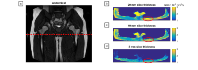 |
ADC quantification of lipids with high b-value stimulated echo-prepared diffusion-weighted 2D single shot TSE
Dominik Weidlich, Stefan Ruschke, Barbara Cervantes, Andreas Hock, Dimitrios Karampinos
The prevalence of the metabolic syndrome is rapidly growing over the past decade. Fat plays a central role in the incidence and the progression of the metabolic syndrome and despite the successful clinical translation of quantitative fat MRI biomarkers into applications, current MRI biomarkers cannot answer questions about fat cell microstructure in different fat depots. This work proposes an acquisition imaging method that probes the diffusion properties of lipids, compares the proposed method to single-voxel diffusion-weighted MRS in vivo in the tibia bone marrow and investigates in vivo the dependency of ADC quantification on voxel size in gluteal fat.
|
|
Body: Animal Models
Traditional Poster
Body: Breast, Chest, Abdomen, Pelvis
Wednesday, 20 June 2018
| Exhibition Hall 2512-2517 |
16:15 - 18:15 |
|
2512.
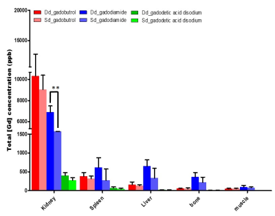 |
Quantitative evaluation of gadolinium deposition after various gadolinium-based contrast agent injection in the rat abdominal organs .
Hyewon Oh, PanKi Kim, ChanGyu Joo, YongEun Chung
Gadolinium-based Contrast agent (GBCA) is likely to deposit in the rat abdominal organs.
|
|
2513.
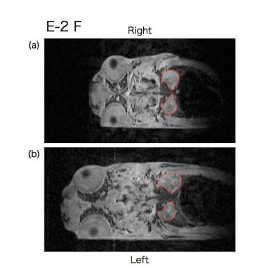 |
Individual Time Series Analysis of p53 Knockout Medaka by in vivo Magnetic Resonance Microscopy
Hajime Morizumi, Takahiro Nishigaki, Naozo Sugimoto, Tomohiro Ueno
Tumor suppressor gene p53 knockout medaka has been generated. The tumor spectrum of this medaka model, however, remains unknown. In this study, we performed individual time series analysis of p53 knockout medaka using a 14.1T MR microscopy. Extracting size change of kidney of the medaka model, we found early indications of disease and difference in phenotype due to location difference of point mutation in the p53 gene. Since p53 knockout medaka showed rather large variations in kidney slice, importance of individual time series analysis was confirmed.
|
|
2514.
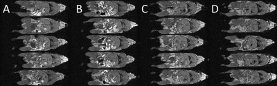 |
Mn2+-free chow reduces gastrointestinal signal for T1-weighted MRI of the mouse abdomen
Veerle Kersemans, Stuart Gilchrist, Paul Kinchesh, Sean Smart
Standard commercial chow given to laboratory animals may contain high levels of paramagnetic Mn2+-ions which act as a T1-reducing contrast agent. Signal intensities where Mn2+ is present are increased when using short-TR, T1W-MRI imaging and the GI-tract appears brighter than the rest of the body. As peristalsis is an inherently unstable motional process, high intensity and temporally unstable signals are formed in the GI-tract, creating image-ghosting and decreasing resolution from that prescribed. We present images acquired before and after transition from Mn2+-bearing to Mn2+-free food to show that these deleterious image effects can be reduced through simple dietary formulation change.
|
|
2515.
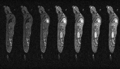 |
Whole-Body Cardio-Respiratory Synchronised DCE-MRI in the Mouse
Veerle Kersemans, Philip Allen, Stuart Gilchrist, Ana Gomes, Paul Kinchesh, Sean Smart
Prospective gating of constant, short TR scans enables rapid imaging to be performed in conjunction with cardiac and respiratory synchronisation. We show that prospectively-gated, dynamic contrast enhanced MRI (DCE-MRI) can be performed over the whole mouse body with a time resolution of ca. 15 s/frame such that multiple organs can be examined simultaneously.
|
|
2516.
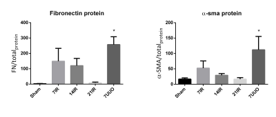 |
Evaluation of fibrosis models using 1H T1 mapping and 23Na T2*.
Per Nielsen, Christian Mariager, Christoffer Laustsen, Marie Mølmer, Rikke Nørregaard
In this study we try to develop a renal IRI model which leads to fibrosis. Fibrosis markers indicate the best effect after 7 days of reperfusion. We also investigate the possibility of using 23Na T2* to evaluate fibrosis, this gave rise to a correlation with fibrosis markers only when normalizing to water transport from cortex to medulla.
|
|
2517.
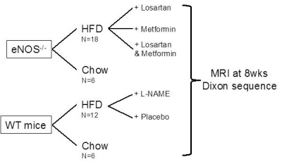 |
eNOS-/- mice fed with HFD develop progressive non-alcoholic fatty liver disease (NAFLD) which is partially reversible with antihypertensive and hypoglycemic therapy
Begoña Lavin Plaza, Marcelo Andia, Thomas Eykyn, Alkystis Phinikaridou, Aline Xavier, Rene Botnar
Liver steatosis or non-alcoholic fatty liver disease (NAFLD) is the most common liver disease in Western countries. However, the cause and treatments are still controversial. Nitric oxide (NO) and its derivatives play important roles in the physiology and pathophysiology of the vascular system and liver metabolism. We quantified intraperitoneal fat and liver fat-fraction using 3T MRI in eNOS-/- mice fed with HFD and investigated (1) whether pharmacological treatments for type 2 diabetes and hypertension reduced fat deposition and (2) if the phenotype could be recapitulated by administration of an inhibitor of endothelial NO synthesis (L-NAME) in wild type mice.
|
|
Body: Technical Advances
Traditional Poster
Body: Breast, Chest, Abdomen, Pelvis
Wednesday, 20 June 2018
| Exhibition Hall 2518-2535 |
16:15 - 18:15 |
|
2518.
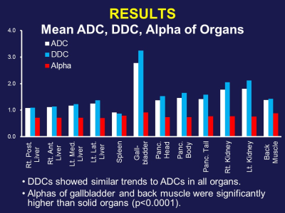 |
Stretched-Exponential Diffusion-Weighted Imaging Model for Abdominal MRI
Takeshi Yoshikawa, Katsusuke Kyotani, Yoshiharu Ohno, Yoshimori Kassai, Seiya Kai, Eiji Takeda, Shinichiro Seki, Yuji Kishida
Stretched-exponential model can be used as an excellent alternative to mono-exponential model in evaluation of abdominal organs and diseases.
|
|
2519.
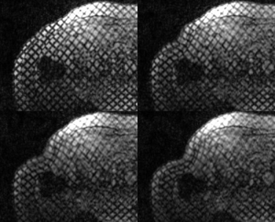 |
Measuring Abdominal Wall Muscle Deformation using MR Tissue Tagging
Lawrence Dougherty, Pilla James, Anil Chauhan
The current state of hernia repair relies heavily on clinical evaluation of patients, which is ultimately a poor predictor of outcomes for patients going into surgery. There are currently no reliable data, standard imaging modalities, or guidelines available to predict successful fascial closure in hernia repair. A method using MR tissue tagging with synchronous displacement of the abdominal wall was developed. This will allow analysis of the mechanical properties of muscle for noninvasive, diagnostic tool for pre-operatively predicting successful fascial closure in hernia repair.
|
|
2520.
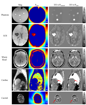 |
Whole body Quantitative Susceptibility Mapping using Automated Preconditioning
Zhe Liu, Yan Wen, Pascal Spincemaille, Thanh Nguyen, Yi Wang
An automated method is proposed for generating an optimal preconditioner for a given field input for performing preconditioned total field inversion quantitative susceptibility mapping. In gradient echo data acquired in healthy subjects and patients in various anatomic regions, the obtained preconditioner leads to the same optimal susceptibility map quality as a manually selected preconditioner.
|
|
2521.
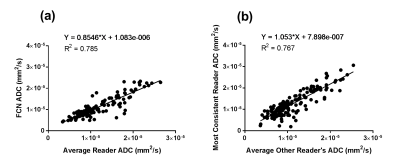 |
Automated contouring and ADC measurement of esophageal cancer with a fully convolutional network
Benjamin Musall, Steven Lin, Penny Fang, Brett Carter, Amy Moreno, Jong Bum Son, Jeremiah Sanders, Jingfei Ma
A Fully Convolutional Network (FCN) was developed and applied to the task of contouring esophageal tumors on diffusion weighted images. After proper training, tumor classification by the FCN demonstrated excellent agreement with tumor contours from an inter-reader agreement study in the validation images. The FCN was able to achieve correct tumor classification in most cases with respect to different tumor position and shapes, and in the presence of intratumoral esophageal lumen.
|
|
2522.
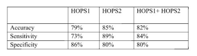 |
Detection of liver fibrosis from MRIusing histogram of strains
Yasmine Safwat, Rasha Hussein, Ayman Khalifa, Ahmed Ibrahim, Ahmed Samir, Heba Abdallah, Ahmed Fahmy
In this work, we present the results of a novel method for detecting liver fibrosis from tagged MRI images. The method is based on extracting a set of features representing the liver deformations induced by the heart motion. First, the tagged MRI images are analyzed to calculate the liver tissue strain induced by the heart motion. The histogram of the peak strain values at each point within the liver are used as feature vectors to classify normal from patients with liver fibrosis. Classification using support-vector-machines using data of 34 subject (15 normal, 19 patients) showed sensitivity and specificity of 89%, and 80% respectively.
|
|
2523.
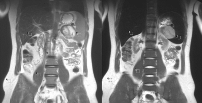 |
Changes of T2 signal intensities of abdominal organs between pre- and post-enhanced HASTE using ferumoxytol
Woo Kyoung Jeong, Kim-Lien Nguyen, Puja Shahrouki, J. Paul Finn
This study was designed to investigate the signal intensities (SI) of abdominal organs on pre- and post-enhanced HASTE images using ferumoxytol at a dose of 4mg /kg, and to compare the differences of enhancement effect among the organs. We found that the SI of liver, spleen, and pancreas were significantly decreased on HASTE after ferumoxytol administration. The greatest effects on SI were observed in liver and spleen. Little change in SI of muscle and fat was noted. The findings suggest that normal liver and spleen undergo profound decrease in signal intensity following ferumoxytol injection, likely reflecting their high blood volume. This observation suggests a potential role for ferumoxytol in detection and characterization of focal lesions of the liver and spleen.
|
|
2524.
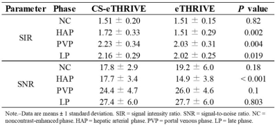 |
Gadoxetic acid-enhanced dynamic MR imaging using optimized integrated combination with parallel imaging and compressed sensing technique.
Nobuyuki Kawai, Satoshi Goshima, Kimihiro Kajita, Tomoyuki Okuaki, Masatoshi Honda, Hiroshi Kawada, Yoshifumi Noda, Yukichi Tanahashi, Shoma Nagata, Masayuki Matsuo
Gadoxetic acid-enhanced MRI represents an essential part in the assessment of hepatic diseases, however, dynamic imaging especially in hepatic arterial phase is still challenging for patients with limited breath-hold capabilities. We assessed prototype sequence using optimized integrated combination with parallel imaging and compressed sensing technique (Compressed-SENSE) for liver imaging, which enabled significant reduction of acquisition time resulting in excellent image quality with less motion artifact, especially in hepatic arterial phase, compared with conventional method. Our results demonstrated the significance and usefulness of Compressed-SENSE in clinical use for gadoxetic acid-enhanced dynamic MR imaging.
|
|
2525.
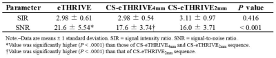 |
Hepatobiliary phase imaging using optimized integrated combination with parallel imaging and compressed sensing technique.
Nobuyuki Kawai, Satoshi Goshima, Kimihiro Kajita, Tomoyuki Okuaki, Masatoshi Honda, Hiroshi Kawada, Yoshifumi Noda, Yukichi Tanahashi, Shoma Nagata, Masayuki Matsuo
Gadoxetic acid-enhanced MRI plays an important role in the assessment of hepatic diseases. Hepatobiliary phase image has an amazing tissue contrast for the lesions with or without functional hepatocytes, however, which is still challenging for patients with limited breath-hold capabilities. We assessed prototype sequence using optimized integrated combination with parallel imaging and compressed sensing technique (Compressed-SENSE) for liver imaging. Our results demonstrated that Compressed-SENSE technique enabled significant reduction of acquisition time without image quality degradation resulting in higher spatial resolution and excellent image quality compared with conventional method.
|
|
2526.
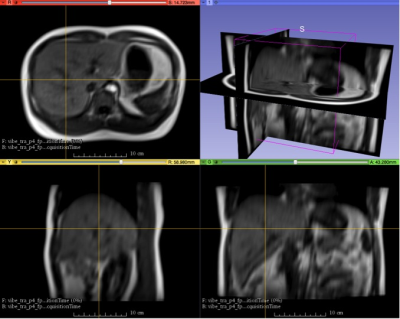 |
Development of a fast 4D-MRI with sub-second volumetric frame rate for respiratory motion tracking in abdominal radiotherapy
Jing Yuan, Yihang Zhou, Oilei Wong, KinYin Cheung, Siu Ki Yu
Time-resolved volumetric MRI (4D-MRI) is gaining more interests for better tumor motion characterization than 4D-CT in abdominal radiotherapy, while 3D sequence has limited use for 4D-MRI acquisition due to its slow volume-frame-rate (VFR) and various motion artifacts. We developed a fast 4D-MRI technique based on CAIPIRINHA accelerated 3D spoiled gradient echo sequence and a 1.63 frames-per-second (615ms/frame, ~1/7 of normal respiratory cycle of 4-5s) VFR was achieved. This 4D-MRI was demonstrated for whole abdomen respiratory motion tracking in healthy volunteers, indicating its great potentials for internal-target-volume definition in radiotherapy treatment planning and image guidance of MR-guided-radiotherapy.
|
|
2527.
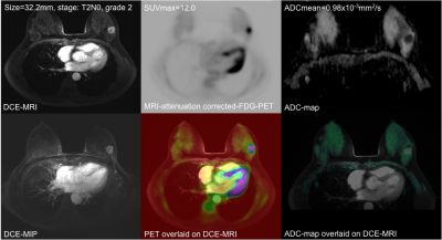 |
A study of correlation of the SUVmax and ADC in malignant breast tumors using simultaneous PET-MRI
Jing Yuan, Gladys Lo, Garrett Ho, Sirong Chen, Helen Chan, Victor Ai, William Cheung, Catherine Wong, Suk Yee Polly Cheung, Ting Ting Wong
We studied the correlation of simultaneous DWI-ADC and 18F-FDG SUVmax in invasive ductal carcinoma (IDC) tumors (n=41), and their association with different diagnostic factors using integrated PET-MRI. An insignificant inverse correlation was found (r=-0.214, p=0.179) between SUVmax and ADCmean. SUVmax was significantly associated with tumor T-stage (p=0.024). ADCmean of the index IDC was significantly smaller in the patients with pathologically confirmed regional lymph node metastasis (p=0.0488) and estrogen receptor status (p=0.0254). An insignificantly larger SUVmax (p=0.1352) was found in triple negative IDCs. Our results showed that SUVmax and ADCmean might potentially have complementary roles in breast cancer characterization.
|
|
2528.
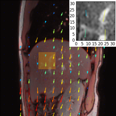 |
Comparison of liver motion measured by dynamic MRI and respiration signals obtained by an optical sensor
Julien Sénégas, Sascha Krueger, Daniel Wirtz, Ger Kersten, Mukul Rocque, Ivan Dimitrov, Andrea Wiethoff, Keith Hulsey, Ivan Pedrosa, Ananth Madhuranthakam
To monitor the breathing status of a subject and to synchronize data acquisition with respiration, external sensors, such as respiratory bellows, are routinely used. These sensors probe only the local breathing motion, and, hence, their signal quality can vary significantly depending on the individual subject’s physiology and morphology as well as the experience of the MR operator. Recently, optical sensors were proposed as an alternative. The purpose of this work was to compare the signals obtained by an optical sensor and by a pressure-based sensor with respect to their ability to represent the true liver motion during breathing.
|
|
2529.
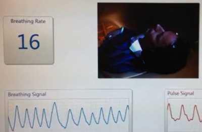 |
Optical unobtrusive physiology sensor for respiratory-triggered MRI acquisitions
Sascha Krueger, Julien Sénégas, Daniel Wirtz, Marek Bartula, Vincent Jeanne, Thiru Kanagasabapathi, Ger Kersten
A prototype in-bore camera-based physiology sensor was developed and applied for respiratory-triggered MR acquisitions on volunteers. The camera-based physiology sensor allows unobtrusive measurement of breathing activity by derivation of respiratory signals from video stream in real-time. The camera-based breathing sensor provided high quality breathing signal reliably under all tested circumstances of a volunteer study. The breathing signal quality was rated to be superior compared to the bellows in terms of SNR and signal characteristics. Potential false triggers where significantly reduced by the camera. The resulting image quality was on average superior when triggering off the camera compared to when triggering off the bellows.
|
|
2530.
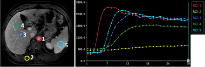 |
Towards efficient free breathing dynamic liver MRI using Cartesian k-space sampling with compressed sensing
Caizhong Chen, Shengxiang Rao, Guobin Li, Zhaopeng Li, Jiayu Zhu, Jinguang Zong, Xixi Wen, Mengsu Zeng
An efficient imaging method using Cartesian k-space undersampling with compressed sensing, and automatic detection of respiration was proposed to enable free-breathing dynamic liver imaging with a temporal resolution up to 1.0 sec/phase
|
|
2531.
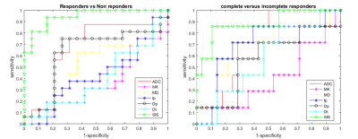 |
MR Imaging Perfusion and Diffusion analysis to assess preoperative Short Course Radiotherapy response in locally advanced rectal cancer: Standardized Index of Shape by DCE-MRI and Intravoxel Incoherent Motion derived parameters by DW-MRI
Roberta Fusco, Antonella Petrillo
Aim of this study is to determine the diagnostic performance of MR imaging for the assessment of tumor response after Short Course Radiotherapy (SCR) in patients with LARC using Standardized Index of Shape (SIS) obtained by DCE-MRI and using ADC, DKI and IVIM derived parameters obtained by DW-MRI. We demostrated that SIS is a hopeful DCE-MRI angiogenic biomarker to assess preoperative treatment response after SCR with delayed surgery and it permits to discriminate pCR allowing to direct surgery for tailored and conservative treatment.
|
|
2532.
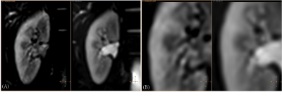 |
Validation of Reproducibility of Both Zoom Diffusion Imaging And Conventional Full Field of View Method in The Kidney Study
Hsuan Wen Yu, Feng Mao Chiu, Cheng Ping Chien, You Yin Chen
Diffusion Tensor Imaging (DTI) is a reliable tool for investigating renal microstructure and renal function, the imaging stability remains challenging. Recently, the image-quality improvement by zoom DTI technique (reduced Field-Of-View diffusion) is reported1. We scanned 10 healthy volunteers by this technique via the respiration-triggered acquisition, and we assessed different ROIs within the medulla and the cortex of the kidney. In this study, the reproducibility between different subjects in zoom DTI was more promising when compared to full-FOV DTI. More DTI scalars were compared between zoom and full-FOV DTI in cortex and medulla and these may be potential parameters to detect pathological changes in kidney.
|
|
2533.
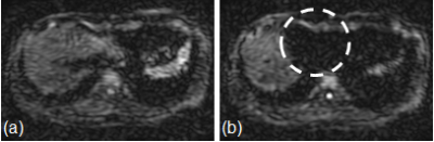 |
Improvement of ADC Precision in Left Liver Lobe by Weighted Averaging
Takashi Nishihara, Masahiro Takizawa, Ryuji Shirase, Takenori Murase, Masayuki Isobe
The signal intensity in liver DWI was induced by the cardiac motion. A new post-processing method using weighted image averaging is evaluated to mitigate these signal loss of pixels. The proposed method suppressed the signal loss and the precision of ADC was improved.
|
|
2534.
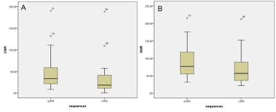 |
Comparison of quiet diffusion-weighted imaging with standard DWI in the abdomen: preliminary evaluation in the assessment of abdominal organs
Xianyun Cai, Guangbin Wang, Tianyi Qian , David Grodzki, Sai Shao, Cong Sun, Huihua Li
This study aimed to evaluate the diagnostic value of a quiet DWI (q-DWI) sequence in abdominal organs. Twenty-four patients underwent MR scans, including standard DWI and q-DWI. Quantitative and qualitative assessments regarding the signal-to-noise ratio (SNR), contrast-to-noise ratio (CNR), lesion conspicuity, the level of artifacts, and overall image quality, were measured. The qualitative rating by two radiologists shows that there were differences in lesion conspicuity, but these were not significant. The CNR and SNR of q-DWI were significantly higher than those of regular-DWI(r-DWI). For those patients who were intolerant to noise , the q-DWI technique could be more suitable.
|
|
2535.
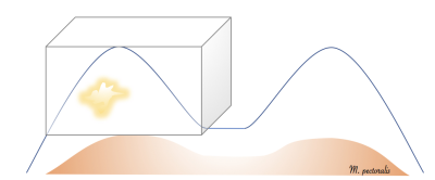 |
Computer aided cancer detection based on volumetric DCE-MRI analysis
Barbara Bennani-Baiti, Pascal Baltzer
While CAD is already routinely employed in conventional mammography, the data available on CAD cancer detection at MRI so far are limited and mostly include evaluation of lesion size, vascularization kinetics and tumor extent. Our data from two different approaches based on the percentage of voxel volume enhancement of either the ipsilateral breast alone or accounting for background parenchymal enhancement measured in the contralateral breast suggest both to be viable approaches for breast cancer detection with excellent reproducibility, that should be further developed.
|
|
Body: Liver
Traditional Poster
Body: Breast, Chest, Abdomen, Pelvis
Wednesday, 20 June 2018
| Exhibition Hall 2536-2552 |
16:15 - 18:15 |
 |
2536.
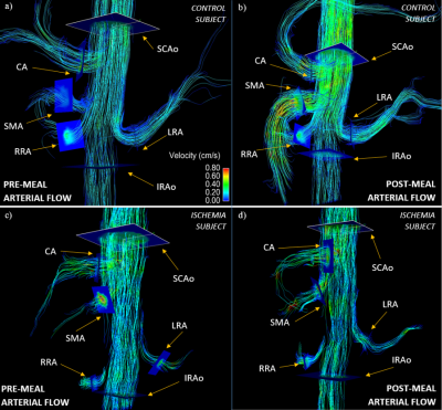 |
Non-Invasive Assessment of Mesenteric Hemodynamics with 4D flow MRI
Grant Roberts, Alejandro Roldan-Alzate, Christopher Francois, Oliver Wieben
Chronic mesenteric ischemia (CMI) is caused by inadequate blood flow to the intestines. This study investigates the use of 4D flow MRI to non-invasively assess the hemodynamics of the mesenteric circulation in patients with CMI and controls. Flow was measured in 9 vessels before and after meal challenges for 19 subjects suspected of CMI and 6 controls. Post-prandial flow increased significantly in the supraceliac aorta, superior mesenteric artery, superior mesenteric vein, and portal vein. The flow increase was drastically stunted in patients with CMI. This demonstrates the potential for 4D flow MRI in assisting the challenging diagnosis of CMI.
|
|
2537.
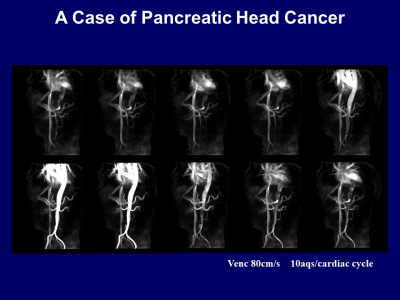 |
Hemodynamic Evaluations of Hepatic Vasculatures using 4D-PCA and MRFD for Liver Disease Assessment
Takeshi Yoshikawa, Katsusuke Kyotani, Yoshiharu Ohno, Shinichiro Seki, Yuji Kishida, Eiji Takeda
4D-PCA and MRFD can characterize liver vessels and measured WSSs provide additional information in liver disease assessments.
|
|
2538.
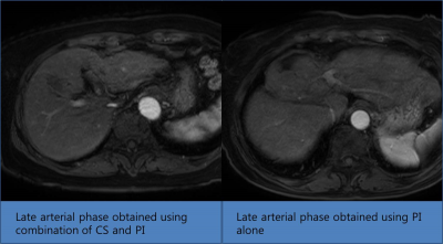 |
Combination of compressed sensing and two-dimensional parallel imaging can reduce the scan time for arterial phase image of gadoxetic acid enhance liver MR without degradation of image quality compared to parallel imaging alone
Dong Ho Lee, Hyo-jin Kang, Eun Ju Kim, Jeong Min Lee, Hwaseong Ryu
Using combination of compressed sensing and parallel imaging for arterial phase image acquisition in gadoxetic acid enhanced liver MR
|
|
2539.
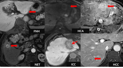 |
Ancillary imaging features for differentiation of hypervascular hepatic tumors on Gadoxetic acid-enhanced MR imaging
Hyun Jeong Park, Young Kon Kim
There are many types of hypervascular tumors that need to be differentiated from hepatocellular carcinoma (HCC) including focal nodular hyperplasia (FNH), hepatocellular adenoma (HCA), neuroendocrine tumor (NET), and intrahepatic cholangiocarcinoma (ICC). Since each tumor requires different treatment strategies, awareness and recognition of reliable imaging features that help precisely distinguish among these hypervascular tumors. Since these hypervascular tumors occasionally manifest overlapping imaging features, the accurate diagnosis of these tumors can still be challenging on MRI. Therefore, we conducted this study to determine ancillary imaging features that help differentiation of hypervascular hepatic tumors on gadoxetic acid-enhanced MRI.
|
|
2540.
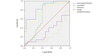 |
Analysis of the Value of Texture Feature Calculated From Contrast-Enhanced MR Images in Differentiating FNH and HCC
Zhuo Shi, Lizhi Xie, XinMing Zhao, Han Ou-Yang
For atypical FNH and HCC, conventional MR still has some limitations in differential diagnosis. Texture features can reflect the internal heterogeneity of the lesions. The purpose of this study was to find the texture features’ differences between FNH and HCC, in order to provide auxiliary diagnosis for the lesions which have difficulties in identification.
|
|
2541.
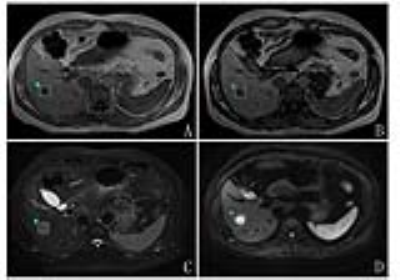 |
Precontrast MRI based radiomics in differential diagnosis of hepatocellular carcinoma and hepatic cavernous hemangioma using a logistic regression classifier
Jingjun Wu, Ailian Liu, Jingjing Cui, Lizhi Xie
Recently, radiomics has drawn attention in radiological research. Many scholars believe that radiomics may provide effective information for cancer diagnosis. In present study, we aim to distinguish hepatocellular carcinoma (HCC) and hepatic cavernous hemangioma (HCH) by precontrast MRI based radiomics and conclude that T2WI based radiomics using logistic regression classifier showed optimal diagnostic performance.
|
|
2542.
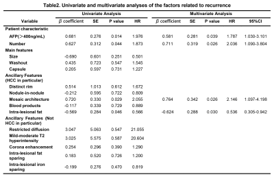 |
Liver Imaging Reporting and Data System Category 5: 3.0 T MR Predictors of Microvascular Invasion and Early Recurrence after Hepatectomy for Hepatocellular Carcinoma
Jingbiao Chen, Qungang Shan, Yao Zhang, Hao Yang, Ying Deng, Jun Wu, Bingjun He, Sichi Kuang, Claude B Sirlin, Jin Wang
Hepatocellular carcinoma (HCC) is the fifth most common malignancy worldwide. Tumor microvascular invasion (MVI) predicts early posthepatectomy HCC recurrence, but usually cannot be determined until the tumor is surgically removed and analyzed histologically. The capability preoperatively to predict MVI and early postsurgical recurrence would represent an advance by informing optimal selection of surgical candidates. Here we show that in combination with a AFP (a tumor biomarker), two Liver Imaging Reporting and Data System (LI-RADS) imaging features (mosaic architecture, corona enhancement) can predict MVI and three features (tumor number, mosaic architecture, absence of intralesional fat) can predict early recurrence.
|
|
2543.
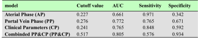 |
Validation of a radiomics nomogram for preoperative prediction of early recurrence in hepatocellular carcinoma less than 5cm
Xiaohong Ma, Jianyong Zhu, Shuang Wang, Meng Liang, Bing Feng, Jiangfen Wu, Chunwu Zhou, Xinming Zhao
A high early recurrence (ER) (≤ 1 year) rate of hepatocellular carcinoma (HCC) remains a significant concern. It is an important problem to find a powerful preoperative tool for predicting ER. This study aimed to development and validation of a Radiomics Nomogram for Preoperative Prediction ER in hepatocellular carcinoma less than 5cm. We found that the textural signature was a significant predictor for ER in HCC, and Radiomics nomogram performed better for preoperative prediction of ER in HCC.
|
|
2544.
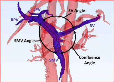 |
The Effects of Helical Flow Patterns, Confluence Angle, and Flow Distribution in the Portal Vein
David Rutkowski, Scott Reeder, Alejandro Roldán-Alzate
The hemodynamics of the liver in normal and diseased conditions are not fully understood. In this study, 4D flow MRI and computational modeling were used to analyze the effects of portal venous flow patterns at the spleno-mesenteric confluence. Specifically, the geometric configuration of the confluence on intra-hepatic portal circulation in healthy subjects and cirrhotic patients before and after a meal challenge was analyzed. Significant correlations between flow distribution, helicity, geometry, and flow patterns were observed, and differences between normal and pathological flow were also characterized.
|
|
2545.
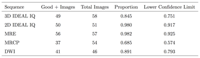 |
MRI in the evaluation of liver involvement in pediatric patients with cystic fibrosis
Katherine Carey, Scott Reeder, Mark Kliewer , R. Guillerman , Diego Hernando, Scott Nagle
In this prospective study of 15 pediatric cystic fibrosis subjects, we show that non-sedated comprehensive quantitative liver MRI is feasible. Furthermore, free-breathing 2D IDEAL IQ outperformed breath-held 3D IDEAL IQ in both image quality and repeatability of proton density fat fraction. Short term 1-2 week repeatability of MR elastography stiffness measurements were comparable with ultrasound elastography. Quantitative liver MRI in the pediatric cystic fibrosis population offers the ability to visualize structure and quantify hepatic steatosis and liver stiffness in a single exam.
|
|
2546.
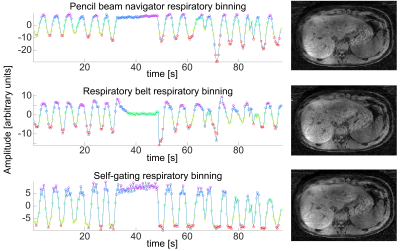 |
Respiratory binning showdown; self-gated, respiration belt or pencil beam?
Paul Heer, Anne-Sophie Schelt, Jasper Schoormans, Gustav Strijkers, Bram Coolen, Jurgen Runge, Jaap Stoker, Aart Nederveen
In body MR many acquisitions respiratory motion correction is of great importance. In this study we compared self-gated motion-state binning to binning by both the pencil-beam navigator and a respiration belt by looking at the resulting image quality for each method. A 3D T1-weighted radial stack-of-stars turbo field echo (TFE) was acquired in three volunteers. The self-gated respiratory motion binning outperformed the other two methods in image quality and smoothness between the respiratory states. More subjects should be included in the study but for now it can be concluded that self-gating would be the preferred method of respiratory binning.
|
|
2547.
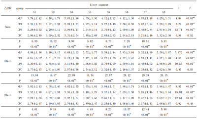 |
Evaluating of segmental liver function by using Gd-EOB-DTPA-enhanced MRI
Jiyun ZHANG, Jian LU
The aim of this study is to investigate the value of Gd-EOB-DTPA-enhanced MRI in evaluating segmental liver function. Statistical analysis was used to evaluate the relationship between the ?LMR of each liver segment and liver function,as well as the?LMR of different liver segments. Our quantitative study demonstrated that Gd-EOB-DTPA intake into hepatocytes was strongly affected by liver function .The segmental liver function can be evaluated via Gd-EOB-DTPA-enhanced MRI and calculation of the ?LMR may be a novel optional.
|
|
2548.
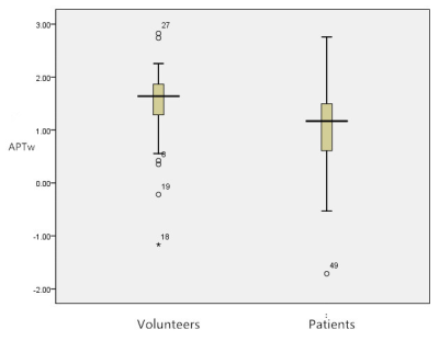 |
Amide Proton Transfer (APT) MR imaging and Magnetization Transfer (MT) MR imaging of liver cirrhosis: a clinical feasibility study
Xin Chen, Guangbin Wang, Jinyuan Zhou, Yi Zhang, Weibo Chen, Huihua Li
This study aimed to demonstrate the feasibility of the APT and MT MR imaging in depicting the liver cirrhosis at 3.0T. We compared MTR and APTw values in 11 healthy livers and 8 liver cirrhosis. The patients with liver cirrhosis showed a lower APTw values than healthy volunteers indicating APT imaging detectable mobile protein levels in the liver tissues. The patients with liver cirrhosis exhibited a higher MTR values than the healthy volunteers. Liver cirrhosis exhibited a significantly higher MTR, indicating indicating a higher concentration of biochemistry components in liver cirrhosis. We have shown that it is clinically feasible to perform APT and MT MR imaging of liver cirrhosis.
|
|
2549.
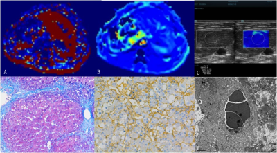 |
Vessel Size Imaging for Liver Fibrosis Staging Based on Dynamic Susceptibility Contrast Using SE/GRE-EPI Sequence:Comparison with US Elastography and Histopathological Correlation
Ruo-kun Li, Fu-hua Yan, Wei-bo Chen, He Wang
The study investigated the value of vessel size imaging (VSI) based on dynamic susceptibility contrast using SE/GRE-EPI sequence for liver fibrosis staging, compared with US elastography and correlated with histopathological results. We found that VVF and Nu value based on VSI were independent predicative factors of liver fibrosis (R2=0.566,P=0.002). They had correlation with hepatic sinusoidal structures including parenchymal area (PA), sinusoidal area (SA), hepatocyte area (HA), sinusoidal perimeter (SP), SA/SP ratio, SA/SP index, and HA/SP index. Microvessel density (MVDdensity) and area (MVDarea). VSI has potential for liver fibrosis staging with good diagnostic capability similar to US elastography.
|
|
2550.
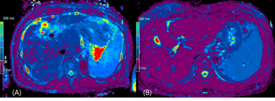 |
Model-based volumetric T2 mapping of the liver
Jeong Hee Yoon, Yohan Son, Berthold Kiefer, Jeong Min Lee
T2 relaxation time estimation is able to aid liver tissue characterization by providing quantitative information of the tissue.
|
|
2551.
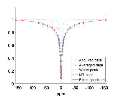 |
Magnetisation transfer in human liver and kidney through acquisition of the z-spectrum
Andrew Carradus, Simon Shah, Olivier Mougin, Caroline Hoad, Penny Gowland
This study explores the feasibility of measuring magnetisation transfer in both the liver and kidney through acquisition of a full z-spectrum. In this study we developed a protocol to reduce artefacts from respiration and blood flow pulsatility and measured relative amounts of MT in both the liver and kidney medulla by modelling MT with a super-Lorentzian lineshape and fitting to the acquired spectrum. This will be relevant in monitoring fibrosis in abdominal organs.
|
|
2552.
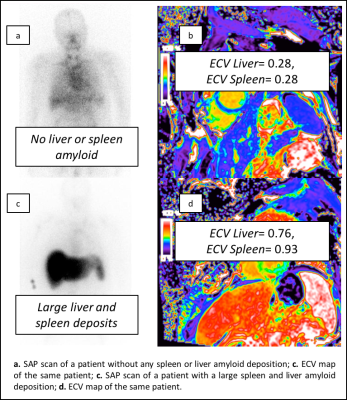 |
Expanding the limits of cardiovascular MR: amyloid detection in the liver and spleen
Michele Boldrini, Andrea Baggiano, Ana Martinez-Naharro, Tushar Kotecha, Tamer Rezk, Daniel Knight, James Moon, Peter Kellman, Julian Gillmore, Philip Hawkins, Marianna Fontana
In this study we evaluated the utility of bolus-only ECV maps in extra-cardiac AL amyloidosis by comparing it with SAP scintigraphy findings in liver and spleen. These two techniques where performed in a large prospective cohort of patients with suspected systemic AL amyloidosis and where compared in terms of (1) diagnostic accuracy in liver and spleen amiloidosis; (2) quantification of the liver and spleen amyloid deposits. This was done using a standard acquisition for cardiac studies, with no extra image acquisition or optimization for hypochondriac regions.
|
|
Prostate
Traditional Poster
Body: Breast, Chest, Abdomen, Pelvis
Wednesday, 20 June 2018
| Exhibition Hall 2553-2577 |
16:15 - 18:15 |
|
2553.
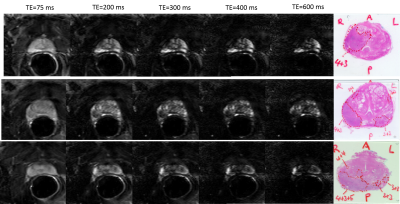 |
Optimization of the Contrast-to-noise Ratio between Malignant and Non-malignant Prostate Tissue in T2-weighted MRI.
Shirin Sabouri, Silvia Chang, Edward Jones, S. Goldenberg , Peter Black, Piotr Kozlowski
T2W imaging is an important sequence in the PIRADSv2 guideline for scoring prostatic lesions. The apparent contrast between malignant and non-malignant tissues on T2W images depends on the time of echo (TE). In this study we have investigated the effect of TE on the contrast-to-noise ratio (CNR) between malignant and non-malignant tissues. We have acquired and analyzed T2W data from 12 patients. Our results show that CNR increases abruptly for TEs between 25 and 175ms. After CNR reaches its maximum at 175ms it gradually decreases. Our findings may be used toward improvement of T2W protocols for diagnosis of prostatic carcinoma.
|
|
2554.
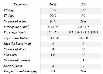 |
The Influence of Temporal Resolution on the Diagnostic Accuracy of DCE-MRI in Evaluation of Prostate Cancer.
Shirin Sabouri, Silvia Chang, Edward Jones, S. Goldenberg , Peter Black, Piotr Kozlowski
DCE-MRI is widely used for cancer detection, and is a part of PIRADS v2 guideline for scoring prostatic lesions. Diagnostic accuracy of DCE-MRI may depend on the rate of temporal sampling. In this study we have investigated the relationship between the rate of temporal sampling of DCE-MRI and the accuracy of detection of prostatic carcinoma. We have acquired and analyzed DCE-MRI data from 15 patients. Our results show that the accuracy of DCE-MRI in detection of prostatic carcinoma is not affected by sampling rates between 3.4 to 13.6 seconds.
|
|
2555.
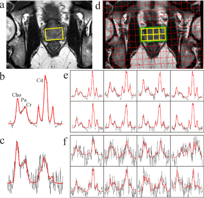 |
Magnetic resonance spectroscopy of localized prostate cancer: assessment of antitumor effects of intra-prostatic hormone deprivation therapy
Jan Weis, Michael Häggman, Sam Ladjevardi, Niklas Axen, Carl-Gustaf Gölander
A novel controlled release formulation based on calcium sulphate as drug carrier loaded with the antiandrogen 2-hydroxiflutamide as the active pharmaceutical agent was injected locally into the prostate in patients with prostate cancer. Single-voxel and 2D MRSI using a surface coil were used to investigate the treatment efficiency. The results demonstrate usefulness of both MRS techniques to detect metabolic atrophy caused by long-term local hormone-deprivation therapy. The presence of metabolic atrophy reflects the antitumor effects of the study drug formulation 6 weeks after the intraprostatic injections.
|
|
2556.
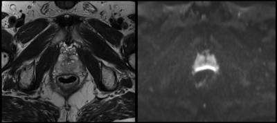 |
Effect of Rectal Gas on Susceptibility Artifact in Prostate DWI
Eun Bin Lee, Ely Felker, David Lu, Kari Sorge, Kyunghyun Sung
Diffusion-weighted imaging (DWI) is a key component of multi-parametric prostate MRI; however, DWI is prone to susceptibility artifact occurring in the peripheral zone, where 70% of tumors are found. The purpose of this study was to qualitatively and quantitatively assess the effect of rectal gas on the presence of this artifact. The study found that in cases with no rectal gas (<2cm3), image quality is excellent as a rule. When more than 2cm3 of gas is present, a range of image quality is seen that is not correlated to the amount of gas present.
|
|
2557.
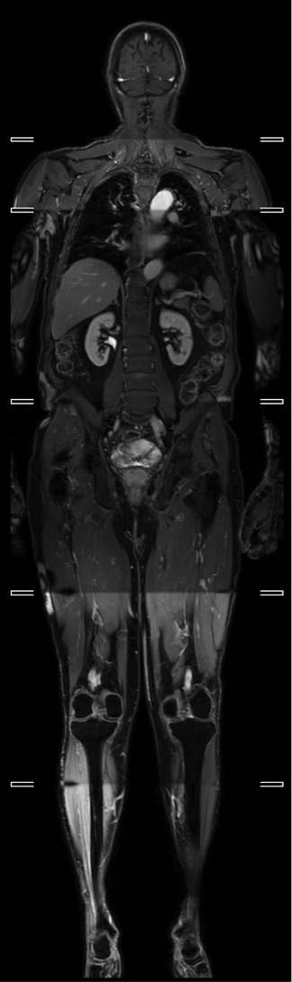 |
Whole-body MRI for prostate cancer at primary staging: interobserver concordance, diagnostic accuracy and protocol optimisation
Edward Johnston, Arash Latifoltojar, Harbir Sidhu, Elisenda Bonet-Carne, Magdalena Sokolska, Alan Bainbridge, Shonit Punwani
Whole body (WB) MRI is developing as a cancer staging platform in primary prostate cancer, although has not yet been adopted into clinical practice. In this study, we show that WB-MRI provides high levels of interobserver concordance, intermodality concordance and diagnostic accuracy for both nodal and metastatic bone disease, with higher levels of sensitivity than BS for metastatic disease, and similar performance to PET/CT. We also show that T2W and post contrast mDixon have no additive diagnostic value above T1W and DWI alone.
|
|
2558.
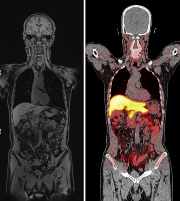 |
Image quality of WB-MRI in staging recurrent prostate cancer: a multicentre, multinational, multivendor, multiscanner study.
Edward Johnston, Alan Bainbridge, Glenn Bauman, Sue Chua, Ian Davis, Rod Hicks, Ur Metser, Frederic Pouliot, Andrew Scott, Jonathan Thiessen, Nina Tunariu, Andrew Weickhardt, Louise Emmett, Shonit Punwani
Whilst whole body (WB) MRI offers substantial promise in cancer staging, considerations regarding image quality are lacking in the literature, yet are essential for the effective delivery of the technique. Here we report the image quality of WB-MRI in 86 patients with suspected biochemical recurrence in prostate cancer in a trial carried out over 3 continents (Australia, America and Europe). We show that the image quality of WB-MRI varies substantially between anatomical sites and centres, particularly for diffusion-weighted sequences, which emphasizes the need to optimise sequences carefully prior to establishing a WB-MRI practice.
|
|
2559.
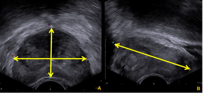 |
Comparison of Prostate Volume Measured by Transrectal Ultrasonography and Magnetic Resonance Imaging with the Actual Prostate Volume Measured after Radical Prostatectomy
Sung Bin Park, Haesun Choi
A determination of prostate gland volume facilitates an assessment of prostate disorders and, for prostate cancer, in conjunction with other parameters, can help predict the pathologic stage of disease, offer insights into the prognosis, and help predict treatment response Prostate volume can also be used for calculating prostate-specific antigen density (PSAD) when selecting active surveillance candidates.The measuring the volume of prostate removed by radical prostatectomy as performed in the present study may be an appropriate way to assess the actual prostate volume even though it may be cancerous. The present study aimed to compare the prostate volume, as measured by TRUS and by MRI, with that of the actual prostate volume measured after a radical prostatectomy.
|
|
2560.
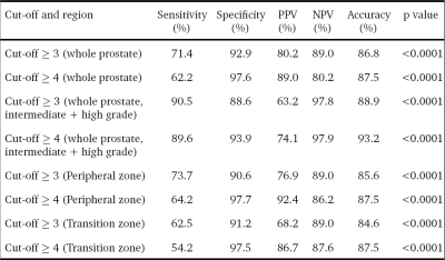 |
Performance of PIRADSv2 =3 and =4 scores as cut-offs for the detection of prostate cancer
Chandan Das, Vivek Lanka, Sanjay Sharma, Rishi Nayyar, Virendra Kumar
We report a prospective evaluation of prostate imaging reporting, archiving and data system version 2 (PIRADSv2) for multiparametric MRI (mpMRI) taking histopathology of radical prostatectomy specimens as reference standard. PIRADSv2 for mpMRI is easy to apply for detection of cancer of prostate (CaP) at optimal cut-offs of ≥3 for cancer as a whole and ≥4 for intermediate and high grade cancers. It is an accurate system to diagnose clinically significant disease. 26 patients having a biopsy-proven CaP, were investigated at 3.0T using mpMRI, followed by radical prostatectomy within 1 month. Gleason grade group from radical prostatectomy specimens and ROC curve analysis was used to determine the accuracy for cut-offs for scores of PIRADSv2.
|
|
2561.
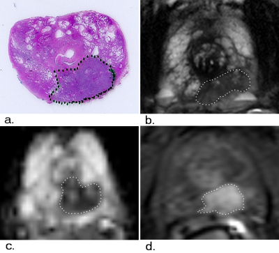 |
Multi-parametric MRI evaluation of prostate cancer volume: correlation with whole mount pathology
Chongpeng Sun, Aritrick Chatterjee, Ambereen Yousuf, Tatiana Antic, Scott Eggener, Gregory Karczmar, Aytekin Oto
This study compared prostate cancer volume determined on different multi-parametric MRI sequences: T2W imaging, ADC map and DCE-MRI with whole mount pathology in 17 patients. Tumor volumes were measured on T2W images, ADC maps and DCE-MRI by 2 radiologists and compared with reference standard volume measured from pathology. While lesion volume estimated using mpMRI sequences showed good correlation with pathology, T2W and ADC significantly underestimated, whereas DCE-MRI showed no significant difference. Therefore, DCE-MRI is the most effective sequence for estimating PCa volume with the highest accuracy compared to T2W-imaging and ADC maps and has similar good correlation and precision.
|
|
2562.
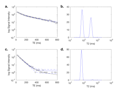 |
A comparison of biexponetial fitting and spectral modelling methods for T2 mapping of prostate cancer
Dominic Carlin, Matthew Orton, Veronica Morgan, David Collins, Nandita deSouza
Spectral modelling and model fitting were compared for quantitative T2 mapping of the prostate. 32-echo data were acquired from 11 patients with biopsy-proven prostate cancer at 3T. There was excellent correlation between the two approaches for estimates of T2-short, T2-long and luminal water fraction (r=0.96, 0.71, 0.94 respectively). Luminal water fractions were significantly higher in normal peripheral and transition zones using the model fitting approach (P = 0.04 and <0.01 respectively), but were comparable in tumor. The larger quantitative difference between tumour and normal tissue could mean model fitting is superior for qualitative assessment in prostate cancer.
|
|
2563.
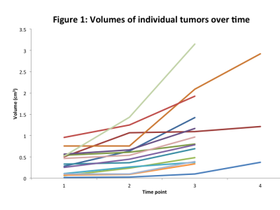 |
Characterising prostate tumour growth patterns in men on active surveillance: linking ADC features to growth kinetics
Dominic Carlin, Veronica Morgan, Chris Parker, Nandita deSouza
Tumor growth kinetics of low-risk prostate cancer in 15 men managed by active surveillance with size increase on repeat MRI were correlated with ADC histogram metrics. Measurements were made over 3 time-points at least 1 year apart (mean 3.6 ± 0.95 years). Median growth was 23.1% in the first interval and 49.8% in the second. ADC reduced over time. Accelerated growth during the second time interval correlated with the increase in interquartile range (r=0.6, p=0.02) and shift to more positive skew (r=-0.56, p=0.03) seen during the first time interval, suggesting that increasing heterogeneity and reducing ADC may signal accelerated growth.
|
|
2564.
|
Comparison of multiparametric MRI and MRI-ultrasound fusion guided biopsy for prostate cancer diagnosis
Renee Cattell, James Kang, Sarah Dacosta, Matthew Barish, Howard Adler, Massimiliano Spaliviero, Marlene Zawin, Haifang Li, Tim Duong
High false-positive rates of prostate cancer diagnosis techniques have resulted in unnecessary biopsies and increased costs of care. This study compared the prostate cancer diagnosis by multiparametric MRI and MRI-ultrasound fusion guided biopsy at our institution, with the ultimate goal of improving MRI diagnosis of prostate cancer. At our institution, multiparametric MRI PI-RADS scores and MRI-ultrasound fusion guided biopsy Gleason scores agreed 46-57% of the time which falls within the ranges in literature.
|
|
2565.
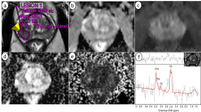 |
Multi parametric magnetic resonance imaging for the detection of prostate cancer: combination of T2-weighted, diffusion tensor imaging and magnetic resonance spectroscopic imaging
Neda Gholizadeh, Peter Greer, John Simpson, Jim Denham, Peter Lau, Saadallah Ramadan
The aim of this study was to determine the diagnostic performance of mp-MRI using T2WI, DWI, DTI and MRSI for prostate cancer patients with various Gleason scores. mp-MRI using T2WI, DWI, DTI and MRSI on 12 prostate cancer patients. The area under receiver operating characteristic (ROC) curve of T2WI+DWI and T2WI+DWI+DTI+MRSI images were generated and used to evaluate the performance of mp-MRI for discriminating cancer and healthy regions. Our results suggest that mp-MRI using DWI, DTI and MRSI in combination with structural T2WI improve performance for discrimination of cancer and healthy prostate tissues.
|
|
2566.
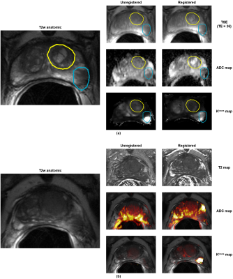 |
A framework for intensity-based affine registration of multiparametric prostate MRI via mutual information and genetic algorithms
Ethan Leng, David Porter, Andrew Larson, Xiaoxuan He, Benjamin Spilseth, Gregory Metzger
An image registration framework was developed to perform 3D, affine, intensity-based co registration of multiparametric MRI series using mutual information as the similarity metric. The proposed methods include corrections to compensate for the effects of an endorectal coil, which is commonly used in prostate MRI. Experiments to characterize the registration method demonstrate that it is theoretically accurate to within 1.0 mm (when estimating the translation component). Qualitatively, significant improvements are seen in the co-localization of parametric maps with the anatomic images. The proposed framework may readily be integrated into a CAD system for prostate cancer detection.
|
|
2567.
|
Radiomics assessment of prostate cancer grade using texture features from DWI,T1WI and T2WI
ZHANG LI, ZHANG XIAOLING, ZHUO ZHIZHENG
The purpose of this study was to investigate the value and diagnostic efficiency of DWI,T1WI and T2WI using texture analysis for discriminating the gleason scores of prostate cancer. The results of this study indicate that texture analysis may provide a new method for Gleason classification of prostate cancer. A radiomics model of textural features from T2WI and ADC maps have a good diagnostic accuracy in patients of a prostate cancer. Quantitative textural analysis may help distinguish low cancers form high- or intermediate-grade cancer with high sensitivity and moderate specificity.
|
|
2568.
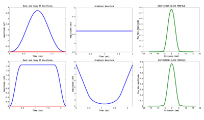 |
Prostate imaging at 7T using multi-acquisition SSFP with parallel transmission and low SAR RF pulses.
Benjamin Knowles, Arthur Magill, Mark Ladd
Prostate imaging at Ultra High Field suffers from SAR limitations and B1 inhomogeneity. This is especially effects Turbo Spin Echo for T2 weighted imaging, although this contrast holds significant clinical value. Steady-State Free Precession offers a T2/T1 contrast with lower flip angles and potentially lower SAR. In this study, multi-contrast CISS imaging was investigated for prostate imaging. VERSE pulses were implemented to reduce SAR. Results show good contrast in the prostate between the peripheral and transitional zones, comparable to that observed in TSE images. The use of VERSE pulses greatly reduces SAR and maintains contrast.
|
|
2569.
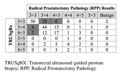 |
Evaluating the Role of PIRADS V2 and TRUSgBX for Improving Detection of Clinically Significant Prostate Cancer at Radical Prostatectomy
Alireza Ziaei, Francesco Alessandrino, Mark Vangel, Tina Kapur, Clare Tempany, Fiona Fennessy
The aim of this retrospective study was to determine a role for PIRADS V2 in conjunction with TRUSgBX to predict the presence of clinically significant prostate cancer (csPCa) in treatment naïve men with pathology-proven prostate cancer who underwent TRUSgBX, followed by 3T mp-MRI prostate, and subsequently underwent RP. Our findings suggest that adding PIRADS V2 assessment to TRUSgBX improves the prediction of final pathology for presence of indolent disease and csPCa, and may help alleviate the rate of upgrading at RP.
|
|
2570.
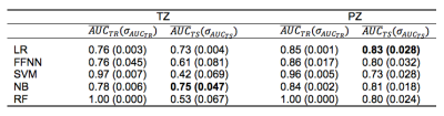 |
mpMRI-based Machine-Learning Classifier Comparison for Gleason 4 Pattern Detection in Transition Zone and Peripheral Zone Prostate Lesions
Michela Antonelli^, Edward Johnston^, Sebastien Ourselin*, Shonit Punwani*
Multi-parametric MRI (mpMRI) can be used to non-invasively predict the presence of a Gleason 4 pattern in transition zone (TZ) and peripheral zone (PZ) prostate cancers. Here the performance of five machine-learning classifiers, which use mpMRI and clinical features, were compared. Analysis included a five-fold cross validation and a temporally separated validation to prove the generalisability of the classifiers. The results showed that PZ models can predict the presence of a Gleason 4 pattern better than TZ models. The statistically better PZ classifier is a linear regression model while for TZ the best classifier is Naïve Bayes model.
|
|
2571.
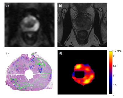 |
Ex vivo ultra-high-field 9.4-Tesla magnetic resonance elastography (MRE) in comparison to whole-mount pathology for improved prostate cancer diagnostics.
Rolf Reiter, Shreyan Majumdar, Steven Kearney, Thomas Royston, Brandon Caldwell, Rong-Wen Tain, Kejia Cai, Cristian Luciano, Andre Kajdacsy-Balla, Winnie Mar, Michael Abern, Dieter Klatt
Despite the success of multiparametric magnetic resonance imaging (mpMRI) for the assessment of prostate cancer, it suffers from limitations such as a moderate inter-reader reliability and sub-optimal diagnostic accuracy. This is the first study for the assessment of 6 human prostate specimens without pathology fixation or prior radiation therapy using ex vivo 9.4-Tesla magnetic resonance elastography (MRE). Using whole-mount pathology as a reference, preliminary results show a sensitivity and specificity of 86 % and 52 %, respectively. MRE has the potential to improve the differentiation of benign prostatic hyperplasia nodules from malignant lesions, which is a known limitation of mpMRI.
|
|
2572.
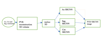 |
Motion-tolerant super-resolution reconstruction from multi-stack MR data
Sachin Jambawalikar, Daniel Litwiller, Michael Liu, Rami Vanguri, Simukayi Mutasa, Zhengchao Dong, Hiram Shaish
Image super resolution reconstruction (ISRR) is a technique that may be useful for generating fast, motion tolerant 3D reconstructed images from multi stack data. We provide initial results of a multi-step ISRR approach using patch-to-volume reconstruction(PVR) followed by a slice-by-slice convolutional neural network to further improve spatial resolution. Our methods provide improved measures of peak-SNR, and could be used to rapidly generate 3D volumes from multiple 2D stacks in fetal and abdominal imaging where constant motion requires short scan times as well as in pelvic imaging where high SNR requirements lead to long scan times and motion artifact. Motion artifact is a significant obstacle in these MRI applications resulting in image quality degradation and potentially limited diagnostic ability.
|
|
2573.
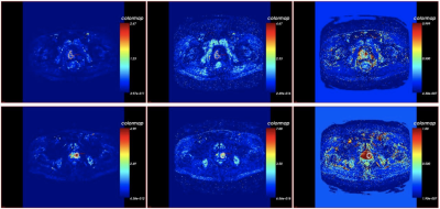 |
Correlation of Perfusion Parameters Between Intravoxel Incoherent Motion Diffusion-Weighted Imaging and Texture Analysis of Dynamic Contrast Enhancement Imaging for Diagnosis of Prostate Cancer in Central Zone and Hyperplasia
Dan Guo, Ailian Liu, Lihua Chen, Lizhi Xie
This work assessed the diagnostic value of intravoxel incoherent motion diffusion-weighted (IVIM-DWI) and texture analysis of dynamic contrast enhancement (DCE) in prostate cancer in central zone(CZ) and hyperplasia and the correlation of perfusion parameters of them.
|
|
2574.
|
Comparison of Radiomics and Quantitative ADC Measurements of Prostate PI-RADS v2 Lesions to Prospective Radiologist Performance
David Bonekamp, Simon Kohl, Manuel Wiesenfarth, Patrick Schelb, Jan-Philipp Radtke, Michael Götz, Philipp Kickingereder, Kaneschka Yaqubi, Bertram Hitthaler, Nils Gählert, Tristan Kuder, Fenja Deister, Martin Freitag, Markus Hohenfellner, Boris Hadaschik, Heinz-Peter Schlemmer, Klaus Maier-Hein
Multiparametric MRI (mpMRI) has recently seen further standardization by introduction of the PI-RADS version 2 system. mpMRI/transrectal ultrasound (TRUS)-guided fusion biopsies have demonstrated ability to closely match the histopathology seen after radical prostatectomy. Radiomics is a novel approach to extract a large number of quantitative features from medical imaging and combination with machine learning has demonstrated potential in the classification of mpMRI of the prostate. Here, we aim to compare state of the art radiomics and machine learning with ADC measurements,and prospective radiologist assessment using PI-RADS version 2 (PIRADSv2) in the evaluation of cancer suspicious lesions of the prostate.
|
|
2575.
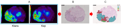 |
Voxel Level Radiologic–Pathologic Validation of DCE-MRI with ISUP Grade in Prostate Cancer
Qing Zhang, Xiaoyu Lv, Chengwei Zhang, Qinglei Zhang, Ming Li, Yao Fu, Jun Xie, Jiangfen Wu, Bing Zhang, Hongqian Guo
The biggest challenge in patients with newly diagnosed PCa is shifting from cancer detection or staging alone to identifying them with aggressive disease. The PI-RADS v 2 recognizes the role of DCE-MRI is limited but is essential. This work presented a radiology pathology correlation framework that enabled identification of promising in vivo DCE MRI markers of PCa risk at voxel level. The relationship between ISUP grade and DCE-MRI (Ktrans and Kep) suggests that it may be used as a component of active surveillance to noninvasively detect high-grade PCa and affect staging and treatment.
|
|
2576.
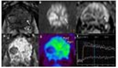 |
In-bore MR guided prostate biopsy using multiparametric MRI to avoid unnecessary biopsies
Sujeet Mewar, Sanajy Sharma, Ekta Dhamijia, Rupsa Bhattacharjee, Sanjay Thulkar, Pradeep Kumar, Virendra Kumar, Senthil Kumaran, Siddhartha Gupta, Rajeev Kumar, Naranamangalam Jagannathan
We report the results of the pilot study carried out using multiparametric (mp) MRI and in-bore MRI-guided prostate biopsy for detection of PCa to reduce the number of unnecessary biopsies. 11 patients were recruited based on prostate specific antigen > 4ng/ml and abnormal digital rectal examination. In-bore MRI targeted lesions with high PIRADS scores (3 to 5) were correlated with the histopathological findings. The average ADC in PCa patients was significantly lower than the prostatitis and BPH patients. Out of 11 patients, 3 showed adenocarcinoma, 5 prostatitis and 3 BPH.
|
|
2577.
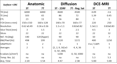 |
Multiparametric MRI methods development for clinical prostate imaging at 7T
Gregory Metzger, Ryan Kalmoe, Arcan Erturk, Xiaoxuan He, Sudhir Ramanna, Ethan Leng, Christopher Warlick, Benjamin Spilseth
The advantages of increased SNR drive the spread of applications to 7T. While methods and hardware continue to improve, the potential to perform a full multiparametric exam exploiting the advantages of ultrahigh magnetic fields becomes possible but has yet to be investigated. We explore a full MRI exam including anatomic, diffusion and dynamic contrast enhanced MRI (DCEMRI) methods at 7T and compare them against 3T acquisitions in a patient population with various coil configurations: surface coils and surface combined endorectal coils.
|
|
Body: Liver Fat & NASH
Traditional Poster
Body: Breast, Chest, Abdomen, Pelvis
Wednesday, 20 June 2018
| Exhibition Hall 2578-2587 |
16:15 - 18:15 |
|
2578.
 |
In Vivo MRI Monitoring of the Induction and Reversal of Non-Alcoholic Steatohepatitis in a Rat Model
Amy Herlihy, Antigoni Ekonomou, Camilla Simmons, Matteo Milanesi, Catherine Kelly, Po-Wah So
Steatosis and steatohepatitis (NASH) may be attenuated by calorie restriction and/or exercise if intervention is early enough. However, steatosis/NASH is generally asymptomatic, and when clinical signs are observed, simple lifestyle interventions are no longer effective. Thus, there is a real clinical need to detect steatosis/NASH early but also to monitor putative therapies. A methionine-choline-deficient diet leads to NASH in rats, that is readily reversible when rats are placed back on a methionine-choline replete diet. This model will be used to assess the ability of MRI to detect the induction of, and reversal of steatosis, in vivo.
|
|
2579.
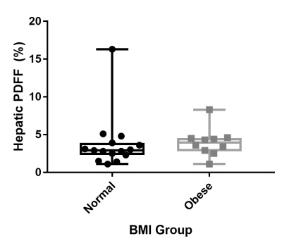 |
Non-alcoholic Fatty Liver Disease Assessment in Obese and Non-obese Pregnant Women with Water-Fat MRI
Stephanie Giza, Simran Sethi, Takashi Hashimoto, Barbra de Vrijer, Charles McKenzie
Proton density fat fraction (PDFF) was used to assess fatty liver of pregnant women with normal and obese body mass indexes (BMI). No significant difference was found in the mean hepatic PDFF between the two groups (p=0.28). One normal BMI woman and one obese woman had elevated hepatic PDFF measurements.
|
|
2580.
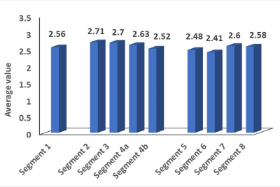 |
Validation of magnetic resonance imaging-proton density fat fraction for hepatic fat content in healthy Asian population
Sonal Krishan
The primary purpose of this work was to determine the precision of clinical MR imaging-PDFF hepatic fat quantification, to look at spatial heterogeneity in all the Couinad segments, to establish normative data and least significant change in Indian population. Our study has shown that there is no systematic or significant difference in the right versus left lobe, or any of the liver segments in patients with grade 0 steatosis. The least significant change of liver fat that can be measured reliably using MR imaging-PDFF is 2.1%. Mean hepatic fat content calculated by MR imaging-PDFF is 2.89% (95%CI, 1% - 6.8%) in normal Indian population.The current study is the first study determining normative data of hepatic fat content in histologically proven grade 0 steatosis population from India.
|
|
2581.
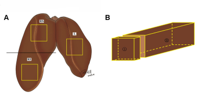 |
Validation of goose liver fat measurement by CSE-MRI with biochemical extraction as reference
Li Xu, Yangyang Duanmu, Xiaoqi Wang, Glen Blake, Peng Wang, Manling Zhang, Chao Wang, Xiaoguang Cheng
This study aimed to validate chemical shift encoded magnetic resonance imaging (CSE-MRI) to assess hepatic steatosis. Twenty-two geese with a wide range of hepatic steatosis were collected, and proton density fat fraction by MRI (MRI-PDFF), biochemical triglyceride content, and histology were performed within the left lobe, upper and lower half of the right lobe of the geese livers. MRI correlated highly with chemical extraction (r = 0.949 (p < 0.001)). Chemically extracted triglyceride was accurately predicted by MRI-PDFF (Y = -1.8 + 0.773?X). In conclusion, CSE-MRI measurement of goose liver fat was accurate and reliable compared with biochemical measurement.
|
|
2582.
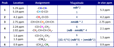 |
Relationship between Proton Density Fat Fraction and Liver Triglyceride Composition estimated by 1H MR Spectroscopy
Gavin Hamilton, Alexandra Schlein, Adrija Mamidipalli, Yesenia Covarrubias, Jonathan Hooker, Walter Henderson, Ethan Sy , Jennifer Cui, Rohit Loomba, Claude Sirlin
Liver triglyceride composition was estimated using 1H MRS and compared to MRS estimated Proton Density Fat fraction (PDFF) to see if liver fat composition changes with PDFF. STEAM liver spectra were acquired in 263 adult subjects at 3 Tesla using breath-held, long-TR, multi-TE MRS to estimate PDFF and respiratory gated water-sated single TE MRS to estimate triglyceride composition. There is a significant change in the triglyceride composition of liver with changing PDFF, with the liver fat becoming more saturated as PDFF increases.
|
|
2583.
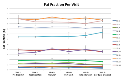 |
Diurnal Variation of Liver Fat Concentration
Timothy Colgan, Andrew Van Pay, Samir Sharma, Scott Reeder
Abnormal accumulation of intracellular triglycerides (hepatic steatosis) is the earliest and hallmark feature of nonalcoholic fatty liver disease (NAFLD). Confounder-corrected quantitative chemical-shift encoded MRI (CSE-MRI) is an accurate, precise and reproducible biomarker of hepatic steatosis as quantified by the proton density fat fraction (PDFF). However, the effect of meals and diurnal variability has not been established. In this study, we examined the variability of PDFF measurements resulting from meals, diurnal variation and between visits on different days. This study demonstrates that CSE-MRI liver fat estimation is not significantly affected by diurnal changes.
|
|
2584.
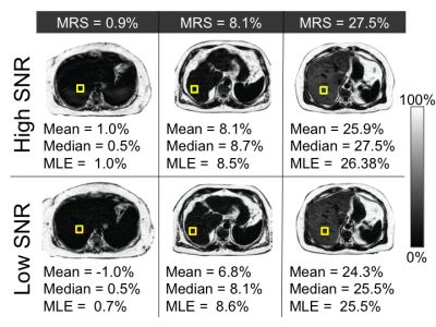 |
Effect of Signal to Noise Ratio and Estimator Type on Bias of Hepatic Proton Density Fat Fraction Measurement
Edward Lawrence, Nathan Roberts, Diego Hernando, Scott Reeder
Proton-density fat-fraction (PDFF) is typically measured by calculating the mean PDFF value within a region of interest (ROI). However, the mean estimator has been shown to result in bias when signal-to-noise ratio (SNR) is low. This work characterizes the accuracy of median and maximum likelihood estimator (MLE) as alternative estimators for the measurement of liver PDFF. Our results demonstrate that at low-SNR, the mean estimator has a larger error than either the median or MLE values obtained from the same ROIs, when compared to the PDFF value obtained from spectroscopy, and had a bias of approximately -1%.
|
|
2585.
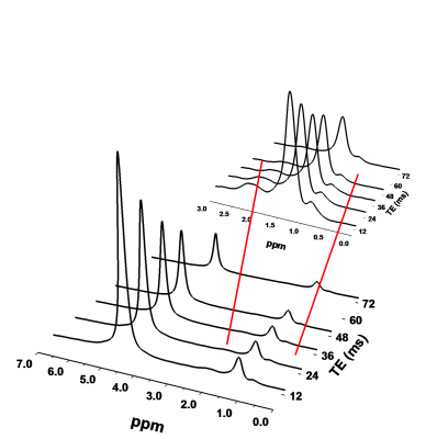 |
Measurement of Hepatic Lipid During Free Breathing with T2-Corrected Multiecho 1H MR Spectroscopy
Jack Knight-Scott, Adina Alazraki, Miriam Vos, Xiaodong Zhong, Brian Dale
Current MR techniques for quantifying hepatic fat through measurement of the proton density fat fraction (PDFF) require a breath hold that many patients find challenging. In this work, we show that when employing single voxel multiecho spectroscopy for measurement of the liver PDFF, breath holding and free breathing acquisitions yield similar results.
|
|
2586.
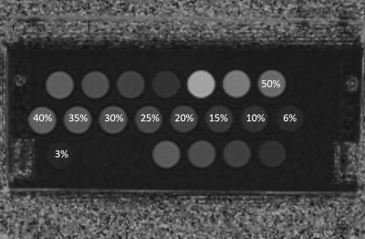 |
Long-term and short-term repeatability of hepatic proton density fat fraction measurement across MR field strengths in nonalcoholic fatty liver disease subjects and a phantom
Bohyun Kim, Hye Jin Kim, Jei Hee Lee, Hyo Jung Cho, Jai Keun Kim
Long-term and short-term repeatability of hepatic proton density fat fraction measurements was assessed across MR field strengths in nonalcoholic fatty liver disease subjects and a phantom. Our results showed that PDFF measurement have high short-term and long-term repeatability across the fields strengths, and patients undergoing a longitudinal PDFF measurement may be scanned regardless of MR field strength.
|
|
2587.
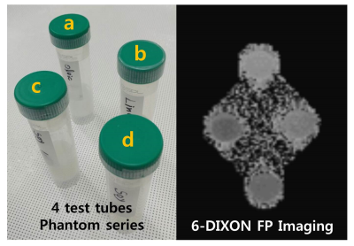 |
A study on the weighting factor investigating of liver parenchyma for 6-point interference Dixon fat percentage imaging accuracy in non-alcoholic fatty liver disease
Seung-Man Yu
The aim of this study was to determine the most accurate weighting factor for precise quantification of fatty liver when the 6-point interference Dixon fat percentage imaging technique is used by analyzing changes in WFs of fatty acid metabolites in liver. The importance of accurate WFs in the calculation of 6-pt-DIXON-based FP was confirmed in the phantom experiment. This study proposes average WF values that can be effectively used to acquire accurate 6-pt-DIXON FP images for non-alcoholic fatty liver. In addition, if the WFs of liver parenchyma FMs are applied, the accuracy of 6-pt-DIXON FP imaging can further increase.
|
|
Body: MRE
Traditional Poster
Body: Breast, Chest, Abdomen, Pelvis
Wednesday, 20 June 2018
| Exhibition Hall 2588-2596 |
16:15 - 18:15 |
|
2588.
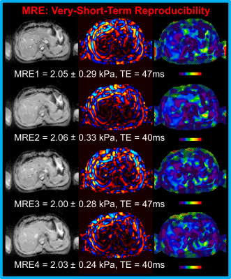 |
11-Second Hepatic MR Elastography in Clinical Research Trials
Jun Chen, Robert Laird, Qingyun Liu, Brad Jr. Bolster, Kevin Glaser, Marianna Baum, Richard Ehman
As a non-invasive imaging technique for detecting and staging liver fibrosis, MR Elastography (MRE) is highly sensitive and specific. Conventional 2D liver GREMRE is very effective, and only takes about 1-2 minutes with multiple breath-holds (11-16 seconds, each). However, shorter acquisition times and fewer breath-holds are always desired for these examinations, especially when patients have difficulty holding their breath. In this study, we developed an 11-second hepatic MRE protocol based on SE-EPIMRE sequence, which was performed in a single breath-hold comfortably; the repeatability of repeated MRE scans was also assessed.
|
|
2589.
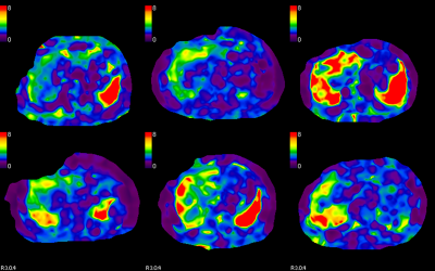 |
MR Elastography in Primary Sclerosing Cholangitis: Interobserver Agreement for Liver Stiffness Measurement
Safa Hoodeshenas, Bogdan Dzyubak, John Eaton, Richard Ehman, Sudhakar Venkatesh
Primary sclerosing cholangitis (PSC) is a chronic liver disease characterized by heterogeneous distribution of increased stiffness in periphery, segmental or lobar pattern. The heterogeneity of liver stiffness has raised concerns for reproducibility of liver stiffness measurement (LSM). We performed interobserver agreement analysis for LSM with two readers drawing manual regions of interest (ROI) and with an automated algorithm. Our study results show that large geographical ROIs including the focal regions of increased liver stiffnesses have excellent agreement between readers and automated method. Therefore large geographical ROIs using either manual or automated methods should be used for LSM in PSC patients.
|
|
2590.
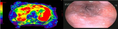 |
The role of MRE in predicting the degree of esophageal varices in patients with hepatitis B cirrhosis
Da-wei Yang, zheng-han Yang, zhen-chang Wang, Hon You
This abstract showed that liver and spleen stiffness value based on MRE was correlated well with the degree of esophageal varices, and they can be used to predict the degree of esophageal varices on hepatitis b cirrhosis patients.
|
|
2591.
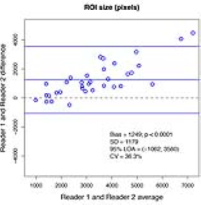 |
Inter reader agreement for liver Magnetic Resonance Elastography region-of-interest (ROI)-size, -overlap, -placement, and stiffness estimation in adults in a clinical trial
Adrija Mamidipalli, Walter Henderson, Jonathan Hooker, Tanya Wolfson, Yesenia Covarrubias, Anthony Gamst, Nikolaus Szeverenyi, Gavin Hamilton, Rohit Loomba, Claude Sirlin
MR elastography (MRE) is an established technique for the non-invasive assessment of hepatic stiffness and fibrosis, and is commonly performed using a gradient-echo-acquisition of four slices through the widest portion of the liver. The mean liver-stiffness is calculated as the average of the ROI pixel values over all four stiffness map slices. Identification (drawing) of these ROIs is subjective, relying on reader judgment to assess the wave-quality. This study examines the inter-reader agreement of MRE-ROI-size, overlap and placement, and how they affect the MRE shear-stiffness values in adults with known or suspected nonalcoholic fatty liver disease.
|
|
2592.
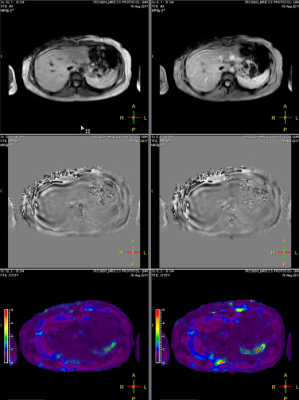 |
Comparison of Breath-Hold (BH) and Respiratory-Triggered (RT) Fast Field Echo (FFE) Hepatic MR Elastography (MRE)
Hui Wang, Tom Cull, Jean Tkach, Suraj Serai, Andrew Trout, Charles Dumoulin, Jonathan Dillman
We compared breath-hold (BH) and respiratory-triggered (RT) two-dimensional (2D) fast field echo (FFE) MR elastography (MRE) liver stiffness measurements in adult volunteers showing comparable results between t techniques.
|
|
2593.
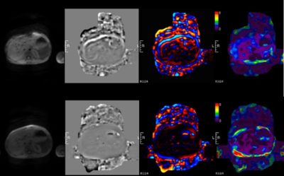 |
Can MR elastography be used to measure liver stiffness in patients with iron overload?
Suraj Serai, Andrew Trout
Untreated, iron overload causes hepatic fibrosis and cirrhosis, diabetes mellitus, hypogonadism, cardiomyopathy, dysrhythmias, and sudden death. In patients with liver iron overload, GRE based MRE techniques most likely fail due to very low signal from the liver. 2D Spin echo echo planar imaging (SE-EPI) based sequences have higher wave SNR compared with 2D GRE based MR elastography because of a higher number of wave cycles encoded per trigger (60 wave cycles per trigger vs three wave cycles per trigger in the typical 2D GRE acquisition sequence), which enables higher signal-intensity sampling of the phase waveform used to calculate the shear stiffness. In this study, our goal was to assess and demonstrate the applicability of a modified short TE, SE-EPI based MRE for staging liver fibrosis in select patients with liver iron overload conditions.
|
|
2594.
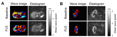 |
Assessment of Treatment Outcome in Chronic Hepatitis C Virus Infected Patients with Liver Stiffness Measured by Magnetic Resonance Elastography
Stephan Marticorena Garcia, Heiko Tzschätzsch, Christian Althoff, Christian Burkhardt, Michael Dürr, Fabian Halleck, Klemens Budde, Korinna Jöhrens, Bernd Hamm, Jürgen Braun, Thomas Fischer, Ingolf Sack, Jing Guo
High-resolution stiffness maps of the liver and kidney transplant (KTx) were generated after direct-acting antiviral therapy using multifrequency magnetic resonance elastography (MRE) and tomoelastography data processing in KTx recipients with chronic hepatitis C infection. Changes in liver stiffness after viral clearance were related to the immediate reduction in the inflammatory response in the early period and were stable until one year after end of treatment. MRE promises to be an early predictor for therapeutic success in HCV treatment.
|
|
2595.
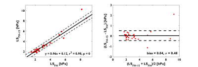 |
Impact of Motion-Encoding Gradient (MEG) Direction, Slice Position and Slice Orientation on the estimation of Liver Stiffness using Magnetic Resonance Elastography (MRE) in clinical patients
Jiming Zhang, Claudio Arena, Debra Dees, Melissa Andrews, Afis Ajala, Raja Muthupillai
As an extension of our previous work done in healthy subjects, we evaluate the impact of the direction of motion-encoding gradient (MEG), slice orientation, and coverage on the estimation of LS in 99 clinical patients referred for MRE. The results from the study show that: (a) liver stiffness (LS) measured with MEG superimposed over RL and AP directions was higher than that of LS measured with MEG in the FH direction; (b) Slight variations in the angulation of the transverse slice has negligible impact on LS estimates; and (c) The percentage area of the liver in which LS can be confidently measured (confidence map area) can have substantial variations (independent of direction of MEG) between slices and therefore, it may be beneficial to acquire more than one slice in a clinical setting.
|
|
2596.
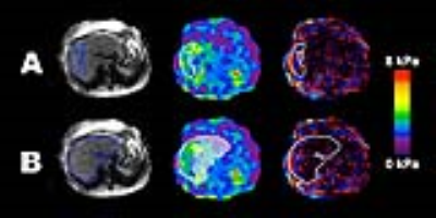 |
Comparison of the QIBA MRE ROI-drawing method to a method standardized by tracing liver parenchyma boundaries
Walter Henderson, Alexandra Schlein, Jonathan Hooker, Yesenia Covarrubias, Tanya Wolfson, Adrija Mamidipalli, Jennifer Cui, Yingzhen Zhang, Ethan Sy, Nikolaus Szeverenyi, Rohit Loomba, Claude Sirlin
While the Quantitative Imaging Biomarker Alliance (QIBA) draft recommendations on ROI placement in 2D MRE image analysis prescribe that only linear waves and parenchyma at least 1 cm from the liver edge be included, another abstract submitted to this meeting (Mamidipalli et al.) has found that analysts with equivalent experience and skill level draw significantly different ROIs when using these guidelines. This study compares the QIBA method of ROI placement to a method that is more standardized and inclusive, and compares the agreement and bias between each method on 2D MRE liver stiffness measurements.
|
|
Body: Liver Iron
Traditional Poster
Body: Breast, Chest, Abdomen, Pelvis
Wednesday, 20 June 2018
| Exhibition Hall 2597-2609 |
16:15 - 18:15 |
|
2597.
 |
Inter-method Reproducibility of Biexponential R2 Magnetic Resonance Relaxometry for Estimation of Liver Iron Concentration
Ali Pirasteh, Qing Yuan, Ivan Pedrosa, Diego Hernando, Scott Reeder, Takeshi Yokoo
Non-invasive estimation of liver iron concentration (LIC) by R2-MRI is often used for detection, grading and treatment monitoring in patients with suspected or known iron overload. The only current R2-MRI LIC estimation method with regulatory clearance is FerriScan®, a proprietary analysis for biexponential R2-relaxometry. We implemented a nonproprietary biexponential R2-relaxometry using a "dictionary-search" algorithm, to reproduce the FerriScan® results. In 38 patients with known or suspected iron overload, we demonstrated excellent reproducibility (by linearity and absolute agreement) in R2 and LIC between FerriScan® and dictionary-search analyses, suggesting generalizability of the R2-MRI approach for LIC estimation.
|
|
2598.
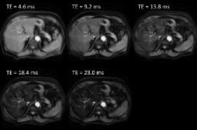 |
R2*-Relaxometry Can Replace Histology for Detecting Slight Iron Overload in Patients with Early Stage Chronic Liver Disease: A Comparison of R2*, Histology, and Mass-Spectrometry
Markus Karlsson, Mattias Ekstedt, Mikael Forsgren, Nils Dahlström, Bengt Norén, Olof Dahlqvist-Leinhard, Stergios Kechagias, Peter Lundberg
R2*-relaxometry can be used to non-invasively detect hepatic iron overload. However, most previous studies included patients with very high iron content. We sought to investigate if relaxometry reliably can detect lower levels of hepatic siderosis. R2* was therefore measured in patients with suspected chronic liver diseases of varying etiologies. We compared the relaxation rates to histological semiquantitative assessment as well as total liver iron content using mass spectrometry. There was good correlation between R2* and liver iron content. We also showed that R2*-relaxometry is better than histology when detecting slight iron overload.
|
|
2599.
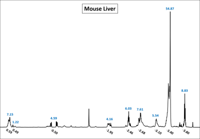 |
Dynamic Monitoring of Liver Iron Overload and Chelation Therapy using Magnetic Resonance Imaging
Gregory Simchick, Zhi Liu, May Xiong, Qun Zhao
Many diseases have been associated with excessive iron in the liver. Therefore, the non-invasive detection of liver iron overload and the monitoring of iron chelation therapy is highly desirable. Presented here is a method to demonstrate the feasibility of this using MR-based $$$R^{*}_{2}$$$ and magnetic susceptibility quantification. Significant increases in $$$R^{*}_{2}$$$ and susceptibility (Glass’ Δ values in the ranges of [-4.29 -3.23] and [-2.55 -2.23], respectively) are observed in iron overloaded livers in comparison to baseline measurements. After six doses of Polyrotaxane conjugated with Deferoxamine (rPR-DFO) iron chelation therapy administered over twelve days, Δ values of 0.13 and -0.09 are observed for $$$R^{*}_{2}$$$ and susceptibility, respectively, indicating that the differences are no longer significant and the treatment is effective.
|
|
2600.
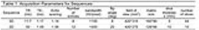 |
Noise-corrected R2* estimation using 3D multi-gradient-echo Dixon for hepatic iron overload: Comparisons with 2D multi-gradient-echo sequences
Huimin Lin, Stephan Kannengiesser, Caixia Fu, Jun Shen, Fuhua Yan
Different combinations of acquisition and postprocessing for R2* estimation were compared: 3D multi-gradient-echo Dixon vs. 2D multi-gradient-echo, with/without fat saturation (FS); noise-corrected (NC) vs. fat-and-noise-corrected (FNC) fitting. Twenty patients suspected of hepatic iron overload, but not having steatosis, were included. 3D_NC_R2* showed excellent agreement with 2D_NC_R2*. Up to medium R2*, this held also for 3D_FNC_R2* vs. 2D_NC_R2*; at high R2*, fat modeling reduced R2*. 2DFS_NC_R2* was also reduced. R2* standard deviation was lowest in 3D_FNC, and highest in 2DFS_NC. 3D multi-echo Dixon with noise correction is a promising technique for whole-liver iron quantification, but further analyses are necessary.
|
|
2601.
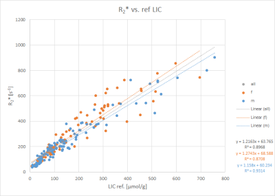 |
Gradient-Echo MRI for Liver Iron Content Determination employing R2* Relaxometry: Influence of Gender and Disease
Arthur Wunderlich, Sabrina Schweyer, Daniel Frisch, Justin Brosig, Holger Cario, Meinrad Beer, Stefan Schmidt
To investigate the relation between R2* gained from gradient echo (GRE) MRI and liver iron concentration (LIC), we studied the influence of patient characteristics. 205 patients (92 f, 113 m; 98 with Thalassemia major, 31 with Sickle Cell Anemia, 15 with Diamond-Blackfan-Anemia) suspected for liver iron overload were scanned according to Ferriscan® with spin echo MRI to obtain reference LIC values, and GRE protocols suitable for LIC determination. GRE analysis based on manually drawn liver ROIs and relaxometry yielded R2* values. Correlation analysis of R2* to reference LIC revealed different correlation parameters between patient subgroups concerning disease and gender.
|
|
2602.
 |
Comparison of Hepatic Quantitative Susceptibility Mapping and R2* for Iron Measurement in Presence of Fat and Fibrosis; an Ex-Vivo Study
Ramin Jafari, Anne Koehne de Gonzalez, Yi Wang, Thanh Nguyen , Alexey Dimov, Kofi Mawuli Deh, Zhe Liu, Gary Brittenham, Martin Prince, Pascal Spincemaille
Precise measurement of liver iron content (LIC) in patients with transfusional iron overload is important in iron-chelation therapy. MRI can be used as a non-invasive method to measure iron levels in the liver. Typically, R2 and R2* based methods are used for this purpose. In this work, we use human liver explants to demonstrate the degree to which steatosis and fibrosis are confounding factors for R2* and quantitative susceptibility mapping in LIC measurement.
|
|
2603.
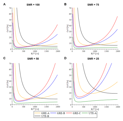 |
Ultrashort Echo Time Imaging for Quantification of Hepatic Iron Overload: Comparison of Current Acquisition and Fitting Methods via Simulations and Phantom Data
Aaryani Tipirneni-Sajja, Ralf Loeffler, Andrea Sajewski, Jane Hankins, Claudia Hillenbrand
Assessment of hepatic iron content by R2*-MRI is a non-invasive alternative to liver biopsy. R2* is typically measured by a multiecho gradient-echo (GRE) sequence, however, GRE fails in high iron cases when T2* decay is rapid. In recent years, ultrashort echo time (UTE) imaging has been proposed to increase the accuracy in R2* measurements in high and massive iron overload. Still, the accuracy of R2* measurements depends on acquisition parameters and curve fitting algorithms, which vary between institutions. The purpose of this study is to compare current R2* acquisition and fitting methods, and identify the optimal acquisition and fitting methods for clinical use.
|
|
2604.
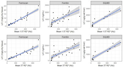 |
Demonstration of linear correlation between R2* and liver iron concentration across multiple MR acquisition parameters at 1.5T and 3T.
Richard Jones, Jason Bentley, Valentina Taviani, Diego Hernando, Scott Reeder, Shreyas Vasanawala
We demonstrate a robust linear relationship between the concentration of liver iron and R2* measurements taken in any liver segment, with various planes of acquisition, slice thickness, flip angle, and echo spacing at 1.5T or 3T. As compared with Ferriscan, R2* imaging is faster, lower-cost, and requires no post-processing, and has better geographic availability compared to the gold standard of superconducting quantum interference devices.
|
|
2605.
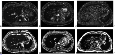 |
Quantification of multiple organ iron deposition in transfusion dependent diseases using mDIXON-Quant technique
Qiaoling Wu, Zhizheng Zhuo, Hongyan Ni
Iron overload is a common complication of transfusion dependent patients. Magnetic resonance imaging can be used for quantitative detection of iron deposition in transfusion dependent patients. A total intake of iron for transfusion was evaluated based on the mDIXON-Quant and 3D-FFE sequence respectively. Because the mdixon-quant can avoid the effect of fat on the iron overload evaluation, the mDIXON-Quant sequence can more accurately quantify iron deposition in liver and pancreas than 3D-FFE sequence. The quantitative application of mDIXON-Quant in detection of iron deposition in patients can provide reliable basis for iron chelation therapy in clinic.
|
|
2606.
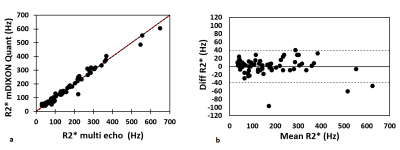 |
Hepatic Iron overload estimation by proton density mDIXON Quant technique
Ane Ugarte, Javier Sánchez-González, Coloma Álvarez-de-Eulate , José Alustiza, Jose Emparanza
This work evaluates the utility of R2* obtained from multi-point multi-peak proton density fat fraction to assess iron overload and the accuracy of provided relaxation maps compared with more established multi-echo gradient echo sequence.
|
|
2607.
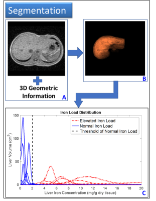 |
Global Measures of Liver Iron Content Based on T2* mapping and Dual Clustering Segmentation
Mitchell Horn, Ning Hua, Chad Farris, Adam Aakil, Ilse Castro-Aragon, Hernán Jara
Purpose: To develop a method to estimate total iron load of the whole liver. Methods: Multi gradient-echo pulse sequence was applied to 17 patients with varying degrees of liver iron content (LIC). LIC was measured via T2* mapping on a voxel-by-voxel basis. Liver was segmented with a semi-automated dual-clustering method. Total iron load was estimated by numerically integrating the LIC histogram. Results: This assessment of iron load presents a noninvasive whole liver alternative to liver biopsies. Conclusion: T2* relaxometry and segmentation provide a novel method for iron content quantification at the organ level that can easily be adapted in clinics.
|
|
2608.
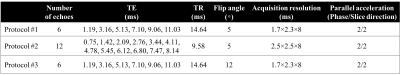 |
Test-retest Repeatability of R2* Mapping and Quantitative Susceptibility Mapping for Liver Iron Quantification
Ante Zhu, Timothy Colgan, Scott Reeder, Diego Hernando
Liver iron concentration is widely recognized as the best overall metric of total body iron content. Accurate and precise (repeatable) non-invasive measurements of liver iron concentration are needed. In this work, we assessed the test-retest repeatability of R2* mapping and quantitative susceptibility mapping (QSM) in patients with liver iron overload at 1.5T. Our test-retest measurements demonstrate good agreement in different protocols for R2* quantification but large limits of agreements for QSM susceptibility estimates. Further optimization of QSM techniques is needed to improve test-retest repeatability.
|
|
2609.
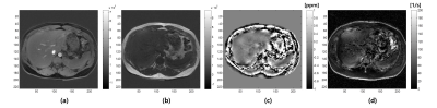 |
Robust multi-parametric mapping for abdomen imaging
Young-Joong Yang, Jong-Hyun Yoon, Jin-Soo Kim, Chang-Beom Ahn
A robust abdominal multi-parametric mapping using multi-echo data is proposed. Reconstructed maps are water, fat images, quantitative susceptibility map (QSM), and R2* map. Fat fraction and iron deposition in the liver may be important parameters for diagnosis. Challenges to the abdominal mapping include large field inhomogeneity, phase wrapping, phase variations from water and fat signal, chemical shift, and physiological motions. We applied simultaneous unwrapping phase and error recovery from inhomogeneity (SUPER) technique to correct field inhomogeneity and phase wrapping. The technique is stably applicable to objects containing water and fat signal, and is also useful as a preprocessing for QSM.
|
|
Body: Liver Imaging Using Perfusion, Diffusion, T1, & T1rho
Traditional Poster
Body: Breast, Chest, Abdomen, Pelvis
Wednesday, 20 June 2018
| Exhibition Hall 2610-2626 |
16:15 - 18:15 |
|
2610.
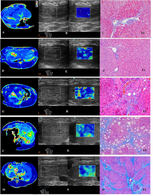 |
Liver Fibrosis Detection and Staging: A Comparative Study of T1? MR Imaging and 2D Real-time Shear-wave Elastography
Ruo-kun Li, Fu-hua Yan, Xin-pin Ren, Wei-bo Chen
There was moderate positive correlation between fibrosis stage and T1ρ values (r=0.566; 95% CI 0.291-0.754; P<0.0001), and LS value (r=0.726; 95% CI 0.521-0.851; P=0.003). T1ρ values showed moderate positive correlations with LS values (r=0.693; 95% confidence interval [CI]: 0.472-0.832; P<0.0001). Areas Under ROC (AUROCs) were 0.861 (95% CI: 0.705-0.953) for SWE and 0.856 (95% CI: 0.698-0.950) for T1ρ (P = 0.940), 0.906 (95% CI: 0.762-0.978) for SWE and 0.849 (95% CI: 0.691-0.946) for T1ρ (P = 0.414), 0.870 (95% CI: 0.716-0.958) for SWE and 0.799 (95% CI: 0.632-0.913) for T1ρ (P = 0.422), and 0.846 (95% CI: 0.687-0.944) for SWE and 0.692 (95% CI: 0.517-0.835) for T1ρ (P = 0.137), when diagnosing liver fibrosis with ≥F1, ≥F2, ≥F3 and F4, respectively. There was moderate positive correlation between inflammatory activity and T1ρ values (r=0.520; 95% CI 0.158-0.807; P=0.013).
|
|
2611.
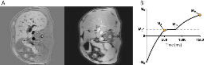 |
Early Stage Chronic Liver Disease: T1 Relaxation and Hepatic Fibrosis
Markus Karlsson, Thobias Romu, Amir Razavi, Nils Dahlström, Mikael Forsgren, Olof Dahlqvist-Leinhard, Bengt Norén, Mattias Ekstedt, Stergios Kechagias, Peter Lundberg
We measured T1 relaxation times in a prospectively recruited cohort of patients with suspected chronic liver disease. Our aim was to investigate the predictive value of T1 for staging hepatic fibrosis. Furthermore, we sought to test if T1 values are confounded by inflammation or presence of iron. We found that T1 was confounded by iron and that T1 alone had a poor ability to stage hepatic fibrosis.
|
|
2612.
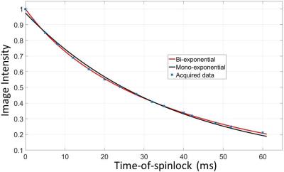 |
Bi-exponential T1rho Relaxation of In Vivo Human Liver
Weitian Chen, Vincent Wong, Queenie Chan, Yi-Xiang Wang, Winnie Chu
Quantitative T1rho imaging is reported a promising non-invasive diagnostic tool for detection of liver fibrosis at its early stage. T1rho relaxation is often estimated by a mono-exponential relaxation model. However, bi-exponential relaxation may occur due to compartmentation of the liver tissue. Bi-exponential T1rho relaxation has been reported in rat muscle and human knee cartilage. In this work, we provided our observation and analysis of bi-exponential T1rho relaxation of in vivo human liver.
|
|
2613.
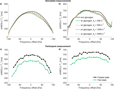 |
The influence of glycogen on shortened modified Look-Locker inversion recovery (shMOLLI) T1 maps of the liver
Ferenc Mozes, Elizabeth Tunnicliffe, Michael Pavlides, Matthew Robson
Dynamic physiological changes in the liver may influence the increased variability of shMOLLI T1 of healthy livers relative to normal myocardial shMOLLI T1 variability. Since glycogen concentration varies over relatively short time periods, this may contribute to the variability. This study explores two possible pathways by which glycogen might influence shMOLLI measurements: chemical exchange saturation transfer (CEST) effects and direct change of liver water relaxation. Simulations, phantom and human experiments suggest that the CEST effect is negligible in vivo and a 7% shortening of T1 at high glycogen concentration is driven by direct relaxation effects.
|
|
2614.
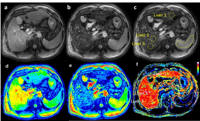 |
More than Hepatobiliary Relative Enhancement Ratio by Gd-EOB-DTPA for Liver Fibrosis Estimation, Hepatocyte Fraction method by T1 mapping measurement
Shen Pan, Xiaoqi Wang, Qiyong Guo
We calculated the hepatocytefraction (Hep) and reduction rate of T1 relaxation time (RE) based on T1changes in the hepatocyte due to pharmacokinetics of gadoliniumethoxybenzyl-diethylenetriamine pentaacetic acid (Gd-EOB-DTPA) uptake in liver.Both Hep and RE were compared with liver fibrosis stage according to theMETAVIR scoring system. And we found that Hep significantly correlated withfibrosis stage, and indicate it a good quantitative biomarker for liver fibrosis estimation.
|
|
2615.
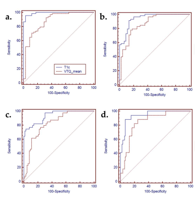 |
Comparison between MR T1? imaging and acoustic radiation force impulse for noninvasive assessment of liver fibrosis: repeatability, reproducibility, and diagnostic performance in rat models
Jinning Li, Huanhuan Liu, Caiyuan Zhang, Shuyan Yang, Yanshu Wang, Weibo Chen, Dengbin Wang
The purpose of this study was to validate the repeatability, reproducibility, and diagnostic performance of MR T1ρ imaging for staging liver fibrosis, compared with ultrasound-based acoustic radiation force impulse (ARFI). The cross-sectional study was performed in rat models with carbon tetrachloride (CCl4). The results of histopathological analysis were used as reference standard. T1ρ imaging showed comparable repeatability and reproducibility with ARFI, however, manifested more accurate diagnostic performance for staging liver fibrosis, especially for detecting early stage of fibrosis.
|
|
2616.
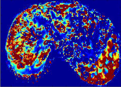 |
Assessment of Liver fibrosis using Exchange dual-input dual-compartment pharmacokinetic model of Dynamic Contrast-enhanced MRI
Lan Zhang, Zhi Zheng Zhuo
The clinical need in the development of non-invasive methods for liver fibrosis assessment has emerged. At 3.0T, human in-vivo studies have demonstrated DCE-MRI using Exchange dual-input and dual-compartment pharmacokinetic model has potential to detect and assess the vascular permeability modification of liver fibrosis. DCE-MRI pharmacokinetic quantitative parameters including Ktrans, Ve and Vp can be used for diagnosing and staging liver fibrosis. Ktrans is the best index and predictor for discriminating normal livers from fibrotic livers.
|
|
2617.
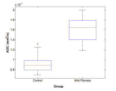 |
Diffusion MRI alteration following the induction of mild liver fibrosis in a rabbit model
Matteo Figini, Liang Pan, Chong Sun, Bin Wang, Junjie Shangguan, Kang Zhou, Na Shang, Quanhong Ma, Zhuoli Zhang
Nine rabbits were injected with carbon tetrachloride for 6 weeks to induce liver fibrosis. At the end of this period, these rabbits and 15 controls injected with saline underwent an imaging protocol including diffusion MRI. Histology showed mild fibrosis throughout the liver of the CCl4-injected animals. The Apparent Diffusion Coefficient in the liver of the fibrotic rabbits was significantly higher than in the controls. This counterintuitive result can be explained by the presence of many conflicting mechanisms during the early stage of fibrosis. If confirmed, the ADC could become a valuable tool for the early detection of liver fibrosis .
|
|
2618.
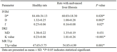 |
Comprehensive analysis of advanced liver fibrosis in rats using multi-parameter MRI
Qing Li, Shuangshuang Xie, Hanxiong Qi, Zhizheng Zhuo, Yue Cheng, Wen Shen
This study investigated the value of multi-parametric analysis using IVIM, DKI and MR T1ρ for the diagnosis of advanced liver fibrosis. Sixteen healthy rats and fifteen rats with advanced liver fibrosis (F4 confirmed by liver pathological examination) were scanned with IVIM, DKI and MR T1ρ. IVIM derived D*, D, f, DKI derived MD, K value and MR T1ρ derived T1ρ value were compared between the above two groups. Our results showed D*, D, f and MD decreased while K value and T1ρ value increased in rats with advanced liver fibrosis. And D*, D, f, K value and T1ρ value demonstrated significant difference (P<0.05). We therefore conclude that decreased D*, D and f and increased K value and T1ρ value could be useful in the diagnosis of liver fibrosis.
|
|
2619.
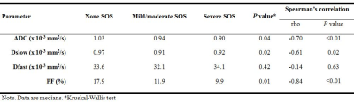 |
Diagnostic value of intravoxel incoherent motion (IVIM) diffusion-weighted imaging in hepatic sinusoidal obstruction syndrome: an experimental study in a rat model – preliminary results
Eun Kyoung Hong, Ijin Joo, Kyoungbun Lee
Hepatic sinusoidal obstruction syndrome (SOS), a toxic liver injury, needs an accurate diagnosis and serial monitoring for an effective management. Intravoxel incoherent motion (IVIM) DWI, which allows separate estimation of molecular diffusion and microcirculation, potentially provides information regarding hepatic parenchymal abnormalities. This study investigated the diagnostic value of IVIM-DWI in the assessment of hepatic SOS using a monocrotaline-induced rat SOS model. Our study results showed that ADC, true diffusion coefficient, and perfusion fraction showed significant correlation with the severity of SOS, which would suggest that IVIM-DWI may serve as a noninvasive method in the quantitative assessment of hepatic SOS.
|
|
2620.
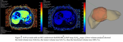 |
Assessment of the Hepatocyte Fraction Combined with Liver Volume for the estimation of liver function
Ke Wang, Xiaochao Guo, He Wang, Zhizheng Zhuo, Xiaoying Wang
Hepatocyte fraction (HeF) and uptake function based on k map have becoming new biomarkers based on EOB-MR in estimation of hepatic function. Liver volume is another factor that influence the liver function. The purpose of our study was to determine whether liver function can be estimated quantitatively from EOB-MR combined with liver volume.
|
|
2621.
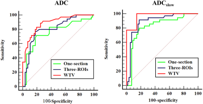 |
Comparison of the diagnostic performances of three methods of ROI placement for the measurements of Intravoxel incoherent motion diffusion-weighted MR imaging parameters in hepatocellular carcinoma
Yi Wei, Bin Song, Dandan Zheng
The pathological differentiated grade is heavily associated with the hepatocellular carcinoma prognosis. Through a prospectively research, we sought to determine the diagnostic performances of three methods of ROI placement for measurements of IVIM parameters in the grading of hepatocellular carcinoma. According to the results, we found that different ROI positioning methods used significantly affects the IVIM and ADC parameters measurements. Measurements of ADCslow value derived from whole tumor volume method entailed the highest diagnostic performance in grading hepatocellular carcinoma. These results suggested that ADCslow value derived from whole tumor volume method might be useful in assessing the differentiated grade of carcinoma, and which might be helpful in predicting the patients’ prognosis.
|
|
2622.
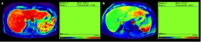 |
In primary sclerosing cholangitis, diffusion weighted magnetic resonance imaging correlates better with liver stiffness than Gadoxetate disodium enhanced MR imaging
Jin Yamamura, Jan Sedlacik, Tillmann Schuler, Ralph Buchert, Dr. Maxim Avanesov, Hendrik Kooijman-Kurfuerst, Christoph Schramm, Gerhard Adam, Sarah Keller
Several disadvantages of DCE-MRI, such as long examination time, application of intravenous contrast agents and elaborative postprocessing and the higher sensitivity of the ADC to differentiate several stages of fibrosis, favorites DWI over DCE-MRI for diagnosis and staging of fibrosis in routine clinical MRI of PSC patients.
|
|
2623.
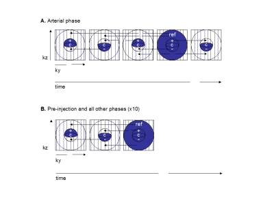 |
Dynamic Contrast-Enhanced MRI to Assess Hepatocellular Carcinoma Response to Transarterial Chemoembolization: a Pilot Study.
Alana Thibodeau-Antonacci, Léonie Petitclerc, Guillaume Gilbert, Laurent Bilodeau, Hélène Castel, Simon Turcotte, Damien Olivié, Catherine Huet, Pierre Perreault, Gilles Soulez, An Tang, Samuel Kadoury
Hepatocellular carcinoma response to transarterial chemo-embolization is traditionally assessed by qualitative interpretation of imaging features and enhancement dynamics. However, quantitative parameters derived by fitting a dual-input single-compartment model on dynamic contrast-enhanced-MRI data show promise, as they may help discriminate non-viable from viable tumors after treatment. Peak enhancement ratio significantly decreased after transarterial chemo-embolization in tumors with complete response (i.e. non-viable tumor group). This pilot study suggests that quantitative dynamic contrast-enhanced-MRI parameters may be used to assess treatment response.
|
|
2624.
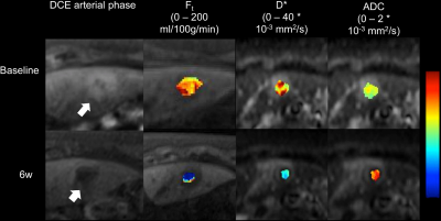 |
Dynamic contrast-enhanced MRI and intravoxel incoherent motion diffusion-weighted imaging for the evaluation of HCC response to 90Yttrium radioembolization
Stefanie Hectors, Paul Kennedy, Octavia Bane, Maxwell Segall, Sara Lewis, Myron Schwartz, Edward Kim, Bachir Taouli
The goal of this study is to assess whether DCE-MRI and IVIM-DWI can be used to predict response of hepatocellular carcinoma (HCC) to 90Yttrium radioembolization (RE). In a preliminary cohort, significant changes were observed in both DCE-MRI and IVIM-DWI parameters at 6 weeks after treatment, which suggest that both techniques are sensitive to treatment effects of RE to HCC tissue. The exact utility of the DCE-MRI and IVIM-DWI parameters will be tested in a larger cohort.
|
|
2625.
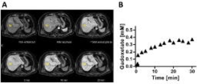 |
Estimating Liver Function by Gadoxetate Enhanced MRI: Comparison of Pharmacokinetic Models in a Clinical Setting
Markus Karlsson, Gunnar Cedersund, Peter Lundberg
The hepatic uptake rate of Gadoxetate is a possible biomarker for liver function and several different pharmacokinetic models have been developed. However, no one has ever compared these models using the same data. We compared three different models using imaging data with low temporal, but high spatial resolution. We showed that two of the models estimates almost the same values of the hepatic uptake rate. The fact that two different pharmacokinetic models can produce the same parameter values validates the entire pharmacokinetic modelling approach, indicating that it is not just a model-specific parameter being estimated, but the actual transport rate.
|
|
2626.
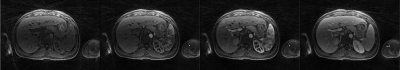 |
Quantifying hepatic fibrosis using a 3D radial golden angle stack-of stars acquisition and a dual-input two compartment model
Abhishek Pandey, Manojkumar Saranathan, Wyatt Unger, Mahesh Keerthivasan, Jean-Philippe Galons, Diego Martin, Ali Bilgin, Kevin Johnson, Maria Altbach
Chronic liver disease (CLD) is known to affect 3.9 million of Americans. Collagen deposition in CLD affects the perfusion of the liver parenchyma and dynamic contrast enhanced MRI (DCE-MRI) can be used for the non-invasive diagnosis of CLD. Here we present a liver perfusion technique based on a free-breathing 3D radial golden-angle stack-of-stars acquisition along with a compressed sensing reconstruction to generate DCE data with 4-sec temporal resolution. Perfusion parameters are estimated by fitting the DCE data to a dual-input two compartment pharmacokinetic model and used to evaluate hepatic fibrosis in CLD.
|
|
| Back |
| The International Society for Magnetic Resonance in Medicine is accredited by the Accreditation Council for Continuing Medical Education to provide continuing medical education for physicians. |
