|
Traditional Poster Session
Acquisition, Reconstruction & Analysis |
Thursday, 21 June 2018
Traditional PosterAcquisition, Reconstruction & Analysis
2648 -2660 Motion Correction: Cleaning up in the Brain
2661 -2710 Pulses, Sequences, Motion & Artefacts
2711 -2725 Machine Learning for Cancer Applications
2726 -2738 Machine Learning for Tissue Segmentation & Classification
2739 -2751 Classification & Prediction for Function & Disease
2752 -2780 Quantitative MRI
2781 -2807 Learning Image Reconstruction
2808 -2827 Acquisition, Reconstruction & Analysis: Sparse & Low-Rank Models
2828 -2863 Image Analysis & Post-Acquisition Computing
2864 -2895 Acquisition, Reconstruction, Analysis |
| |
Motion Correction: Cleaning up in the Brain
Traditional Poster
Acquisition, Reconstruction & Analysis
Thursday, 21 June 2018
| Exhibition Hall 2648-2660 |
08:00 - 10:00 |
|
2648.
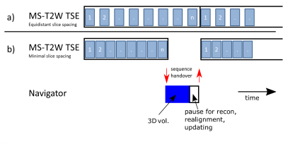 |
High resolution imaging at 7T using interleaved prospective motion correction (iMOCO)
Vincent Boer, Mads Andersen, Anouk Marsman, Esben Petersen
Subject motion is a major problem in MRI, leading to less diagnostic information in the clinic and lowering data quality in research. Especially at high field, the relatively long scan times applied for high resolution imaging makes motion one of the major challenges. A promising solution is to update the field-of-view in real time based on tracking with MRI-based navigators. Here we show an implementation for prospective motion correction using MRI navigators at 7T. The framework was very flexible, as the navigator and target sequence are simply defined as two different scans, which can be interleaved at any sequence level.
|
|
2649.
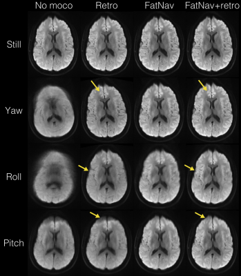 |
Prospectively Motion Corrected DWI by Projection Fat Navigators
Johan Berglund, Henric Rydén, Enrico Avventi, Tim Sprenger, Ola Norbeck, Stefan Skare
A projected fat navigator module was added to a diffusion weighted EPI sequence to allow prospective rigid body motion correction without additional hardware. Improved image quality was demonstrated by imaging the brain of a volunteer subject who performed prescribed patterns of large motion with and without prospective correction. Improvements were most evident for through-plane motion. For in-plane motion only, the image quality was comparable to images acquired without motion. Ghosting due to gradient delays following FOV updates was avoided by acquiring phase reference lines directly after the excitation pulse.
|
|
2650.
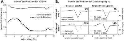 |
Comparing TAMER (TArgeted Motion Estimation and Reduction) reduced modeling to alternating minimization for data consistency based motion mitigation
Melissa Haskell, Stephen Cauley, Lawrence Wald
Retrospective motion correction techniques offer minimal disruptions to sequences and clinical workflows. The computational burden of retrospective techniques can be eased either with alternating minimizations, or true joint estimation but on a reduced model. We provide computational experiments demonstrating the tightly coupled nature of the optimization variable types (motion and voxel values) which hinders the alternating based approaches. The alternating techniques can have an average search direction error of 75%, vs. 22% with reduced modeling. We demonstrate a computational speedup of 17x using our reduced model approach, and present in vivo imaging results comparing TAMER to a state-of-the-art alternating minimization.
|
|
2651.
 |
Optical prospective motion correction for brain imaging at 7T without a mouthpiece
Phillip DiGiacomo, Julian Maclaren, Murat Aksoy, Brian Burns, Roland Bammer, Brian Rutt, Michael Zeineh
The advancements in signal to noise ratio, contrast, and resolution enabled by high-field MR systems provide great potential for visualizing more nuanced brain anatomy. However, in order to translate these advancements to the discovery and clinical implementation of novel neuroimaging biomarkers, motion artifact resulting from long scan times must be addressed. Here, we demonstrate proof-of-concept of a novel prospective optical motion tracking and correction system using a coil-mounted camera without a mouthpiece, visualizing an optical marker placed on the cheek of human subjects in a 7T MR system.
|
|
2652.
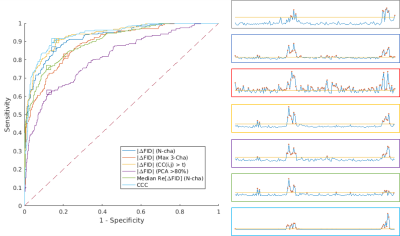 |
Pediatric Head Motion Detection using Free Induction Decay Navigators
Tess Wallace, Kristina Pelkola, Monet Dugan, Simon Warfield, Onur Afacan
Free induction decay navigators (FIDnavs) are sensitive to head motion and can be rapidly acquired using standard scanner hardware, making them an attractive approach for motion detection in pediatric MRI. In this study, we perform a head-to-head comparison of various FIDnav motion detection algorithms in controlled volunteer experiments and in pediatric patients scanned under typical conditions using a modified MPRAGE sequence. We demonstrate that computing the change in cross-correlation coefficient between FIDnav signal vectors results in excellent detection accuracy in both volunteers and patients, based on concurrent ground-truth RMS displacements measured using an electromagnetic tracking system.
|
|
2653.
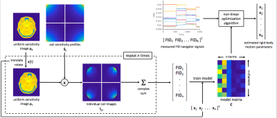 |
A Novel Framework for Head Motion Measurement using Free Induction Decay Navigators from Multi-Channel Coil Arrays
Tess Wallace, Onur Afacan, Simon Warfield
FID navigators (FIDnavs) encode substantial quantitative rigid-body motion information; however, current implementations require subjects to cooperate for a choreographed training session, which is impractical in many clinical scenarios. We present a new approach that uses simulation of the acquisition physics and effect of motion on the measured FIDnav from each coil. This method is tested in three volunteers scanned at 3T with a 32-channel head coil using a 3D FLASH sequence, each performing a series of repeating motion patterns. Sub-millimeter and sub-degree tracking accuracy was achieved across all volunteers, demonstrating the efficacy of this approach for real-time head motion measurement.
|
|
2654.
 |
Motion correction of PET images using Spherical Navigator echoes (SNAVs) on a hybrid PET-MR scanner
Patricia Johnson, Reggie Taylor, Tim Whelan, Maria Drangova
Head motion during brain imaging with hybrid PET-MR degrades the quality of both the PET and MR images. Simultaneous acquisition with the two modalities provides the opportunity for MR motion measurement techniques to be used for correction of PET data. In this study, spherical navigator echoes (SNAV) were used for retrospective motion correction of PET images. A phantom was repositioned several times during a list mode acquisition. The list mode data was binned into motion states based on the SNAV measured motion, and a motion-corrected PET reconstruction was performed. SNAV motion correction successfully removed blurring in the PET images.
|
|
2655.
 |
Artifact Detection Using Correlation Analyses Applied to MEGA-PRESS Data Containing Subject Head Movements
Sofie Tapper, Anders Tisell, Gunther Helms, Peter Lundberg
Subject movements and other disturbances might contaminate the Magnetic Resonance Spectroscopy data, and these artifacts can be misinterpreted as actual metabolite signals by the quantification program. Thus, an automatic method could be very helpful for finding artifacts and eliminating them. In this work, an approach of using correlation analyses was tested in order to evaluate if motion contaminated data could be identified. A total of 296/320 spectra were correctly categorized according to the movement-paradigm. This procedure could be suitable for identifying data that are affected by subject motion or other artifacts that would reduce the quality of the result.
|
|
2656.
 |
Motion correction of T2*-weighted MRI with consideration of B0 and B1 effect
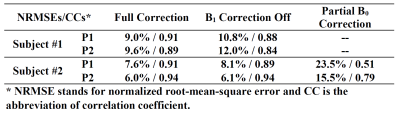
Jiaen Liu, Peter van Gelderen, Jacco de Zwart, Jeff Duyn
T2*-weighted MRI has broad applications in the brain and can provide both functional and (micro) anatomical information. Unfortunately, it has proven rather sensitive to subtle head motion, and the associated changes in B0 and to a lesser extent B1. In this study, the collective impact of pose-dependent B0 and B1 on T2*-weighted gradient echo MRI was investigated. A conjugate-gradient method was utilized for reconstructing MR images collected during variation of head poses.
|
|
2657.
 |
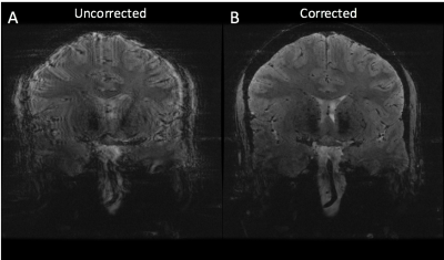
Retrospective motion correction of head motion using electromagnetic sensors
Onur Afacan, Tess Wallace, Simon Warfield
Motion artifacts pose significant problems for the acquisition of MR images, especially in pediatric populations. In this work we developed a retrospective motion correction framework that uses motion information from two electromagnetic sensors attached to the forehead of subjects. We evaluated our retrospective motion correction strategy on 12 different cases and show that that motion traces from the EM tracker can be used to retrospectively improve image quality.
|
|
2658.
 |
Blurring and Ghosting Effects Under Beats Formation in Magnetic Resonance Imaging Under Source Vibration
Dhiraj Sinha, Pranay Prateek, Simon Lui, Shaoying Huang
A key challenge of MRI is development of an accurate model of noise generation which are integral to generation of high-resolution images. Currently, motion induced noise is addressed at algorithmic level. Here, we present a novel physical model which incorporates the role of mechanical vibration of body parts in generation of additional frequency components in the emitted radio frequency spectrum around the precession frequency. The mathematical model was validated through a computational simulation which led to the discovery that beats generated as a result of mechanical vibrations of the source lead to ghosting and blurring effects.
|
|
2659.
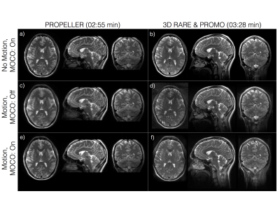 |
Pseudo-3D PROPELLER
Ola Norbeck, Enrico Avventi, Henric Ryden, Johan Berglund, Tim Sprenger, Stefan Skare
A thin-sliced (pseudo-3D) SMS accelerated PROPELLER with retrospective motion correction is demonstrated and compared to prospectively motion corrected 3D RARE using spiral navigators. The results show that our pseudo 3D PROPELLER sequence can produce higher image quality than 3D RARE, even in reformatted views, with and without the presence of head motion.
|
|
2660.
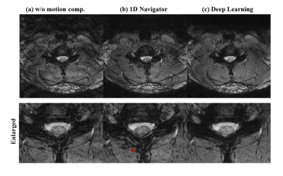 |
Reduction of respiratory motion artifact in c-spine imaging using deep learning: Is substitution of navigator possible?
Hongpyo Lee, Kanghyun Ryu, Yoonho Nam, Jaeho Lee, Dong-Hyun Kim
Deep learning methods are starting to be widely used in medical images. Here, we propose a deep learning approach to compensate respiratory induced artifacts. A deep convolutional neural network was designed to train the ghosting artifact caused by respiratory motion in c-spine imaging. Using deep learning, compensation can be applied without additional data such as navigator echo.
|
|
Pulses, Sequences, Motion & Artefacts
Traditional Poster
Acquisition, Reconstruction & Analysis
Thursday, 21 June 2018
| Exhibition Hall 2661-2710 |
08:00 - 10:00 |
|
2661.
 |
Can scans with different TR be combined to improve UTE T2* measurements?
Dirk Poot, Paul Baron, Juan Hernandez-Tamames
We investigated combining UTE sequences with different TR without requiring knowledge of T1, to enable increasing the number of short TE scans for T2* quantification. Many short T2* tissues have multiple compartments with ultra-short and somewhat longer T2 values. To quantify both a substantial number of images with ultra-short TE and images with a substantial maximal TE are required. The large maximal TE requires relatively large TR and hence long scan times, while the ultra-short TE scans have to be acquired separately. Hence, being able to combine images with different TR would be beneficial for such studies.
|
|
2662.
 |
Image reconstruction in low field MRI: a super-resolution approach
Merel de Leeuw den Bouter, Martin van Gijzen, Andrew Webb, Rob Remis
Inexpensive MRI scanners based on permanent magnets present a promising diagnostic tool for developing countries. For very inhomogeneous fields an ill-posed system of equations has to be solved in order to obtain an image. Due to the low signal-to-noise ratio, direct attempts at generating high resolution images yield poor results. In this research, super-resolution reconstruction is considered as an alternative. By first obtaining low resolution images and then applying super-resolution, high resolution images of better quality can be obtained.
|
|
2663.
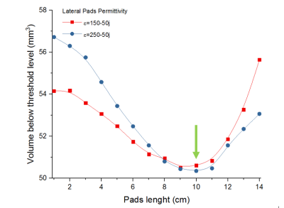 |
Properties optimization of pads configurations on CST to minimize B1+ field inhomogeneities at 7T in the temporal lobes and cerebellum
Zo Raolison, Marc Dubois, Luisa Neves, Stefan Enoch, Nicolas Malléjac, Pierre Sabouroux, Anne-Lise Adenot-Engelvin, Alexandre Vignaud, Redha Abdeddaïm
A simple and efficient way to enhance the B1+ field dark areas appearing in the temporal lobes and cerebellum at 7T in MRI is to use pads with relative High-Dielectric Constant materials. We present here simulations of different pads configurations aiming to reduce those dark areas. It has been found that the educated guess consisting in using a three pads configuration localized in front of each area is less efficient than two pads above the ears for the temporal lobes or a single pad on the neck for the cerebellum.
|
|
2664.
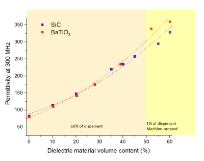 |
Evaluation of a new long-lasting Silicon Carbide based dielectric pad for ultra-high field MRI
Zo Raolison, Redha Abdeddaïm, Marc Dubois, Lisa Leroi, Luisa Neves, Franck Mauconduit, Stefan Enoch, Nicolas Malléjac, Pierre Sabouroux, Anne-Lise Adenot-Engelvin, Alexandre Vignaud
A simple and efficient way to enhance the B1+ field dark areas appearing in the temporal lobes at 7T in MRI is to use pads with relative High-Dielectric Constant materials which most promising ones are perovskites mixed with water. As their performance drops over time, those materials are still not currently used in clinical routine. A novel high lifespan material made of 4-Fluoro 1.3-dioxalan-2-one and Polyethylene glycol mixed with silicon carbide particles is presented here. It is shown that their performances are on pair with BaTiO3 water mixture through permittivity measurements and MRI scans a 7T.
|
|
2665.
 |
Enabling long excitation pulses in algebraic ZTE imaging by dead-time reduction via dual acquisition with alternative RF modulations
Romain Froidevaux, Markus Weiger, Klaas Pruessmann
MRI of tissues with short transverse relaxation times raises both scientific and clinical interest and can be performed with zero echo time MRI. However, as RF excitation is done under the radial encoding gradient, flip angle amplitudes and uniformity are limited. This issue can be circumvented by using longer modulated pulses. However, pulse length is limited by dead-time-induced central k-space gaps getting too large for robust image reconstruction. In this work, we propose a new approach that enables the use of long RF pulses in algebraic ZTE by utilizing their intrinsic encoding properties to fill part of the dead-time gap.
|
|
2666.
 |
Distribution-controlled and optimally spread non-Cartesian sampling curves for accelerated in vivo brain imaging at 7 Tesla
Carole Lazarus, Pierre Weiss, Loubna El Gueddari, Franck Mauconduit, Alexandre Vignaud, Philippe Ciuciu
This work reports the use of new non-Cartesian k-space trajectories whose improved efficiency allows to significantly reduce MR scan time with minimum deterioration of image quality. Instead of using simple geometrical patterns, we introduce an approach inspired from stippling techniques, which automatically designs optimized sampling patterns along any distribution by taking full advantage of the hardware capabilities. Our strategy leads to drastically accelerated acquisitions, as demonstrated by our experimental results at 7T on in vivo human brains. We compare our method to widely-used non-Cartesian trajectories (spiral,radial) and demonstrate its superiority regarding image quality and robustness to system imperfections.
|
|
2667.
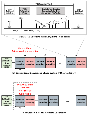 |
Accelerated SMS-FSE with Long Hard Pulse Trains and Spatially Invariant FID Suppression
Eun Ji Lim, Jaeseok Park
Simultaneous multi-slice (SMS) FSE in [1] was shown to be efficient for slice acceleration without much loss of signals. Despite its gains, conventional SMS-FSE, which employs high-flip-angle, spatially selective multi-band RF pulses in both excitation and refocusing, remains challenging particularly on high magnetic field due to high energy deposition and limited echo train length (ETL), eventually leading to low imaging efficiency. To alleviate this problem, we recently introduced a variable-flip-angle (VFA) SMS-FSE imaging with long hard pulse trains in which spatially selective multi-band RF pulses are used only for excitation while all refocusing RF pulses are short and non-selective2. Nevertheless, this approach still remains sub-optimal due to the 180° phase cycling in the refocusing pulse trains over two averages for FID suppression. Thus, the purpose of this work is to develop a novel, accelerated SMS-FSE with long hard pulse trains and spatially invariant FID suppression in which sharable FID artifacts are directly constructed using only 2-TR calibration scan instead of 2-average phase cycling scan and then subtracted. It is demonstrated that the proposed SMS-FSE with an SMS factor of 7 makes it possible to complete whole brain imaging only in 15 sec without apparent artifacts and noise.
|
|
2668.
 |
Rapid dynamic contrast-enhanced MRI for small animals at 7T using 3D UTE-GRASP
Jin Zhang, Li Feng, Ricardo Otazo, Sungheon Kim
It remains challenging to achieve simultaneous high spatial isotropic resolution and high temporal resolution in dynamic contrast enhanced (DCE) MRI of small animals, due to the relatively low signal to noise ratio (SNR) from small voxels. The purpose of this study is to develop a highly accelerated, high-spatial and high-temporal resolution DCE-MRI method for small animal imaging at 7T using 3D ultrashort echo time (UTE) golden-angle radial sampling with a combined compressed sensing and parallel imaging approach based on the GRASP technique. Our preliminary results demonstrate that the proposed UTE-GRASP method has the potential to improve both spatial and temporal resolution.
|
|
2669.
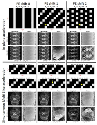 |
Improving image reconstruction with Phase Encoding Shifting of Successive IMaging slices (PESSIM)
José Marques, Daniel Gomez, David Norris
In this work we explore the added incoherence introduced when shifting the undersampling pattern in the phase enconding direction in successive slices, both when doing standard in-plane acceleration in 2D imaging or Simultaneous Multi-Slice (SMS) imaging with CAIPI trajectories. To be able to explore this incoherence, we treat both the 2D imaging and SMS imaging as one volumetric problem where the physically successive slices are forced to be coherent.
|
|
2670.
 |
Concomitant B1 Field in Low-Field MRI: Potential Contributions to TRASE Image Artefacts
Christopher Bidinosti, Pierre-Jean Nacher, Geneviève Tastevin
TRansmit Array Spatial Encoding (TRASE) MRI uses trains of rf pulses produced by transmit coils which generate transverse fields of uniform magnitude and spatially varying directions. These coils also unavoidably generate concomitant rf fields, which in turn affect magnetisation dynamics during rf flips in low-field NMR. Bloch’s equation are numerically solved to show that π-pulses imperfectly reverse transverse magnetisation and that the resulting error in azimuthal angle linearly increases with B1/B0, with the number of pulses in the TRASE pulse train, and with distance from the coil axis in the sample. This may induce significant image distortions or artefacts. Supporting experiments performed at 2 mT will be reported.
|
|
2671.
 |
Exploring the Limits of Super-Resolution MRI with Phaseless Encoding
Rui Tian, Franciszek Hennel, Klaas Pruessmann
The recently proposed method of Super-resolution (SR) MRI with phaseless subpixel encoding simultaneously samples three neighboring k-space bands and provides resolution enhancement factor up to 3.0. We now demonstrate an almost five-fold resolution enhancement by applying additional encoding steps of higher modulation frequency, which allows five bands to be acquired without compromising the methods’ immunity to phase fluctuations. Since the signal-to-noise ratio at high resolution becomes critical, we derived and experimentally verified the optimum flip angle of the encoding (tagging) sequence. A possibility to correct artefacts caused by flip angle inhomogeneity is also shown based on simulation.
|
|
2672.
 |
Banding-Free Balanced SSFP Cardiac Cine using Frequency Modulation and Phase-Cycle Redundancy
Anjali Datta, Dwight Nishimura, Corey Baron
For banding-artifact reduction in cardiac cine bSSFP imaging, we present a highly accelerated frequency-modulated sequence that can be used to acquire three phase-cycles within a short breath-hold. A reconstruction that exploits redundancies between the phase-cycles enables the high acceleration. Acquiring more phase-cycles facilitates a flatter spectral profile after phase-cycle combination. We formulate a regularization term for the reconstruction that is general to any number of phase-cycles to consistently achieve good image quality in multiple subjects.
|
|
2673.
 |
T1-weighted bipolar fat/water separated spin-echo PROPELLER acquired with dual bandwidths
Henric Rydén, Johan Berglund, Enrico Avventi, Tim Sprenger, Ola Norbeck, Stefan Skare
A bipolar fat/water separated T1-weighted dual-bandwidth spin-echo PROPELLER sequence is proposed which achieves strong and homogenous fat suppression without any dead time. Dual bandwidth sequences are compared against a corresponding fat saturated sequence in terms of SNR and CNR efficiency.
|
|
2674.
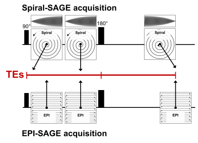 |
Development of a spiral spin- and gradient-echo (spiral-SAGE) approach for improved dynamic contrast neuroimaging
Ashley Stokes, Ryan Robison, Ashley Anderson III, James Pipe, C. Quarles
The purpose of this study is to develop a spiral-based combined spin- and gradient-echo (spiral-SAGE) pulse sequence for simultaneous dynamic contrast-enhanced (DCE-MRI) and dynamic susceptibility contrast MRI (DSC-MRI). Using this sequence, we obtained gradient-echo TEs of 1.69 and 26 ms, a SE TE of 87.72 ms, with a TR of 1663 ms. Using an iterative SENSE reconstruction followed by deblurring, spiral-induced image artifacts were minimized. Comparison of spiral-SAGE images with conventional EPI-SAGE images illustrates substantial improvements in image distortion and image intensity variations. Spiral-SAGE provides a significant improvement for the assessment of perfusion and permeability in various neuropathologies.
|
|
2675.
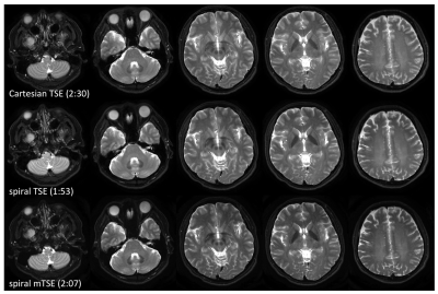 |
A Two-Dimensional Spiral Multi-Echo Turbo-Spin-Echo Technique
Zhiqiang Li, Ashley Anderson III, Melvyn Ooi, James Pipe
TSE is widely used for T2 weighted imaging in routine clinical neuro exams. However, the concerns with TSE include its high specific absorption rate (SAR), and difference in contrast compared to conventional SE. In this work we propose a 2D spiral multi-echo TSE technique, which is insensitive to the T2-decay induced signal variation that affects other spiral TSE techniques. This technique provides improved contrast, high signal to noise ratio, and substantially reduced SAR, compared to Cartesian TSE.
|
|
2676.
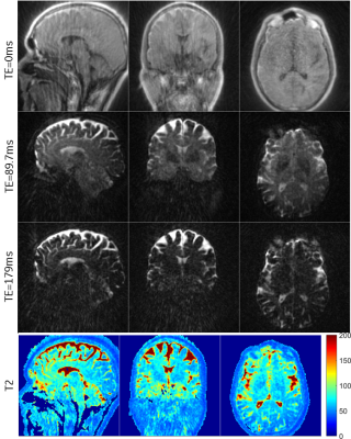 |
T2 Mapping Using ZTE Combined with Burst Encoding (BURZTE)
Rolf Schulte, Ana Beatriz Solana
ZTE acquisition is combined with spin-echo burst encoding for quiet T2 mapping. An initial ZTE excitation train encodes multiple 3D radial spokes, which get refocused by reversing the gradients. A double spin-echo leads to T2 decay, from which T2 maps are extracted by exponential fitting. Accuracy is validated in the Eurospin TO5 relaxation phantom, while in vivo feasibility is demonstrated by T2 mapping in healthy brains.
|
|
2677.
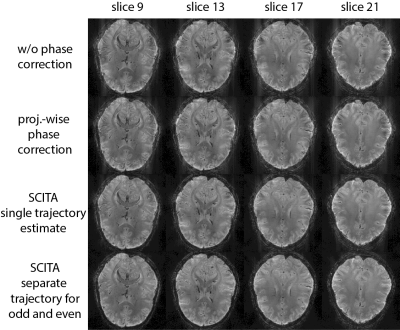 |
A Data Driven Nyquist Ghost and Gradient Delay Correction for Navigator-Free 3D Planes on a Paddlewheel (POP) EPI
Daniel Stäb, Tobias Wech, Markus Barth
3D planes-on-a-paddlewheel (POP) echo-planar imaging (EPI) is an effective non-Cartesian readout scheme realized by rotating conventional EPI readout planes about the phase encoding axis. Navigator based phase correction schemes are typically employed to account for gradient timing errors, associated trajectory errors and artifacts. In this work, we propose to use “Self Consistency for an Iterative Trajectory Adjustment” SCITA for an improved and purely data-driven removal of trajectory misalignment artifacts. As the actual k-space trajectory is derived from the imaging data, navigator acquisitions can be omitted and echo, repetition and acquisition times may be considerably shortened.
|
|
2678.
 |
Tailored SEMs for wave modulations in SMS imaging
Sebastian Littin, Stefan Kroboth, Huijun Yu, Feng Jia, Ying-Hua Chu, Yi-Cheng Hsu, Maxim Zaitsev
The use of a matrix gradient coil enables to tailor spatial encoding magnetic Fields (SEMs) for slice specific frequency shifts. Applying such shifts in oscillatory manner allows for novel methods of signal separation in SMS imaging.
|
|
2679.
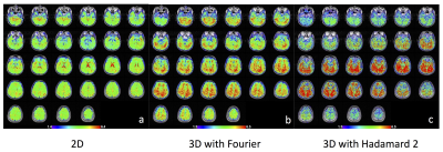 |
Phase corrected Hadamard acquisition compared with three-dimensional (3D) Fourier encoding for functional MRI
Seul Lee, Gary Glover
Three-dimensional (3D) functional MRI (fMRI) can be superior in localization of activation signals compared to two-dimensional (2D) fMRI because higher spatial resolution can be acquired due to potentially higher signal-to-noise ratio (SNR) and thinner slices. However in 3D, physiological noise reduces SNR due to higher signal at the k-space center; thus the number of slices should be decreased to reduce physiological noise. With Fourier encoding, acquiring a small number of slices results in excessive Gibbs ringing. In this study, we propose Hadamard reconstruction for 3D fMRI acquisition to avoid the artifact caused from Fourier encoding and return higher SNR.
|
|
2680.
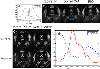 |
Improved Automatic Deblurring Using a Novel Objective Function Paired with a Retraced Spiral Acquisition Trajectory
Steven Allen, Xue Feng, Samuel Fielden, Craig Meyer
We introduce a novel objective function for automatic deblurring of images acquired with non-2DFT trajectories. When paired with the recently introduced retraced, spiral-in-out trajectory, this objective function provides two advantages over previously established functions: it is invariant with incidental phase and is less susceptible to spurious extrema. These advantages lead to effective deblurring over a larger range of off resonance conditions and readout durations. Here, using simulations and phantom studies, we compare the sensitivity of this objective function to spurious extrema to a previously proposed function.
|
|
2681.
 |
Influence of Parameter Optimization and Segmentation on the Accuracy of Various Registration Approaches for Multi-parametric 3D Breast MRI Data
Subhajit Chatterjee, Snekha Thakran, Rakesh Gupta, Anup Singh
Registration of human Breast MRI images is challenging due to its elastic deformable nature. In this study, various existing rigid and non-rigid registration methods were evaluated and compared in terms of accuracy and computation time. This work investigated influence of different registration parameters and showed possible ways to achieve better registration results. Experiential result revealed that the combination of Affine and B-spline method provided more time efficiency and accuracy than other methods.
|
|
2682.
 |
Radius Segmented Multi-shot Spiral for Diffusion Imaging
Yukari Yamamoto, Shinji Kurokawa, Yoshitaka Sato, Hisaaki Ochi
Single-shot echo-planar imaging (EPI) is usually used in diffusion-weighted imaging (DWI); however, it is difficult to apply to examining the entire brain because of image distortion due to susceptibility inhomogeneity. In addition, multi-shot imaging, in which image distortion is relatively small, is affected by pulsation artifacts and aliasing. We propose a multi-shot spiral method in which a spiral trajectory is divided in the radial direction. DWI studies were performed on the brain of a healthy volunteer. The proposed method could sample k-space data for each shot without aliasing, and sufficient correction for pulsation artifacts could be obtained.
|
|
2683.
 |
PET/MR dynamic imaging of an inflatable phantom with self-gated UTE-MRI
Fatiha Andoh, Tanguy Boucneau, Marina Filipovic Pierucci, Simon Stute, Brice Fernandez, Peder Larson, Xavier Maître
MRI offers many advantages for chest imaging such as the absence of irradiation and the opportunity to obtain images with various contrasts in soft tissues. Developing MRI lung imaging would provide solutions to a real public health problem related to lung disease. Besides, PET is relevant for the study of metabolic changes caused by parenchymatous affections. Hence PET-MRI is a promising route for the characterization of lung diseases. One of the immediate issues lung imaging raises is motion. Physiological motion needs to be taken into account during the imaging process to avoid blurring or ghosting artifacts in both imaging modalities.
|
|
2684.
 |
Motion Correction for Quantitative 3D UTE Cones Magnetization Transfer (3D UTE-Cones-MT) Imaging and 3D UTE Cones Adiabatic T1? (3D UTE-Cones-AdiabT1?) Imaging of the Knee Joint
Wei Zhao, Yajun Ma, Michael Carl, Xing Lu, Eric Chang, Jiang Du
Conventional T2 and T1ρ have limited values in evaluating short T2 tissues, and are affected by the magic angle effect. Ultrashort echo time (UTE) sequences can detect short T2 tissues. Magnetization transfer (MT) modeling and adiabatic T1ρ (AdiabT1ρ) seem to be insensitive to the magic angle effect. The combination of 3D UTE-Cones sequence with MT (3D UTE-Cones-MT) and AdiabT1ρ (3D UTE-Cones-AdiabT1ρ) may resolve those limitations. However, patient motion may occur during the relatively long scan time. This study aims to develop 3D UTE-Cones-MT and UTE-Cones-AdiabT1ρ with an elastix registration technique to compensate for motion during the scans.
|
|
2685.
 |
Rotating Outer Volume Suppression for Reduced Field of View PROPELLER Imaging
Daniel Litwiller, Valentina Taviani, Suchandrima Banerjee, Lloyd Estkowski, Yuval Zur, Ali Ersoz, Ersin Bayram
We present a modified PROPELLER pulse sequence that incorporates rotating outer volume suppression for reduced field of view imaging. In vivo results are presented, demonstrating comparable imaging performance with conventional PROPELLER imaging.
|
|
2686.
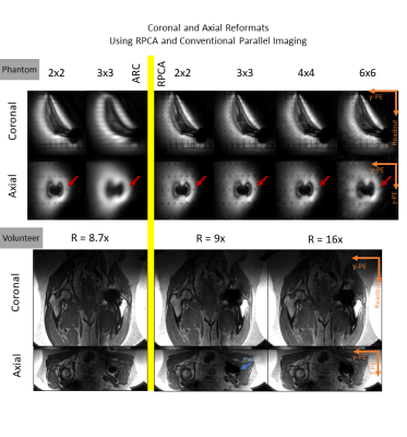 |
Reformattable MAVRIC-SL Using Robust Principal Component Analysis and Variable Density Complementary Poisson Disc Sampling
Philip Lee, Daehyun Yoon, Xinwei Shi, Evan Levine, Yuxin Hu, Brian Hargreaves
MAVRIC-SL resolves metal-induced artifacts at the cost of additional scan time. A reconstruction using Robust Principal Component Analysis (RPCA) has been shown to considerably reduce scan times with minimal loss in image quality. We apply this scan time reduction to acquire isotropic MAVRIC-SL data that can be reformatted to all three planes, combining multiple high-resolution scans into a single, short, isotropic scan. We show retrospectively undersampled isotropic MAVRIC-SL RPCA reconstructions reformatted to three planes for the case of a hip phantom, and a volunteer with a titanium hip replacement. The RPCA reconstruction offers good image quality in multiple planes at clinically feasible scan times, with shorter scan times than separate high-resolution acquisitions.
|
|
2687.
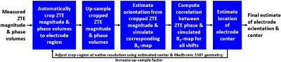 |
Accurate localization of individual DBS contacts by MRI using zero-TE phase images
Sathish Ramani, Rolf Schulte, Graeme Mckinnon, Jeffrey Ashe, Julie Pilitsis, Ileana Hancu
The goal of our work was to demonstrate improved DBS contact visualization and localization by using a zero-TE (ZTE) acquisition. Signal dephasing during sequence readout, proportional to the electrode-induced field inhomogeneity, enables high-contrast visualization of individual electrode contacts. Matching measured ZTE-phase maps to simulations of orientation dependent, susceptibility induced field inhomogeneity created by the electrode is shown to result in significantly more accurate and precise contact localization than by using standard SPGR acquisitions. Electrode center differences of 0.69±0.45mm/0.32±0.09mm were seen between SPGR/ZTE[phase] and CT.
|
|
2688.
 |
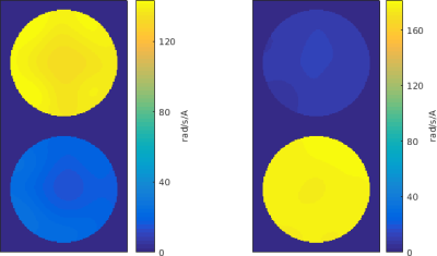
Measured k-space based RF Compensation Effect Analysis within Various 2D Excitation Volume in 7T pTx system
Sanghoon Kim, Mark Lowe
This work presents simple method for RF compensation effect analysis. For the RF compensation, we used previously presented measured k-space based method. We analyzed three different 2D excitation volume data using simple histogram based method and found that not only for small volume excitation region, larger volume excitation region shows significant and more dominant compensation effect. This finding will help inform the design of RF profiles in In-vivo 2D excitation applications in pTx system.
|
|
2689.
 |
KT-Points Pulses Reduce B1 Shading at 3T: Demonstration in Routine Abdominal DCE-MRI and Evaluation of Reliability
Raphaël Tomi-Tricot, Vincent Gras, Franck Mauconduit, François Legou, Nicolas Boulant, Matthias Gebhardt, Dieter Ritter, Berthold Kiefer, Pierre Zerbib, Alain Rahmouni, Alexandre Vignaud, Alain Luciani, Alexis Amadon
At high field, MRI systems offer a higher signal-to-noise ratio, but B1+-inhomogeneity-induced artefacts in large organs can lead to shading and erroneous contrast. In this work, subject-tailored kT-points pulse design performance was evaluated in clinical routine on liver DCE-MRI at 3T, against that of patient-specific RF shimming. Both excitation homogeneity simulation and image quality assessment were performed on a variety of patients. The interest of kT-points is clearly demonstrated, as well as the reliability of the approach.
|
|
2690.
 |
kT-spokes: combining kT-points with spokes to ease ramp pulse design for TOF slab selection with parallel transmission at 7T
Gaël Saïb, Vincent Gras, Franck Mauconduit, Alexandre Vignaud, Denis Le Bihan, Laurent Le Brusquet, Nicolas Boulant, Alexis Amadon
TONE pulses counteract blood saturation through the imaged slab in TOF sequences, but their ramp profile is hampered by RF inhomogeneities at Ultra High Field. On the other hand, kz-spokes are known to compensate for in-plane B1+ heterogeneities in slice or slab selection. However, their design doesn’t address thru-slab heterogeneities. To address them, a new pulse type called “kT-spokes” is introduced. As TONE pulses, kT-spokes efficacy is demonstrated with pTx at 7T in comparison with mere equivalent kz-spokes.
|
|
2691.
 |
K-Space Trajectory Correction for UTE Sequence with Multi-Echo Radial Acquisition
Liao Ying, Paul Han, Shuang Hu, Kui Ying, Chao Ma, Georges El Fakhri
UTE allows imaging of rapidly decaying short-T2 components and are often combined with multi-echo radial acquisition for PET attenuation correction applications. However, UTE is inherently susceptibility to gradient errors due to the usage of radial acquisition and simple time delay corrections render impractical to correct deviations from the ideal trajectory when UTE is combined with multi-echo radial acquisition scheme. In this work, we describe a simple, one-time calibration method that allows k-space trajectory correction for UTE sequence combined with multi-echo radial acquisition. The performance of the proposed method is shown via a phantom and an in vivo experiment, using a calibration scan previously acquired from a water phantom.
|
|
2692.
 |
Spin Lock Adiabatic Correction (SLAC) Excitation
Edward Green, James Korte, Bahman Tahayori, Peter Farrell, Leigh Johnston
A new form of B1-insensitive excitation is introduced, termed Spin-Lock Adiabatic Correction (SLAC) excitation, that combines a Spin-Locking excitation with an orthogonal Adiabatic Correction to more uniformly flip the magnetisation across a range of B1 strengths. SLAC pulses achieve adiabatic-like excitation, in terms of B1-insensitivity, in faster excitation time while not increasing the delivered power. We demonstrate the advantages of SLAC pulses in both simulation and phantom experiments. Decreasing the pulse duration causes performance breakdown of the adiabatic pulse due to violation of the adiabatic condition, while the SLAC pulse maintains control of magnetisation across the range of B1 strengths.
|
|
2693.
 |
Comparison of Efficacy of Multiple EPI Distortion Correction Techniques on Toddler Data
Vinai Roopchansingh, Jerry French Jr., Daniel Glen, Richard Reynolds, Dylan Nielson, Robert Cox, Audrey Thurm, Susan Swedo
Echo-planar data acquired from a group of toddlers was distortion corrected using combinations of different data, algorithms, and software packages. Performance was evaluated by comparing mutual information scores of how well corrected versus uncorrected EPI data aligned with structural T 1-weighted data.
|
|
2694.
 |
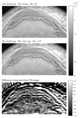
Evaluating T2* bias impact and correction strategies in quantitative proton density mapping
Evelyne Balteau, Tobias Leutritz, Nikolaus Weiskopf, Enrico Reimer, Antoine Lutti, Martina Callaghan, Siawoosh Mohammadi, Karsten Tabelow
Bias correction is an important step for achieving accurate and precise parameter quantification in MRI. Residual T2*-weighting in quantitative proton density maps estimated from short echo time FLASH images is often considered negligible, despite the potential bias. Using the hMRI toolbox, we analyse simulated FLASH-based multiparameter mapping datasets with variable noise levels. Using the quantitative maps on which the simulations are based as a gold standard, we quantified the bias caused by residual T2*-weighting. Furthermore, we evaluated a number of estimation methods in terms of their sensitivity and/or effectiveness at correcting this T2*-weighting bias, and in terms of their robustness to background noise.
|
|
2695.
 |
A Simple Method for Improved Correction of EPI Odd-Even Line Inconsistency
Yuan Zheng, Yu Ding, Qing Wei, Weiguo Zhang
We have developed a simple method for EPI Nyquist ghosting artifacts removal. Our technique borrows the idea of GRAPPA, and extracts a non-biased kernel from imperfect multichannel EPI data to correct the odd-even line inconsistency. We have demonstrated both in-vivo and in-vitro that this strategy can significantly reduce Nyquist ghosts. The proposed method is quite simple and can be conveniently used with many current EPI correction techniques to generate ghosting-free images.
|
|
2696.
 |
Real-time cardiac MR imaging based on a radial bSSFP sequence with trajectory auto-correction
Guoxi Xie, Xiaoyong Zhang, Wenlong Lv, Caiyun Shi, Shi Su, Bensheng Qiu, Xin Liu
Conventional cardiac cine imaging is based on ECG-triggering, which is difficult to be used in arrhythmic patients. Real-time cardiac cine technique based on radial sampling scheme is an alternative approach for imaging the arrhythmic patients. However, the technique is often hampered in trajectory error due to system gradient delay. To address this issue, a novel real-time cardiac cine technique was developed based on a radial bSSFP sequence with trajectory error auto-correction. Preliminary results demonstrated that the proposed technique can improve the image quality and has potential to be clinically useful for the arrhythmic patients.
|
|
2697.
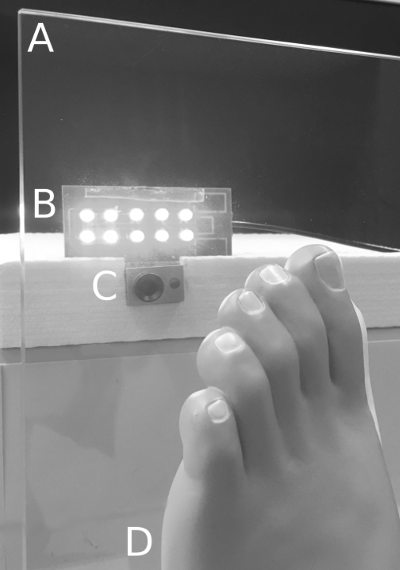 |
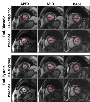
A novel method for video-based cardiac gating in 7T MR angiography using a video of the foot
Nicolai Spicher, Stephan Orzada, Stefan Maderwald, Mark Ladd, Markus Kukuk
In ultra-high-field MRI, cardiac gating is problematic because electrocardiography is prone to magnetohydrodynamic artifacts and pulse oximetry suffers from signal loss during long examinations. The goal of this work is to investigate practical feasibility of cardiac gating based on a video from the sole of the foot that is leaned to a glass plate. We combined this novel setup with an open-source software for video-based cardiac gating (https://github.com/nspi/vbcg) and performed ultra-high-field non-enhanced angiography in one volunteer. As reference, we performed pulse oximetry gating and comparison of maximum intensity projection images shows a similar image quality. Future work will evaluate the feasibility of this novel cardiac gating method in a larger cohort.
|
|
2698.
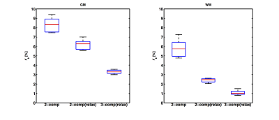 |
Multi-compartment relaxation-compensated IVIM imaging of the human brain
Anna Rydhög, Ofer Pasternak, Freddy Ståhlberg, Ronnie Wirestam, Linda Knutsson, André Ahlgren
In conventional intravoxel incoherent motion (IVIM) imaging, the blood fraction is estimated using a two-compartment model (blood and tissue). However, blood fraction estimation is hampered by cerebrospinal fluid (CSF) contamination and tissue-dependent relaxation times. We propose a three-compartment model (blood, tissue, CSF), which accounts for compartment-specific diffusion and relaxation properties. Estimation of gray and white matter blood fractions using this model is demonstrated with in-vivo human data of variable diffusion weightings, echo times and inversion times. In comparison with two-compartment models (with and without relaxation), the proposed three-compartment model yielded lower estimates of the blood fraction, suggesting a better separation from CSF.
|
|
2699.
 |
Data driven sampling of k-space using GO-Active technique
Pavan poojar, Ashok reddy, Amaresha Konar, Ramesh Venkatesan, Sairam Geethanath
The extensive coverage of k-space data on a standard MRI scanner requires long acquisition times. In dynamic MRI methods such as DCE-MRI, cardiac MRI, DWI, etc., the shape of the significant values in k-space depends on the structure of the organ and temporal events. The proposed method generates the arbitrary k-space trajectory and optimizes the gradient waveforms by utilizing GO-Active. Design constraints of gradient system are slew rate and gradient amplitude are accounted for by using convex optimization. All images were acquired on a GE 1.5T scanner. Image reconstruction was performed in graphical programming interface.
|
|
2700.
 |
Optimal Choice of Echo Times for Gradient Echo B0 Field Mapping
Yasmin Geiger, Assaf Tal
Field maps are essential in spectroscopy, shimming, MR thermometry and geometric distortion correction. Minimizing the noise in acquired field maps is therefore potentially important to all of these applications. When using a multi-gradient echo, the choice of echo times has a marked effect on the noise on the acquired field maps. Here, we derive the optimal echo times which minimize the amount of noise in the resulting field maps.
|
|
2701.
 |
Unconventional trajectories on the Bloch Sphere: A closer look at the effects and consequences of the breakdown of the rotating wave approximation
Christopher Bidinosti, Pierre-Jean Nacher, Geneviève Tastevin
TRASE MRI uses rapid π-pulses of phase gradient fields, and in general requires as many as two distinct phase-gradient coils per encoding direction. This tends to restrict one to large amplitude, linear B1 fields, which in low B0 field leads to a breakdown of the rotating wave approximation. We have studied this regime both numerically and experimentally. Our results show a rich behavior involving a complex interplay of the Bloch-Siegert shift, the B1 start and stop phase, and B1 amplitude transients.
|
|
2702.
 |
Respiratory-Gated B0 Field Stabilisation for High Resolution Mouse Brain Imaging
Paul Kinchesh, Stuart Gilchrist, Niloufar Zarghami, Alexandre Khrapitchev, Nicola Sibson, Sean Smart
The echoes of a 3D multi gradient echo (MGE) scan are typically combined for detection of USPIO and MPIO. The echo combination requires B0 to be constant throughout the scan to achieve good image fidelity at high resolution. A navigator acquisition embedded in the MGE scan maintains the MR steady state and enables a real-time adaptive B0 correction. It is demonstrated that a respiratory-gated correction scheme outperforms ungated correction in mouse brain for the detection of micron sized iron-oxide particles coupled with anti-vascular cell adhesion molecule antibody (VCAM-MPIO) to identify inflammation in vessels.
|
|
2703.
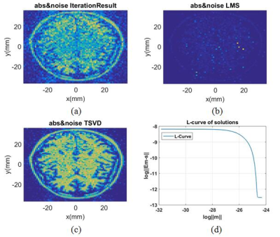 |
Flexible spatial encoding strategy using receive coil aggregates for Halbach magnet array based magnetic resonance imaging
Dong Wei Lu, Zhi Hua Ren, Shao Ying Huang
To make a MRI system portable, a practical approach is applying Halbach magnet array and nonlinear spatial encoding strategy. Here, the rotation of a magnet array for imaging is replaced by electrically forming RF receive coil aggregates with phase delay. For the resultant system with a new encoding matrix, Truncated-Singular-Value-Decomposition with an optimal regularization parameter is proposed which reconstructs images with good quality. An accelerated L-curve method is proposed to obtain the optimal regularization parameter. Results show that the proposed strategy provides considerable improvement of the image quality compared to existing method, e.g. Kaczmarz iteration, without rotating the magnet array.
|
|
2704.
 |
Simultaneous Multi-Contrast Imaging in Combination with in-plane Parallel Imaging
Nora-Josefin Breutigam, Matthias Günther, David Porter
Simultaneous Multi-Contrast (SMC) Imaging enables a synchronous acquisition of multiple image contrasts within one measurement. The technique reduces patient examination times and facilitates accurate image registration between contrasts. Previous work used readout-segmented EPI (rs-EPI) to perform high-resolution, navigator-corrected, diffusion-weighted imaging simultaneously with a T2*-weighted acquisition. This combination of contrasts has clinical significance in acute stroke. These previous studies did not use in-plane acceleration to reduce spatial distortion caused by the EPI readout. This study introduces an updated version of the SMC technique that incorporates in-plane acceleration with GRAPPA to allow an improved image quality for future clinical studies.
|
|
2705.
 |
3D Cones acquisition for human extremities using a 1.5 T compact superconducting magnet and unshielded gradient coil
Ayana Setoi, Katsumi Kose
We developed 3D Cones sequences for human extremities on a 1.5 T MRI system using a compact superconducting magnet (280 mm bore) equipped with an unshielded gradient coil. Linear eddy fields were measured using a spherical phantom and eddy current effects on the 3D Cones sequences were evaluated using a 3D water phantom. As a result, effects of higher-order eddy fields proportional to z2x and z2y spatial distributions were clearly observed. The 3D Cones sequences were applied to UTE imaging of a porcine hoof sample and a human forearm, which demonstrated their promise in UTE imaging.
|
|
2706.
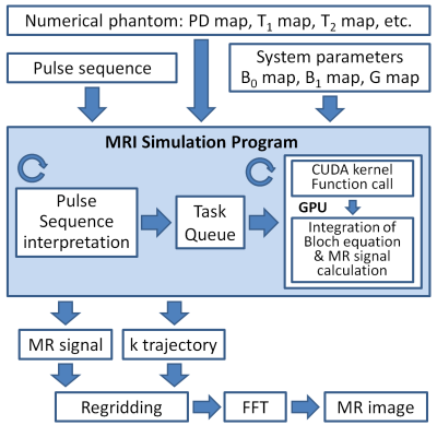 |
GPU-optimized fast 3D MRI simulator for arbitrary trajectory sampling
Ryoichi Kose, Ayana Setoi, Katsumi Kose
We developed a GPU-optimized fast 3D MRI simulator for arbitrary trajectory sampling. The performance of the simulator was evaluated using stack of 2D spiral and 3D Cones sequences. The result demonstrated that our simulator is a powerful tool for studies of non-Cartesian sampling as well as Cartesian sampling imaging sequences.
|
|
2707.
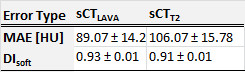 |
DIXON-type pulse sequence for MRI-only external beam radiotherapy of prostate cancer
Souha Aouadi, Satheesh Paloor, Ana Vasic, Tarraf Torfeh, Maeve McGarry, Primoz Petric, Hadi Fayad, Rabih Hammoud, Noora Al-Hammadi
Water-fat separated images provided by the DIXON-type pulse sequence were combined with the multi-scale and dual-contrast patch-based method to generate synthetic-CT (sCT) for MR-only external beam radiotherapy treatment planning of prostate cancer. The benefit of such sequence was demonstrated by retrospective geometric and dosimetric evaluation of sCT on five patients. Compared to reference CT, the mean absolute error was 89.07±14.2HU, the dice coefficient in soft tissues was 0.93±0.01. Good agreement with conventional planning techniques was obtained; the highest percentages of dose metrics deviations were below 0.7% for PTV, 0.05% for the rectum, and 0.01% for the bladder.
|
|
2708.
 |
Simultaneous Multi-Slice fMRI of the Mouse Brain Using POMP-EPI at 9.4T
Hsu-Lei Lee, Zengmin Li, Kai-Hsiang Chuang
Acceleration of rodent brain functional MRI using parallel imaging techniques is not widely used due to the limited availability of high-density phased-array coil on pre-clinical scanners. In this study we demonstrated a POMP-EPI method to enable simultaneous multi-slice acquisition for fast mouse brain imaging without a phased array coil. A four-fold multiband acceleration was achieved without using coil sensitivity information. This method can be used to increase the spatial or temporal resolution of mouse fMRI acquisition, which will benefit the study of dynamics of neural activity and connectivity.
|
|
2709.
 |
Analysis of diffusion effects in SSFP sequences with extended phase graphs
Yangzi Qiao, Chao Zou, Chuanli Cheng, Qian Wan, Changjun Tie, Xin Liu, Hairong Zheng
EPG simulation was applied to analysis the diffusion effect of two SSFP-FID signals, FISP and ES. The influence of T1, T2, and unbalanced gradient on signal intensity with consideration of diffusion effect was studied. The EPG simulation have a good consistency with the experimental data, indicating it can efficiently and precisely calculate the diffusion effect of SSFP signals. Both the simulation and phantom study reveals that for some specific tissues and imaging parameters, positive diffusion contrast can be obtained in FISP and ES sequence. For quantitative method based on SSFP signals, such as TESS relaxometry, the diffusion effect should be considered while large unbalanced gradients and small flip angle were employed for high resolution imaging in high field system.
|
|
2710.
 |
Simple algorithm for the correction of MRI image artefacts due to random phase fluctuations
P. Ross, Lionel Broche, David Lurie
Here we present a simple post-processing algorithm that is able to correct ghosting caused by a slow off-resonance drift caused by the use of a resistive magnet. The algorithm is described and validated in simulations, phantoms and in vivo.
|
|
Machine Learning for Cancer Applications
Traditional Poster
Acquisition, Reconstruction & Analysis
Thursday, 21 June 2018
| Exhibition Hall 2711-2725 |
08:00 - 10:00 |
|
2711.
 |
Radiomics analysis for preoperative prediction of synchronous distant metastasis in patients with rectal cancer
Huanhuan Liu, Caiyuan Zhang, Jinning Li, Weibo Chen , Dengbin Wang
Rectal cancer is one of the most common malignant tumors in gastrointestinal tract. Tumor metastasis is still a major cause of death in patients with rectal cancer. The distant metastasis rate for rectal cancer remains constant at 20-50%1. Prediction of synchronous distant metastasis is important for the choice of personalized treatment strategies. Radiomics can extract quantitative features from digital images, which are related to the underlying pathophysiology2. We developed a radiomics model based on the MR radiomics features in combination with independent clinico-radiologic risk factors, which help to predict the synchronous distant metastasis in patients with rectal cancer.
|
|
2712.
 |
Computer-aided diagnosis of hepatocellular carcinoma and hepatic cavernous hemangioma using non-enhanced MRI with a random forest classifier
Jingjun Wu, Ailian Liu, Jingjing Cui, Lizhi Xie
The current study aims to develop a computer-aided diagnosis (CAD) system and assess its ability in identification of hepatocellular carcinoma (HCC) and hepatic cavernous hemangioma (HCH) using non-enhanced MRI with a random forest classifier. Good performance was observed in this CAD system based on out-phase images.
|
|
2713.
 |
MR Image Synthesis For Glioma Segmentation
Ken Chang, Andrew Beers, James Brown, Elizabeth Gerstner, Bruce Rosen, Jayashree Kalpathy-Cramer
Deep learning has become the method of choice for tumor segmentation. Most deep learning algorithms incorporate a multi-modal approach, as different MR modalities are optimized to detect different aspects of tumor. However, modalities are often missing or unusable due to artifacts. In such cases, it is difficult to perform robust automatic tumor segmentation. We demonstrate that a convolutional neural network can be used to synthesize FLAIR MR images that have high similarity with real FLAIR images. Furthermore, we show that the use of these synthetic images can improve segmentation performance.
|
|
2714.
|
Development and Validation of a Classifier for Prediction of Distant Metastasis in Nasopharyngeal Carcinoma at Initial Staging
Bin Zhang
we sought to improve the prediction of DM in NPC patients by developing a novel combined classifier to stratified patients into high-risk and low-risk groups with significant differences in 5-year survival. To our best of knowledge, our study is the first to integrate intratumor heterogeneity with EBV DNA for predicting DM in NPC patients, and found the combined classifier achieved superior prognostic performance than either the radiomic signatures or the clinical variables alone, which with a higher AUC, sensitivity, and specificity improvement.
|
|
2715.
 |
Motion Detection and Quality Assessment of MR images with Deep Convolutional DenseNets
Sandro Braun, Xiao Chen, Benjamin Odry, Boris Mailhe, Mariappan Nadar
We use simulated motion-corrupted images to compute associated image quality metrics and quantify the corresponding severity of motion. We train models with four different inputs (full image, Foreground only, Background only or both Foreground and Background in two channels) to regress to those metrics. To obtain a ground-truth as acceptable or not acceptable image quality, we choose acceptance thresholds within a reasonable range, depending on the level of tolerable motion. The network shows high accuracy within this range. For both metrics used (MSSIM and NRMSE), BG-models perform better than FGBG-models.
|
|
2716.
 |
A multi-channel convolutional neural network for segmentation of breast lesions in DCE-MRI
Karl Spuhler, Mario Serrano Sosa, Jie Ding, Tim Duong, Chuan Huang
Radiomics offers a highly quantitative and high-dimensional view of the tumor microenvironment which no conventional imaging technique allows. It is the ideal strategy for personalizing care in heterogeneous cancers such as in the breast. Most approaches require time consuming, manual region of interest segmentation. Here, we present a fast and accurate neural network approach for breast lesion segmentation which can be adapted to accept any number of imaging modalities and shows reliability across many types of lesion.
|
|
2717.
 |
Segmentation of Bone Tumor with MR imaging using Machine Learning
Amit Mehndiratta, Akshay Gupta, Esha Kayal, Devasenathipathy Kandasamy, Sameer Bakhshi, Raju Sharma
There has been a lot of work in segmentation of tumors in organs like the brain. Segmentation of bone tumor with MRI is not widely studied. Manual segmentation can be costly and time consuming. We study three automatic 3D segmentation techniques: Energy-based graph cuts, deep feed forward neural networks and mean shift clustering. Results show that, these methods can perform good quality segmentation (dice coefficient >70%) even with no human intervention. Tumor ADC values computed using these methods are comparable with those obtained from manual segmentation, showing that these methods can be used as a screening tool.
|
|
2718.
 |
Noninvasive Identification of IDH-mutational Status from 1H-MRS Spectra by Deep Learning
Hyeonghun Lee, Hyeonjin Kim
Noninvasive identification of IDH-mutational status in glioma patients using 1H-MRS is diagnostically and prognostically valuable. However, the most widely used short TE method is reported to be more subject to false diagnosis due to the severe spectral overlap of 2HG. We explored the potential applicability of deep learning in addressing this issue. A deep neural network that was trained on a large number of simulated spectra substantially improved the overall diagnostic accuracy on the patient spectra, compared to the LCModel analysis. As no spectral fitting is involved, our results are not subject to ambiguity arising from the CRLB-based data interpretation.
|
|
2719.
 |
Evaluation of 2D and 3D convolutional neural network methods for generating pelvic synthetic CT from T1-weighted MRI
Jie Fu, Yingli Yang, Kamal Singhrao, Dan Ruan, Daniel Low, Anand Santhanam, John Lewis
Synthetic CT (sCT) must be generated directly from MRI scans to achieve MRI-only radiotherapy. We propose 2D and 3D convolution neural network models for generating pelvic sCT and evaluate their performance. Five-fold cross-validation is performed using paired T1-weighted MRI and CT scans from 20 patients. Our results show the 2D model generates accurate sCT for all patients in this study. The average mean absolute error (MAE) between CT and sCT across all patients is 38.0±3.9 HU in the 2D model. The average MAE is 55.9±28.4 HU in the 3D model. This large variation is possibly due to the limited number of 3D training volumes.
|
|
2720.
 |
The Weakest Link in the Chain: How MR Data Quality influences Convolutional Neural Network Performance
Lars Bielak, Hatice Bunea, Nicole Wiedenmann, Anca-Ligia Grosu, Michael Bock
In this work, tumor segmentation performance of a convolutional neural network is tested with respect to input data quality. 19 patients suffering from head and neck tumors underwent multi-parametric MRI including diffusion weighted imaging. The network was trained on multiparametric MR images with and without geometrically corrected diffusion data. With distortion correction, the Dice coefficient could be increased by 22% over uncorrected data showing the necessity for geometric image pre-processing in neural network analysis.
|
|
2721.
 |
Computer aided quantification of prostate cancer diffusion-weighted imaging: repeatability analysis of radiomics as biomarkers for Gleason score prediction
Ileana Montoya Perez, Jussi Toivonen, Parisa Movahedi, Harri Merisaari, Janne Verho, Pekka Taimen, Peter Boström, Tapio Pahikkala, Hannu Aronen, Ivan Jambor
We evaluated the repeatability of apparent diffusion coefficient, derived using monoexponential function (ADCm) from prostate cancer DWI (12 b values, 0-2000 s/mm2), radiomics of prostate cancer and their potential to predict prostate cancer Gleason score (histological grading system of prostate cancer aggressiveness). Statistical features (mean, median, 10th, 25th percentile) and Gabor texture feature of DWI ADCm parametric maps showed high repeatability and correlated significantly with Gleason score. In contrast, homogeneity gray-level co-occurrence matrix showed low repeatability despite having significant correlation with Gleason score.
|
|
2722.
 |
Locating hypoxia-related tumour regions in NSCLC: utility and repeatability of data-driven segmentation of combined OE/DCE-MRI data
Adam Featherstone, Ahmed Salem, Ross Little, Yvonne Watson, Susan Cheung, Corrine Faivre-Finn, James O'Connor, Julian Matthews, Geoff Parker
There is a need to develop tumour hypoxia biomarkers for patient stratification and for tracking tumour response to therapy. We apply our preclinically-optimised, data-driven segmentation of combined OE-MRI/DCE-MRI data to a cohort of non small-cell lung cancer (NSCLC) patients, aiming to map tumour hypoxia non-invasively. Tissue classes with different oxygenation and perfusion characteristics are located, and we discuss challenges specific to use in the clinical setting. Further optimisation of the technique is needed to improve its repeatability and its ability to enable the identification of definitively hypoxic regions in these types of data.
|
|
2723.
 |
Improving the image quality of liver DWI using the convolutional neural network-based selection algorithm
Daiki Tamada, Utaroh Motosugi, Hiroshi Onishi
Diffusion-weighted imaging (DWI) of the liver using a single-shot EPI sequence suffer from motion artifact caused by cardiac motion. The reconstruction of DWI with multiple numbers of excitation including the corrupted echoes due to systolic cardiac motion results in a severe signal loss in the left lobes, even if other echoes in diastolic phase had no artifact. In this study, we propose a selection algorithm to reject the corrupted echoes using convolutional neural network was proposed. The volunteer studies demonstrated that the proposed method improves the image quality of liver DWI.
|
|
2724.
 |
Repeatability of Selected Multiparametric Prostate MRI Radiomics Features
Michael Schwier, Joost van Griethuysen, Mark Vangel, Steve Pieper, Sharon Peled, Clare Tempany, Hugo Aerts, Ron Kikinis, Fiona Fennessy, Andrey Fedorov
In this study we assess the repeatability of selected radiomics features for small prostate tumors in ADC and T2-weighted images. We used a prostate mpMRI test-retest dataset for our evaluation. Different configurations of preprocessing were compared. The intraclass correlation coefficient was employed as a measure of repeatability. Our results show that several of the selected features have good repeatability, however, only when specific preprocessing was applied. Based on our data, texture computation should be done in 2D. Normalization improves repeatability for ADC features, but not in T2-weighted images.
|
|
2725.
 |
Quantitative texture analysis of apparent diffusion coefficient (ADC) for evaluating histologic differentiated grade of head and neck squamous cell carcinoma
Yu Chen, Yanan Zhao, Huadan Xue, Zhuhua Zhang, Zhengyu Jin
To investigate the feasibility of using texture analysis (TA) of apparent diffusion coefficient (ADC) to distinguish between well- and moderate- differentiated head and neck squamous cell carcinoma (HNSCC). A total of 22 patients were retrospectively analyzed, including: well-differentiated degree SCC (WSCC, n=11) and moderate-differentiated degree SCC (MSCC, n=11). A Mean>101.38 at coarse texture scale (SSF=6mm) identified WSCC and MSCC with the highest AUC of 0.843±0.083 (Se=72.7%, Sp=81.8%, PPV=80%, PV=75%, and accuracy=77.3%). Texture analysis of ADC proved to be a feasible tool for differentiating WSCC from MSCC, and had better diagnostic performance than ADC value.
|
|
Machine Learning for Tissue Segmentation & Classification
Traditional Poster
Acquisition, Reconstruction & Analysis
Thursday, 21 June 2018
| Exhibition Hall 2726-2738 |
08:00 - 10:00 |
|
2726.
 |
Deep learning-based whole head segmentation for simultaneous PET/MR attenuation correction
Jakub Baran, Kamlesh Pawar, Nicholas Ferris, Sharna Jamadar, Marian Cholewa, Zhaolin Chen, Gary Egan
Estimation of an accurate PET attenuation correction factor is crucial for quantitative PET imaging, and is an active area of research in simultaneous PET/MR. In this work, we propose a deep learning-based image segmentation method to improve the accuracy of PET attenuation correction for simultaneous PET/MR imaging of the human head. We compare segmentation methods for accurate tissue segmentation and attenuation map generation. We demonstrate improved PET image reconstruction accuracy using the proposed deep learning-based method.
|
|
2727.
 |
Generalized AI for Organ Invariant Tissue Segmentation and Characterization of Multiparametric MRI: Preliminary Results
Vishwa Parekh, Katarzyna Macura, Michael Jacobs
Artificial intelligence(AI) and deep learning techniques are increasingly being used in radiological applications. The true potential of deep learning in MRI applications can only be achieved by developing an AI that can learn the underlying MRI physics rather than a task that is specific to an organ or a particular tissue pathology. To that end, we developed and tested a multiparametric deep learning model capable of tissue segmentation and characterization in both breast cancer and stroke.
|
|
2728.
 |
Brain Segmentation in Rodent MR-Images Using Convolutional Neural Networks
Björn Sigurðsson, Sune Darkner, Stefan Sommer, Kristian Mortensen, Simon Sanggaard, Serhii Kostrikov, Maiken Nedergaard
This study compares two different methods for the task of brain segmentation in rodent MR-images, a convolutional neural network (CNN) and majority voting of a registration based atlas (RBA) , and how limited training data affect their performance. The CNN was implemented in Tensorflow. The RBA performs better on average when using a training set with fewer than 20 images but the CNN achieves a higher median dice-score with a training set of 19 images.
|
|
2729.
 |
A Comparison of Deep Learning Convolutional Neural Networks for Liver Segmentation in Radial Turbo Spin Echo Images
Lavanya Umapathy, Mahesh Bharath Keerthivasan, Jean-Philippe Galons, Wyatt Unger, Diego Martin, Maria Altbach, Ali Bilgin
Motion-robust 2D-RADTSE can provide a high-resolution composite, T2-weighted images at multiple echo times (TEs), and a quantitative T2 map, all from a single k-space acquisition. We use deep-learning CNN for segmentation of liver in abdominal RADTSE images. An enhanced UNET architecture with generalized dice loss based objective function was implemented. Three nets were trained, one for each image type obtained from the sequence. On evaluating net performances on the validation set, we found that nets trained on TE images or T2 maps had higher average dice scores than the one trained on composites, implying information regarding T2 variation aids in segmentation.
|
|
2730.
 |
Deep learning Based Liver Segmentation from MR Images Using 3D Mutli-Resolution Convolutional Neural Networks
Mootaz Eldib, Jonathan Riek
A deep learning based image segmentation algorithm is presented for the liver in volumetric MRI data. The fully automated state-of-the-art algorithm was trained with a large dataset resulting in excellent segmentation accuracy as compared to the trained radiologist performance.
|
|
2731.
 |
2D Single Plane Big Data Convolutional Neural Network for Skull-Stripping
Oeslle Lucena, Roberto Souza, Richard Frayne, Letícia Rittner, Roberto Lotufo
Convolutional neural networks for MR image segmentation require a large amount of labelled data. Nevertheless, medical image datasets with expert manual segmentation, which is usually the gold standard for that task, are scarce as this step is both time-consuming and labor intensive. We propose a deep-learning-based skull-stripping (SS) method trained using data provided by consensus-based data augmentation through silver standard masks. Silver standard masks are generated using Simultaneous Truth and Performance Level Estimation (STAPLE) consensus algorithm. Our results indicate comparable performance to state-of-the-art-methods, but computationally effcient even under CPU-based processing.
|
|
2732.
 |
Accurate Cerebellum segmentation using a 3D Convolutional Neural Network and fully connected CRF
Nina Jacobsen, Andreas Deistung, Dagmar Timmann, Jürgen Reichenbach, Daniel Güllmar
Subject-specific information about the cerebellum serves as an important biomarker in the clinical setting, however segmentation of the cerebellum is a challenging task. We demonstrate the feasibility of automatic cerebellum segmentation using a 3D convolutional neural network followed by a fully connected conditional random fields algorithm. The network was trained using 12 preprocessed T1-weighted images and corresponding manually refined ground truth segmentations. The new approach revealed robustness and similar DICE coefficients with respect to the conventional FreeSurfer approach.
|
|
2733.
 |
Sciatic Nerve Segmentation in MRI Volumes of the Upper Leg via 3D Convolutional Neural Networks
Matthew Hancock, Shashank Manjunath, Jun Li, Richard Dortch
In Charcot-Marie-Tooth disease (CMT) diseases, sciatic nerve (SN) hypertrophy may be a viable biomarker of patient impairment. Estimating nerve diameters currently requires labor-intensive manual segmentations. Our goal was to use 3D convolutional neural networks (CNN), which have been applied successfully in other biomedical imaging applications, to segment the SN. Using a 3D U-Net architecture developed in Keras 2.0 and Python 2.7, we trained CNNs on data partitioned from 38 control and 34 CMT patients with manually defined region-of-interests (ROI). We found that batch-normalizing 3D CNNs achieved the highest performance, demonstrating CNN’s ability to automatically produce high-quality segmentations of the SN.
|
|
2734.
 |
Automatic Myocardium Segmentation using Fully Conventional Network (FCN)
Yan Wang, Peng Cao, Karen Ordovas, Jing Liu
We introduce a new methodology that combines deep learning and level set for the automated segmentation of the myocardium from cardiac cine magnetic resonance (MR) data. The method employs deep learning algorithm to learn the segmentation task from the ground truth data. The inferred shape is incorporated into level set model to improve the accuracy and robustness of the segmentation.
|
|
2735.
 |
U-net: Convolutional Networks for Carotid Artery Wall Segmentation in Simultaneous Non-Contrast Angiography and intra-Plaque hemorrhage (SNAP) imaging
Mingquan LIN, Bernard Chiu , Qiang Zhang, Huiyu Qiao, Jiaqi Dou, Binbin Sui, Shuo Chen, Xihai Zhao, Zhensen Chen, Huijun Chen
The purpose of this study is to develop a U-net deep learning model to segment the carotid artery wall using a single 3D Simultaneous Non-Contrast Angiography and intra-Plaque hemorrhage (SNAP) acquisition. Using U-net convolutional Networks can achieve acceptable dice similarity coefficient. In addition, by adding more SNAP imaging such as phase-corrected images (CR), the magnitude of REF and the real part of IR as well as excluded the slice that cannot register and has low image quality may further improve the result.
|
|
2736.
 |
Breast MRI Tissue Classification and Partial Volume Estimation using Different Methods: Evaluation on T1, T2 and PD-weighted TSE Images
Subhajit Chatterjee, Snekha Thakran, Rakesh Gupta, Anup Singh
Partial volume effect(PVE) is caused by the insufficient spatial resolution of MRI images. Boundaries of different tissue-types are considered as partial volume(PV) prone area where each voxel can be mixture more than one tissue-type. PVE can introduce errors in inner segmentation and Breast density estimation. In this study we have identified PV voxels and estimated the proportion of each tissue-type within a PV voxel using fat and nonfat saturated MRI data. Experimental results revealed that difference method (difference between nonfat and fat saturated images) can provide similar tissue classification and estimation accuracy as compared to existing methods.
|
|
2737.
 |
Skull Segmentation for MR-Only Radiotherapy Simulation using An Unsupervised-Learning Multi-Sequence Analysis Framework
Max Law, Jing Yuan, Oilei Wong, Ben Yu
MR-only simulation is increasingly more popular because of superior soft-tissue contrast and radiation dose-free for conventional and adaptive radiotherapy, as compared to CT simulation. Identifying bones is crucial towards successful MR-only simulation, particularly in cranial and head-and-neck regions where radio-sensitive soft-tissues densely present. This abstract proposed a framework exhibiting self-learning compatibility to capture case-specific information to perform skull segmentation. Without manual input and training information, the proposed framework utilized a clustering technique to collectively analyze images from multiple MR sequences. Evaluated in eight volunteer cases, it was shown that the proposed unsupervised-learning framework well-suited MR-based skull segmentation.
|
|
2738.
 |
Reconstruction of MR images by combining k-spaces of multi-contrast MR data through deep learning
Won-Joon Do, Yo Seob Han, Seung Hong Choi, Jong Chul Ye, Sung-Hong Park
We propose a new deep neural network (Y-net) that can utilize images acquired with a different MR contrast for reconstruction of down-sampled images. K-space center of down-sampled T2-weighted images and k-space edge of full-sampled T1-weighted images were combined through one Y-net, and desired high-resolution T2-weighted images were generated by another Y-net. The proposed network not only improved spatial resolution but also suppressed ringing artifacts caused by the down-sampling at the k-space center. The developed technique potentially enables to accelerate the multi-contrast MR imaging in routine clinical studies.
|
|
Classification & Prediction for Function & Disease
Traditional Poster
Acquisition, Reconstruction & Analysis
Thursday, 21 June 2018
| Exhibition Hall 2739-2751 |
08:00 - 10:00 |
|
2739.
 |
Application of machine learning for MRI case studies
Nagesh Adluru, Cole Korponay, Robin Goldman, Andrew Alexander, Richard Davidson
Machine learning can be used to train a model that maps MRI features to clinical phenotype covariates. We present the application of such a framework in the context of MRI case studies. While the presented framework is general in its applicability for individual level analysis, it has particular appeal in the context of case studies where the data can be extraordinarily rare or precious. Specifically, the framework was applied to study the case of an extraordinary long term meditator whose MRI data was acquired over four different time points over a period of fifteen years. Thanks to standardization of image processing and sparsity enhancing regularization methods in machine learning, the case study was performed by including the existing prior data in training the model.
|
|
2740.
 |
Deep Recurrent Neural Network Based Learning for Determining Structural Changes in Brain MRE: Towards Early Detection of Alzheimer’s
Raghuprasad M S
Alzheimer’s Disease (AD) is a type of dementia which is now known to be the leading cause of death in the United States. Hence, early detection of AD is crucial for treatment planning and preventive measures before patient develops irreversible brain trauma. Deep learning (DL) is a robust machine learning technique used for classification to extract low-to high-level features. Previous studies have used DL to classify functional MRI data of Alzheimers subjects. However, none have employed DL to classify the ealsticity changes in brain MRE data. As a first step towards early diagnosis of AD we have developed a deep recurrent neural learning scheme to classify structural and elasticity changes in brain MRE.
|
|
2741.
 |
Is it possible to estimate recanalization effect for acute ischemic stroke patients using a single deep learning model?
Anne Nielsen, Mikkel Hansen, Kim Mouridsen
Every year, 13 million people suffer acute ischemic stroke. Brain tissue infarcts permanently within hours after stroke onset and rapid recanalization is therefore of utmost importance. In this project, we aim to estimate recanalization effect by a single convolutional neural network customized to include magnetic resonance imaging biomarkers as well as individual recanalization information. This is in contrast to the traditional approach which is splitting the data set according to the recanalization information and training several models. We find a significant recanalization effect and believe this to be an important step towards an automated decision support system.
|
|
2742.
 |
Prognostic-value of imaging markers for the prediction of the clinical evolution in Alzheimer’s disease
Cécilia Damon, Guillaume Magnien, Urielle Thoprakarn, Bruno Vegreville, Jinpeng Li, Jean-Baptiste Martini, Clarisse Longo dos Santos
Predicting the individual clinical course remains a major issue in biomarker research in Alzheimer’s disease to adapt the therapeutic care of patients. Imaging data may contain valuable early markers of the clinical evolution of AD. In this study, we investigated the prognostic value of some imaging markers for the prediction of the clinical evolution of mild cognitive impairment (MCI) and AD patients over 24 months through both the conversion and the cognitive decline problems. With a rigorous validation scheme, for each clinical outcome, we built competitive predictive models on the ADNI cohort which are highly generalizable to other independent cohorts (OASIS and AddNeuroMed).
|
|
2743.
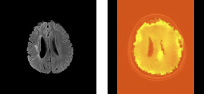 |
Optimization of Asymmetric Spin Echo MRI for Oxygen Extraction Fraction Mapping in the Brain and Initial Experience with Moya-Moya Patients
Dharmesh Tailor, John Lee, Hongyu An, Colin Derdeyn
Asymmetric Spin Echo (ASE) MRI has been previously applied for quantitative cerebral oxygen extraction mapping. In this study we optimize this technique using O-15 PET as a gold standard for oxygen extraction fraction (OEF) quantitation and apply the optimized ASE approach for studying brain lesions in Moya-Moya patients. Results suggest that optimized OEF maps from ASE MRI have the potential to detect brain lesions unseen with conventional MRI sequences. These lesions detected by ASE appear to provide information that is statistically independent from the information provided by conventional MRI approaches.
|
|
2744.
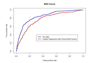 |
Early Prediction of Total Knee Replacement using Structural MRI and 3D Deep Convolutional Neural Networks
Kevin Leung, Gregory Chang, Kyunghyun Cho, Cem Deniz
The early prediction of individuals who will eventually require total knee replacement (TKR) remains a challenging problem. In this project, we propose to use 3D deep convolutional neural networks (CNN) to predict the likelihood of a patient receiving a TKR within nine years using 718 subjects from the Osteoarthritis Initiative1 (OAI) dataset. We found that our model results in better performance compared to a logistic regression model using clinical risk factors2 (AUC: 0.8480±.0173 vs 0.7716±.0229 and accuracy: 77.15±1.88% vs. 71.16±2.70%).
|
|
2745.
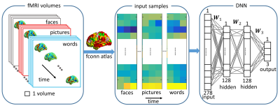 |
Classification of Different Episodic Memory Tasks by Time Points using a Deep Neural Network
Zhengshi Yang, Xiaowei Zhuang, Karthik Sreenivasan, Virendra Mishra, Christopher Bird, Tim Curran, Sarah Banks, Dietmar Cordes
Classification of different episodic memory tasks by time points is challenging because the signal-to-noise ratio in affected brain regions of the medial temporal lobes is low and similar brain regions (such as the hippocampus) contribute to memory activation. No studies have implemented a deep neural network (DNN) to classify memory tasks at each fMRI time point using whole-brain data. We have implemented a region-of-interest based DNN framework and applied it to classify three different episodic memory tasks. Results indicate that this DNN classifier can accurately discriminate between all these tasks.
|
|
2746.
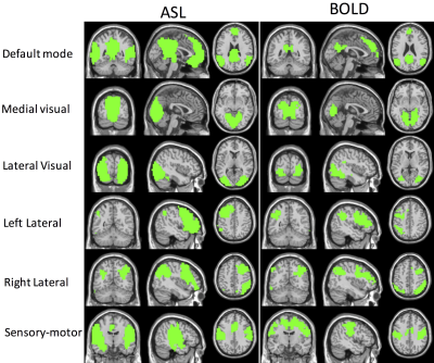 |
Resting-state Brain Networks using Spectral Clustering Analysis
Jason Barrett, Haomiao Meng, Song Chen, Li Zhao, David Alsop, Xingye Qiao, Weiying Dai
Seed-based correlation method and independent component analysis (ICA)-based method have been used to extract the resting-state brain networks from fMRI data. Both methods require either prior knowledge of brain anatomy or selection of unordered spatial sources. Here, we investigate a data-driven spectral clustering algorithm to study brain networks for resting-state arterial spin labeling (ASL) and blood-oxygen-level dependent (BOLD) fMRI data. The spectral clustering algorithm successfully separates the brain resting-state networks and rank the non-neural noises at last. It is of great benefit to use ASL to study brain resting-state networks because of the largely reduced non-neural noise sources.
|
|
2747.
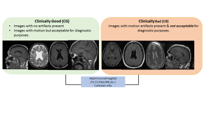 |
Deep learning based MR image diagnostic quality deduction to reduce patient recall
Arathi Sreekumari, Ileana Hancu, Dirk Beque, Keith Park, Uday Patil, Desmond Teck Beng Yeo, Thomas Foo, Dattesh Shanbhag
In this abstract, we describe a fast and robust methodology to highlight on-console, the diagnostic quality of acquired MRI imaging data. Specifically, using convolutional neural networks we flag the MRI volumes affected by motion and consequently hinder the diagnosis by clinician at the time of reading the exam. By prospectively flagging such exams at acquisition console itself and re-acquiring them with improved protocol will obviate the need for costly patient recall and re-scan in clinical setting.
|
|
2748.
|
MRI-based radiomics signature for head and neck squamous cell carcinoma patients
Ling Dong, Ying Yuan, Xiaofeng Tao, Di Dong, Zhenyu Liu, Yali Zang, Jie Tian
To assess overall survival (OS) of head and neck squamous cell carcinoma (HNSCC) patients and the radiomics features, a large number of quantitative radiomics features were extracted from MRI and selected by machine learning methods. Based on these features, a multivariate Cox proportional hazards model was built as a independent predictor to identify patients. Seven features was found to have association with OS (training cohort, P < 0.0001; testing cohort, P = 0.0013). In the training cohort, the radiomics signature yielded a C-index of 0.73 (95% CI, 0.63-0.84), which was 0.71 (95% CI: 0.59-0.82) in the testing cohort. The potential association between MRI-based radiomics signature and OS was explored.
|
|
2749.
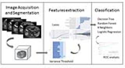 |
Radiomics based strategy for identifying poorly differentiated HCC by using precontrast MRI
Jingjun Wu, Ailian Liu, Jingjing Cui, Lizhi Xie
This work aimed for a radiomics based strategy to identify poorly differentiated hepatocellular carcinoma (HCC) which may own a high risk of recurrence or metastasis. By comparing the performance of four classifiers (decision tree, DT; random forest, RF; k-nearest neighbors, KNN; logistic regression, LR) on dual-echo T1WI (in-phase and out-phase), T2WI and DWI images, we found that LR achieved the best result (AUC: 0.95; sensitivity: 0.75; specificity: 0.85) on DWI images, forming a valuable strategy for clinical practice.
|
|
2750.
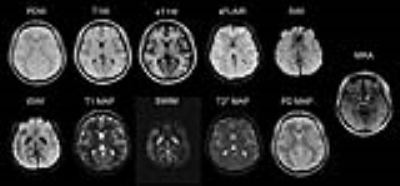 |
STAGE Imaging at 1.5T: A Rapid Brain Protocol Providing More Images As Well As Quantitative Data
Yu Wang, Feng Huang, Wei Xu, Tiecheng Li, Hongyu Guo, Yongsheng Chen, Ewart Haccke
Many image contrasts are necessary in clinical magnetic resonance imaging (MRI) including qualitative and quantitative images, which traditionally take a long acquisition time. STrategically Acquired Gradient Echo (STAGE)1,2,3 is a rapid imaging method which can acquire multiple qualitative and quantitative images with good resolution and SNR in just 5 minutes at 3T. In this work, the STAGE concept is optimized, and further extended to 1.5T. A total of 11 high quality clinically meaningful images, and 2 field maps, were produced with 0.67x1.33x2.7 mm3 resolution in a single 9-min scan on a NMS 1.5T system covering the whole brain.
|
|
2751.
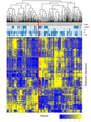 |
Radiomics using multi parametric MRI for pre-treatment prediction of complete response to neo-adjuvant treatment in locally advanced rectal cancer
Stefano Trebeschi, Joost van Griethuysen, Doenja Lambregts, Max Lahaye, Frans Bakers, Roy Vliegen, Emile Voest, Regina Beets-Tan, Hugo Aerts
Aim of this investigation was to assess the predictive value of MR Radiomics as predictive biomarker for locally advanced rectal carcinoma. Through univariate analysis and unsupervised biclustering we found significant associations between diffusion radiomic textures and complete response in a multi-center cohort. The results suggest the viability of Radiomics as biomarker and puts emphasis on image quality.
|
|
Quantitative MRI
Traditional Poster
Acquisition, Reconstruction & Analysis
Thursday, 21 June 2018
| Exhibition Hall 2752-2780 |
08:00 - 10:00 |
|
2752.
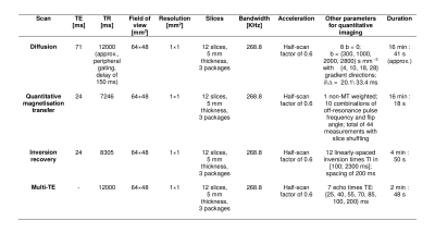 |
A unified signal readout improves denoising of multi-modal spinal cord MRI
Francesco Grussu, Marco Battiston, Jelle Veraart, Torben Schneider, Julien Cohen-Adad, Manuel Jorge Cardoso, Daniel Alexander, Dmitry Novikov, Els Fieremans, Claudia Gandini Wheeler-Kingshott
Denoising based on Marcenko-Pastur principal component analysis (MP-PCA) is a versatile model-free method proposed for brain imaging. Here, we assess the potential of the technique for multi-modal quantitative spinal cord MRI. We analyse a unique data set consisting of multi-modal cervical scans obtained with a unified signal readout, and corroborate in vivo findings with simulations. We show that MP-PCA denoising is a valid tool for pre-processing a variety of signal contrasts in the spinal cord. In particular, the overall performance of denoising can be enhanced further on multi-modal acquisitions with matched signal readout, due to increased data redundancy.
|
|
2753.
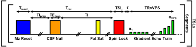 |
SNR-Efficient 3D GRE T1? Mapping of the Brain using Tailored Variable Flip Angle Scheduling
Casey Johnson, Daniel Thedens, Vincent Magnotta
We introduce a new 3D GRE acquisition strategy to greatly improve the SNR efficiency of quantitative 3D T1ρ mapping. Unlike the state-of-the-art 3D MAPSS method, the proposed approach assigns a unique variable flip angle schedule for each spin-lock preparation pulse duration. This enables the use of larger flip angles and greater flexibility in selection of imaging parameters to improve SNR efficiency. In this work, we evaluate this technique for T1ρ mapping of the brain, but this method can also be applied to other regions of the body and used with a variety of magnetization preparation pulses.
|
|
2754.
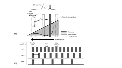 |
Rapid whole brain qMT imaging with inter-slice MT effects and database-driven fitting approach
Jae-Woong Kim, Sul-Li Lee, Seung Hong Choi, Sung-Hong Park
Quantitative magnetization transfer (qMT) imaging provides unique tissue contrast, but suffers from prolonged scan time and processing time. The current study suggests inter-slice MT acquisition and database-driven qMT parameter fitting in order to mitigate the problems. Inter-slice scanning takes advantage of incidental MT effects, and thus does not require separate MT preparation. It enabled us to complete the whole brain data acquisition within a clinically reasonable scan time of ~10 min. The employment of pre-defined database also greatly reduced the qMT processing time, while revealing consistent qMT maps compared to those from the conventional method. The proposed database-driven inter-slice qMT method can be a promising alternative of qMT imaging.
|
|
2755.
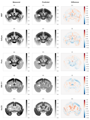 |
Predicting Histological Stainings of Brain Tissue from MRI Data using Artificial Neural Networks
Riccardo Metere, Henrik Marschner, Katja Reimann, André Pampel, Harald Möller
The generation of contrast in MRI relies on a variety of physical processes (e.g. relaxation, magnetization transfer, etc.) that produces a relatively rich amount of information for biological samples. However, given the complex microstructure of tissues, some histological information of relevance in biology and medicine are obtained more easily using optical acquisition techniques on specifically stained specimens. Here, we propose a machine-learning-based method of replicating the contrast information from optical microscopy by exploiting the richness of MRI acquisitions (which will limit the final resolution). The approach exploits the properties of multi-layer feed-forward neural networks as universal function approximators.
|
|
2756.
 |
Multiple dynamics gradient-echo EPI acquisitions for quantitative susceptibility mapping
Vanessa Wiggermann, Enedino Hernández-Torres, Christian Kames, Alexander Rauscher
In this work we demonstrate the feasibility to utilize EPI read-out schemes in combination with multiple dynamics to acquire multi-echo like data sets with the freedom of variable echo times, allowing to acquire fast, high-resolution quantitative susceptibility maps (QSM) images. Assessing the quality of the QSM scans in a region-of-interest based analysis as well as via structural and feature similarities we observed high qualitative and quantitative agreement between QSM images from multi-dynamic EPI acquisitions and multi-echo gradient echo scans.
|
|
2757.
 |
Evaluation of Marchenko-Pastur PCA denoising on Multi-Exponential Relaxometry
Mark Does, Jonas Olesen, Kevin Harkins, Teresa Serradas-Duarte, Sune Jespersen, Noam Shemesh
MRI relaxometry is a powerful tool for characterizing tissue at the sub-voxel level, such as for myelin water imaging. However, a major impediment to its use is the high signal-to-noise ratio requirement. Here, we propose Marchenko-Pastur principal component analysis—previously proposed for diffusion MRI—to denoise relaxometry data. Experimental studies and simulations exemplify the utility of this denoising, and its potential to accelerate data acquisition by 6-8X or more without bias in fitted relaxometry measures or degradation of image resolution. This simple yet important denoising step thus paves the way for broader applicability of relaxometry.
|
|
2758.
 |
A novel strategy for rapid multiparameter mapping based on SPGR with continuous steady state longitudinal magnetization
Jinhyeok Choi, Hyeonjin Kim
A method is proposed for simultaneous T1, T2* and M0 mapping on a single scan by removal of inter-scan time delays based on the analytically found arrays of flip angles and TRs that maintain longitudinal magnetization in a steady state throughout the scan. Our preliminary results are in support of potential application of the proposed method in rapid multiparametric MRI in combination with a suitable undersamping strategy.
|
|
2759.
 |
MAGNETIC SUSCEPTIBILITY OF HUMAN KNEE AT 7T USING ULTRASHORT ECHO MR DATA
Shaeez Abdulla, David Reutens, Viktor Vegh
Ultra-short echo time quantitative susceptibility mapping (QSM) is a promising tool for the study of tissues with short relaxation times. At ultra-high field, the reconstruction of quality phase images is challenging because of the absence of a reference coil. We propose the use of selective channel combination of phase-offset-corrected signal phase data for ultra-short echo time QSM. We compared our findings against an established channel combination method. Qualitative and quantitative analyses of combined phase and QSM images were performed at three echo times. Selective combination of individual channel phase images results in improved ultra-short echo time susceptibility maps.
|
|
2760.
 |
Multi-Parameter Mapping with 500 µm Resolution Using a Flexible 23-Channel RF Coil
Kerrin Pine, Lenka Vaculciakova, Evgeniya Kirilina, Nico Scherf, Nikolaus Weiskopf
To better understand the human brain’s microstructure, there is a need for in-vivo myelin and iron mapping methods which have sufficient resolution to map mesoscopic intra-cortical structures (e.g. lamina). However, resolution is critically SNR-limited. We show that by using a mechanically flexible RF coil array which conforms to the subject’s own individual skull shape, sufficient SNR is gained to map the main MR contrast parameters and the line of Gennari within the superficial primary visual cortex. The work demonstrates the feasibility of laminar analysis of myelination at widely available modest field strengths.
|
|
2761.
 |
Lateralization of Temporal Lobe Epilepsy Using Multimodal Neuroimaging Models
Mohammad-Reza Nazem-Zadeh, Kost Elisevich, Hamid Soltanian-Zadeh
In this work, multivariate response-driven lateralization models were developed using MRI, DTI, and SPECT attributes and logistic regression, to determine the side of epileptogenicity in TLE patients. The proposed response models were capable of handling missing data points using imputation of missing attributes by their mean values measured on a control cohort. Additionally, the proposed response model can be further generalized by integrating attributes of additional modalities (such as PET- positron emission tomography) into the process. Increased reliability in lateralizing TLE cases using the proposed response model reinforces the notion that ECoG in a number of cases may be circumvented.
|
|
2762.
 |
Body Phantom with Prostate Mimic for Evaluation of Quantitative MRI
Ryan Kalmoe, Elizabeth Mirowski, Gregory Metzger
A body phantom, containing a prostate mimic with traceable T1/T2/ADC standards, was designed and manufactured to assess acquisition-, system-, and RF coil- dependent variances of quantitative MRI parameters. In order to explore the potential of the phantom as a quality assurance tool, two phantoms were constructed and evaluated with two receive coil configurations across two scanners over a period of three weeks. It is demonstrated that this phantom is a useful prostate specific quality assurance tool and provide the information needed to harmonize results thus minimizing the impact of multiple dependencies on quantitative results.
|
|
2763.
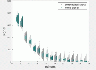 |
Improved muscle T2 estimation by maximum-likelihood parameter estimation using an extended-phase-graph signal model with locally estimated Rician noise levels
Nick Zafeiropoulos, Stephen Wastling, Christopher Sinclair, Tarek Yousry, Enrico De Vita, Robert Janiczek, John Thornton
Maximum likelihood model parameter estimation accounting for the Rician noise distribution in MRI acquisitions, combined with the extended graph formalism and incorporating slice profile considerations, offers higher precision and less bias with regards to the predicted parameters in T2 relaxometry. In this work this was tested by simulations and validated in phantom and in vivo data from healthy volunteers.
|
|
2764.
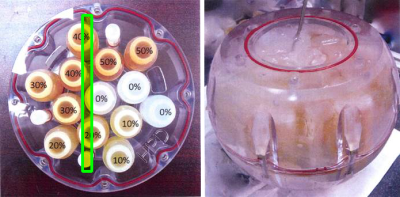 |
Improved ADC Estimation Technique Using Regularized Nonlinear Least Squares Fitting
Eric Borisch, Adam Froemming, Roger Grimm, Yunhong Shu, Ashley Tao, Stephen Riederer, Joshua Trzasko
A high-performance model-based regularized non-linear-least-squares ADC fitting technique has been designed and implemented. Phantom testing shows a reduction in noise with significant retention of detail, while providing < 10 sec computation for 3D acquisitions with 4 b-values.
|
|
2765.
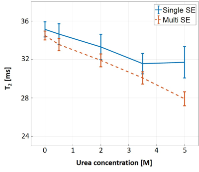 |
Analysis of magnetization transfer (MT) effect on Bloch-simulation based T2 mapping accuracy, demonstrated on in vitro urea phantom
Dvir Radunsky, Noam Ben-Eliezer
Accurate quantification of T2 values hold high value for a variety of clinical and research applications, yet is highly challenged by the inherent bias of rapid multi-SE (MSE) protocols due to stimulated and indirect echoes. Recently, we introduced the echo modulation curve (EMC) algorithm, which successfully overcomes this problem to produce accurate quantification of T2 values that are stable across scanners and scan settings. In this work, we investigate the effect of magnetization transfer on MSE signal, and specifically on EMC-derived T2 values for different T2 baselines, number of slices, and slice gaps, using an in vitro urea model.
|
|
2766.
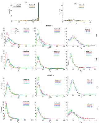 |
A new method to generate a voxel-specific input function for the analysis of dynamic contrast-enhanced MRI data in patients with brain tumours
Georgios Krokos, Neil Thacker, Ibrahim Djoukhadar, David Morris, Alan Jackson, Asselin Marie-Claude
The parameters estimated in DCE-MRI studies vary greatly depending on the arterial input function. Moreover, a more complex model than the extended Tofts’ model (ETM) is needed in glioma patients. In this work, a method to generate a voxel-specific input function (VIF) is introduced and used with a two-tissue compartment model (2TCM) that separates the fast and slow kinetics of the Gd contrast agent. The VIF provided more accurate results in the superior sagittal sinus (SSS) than the internal carotid artery and combined with the 2TCM, significantly improved the fits to the tumour over the ETM using the SSS.
|
|
2767.
 |
Joint T1/T2 mapping with frequency-modulated SSFP, radial sampling, and subspace reconstruction
Volkert Roeloffs, Jost Kollmeier, Nick Scholand, Dirk Voit, Sebastian Rosenzweig, H. Christian Holme, Martin Uecker, Jens Frahm
In this work, we propose frequency-modulated SSFP imaging with 3D stack-of-stars encoding to perform joint T1/T2 mapping. In contrast to phase-cycled SSFP, inefficient preparation phases are avoided and a subspace-constrained reconstruction allows efficient handling of large data sets. Quantitative mapping is realized by projecting the reconstructed subspace coefficients onto a precomputed piece-wise linear approximation of the Bloch-response manifold. General feasibility is proven by comparison to Gold Standard measurements on a home-brew T1/T2 phantom. The investigated approach is a promising candidate for multi-parametric mapping in vivo.
|
|
2768.
 |
Accurate and rapid dictionary-based T2 mapping using multi-echo turbo spin echo sequences with reduced refocusing angle
Julian Emmerich, Sina Straub
In this work, we present a fast and accurate T2 mapping method based on standard multi-echo turbo spin echo sequences (ME-TSE) that are widely available on clinical scanners. Estimation of T2 values is done by a Bloch simulation-based algorithm. As this method can account for stimulated echoes that occur during the echo train within a TSE sequence, its use for sequences with reduced refocusing flip angle is feasible to avoid SAR problems at higher field strength.
|
|
2769.
 |
Towards measurement of normal Blood-Brain Barrier leakage in individual subjects using DCE-MRI
Nicholas Dowell, Samira Bouyagoub, Naji Tabet, Neil Harrison, Mara Cercignani, Paul Tofts
The ability to measure normal BBB leakage in individual subjects would provide a technique to quantify extremely subtle BBB abnormalities in neurological disease. The technique is extremely demanding of scanner stability and vulnerable to low-level (invisible) artefacts. In phantom and healthy control scans without Gd machine stability is good when using Ernst-angle scanning. Image artefact currently limits precision in measuring BBB permeability, and is probably caused by pulsatile motion of the Superior Sagittal Sinus (SSS). Image noise is insignificant when optimised imaging parameters (e.g. FA=30o, TR=30ms) are used. Blood signal is significant and can probably be modelled using SSS signal.
|
|
2770.
 |
Analytical Characterization of Statistical Bias in Multi-Point Apparent Diffusion Coefficient (ADC) Measurements: Application to Prostate Cancer Imaging
Joshua Trzasko, Brent Warndahl, Stephen Riederer, Adam Froemming
In most diffusion studies, two or more DW images are acquired and an apparent diffusion coefficient (ADC) map is generated, with the goal of providing quantitative diffusion information that is independent of acquisition settings or secondary tissues properties. However, ADC values can vary significantly following protocol changes. In this work, we analytically determine the statistical bias in ADC maps generated from multi-point DWI acquisitions, and show how the derived model rationalizes noise-based error propagation as the source of ADC inconsistencies observed in our own clinical practice.
|
|
2771.
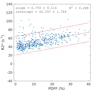 |
The change in R2* with PDFF in liver can be explained by the water/fat susceptibility difference
Mark Bydder, Ludovic de Rochefort, Gavin Hamilton, Nikolaus Szeverenyi, Claude Sirlin
Proton density fat fraction (PDFF) measurements can be confounded by small effects that are not properly accounted for in modeling. This abstract seeks to understand the empirically observed correlation between R2* and PDFF in terms of the susceptibility difference between water and fat. Numerical fitted values were found to be close to literature values for triglyceride unsaturation and magnetic susceptibility in liver.
|
|
2772.
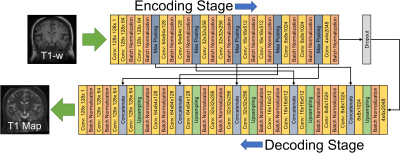 |
Quantitative Synthetic T1 Mapping of the Brain from Structural Imaging using Deep Learning
Samuel Hurley, Jacob Johnson, Barbara Bendlin, Alan McMillan
We propose a method to generate synthetic T1 maps directly from conventional T1-weighted imaging. Rather than rely on fitting an explicit signal model or precomputing a dictionary from a closed form equation (e.g. Bloch equations or extended phase graph), we employ deep learning combined with training data from variable flip angle (VFA) T1 mapping experiments to generate an implicit machine learning model of T1 signal. The use of deep learning to enable quantitative imaging directly from an acquired T1-weighted image is a provocative approach with promising capability, as demonstrated herein with less than 3% error compared to a VFA approach.
|
|
2773.
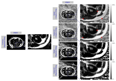 |
Fat Content and Fatty Acid Composition Quantification Using a 3D Stack-of-Radial Trajectory With Adaptive Gradient Calibration
Manuel Schneider, Felix Lugauer, Elisabeth Hoppe, Dominik Nickel, Brian M Dale, Berthold Kiefer, Andreas Maier, Mustafa R Bashir
The purpose of this study was to evaluate the effect of an adaptive gradient calibration technique for a 3D stack-of-radial sequence with regard to magnitude- and complex-based fat content quantification and triglyceride saturation estimation. In-vivo measurements in two healthy volunteers showed that gradient calibration improved the accuracy of complex fitted fat fraction and fatty acid maps. Gradient calibration only had a minor impact on magnitude-based fat fraction results.
|
|
2774.
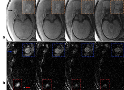 |
T2-based MR oximetry with background-suppressed T2-bSSFP to reduce partial volume errors
Michael Langham, Ana Rodríguez-Soto, Nadav Schwartz, Felix Wehrli
In small tortuous vessels in the presence of motion it is not possible to prescribe the imaging slice perpendicular to minimize the partial volume effect, which is a significant source of error in T2-based oximetry. We propose background suppression (BS) commonly used in ASL prior to T2-preparation. BS reduces SNR but can be compensated with increased slice thickness and reduced inplane resolution. We tested the method in a controlled experiment via quantification of femoral vein blood oxygenation, which has been measured extensively in our laboratory. The utility of the method is further demonstrated in human umbilical vessels in vivo.
|
|
2775.
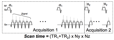 |
In vivo feasibility of T1-corrected Dual-TR Chemical Shift Encoded Fat Quantification Method
Xiaoke Wang, Diego Hernando, Scott Reeder
In chemical shift encoded (CSE) fat quantification techniques, a low flip angle is most commonly used to avoid T1 bias at the expense of SNR. Alternatively, dual flip angle (DFA) acquisitions can be used for T1-corrected fat quantification, however DFA doubles the scan time. A dual TR (DTR) method is proposed where a small percentage increase of scan time allows the independent estimation of T1 of water and fat, and T1-correced fat quantification. This work demonstrates the feasibility of DTR in phantoms and liver imaging.
|
|
2776.
 |
Simultaneous acquisition of MR angiography and 3D quantitative MR parameter maps
Tomoki Amemiya, Suguru Yokosawa, Yo Taniguchi, Toru Shirai, Ryota Sato, Yoshihisa Soutome, Hisaaki Ochi
We proposed a method to obtain MRA simultaneously with 3D quantitative MR parameter maps. The method calculates MRA by combining images and maps obtained using MR parameter mapping with weights that change in the head-to-neck direction in order to correct for the effect of blood flow. The method was evaluated with five healthy volunteers. It visualized the visibility of blood vessels and correlation of intensity with time-of-flight MRA more effectively than conventional calculation method. This suggests that the proposed method is effective for simultaneously obtaining computational MRA and MR parameter maps.
|
|
2777.
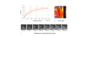 |
Reproducibility of Native Renal T1 mapping for Renal Tissue Characterization
Ilona Dekkers, Elisabeth Paiman, Aiko de Vries, Hildo Lamb
Advanced renal disease is characterized by adverse changes in renal structure, however non-invasive diagnostic imaging techniques are currently lacking. Here we describe the assessment and reproducibility of native T1 mapping for renal tissue characterization. Renal native T1 mapping was performed in 15 healthy human volunteers using the Modified Look-Locker Imaging (MOLLI) 5s(3s)3s sequence on a clinical 3.0 T MR system. Found intra- and inter-examination ICCs for renal cortex (0.77, 0.65) and medulla (0.65, 0.99) indicate good intra- and inter-examination reproducibility, combined with the Bland-Altman analysis showing good agreement. Renal native T1-mapping is a promising reproducible technique for renal tissue characterization.
|
|
2778.
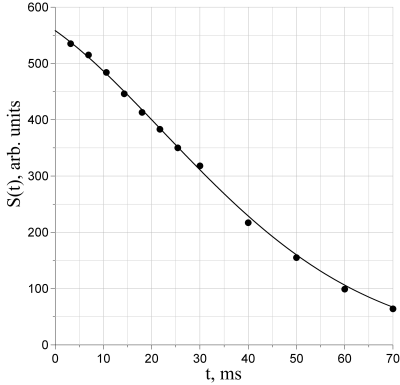 |
Physical parameterization of relaxation curves in GRE sequences
Alexey Protopopov, Michael Bock
The parameter T2* is often used to describe the apparent rate of spin-spin relaxation in the presence of local magnetic field gradients, which is commonly assumed to be mono-exponential. However, the behavior of the transverse relaxation is more complex, since structural characteristics of biological tissues are encoded in the shape of relaxation curve which cannot be described by a single parameter. Several attempts have been made to introduce more accurate relaxation models. In this work we present a concept for the quantitative analysis of the relaxation curve shape in gradient-recalled echo (GRE) imaging based on physical parameters of the signal.
|
|
2779.
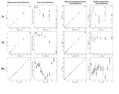 |
Synthetic MRI of the Knee: ISMRM/NIST Phantom Validation and In-Vivo Qualitative, Quantitative and Diagnostic Comparison with Conventional MRI of the Diagnosis of Internal Derangement
Neil Kumar, Benjamin Fritz, Steven Stern, Marcel Warntjes, Yen Chuah, Jan Fritz
Knee MRI protocols containing morphologic and quantitative pulse sequences allow comprehensive evaluation of multiple tissues. However, separate quantitative and qualitative image acquisitions are time consuming. We demonstrated excellent native and error-calibrated accuracy of synthetic MRI of the knee for T1, T2 and proton density quantification with use of an ISMRM/NIST phantom, and show excellent intra-day and inter-day repeatability in living human subjects. Synthetic MRI improves contrast-to-noise ratios of cartilage and menisci and yields improvements in artifact reduction and fat suppression. We demonstrate equivalent subjective ratings and diagnostic performance for internal derangement between conventional and synthetic MRI.
|
|
2780.
 |
The statisitical error in FISP-MRF experiments
Danielle Kara, Jesse Hamilton, Mingdong Fan, Nicole Seiberlich, Robert Brown
The MRF framework has significant freedom in sequence design, increasing its utility and scope, but also the difficulty of determining an optimally efficient experiment. To address this challenge, a statistical analysis of MRF is used to develop a model relating the error in relaxation time quantification and the resulting experimental efficiencies to the number of repetitions in a FISP-MRF experiment. In general, T1 and T2 efficiencies peak prior to 1000 time steps, then decrease to constant values for larger time step totals. Therefore, the derived model can be used to design efficient MRF experiments.
|
|
Learning Image Reconstruction
Traditional Poster
Acquisition, Reconstruction & Analysis
Thursday, 21 June 2018
| Exhibition Hall 2781-2807 |
08:00 - 10:00 |
|
2781.
 |
Complex-valued residual network learning for parallel MR imaging
Shanshan Wang, Huitao Cheng, Ziwen Ke, Leslie Ying, Xin Liu, Hairong Zheng, Dong Liang
Applying deep learning to fast MR imaging has been new and highly evolved. This direction utilizes networks to draw valuable prior information from available big datasets and then assists fast online imaging. Nevertheless, most existing works adopt real-valued network structures while MR images are complex-valued. This paper proposes a complex-valued residual network learning framework for parallel MR imaging. Specifically, complex-valued convolution and initialization strategy are provided. Residual connections are also adopted to learn a more accurate prior. Experimental results show that the proposed method could achieve improved complex-valued image reconstruction with much less time compared to GRAPPA and SPIRiT.
|
|
2782.
 |
A Neural Network for Referenceless Reconstruction in Simultaneous Multi-Slice Imaging
Klaus Eickel, Matthias Günther
The unwrapping of simultaneous multi-slice images without extra reference data is presented. A trained deep neural network disentangles overlapping image content and creates the final magnitude images. The results are compared to established techniques (split slice-GRAPPA), especially where correct reference data are missing.
|
|
2783.
 |
Deep Generative Adversarial Networks for High Resolution fMRI using Variable Density Spiral Sampling
Tianle Cao, Xuesong Li, Yan Tong, Hua Guo
An approach to fMRI image reconstruction for variable density radial trajectories is proposed in this abstract. We have employed Generative Adversarial Networks (GAN), which is made up of a generator and a discriminator, to map input aliasing images to gold standard images. Different from the large computation requirements of CS-based methods, the proposed method is able to both boost reconstruction efficiency and achieve a good image quality in the meantime.
|
|
2784.
 |
Auto-calibrated Parallel Imaging Reconstruction using Fully Connected Recurrent Neural Networks
Tianle Cao, Jiahao Lin, Kyunghyun Sung
A new approach to auto-calibrating, coil-by-coil parallel imaging reconstruction is presented. It is a generalized reconstruction framework based on deep learning. A neural network consisting of three Dense layer (Fully connected layer) units, an RNN layer and an output Dense unit is designed and trained to identify the mapping relationship between the zero-filled and fully-sampled k-space data. The training process could be separated into two steps: pre-training and fine-tuning. Results show our proposed model could be robust to arbitrary undersampling patterns in k-space and shows a higher structural similarity index compared with traiditional k-space based methods.
|
|
2785.
 |
FLAIR MR Image Synthesis By Using 3D Fully Convolutional Networks for Multiple Sclerosis
Wen Wei, Emilie Poirion, Benedetta Bodini, Stanley Durrleman, Olivier Colliot, Bruno Stankoff, Nicholas Ayache
Fluid-attenuated inversion recovery (FLAIR) MRI pulse sequence is used clinically and in research for the detection of WM lesions. However, in a clinical setting, some MRI pulse sequences can be missing because of patient or time constraints. We propose 3D fully convolutional neural networks to predict a FLAIR MRI pulse sequence from other MRI pulse sequences. We evaluate our approach on a real multiple sclerosis disease dataset by assessing the lesion contrast and by comparing our approach to other methods. Both the qualitative and quantitative results show that our method is competitive for FLAIR prediction.
|
|
2786.
 |
Investigation of convolutional neural network based deep learning for cardiac imaging
Shanshan Wang, Ziwen Ke, Huitao Cheng, Leslie Ying, Xin Liu, Hairong Zheng, Dong Liang
Deep learning based fast MR imaging has been very popular lately. Nevertheless, the empirical nature of existing approaches still leave quite a few questions open. To address this, this paper designs different convolutional neural networks to investigate various factors, such as direct CNN mapping, noise stimulation, data consistency and data sharing, for deep learning based cardiac imaging. We find out that if K-space manipulation strategy is not adopted, CNN still needs dedicated sampling patterns or more complicated structures to remove global corruptions. Furthermore, K-space updating strategy are encouraged to be incorporated with deep learning for better final performances.
|
|
2787.
 |
Synthetic CT Generation using MRI with Deep Learning: How does the selection of input images affect the resulting synthetic CT?
Andrew Leynes, Peder Larson
Most recently, synthetic CT generation methods have been utilizing deep learning. One major open question with this approach is that it is not clear what MRI images would produce the best synthetic CT images. We investigated how the selection of MRI inputs affect the resulting output using a fixed network. We found that Dixon MRI may be sufficient for quantitatively accurate synthetic CT images and ZTE MRI may provide additional information to capture bowel air distributions.
|
|
2788.
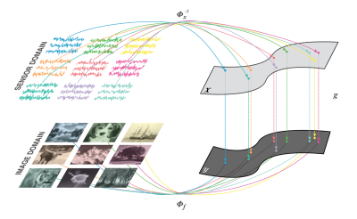 |
Learning multichannel coil combination with Automated Transform by Manifold Approximation (AUTOMAP) using complex-valued neural networks
Bo Zhu, Stephen Cauley, Bruce Rosen, Matthew Rosen
End-to-end learning of the image reconstruction domain transform with AUTOMAP (Automated Transform by Manifold Approximation) has been demonstrated on a variety of spatial encoding strategies previously limited to single-channel data. We extend this framework to learning reconstruction of highly undersampled multichannel k-space data solely from pairs of multichannel k-space and image training data without employing conventional parallel imaging formulations such as SENSE or GRAPPA, and show improved RMSE and artifact reduction with the trained AUTOMAP reconstruction network.
|
|
2789.
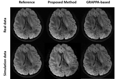 |
Accelerated EPI DWI using a Deep-learning-based Reconstruction.
Yuhsuan Wu, Erpeng Dai, Chun Yuan, Hua Guo
In this work, we preliminarily demonstrate the deep-learning-based reconstruction can be used for under-sampled diffusion imaging. By integrating the sharable information from multiple diffusion directions, the under-sampled data can be nicely recovered.
|
|
2790.
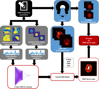 |
Deep Learning Reconstruction for Tailored Magnetic Resonance Fingerprinting
Amaresha Konar, Vineet Bhombore, Imam Shaik, Seema Bhat, Rajagopalan Sundaresan, Sachin Jambawalikar, Ramesh Venkatesan, Sairam Geethanath
Magnetic Resonance Fingerprinting (MRF) is an accelerated acquisition and reconstruction method employed to generate multiple parametric maps. Tailored MRF (TMRF) coupled with deep learning based reconstruction has been proposed to overcome the shortcoming of T2under estimation and the need for dictionaries respectively. A generalized approach with training of natural images and a specific approach with training of brain data are detailed in this work. Both approaches are demonstrated, compared and quantified.
|
|
2791.
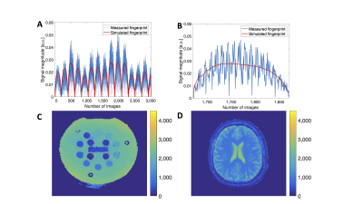 |
Deep Learning for Magnetic Resonance Fingerprinting: Accelerating the Reconstruction of Quantitative Relaxation Maps
Elisabeth Hoppe, Gregor Körzdörfer, Mathias Nittka, Tobias Würfl, Jens Wetzl, Felix Lugauer, Manuel Schneider, Josef Pfeuffer, Andreas Maier
This work demonstrates the successful application of Deep Learning with phantom and human measurements for the reconstruction in Magnetic Resonance Fingerprinting (MRF). State-of-the-art MRF reconstruction yields quantitative maps of e.g. T1 and T2 by acquiring multiple undersampled images with various acquisition parameters, commonly referred to as fingerprints. Every measured fingerprint (per voxel) is compared with a dictionary of simulated fingerprints for possible parameter combinations. This time-consuming step can be replaced with a neural network, which directly predicts the parameters from a fingerprint. This was previously shown with simulated data. Here, we extend this approach to real measurements.
|
|
2792.
 |
Tailored Magnetic Resonance Fingerprinting: optimizing acquisition schedule and intelligent reconstruction using a block approach
Imam Shaik, Amaresha Konar, Vineet Bhombore, Rajagopalan Sundaresan, Shivaprasad Chikop, Gul Moonis, Prachi Dubey, Sachin Jambawalikar, Ramesh Venkatesan, Sairam Geethanath
Magnetic Resonance Fingerprinting technique concurrently generates multiple parametric maps providing for accelerated quantitative imaging. However, quantification of tissues with long T2 such as Cerebrospinal Fluid (CSF) remains a challenge. The main aim of this study is to design acquisition parameters to quantify tissues with long T2 values employing a block based, contrast Tailored MRF (TMRF) approach. In addition, this work emphases on a Neural Network (NN) approach that does not demand noise simulation and/or dictionaries.
|
|
2793.
 |
Data-Driven Image Contrast Synthesis from Efficient Mixed-Contrast Sequences
Jonathan Tamir, Valentina Taviani, Shreyas Vasanawala, Michael Lustig
Synthetic MR is an attractive paradigm for generating diagnostic MR images with retrospectively chosen scan parameters. Typically, synthetic MR images are produced by collecting measurements at multiple measurement times and fitting to a physical model. Here we propose a two-step approach to contrast synthesis. First, we solve a regularized linear inverse problem to reconstruct images at multiple measurement times. Second, we classify spatio-temporal signals and apply different linear combinations based on the classification. We demonstrate the approach on retrospectively under-sampled T1 Shuffling data, in which 3D FSE is collected at relatively short repetition times (TR), and combined to synthesize image contrast with a long TR. The data-driven approach may be useful for synthesizing MR contrasts from acquisitors with varying measurement parameters.
|
|
2794.
 |
Synthetic FLAIR image from multi-echo GRE using U-Net
Jiyong Park, Kanghyun Ryu, Yoonho Nam, Jaewook Shin, Jaeho Lee, Dong-Hyun Kim
The fluid-attenuated inversion recovery(FLAIR) image is one of the most frequently scanned images useful for detecting and diagnosing various lesions. The FLAIR technique suppresses cerebrospinal fluid(CSF) signal by using specific TR and long TE. The WM-GM contrast is similar to the T2-weighted image, except that CSF signal is suppressed. Multi-echo GRE(mGRE) has increasingly been used for medical diagnosis. Here, we used the mGRE images to create a synthetic FLAIR image using deep learning.
|
|
2795.
 |
Improved Synthetic MRI from Multi-echo MRI Using Deep Learning
Enhao Gong, Suchandrima Banerjee, John Pauly, Greg Zaharchuk
Synthetic MRI enables reconstruction of multiple MRI contrasts from a single (multi-echo) scan which significantly improves scanning efficiency. However, the existing state-of-the-art voxel-wise model-fitting method is not optimal. The model-fitting method often results in inaccurate parameter estimation and undesired artifacts, especially for T2-FLAIR synthesis as shown in clinical studies. Here a deep learning method is proposed to improve the contrast synthesis from multi-delay multi-echo MR imaging. With T2-FLAIR synthesis as an example, the proposed method outperforms existing model-fitting based method to overcome artifacts and improve synthesis accuracy. The proposed method is an essential component for delivering reliable and accurate synthetic MRI, further accelerating scanning and improving quantitative parameter mapping.
|
|
2796.
 |
Integrating Spatial and Temporal Correlations into a Deep Neural Network for Low-delay Reconstruction of Highly Undersampled Radial Dynamic Images
Hidenori Takeshima
This paper proposes a novel method for the reconstruction of dynamic images from highly undersampled radial k-space data. In order to take advantage of spatial and temporal correlations and reducing the reconstruction time delay, a deep neural network (DNN) was trained with additional input images displaying the aforementioned correlations. It is shown that the image quality from the proposed method is superior to that of the method based on the conventional DNN reconstruction scheme from a single input to a single output.
|
|
2797.
 |
Noise Level Adaptive Deep Convolutional Neural Network for Image Denoising
Kenzo Isogawa, Takashi Ida, Taichiro Shiodera, Tomoyuki Takeguchi, Yuichi Yamashita, Hiroshi Takai
For integrated diagnosis, MRI provides various types of images related to different acquisition parameters. The change of the acquisition parameters affects noise levels of the provided image in meaningful ways. To adapt the change of the noise level, it is desirable for denoising methods to be adaptive to the noise level, but deep neural network methods are not adaptive, despite their high performance. We propose a deep convolutional neural network (CNN) adjustable to noise levels. The activation functions of the CNN use soft shrinkage whose threshold is proportional to noise level of the input image.
|
|
2798.
 |
Iterative Cross-Domain Deep-Learning Approach for Reconstructing Undersampled Radial MRI
Doohyun Park, Taejoon Eo, Taeseong Kim, Jinseong Jang, Dosik Hwang
The purpose of this study is to eliminate the aliasing artifacts in accerelated radial MRI. We designed a Cross-Domain deep-learning network, called SISI-Net(Sinogram-Image-Sinogram-Image Network). This is an architecture to gradually solves data sparsity problems by iteratively learning the radial sampling data in the sinogram domain and the reconstructed data in the image domain. As a result, proposed network could remove aliasing artifacts effectively while maintaining structural information.
|
|
2799.
 |
Deep Sinogram Learning for Radial MRI: Comparison with k-space and Image Learning
Taeseong Kim, Taejoon Eo, Doohyun Park, Yohan Jun, Dosik Hwang
Deep Sinogram Learning for Radial MRI: Comparison with k-space- and image learning. We demonstrated that singoram learning was more effective than k-space- or image learning in terms of restoring tissue structures and removal of streaking artifacts while not making those as real structures.
|
|
2800.
 |
Convolutional neural network segmentation of skeletal muscle NMR images
Eduard Snezhko, Noura Azzabou, Pierre-Yves Baudin, Pierre Carlier
The purpose of this work was to investigate the ability of deep convolutional neural networks (CNN) to segment muscle groups in NMR images. To this end, we used lower limb scans of patients with different neuromuscular diseases and various levels of fatty infiltration. Thigh and leg muscle groups were first segmented manually and then used in the training and validation processes of the CNN. The mean Dice coefficient of the obtained segmentations was 0.9, demonstrating the effectiveness of the technique in automatically segmenting both healthy and pathological muscle groups.
|
|
2801.
 |
AUTOMAP Image Reconstruction of Low Signal-to-Noise MR Data at 6.5 mT
Neha Koonjoo, Bo Zhu, Matthew Rosen
Due to very low Boltzmann polarization, MR images acquired at ultra-low field (ULF), MR images are mostly corrupted with noise, thus resulting in low signal-to-noise. In the aim of improving image quality at ULF, we apply the deep neural network image reconstruction technique, AUTOMAP, to low SNR datasets acquired at 6.5 mT. The performance of AUTOMAP (Automated Transform by Manifold Approximation) versus the conventional Inverse Fast Fourier Transform (IFFT) on this data was evaluated. The results for AUTOMAP reconstruction show a significant noise reduction, leading to more than 30% gain in signal to noise ratio as compared to standard IFFT.
|
|
2802.
 |
Machine Learning Using the BART Toolbox - Implementation of a Deep Convolutional Neural Network for Denoising
Martin Uecker
Deep convolutional neural networks (DCNNs) tend to outperfom conventional image processing algorithms in recent benchmarks for classifcation, segmentation, denoising, and many other image processing tasks. Here, we show how DCNNs can be implemented using existing building blocks already provided by the BART image reconstruction toolbox. As proof-of-principle we discuss the implementation of an image denoising tool based on a pre-trained DCNN.
|
|
2803.
 |
MR Image Super-resolution Reconstruction Via Enhanced Recursive Residual Network
Ye Fuze, Lijun Bao
Magnetic resonance image (MRI) super-resolution (SR) algorithms have been applied to increase the spatial resolution of scans after acquisition, thus facilitating the clinical diagnosis. Motivated by the great success of deep convolutional neural network in computer vision, we introduced an Enhanced Recursive Residual Network (ERRN) for MRI SR. We show that the performance of our method exceeds conventional learning based methods (sparse coding-based ScSR, CNN-based SRCNN and VDSR) in terms of reconstruction error, peak-signal-to-noise-ratio (PSNR) and structure similarity index (SSIM) value.
|
|
2804.
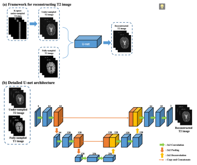 |
Reconstruction in deep learning of highly under-sampled T2-weighted image with T1- weighted image
Lei Xiang, Weitang Chang, Yong Chen, Weili Lin, Qian Wang, Dinggang Shen
T1-weighted image (T1WI) and T2-weighted image (T2WI) are routinely acquired in MRI protocols, which can provide complementary information to each other. However, the acquisition time for each sequence is non-trivial, making clinical MRI a slow and expensive procedure. With the purpose to shorten MRI acquisition time, we present a deep learning approach to reconstruct T2WI from T1WI and highly under-sampled T2WI. Our results demonstrate that the proposed method could achieve 8 or higher acceleration rate while keeping high image quality of the reconstructed T2WI.
|
|
2805.
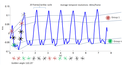 |
Real-time cardiac cine using supervised machine learning and compressed sensing with radial trajectory
Jingyuan Lyu, Yu Ding, Qi Liu, Jian Xu
2D Real-time cardiac cine imaging is valuable for myocardiac function studies. Compared with Cartesian trajectory, Golden-angle (GA) radial acquisition is promising in patients with impaired breath-hold capacity [1]. The GA radial acquisition is an easy-to-implement and promising technique that features improved spatial-temporal resolution, and overcuts Cartesian sampling trajectories in reducing motion artifacts.
|
|
2806.
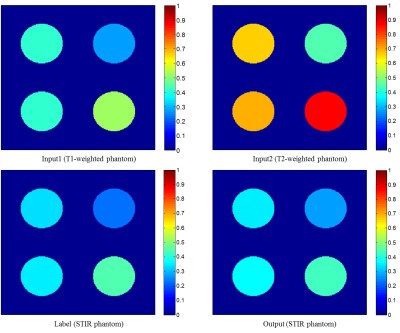 |
Deep-learned STIR imaging via Deep Learning with multi-contrast MRI
Hanbyol Jang, Jinseong Jang, Kihun Bang, Dosik Hwang
The goal of this study is to make STIR MRI using deep learning with multi-contrast MRI. First, we simulated the phantom image created by the bloch equation, which is the basic formula for making MRI, and confirmed that the convolution neural network learns the bloch equation. We also showed the feasibility of making STIR image with in-vivo T1- and T2-weighted, and GRE images in the knee.
|
|
2807.
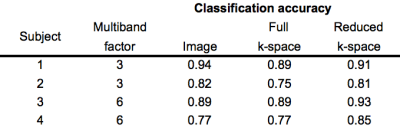 |
Multivariate pattern analysis of multi-band MRI k-space
Scott Peltier, Krisanne Litinas, Anne Gu, Jonathan Lisinski, Stephen LaConte
Multi-band MRI allows for accelerated MR acquisition. However, the reconstruction algorithms, being more complex, require increased reconstruction time without advanced hardware. In this work, we extend classification of MR k-space data to multi-band imaging, enabling rapid prediction of brain state without the need for image reconstruction. We also demonstrate that high prediction accuracy can be achieved even with reduced k-space coverage.
|
|
Acquisition, Reconstruction & Analysis: Sparse & Low-Rank Models
Traditional Poster
Acquisition, Reconstruction & Analysis
Thursday, 21 June 2018
| Exhibition Hall 2808-2827 |
08:00 - 10:00 |
|
2808.
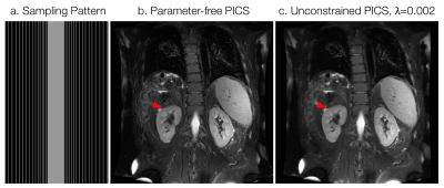 |
Parameter-free Parallel Imaging and Compressed Sensing
Jonathan Tamir, Frank Ong, Shreyas Vasanawala, Michael Lustig
We demonstrate an end-to-end parallel imaging and compressed sensing reconstruction that does not rely on parameter tuning. We combine noise pre-whitening, auto-tuned coil sensitivity estimation, and a noise-constrained compressed sensing reconstruction to eliminate the need to select parameters such as soft threshold regularization. The method is validated across a large corpus of phantom and in vivo data at different levels of SNR and with different types of coils in 2D and in 3D. An end-to-end reconstruction is shown for 2D variable density single-shot fast spin-echo with reconstruction times of less than one minute.
|
|
2809.
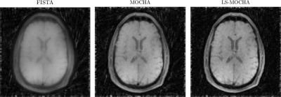 |
Accelerating Non-Cartesian, Sparsity-Promoting Image Reconstruction Via Line Search FISTA
Matthew Muckley, Jeffrey Fessler, Marcelo Zibetti
Iterative reconstruction algorithms for non-Cartesian MRI can have slow convergence due to the nonuniform density of k-space samples. Convergence speed can be improved by including the density compensation function into the algorithm, but current techniques for doing so can lead to SNR penalties or algorithm divergence. Here, we combine the use of density compensation with a line search under the MFISTA framework. The method has the convergence guarantees of MFISTA while gaining the speed improvements of using the density compensition function. The algorithm generalizes further to any FISTA algorithm.
|
|
2810.
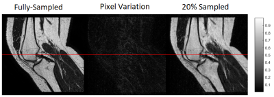 |
Rapid acquisition for MSK applications using compressed sensing with small coils
Laura Lane, Nicolás Schlotterbeck, Gabriel della Maggiora, Carlos Castillo-Passi, Pablo Besa, Sebastián Irarrazaval, Alvaro Burdiles, Cristián Montalba, Pablo Irarrazaval
There is a need for faster acquisitions of the MSK system. Particularly, for assessing the ACL at different degrees of flexion, and even better, for dynamic studies. Our work proposes how to obtain quasi-static images of the MSK, in particular for the study of the Anterior Cruciate Ligament (ACL), using smaller and less rigid coils by undersampling and compressed sensing reconstruction.
|
|
2811.
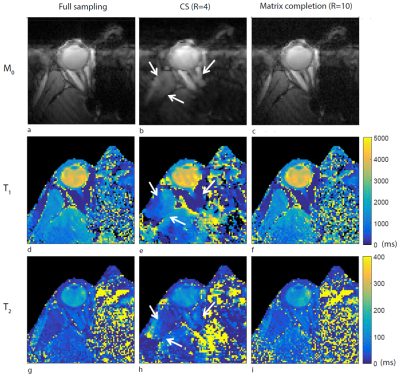 |
A Matrix Completion-Based Reconstruction of In Vivo Eye Images from Undersampled Cartesian 7T MRF Data
Kirsten Koolstra, Andrew Webb, Jan-Willem Beenakker, Peter Koken, Mariya Doneva, Peter Börnert
Eye motion is the main challenge in ocular MRF scans. To achieve good MRF image quality on one side and to improve patient comfort on the other side, scan times need to be reduced. In this single-channel coil approach with Cartesian sampling, high undersampling can be supported by using the appropriate reconstruction approach. In this work, a matrix completion-based reconstruction was adopted. Resulting parameter maps are compared to maps obtained after a compressed sensing reconstruction, showing that for matrix completion even much greater undersampling factors result in more accurate parameter maps.
|
|
2812.
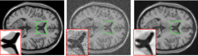 |
MRI denoising using image patch prior based on Gasussian mixture model
Yuhan Zhang, Shurong Zou, Ying Fu, Jia He
MRI is prone to noise pollution in imaging process.MRI with noise seriously affects the doctor's diagnosis of disease.In order to remove noise in MRI,this abstract considers a patch-based method that integrates Gaussian mixture models(GMMs) learning its parameters from external MRI patches with the clustering of desired patches guided by learned GMMs.The last step is to estimate the clear image by low-rank approximation process.Experimental results show the effectiveness of our method.Compared with the classical MRI denoising algorithm—NLM(Non Local Mean) and ADF(Anisotropic Diffusion Filter), our method achieves better results both visually and numerically.
|
|
2813.
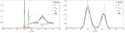 |
L1, Lp, L2, and Elastic Net Penalties for Regularization of Two-Gaussian Component Distributions in One-dimensional Magnetic Resonance Relaxometry
Christiana Sabett, Ariel Hafftka, Kyle Sexton, Richard Spencer
Magnetic resonance (MR) relaxometry time distributions are recovered via the inverse Laplace transform (ILT), an ill-posed problem that is generally stabilized using Tikhonov regularization. Recent work has considered other penalties, such as the L1 penalty for locally narrow distributions. Lp penalties, 1<p<2, may be appropriate for distributions consisting of both narrow and broad components; a linear combination of L1 and L2 penalties, the elastic net (EN), may similarly be useful. However, there is little guidance regarding the choice of regularization penalty for the recovery of transverse relaxation distributions. We compare the effectiveness of each penalty for representative relaxation data.
|
|
2814.
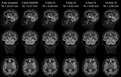 |
Compressed Sensing 3D Double Inversion Recovery (DIR) in the Brain
Tom Hilbert, Esther Raitel, Jean-Philippe Thiran, Reto Meuli, Christoph Forman, Tobias Kober
Double Inversion Recovery (DIR) provides a clinical valuable contrast, especially to study gray matter tissue alterations. However, long acquisition times hinder its use in clinical routine. Assuming that the inherent sparsity of the contrast is well suited for compressed sensing, we tested an incoherently undersampled 3D variable-flip-angle fast spin echo sequence with subsequent compressed sensing reconstruction. The reconstructed fourfold accelerated images exhibit image quality similar to both the clinical standard (twofold GRAPPA-accelerated) and to the fully sampled acquisition. The proposed sequence with ~4 min acquisition time may allow a more frequent use of DIR in clinical routine.
|
|
2815.
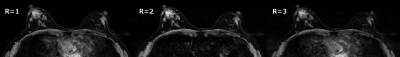 |
Effect of compressed sensing acceleration on high spectral and spatial resolution (HiSS) breast MRI image quality
Milica Medved, Marco Vicari, Gregory Karczmar
Strong T2* weighting has allowed high sensitivity of HiSS breast MRI to cancer, but in whole-breast imaging, contrast is compromised due to necessarily shorter echo trains. k-space under-sampling techniques such as compressed sensing (CS) yield time savings that can be traded for longer echo trains and stronger T2* weighting, potentially increasing breast HiSS MRI performance in screening and diagnostic applications. Our CS simulation resulted in minimal reduction in spatial resolution for acceleration factor R = 2, showing CS to be a promising acceleration strategy for HiSS MRI, allowing longer echo trains and stronger T2* weighting.
|
|
2816.
 |
Automatic Selection of Optimal Regularization Parameters in Compressed Sensing using No Reference Magnetic Resonance Image Quality Assessment.
Kihun Bang, Jinseong Jang, Yohan Jun, Hanbyol Jang, Hojoon Lee, Dosik Hwang
Compressed Sensing can reconstruct image without artifacts from the undersampled data, however setting the regularization parameters in CS optimization problem is difficult. Empirically selected parameters or extracted from L-curve method have less reliability. This abstract proposes CS reconstructed MR image quality assessment without ground truth and it can select proper regularization parameters automatically much faster and much reliable.
|
|
2817.
 |
Real-time 4D Flow MRI with Arbitrary Acquisition Duration
Yichen Zheng, Aiqi Sun, Shuo Chen, Xiaole Wang, Chun Yuan, Rui Li
Real-time 4D flow MRI, without ECG gating and respiration control, has been developed as an effective tool to evaluate hemodynamics. With the benefits of low rank and partial separable model, it could be reconstructed with arbitrary acquisition duration. In this study, we investigated the relationship between acquisition duration and image quality of real-time 4D Flow MRI, and proposed optimized acquisition duration considering both image quality and acquisition efficiency.
|
 |
2818.
 |
Combination of Narrow-band KWIC and GROWL for Multiple T1-weighted Images Reconstruction Based on 3D Golden Angle Radial MR Sequence
Yajie Wang, Haikun Qi, Yishi Wang, Feng Huang, Huijun Chen
Radial sampling has been an increased application due to its insensitivity to motion. A reconstruction method combined the 3D GRAPPA operator for wider radial bands (GROWL) and narrow-band k-space weighted image contrast (KWIC) was proposed and used in GOAL-SNAP sequence. The proposed reconstruction method showed lower image RMSE, accurate T1 map estimation in simulation and higher image quality in in-vivo experiments with shorter computation time.
|
|
2819.
 |
Phase sensitive receiver combination using prescan singular value decomposition derived receiver sensitivities
Olivia Stanley, Ravi Menon, L Klassen
Phase sensitive imaging with multi-channel radio-frequency arrays requires sophisticated channel combination. Combining signal from multiple channels without considering the spatial sensitivity profile of those channels can lead to destructive interference and poor quality phase images. This work outlines a phase combination method which interpolates SVD derived relative sensitivity estimates from a prescan using a solid harmonic basis to allow for phase alignment that is extensible to the remainder of the imaging session. Furthermore, this phase alignment method is computationally efficient and applicable to any coil configuration.
|
|
2820.
 |
Impact of ICA-based denoising of ASL data in clinical settings
Davide Carone, George Harston, Thomas Okell, Michael Chappell, James Kennedy
ASL data has a low signal to noise ratio (SNR) and is sensitive to motion. Independent component analysis (ICA) has been successfully applied to denoise similar quality data in fMRI. We explored the effects of using an ICA approach on ASL data acquired in two different clinical settings. Mean cerebral blood flow (CBF) values were identical pre- and post- ICA indicating good signal preservation. However, the variance of CBF and bolus arrival time measures was significantly reduced suggesting a reduction in noise. These results suggest that ICA based denoising represents a promising strategy to improve ASL data quality.
|
|
2821.
 |
A Fast and General Non-Cartesian GRAPPA Reconstruction Method
Tianrui Luo, Douglas Noll, Jeffrey Fessler, Jon-Fredrik Nielsen
Iterative parallel imaging reconstruction can be very time-consuming for dynamic imaging applications such as functional MRI. GRAPPA is non-iterative but is generally not well-suited for non-Cartesian acquisitions. In this work, we propose a generalization of GRAPPA applicable to arbitrary non-Cartesian readouts. Our non-Cartesian GRAPPA method works by associating a unique kernel with each unsampled (missing) k-space location, and synthesizing non-Cartesian autocalibration (ACS) data by phase-shifts. This approach requires calibrating a very large number of distinct patterns, for which we propose an efficient NUFFT-like algorithm. With this approach we demonstrate fast reconstruction of 3D stack-of-spirals and stack-of-stars images.
|
|
2822.
 |
A Fourier Spectrum Features Based Patch Clustering Method for Inverse Problems In MRI Processing
Lijun Bao
Patch clustering is involved into a number of inverse problems in MRI processing, such as image denoising, cross modality synthesis, parallel imaging reconstruction, super-resolution, under-sampled reconstruction, image registration and even segmentation. Considering that the MR signals are acquired in the k-space and then are Fourier transformed into the spatial domain, in this work we propose a new clustering method based on the features extracted from the frequency spectrum, which can be either applied alone for patch or image clustering, or combined with feature descriptors in the spatial domain to facilitate inverse problems processing in MRI.
|
|
2823.
 |
Blind Simultaneous MultiSlice (SMS) Reconstruction with Application to Phase Contrast Flow Imaging
Suhyung Park, Liyong Chen, David Feinberg
Phase contrast MRI (PC-MRI) has been evolved into a practical and widely used technique for quantification of blood flow velocity and volume, which provides useful insights into pathophysiology. However, PC-MRI requires a long acquisition time to build up phase contrast, requiring flow-reference and flow-encoded datasets over multiple heartbeats, and limiting its general use of flow imaging as a clinical routine. To enable higher acceleration rates, in this work we proposed a generalized coil-by-coil approach to simultaneous multislice (SMS) reconstruction in conjunction with inplane acceleration called as bline SMS (b-SMS) by incorporating slice separation and inplane reconstruction into a single optimization framework that is formulated as an inverse problem with data fidelity.
|
|
2824.
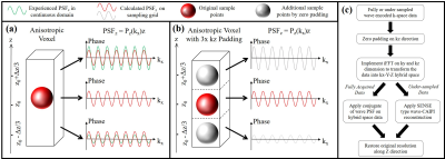 |
Zero-padding reconstruction for wave-CAIPI images with improved accuracy, and its application in ViSTa myelin water images
Zhe Wu, Berkin Bilgic, Hongjian He, Yi Sun, Yiping Du, Kawin Setsompop, Jianhui Zhong
This study presents an intuitive zero-padding (ZP) reconstruction method for wave-encoded images with an improved accuracy. It was shown to be effective in reducing the residual point spread function (PSF) for all wave-encoded images. ZP reduced the errors between the wave-encoded and Cartesian GRE for all wave gradient configurations in simulation and reduced the side-main lobe intensity ratio from 34% to 16% in the thin-slab in vivo Visualization of Short Transverse relaxation time component (ViSTa) images. ZP is applicable for the reconstruction of wave-CAIPI, a recent proposed parallel imaging method using wave-encoding with negligible g-factor penalty under high acceleration factor.
|
|
2825.
 |
Enhanced ADMM-Net for Compressed Sensing MRI
Guanyu Li, Jiaojiao Xiong, Qiegen Liu
Compressed sensing is an effective approach for fast magnetic resonance imaging (CSMRI) that employs sparsity to reconstruct MR images from undersampled k-space data. Synthesis and analysis sparse models are two representative directions. This work aims to develop an enhanced ADMM-Net on the basis of SADN model, which unifies synthesis and analysis prior by means of the convolutional operator. The present SADN-Net not only promotes the generative sparse feature maps to be sparse, but also enforces the convolution between the filter and trained images to be sparse. Besides, it uses optimized parameters learned from the training data. Experiments show that the proposed algorithm achieves higher reconstruction accuracies.
|
|
2826.
 |
DCE-MRI Perfusion Analysis with L1-Norm Spatial Regularization
Michal Bartoš, Michal Šorel, Marie Mangová, Pavel Rajmic, Michal Standara, Radovan Jirík
DCE-MRI perfusion analysis suffers from low reliability, especially when 2nd-generation pharmacokinetic models are used to estimate perfusion parameter maps (voxel-by-voxel estimation) in low SNR conditions. These models provide estimates of plasma flow and capillary permeability in addition to the commonly used parameters Ktrans, kep. This contribution presents a method for estimation of perfusion maps using the tissue homogeneity model with incorporated spatial regularization in the form of total variation. The algorithm is based on the proximal minimization methods well established in image reconstruction problems. The use of state-of-the-art minimization and image regularization techniques stabilizes the estimates of perfusion parameter maps and keeps the computational demands low.
|
|
2827.
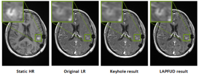 |
Laplacian pyramid based data fusion for high resolution dynamic MRI
Liad Pollak Zuckerman, Lior Weizman, Yonina C. Eldar, Dafna Ben Bashat, Moran Arzi, Michal Irani
Dynamic contrast-enhanced (DCE) MRI is useful for tumor diagnosis and treatment. In DCE, there is a tradeoff between the spatial and temporal resolutions. Improving the spatial resolution while preserving the temporal dynamics is essential for better diagnosis/treatment. We present a method (LAPFUD) for enhancing the spatial frequency without compromising on temporal resolution. LAPFUD combines information from a static high-resolution image acquired at baseline, with each low-resolution frame. By making local decisions it preserves details from both inputs without changing the temporal behavior. Experiments show that LAPFUD provides superior performance (spatially and temporally) compared to the commonly used keyhole method.
|
|
Image Analysis & Post-Acquisition Computing
Traditional Poster
Acquisition, Reconstruction & Analysis
Thursday, 21 June 2018
| Exhibition Hall 2828-2863 |
08:00 - 10:00 |
|
2828.
 |
Quantification of liver function by linearization of a 2-compartment model of gadoxetic-acid uptake using dynamic contrast enhanced magnetic resonance imaging
Josiah Simeth, Adam Johansson, Dawn Owen, Kyle Cuneo, Michelle Mierzwa, Theodore Lawrence, Mary Feng, Yue Cao
This study used the uptake of gadoxetic acid contrast into the hepatocytes as a means of quantifying liver function. A linearized form of the dual-input two-compartment model was developed to estimate the uptake robustly and efficiently. Validation was obtained relative to the predictions of the accepted dual-input two-compartment model, and independent measurements of whole liver function. The linearized approach allows the creation of a spatially resolved quantitative image of liver function, using standard clinical acquisitions, and removes the requirement for impractical, high temporal resolution scans.
|
|
2829.
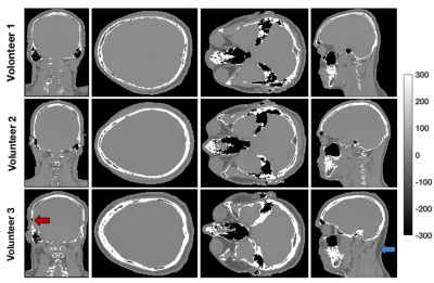 |
Pseudo-CT generation from 3D multi-echo gradient-echo MRI
Véronique Fortier, Ives R. Levesque
MRI-based treatment planning in radiotherapy is limited by the lack of electron density information and by the difficulty to differentiate air from bone regions. A completely automatic method to produce a pseudo-CT from a 3D gradient-echo dataset through voxel-wise assignment of computed tomography (CT) numbers (Hounsfield unit (HU)) was developed. The HU assignment is based on a combination of relative fat and water content and magnetic susceptibility estimates. The proposed method avoids registration errors and allows for HU variability in each tissue class. An improved quantitative susceptibility mapping algorithm for regions with large susceptibility and negligible signal is also presented.
|
|
2830.
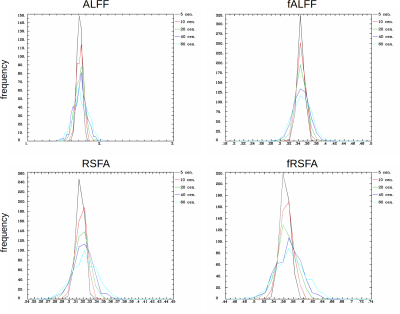 |
Lomb-Scargle your way to RSFC parameter estimation in AFNI-FATCAT
Paul Taylor, Gang Chen, Daniel Glen, Richard Reynolds, Robert Cox
We propose a new tool in AFNI-FATCAT to estimate the above RSFC parameters even when time series are censored, using the Lomb-Scargle (L-S) periodogram. The L-S approach for estimating RSFC parameters is useful and generalizable for FMRI data, where censoring is nearly always performed during processing. The method shows minimal bias of parameter estimation, and also allows for the estimation of confidence intervals for the parameters.
|
|
2831.
 |
The fractal dimension of the tendon-microstructure and its relevance for the detection of permanent changes in micromorphology due to strong mechanical load: a T2* MR-microscopy study using very short detection time
Andreas Berg, Martin Stoiber
The tendon-structure is hierarchically organized: the endotenon soft tissue separates the collagen fibre bundles in sub-segments with decreasing diameter. MR-microscopy at pixel size below 80µm is capable to differentiate microstructure up to the second hierarchical level and demonstrate self-similarity of the sub-segmentations. Can this self-similarity of the tendon be characterized by a fractal dimension? Is the fractal dimension sensitive to microstructural permanent changes as a consequence of strong mechanical load? We present our investigations obtained within a pilot study using short-TE Multislice-T2*-microscopy with a pixel-size of 39x35µm2 indicating the importance of the crimp filament structure.
|
|
2832.
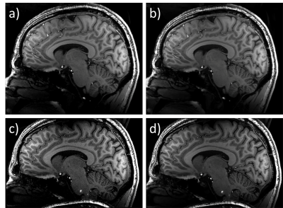 |
Noise mitigation of high-resolution 7T MRI images
Tales Santini, Fabrício Brito, Sossena Wood, Tiago Martins, Joseph Mettenburg, Howard Aizenstein, Marcelo Vieira, Tamer Ibrahim
High-resolution images typically present lower signal-to-noise ratio (SNR) due to the reduced voxel size. In this work, the BM4D filter was applied to high-resolution MPRAGE images acquired at 7T MRI. Original and denoised images were compared using two different acquisition resolutions: 0.7mm isotropic and 0.54mm isotropic. The method shows good results for higher-resolution images, greatly improving the SNR while keeping the useful clinical information and the small details which are not discerned using the lower-resolution acquisitions.
|
|
2833.
 |
A Comparison of Brain Subnetwork Extraction Methods
Elizabeth Powell, Ferran Prados, Baris Kanber, Wallace Brownlee, Sara Collorone, Sebastien Ourselin, Olga Ciccarelli, Jonathan Clayden, Ahmed Toosy, Claudia Gandini Wheeler-Kingshott
In the complex network model of the brain it is often noted that a subset of nodes, or subnetwork, plays a central role in network architecture, whose damage could have a disproportionate effect on network resilience to injury. The identification of "important" nodes in a network is non-trivial though, and several fundamentally different methods exist; it is currently unclear to what extent these methods agree. In this work we demonstrate that subnetworks extracted using rich club and principal network analysis share 60% of nodes, suggesting a core subset of nodes are important to network architecture independently of analysis model.
|
|
2834.
 |
Beyond high resolution: Pitfalls in quantification of cortical thickness based on higher and ultra-high resolution data
Falk Lüsebrink, Oliver Speck
It was shown that higher resolution data increases the accuracy of the brain segmentation resulting in a decrease of cortical thickness estimates. However, data is still mostly acquired at a spatial resolution of 1 mm for quantifying cortical thickness. Several software packages allow processing of higher resolution data. However, FreeSurfer constitutes the de facto standard due to its prevalence. Therefore, we investigate the effects of resolution and SNR at two important stages of its standard processing pipeline: the skull stripping and white matter segmentation.
|
|
2835.
 |
An efficient facial de-identification method for structural 3D neuroimages
Ke Gan, Weitian Chen
A major challenge to facial de-identification in 3D brain MR images is to find a trade-off between patient privacy protection and retaining the usefulness of the image data. An efficient facial de-identification method is proposed. The method can efficiently conceal identifiable facial details in the 3D brain MR images while maintaining the usefulness of the data. The experimental results indicated the proposed method can achieve the state-of-the-art performance and retain more image data in comparison with the currently available tools.
|
|
2836.
 |
Segmentation of Gray Matter, White Matter and Cerebrospinal Fluid with MP2RAGE
Yishi Wang, Yajie Wang, Zhe Zhang, Yuhui Xiong, Qiang Zhang, Chun Yuan, Hua Guo
Segmentation of gray matter (GM), white matter (WM) and cerebrospinal fluid (CSF) is an important tool for brain MRI research. A few challenges remain for the segmentation such as the image intensity non-uniformity induced by B1 field inhomogeneity, suboptimal data acquisition protocols and long processing time. We propose a fast automatic method which combines the data acquisition with segmentation and is insensitive to B1 field inhomogeneity using MP2RAGE. The proposed method has high accuracy and superior performance for the segmentation of subcortical gray matter and is applicable for a wide age range.
|
|
2837.
 |
A software package designed to integrate advanced fMRI methods for presurgical mapping and clinical studies (IClinfMRI)
Ai-Ling Hsu, Ping Hou, Jason Johnson, Changwei Wu, Kyle Noll, Sujit Prabhu, Sherise Ferguson, Vinodh Kumar, Donald Schomer, John Hazle, Jyh-Horng Chen, Ho-Ling Liu
Task-evoked and resting-state (rs) fMRI techniques have been applied to clinical management of neurological diseases, exemplified by pre-surgical functional mapping. Moreover, recent studies recommended incorporating cerebrovascular reactivity imaging into clinical fMRI to evaluate the risk of lesion-induced neurovascular uncoupling. However, a specialized clinical software that integrates the three complementary fMRI techniques and promptly outputs results to clinical PACS and surgical navigation system remains lacking. Here, we developed the Integrated fMRI for Clinical Research (IClinfMRI) software package to incorporate these advanced fMRI methods with streamlined processing and shortened the processing time for pre-surgical mapping and other clinical applications.
|
|
2838.
 |
The Change of Adipose Tissues and Organ Fat-fraction in Patients with Morbid Obesity Before and After Bariatric Surgery
Steve Hui, Simon Wong, Qiyong Ai , David Yeung, Winnie Chu
The purpose of this study was to investigate the change of brown and white adipose tissue, as well as fat content in liver and pancreas, in patients with morbid obesity before and after bariatric surgery. mDixon sequence and proton MRS were used to measure fat content. Results indicated that weight, BMI, waist circumference, pancreatic fat, liver fat, subcutaneous and visceral adipose tissues were significantly reduced 6 – 12 months after surgery. The present study suggested that bariatric surgery effectively reduced the weight in patients with morbid obesity.
|
|
2839.
 |
Infant brain extraction in T2 weighted MR images using k-means clustering and spatial information
Inyoung Bae, JungHyun Song, Seonyeong Shin, Jun-Young Chung, Sung-Ho Woo, Dongchan Kim, Yeji Han
Brain extraction is an essential pre-processing step for brain image analysis. In this work, a new brain extraction technique for T2 weighted image of an infant brain with pathological characteristics is proposed to reduce the error of conventional techniques caused by variations in contrast and brain size of infant brain from that of the adult brain. We used k-means clustering, spatial information, and morphological approaches to improve brain extraction technique. Quantitative analysis was conducted using the dice ratio compared with the results of manual segmentation.
|
|
2840.
 |
Random Forest based Calf Muscle Segmentation from MR data incorporating Prior Information
Marc Fischer, Martin Schwartz, Bin Yang, Fritz Schick
Delineation of muscle structures from MR images is an intricate but essential step for quantitative morphological assessment in many areas. In this work segmentation of muscles in the right calf from 2D MR data has been performed. Since challenging conditions prevail, prior information was incorporated in a Machine Learning driven approach. Versatile Random Forests were employed making use of annotated atlases as well as defined landmarks. It was demonstrated that incorporation of this prior information results in a feasible and fully automatic muscle segmentation.
|
|
2841.
 |
Volumetric Mesh-based Mapping of the Placenta to a Canonical Template for Visualization of Regional Anatomy and Function
S. Mazdak Abulnaga, Esra Abaci Turk, Jie Luo, Justin Solomon, Lawrence Wald, Elfar Adalsteinsson, Carolina Bibbo, Julian Robinson, William Barth, Jr., Drucilla Roberts, P. Ellen Grant, Polina Golland
We demonstrate a volumetric mesh-based mapping of the placenta to a canonical template that resembles the better-known ex vivo shape. Placental shape presents significant challenges for visualization of the associated signals. No standard framework exists for visualizing the organ in vivo. Our approach is to flatten a volumetric mesh that captures subject-specific placental shape while penalizing local distortion to maintain anatomical fidelity. The resulting algorithm produces an invertible transformation to the canonical template. To demonstrate the promise of the proposed approach, we present visualization of BOLD MRI intensity and oxygenation measures after mapping them to a flattened placenta template.
|
|
2842.
 |
Interactive and flexible quality control in fMRI sequence evaluation: the uniQC toolbox
Saskia Bollmann, Lars Kasper, Klaas Pruessmann, Markus Barth, Klaas Stephan
We present a unified neuroimaging quality control (uniQC) toolbox that enables flexible, interactive assessment of various quality measures on n-dimensional imaging data in Matlab. Key features are its seamless integration in the interactive Matlab command window and the intuitive concatenation of imaging and plot operations using operator overloading that enables fast prototyping of artefact detection and data analysis pipelines. The object-oriented design provides a general framework for n-dimensional data handling that can be utilized for fMRI sequence development and quality control.
|
|
2843.
 |
Interactive Tool to Create Adjustable Anatomical Atlases for Mouse Brain Imaging
Markus Sack, Lei Zheng, Natalia Gass, Alexander Sartorius, Gabriele Ende, Wolfgang Weber-Fahr
Brain atlases enable researchers to focus their investigations on specific anatomically defined brain regions and are used in many MRI applications like fMRI, morphometry, whole brain spectroscopy, et cetera. Despite their great use and numerous variants they usually consist of rigid predefined brain regions with a given level of detail often degrading them a non-ideal tool in special research topics. We present a GUI application which allows researchers to easily create mouse brain atlases with an adjustable level of detail and coverage to match specific research questions.
|
|
2844.
 |
A High Performance Computing Cluster Implementation Of Compressed Sensing Reconstruction For MR Histology
Robert Anderson, Nian Wang, James Cook, Gary Cofer, Russell Dibb, G. Johnson, Alexandra Badea
We report the generation of a software pipeline for accelerated MR image reconstruction in a high-performance computing environment, motivated by the shift in time demands from the acquisition to the computational burden of reconstruction in compressed sensing.
|
|
2845.
 |
Cerebral white matter lesions in multiple sclerosis: optimized automated segmentation and longitudinal follow-up
Philippe Tran, Domitille Dempuré, Ludovic Fillon, Marie Chupin, Urielle Thoprakarn, Jean-Baptiste Martini
In Multiple Sclerosis (MS), detection of T2-hyperintense white matter lesions on MRI has become a crucial criterion for early diagnosis and monitoring. In this study, we propose an accurate and reliable automated method for lesion segmentation and longitudinal follow-up, using color-scaled maps of lesion evolution depicting increasing and decreasing patterns. Validation of the cross-sectional segmentation has been performed on large samples of MS patients and shows good agreement with manual tracing. Through its reliability and robustness, the measures provided by our automated method of lesion quantification could be a valuable tool for clinical routine and clinical trials.
|
|
2846.
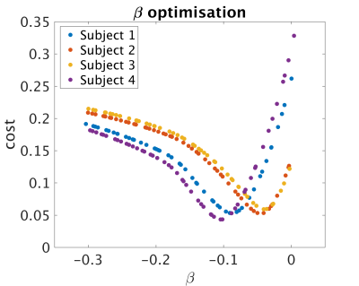 |
Reconstruction of quantitative proton density maps from routine clinical data
Antonio Ricciardi, Francesco Grussu, Rebecca Samson, Daniel Alexander, Claudia Gandini Wheeler-Kingshott
Quantitative proton density (qPD) mapping can be used to measure tissue water content, whose alteration are often linked to pathological conditions. Quantitative MRI methods have been developed in order to make results numerically coherent, but require specific sequences often missing in standard clinical protocols. In this study, an existing approach for the reconstruction of qPD maps from clinical data was corrected to take into account excitation B1 field inhomogeneities, and compared to qPD maps obtained via multi parametric mapping (MPM). The applied correction made clinical-derived qPD maps more similar to the MPM reference than the uncorrected method, without the need of additional specific sequences.
|
|
2847.
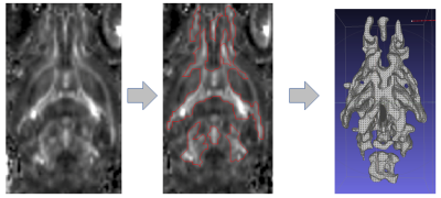 |
Morphometric Thresholded Fractional Anisotropy for robust quantitative assessment and enhanced visualization of whole-brain white matter alterations in rodent models of Lupus
Daniele Procissi, Hadijat-Kubura Makinde, Nicola Bertolino, Bradley Allen , Cynthia Yang, Carla Cuda
The study describes a novel voxel-wise brain fractional anisotropy (FA) analysis approach based on morphometrics evaluation of 3D surfaces. These surfaces were generated using thresholded segmentation of two dimensional FA maps and used for both quantitative analysis and enhanced visualization of differences between a control group and two rodent Lupus models with different degrees of white matter alterations. The methods described, if appropriately translated, could enable integration of DTI-MRI in the diagnostic pipeline in a clinical setting.
|
|
2848.
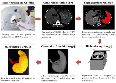 |
A Simplified Framework for MR Image Processing & 3D Printing in Healthcare Applications
Amit Mehndiratta, Manish Arya, Ravi Singh , Shankhabrata Nag, Sonal Krishan
This work summarized a simplified framework that can be used to generate 3D printed prototype from 3D MRI images, with the help of widely available processing tools. The process is conceptually divided into three steps: image acquisition, image post-processing and 3D printing. The utility of the streamlined framework is demonstrated by building 3D prototype of Liver, Spleen and Kidneys using Selective Laser Sintering (SLS) and Fused Deposition Modeling (FDM) technology based 3D printers. The simplified approach has been suggested to assist users in creating 3D anatomical model from medical imaging data using relevant open source tools.
|
|
2849.
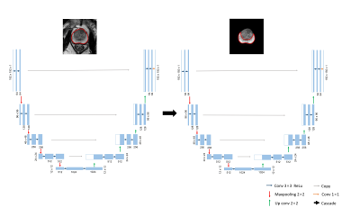 |
An automatic prostate gland and peripheral zone segmentations method based on cascaded fully convolution network
Yi Zhu, Rong Wei, Lian Ding, Ge Gao, Xiaodong Zhang, Xiaoying Wang, Jue Zhang, Jing Fang
Automatic segmentation both in the whole prostate gland and the peripheral zone is a meaningful work, because there are different evaluation criteria for different regions according to prostate imaging reporting and data system's advice. Here we show a new method base on deep learning which can get the prostate outer contour and the peripheral zone contour fast and accurately without any manual intervention. The mean segmentation accuracies for 262 images are 94.87% ( the whole prostate gland) and 85.66% (the peripheral zone). Even in some extreme cases, such as hyperplasia and cancer, our method shows relatively good performance.
|
|
2850.
 |
A STATISTICAL FRAMEWORK FOR EVALUATING THE RELIABILITY OF MYELIN IMAGING
Agah Karakuzu, Cyril Pernet, Tanguy Duval, Julien Cohen-Adad, Nikola Stikov
Given the importance of myelin in brain structure and function, the advancement of MR-based myelin imaging techniques has drawn a great deal of attention. In this abstract we propose a statistical framework for analyzing myelin imaging, taking us one step closer to standardizing and industrializing MR-based myelin biomarkers. In a nutshell, we are computing Pearson correlation coefficients for scan-rescan reliability and taking their differences to determine if some myelin techniques are more reliable than others. We tested this framework in ex vivo dog spinal cord and found the differences between myelin metrics to be subtle, indicating that one metric can often serve as a surrogate for another.
|
|
2851.
 |
QuantiCEST: Bayesian Model-based Analysis of CEST MRI
Paula Croal, Yunus Msayib, Kevin Ray, Martin Craig, Michael Chappell
QuantiPhyse, a python-based software tool for quantitative image processing, has recently been released, to increase the accessibility of physiological modelling and quantification. Here, we present QuantiCEST, a QuantiPhyse plug-in offering Bayesian model-based analysis for quantification of CEST MRI. Using either the graphical user interface or command line, users can easily specify a multipool model of the Bloch-McConnell equations to quantify CEST data acquired with any combination of offset frequencies, saturation power and field strength. Additional information, such as relaxation times, can also be incorporated in the model, allowing flexibility to suit individual research needs. A typical analysis pipeline is presented.
|
|
2852.
 |
A fully automatic territory segmentation method for prostate MR images by multi-atlas matching
Lian Ding, Ge Gao, Yi Zhu, Xiaodong Zhang, Jue Zhang, Jing Fang, Xiaoying Wang
Approximately 70%-75% of prostate cancers originate in the peripheral zone (PZ) and 20%-30% in the transition zone (TZ). According to the Prostate Imaging Reporting and Data System (PIRADS), the diagnostic criteria for PZ and TZ is different. To accomplish fully automatic segmentation for the PZ and TZ, we proposed a territory segmentation method for prostate MR images by multi-atlas matching. This novel segmentation method could not only segment the whole prostate (WP) region, but also the PZ and TZ. The proposed method is fully automatic and could achieve high segmentation accuracy.
|
|
2853.
 |
Making Quantitative Susceptibility Mapping (QSM) a clinical reality: a one minute Morphology Enabled Dipole Inversion using GPU computing
Mengyuan Wan, Zhe Liu, Pascal Spincemaille, Yi Wang
In this work, we demonstrate the feasibility of using GPU computing to achieve a 15 fold acceleration of the most time consuming parts of the Morphology Enabled Dipole Inversion (MEDI) method for Quantitative Susceptiblity Mapping (QSM) leading to an overall 5 fold reduction in total processing time, allowing a one minute susceptibility map reconstruction.
|
|
2854.
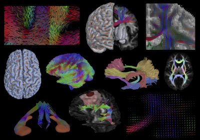 |
Quantitative Imaging Toolkit: Software for Interactive 3D Visualization, Data Exploration, and Computational Analysis of Neuroimaging Datasets
Ryan Cabeen, David Laidlaw, Arthur Toga
Computational tools are increasingly important to MR imaging research, as they can make experiments more reproducible, improve our ability to share our findings and methods, and facilitate hypothesis generation. We aim to contribute a software package to the research community named the Quantitative Imaging Toolkit (QIT). QIT was developed to provide tools for 3D visualization, data exploration, and computational analysis of neuroimaging datasets. While meant to be generally useful for neuroimaging, the tools have extensively developed features for analyzing diffusion MRI data, running large population imaging analyses, and developing new algorithms.
|
|
2855.
 |
ISMRM Raw Data Viewer
Benjamin Dietrich, Bertram Wilm, Klaas Prüssmann
The ISMRM raw data format enables vendor agnostic, reproducible image reconstruction research. So far, the ISMRM raw data format ecosystem did not have a format specific, fast data browser, which is capable of handling large datasets and displaying the data in a convenient form. In this work, we present such a software tool, open-source and platform independent.
|
|
2856.
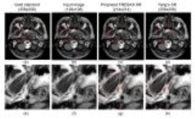 |
Single Image Super-Resolution using the Similarity of Sub-Images in FREBAS Transformed Space
satoshi ITO
In this paper, we propose a new fast image interpolation method involving super-resolution effects. We use FREBAS transform to obtain multi-directional multi-resolution sub-images. By using the similarity of sub-images between different size images, sub-images beyond the Nyquist frequency is estimated using the FREBAS transformed images corresponding scaling parameter. Experiments showed that obtained images have much more sharpened structure than super resolution method based on dictionary learning. PSNR and SSIM are improved and calculation cost is very small compared to learning based method.
|
|
2857.
 |
FPGA based real-time sensitivity maps estimation using pre-scan method.
Tooba Khan, Muhammad Faisal Siddiqui, Hammad Omer
Accurate estimation of the receiver coil sensitivities is critical for an error-free image reconstruction from under-sampled data in SENSE. This work proposes an FPGA (Field Programmable Gate Array) based application specific hardware, for real-time sensitivity maps estimation using pre-scan method. In the proposed work, SENSE reconstructions are performed using the sensitivity maps (computed from the proposed design) and the under-sampled data. The results show that the proposed architecture computes receiver coil sensitivity maps in only 1.466 ms for 8 receiver coils. Also, SENSE reconstructed images show a good mean SNR (30+dB) and low artefact-power (<6×10⁻⁴).
|
|
2858.
 |
Active learning for automated reference-free MR image quality assessment: decreasing the number of required training samples by reduction of intra-batch redundancy.
Annika Liebgott, Damian Boborzi, Sergios Gatidis, Fritz Schick, Konstantin Nikolaou, Bin Yang, Thomas Küstner
Active learning aims to reduce the amount of labeled data required to adequately train a classifier by iteratively selecting samples carrying the most valuable information for the training process. In this study, we investigate the influence of redundancy within the batch of selected samples per iteration, aiming to further reduce the amount of labeled data for automated assessment of MR image quality. An SVM and a DNN are trained with images labeled by radiologists according to the perceived image quality. Approaches to reduce redundancy are compared. Results indicate that reducing the intra-batch correlation for SVM needs fewest labeled samples.
|
|
2859.
 |
Generic feature extraction accompanied by support vector classification: an efficient and effective way for MR image quality determination
Dirk Bequé, Arathi Sreekumari, Dattesh Shanbhag, Keith Park, Desmond Teck Beng Yeo, Thomas Foo, Ileana Hancu
Support vector machine image classification is performed on MR brain images to determine the need to repeat the MR acquisition. However, the image feature extraction is completely brain image agnostic. It is performed either directly on image slices or simple transformations thereof, like e.g. by fore/background thresholding or 1-level wavelet decomposition. 120 image features and meta-data entries are used to classify images as sufficient to diagnose or not. 84% accuracy is demonstrated, even after reducing the feature space to only 20 features. Such feature computation is fast enough to perform image quality assessment in real time, immediately after scan completion.
|
|
2860.
 |
Automatic Brain MR Sequence Classification for Quality Control using Support Vector Machines and Convolutional Neural Networks
Luis Souto Maior Neto, Heather Charette, Marina Salluzzi, Mariana Bento, Richard Frayne
Medical imaging core lab centres face increasing quality control (QC) challenges as studies/trials become larger and more complex. Many QC processes are performed manually by experts, a time-consuming process. Most of the work on automated medical image QC in the literature focuses on text-based metadata correction, thus automated QC algorithms that are able to detect inconsistencies with image data only are needed. We propose two different methods for classification of anonymized MR images by acquisition method (T1-w, T2-w, T1 post contrast, or FLAIR). The classifiers were trained on the MICCAI-BRATS 2016 dataset and achieved accuracies of 85.7% and 93.8%.
|
|
2861.
 |
Integration of the BART Toolbox into Gadgetron Streaming Framework for Inline Cloud-Based Reconstruction
Mahamadou Diakite, Adrienne E. Campbell-Washburn , Hui Xue
BART toolbox is a free open-source framework that consists of a rich set of libraries for common operations in medical image reconstruction. Although the libraries provide highly efficient image reconstruction algorithms and toolbox of command-line programs, it does not, by itself, provide seamless integration with commercial MRI systems. Therefore, the goal of the present work is to enable the deployment of BART in clinical research environment for real-time image reconstruction using Gadgetron streaming framework.
|
|
2862.
 |
MR-only Radiation Therapy Planning workflow optimization for Head and Neck: Zero TE based pseudo CT conversion with body coil.
Cristina Cozzini, Sandeep Kaushik , Florian Wiesinger
Proton Density (PD) weighted Zero Echo Time (ZTE) imaging has been recently developed to provide bone, soft-tissue and air classification suitable for PET/MR Attenuation Correction and Radiation Therapy Planning (RTP). Here we demonstrate ZTE based derivation of pseudo CT using an optimized body coil protocol, which enables patient positioning in the MRI with the RT fixation devices, while preserving the image quality and reproducibility needed for pseudo CT conversion. The method was tested for the head and neck application on N=5 volunteers for different resolutions. The results were compared versus a high SNR surface coil, previously demonstrated suitable for pseudo CT conversion and dose calculation.
|
|
2863.
 |
Data processing methods for the extraction of novel FFC-MRI biomarkers
Lionel Broche, Vasileios Zampetoulas, David Lurie
Fast Field-Cycling (FFC) MRI generates images with T1-dispersion contrast that provide new insights for medical applications. No model of such dispersion data exists for biological tissues therefore a phenomenological approach is chosen here that minimises data loss while isolating meaningful information by curve fitting. This approach provided promising biomarkers in several pilot studies spanning a range of applications: osteoarthritis, liver fibrosis, breast cancer, glioma and fatty tissues. This shows that a dispersion-based approach of FFC-MRI data is an interesting and novel approach for the discovery of novel biomarkers.
|
|
Acquisition, Reconstruction, Analysis
Traditional Poster
Acquisition, Reconstruction & Analysis
Thursday, 21 June 2018
| Exhibition Hall 2864-2895 |
08:00 - 10:00 |
|
2864.
 |
A Principled Approach to Combining Inversion Recovery Images
Antal Horvath, Christoph Jud, Simon Pezold, Matthias Weigel, Charidimos Tsagkas, Katrin Parmar, Oliver Bieri, Philippe Cattin
The averaged magnetization inversion recovery acquisitions (AMIRA) spinal cord imaging sequence acquires images of different inversion contrasts. Despite the different contrasts the images can be combined to even enhance tissue contrast. We give a principled justification for such averaging. Using energy optimization, we describe how to automatically optimize the contrast-to-noise ratio between different tissues using a compressed sensing inspired approach. We show that the uniform weights in the recently proposed AMIRA sequence are close to the optimum but can nevertheless still be improved. As an example we optimize the contrast-to-noise ratio between different compartments in the spinal cord.
|
|
2865.
 |
High Resolution Restoration of Neonatal Images: Matlab Based Framework
Nurten Askin, Peter Lichard, Sebastien Courvoisier, Petra Huppi, Michel Kocher, Francois Lazeyras
A MATLAB based graphical user interface (GUI) was created to apply super resolution (SR) technique on low resolution neonate MR images for obtaining high resolution volume. The user has options to compute HR volume with registration and different reconstruction and regularization methods. In quantitative analysis section, root mean square error, signal-to-noise ratio values could be computed.
|
|
2866.
 |
Correcting for contrast differences across 3D T1 acquisitions
Sean Hatton, Donald Hagler, Joshua Kuperman, William Kremen, Anders Dale
The aim of this study was to correct volumetric differences between images acquired with different MRI parameters. We scanned six subjects on the same 3.0T MRI scanner using different T1-weighted imaging sequences. Images were corrected for gradient warping and intensity inhomogeneity, then we applied a novel white matter intensity scaling and a voxel-wise image intensity normalization process. The correction improved the goodness of fit, precision and accuracy of the volumetric segmentation of the target image to each test sequence (typically < 1% difference). This procedure is particularly effective for voxel-wise segmentation techniques over surface-based approaches.
|
|
2867.
 |
A Tailored Functional Form for Increased Accuracy in CEST B1 Calibration Curves
Abigail Cember, Hari Hariharan, Ravinder Reddy
Correction for the amplitude of B1 (RF pulse) in CEST experiments is currently done using calibration curves fitted ad hoc with polynomial functions. We have previously found that these polynomial-based correction curves sometimes produce unreasonable results, especially in measurements with large B1 variation. Here, we use Bloch-McConnell simulations of CEST as a function of B1 strength to demonstrate a new, Lorentzian-type functional form and fitting strategy, expected to lead to an increase in both accuracy and precision in processing of CEST data.
|
|
2868.
 |
Spatially regularized multi-exponential transverse relaxation times estimation from magnitude MRI images under Rician noise.
Christian EL HAJJ, Guylaine COLLEWET, Maja MUSSE, Saïd MOUSSAOUI
This work aims at improving the estimation of multi-exponential transverse relaxation times from noisy magnitude MRI images. A spatially regularized Maximum-Likelihood estimator accounting for the Rician distribution of the noise was introduced. This approach is compared to a Rician corrected least-square criterion with the introduction of spatial regularization. To deal with the large-scale optimization problem, a majoration-minimization approach was used, allowing the implementation of both the maximum-likelihood estimator and the spatial regularization. The importance of the regularization alongside the rician noise incorporation is shown both visually and numerically on magnitude MRI images acquired on fruit samples.
|
|
2869.
 |
Multi-Contrast 3D MR Image Reconstruction from Incomplete Measurements with Spatially Adaptive Priors
Hyunkyung Maeng, Sugil Kim, Suhyung Park, Eun ji Lim, Jaeseok Park
Magnetic resonance imaging (MRI) is well established as a clinical routine in which multiple sets of data are typically acquired to produce various image contrasts such as T1, T2, FLAIR, etc. Despite the versatile nature of MRI, multi-contrast data acquisition is highly time consuming particularly when 3D encoding is needed. To address this issue, in this work we propose a novel, multi-contrast 3D MR image reconstruction with spatially adaptive priors by exploiting sharable information across the contrast dimension: edge and coil sensitivity maps. The proposed method consists of the following three steps: 1) estimation of edge maps common over all contrasts, 2) estimation of contrast-specific edge maps, and 3) multi-contrast image reconstruction with spatially adaptive, contrast-specific edge priors. In vivo experimental studies show that the proposed method enables T1, T2, and FLAIR 3D isotropic (1mm 3) imaging roughly in 5-6 minutes.
|
|
2870.
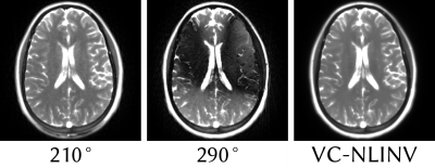 |
Banding-Free Reconstruction in Frequency-Modulated bSSFP using Virtual Coils with Regularized Non-Linear Inversion
H. Christian M. Holme, Nick Scholand, Sebastian Rosenzweig, Robin Wilke, Martin Uecker
We propose a method for banding-free reconstruction of bSSFP images using Regularized Non-Linear Inversion (NLINV). Instead of using only a few different phase cycles, we dynamically change the phase cycle in each frame, which only slightly changes the steady state. By stacking all frames together as virtual coils and using NLINV, images free of banding artifacts can be reconstructed. Since no new steady states need to be prepared, dead time is eliminated from the acquisition.
|
|
2871.
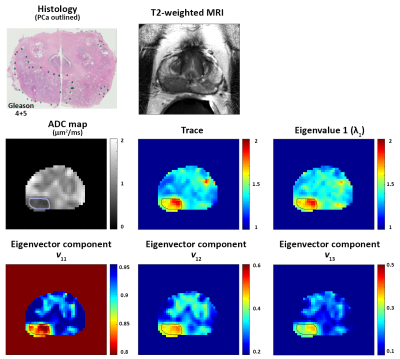 |
Matrix analysis of Hybrid Multidimensional MRI for the diagnosis of prostate cancer
Aritrick Chatterjee, Xiaobing Fan, Shiyang Wang, Tatjana Antic, Scott Eggener, Aytekin Oto, Gregory Karczmar
This study investigates the feasibility of diagnosing prostate cancer through matrix analysis of Hybrid Multidimensional MRI (HM-MRI) data. Data was acquired with all combinations of TE (47,75,100ms) and b-values (0,750,1500s/mm2), resulting in a 3×3 matrix associated with each voxel. Matrix analysis parameters: trace, eigenvalues and eigenvectors were calculated for benign tissue and prostate cancer. Prostate cancer showed significantly increased trace, eigenvalue 1, eigenvector components v12 and v13 and reduced v11 compared to normal tissue. PCa diagnosis is feasible using matrix analysis of HM-MRI data with parameters showing good differentiation between PCa and benign prostatic tissue (AUC 0.80-0.96 on ROC analysis).
|
|
2872.
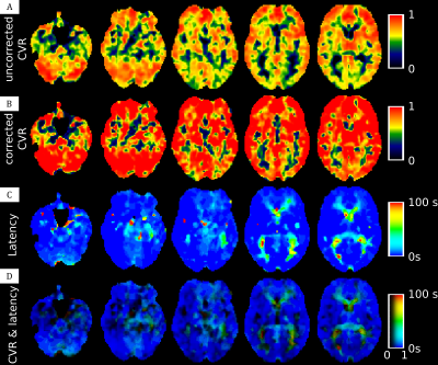 |
Cerebral vasoreactivity latency correction: a clinical case study
Olivier Rossel, Jérémy Deverdun, Amel Benali, Victor Vagné, Nicolas Menjot de Champfleur, Emmanuelle Le Bars
In MR cerebral vasoreactivity (CVR) experiments, responses to the gas stimuli are expected to be with no major time lag. However, it has been shown that different brain regions could respond with different timing during a vasoreactivity. The presence of potential latencies could lead to a misinterpretation of the resulting CVR maps. We attempt to correct CVR maps for physiological latency differences, and propose an alternative way to display both corrected CVR and CVR latency. The data from a Moya Moya patient highlight that even without a strong perfusion alteration, the vasoreactivity is strongly delayed or even completely disrupted.
|
|
2873.
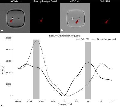 |
Differentiating Brachytherapy and Intraprostatic Gold Fiducial Markers with Varying Off-Resonant Frequency Offsets
Evan McNabb, Raimond Wong, Michael Noseworthy
The dual-plane co-RASOR sequence is able to differentiate between a LDR brachytherapy seeds and gold fiducial markes used in prostatic imaging, by exploiting signal pileups and rewinding them radially inwards using different off-resonant frequency offsets and has the potential to avoid a radiological CT scan that’s clinically used to differentiate the two in boost therapy.
|
|
2874.
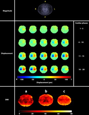 |
SNR analysis of retrospectively gated DENSE at 7T for the measurement of brain tissue pulsatility
Ayodeji Adams, Jacob-Jan Sloots, Peter Luijten, Jaco Zwanenburg
Measurements of brain tissue pulsatility can provide information about viscoelastic tissue properties and assess microvasculature blood volume pulsations as a biomarker. VCG-triggered DENSE is capable of acquiring micrometer-level tissue displacements and volumetric strain. However, it is slow and suffers from triggering issues, especially at 7T. In this work, retrospectively-gated DENSE using a pulse oximeter was implemented at 7T. Assessment of its performance showed maintained SNR within half the scan time of triggered DENSE. The high SNR and reduced scan time simplifies its application in future studies assessing the potential of DENSE-derived brain tissue displacements as a biomarker for neurological diseases.
|
|
2875.
 |
Quantification and management of MR image spatial accuracy for applications in radiation therapy
Teo Stanescu, Joanne Moseley, Callum Moseley, Mostafa Shahabi, David Jaffray
An automated imaging pipeline was developed and validated to handle the management of MR image spatial accuracy with a focus on applications for radiation therapy (RT). Protocol enforcement was implemented to accept/reject datasets based on expected clinical sequence parameters. System and patient related image spatial distortions were quantified using numerical simulations and measurements. Vector field maps were rendered and stored for automatic filtering and correction of patient MR images. Data and process monitoring was enabled via a web application. The imaging pipeline was deployed clinically to automatically validate patient image data required for RT planning and in-room MR-guided treatment delivery.
|
|
2876.
 |
Quality Assurance of physiologic signal measures in HCP resting state fMRI data
Wanyong Shin, Mark Lowe
Recently, a physiological log file timing error with fMRI acquisition was fixed and physiologic data with corrected timing was uploaded to the WU-MINN human connectome project (HCP) cloud. While HCP preprocessing pipeline for resting state (rs-) and fMRI has been proposed, the physiologic noise correction sub-pipeline has not been established yet and the physiologic noise data has had less attention in HCP community. In this study, we investigate the quality of HCP physiologic data and propose a standard physiological noise quality assurance.
|
|
2877.
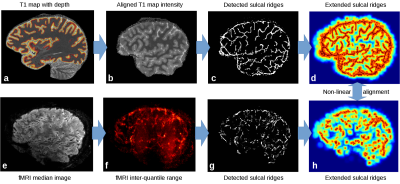 |
Sulcal ridge alignment for laminar fMRI at 7T
Pierre-Louis Bazin, Wietske van der Zwaag, Ritu Bhandari, Christian Keysers, Valeria Gazzola
Laminar fMRI at 7T typically involves imaging small slabs of cortex, and requires precise alignment of the anatomical and functional data to transfer intra-cortical depth information to the fMRI data. We present a method taking advantage of the high resolution of the fMRI data and extracted sulcal patterns.
|
|
2878.
 |
Short-term Fourier Transform Analysis of Respiratory- and Cardiac-driven Pulsation of Cerebrospinal Fluid under Free Breathing
Tetsuya Tokushima, Satoshi Yatsushiro, Saeko Sunohara, Mitsunori Matsumae, Hideki Atsumi, Kagayaki Kuroda
To separate the respiratory- and cardiac-driven motions of cerebrospinal fluid (CSF) under free breathing, CSF velocity in 7 healthy volunteers and 3 hydrocephalus patients were observed by asynchronous phase contrast (PC) technique with monitoring respiratory and ECG signals. Spectrograms of CSF velocity and respiratory signal obtained by short-term Fourier transform (STFT) with 8-sec length Hamming window revealed that the peak respiratory motion appeared in 0-0.5 Hz band, while the cardiac motion appeared around 1-1.5 Hz. These results suggest that the separation of the two motion components is possible by sliding the frequency bands temporarily according to the spectrogram.
|
|
2879.
 |
Improved MR Neurography of Brachial Plexus at 3.0 T with iMSDE and DIXON methods
Xiaoqi Wang, Li Xu
In this study we developed a sequence for neurography with robust fat and blood suppression for increased conspicuity of nerves. Improved Motion-Sensitized Driven-Equilibrium Preparation (iMSDE) was applied to null blood and lymphatic motions. DIXON TSE readout with long TE was used to generate water images representing nerves, in brachial plexus where other fat suppression pulses were challenged by B0 and B1 complexities. Preliminary test in brachial plexus showed its advantages and stability.
|
|
2880.
 |
Towards a Routine Clinical Application of Chemical Exchange Sensitive MRI
Patrick Schuenke, Ralf Floca, Caspar Goch, Marco Nolden, Moritz Zaiss, Johannes Windschuh, Hoai Nam Dang, Christian David, Volkert Roeloffs, Peter Bachert, Mark Ladd
Chemical exchange saturation transfer (CEST) and chemical exchange sensitive spinlock (CESL) were shown to have potential to provide molecular information for diagnosing a wide range of diseases. However, the lack of standardized acquisition protocols and freely available post-processing software prohibited the widespread application of these promising techniques until now. In this work, we present a modularly designed CEST/CESL preparation block that is easy to operate and can be used with arbitrary MRI readouts. Further, we developed and provide a C++ based open-source software that offers many CEST/CESL specific functionalities for the post-processing of the acquired data.
|
|
2881.
 |
Observation of the kinetics of antioxidant action in blood serum as measured by NMR relaxation
Magdalena Witek, Dorota Wierzuchowska, Barbara Blicharska
The exposure of blood serum to reactive oxygen species creates free radicals and damages the structure of biomolecules. It causes proton NMR relaxation times to change over time. In aqueous protein solutions, T1 and T2 decay curves were fitted by single exponential components. Following addition of hydrogen peroxide to blood serum which contained endogenous antioxidants, relaxation times initially decreased then increased towards initial values. The character of these curves and their fitting parameters depend on kinetics of oxidation processes. We hope that MR imaging may help to investigate these important processes also in vivo.
|
|
2882.
 |
Visualizing the Spatial Propagation of Ventilation and Perfusion Signal with Fourier-Decomposition MRI
David Bondesson, Thomas Gaaß, Moritz Schneider, Bernd Kühn, Olaf Dietrich, Julien Dinkel
Fourier decomposition (FD) MRI offers functional imaging without exposing patient to contrast agents during free breathing measurements, facilitating the examination of patients with impaired respiration. The presented method visualizes signal progression in the lung by using the phase information of the FD-method and addresses recently raised concern that variable-frequency signals can lead to errors in ventilation and perfusion phase estimates. With the signal progression maps it is further shown how localized signal delays caused by pathologies can be identified.
|
|
2883.
 |
Dynamic field map correction based on reversed-gradient design for non-Cartesian single-shot fast fMRI
Fei Wang, Bruno Riemenschneider, Juergen Hennig, Pierre LeVan
A dynamic field map correction technique based on reversed-gradient design is introduced to non-Cartesian single-shot fast fMRI to correct the off-resonance artifacts. The field map estimated from dual-TE GRE scan could not capture that from field drift and eddy currents, so the off-resonance artifacts could not be corrected completely. This technique acquires two images with reversed slow-encoding directions in each time frame, which is generally used in EPI, and updates field map iteratively based on conjugate gradient reconstruction. After correction, the off-resonance artifacts are significantly reduced.
|
|
2884.
 |
Improving radial cardiac cine with higher-order total-variation regularizations
Renjie He, Qi Liu, Yao Ding, Ruobing He
In cardiac cine MRI, radial data acquisition will make the motion effects being more noise-like in image domain, and to achieve high temporal resolution, sparse sampling will inevitably lead to streaking artifacts using conventional image reconstruction methods. Golden angle radial reordering which provides continuously change in angle direction will eliminate the coherence of (streaking) artifacts in the temporal dimension. While GRASP-like reconstruction method applies 1D total-variation (TV) regularization on the reconstructed temporal signal, the spatial consistence of the reconstructed images are not ensured. Here we propose a reconstruction strategy using a higher-order TV to promote the spatial imaging quality.
|
|
2885.
 |
Correction of Ferromagnetic Object Artifacts Using Simulated Off-Resonance Map
Sina Amirrajab, Vahid Ghodrati, Abbas Moghaddam
The aim of this study is to quantitatively investigate the potential of field-mapping method in areas around a ferromagnetic object in the post processing approach. As a worst-case scenario, for regions farther than around 10 times the radius of a ferromagnetic foreign object, the post processing approach (based on the simulated off-resonance map) can be useful, even at 3 Tesla, to correct image distortions. At this distance, the exact shape of the object is not important and results obtained for the sphere, remain valid for most objects with the same volume.
|
|
2886.
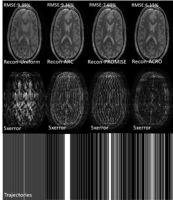 |
Real-time Personalized Acquisition Optimization: 30%-50% reconstruction improvements from a 10-second undersampling optimization
Ke Wang, Enhao Gong, Suchandrima Banerjee, John Pauly, Greg Zaharchunk
Improved undersampling designs can effectively improve the acquisition and resulting reconstruction accuracy. However, existing undersampling optimization methods are time-consuming and the limited performance prevent their clinical applications. Here we proposed an improved undersampling trajectory optimization scheme to generate an optimized trajectory within seconds and apply for subsequent multi-contrast MRI datasets on a per-subject basis. By using a data-driven method combined with improved algorithm design, GPU acceleration and more efficient computation, the proposed method can optimize a trajectory within 5-10 seconds and achieve 30% - 50% reconstruction improvement with the same acquisition cost, which makes real-time under-sampling optimization possible for clinical applications.
|
|
2887.
 |
Speedup of iterative k-t SENSE reconstruction using the multidimensional fast Fourier transform for arbitrary periodic grids
Adam Johansson, James Balter, Yue Cao
The multidimensional fast Fourier transform (MFFT) for arbitrary n-dimensional periodic grids allows images with non-cuboid voxels and fields of view (FOV) to be reconstructed efficiently. In this study, we apply the MFFT with a modified Smith normal form to iterative k-t SENSE reconstruction of data from a golden-angle stack-of-stars DCE-MRI sequence to produce a 4-D image with a non-cuboid FOV tailored to fit tight around the imaged subject. Compared to FFT-based reconstruction on a tight-fitting cuboid FOV, the non-cuboid MFFT reduced reconstruction time by 18% and memory usage by 11% while producing voxel values identical to those found in the reconstruction domain of the cuboid FOV.
|
|
2888.
 |
MR Fingerprinting Reconstruction using Convolutional Neural Network (MRF-CNN)
Qiang Zhang, Rui Guo, Huikun Qi, Di Cui, Edward S Hui, Shuo Chen, Hua Guo, Huijun Chen
The purpose of this study is to develop a MR fingerprinting (MRF) reconstruction algorithm using convolutional neural network (MRF-CNN). Better MRF reconstruction fidelity was achieved using our MRF-CNN compared with that of the conventional approach (R2 of T1: 0.98 vs 0.97, R2 of T2: 0.97 vs 0.59). This study further demonstrated the performance of our MRF-CNN, which was retrained using MR signal evolutions in the continuous parameter space with various levels of Gaussian noise, amidst noise contamination, suggesting that it may likely be a better alternative than the conventional MRF dictionary matching approach.
|
|
2889.
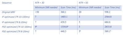 |
Interior-Point and Particle-Swarm Optimization of an Inversion-Recovery Prepared Spoiled Gradient Echo Magnetic Resonance Fingerprinting Sequence
Jingwen Yao, Zhaohuan Zhang, Kyunghyun Sung, Holden Wu
Magnetic Resonance Fingerprinting (MRF) permits simultaneous quantitative mapping of multiple MR parameters. We propose a framework for optimizing the sequence parameters of an inversion recovery prepared spoiled gradient echo (IR spoiled-GRE) based MRF sequence, with interior-point (IP) method and particle-swarm optimization (PSO). By using an designed exponential cost function to maximize the discrimination between tissue types, with combined sinusoidal wave functions as input to generate sequence parameters, substantial improvement of accuracy for T1 and T2* quantification can be achieved. Simultaneous high accuracy of T1 and T2* estimations can be achieved within 0.7s for SNR≥5; within 0.5s for SNR≥10.
|
|
2890.
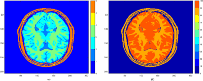 |
CoverBLIP: scalable iterative matched-filtering for MR Fingerprint recovery
Mohammad Golbabaee, Zhouye Chen, Yves Wiaux, Michael Davies
Current popular methods for Magnetic Resonance Fingerprint (MRF) recovery are bottlenecked by the heavy computations of a matched-filtering step due to the size and complexity of the fingerprints dictionary. In this abstract we investigate and evaluate the advantages of incorporating an accelerated and scalable Approximate Nearest Neighbour Search (ANNS) scheme based on the Cover trees structure to shortcut the computations of this step within an iterative recovery algorithm and to obtain a good compromise between the computational cost and reconstruction accuracy of the MRF problem.
|
|
2891.
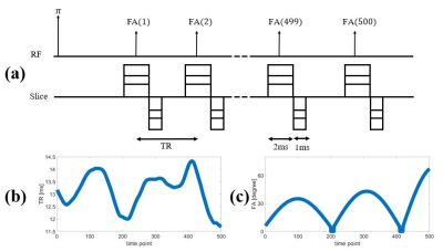 |
Matching Error Evaluation in Magnetic Resonance Fingerprinting with a Fast Imaging with Steady Precession sequence using Bloch Equation Simulations with a Diffusion Propagator
Shota Hodono, Yun Jiang, Gregor Körzdörfer, Naren Nallapareddy, Vikas Gulani, Mark Griswold
The robustness of T1, T2 values derived from Magnetic Resonance Fingerprinting (MRF) is limited in certain situations because MRF dictionaries have in general not included apparent diffusion coefficients (ADC). In this study, the potential estimated T1, T2 errors due to the omission of diffusion were evaluated for the MRF-fast imaging with steady precession sequence. Dictionaries with ADC values were generated by using Bloch equations with a diffusion propagator. The generated signal evolutions with ADC were matched to those generated by Bloch equation simulations without ADC by employing a template-matching algorithm.
|
|
2892.
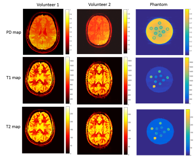 |
In vivo parametric mapping using piecewise constant flip angle and multi shot EPI MR Fingerprinting
Zaid Mahbub, Mohammad Golbabaae, Arnold Julian Benjamin, Mike Davies, Ian Marshall
Previous MR fingerprinting studies have used smooth variations in TR and flip angles. In this study we introduce a piecewise constant flip angle train into a standard gradient echo multi shot EPI sequence. The resulting T1, T2 and proton density maps were obtained from a phantom and healthy volunteers using only 3 distinct flip angle values (obtained by optimization over 8 different flip angles) and using iterative reconstruction. The method generates steady states covering full k-space, producing alias-free maps.
|
|
2893.
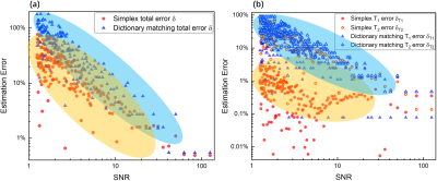 |
Fast Dictionary-free Reconstruction in MR Fingerprinting
Tianyu Han, Teresa Nolte, Nicolas Gross-Weege, Volkmar Schulz
MR fingerprinting offers a rapid way to accurately map multiple tissue parameters. The dictionary based reconstruction under the influence of Gaussian noise is identified as a convex optimization problem and solved by a Nelder-Mead simplex algorithm. Instead of a lengthy and uniform sampling proposed by dictionay matching, the new approach using a heuristic and incoherent sampling in the $$$T_1$$$-$$$T_2$$$ space. More robust $$$T_1$$$ estimations are obtained even under severe noise environments. Thus, a robust and fast MR fingerprinting reconstruction can be made without any dictionary.
|
|
2894.
 |
Towards Unified Colormaps for Quantitative MRF Data
Mark Griswold, Jeffrey Sunshine, Nicole Seiberlich, Vikas Gulani
The goal of quantitative methods such as MRF is to provide a quantitative characterization of tissue physiology and pathology. These data are displayed as images to convey both geographic/anatomical information and quantitative physical property measurements. But this also means that the manner in which the information is displayed is critical. Here we propose several color map alternatives that have been optimized for use in MRF. It is hoped that the use and further optimization of these maps by the community will further improve our ability to visualize and understand this kind of quantitative data.
|
|
2895.
 |
Simultaneous Quantification of T1, T2, and Off-resonance Using FISP-MRF with a Rosette Trajectory and Readout Segmentation
Yuchi Liu, Jesse Hamilton, Mark Griswold, Nicole Seiberlich
Artifacts due to off-resonance effects are a significant challenge for non-Cartesian MRI. In FISP-based MRF sequences, if the entire spiral readout is employed to generate a highly undersampled image, any off-resonance during the readout will lead to blurring. However, short portions of the readout will be mostly free of dephasing due to off-resonance effects. By gridding only segments of the readout, it may be possible to quantify the resonance frequency along with T1 and T2. This work shows a proof-of-principle application of this idea using the cardiac MRF sequence with the rosette trajectory in simulations.
|
|
| Back |
| The International Society for Magnetic Resonance in Medicine is accredited by the Accreditation Council for Continuing Medical Education to provide continuing medical education for physicians. |
