|
Traditional Poster Session
Molecular Imaging |
Thursday, 21 June 2018
Traditional PosterMolecular Imaging
3020 -3032 MR/PET
3033 -3051 Molecular Imaging
3052 -3074 Hyperpolarised MR |
| |
MR/PET
Traditional Poster
Molecular Imaging
Thursday, 21 June 2018
| Exhibition Hall 3020-3032 |
13:15 - 15:15 |
|
3020.
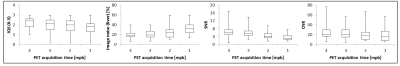 |
Radiotracer dose reduction in 18F-FDG whole-body PET/MR: Effects on image quality and quantification
Maike E. Lindemann, Vanessa Stebner, Alexander Tschischka, Julian Kirchner, Lale Umutlu, Harald H. Quick
The study goal is to investigate how the simulated reduction of injected radiotracer affects PET image quality and quantification in whole-body PET/MR in patients with oncologic findings. PET data of fifty-one patients was reconstructed with 4, 3, 2 and 1 minute/bed time interval. Image quality parameters were analyzed. As expected, the image quality decreases with shorter PET image acquisition times. Besides the two key factors acquisition time and injected activity, the image quality is influenced by the BMI. A lower BMI results in better image quality parameters. 2 minutes acquisition time per bed is sufficient to provide accurate lesion detection.
|
|
3021.
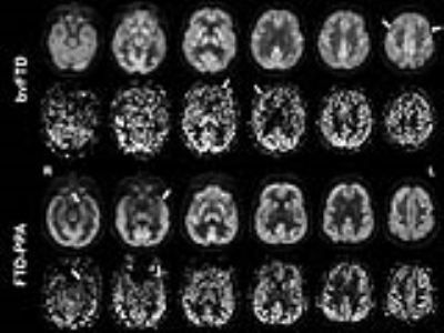 |
Investigating the relationship between perfusion and glucose metabolism by simultaneous PET/MRI in frontotemporal dementia.
Rebecca Steketee, Mariachiara Longarzo, Vincenzo Alfano, Carlo Cavaliere, Dario Grossi, Marion Smits, Marco Aiello
Arterial spin labeling (ASL)-magnetic resonance imaging (MRI) and fluorodeoxyglucose (FDG)-positron emission tomography (PET) both have diagnostic value for dementia, particularly frontotemporal dementia (FTD). By using simultaneous FDG-PET/ASL-MRI, we investigated the relationship between brain metabolism and perfusion in FTD, to evaluate their suitability and complementarity. Exploratory analysis of simultaneous FDG-PET/ASL-MRI in 15 dementia patients showed that metabolism and CBF correlate well on a global level, both visually and quantitatively. On a regional level, one-on-one correlations are limited, supposedly to disease-specific regions such as frontotemporal, subcortical and parietal regions. These results will be substantiated in a larger and better differentiated dementia cohort.
|
|
3022.
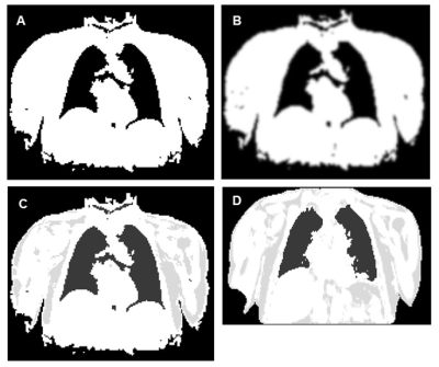 |
An Evaluation of Radial GRE Attenuation Correction Maps for Cardiac and Coronary PET-MRI Studies
Gillian Macnaught, Jack Andrews, David Brian, Kenneth Dolan, Philip Robson, Zahi Fayad, Tim Clark, Alison Fletcher, Matthias Fenchel, Scott Semple, Edwin van Beek, David Newby, Marc Dweck
MR-based attenuation correction of PET images is essential for PET-MRI studies. An intensity threshold method for creating attenuation correction maps (µmaps) from 3D golden-angle radial spoiled gradient echo (radial GRE) images is presented. PET reconstructions using the Threshold µmaps, an existing radial GRE method for creating µmaps and the manufacturer Dixon VIBE µmaps are compared for quantification of 18F Sodium Fluoride (18F NaF) uptake in the aorta. Radial GRE µmaps better delineate the trachea and heart-lung boundaries. Dixon µmaps produced PET images with significantly lower aorta wall SUVmax values than radial GRE µmaps. µmaps must be characterised prior to implementation.
|
|
3023.
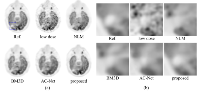 |
Multi-contrast MRI Enhance Ultra-low-dose PET Reconstruction
Junshen Xu, Enhao Gong, Mehdi Khalighi, John Pauly, Greg Zaharchuk
Simultaneous PET/MRI is a powerful multimodality imaging technique for both anatomical and functional imaging. Here we propose a novel method for high-quality PET image reconstruction from ultra-low-dose (more than 99% reduction compared to current practice) PET scanning by using multi-contrast-MRI. A multi-scale fully convolutional network was developed for solving the reconstruction. The proposed method is compared with other methods on a Glioblastoma(GBM) clinical dataset. Results show that our method achieves superior image quality compared with state-of-the-art methods in low-dose PET reconstruction. Besides, quantitative and qualitative evaluations indicate that multi-contrast MRI significantly improves the reconstruction quality with better structural details.
|
|
3024.
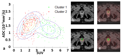 |
A Voxelwise Analysis of PET/MR DATA towards Characterization of Prostate Cancer
Yachao Liu, Mu Lin, Xu Yan, Baixuan Xu
The combined use of diffusion-weighted and 11C-Choline PET images can provide complementary information on prostate cancer. However, it is still unknown how to combine these multiple parameters to give a simple indication for malignant lesions. Based on a scatterplot analysis of standardized uptake values (SUVs) and apparent diffusion coefficient (ADC) values, we clustered voxels into groups corresponding to different tissue types. The proposed method shows promising results in differentiating the lesion of tumor from normal tissue.
|
|
3025.
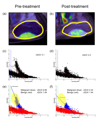 |
ADC-corrected SUV derived from voxel-based SUV-ADC scatter plots for the evaluation of soft-tissue tumor treatment response in FDG-PET/MR hybrid imaging
Sungtak Hong, Yuji Watanabe, Daiki Shinyama, Keisuke Ishimatsu, Koji Sagiyama, Hiroshi Honda
It is often difficult to quantify tumor treatment response with SUVmax because a single voxel measurement does not always represent a whole tumor. In this study, we developed a new parameter called cellular SUV (cSUV) from the SUV-ADC scatter plots. Cluster analysis also applied to the cSUV-measurement of a tumor consisting of multiple components such as liposarcoma, necrotic tumor after treatment, etc. The percent change in cSUV between pre- and post-treatment correlated better with the RECIST1.0 assessment than that of SUVmax. The cSUV combined with cluster analysis could be a promising bio-imaging marker for monitoring treatment response of soft-tissue tumors.
|
|
3026.
 |
Hybrid Liver Multiparametric MRI and F18-FDG PET/MR in Diagnosing and Staging of Intrahepatic Cholangiocarcinoma: An initial Experience
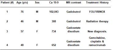
Ming Yang, Alvin Silva, Mitesh Borad, Andrew Liguori, Anshuman Panda, Ba Nguyen, Thomas DeLeon, Michael Roarke, Yuxiang Zhou
Intrahepatic cholangiocarcinoma (ICC) is an uncommon biliary tract malignancy with an unfavorable prognosis given its complicated clinical and imaging manifestations. In this preliminary study, we investigated the role of hybrid liver mpMRI and F18-FDG PET/MR in diagnosing and staging of ICC. Our preliminary data show promising value of this “one-stop” imaging modality in providing complementary morphological and functional information in detecting viable tumor burden, defining nodal and distant metastasis utilizing both MRI and PET molecular imaging biomarkers.
|
|
3027.
 |
A Motion Correction Method Based on Navigator for Simultaneous PET/MR abdominal Imaging
Ke Meng, Lingzhi Hu, Qun Chen
The integrated PET/MR combines the advantages of functional imaging device PET and high resolution high contrast MRI, simultaneously acquiring PET and MR images at the same position, improving image fusion accuracy. However, respiratory motion during abdominal imaging causes notorious motion artifact in the MRI images and blurring the PET images. A PET/MR motion correction method based on real-time 2D excitation navigator has been implemented and evaluated. Phantom and human imaging result implies that this technique can precisely acquire object motion and effectively eliminate motion blurring. Without additional operation and device, it offers a simple and cost-down way for clinical use.
|
|
3028.
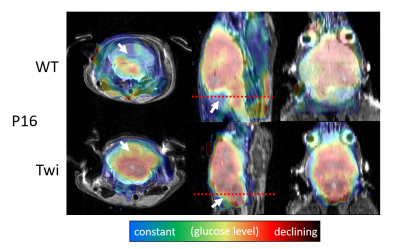 |
Truly simultaneous preclinical PET-MRI in a 20cm 9.4 Tesla magnet with a retrofitted miniature detector: Initial results in the twitcher mouse model of Krabbe disease
Ferdinand Schweser, Akshay Dhamankar, Poonam Choudhary, Nadav Weinstock, Cheryl Knapp, Marilena Preda, Daesung Shin, Robert Zivadinov, Lawrence Wrabetz
While the potential of PET-MRI is increasingly being explored in the clinical setting, preclinical PET-MRI is only slowly leaving the proof-of-concept stage, which may be explained by technical difficulties due to the size-constraints and strong magnetic fields used in preclinical MRI. In the current work, we present results from a first in vivo application of 18F-FDG PET-MRI using a retrofitted micro-PET detector in a commercial 9.4T magnet. We studied the twitcher mouse model of Krabbe disease, in which an altered glucose metabolism had been suggested.
|
|
3029.
 |
Assessment of Metastatic Lymph Node in Head and Neck Cancer Using Simultaneous 18F-FDG-PET and DCE-MRI
Akshay Wadera, Mari Hagiwara, Roy Raad, Kent Friedman, Brian Schmidt, Babak Givi, Adam Jacobson, Theresa Tran, Mark DeLacure, Cheng Liu, Elcin Zan, S. Gene Kim
Regional lymph node metastasis is one of the important predictors of poor prognosis in head and neck cancer. Detecting small nodes with micro-metastases remains challenging for currently available diagnostic imaging methods, including positron emission tomography with 18F-fluorodeoxyglucose (18F-FDG PET) and dynamic contrast enhanced magnetic resonance imaging (DCE-MRI). The purpose of this study is to demonstrate the synergistic role of FDG PET and DCE-MRI in detecting lymph nodes with metastatic potential. Our preliminary results demonstrate that the combined modeling of MR and FDG PET kinetic parameters has the potential to detect lymph node microenvironment changes and assess potentially metastatic lymph nodes.
|
|
3030.
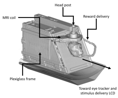 |
PET/MR Platform for Neuroscience in Awake Behaving Non-Human Primates
Rasmus Birn, Samuel Hurley, Abigail Rajala, Caitlynn Filla, Austin Patrick, Dillon Gwozdz, Walter Block, Andrew Alexander, Alexander Converse, Rick Jenison, Bradley Christian, Alan McMillan, Luis Populin
Higher-order cognitive functions result from dynamic interactions of distributed networks comprised of anatomically, physiologically, and pharmacologically separate components of the nervous system. To further our understanding the basic mechanisms and functions of such networks, as well as how they are affected by the administration of therapeutic drugs, we have developed a PET-fMRI platform to take simultaneous measurements of neural activity (fMRI), and concentration of dopamine (PET) during the same physiological state, and without the confounding effects of anesthetics. With this platform, we have measured for the first time in a primate brain the effects of administering different doses of methylphenidate on extracellular levels of dopamine and functional connectivity.
|
|
3031.
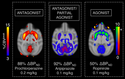 |
Neurovascular coupling to D2/D3 partial agonist antipsychotic drug occupancy using simultaneous PET/fMRI
Christin Sander, Bruce Rosen, Joseph Mandeville
Drug-receptor interactions are the basis of signal modulation in the brain, yet, in vivo mechanisms of the action of many drugs are not well understood. In this study, we characterize the in vivo profile of a current third generation antipsychotic drug at the D2/D3 dopamine receptor using simultaneous PET and fMRI. The results are compared to full D2/D3 antagonists and agonists profiles and show that functional differences can be distinguished with occupancy-matched fMRI responses.
|
|
3032.
 |
Feasibility study of multinuclear MR at 9.4T and PET in a rat brain tumour model
Chang-Hoon Choi, Carina Stegmayr, Aliaksandra Shymanskaya, Wieland A. Worthoff, Nuno A da Silva, Jörg Felder, Karl-Josef Langen, N. Jon Shah
Multinuclear MR provides important information concerning cell integrity or energy metabolism. PET uses radioactive tracers to gain valuable insights into physiological and metabolic processes with both a high level of sensitivity and specificity. Here, we explored the combination of sequential multinuclear MR and PET in a rat brain tumour model. This allows in vivo multinuclear MR-PET experiments to be carried out without compromising the performance of either multinuclear MR or PET. In vivo multinuclear MR and PET images and spectra from rats with/without brain tumours confirmed the potential use of the different X-nuclei derived metabolic information.
|
|
Molecular Imaging
Traditional Poster
Molecular Imaging
Thursday, 21 June 2018
| Exhibition Hall 3033-3051 |
13:15 - 15:15 |
|
3033.
 |
Smart thermosensitive liposomes for effective solid tumor therapy with MRI tracking at 21.1 T
Jens Rosenberg, Kevin Affram, Ofonime Udofot, Mandip Singh, Sunil Krishnan, Renee Reams, Edward Agyare
Here we show the ability of using Gadolinium (Gd) labeled thermosensitive liposomal nanoparticle (TSLnp) as a delivery system for anticancer drug, gemcitabine (Gem) to human pancreatic tumors. Pancreatic cancer (PCa) due to its high malignancy, poor prognosis and resistance to chemotherapy is one of the leading cancer-associated death in the United States. The proposed agent showed significant Gem accumulation in heated tumor relative to free Gem. Gd labeled TSLnp (Gd-TSLnp) show contrast in ex vivo tumor tissue. The Gd-TSLnp show increased T1 contrast in vivo with an implanted tumor compared to Gd and targets the tumor tissue.
|
|
3034.
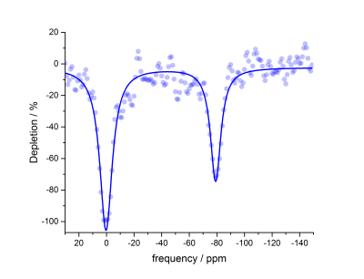 |
Benzene-Appended Cucurbit[6]uril as a Potential Biosensor Scaffold for Hyperpolarized 129Xe MRI Molecular Contrast Agents
Braedan Prete, Dave Robinson, Ashvin Fernando, Yurii Shepelytskyi, Alanna Wade, Francis Hane, Brenton DeBoef, Mitchell Albert
We have recently advanced the field of hyperpolarized (HP) 129Xe magnetic resonance imaging (MRI) with the in vivo detection of cucurbit[6]uril (CB6), a highly sensitive MR contrast agent. CB6 is biochemically inactive, which makes its natural bio-distribution non-specific; thus, it cannot be precisely localized within a living mammalian body using HP 129Xe MRI. We have previously identified cyclodextrin-based pseudorotaxanes as conjugatable scaffolds for xenon biosensors; in this work, we introduce a second class of conjugatable scaffolds, with the hyperCEST detection of benzene-appended CB6, a potential precursor to a wide variety of targeted molecular imaging probes.
|
|
3035.
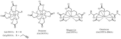 |
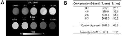
Prospects of 31P Contrast Media for 31P-MRS
Louise Tear, Mahon Maguire, Gogulan Karunanithy, Deborah Sneddon, Nicola Farrer, Andrew Baldwin, Stephen Faulkner, Jurgen Schneider
31P-MRS can be used to determine the relative ratios of phosphate species in vivo to aid clinical diagnosis, but is limited by poor SNR and long acquisition times associated with the 31P nucleus. This work investigates the potential of 31P T1 contrast agents based on Gd.DO3A derivatives by using 31P-MRS. These compounds demonstrate significant relaxation enhancement of 31P R1 for ATP, PCr and Pi, therefore showing excellent potential as 31P contrast agents. Cell studies indicate the Gd.DO3A derivatives investigated do not come into contact with intracellular phosphate metabolites, which limits these initial complexes to use as extracellular contrast agents.
|
|
3036.
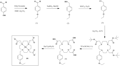 |
Dysprosium based liver-specific ultra-high field MRI T2 contrast agent
Ah Rum Baek, Heekyung Kim, Soyeon Kim, Garam Choi, Bokyung Sung, MD. Kamrul Islam, Taekwan Lee, DongKyu Kim, Hoesu jung, Yongmin Chang
[Dy(EOB-DO3A)] is prepared according to the general synthetic methods, and characterized by spectroscopic analysis.Therelaxivities that measured at 9.4 T animal MRI are r1 = 1.01, r2 = 2.80 mM-1s-1.We observe acceptablenegative-enhancement with liver T2-weighted image, alsoconfirm about 30% liver accumulation within 1 h post-injection at inductively coupled plasma (ICP) spectrometer data.
|
|
3037.
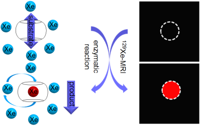 |
A Hyperpolarized 129Xe “OFF-ON” MRI Biosensor Triggered by Diamine Oxidase
Bin Zhang, Qing Luo, Qianni Guo, Xiaoxiao Zhang, Qingbin Zeng, Longhui Zhao, Yaping Yuan, Weiping Jiang, Chaohui Ye, Xin Zhou
Benefiting from ultra-high sensitivity of Hyper-CEST method, 129Xe biosensors possess an obvious advantage in sensitivity over other MRI sensors. However, due to its indirect detection mode the Hyper-CEST spectra resolution is relatively low limiting chemical shift to be an effective indicator in traditional NMR. In order to solve this problem, a 129Xe biosensor based on a new “turn-on” strategy is designed, which exhibits high detection specificity for an enzyme diamine oxidase (DAO). This 129Xe biosensor possesses very high detection sensitivity, and can be tested in Small intestinal villus epithelial cells. Using this strategy, lots of disease-related enzyme can be detected.
|
|
3038.
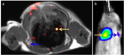 |
Fluorine-19 MRI hot-spot imaging of lung metastasis in rodents
Deanne Lister, Hongyan Xu, Eric Ahrens
Lung cancer is the leading cause of cancer deaths. Safe and specific MRI probes are needed to enable early detection of lesion presence and therapeutic response. Injected PFC nanoemulsion, taken up by tumor associated macrophages (TAMs), can be used as a biomarker to detect metastases using 19F MRI. In a metastatic lung cancer mouse model, we show that PFC is effectively taken up by TAMs and vividly displays lung metastasis using 19F MRI. Validation assays using in vivo bioluminescence and histology support the MRI findings. Overall, 19F hot-spot imaging offers a highly-specific marker of tumor burden in lung parenchyma.
|
|
3039.
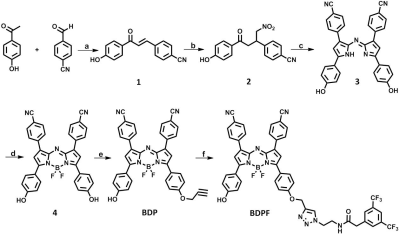 |
A Small Molecule NIR-19F MR Contrast Agent of Aza-BODIPY for Bimodal In Vivo Imaging
Lianhua Liu, Yaping Yuan, Yuqi Yang, McMahon Michael , Shizhen Chen, Xin Zhou
Accurate and early diagnosis of diseases is most import for medical imaging. MRI is one of the most promising techniques for the non-invasive visualization. Compared to 1H MRI, 19F MRI provides high-contrast images without endogenous background signals, but low sensitivity. To address the limitation, our strategy is to combination of 19F MRI and a more sensitive NIR fluorescence imaging technique to develop a bimodal contrast agent BDPF. Both ex vivo and in vivo experimental results indicated BDPF had excellent optical and 19F MRI properties. Thus, the NIR-19F MR bimodal imaging may provide a new way to detect tumor.
|
|
3040.
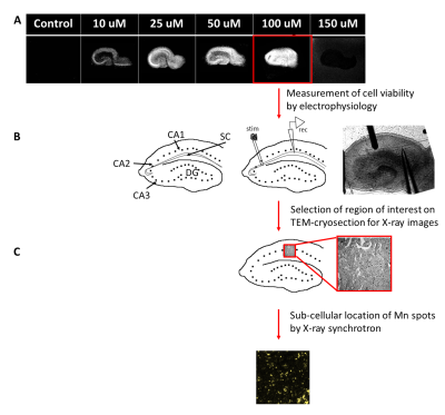 |
Manganese enhanced MRI in organotypic rat hippocampus slices: A correlative study with synchrotron X-ray nanoprobe analysis and electron microscopy.
Alexia Daoust, Natalia Pivovarova, Emily Petrus, S Andrews, Barry Lai, Si Chen, Maria Aronova, Richard Leapman, Alan Koretsky
Manganese (Mn2+) Enhanced Magnetic Resonance Imaging (MEMRI) can be used for different applications such as tracing neuronal connections or functional imaging. However, Mn2+ uptake and transport mechanisms are still unclear. These mechanisms were studied by imaging sub-cellular Mn2+ in an organotypic hippocampal slice culture by coupling MEMRI, TEM and X-ray methodologies. The data indicates that Mn2+ is located at synapses but not in mitochondria.
|
|
3041.
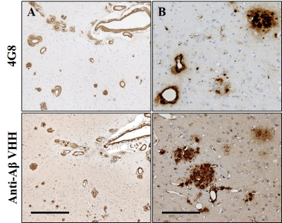 |
Camelid single-domain antibodies bioconjugate for the magnetic resonance imaging of Alzheimer’s disease.
Clémence Dudeffant , Matthias Vandesquille, Tengfei Li, Christelle Ganneau, Ihsen Youssef, Benoît Delatour, Pierre Lafaye, Sylvie Bay, Marc Dhenain
Detection of intracerebral targets with imaging probes is challenging due to the non-permissive nature of blood-brain barrier (BBB). Camelid single-domain antibody-fragments (VHH) are small and stable antibodies able to potentially cross the BBB. Here, we selected VHH specifically targeting amyloid-beta deposits, one of the main lesions of Alzheimer’s disease and labeled them with the contrastophore gadolinium. These innovative contrast agents allowed MRI detection of amyloid deposits in postmortem brain tissues of a mouse model of amyloidosis. The ability to produce VHH conjugates that cross the BBB opens the way for future development of tailored imaging probes targeting intracerebral antigens.
|
|
3042.
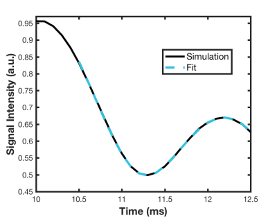 |
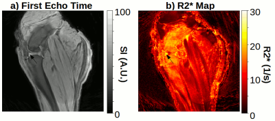
Investigating Off-Resonance Fat Modulations in the TurboSPI Signal to Improve R2* Mapping for Quantitative Cell Tracking
Zoe O'Brien-Moran, Chris Bowen, James Rioux*, Kimberly Brewer*
TurboSPI has the potential to offer quantitative cell tracking with high fidelity R2* mapping. However, early in vivo studies demonstrated that accuracy of the R2* fitting deteriorates in the presence of off-resonance fat signal. In this work, we investigate these findings further with an in vitro study. We used in silico and in vitro data to develop and test a more comprehensive decay model that accounts for fat oscillations in the TurboSPI signal. The proposed model results in improved R2* estimates in the presence of fat.
|
|
3043.
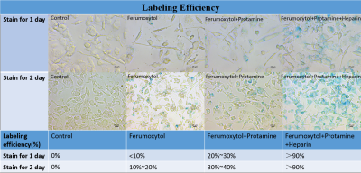 |
Improved Sensitivity of Cellular MRI Using Phase-cycled Balanced SSFP of Ferumoxytol Nanocomplex Labeled Macrophages at Ultra-high Field
Yelong Shen, Lirong Yan, Xingfeng Shao, Bin Zhao, Jinlun Bai, Wange Lu, Danny Wang
This study aimed to investigate the feasibility and sensitivity of cellular MRI with ferumoxytol nanocomplex labeled macrophages at ultrahigh magnetic field of 7T. Different labeling strategies, labeling times, magnetic field strengths, imaging sequences and post processing methods were evaluated to achieve the optimal protocol. Combining ferumoxytol, heparin and protamine (HFP nanocomplex) labeled macrophages with balanced steady-state free precession (bSSFP) sequence on a 7T MRI scanner and post processed by root mean square (RMS) combination of multiple phases showed the best contrast in phantom and ex vivo experiments, reaching a sensitivity for detecting a few tens of cells.
|
|
3044.
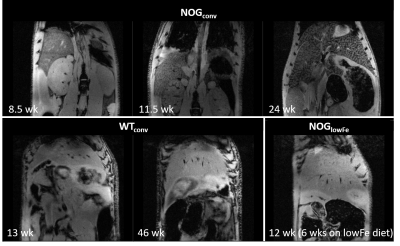 |
Unexpected accumulation of iron in liver of immune compromised mice: Implications for cell tracking experiments
Christiane Mallett, Matti Kiupel, Erik Shapiro
An increased iron load was observed in immune-deficient mice which may mean that they are not suitable for iron oxide based cell tracking experiments. This was not seen in healthy controls fed a similar diet. It was resolved by feeding a low-iron diet.
|
|
3045.
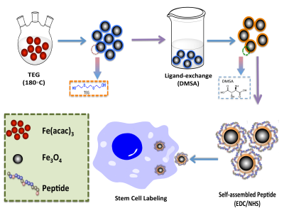 |
Stem Cell Tracking Using Effective Self-Assembled Peptide-Modified Superparamagnetic Nanoparticles
Lei Gu, Min Wu
In cell therapies and regeneration medicine, superparamagnetic iron oxide nanoparticles (SPIONs) have been developed as excellent magnetic resonance imaging (MRI) contrast agents for stem cell labeling and tracking due to their biocompatibility. Here, we designed a self-assembled peptide amphiphile (PA) replace the transfection agents. This PA was conjugated to the surfaces of SPIONs to label rat mesenchymal stem cells (MSCs), which enhanced the contrast and labeling effects. The labeled cells showed that peptide-SPIONs had improved internalization, efficiency and T2-weight relaxivity and were nontoxic to the MSCs. The results demonstrated that these self-assembled peptide-modified SPIONs are potential candidates to label MSCs for tracking stem cells using MRI in vivo.
|
|
3046.
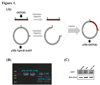 |
A proof-of-concept study on the quantification of gene expression levels with doxycycline-inducible MR reporter gene
Seul-I Lee, Jeeheon Kang, Yoonseok Choi, Jinil Kim , Jae-Im Kwon, Ho-jin Kim, Su Jung Ham, Sang-Tae Kim, Chul Woong Woo, Do-Wan Lee, Dong-Cheol Woo, Kyung Won Kim
Recent research on MR reporter genes has demonstrated their potential for use in transgene expression monitoring. We have conducted a preliminary study on the development of a new MRI reporter system [organic anion transporting polypeptide (OATP) 1B1] that can analyze gene expression level using MR reporter genes. By establishing doxycycline-inducible cell line, we observed T1 shortening on MRI, which indicated increased expression level of the OATP1B1 gene. A strong correlation was observed between conventional methods for measurement of gene expression and rrT1 of MR imaging. In this study, we provide preliminary evidence of the potential application of MRI to determine gene expression.
|
|
3047.
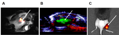 |
GMP-grade nanoparticle imaging agent for 19F MR, photoacoustic, and fluorescence imaging
Edyta Swider, Khalid Daoudi, Eric van Dinther, Alexander H. J. Staal, N. Koen van Riessen, Olga Koshkina, I. Jolanda M. de Vries, Chris L. de Korte, Mangala Srinivas
Cellular therapies hold great promise for the treatment of various diseases. Its success strongly depends on the imaging modality and cell tracking, which can be achieved by the addition of an imaging label to cells, for example in the form of nanoparticles. Here, we report on polymeric nanoparticles encapsulating perfluorocarbon and dye, which can be used for cell loading and can be detected with several imaging modalities. This will further give information about cell numbers and localization in vivo.
|
|
3048.
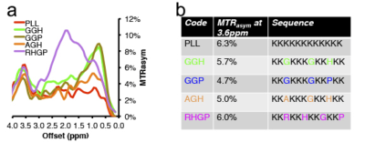 |
An Improved CEST MRI Reporter Gene for Molecular Imaging of Cell and Viral Based Therapeutics
Christian Farrar, Hirotaka Ito, Hiroshi Nakashima, E. Chiocca, Assaf Gilad
The ability to image cell- or viral-based therapeutics is critical for optimizing therapeutic strategies and assessing efficacy. A lysine rich protein (LRP) chemical exchange saturation transfer (CEST) MRI reporter gene has previously been developed and successfully used to image oncolytic viruses and tumor cells. However, the highly repetitive nature of the LRP reporter gene sequence lead to DNA recombination events and the expression of a range of truncated LRP protein fragments, thereby greatly limiting the CEST sensitivity. Here we report the use of a redesigned LRP reporter (rdLRP), which demonstrated excellent stability and CEST sensitivity.
|
|
3049.
 |
Magnetic Resonance Tracking of Iron-Labeled Stem Cells After Osteochondral Defect in Ovine Model
Joshua Kaggie, Martin Graves, James MacKay, Scott Reid, Hareklea Markides, Alicia El Haj, Stephen McDonnell, Fiona Gilbert, Andrew McCaskie, Frances Henson
Multipotent mesenchymal stem cells (MSCs) can be labeled with superparamagnetic iron-oxide nanoparticles (SPION) particles to track single cells with MRI, and thereby follow MSC infiltration. However, a limitation with conventional MR sequences is that their long echo times are unable to measure fast signal decays, which occur in dense bone tissue and with high SPION infiltration. Ultra-short echo time (UTE) MRI can capture these rapidly decaying signals. In this work, we use 3D cones to track tissue development after injection of SPION labeled MSCs in an ovine model.
|
|
3050.
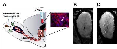 |
Mapping of spatial distribution of the olfactory bulb new neurons at single cell level using iron oxide assisted-MRI
Nikorn Pothayee, Claire Perez, Stephen Dodd, Alan Koretsky
In this study, we aimed to develop a method that could quantitatively track new neurons in the olfactory bulb (OB). We first established that MRI signals detected in the OB were those of single labeled new neurons that migrate from the neurogenic niche into the OB. Further, we combined the anatomical MRI enhancing properties of Mn2+ to evaluate the preference of new neurons for specific layers within the OB and to determine whether sensory enrichment affects distribution of adult-born neurons within the OB layers.
|
|
3051.
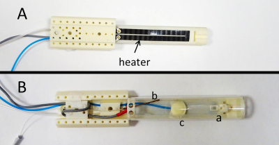 |
A Carbon-Fibre Sheet Resistor for MR, CT, SPECT and PET-compatible Temperature Maintenance in Small Animals
Veerle Kersemans, Stuart Gilchrist, Philip Allen, Paul Kinchesh, Sean Smart
A resistive heater that is compatible with MR, CT, SPECT and PET imaging has been produced from a commercially available carbon-fibre sheet. Adequacy of temperature maintenance and insensitivity of MR and CT imaging to the presence and use of the heater is shown. Multimodal MR-CT-PET-SPECT imaging of the lower abdomen is demonstrated in vivo in the physiologically maintained and viable anaesthetised mouse.
|
|
Hyperpolarised MR
Traditional Poster
Molecular Imaging
Thursday, 21 June 2018
| Exhibition Hall 3052-3074 |
13:15 - 15:15 |
|
3052.
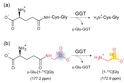 |
Application of a novel 13C hyperpolarized metabolic tracer for ?-Glutamyl transferase activity in vivo tumor xenograft
Tomohiro Seki, Marino Itoda, Shun Kishimoto, Kazu Yamamoto, Yoichi Takakusagi, Jeffery Brender, Ronja Malinowski, Tatsuya Nishihara, Hikari Yoshihara, Hiroshi Nonaka, Keita Saito, Nobu Oshima, Jan Ardenkjaer-Larsen, James Mitchell, Murali Krishna, Shinsuke Sando
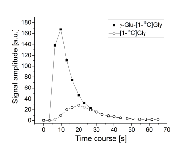
This research aimed to develop the non-invasive in vivo detection of γ-glutamyl transferase (GGT) activity by a novel GGT molecular probe, γ-Glu-[1-13C]Gly, in combination with hyperpolarized (HP) 13C Magnetic Resonance (MR) spectroscopy. We succeed in detecting the strong HP 13C MR signal of γ-Glu-[1-13C]Gly from tumor xenograft in vivo. Detecting HP 13C MR signal from the metabolite of this novel probe in tumor xenograft is our next challenge.
|
|
3053.
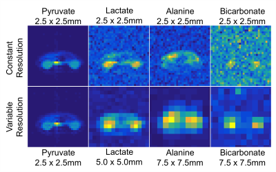 |
Variable Resolution Echo-Planar Imaging for Improved Quantification of Hyperpolarized 13C Metabolism
Jeremy Gordon, Eugene Milshteyn, Daniel Vigneron, Peder Larson
Unlike ionizing imaging modalities, the SNR in MRI is proportional to voxel volume, but downsampling or voxel averaging after acquisition only improves SNR by the square root of the voxel volume. To take advantage of this distinction, we use a frequency selective imaging approach to independently excite the hyperpolarized 13C substrate (pyruvate) and downstream metabolites (lactate, alanine, and bicarbonate). This allows us to tailor the spatial resolution for each metabolic product, yielding high-resolution images for pyruvate as well as quantification at a coarser resolution for the lower SNR metabolites, such as bicarbonate, which would be undetectable at the higher resolution.
|
|
3054.
 |
Dynamic Metabolic Imaging of Co-Polarized [2-13C]Pyruvate and [1,4-13C2]Fumarate Using 3D-Spiral CSI with Alternate Spectral Band Excitation
Maninder Singh, Sonal Josan, Dirk Mayer
Metabolic imaging of biologically-relevant hyperpolarized agents allows measurement of metabolic processes in real time in-vivo. We demonstrate dynamic metabolic imaging of a single bolus of co-polarized [2-13C]pyruvate and [1,4-13C2]fumarate in control as well as in rats with liver necrosis. Chemical shift imaging (CSI) of such a mixture is challenging due to the large spectral dispersion of resulting resonances, which could lead to severe chemical shift displacement artifacts if acquired by conventional slice-selective excitation pulses. Here we obtain CSI information by a volumetric method using alternate 3D spectrally-selective excitation of sub-bands containing fewer resonances.
|
|
3055.
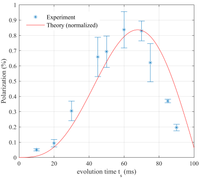 |
Simple and fast hyperpolarization of a biomolecule: Theory and Experiment
Stephan Berner, Stephan Knecht, Andreas Schmidt, Mirko Zimmermann, Jürgen Hennig, Dominik von Elverfeldt, Jan-Bernd Hövener
Hyperpolarization overcomes the biggest limitation of MRI: its low sensitivity, and enables metabolite mapping. Hyperpolarized 13C magnetization can be produced by transferring the spin order of parahydrogen into 13C by hydrogenation followed by a sequence of 1H and 13C pulses. However, it is possible to hyperpolarize AA’X spin systems by two pulses on 13C. Theoretical models were developed to describe the polarization transfer and significant signal increase was observed for the biomolecule succinate after spin order transfer directly in the magnet of a commercial MRI system. The experimental data is well described by theoretical calculations except for an overall scaling factor.
|
|
3056.
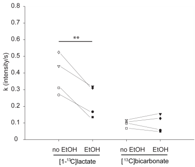 |
Ex-vivo real-time measurement of ethanol induced changes in brain metabolism of hyperpolarized [1-13C]-pyruvate
Benjamin Grieb, Assad Azar, Talia Harris, Gal Sapir, Atara Nardi-Schreiber, Ayelet Gamliel, Sivaranjan Uppala, Jacob Sosna, J. Gomori, Rachel Katz-Brull
Changes in brain metabolism during acute alcohol intoxication either reflect global inhibition or changes in the utilized energy substrates. NMR spectroscopy of hyperpolarized metabolites offer the opportunity to investigate the metabolic changes due to alcohol intoxication in the brain in real time. Here we present preliminary data showing decreased [1-13C]lactate formation from hyperpolarized [1-13C]pyruvate in perfused and ethanol incubated rat brain slices. This approach may offer a future innovative tool to non-invasively image brain metabolism in real-time during alcohol intoxication.
|
|
3057.
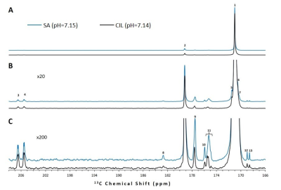 |
Impurities of [1-¹³C]pyruvic acid and their potential effects on the interpretation of hyperpolarized pyruvate metabolism studies
Talia Harris, Ayelet Gamliel, Jacob Sosna, J. Moshe Gomori, Rachel Katz-Brull
Commercially available [1-13C]pyruvic acid contains impurities that have chemical shifts similar to pyruvate’s metabolic products. We show that these observed impurity peaks possess long T1s and for several peaks the chemical shift is very sensitive to the pH in the narrow physiological range measured. We concluded that in order to reliably identify low concentration metabolic products of hyperpolarized pyruvate it is crucial to characterize in situ the pH dependent impurity spectrum of the batch of [1-13C]pyruvic acid used.
|
|
3058.
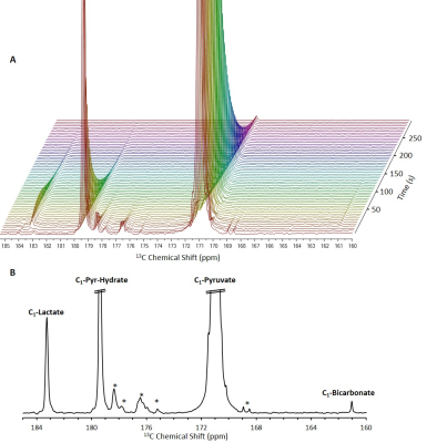 |
Real-time ex-vivo measurement of brain metabolism using hyperpolarized [1-¹³C]pyruvate
Talia Harris, Assad Azar, Gal Sapir, Ayelet Gamliel, Atara Nardi-Schreiber, Jacob Sosna, J. Moshe Gomori, Rachel Katz-Brull
Translating the hyperpolarized signal observed in the brain in vivo to cerebral metabolic rates is not straightforward, as the observed signals reflect also the influx of metabolites produced in the body, the cerebral blood volume and flow, and the rate of transport across the blood brain barrier. We introduce a robust method to study rapid metabolism of hyperpolarized substrates ex vivo in viable rat brain slices and demonstrate its ability to characterize rates of LDH and PDH activities. Despite variations in these measured rates, we saw that the Lactate to Bicarbonate ratio is highly reproducible across all samples.
|
|
3059.
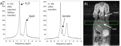 |
In vivo hyperpolarization transfer in a clinical MRI scanner
Cornelius von Morze, Galen Reed, Peder Larson, Daniele Mammoli, Albert Chen, James Tropp, Mark Van Criekinge, Michael Ohliger, John Kurhanewicz, Daniel Vigneron, Matthew Merritt
The purpose of this study was to investigate the feasibility of in vivo 13C->1H hyperpolarization transfer, which has significant potential advantages for detecting the distribution and metabolism of hyperpolarized 13C probes, in a clinical MRI scanner. A standalone pulsed 13C RF transmit channel was developed for operation in conjunction with the standard 1H channel of a clinical 3T MRI scanner. Operation of the custom pulsed 13C RF channel resulted in effective 13C->1H hyperpolarization transfer, as confirmed by the characteristic anti-phase appearance of 1H-detected, 1JCH-coupled doublets. 1H detection of HP [2-13C]lactate generated in vivo was achieved in a rat liver slice.
|
|
3060.
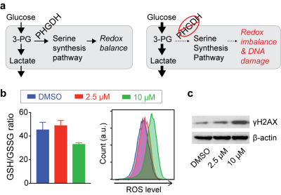 |
Novel Metabolic Markers for Therapeutic Approaches Targeting Serine Synthesis Pathway in Leukemia
Sangmoo Jeong, Madeleine Gao, Alexandra Schurer, Nathaniel Kim, Yuanming Cheng, Roozbeh Eskandari, Michael Kharas, Kayvan Keshari
The serine synthesis pathway (SSP), which provides precursors for redox homeostasis and nucleotide synthesis, has emerged as a critical metabolic pathway in cancer. However, the assessment of therapeutic approaches targeting the SSP has been challenging due to a lack of distinct biomarkers. We have identified that the SSP inhibition increases reactive oxygen species (ROS) levels and, intriguingly, glycolytic rate in leukemia cells. Using hyperpolarized dehydroascorbate and pyruvate magnetic resonance, we assessed therapeutic responses earlier than any significant changes in cell viability. This approach has broad implications as an effective methodology for monitoring therapeutic responses with SSP inhibition in multiple cancers.
|
|
3061.
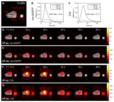 |
In vitro and in vivo 13C metabolic imaging of pyruvate to lactate conversion with high spatial and temporal resolution using a me-bSSFP sequence
Christoph Müller, Christian Hundshammer, Miriam Braeuer, Jason Skinner, Adam Hansen, Sven Mansson, Franz Schilling, Jochen Leupold, Dominik von Elverfeldt, Jan Ardenkjaer-Larsen, Markus Schwaiger, Jürgen Hennig, Jan-Bernd Hövener
In order to use the transient signal of hyperpolarized tracers and their metabolites efficiently, dedicated imaging sequences are required. Here, we present a multi-echo bSSFP sequence with Dixon-based iterative reconstruction to obtain metabolite maps of hyperpolarized [1-13C]pyruvate and the product of an enzymatic conversion [1-13C]lactate on a human 3T PET-MRI system in vitro and in vivo. When comparing to other methods (i.e. CSI and non-localized NMR spectra) we found that me-bSSFP provides good metabolite separation and reliable quantitative kinetic data more than 16 times faster than CSI (350 ms vs. 5.8 s), while consuming a similar amount of hyperpolarized magnetization.
|
|
3062.
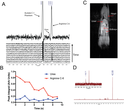 |
In Vivo Spectroscopic Detection of Arginase Enzyme Activity with Hyperpolarized [6-13C,15N3]-Arginine
Andrew Cho, Roozbeh Eskandari, Kayvan Keshari
Aberrations in arginase enzyme expression are associated with a variety of pathologies, and an in vivo probe to quantify flux through this pathway may hold utility towards patient stratification. We propose the use our custom synthesized compound, [6-13C,15N3]-arginine, as a hyperpolarized MRI probe for arginase activity. 15N enrichment reduces quadrupolar relaxation and extends T2, facilitating in vivo imaging. We were able to acquire 13C spectroscopic data on a healthy mouse and detected in vivo conversion of hyperpolarized arginine to urea, which warrants further exploration of this imaging probe in the future.
|
|
3063.
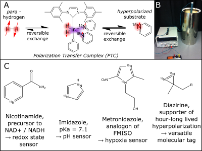 |
Recent advances in low-cost, rapid Hyperpolarization Chemistry: from portable NMR to low-cost molecular MRI.
Thomas Theis, Johannes Colell, Zijian Zhou, Shannon Eriksson, Jacob Lindale, Yi-Fen Yen, Matthew Rosen, Eduard Chekmenev, Warren Warren
NMR and MRI are inherently low sensitivity techniques. Hyperpolarization technology overcomes this problem by enhancing MR signals by 10,000-fold or more. However, most hyperpolarization techniques are complex, expensive and slow. We describe hyperpolarization chemistry that is simple, low-cost, and fast or even continuous. Specifically, we describe recent advances in parahydrogen-induced polarization, combined with various MR detection schemes to establish 1) miniaturized NMR spectrometers, 2) NMR structural elucidation with reduced limits of detection, and 3) low-cost biomolecular imaging.
|
|
3064.
 |
Boosting SABRE-SHEATH hyperpolarization with Coherent Control of Spin Dynamics
Thomas Theis, Shannon Eriksson, Johannes Colell, Zijian Zhou, Jacob Lindale, Warren Warren
Signal Amplification By Reversible Exchange (SABRE) is a parahydrogen based hyperpolarization modality that is particularly simple, low-cost, and fast or even continuous. A more recent variant, SABRE-SHEATH (SABRE in SHield Enables Alignment Transfer to Heteronuclei) enables targeting 15N and 13C nuclei in a wide range of substrates, where hyperpolarization lifetimes can be particularly long. However, both SABRE and SABRE-SHEATH are limited by the incoherent nature of the hyperpolarization transfer process. Here we describe a pulsed variant of SABRE-SHEATH that takes coherent control over the spin dynamics and more than doubles achievable hyperpolarization levels. In addition, the pulsed SABRE-SHEATH experiments provide a new way of probing the hyperpolarization transfer, shedding new light on the limiting factors of this emerging technology.
|
|
3065.
 |
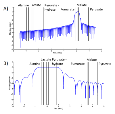
Super-resolution Hyperpolarized C13 Imaging with 2D-Linear Prediction and Trigonometric Curves
Jack Miller, Sofia Dimoudi, Aaron Hess, Damian Tyler
Hyperpolarized $$$^{13}\text{C}$$$-imaging techniques a powerful and clinically translatable method to image metabolism. However, owing to the finite and non-renewable magnetisation available to the technique, all proposed imaging sequences necessarily have a comparatively small matrix size compared to conventional anatomical imaging. Typically hyperpolarized images are therefore reconstructed with a large degree of zero-filling. We show here that a modified form of 2D least-squares linear prediction that uses the known analytic properties of trigonometric curves can extrapolate unmeasured Fourier coefficients and hence improve the apparent reconstructed resolution of hyperpolarized images.
|
|
3066.
 |
Assessing ?-glutamyl transpeptidase activity in kidney using hyperpolarized ?-Glu-[1-13C]Gly
Steffen Frank, Hikari Yoshihara, Marino Itoda, Shinsuke Sando, Rolf Gruetter
Hyperpolarized γ-Glu-[1-13C]Gly provides a non-invasive means to detect γ-glutamyl transpeptidase (GGT) enzyme activity in vivo and indicates its potential for application in functional imaging. Since GGT is most abundant in the proximal tubules of the kidney, and since the properties of γ-Glu-[1-13C]Gly are suitable for in vivo hyperpolarized 13C metabolic analysis, it was proposed as a molecular probe to study kidney function. The aim of the present study is to identify the dose of γ-Glu-[1-13C]Gly that gives high NMR sensitivity in the unsaturated state of the GGT enzyme.
|
|
3067.
 |
Dynamic Hyperpolarized 13C MRSI using the SPICE technique: A feasibility study
JaeEun Song, Jaewook Shin, Hansol Lee, Young-suk Choi, Chan Gyu Joo, Ho-Taek Song, Dong-Hyun Kim
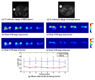
In this study, we investigated the use of SPICE (SPectroscopic Imaging by exploiting spatiospectral CorrElation) technique for dynamic hyperpolarized 13C MRSI by in vitro phantom experiment and in vivo mouse experiment. In vitro phantom experiment, the dynamic images from SPICE were compared to the dynamic data from FIDCSI. In vivo experiment, the dynamic images were acquired in normal and high fat diet (HFD) mouse kidney.
|
|
3068.
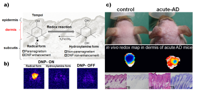 |
Non-invasive redox molecular imaging of atopic dermatitis using in vivo dynamic nuclear polarization MRI
Fuminori Hyodo, Hinako Eto, Gaku Tsuji, Masutaka Furue, Masayuki Matsuo
Atopic dermatitis (AD) is a chronic inflammatory condition with complex etiology. Redox imbalance caused by excessive oxidative stress has been shown to mediate disease activity of AD. We have established such a technique that can detect and visualize the redox status of the skin using in vivo dynamic nuclear polarization(DNP) MRI. We utilized an AD mouse model that was generated by repeated topical application of mite antigen in NC/Nga mice. We revealed that AD skin lesions demonstrated more rapid reduction rates of image intensity than normal skin, indicating that our technique can monitor oxidative stress in AD skin.
|
|
3069.
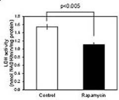 |
Monitoring effect of rapamycin on pyruvate metabolism in SCC tumor using hyperpolarized 13C-MRI
Keita Saito, Shingo Matsumoto, Yoichi Takakusagi, Masayuki Matsuo, Hellmut Merkle, James Mitchell, Murali Krishna
Effect of an mTOR inhibitor rapamycin on pyruvate metabolism in squamous cell carcinoma (SCC) xenografts was investigated using hyperpolarized 13C-MRI. [1-13C]lactate to [1-13C]pyruvate ratio (Lac/Pyr) in the SCC tumors increased as tumor grew in non-treated control mice, whereas it significantly dropped after 2 days of the rapamycin treatment. Inhibition of mTOR caused a drop of LDH protein level and the activity in the SCC tumor, and perfusion in the tumor was improved by the rapamycin treatment. Lac/Pyr monitored using hyperpolarized 13C-MRI would become a useful marker for tumor response to mTOR inhibitors.
|
|
3070.
 |
Multiscale Imaging of Breast Cancer Metabolism using Fluorescence Lifetime Imaging Microscopy and Hyperpolarized Magnetic Resonance Spectroscopy
Sarah Erickson-Bhatt, Ben Cox, Erin Adamson, Suzanne Ponik, Matthew Conklin, Brett Morris, David Inman, Joseph Szulczewski, Patricia Keely, M. Elizabeth Meyerand, Caroline Alexander, Kevin Eliceiri, Sean Fain
Every day in the U.S. 100 women die of metastatic breast cancer. Current clinical methods cannot determine from the primary site which tumors will metastasize and spread to other areas of the body. Herein, multiple imaging scales are used to assess the metabolic signatures of metastatic and dormant tumor cell lines. Fluorescence lifetime imaging microscopy (FLIM) and hyperpolarized magnetic resonance spectroscopy (hMRS) imaging studies are performed in 3D cell culture using an MRI compatible bioreactor and in vivo mouse models to evaluate metabolic signatures at the individual cellular and tumor mass scales to predict metastasis versus dormancy.
|
|
3071.
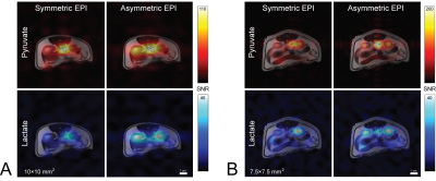 |
Comparison of Asymmetric and Symmetric K-space Sampling in EPI for 3D Time-Resolved Hyperpolarized 13C MRI with [1-13C]Pyruvate
Benjamin Geraghty, Casey Lee, Albert Chen, Charles Cunningham
The minimum echo-time for hyperpolarized 13C echo-planar imaging can be reduced with partial sampling along the blipped direction in k-space. To investigate the extent to which echo-time shortening can improve signal-to-noise ratio, we’ve employed an experimental design that toggles between two different spatial encoding strategies during a time-resolved hyperpolarized [1-13C]pyruvate acquisition. Using clinically approved hardware with a pre-clinical animal model, we compared symmetric with asymmetric echo-planar imaging. Considerable signal-to-noise ratio gains for asymmetric vs symmetric sampling were observed without artifacts. On the basis of this study, our group will employ asymmetric sampling in our forthcoming human trials.
|
|
3072.
 |
Hyperpolarized 13C chemical shift imaging of transient focal ischemia reperfusion injury in developing rat brain
Shu-Juan Fan, Amara Larpthaveesarp, Yiran Chen, Sukumar Subramaniam, Robert Bok, Fernando Gonzalez, Duan Xu
We investigated the use of hyperpolarized [1-13C]pyruvate magnetic resonance chemical shift imaging in monitoring energy metabolism in developing rat brain following transient focal ischemia-reperfusion injury, which is the most common form of stroke in neonates. We show that the conversion from [1-13C]pyruvate to [1-13C]lactate was higher in the injured cerebral hemisphere as compared with that in the contralateral hemisphere, which lasted for up to 7 days after the ischemia-reperfusion injury. This phenomenon can be potentially used as a biomarker to facilitate long-term prognosis, characterize therapeutic responses and study the mechanisms of injury repair in neonates with transient focal ischemic stroke.
|
|
3073.
 |
Quantitative Data Analysis of in vivo Hyperpolarized 13C NMR Data: 1D vs 2D
Frits van Heijster, Ron de Beer, Arend Heerschap, Dirk van Ormondt
13C-DNP hyperpolarization (HP) in MR allows for single shot detection of 13C-labeled metabolites in vivo. The dynamic acquisition of 13C MR signals after injection of a HP 13C-substrate results in a two-dimensional time-domain dataset. Often the 1D NMR time domain is fitted first and the results are fed into a kinetic model. We present a 2D method, in which all data points in both NMR and kinetic time dimensions are fitted simultaneously. This results in an improved accuracy for all determined kinetic parameters compared to the 1D method, in particular for low-SNR metabolites. CRBs are significantly smaller using 2D analysis.
|
|
3074.
 |
HyperSIFT: Temoporal Denoising of Hyperpolarized Data Improves SNR while Perserving Dynamic Information
Kristin Granlund, Kayvan Keshari
We apply a temporal denoising algorithm to hyperpolarized MRI. The SIFT method filters out temporal frequencies with low amplitudes, reducing noise while preserving dynamic information. We demonstrate this in a bioreactor setting, introducing hyperpolarized [1-13C] pyruvate to cells and human subjects and observing the conversion of pyruvate to lactate.
|
|
| Back |
| The International Society for Magnetic Resonance in Medicine is accredited by the Accreditation Council for Continuing Medical Education to provide continuing medical education for physicians. |
