|
Traditional Poster Session
Cardiovascular |
Thursday, 21 June 2018
Traditional PosterCardiovascular
2896 -2923 Tissue Characterization
2924 -2948 Velocity & Flow
2949 -2969 Cardiac Function & Myocardial Perfusion
2970 -2981 Cardiovascular Image Processing
2982 -3007 Vascular
3008 -3019 Novel Concepts, Techniques & Methods |
| |
Tissue Characterization
Traditional Poster
Cardiovascular
Thursday, 21 June 2018
| Exhibition Hall 2896-2923 |
13:15 - 15:15 |
|
2896.
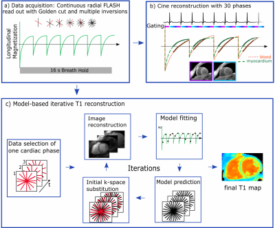 |
Simultaneous high-resolution cardiac T1 mapping and cine imaging using model-based iterative image reconstruction
Kirsten Becker, Jeanette Schulz-Menger, Tobias Schaeffter, Christoph Kolbitsch
Native myocardial T1 mapping provides information for the detection of diffuse fibrosis in different cardiac diseases. Here we present simultaneous T1 mapping and cine imaging with a resolution of 1.3x1.3mm² using model-based iterative reconstruction. T1 times in the septum were 1285±46ms compared to 1240±28ms obtained with MOLLI. In contrast to MOLLI, the proposed approach did not show any heart rate dependence of T1. In addition, the approach allows T1 mapping of challenging structures, such as the right ventricle and the apex. Functional assessment of reconstructed cine images did not show any significant differences compared to a standard Cartesian cine scan.
|
|
2897.
 |
Prospective correction of patient-specific respiratory motion in T1 and T2 mapping
Michael Bush, Rizwan Ahmad, Yingmin Liu, Ning Jin, Juliet Varghese, Orlando Simonetti
Respiratory motion in cardiovascular MRI presents a challenging problem with many solutions. Current approaches require breath-holds, neglect through-plane motion or significantly increase scan time. Our patient-specific prospective motion correction strategy addresses these issues and corrects for respiratory motion in real time. Numerous cardiac imaging applications stand to benefit from our approach, including perfusion imaging, parameter mapping, and late gadolinium enhancement. By modeling on a patient-specific basis, and prospectively correcting for respiratory motion, we expect to significantly improve the reliability and efficiency of CMR. For demonstration, the proposed strategy was applied to improve the accuracy of free-breathing T1 and T2 mapping.
|
|
2898.
 |
Dynamic Nitroxide-Enhanced MRI Detects Oxidative Stress in Myocardial Infarction
Sophia Cui, Soham Shah, Christopher Waters, Lanlin Chen, Rene Roy, Brent French, Frederick Epstein
Oxidative stress plays an important role in the pathogenesis of myocardial repair and remodeling after myocardial infarction (MI). Nitroxide free radicals have been used as redox-sensitive MRI contrasts agents in preclinical studies to assess tumor redox status. We tested the hypothesis that dynamic nitroxide-enhanced MRI can detect oxidative stress in MI. Imaging was performed in healthy control mice and in mice one day post-MI. The ratio of the MRI signal decay between the infarcted anterolateral wall and the noninfarcted septum was significantly higher in mice after MI, indicating that nitroxide-enhanced MRI can detect increased oxidative stress in infarcted myocardium.
|
|
2899.
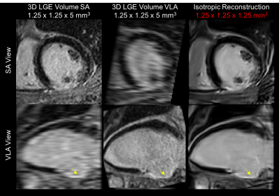 |
Isotropic 3D Late Gadolinium Enhancement Imaging using 3D Patch-Based Super-Resolution
Aurelien Bustin, Damien Voilliot, Jacques Felblinger, Laurent Bonnemains, Freddy Odille
Cardiac late gadolinium enhancement (LGE) imaging has become a reference clinical tool for assessing myocardial scar and viability. Despite superior signal-to-noise-ratio of 3D LGE techniques, current 3D breath-hold acquisitions are still limited by scan time and low-resolution, especially in the through-plane direction. Consequently, most clinical protocols include three anisotropic LGE acquisitions in different views to better visualize myocardial fibrosis in different orientations. Nevertheless, assessing myocardial viability in different views remains tedious and time-consuming. In this study, we sought to achieve isotropic 3D LGE by combining low-resolution anisotropic acquisitions using a 3D patch-based super-resolution reconstruction.
|
|
2900.
 |
Cardiac relaxometry in childhood acute lymphoblastic leukemia survivors.
Delphine Perie-Curnier, Mohamed Aissiou, Louise Leleu, Farida Cheriet, Tarik Hafyane, Maja Krajinovic, Caroline Laverdiere, Daniel Sinnett, Gregor Andelfinger, Daniel Curnier
The aim of this study was to evaluate T1 pre- and post-gadolinium enhancement and T2 relaxation times sensitivity to detect myocardial changes induced by doxorubicin-based chemotherapy in childhood acute lymphoblastic leukemia survivors. Myocardial changes such as increased fibrosis index and injury due to associated changes in myocardial free water content were found between risk groups of cancer survivors, suggesting T2, post-gadolinium T1 and particularly the partition coefficient as early indices for myocardial tissue damages in the onset of doxorubicin-induced cardiotoxicity. These computing tools will be pivotal in patient follow-up to anticipate pathology evolution.
|
|
2901.
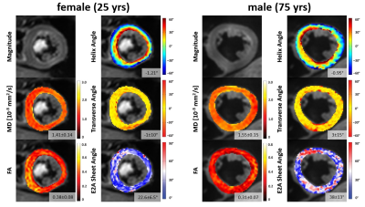 |
Age, gender and heart rate dependency of spin echo based diffusion tensor imaging measurements in healthy hearts
Alexander Gotschy, Constantin von Deuster, Robbert van Gorkum, Ella Vintschger, Robert Manka, Christian Stoeck, Sebastian Kozerke
Cardiac diffusion tensor imaging (cDTI) is a novel non-invasive method that allows assessing changes in myocardial microstructure in various cardiomyopathies. To identify pathologies, however, the distribution of cDTI parameters and their subject specific dependencies in normal hearts need to be known. Therefore, we investigated age, gender and heart rate dependencies of quantitative parameters derived from spin-echo based cDTI in healthy subjects. Our results display the variation of cDTI parameters in normal hearts and thereby allow gauging at which level of expected pathological changes sex and age matched reference values will be needed in future clinical practice.
|
|
2902.
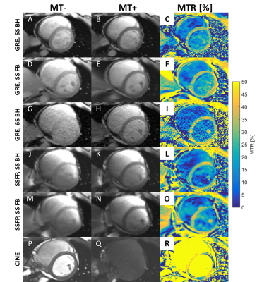 |
Comparison of GRE, SSFP, and CINE CMR Acquisitions for Measuring Magnetization Transfer at 3T
Matthew Van Houten, Yang Yang, Michael Salerno
We developed and compared multiple magnetization transfer (MT) pulse sequence strategies to characterize myocardial fibrosis without the need for gadolinium contrast at 3T. We demonstrated in an initial study of 4 healthy volunteers that a free-breathing, single-shot GRE is the most effective technique for producing high quality myocardial MT ratio maps. We will continue refining and investigating this sequence as a method for quantifying both focal and diffuse fibrosis in patients with heart failure.
|
|
2903.
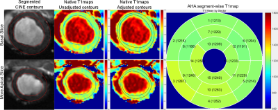 |
Fully Automatic SegmenTal analysis of myocardial Relaxometry (FASTR) - Initial results using T1 mapping
Venkat Ramanan, Nitishkumar Bhatt, LaBonny Biswas, Idan Roifman, Graham Wright, Nilesh Ghugre
Relaxometric techniques, particularly T1 mapping, have gained clinical importance recently. T1 and ECV are usually calculated by manually drawing contours on the maps. This is laborious particularly for large volume studies. Here we present a fully automated framework (FASTR) for segmental analysis of T1 maps (both native and post-contrast) and partition-coefficient values. Since CINE images are usually always acquired in the studies, we use CINE derived epi/endocardial contours and make further adjustments on T1 maps. This results in more accurate and robust segmentation of the myocardial wall, which works even in the presence of edema, infarct and minor artifacts.
|
|
2904.
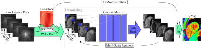 |
High-Resolution T1 Mapping using Parameter-Free Low Rank Denoising
Sebastian Weingärtner, Steen Moeller, Chetan Shenoy, Mehmet Akcakaya
Myocardial T1 mapping has become increasingly established for tissue characterization in numerous cardiomyopathies. However, the commonly used end-diastolic single-shot imaging imposes restrictions on the spatio-temporal resolution. In this work, we explored increased parallel imaging accelerations and higher resolutions, in conjunction with an image denoising technique that exploits inter-dependencies between the multiple images using random matrix theory. Following parallel imaging reconstruction, common noise characteristics across the images are extracted from the singular value decomposition of a Gaussian random matrix and denoised using locally low-rank regularization. Application of this technique to SAPPHIRE T1 mapping shows no corruption of the T1 time and enables parallel imaging acceleration up to 4 with an in-plane resolution of 1.1x1.1mm2 at clinical image quality.
|
|
2905.
 |
Cardiac MR multi-modal imaging: role in diagnosis and differential diagnosis of fulminant myocarditis in children
Cuiyan Wang, Haipeng Wang, Guangbin Wang, Bin Zhao, Bin Zhao
Clinical characteristics, cardiac morphology, function parameters and myocardial tissue characterization on MRI of three groups (FM, AM and CM) were retrospectively compared to find that higher myocardial thickness and T2 ratio were seen in FM than in AM and CM with statistical significance; the LVEF and incidence of LGE in FM were higher than that in CM with statistical significance. So that CMR has values in the diagnosis and differential diagnosis of FM.
|
|
2906.
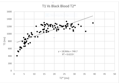 |
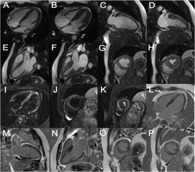
Myocardial T1 Measurement and Relationship with Myocardial T2 and Black Blood T2* at 3.0T MRI for Thalassemia Major Patients
Aamish Kazi, Bhavin Jankharia
Black blood T2* mapping on 1.5T is currently the gold standard for iron load assessment in patients with iron overload and plays a crucial role in patient management. Inaccuracies in T2* quantification at 3T due to greater artifact levels and higher B0 and B1 inhomogeneities have resulted in a lack of multi-center and multi-vendor validation studies to standardize T2* for iron overload assessment on 3T. In-vivo and In-vitro studies on 1.5T have suggested that T1 and T2 could be potential alternatives to T2*. In this study, we have demonstrated linear relationships for T1 Vs T2*, T2 Vs T2* and T1 Vs T2 at 3T suggesting that T1 or T2 could be potential alternatives to T2*.
|
|
2907.
 |
Iron-Ceroid Complex From Apoptotic Siderophage-Derived Foam Cells Promotes Perpetual Macrophage Ingress and Localized Edema Formation in Hemorrhagic Myocardial Infarctions: Histopathology and Immunohistochemistry Findings to MRI Correlates
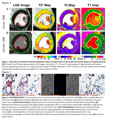
Ivan Cokic, Guan Wang, Kolja Wawrowsky, Hsin-Jung Yang, Richard Tang, Rohan Dharmakumar
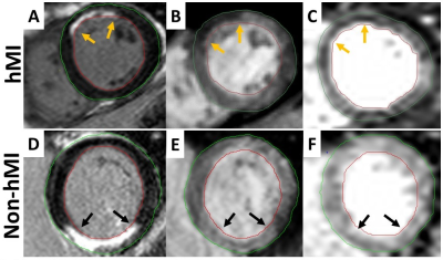
The capacity of macrophages (MΦ) to oxidize LDL, produce ceroid (CR), and transform into foam cells (FC) is enhanced following erythrophagocytosis. During the process of FC formation, part of hemoglobin-derived iron forms a complex with CR. CR is cytotoxic; and over time, it can lead to FC apoptosis. Release of CR from apoptotic FC into the surrounding tissue may cause dysfunction and apoptosis of newly invading MΦ. Given that lipid and iron deposits within hemorrhagic MI (hMI) typically colocalize in the infarct periphery, we hypothesized that CR from apoptotic FC promotes perpetual MΦ ingress and localized edema formation in hMI.
|
|
2908.
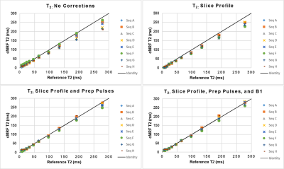 |
Improved T1 and T2 Accuracy for Cardiac MR Fingerprinting Sequences by Including Detailed Modeling of Slice Profile, B1, Inversions, and T2 Preparation Pulses
Jesse Hamilton, Yun Jiang, Dan Ma, Wei-Ching Lo, Mark Griswold, Vikas Gulani, Nicole Seiberlich
Because different cMRF pulse sequences may have different sensitivities to confounding factors, the generation of accurate and precise T1 and T2 maps may require detailed modeling of spin dynamics. This work studies the importance of modeling slice profile, B1, and relaxation during adiabatic inversion and T2 preparation pulses in cMRF. The ISMRM/NIST system phantom was scanned using cMRF sequences with different patterns of flip angles, TRs, and preparation pulses. Modeling these additional effects leads to higher correlation (using linear regression and concordance correlation coefficients) between NIST and cMRF measurements and better consistency between different cMRF sequences.
|
|
2909.
 |
Assessing myocardial infarct in lymphatic insufficient mice by rotating frame relaxation times
Elias Yla-Herttuala, Taina Vuorio, Johanna Laakkonen, Svetlana Laidinen, Seppo Yla-Herttuala, Timo Liimatainen
Relaxation times T2, T1ρ and RAFFn were applied to study alterations in myocardial infarct (MI) in control and in mice with insufficient lymphatic system (VEGFR3). The findings are supported by cardiac functional parameters and histology. We found significant difference between VEGFR3 and control in TRAFF4 (p<0.05) 8 days after MI and between pre-MI and post-MI time points in T2 (p<0.01). Relaxation times increased significantly (p<0.05) after MI in all measurements. We conclude that TRAFF4 gain information of alterations of fibrosis in lymphatic insufficiency after MI.
|
|
2910.
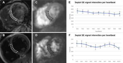 |
Simultaneous Multi-Slice Gradient Echo Spin Echo EPI (SMS-GESE-EPI) enables simultaneous cardiac T2 and T2* imaging and mapping across six slices within a single heartbeat
Maaike van den Boomen, Mary Kate Manhard, Christopher Nguyen, SoHyun Han, Kyre Emblem, Riemer Slart, Ciprian Catana, Niek Prakken, Bruce Rosen, Ronald Borra, Kawin Setsompop
Cardiac T2* and T2-based techniques suffer from variabilities introduced by acquisition over multiple heartbeats and breath holds. We demonstrate the use of a dual-echo SMS-GESE-EPI sequence that can simultaneously provide T2*- and T2-weighted images from six slice locations within a single heartbeat and breath-hold. Introduction of 5-echos also enabled dynamic T2*- and T2-mapping per heartbeat within a breath-hold. These dynamically acquired T2*- and T2-maps remained stable over ten heartbeats. Several applications might benefit from these modified GESE sequences, such as BOLD measurements and vessel architecture imaging of the myocardium.
|
|
2911.
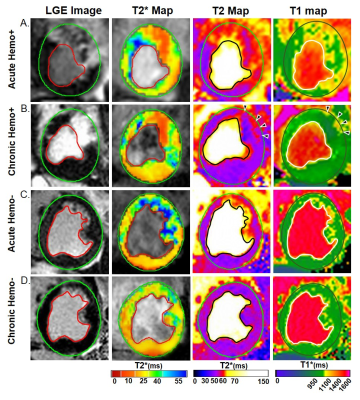 |
Multi-Parametric Cardiac MRI is Needed for Accurate Staging of Reperfused Hemorrhagic Myocardial Infarctions
Guan Wang, Hsin-Jung Yang, Ivan Cokic, Avinash Kali, Richard Tang, Joseph Francis, Songbai Li, Rohan Dharmakumar
Cardiac MRI (CMR) based staging of myocardial infarction (MI) with or without contrast agents relies on the resolution of edema in the chronic phase, which is typically determined on the basis of T2-based MRI. However, whether T2 CMR is sufficient for staging all MI types has not been studied. We investigated this using animal models with and without hemorrhagic MIs. Our results show that non-hemorrhagic MIs can be staged based on T2 changes in the MI territory. However, the incomplete resolution of T2 elevations in the peripheral layers of hemorrhagic MI territories can confound staging of hemorrhagic MIs.
|
|
2912.
 |
Fast and precise myocardial T1 mapping using a segmented golden angle radial MOLLI sequence with bSSFP readout
Jiaxin Shao, Ziwu Zhou, Fei Han, Peng Hu
Among the various myocardial T1 mapping sequences developed, the radial variants of the MOLLI acquisitions (raMOLLIs) are promising. As raMOLLIs can decrease the acquisition time down to a few heartbeats while keeping good T1 estimation precision due to a large number of images reconstructed along the T1 relaxation recovery curve. The previous raMOLLIs use FLASH readout due to the sensitivity of bSSFP readout to image artifacts. As bSSFP readout has high SNR, a variant of radial MOLLI with bSSFP readout was developed to ensure accurate and precise myocardial T1 mapping by using segmented golden angle radial acquisition.
|
|
2913.
 |
Investigating extra-cellular volume fraction in patients with Becker Muscular Dystrophy and Limb Girdle Muscular Dystrophy 2I.
Alex Murphy, David Higgins, Volker Straub, Kieren Hollingsworth
The development of gradual and diffuse myocardial fibrosis is a key pathology in muscular dystrophy and it is possible that extracellular volume (ECV) measurement may be a useful biomarker. Thirteen participants with muscular dystrophy and ten healthy controls were recruited to undergo cardiac MRI, including cardiac tagging, LGE and ECV measurement to determine whether significant global or local differences in ECV could be detected, and their relationship to cardiac dysfunction as indicated by cine imaging and cardiac tagging. Global ECV was not different but there were significant segmental differences between muscular dystrophy and controls.
|
|
2914.
 |
Accelerated 3D saturation-recovery based myocardial T1 mapping using fewer saturation time points and denoising
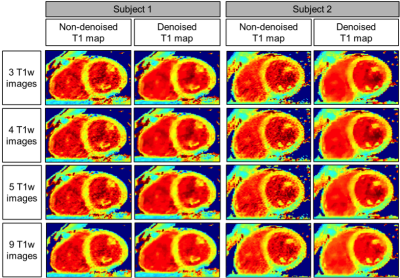
Giovanna Nordio, Aurelien Bustin, Torben Schneider, Markus Henningsson, Claudia Prieto, René Botnar
In this study we propose to accelerate the 3D saturation-recovery (3D SASHA) T1-mapping technique by using a reduced number of saturation time points while maintaining accuracy and precision using a 3D denoising method. No statistical difference was found in terms of accuracy and precision (respectively p=0.14 and p=0.99) between the T1-maps reconstructed after denoising using different number of T1-weighted images (between three and nine). After application of 3D denoising, the precision was independent of the number of T1-weighted images used for the fitting, which may permit to considerably accelerate the 3D SASHA acquisition.
|
|
2915.
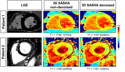 |
3D SASHA myocardial T1 mapping with high accuracy and improved precision
Giovanna Nordio, Aurelien Bustin, Markus Henningsson, Freddy Odille, Claudia Prieto, René Botnar
In this study we propose to further improve the precision of free-breathing 3D saturation-recovery based T1 mapping (3D SASHA), while keeping its high accuracy, by employing a novel 3D denoising method which exploits spatio-temporal correlations in the T1-weighted images. The proposed approach has been tested on ten healthy subjects and four patients with cardiovascular disease. For all subjects, no statistical difference was observed between the precision measured on 3D denoised SASHA and 2D MOLLI T1 maps (p=0.62), while preserving the T1 accuracy. There was an improvement in the precision after denoising on the 3D SASHA T1 maps acquired in healthy subjects and patients.
|
|
2916.
 |
Single breath-hold MR T1-mapping in the heart: comparison of hybrid MOLLI and MOLLI53
Yu Chun-Yang, Huang Teng-Yi, Chung Hsiao-Wen
A hybrid MOLLI method that integrated saturation recovery with the classical inversion recovery sequence was proposed for quantitative T1 mapping in the myocardium within one single breath-hold. By replacing the second inversion pulse of the original MOLLI53 technique with a saturation pulse, the long recovery time could be alleviated in hybrid MOLLI, thereby allowing more images to be sampled from the T1 relaxation curve. Phantom and healthy subject experiments conducted in comparison with the classical MOLLI53 demonstrated that the proposed method was able to provide comparable image quality as well as precise T1 quantification in the myocardium.
|
|
2917.
 |
Golden Angle Radial Chemical Exchange Saturation Transfer for the Rat Heart
Pan ki Kim, Chul Hwan Park, Yoo Jin Hong, Byoung Wook Choi
Chemical Exchange Saturation Transfer (CEST) has been attracting attention as a molecular imaging method to investigate myocardial muscle energetics according to creatine changes. In this study, we proposed a robust CEST imaging technique from cardiac and respiratory motion using golden angle radial readout to achieve CEST imaging at the heart of the rat. We investigated the feasibility of the proposed method for the creatine phantom and a normal rat.
|
|
2918.
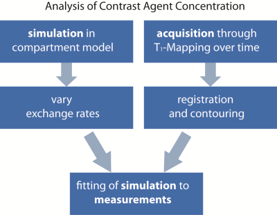 |
Simulation-Aided Contrast Agent Washout Analysis in Patients with Acute Myocarditis
Leili Riazy, Tobias Schaeffter, Marc Olbrich, Johannes Schueler, Florian von Knobelsdorff-Brenkenhoff, Thoralf Niendorf, Jeanette Schulz-Menger
Contrast-enhancement techniques allow the visualization of small myocardial injuries in acute myocarditis, which cannot be detected by any other noninvasive technique. Late Gadolinium Enhancement (LGE) has been shown predictive for the development of heart failure. Early Gadolinium Enhancement (EGE) was identified as parameter for detection of disease activity. We analyze the contrast agent washout during 10 minutes after tracer administration. Our aim is to characterize parameter values of patients with myocarditis in a 3D spatially distributed contrast agent flow model.
|
|
2919.
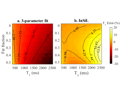 |
Evaluation of MOLLI fitting algorithms robustness to partial volume effects due to fat
Andreia Gaspar , Rita Nunes
The MOLLI sequence for myocardium T1 quantification is widely applied in the clinical setting. The standard 3-parameter fitting algorithm allows a high precision in the T1 estimates but it comes at the cost of a low accuracy. The accuracy can be improved using the instantaneous signal loss (InSiL) approximation method for signal fitting. In this work we evaluated the robustness of the InSiL algorithm when fat also contributes to the signal. The results show that InSiL enables to increase the accuracy of the MOLLI sequence even in the presence of partial volume effects due to fat.
|
|
2920.
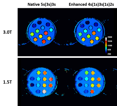 |
Myocardial T1 mapping with second-based MOLLI scheme for reduced heartrate variation: A phantom validation study at 1.5T and 3.0T
Shuo Zhang, Jennifer Bryant, Evelyn Quah, Calvin Chin, Derek Hausenloy, Jouke Smink, Ru San Tan, Yeong Shyan Lee, Stuart Cook
Current standard myocardial T1 mapping is based on the modified Look-Locker inversion recovery (MOLLI) technique and single-shot readout per image acquisition. A second-based scheme has recently been proposed to mitigate the dependence of imaging times on heartrate and also to increase robustness in T1 estimation. Here we report a phantom-based study with ECG simulation comparing it with the original beat-based MOLLI acquisition scheme at both 1.5 and 3.0T. We demonstrate the advantage of this approach with reduced heartrate dependence and variation for an improved T1 quantification reliability.
|
|
2921.
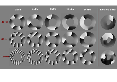 |
Understanding the material behaviour of ex-vivo porcine hearts using MR-Elastography and Rheology
Myrianthi Hadjicharalambous, Adela Capilnasiu, Ayse Sila Dokumaci, Daniel Fovargue, Gerhard Sommer, Gerhard Holzapfel, Ralph Sinkus, David Nordsletten
Myocardial stiffness has been shown to correlate with heart disease, nevertheless, reliable stiffness estimates are hindered by the remarkably complex behaviour and function of the heart muscle. In this work, we use MR-Elastography and rheological experiments to obtain a better understanding of the myocardial material behaviour. MR-Elastography and cyclic shear tests are performed on ex-vivo porcine hearts, under varying frequencies. Our results demonstrate important tissue properties and highlight the viscoelastic properties of the myocardium which are often neglected. Improving our understanding of the interlinked material properties of the heart is a critical step towards the accurate prediction of myocardial stiffness.
|
|
2922.
 |
Assessment of Myocardial Fiber Orientation Using Diffusion Tensor Imaging in Patients with Hypertrophic Cardiomyopathy and Its Correlation with Echocardiographic Strain
Sang-Eun Lee, Christopher Nguyen, Sen Ma, Debiao Li, Hyuk-Jae Chang
In hypertrophic cardiomyopathy, myocardial fiber disarray, and interstitial fibrosis interfere with regional systolic myocardial function despite clinically hyperdynamic systolic function. We quantitatively assessed the difference in myocardial fiber orientation between diseased and normal cardiac segments using diffusion tensor imaging. Further, these fiber microstructure were compared to the regional global longitudinal strain to evaluate whether the structure-function relationship changes according to the disease involvement.
|
|
2923.
 |
Single-shot Radial Fast Spin-Echo T2 Mapping Pulse Sequence
KyungPyo Hong, Hassan Haji-valizadeh, Nivedita Naresh, Daniel Kim
Cardiac T2 mapping is a proven imaging test for myocardial tissue characterization. Standard cardiac T2 mapping involved in cardiac MRI protocol requires a breath-hold duration of 10 sec and usually samples three short-axis planes of the heart. This limited spatial coverage may miss the focal lesion. In this study, we developed a single-shot cardiac T2 mapping pulse sequence and reconstructed multiple T2-weighted images from a single T2-decay data, using k-Space weighted image contrast and compressed sensing technique. We tested its performance in patients with suspected infiltrative cardiomyopathy, and it yielded 7.9% difference in myocardial T2 values compared to standard T2 mapping.
|
|
Velocity & Flow
Traditional Poster
Cardiovascular
Thursday, 21 June 2018
| Exhibition Hall 2924-2948 |
13:15 - 15:15 |
|
2924.
 |
A combined 4D Flow MRI-modelling approach for assessing the subject-specific effects of dobutamine on left ventricular function
Belen Casas, Federica Viola, Gunnar Cedersund, Ann F Bolger, Matts Karlsson, Carl-Johan Carlhäll, Tino Ebbers
This study applies a previously developed imaging-modelling approach to investigate the subject-specific effects of dobutamine on left ventricular contraction and relaxation patterns in healthy subjects. We created personalized models for nine subjects at rest and under dobutamine stress. The personalized parameter values were in agreement with the effects of inotropy and lusitropy reported in previous studies, and demostrated the anticipated variability in individual responses to dobutamine. With further validation, the given approach has the potential to generate advanced metrics of cardiovascular physiology and pathophysiology that could extend beyond conventional techniques for both diagnosis and optimization of a personalized medical regimen.
|
 |
2925.
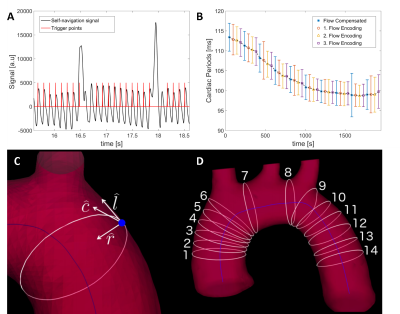 |
Fast Self-Navigated Wall Shear Stress Measurements in the Murine Aortic Arch Using Radial 4D-PC-MRI at 17.6T
Kristina Andelovic, Patrick Winter, Thomas Kampf, Julius Heidenreich, Anton Xu, Peter M. Jakob, Wolfgang R. Bauer, Volker Herold
4D phase contrast (PC)-MRI is a non-invasive tool for the assessment of cardiovascular hemodynamics or the Wall Shear Stress (WSS) to study atherosclerotic risks in vivo. Major limitations of conventional triggered methods are the long measurement times needed for high-resolution data sets and the requirement of stable ECG triggering, which is diffcult at high magnetic field strengths. In this work, an ECG-free, retrospectively synchronized method is presented that enables fast high-resolution measurements of 4D flow and wall shear stress in the murine aortic arch.
|
|
2926.
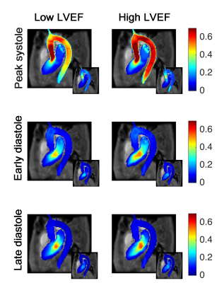 |
The impact of left ventricular ejection fraction on cardiovascular blood flow
Merih Cibis, Carl-Johan Carlhäll, Jan Engvall, Tino Ebbers
The impact of left ventricular (LV) ejection fraction (LVEF) on cardiovascular blood flow is not completely understood. We used a method, called “Atlas heart generation”, to investigate cardiovascular flow of patients with ischemic heart disease (n=62). The patients underwent 4D-Flow MRI and were stratified according to LVEF. We found that the lower LVEF group had lower velocities throughout the aorta, in a portion of LV and left atrium, at peak-systole. At early-diastole, differences were observed in the aortic arch, and in the apical-septal segments of LV. The suggested method can detect changes in cardiovascular flow and add to pathophysiological understanding.
|
|
2927.
 |
4D flow MRI Investigation of link between Aortic Stiffness and Embolic Pathway of Aortic Flow Reversal in Patients with Cryptogenic Stroke
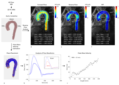
Kelly Jarvis, Alireza Vali, Shyam Prabhakaran, Jeremy Collins, Michael Markl
Reverse aortic flow causing plaque embolization from the descending aorta (DAo) has been identified as a new source of stroke but the underlying cause of flow reversal is unclear. There is evidence that aortic stiffness can cause flow reversal but no study has investigated this relationship in detail. This study used high-temporal resolution 4D flow MRI to evaluate aortic stiffness and regional aortic flow reversal in patients with cryptogenic stroke. Elevated PWV was associated with reverse flow in areas of the aortic arch and DAo providing evidence for aortic stiffness and flow reversal as a potential embolic mechanism.
|
|
2928.
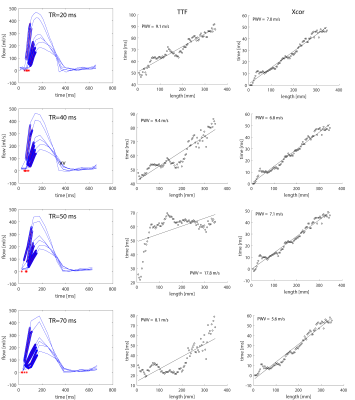 |
4D Flow MRI-Based Aortic Pulse Wave Velocity: Systematic Analysis of the Impact of Temporal Resolution on Estimation in Patients with Aortic Atherosclerosis and Age-matched Controls
Kelly Jarvis, Alireza Vali, Shyam Prabhakaran, Jeremy Collins, Michael Markl
Elevated pulse wave velocity (PWV) is a measure of aortic stiffness and an indicator of cardiovascular disease. Pulse waves propagate quickly along the aorta and high-temporal resolution measurement of velocity data with full spatial coverage is needed to improve the precision of PWV estimation. This study used high-temporal resolution 4D flow MRI to assess PWV and investigate the impact of temporal resolution on PWV estimation methods (i.e. time-to-foot and cross-correlation) in patients with known atherosclerosis. The findings suggest that using cross-correlation to estimate the time-delay between flow waveforms is optimal, particularly at inferior temporal resolutions.
|
|
2929.
 |
4D Flow Imaging with Reduced Field-of-Excitation
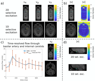
Clarissa Wink, Giulio Ferrazzi, Jean Pierre Bassenge, Sebastian Flassbeck, Simon Schmidt, Tobias Schaeffter, Sebastian Schmitter
4D flow MRI suffers from long scan times which limit maximum spatial resolutions. A promising approach is to restrict the field-of-excitation (FOX) to the region of interest and therefore reduce the field-of-view (FOV) not only in partition/slab direction, but also in phase encoding direction. In this work, we replace the slab-selective excitation of a 4D flow sequence by a 2D spatially-selective excitation with reduced FOX to enable reduced FOV imaging. We investigate the impact of the excitation on velocity encoding and demonstrate correct velocity quantification with $$$10\%$$$ reduced scan times in phantoms and in-vivo.
|
|
2930.
 |
Estimating highly-accurate velocity maps from FVE MRI data using a PDE-constrained optimization
Vinicius Rispoli, Joao Carvalho, Cristiano Miosso, Fabiano Soares, Giordanno Borges, Ivan Siqueira
Fourier velocity encoding (FVE) is a technique capable of delivering clinically treatable data at short acquisition times. FVE resolves the velocity distribution in each voxel of the image with high signal-to-noise ratio. This makes it suitable for the calculation of relevant biomarkers (e.g. wall shear rate and oscillatory shear index). However, it does not provide the blood flow velocity field directly. Techniques to estimate the actual blood flow from FVE velocity distributions have been previously presented. In this work, we present a novel method for velocity map estimation based on a PDE-constrained optimization that provides better results than previous methods.
|
|
2931.
 |
Flow-encoding Arterial Structure Acquired using Silent-MRA: A Preliminary Study
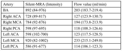
Chia-Wei Li, Chien-Yuan Lin, Charng-Chyi Shieh, Chen-Syuanms Lin, Chia-Yuen Chen, Wing P. Chan
Silent magnetic resonance angiography (Silent-MRA), which combines the Silent Scan algorithm to achieve a zero echo time with an arterial spin-labelling method, has recently been introduced as a novel MRA technique. Many studies of Silent-MRA focused on evaluating vascular structure; however, reports of further investigations into the flow information generated by Silent-MRA cannot be found. To this end, we compared the flow-encoding Silent-MRA signal with phase contrast flow imaging and found a linear correlation between the two. This preliminary study demonstrates the potential power of using flow-encoding Silent-MRA in assessing complicated vascular disease.
|
|
2932.
 |
Wall shear stress analysis after anatomically pre-shaped 90°- and straight ascending aortic grafts: A comparison between prostheses and age-matched volunteers using 4D Flow MRI
Malte Sieren, Jennifer Schlüter, Thekla Oechtering, Michael Scharfschwerdt, Christian Auer, Markus Hüllebrand, Hans-Hinrich Sievers, Jörg Barkhausen, Alex Frydrychowicz
Patients with aortic prostheses following aneurysm/dissection repair demonstrate an increased number of secondary aortic flow patterns. These may result in elevated forces acting on the vessel wall and thus preterm degenerative changes. Anatomically pre-shaped 90°-prostheses promise more physiological flow patterns and wall shear stress (WSS). The aim of this study was to compare WSS of patients with straight prostheses (n=8), 90°-prostheses (n=9) and healthy volunteers (n=12) based on 4D Flow MRI. Results revealed a tendency towards decreased WSS in regions distal to the 90°-prostheses, whereas in comparison to healthy volunteers, WSS values in patients with both prostheses were significantly increased.
|
|
2933.
 |
Measuring cardiac output and leg blood flow with phase-contrast MRI during supine cycling exercise.
Thijs Schoots, Berit Wassenaar, Anita Kuiper, Hareld Kemps, Jeroen Jeneson, Remco Renken
Patients with chronic heart failure suffer from diminished leg blood flow (LBF). Question remains to what extent the distribution or the cardiac output (CO) is responsible. This study investigates whether CO and LBF could be measured reliably using phase contrast MRI during supine exercise. 10 healthy subjects performed a supine exercise test in the MRI at two days at different exercise intensities. Comparison between both days showed promising reproducibility of measuring CO and LBF during supine cycling in the MRI although LBF measurements proved more challenging.
|
|
2934.
 |
A Comparison of PC-MRI Eddy Current Correction Methods in the Presence of Noise
Avinash Chinchali, Michael Loecher, Daniel Ennis
Eddy current induced phase errors lead to PC-MRI velocity errors that must be corrected. Static tissue fitting is commonly implemented to correct these phase errors. The aim of this work was to quantitatively compare corrections made using local and global static tissue fitting techniques over a wide range of SNR. Average correction differences between local and global strategies in static tissue were on the order of 0.9 cm/s for low SNR protocols and 0.1 cm/s for high SNR protocols. Local correction introduced phase error in ~5% ROIs (always when SNR<30). Local correction is therefore suitable for higher SNR PC-MRI acquisitions.
|
|
2935.
 |
Local Pulse Wave Velocity from 4D-Flow MR applied in Familial Hypercholesterolemia patients.
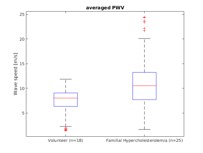
Joaquin Mura, Julio Sotelo, Animesh Tandon, Tarique Hussain, Andrew Tran, Cristian Tejos, Sergio Uribe
We propose an improved methodology1 to automatically estimate local 3D Pulse Wave Velocity (PWV) measurements for quantifying local alterations due to aortic distensibility using 4D flow data. 18 volunteers and 25 patients with Familial Hypercholesterolemia (FH) were evaluated using the proposed method. The results show the prevalence of higher values of PWV in FH patients than volunteers, particularly in the ascending aorta (AAo) and proximal descending aorta (pDAo). This semi-automatic 3D method is less user dependent and uses multiple correlations to improve accuracy. We demonstrate an excellent agreement with expect values.
|
|
2936.
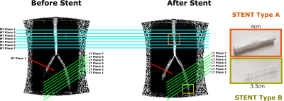 |
Where phase-contrast measurements should be performed in the presence of stents
Ana Beatriz Solana, Fatih Hafalir, Martin Janich, Christian Meierhofer
Here, a Y-shaped pulsatile flow phantom is used to evaluate the flow quantification error, as measured by 2D CINE PC, caused by magnetic susceptibility in the presence of clinically used MR-conditional ferromagnetic stents, even in ROIs where the artifact is not visualized in magnitude image. Our results indicate that flow measurements should be performed more than 12 mm away from the proximal or distal part of the stent to achieve accurate flow measurements.
|
|
2937.
 |
Can cardiac 2D Phase-Contrast MRI velocity measurements be used to characterize left ventricle hemodynamics?
Stephanie Marchesseau, Teresa Yeung, John Totman
Phase-contrast MRI were proposed to estimate intracardiac pressure gradients, but it is still unclear if this acquisition can be reliably used in the assessment of hemodynamics of the left ventricle. In this study, we performed reproducibility and test-retest experiments to evaluate the clinical use of PC-MRI and concluded that pressure and velocity parameters measured from the inflow (from atrium to LV) seem to be reliably measured using PC-MRI but it was not the case for the outflow parameters. Moreover, test-retest experiment showed that individual parameters were not constant over time, which therefore questions the diagnostic value of PC-MRI pressure measurement.
|
|
2938.
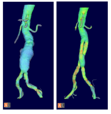 |
Blood flow measurement using 3D cine PC MRI within the abdominal aortic aneurysm and visceral arteries in pre- and post-EVAR condition; blood flow in the SMA might be improved after EVAR.
Masataka Sugiyama, Yasuo Takehara, Tetsuya Wakayama, Atsushi Nozaki, Marcus Alley, Takasuke Ushio, Shinji Naganawa, Harumi Sakahara
The blood flow volume within the visceral arteries were measured and compared between pre-and post-EVAR conditions using 4DFlow MRI. The maximum systolic flow volume ratio in the SMA to that of the aorta showed significant increase after EVAR. 4DFlow might be useful for evaluation of the blood flow dynamics of the aorta and visceral arteries in pre- and post-EVAR condition.
|
|
2939.
 |
Usefulness of 4D flow in the Diagnosis of Atrial Septal Defects in Adults
Mamoru Takahashi, Yasuo Takehara, Norihiro Tooyama, Katsutoshi Ichijo, Tomoyasu Amano, Yoshikazu Nagura, Kouichi Mizuno, Takuya Matsumoto, Tomoyuki Okuaki, Harumi Sakahara
We tested whether 4D flow can offer useful hemodynamic information in the diagnosis of atrial septal defects in Adults. 4D PCA was clearly able to visualize abnormal shunts from the left antrum to the right antrum of ASD patients.
|
|
2940.
 |
Background phase correction in the presence wrap-around artifact: Application in 4D flow imaging
Aaron Pruitt, Ning Jin, Yingmin Liu, Orlando Simonetti, Rizwan Ahmad
Residual background phase offsets due to eddy-currents limit the accuracy of flow quantification in 4D flow imaging. Commonly utilized polynomial regression of stationary voxels to correct background phase, however, is unreliable in the presence of wrap-around artifact. Here, we present an automated approach to identify and exclude regions of wrap-around from the fit, and validate its effectiveness in phantom and in vivo.
|
|
2941.
 |
Hemodynamic Evaluation in Patients with Tetralogy of Fallot after Operation: Repeatability And Internal Consistency of 4D Flow and 2D Phase Contrast by Cardiovascular Magnetic Resonance
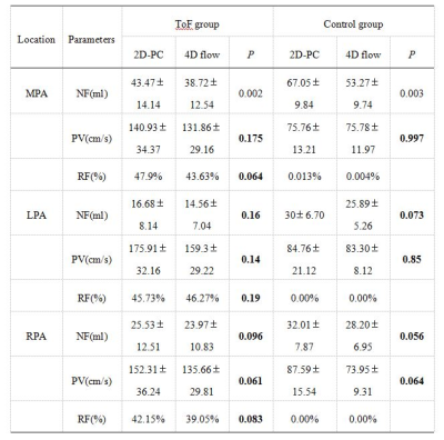
Li-wei Hu, Rong-zhen Ouyang, Yong Zhang, Yu-min Zhong
4D flow MRI offers the ability to measure and visualize the temporal evolution of complex blood flow patterns within an 3D volume. Some studies have been performed to validate 4D PC flow measurements, such as the comparison of 4D PC flow measurements to two-dimensional (2D) flow and to phantoms measurements as a reference standard. We hypothesized that 4D flow could be used to evaluate the hemodynamic parameters in patients with tetralogy of Fallot compare with 2D flow.
|
|
2942.
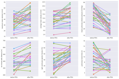 |
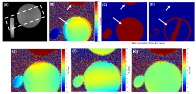
2D phase-contrast MRI as an integrative method for the evaluation of patients with chronic thromboembolic pulmonary hypertension before and after pulmonary endarterectomy
Christoph Czerner, Christian Schoenfeld, Serghei Cebotari, Julius Renne, Till Kaireit, Hinrich Winther, Gesa Hauck, Marius Hoeper, Frank Wacker, Jens Vogel-Claussen
Pulmonary endarterectomy (PEA) is an established method for treatment of chronic thromboembolic pulmonary hypertension (CTEPH). MRI is currently proposed as novel tool for treatment monitoring. We evaluated 2D phase-contrast (PC) MRI of the main pulmonary artery as perioperative monitoring method in relation to cardiac and parenchymal perfusion MRI as well as to clinical parameters. 32 CTEPH patients who underwent MRI before and after PEA were analyzed. Results show improved postoperative pulmonary hemodynamics. 2D PC MRI data correlate well with cardiac as well as perfusion changes and clinical parameters which makes this method a simple tool for treatment monitoring after PEA.
|
|
2943.
 |
Cardiovascular Magnetic Resonance Imaging : a Tool for Non-invasive Absolute Aortic Blood Pressure Estimation
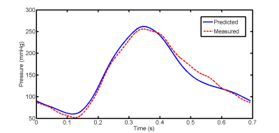
Khalil Rachid, Dima Rodriguez
Cardiovascular Magnetic Resonance Imaging (CMRI) is a well-established modality that allows not only non-invasive accurate blood flow quantification but also provides anatomical and biomechanical information about large vessel properties (e.g. aortic wall elasticity and distension) and central hemodynamics. The aim of our study is to use an MR-compatible aortic flow setup including two different elastic phantoms and validate MR-based pressure waveforms predicted by 1D blood flow model against invasive pressure measurements.
|
|
2944.
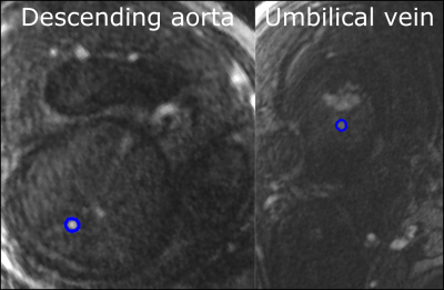 |
Quantitative phase-contrast CMR of blood flow in fetal vessels gated by Doppler ultrasound: comparison with metric optimized gating
Erik Hedström, Katarina Steding-Ehrenborg, Sebastian Bidhult, Christian Ruprecht, Fabian Kording, Anthony Aletras
The recent Doppler UltraSound (DUS) triggering method, which utilizes an MR-compatible ultrasound device to assess fetal heart contractions to provide a triggering/gating signal may improve fetal quantitative flow assessment by phase-contrast CMR. We evaluated the DUS method for blood flow measurements in the fetal descending aorta and umbilical vein, in comparison with the metric optimized gating method. Fetal quantitative blood flow by phase-contrast CMR is feasible using the DUS method. This further increases usability of fetal CMR, as post-processing is not needed.
|
|
2945.
 |
A Validation of MR Flow Velocity Mapping with Automated Phase Offset Correction Using a Gel Flow Phantom Controlled by a Motorized Piston in MR Phase Contrast Cine Flow Measurement
Kwan-Jin Jung, Youssef Jaber, Frank Sup IV
The accuracy of MR flow velocity measurement has been compromised due to a phase offset induced from the eddy current of gradient pulses. An automated correction method of the phase offset had been developed using an image-based algorithm. In order to validate the correction method and the measured velocity accuracy, we developed a flow phantom with a constant flow cross-section and used a servo motor controlled actuator to move the flow phantom accurately. The controlled movement of the new flow phantom allowed us to validate the phase offset correction method and the accuracy of the MR velocity measurement.
|
|
2946.
 |
Accurate MR-based Wall Shear Stress Measurements in Fully Developed Turbulent Flow Using the Clauser-plot Method
Nina Shokina, Waltraud Buchenberg, Marius Menza, Andreas Bauer, Gabriel Teschner, Cameron Tropea, Herbert Egger, Juergen Hennig, Axel Krafft
Wall shear stress (WSS) quantifies the frictional force that flowing blood exerts on a vessel wall and can be estimated from MR-based flow measurements via numerical differentiation. Correct assessment of WSS remains difficult because of the limited spatial resolution, partial volume effects and the per-se unknown position of the wall. It has been shown that such WSS evaluations tend to underestimate. We investigate an alternative method to evaluate WSS using the Clauser-plot method – a graphical way to estimate the WSS in fully developed turbulent stationary flow. We briefly describe the Clauser-plot method and present experimental validation in a straight tube.
|
|
2947.
 |
Correlation of Aortic Flow and Cardiac Function in Patients With Fabry Disease
Yi-Xian Li, Bo-Yan Chuang, Ming-Ting Wu, Hsu-Hsia Peng
We aim to explore the potential correlation of aortic flow and cardiac function in patients with Fabry disease (FD). The decreased total flow and increased maximum acceleration illustrated altered aortic hemodynamics. The left ventricular peak ejection rate (LVPER) negatively associated with the aortic total flow might be a mechanism to compensate the decreased aortic total flow in FD group. Besides, the positive correlation between LVPER and the systolic maximum acceleration described the interaction between cardiac function and aortic flow. In conclusion, the quantitative aortic flow-related parameters could help to elucidate altered aortic characteristics and the possible correlation with cardiac function.
|
|
2948.
 |
Accelerating Dual Venc 4D Flow Using Compressed Sensing with Locally Low Rank along Velocity Encoding
Peng Lai, Fatih Suleyman Hafalir, Joseph Cheng, Jonathan Tamir, Shreyas Vasanawala, Anja Brau, Martin Janich
Dual Venc has been developed to improve the accuracy of conventional 4D flow in high dynamic range of velocity. However, dual Venc acquisition doubles scan time. This work explored a high dimensional compressed sensing method to accelerate dual Venc 4D flow by utilizing additional data redundancy in the velocity encoding dimension.
|
|
Cardiac Function & Myocardial Perfusion
Traditional Poster
Cardiovascular
Thursday, 21 June 2018
| Exhibition Hall 2949-2969 |
13:15 - 15:15 |
|
2949.
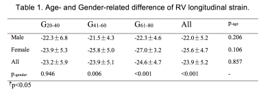 |
Right-ventricular Longitudinal Strain Reference Values of Healthy Volunteers by Age and Gender as Measured with CMR Tissue Tracking
Yangyang Qu, Jan Paul, Dominik Buckert, Genshan Ma, Volker Rasche
Our study measured RV longitudinal strain (RVGLS) by CMR 2D tissue tracking and investigated its diagnostic role in patients with RV heart failure. 150 healthy volunteers in three age groups (G20-40 years, G41-60 years, and G61-80 years) and 30 patients diagnosed as DCM were recruited.
Normal RVGLS was -23.9%±5.2% with significant higher values in females in G41-60 and G61-80. The cut-off value identified as -13.71% showed good sensitivity, specificity, positive and negative predictive value in diagnosing RV contractile dysfunction among DCM patients.
In summary, RVGLS were increased in females, and it benefited the evaluation of RV contractile function.
|
|
2950.
 |
Bias in the assessment of left ventricular function with compressed sensing CINE MRI
Jong-Hyun Yoon, Young-Joong Yang, Jin-Soo Kim, Pan-ki Kim, Jinho Park, Byoung Wook Choi, Chang-Beom Ahn
We report that a bias in the assessment of left ventricular function (LVF) is due to the compressed sensing (CS) CINE. In cardiac CINE MRI EDV (or ESV) is assessed when blood volume is a maximum (or a minimum). Practically a time window (given by VPS) is used to reduce scan time. For CS-CINE the time window is expanded by adopting data at nearby cardiac frames. The expanded acquisition window reduces EDV and increases ESV due to time average effect. Note that the changes of the quantities are not random, thus they should be removed for a better diagnosis.
|
|
2951.
 |
The right ventricular deformation in type 2 diabetes mellitus patients: insights from cardiac magnetic resonance feature-tracking
Bi-yue Hu, Zhi-gang Yang , Xi Liu, Ke Shi, Hua-yan Xu, Ying-kun Guo
Aim of this study was to clarify the feasibility of cardiovascular magnetic resonance (CMR)-derived feature-tracking for assessing right ventricle (RV) myocardial deformation in patients with type 2 diabetes mellitus (T2DM). Seventy T2DM patients and 22 healthy controls were enrolled. Cardiac volumes and function, and RV tissue-tracking parameters were determined by CMR. Compared with healthy subjects, significantly lower values of some global and regional strain parameters in T2DM (all p<0.05). Our results concluded that abnormal RV myocardial deformation could be monitored using CMR feature-tracking in T2DM; and the systolic and diastolic dysfunction was associated with RV volumes, HDL, and HbA1c.
|
|
2952.
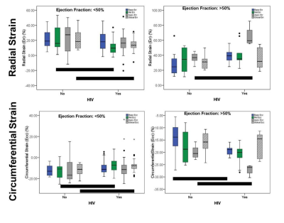 |
Patterns of myocardial strain are unique in HIV+ patients with heart failure with preserved ejection fraction
Bradley Allen, Amer Ahmed Syed, James Carr, Matthew Feinstein, Jeremy Collins
Human immunodeficiency virus (HIV) infection is associated with impaired cardiac function beyond what is expected from coronary artery disease alone. Our aim in the current study was to compare myocardial strain in a cohort HIV+ patients and uninfected controls with adjudicated heart failure (HF) using cardiovascular MRI feature tracking. Our results demonstrate unique Ecc and Err strain patterns in HIV+ patients, with relative apical sparing in HIV+ patients with EF>50%, but relative mid-LV and global strain reduction in HIV+ patients with EF<50%. This constellation of findings suggests that patterns of myocardial functional impairment may be unique in HIV+ HF patients.
|
|
2953.
 |
Cardiac Balanced SSFP 2D Cine DENSE for Myocardial Strain with comparison to Spiral 2D Cine DENSE
Ronald Beyers, Nouha Salibi, Thomas Denney
Quantification of myocardial strain has been previously demonstrated with echo-planar and spiral sequence versions of Displacement Encoding with Stimulated Echoes (DENSE). However, the non-conventional k-space acquisition of these previous efforts has hindered their integration into mainstream cardiac MRI application. Here we present a more conventional balanced SSFP (bSSFP) version of 2D Cardiac Cine DENSE and compare its performance to 2D Spiral Cine DENSE in normal human subjects. In vivo human scans at 3T demonstrated good agreement of myocardial radial (Err) and circumferential (Ecc) strain values between bSSFP Cine DENSE and Spiral Cine DENSE that also agree with previous literature.
|
|
2954.
 |
Left Ventricle 2D and 3D Strain Phantoms Generation Using a Python Finite Element-based Library
Hernán Mella, Joaquin Mura, Julio Sotelo, Sergio Uribe
The strain in the left ventricle is a well-known biomarker for cardiac diseases. Nowadays, several acquisition techniques have been developed to improve the diagnose of this kind of conditions. Usually, strain biomarkers are obtained by mean of image post-processing techniques using different deformation metrics. In this work we present a numerical framework for the generation of left-ventricle strain phantoms using three different acquisition sequences in order to provide a broad database of patients and volunteers with different types of diseases. Our library provides a robust image generation tool to compare and develop new post-processing methods for quantifying strain phantoms
|
|
2955.
 |
Functional cardiac MRI for monitoring progression of hypertrophic cardiomyopathy in Mybpc3 mouse models
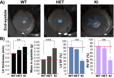
Min-Chi Ku, Till Huelnhagen, Saskia Schlossarek, Andreas Pohlmann, Lucie Carrier, Thoralf Niendorf
Mutations in gene MYBPC3, encoding cardiac myosin-binding protein C, cause hypertrophic cardiomyopathy (HCM), which is characterized by left ventricular hypertrophy (LVH), diastolic dysfunction, increased interstitial fibrosis, and may lead to sudden cardiac death and heart failure. In spite of the advances in translational medicine, we know very little about HCM. The HCM progression is complex and shows heterogeneous phenotypes. The missing linkage of in vivo imaging and pathology has hindered the investigation of detail mechanisms of HCM. We therefore investigated Mybpc3-targeted mouse models using CMR markers for understanding HCM pathophysiology and to get closer to complete pictures of HCM progression.
|
|
2956.
 |
Protective effect of Resveratrol against cardiac dysfunction and impaired energy metabolism of type 2 diabetic female GK rat heart submitted to Ischemia-Reperfusion injury
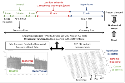
Natacha Fourny, Carole Lan, Eric Sérée, Laurent Pechere, Monique Bernard, Martine Desrois
Type 2 diabetes doubles the risk of myocardial infarction in women. New treatments need to be found to reduce cardiovascular mortality. Consequently, we investigated the effect of Resveratrol (RSV) on the tolerance to ischemia-reperfusion (IR) injury of type 2 diabetic female Goto-Kakizaki (GK) rat heart. We used a multiparametric approach allowing simultaneous measurement of cardiac function, energy metabolism by 31P MRS and endothelial function. Oral RSV treatment improved myocardial performance, coronary flow and energy metabolism during reperfusion in GK rats. Consequently, RSV might be an interesting therapeutic approach to improve survival to myocardial IR injury of type 2 diabetic women.
|
|
2957.
 |
Longitudinal follow-up of endothelial function after ischemia reperfusion injury treated with a novel regenerative therapy by albumin-based DCE MRI
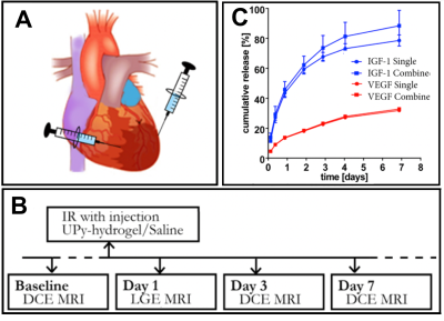
Maaike van den Boomen, Patricia Dankers, Leonie Niesen, Carlijn Bouten, Katrien Vandoorne
Dynamic contrast enhanced (DCE) MRI in combination with gadolinium-labeled albumin enabled longitudinal follow up of a novel hydrogel based regenerative therapy to treat myocardial infarction (MI). The local fractional blood volumes (fBVa measure for microvascular density) and permeability surface areas in the myocardium were increased at day 3 after MI due to the growth factors released from the hydrogel. This increase might indicate angiogenesis, which improves the inflammatory response. At day 7 the vascular density and permeability went back to normal again, which possibly avoid excessive extension of the MI.
|
|
2958.
 |
Global and segmental cardiac magnetic resonance tissue tracking of hypertrophic cardiomyopathy: How does hypertrophy and fibrosis contribute to myocardial deformation?
Ruo-yang Shi, Bing-hua Chen, Dong-aolei An, Rui Wu, Liang Du, Jiani Hu, Meng Jiang, Wei-bo Chen, Lian-ming Wu, Jian-rong Xu
In the patients with HCM, subtle LV deformation can be observed and measured clinically before the onset of general LV functional changes. Both hypertrophy and fibrosis influenced the extent of LV deformation. Our study demonstrated the 2D CS as a stable global parameter to assess LV functional and ECV changes. At the segmental level, hypertrophy and LGE (+) antagonistic affected 2D RS and diastolic RSR. Despite the excitement surrounding these pertinent clinical findings, further research is warranted as the mechanism of the phenomenon still needs to be explored.
|
|
2959.
 |
Oxygenation-sensitive cardiovascular magnetic resonance in Hypertensive Heart Disease with LVMH and Non-LVMH:Insight from altered mechanics and cardiac BOLD imaging
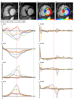
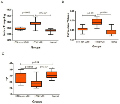
Binghua Chen, Rui Wu, Dong-Aolei An, Ruo-Yang Shi, Qiu-Ying Yao, Qing Lu, Jiani Hu, Meng Jiang, Weibo Chen, James Deen, Ankush Chandra, Jian-Rong Xu, Lian-Ming Wu
According to our study findings, BOLD MRI detected greater deoxygenated hemoglobin in HTN LVMH(measured by T2* BOLD MRI)compared with HTN non-LVMH and control groups. Lower T2* BOLD MRI values were associated with higher ECV values and correlated with reductions in circumferential and longitudinal strain, strain rate and displacement. Higher LVMI was associated with an increase in ECV and nativeT1, and a decrease inT2* BOLD MRI values. To our knowledge, this is the first study to assess the influence of myocardial oxygenation on cardiac function in hypertensive patients by applying combined T2* BOLD MRI, T1mapping and strain analysis. Assessing myocardial capillary oxygenation by BOLD MRI relies on the measurement of BOLD MRI relaxation time through endogenous contrast of deoxygenated hemoglobin. Myocardial microvascular oxygenation could reflect a balance or imbalance between oxygen supply and demand.
|
|
2960.
 |
Pilot Tone Navigation Enables Contactless Prospective Cardiac Triggering: Initial Volunteer Results for Prospective Cine
Mario Bacher, Peter Speier, Jan Bollenbeck, Matthias Fenchel, Matthias Stuber
Pilot Tones are a contactless, electromagnetic navigator that offers monitoring of cardiac and respiratory motion independently of the acquisition. Here we present initial volunteer results in utilizing the cardiac Pilot Tone signal to prospectively trigger a segmented cardiac Cine acquisition without the need for ECG.
|
|
2961.
 |
Simultaneous Multi Slice (SMS) SSFP first-pass myocardial perfusion imaging with iterative reconstruction at 1.5 Tesla.
Muhummad Sohaib Nazir, Radhouene Neji, Peter Speier, Daniel Staeb, Michaela Schmidt, Christoph Forman, Reza Razavi, Sven Plein, Tevfik Ismail, Amedeo Chiribiri, Sebastien Roujol
Myocardial perfusion imaging is recommended for ischaemia testing in patients although spatial coverage is limited to 3 slices in clinical practice. Simultaneous Multi Slice (SMS) imaging combined with iterative reconstruction was evaluated to provide greater heart coverage with minimal signal-to-noise penalty. 8 patients underwent two contrast enhanced dynamic perfusion scans at rest to compare the standard 3 slice with the SMS 6 slice protocol. Subjective image quality was found to be comparable to a standard 3 slice approach. This technique may have clinical utility in patients with suspected coronary artery disease through detection of ischaemia with greater heart coverage.
|
|
2962.
 |
Rest Perfusion within Chronic Infarctions Depends on Type of Acute Myocardial Infarction: Insights from a Serial MRI Study in Patients
Eric Johnson, Andreas Kumar, Rohan Dharmakumar
Excessive iron in tissue can impair endothelial function and reduce microcirculatory blood flow. We hypothesized that resting blood flow in chronic hemorrhagic myocardial infarction (hMI) territories, where iron concentration is known to be significantly elevated, would be lower than in non-hMI territories. We studied this in patients with reperfused myocardial infarction using cardiac MRI over a 6-month period following infarction. Mean relative perfusion index of hMIs were significantly lower than non-hMIs. This finding supports the notion that hypoperfusion within hMI territories may be an important pathological contributor to adverse cardiac remodeling commonly observed in patients with hMIs.
|
|
2963.
 |
Multiple sets of simultaneous multi-slice (SMS) for improved short and long axis coverage of myocardial DCE perfusion
Edward DiBella, Jason Mendes, Mark Ibrahim, Ye Tian, Brent Wilson, Ganesh Adluru
We propose a unique perfusion acquisition that offers improved coverage and confidence of detecting true ischemia and artifacts in cardiac perfusion dynamic acquisitions. Three slices are acquired simultaneously after each saturation pulse, and there is time to acquire 3 sets of such slices even at high heartrates. The ability to simultaneously acquire multiple slices opens up many new possibilities. The approach proposed here can acquire for example 6 short axis slices and 3 long axis slices each heartbeat, which allows detection of small areas of ischemia and can provide additional volume coverage and confidence. Preliminary results show the promise of this multi-plane SMS approach.
|
|
2964.
 |
Evaluation of extended GROG and Toeplitz pre-reconstruction interpolation methods on radial simultaneous multi slice MRI
Ye Tian, Ganesh Adluru, Jason Mendes, Edward DiBella
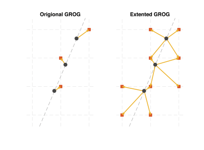
The purpose of this study is to develop and extend GRAPPA operator gridding (GROG) for fast iterative reconstruction of radial SMS data, and to compare extended GROG (EGROG) with GROG, Toeplitz and NUFFT methods. Simulation and in-vivo tests were done to compare these methods. Our results show that EGROG improves reconstruction by providing better Cartesian k-space estimation, it outperforms Toeplitz and GROG at oversampling factor 2, and a speed up factor of ~2 was achieved compared to NUFFT.
|
|
2965.
 |
Hybrid Estimation of the Arterial Input Function Using Blind Deconvolution and the Measured Blood Pool Signal
Radovan Jirik, Jason Mendes, Ye Tian, Ganesh Adluru, Edward DiBella
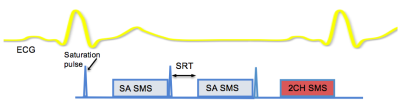
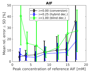
In Dynamic Contrast-Enhanced (DCE) MRI, inaccurate estimation of the arterial input function (AIF) is still a major cause of the low reliability of kinetic parameter estimates. We propose a new method of AIF estimation. It combines AIF measured from the blood-pool signal and multichannel blind deconvolution. The weights of the measured AIF are based on its analytically derived uncertainty and a model relating signal intensity and gadolinium concentration. The method has been evaluated on simulated myocardial perfusion data, mimicking real noise and kinetic parameter distributions. The hybrid method gave better results compared to the blood-pool or blind-deconvolution approaches alone.
|
|
2966.
 |
Fully-automated motion correction and probability-based segmentation of myocardial perfusion MRI data
Cian Scannell, Adriana Villa, Jack Lee, Marcel Breeuwer, Amedeo Chiribiri
This work presents a fully-automated framework for the pre-processing of free-breathing myocardial perfusion MRI data. Image series are first split into low-rank and sparse components using RPCA. This allows estimation of the deformation fields required to motion correct the image series, in the absence of dynamic contrast enhancement. Once motion corrected, pixels are clustered into anatomically relevant clusters using perfusion-superpixels which groups nearby pixels that have similar time dynamics. A LDA classifier is trained which allows the generation of myocardial probability maps and active contours are fit to the high probability regions to give a delineation of the myocardium.
|
|
2967.
 |
Validation of MR multitasking myocardial perfusion reserve measurements against simultaneous 13N-ammonia PET
Anthony Christodoulou, Damini Dey, Behzad Sharif, Richard Tang, Wafa Tawackoli, Rohan Dharmakumar, Piotr Slomka, Daniel Berman, Debiao Li
Measurements from myocardial perfusion MRI have previously been compared against separate PET measurements. However, MR quantification is complicated by signal nonlinearity (leading to a dual-bolus paradigm) and ECG misfires; furthermore, physiological variation in between separate PET and MR assessments are a confounding factor in validation. This work leverages the recent advent of multimodal PET-MR systems to perform a preliminary validation of quantitative MPR measurements from MR multitasking—a new framework allowing single-bolus, non-ECG perfusion quantification—against simultaneous 13N-ammonia PET-MR measurements in pigs. Excellent agreement was found between modalities (no bias, p=0.66; intraclass correlation coefficient=0.95).
|
|
2968.
 |
Left Atrial Surface Strain from Cine MRI Data in Patients with Mitral Regurgitation
Xiaoxia Zhang, Himanshu Gupta, James Davis, Steven Lloyd, Louis Dell’Italia, Thomas Denney Jr.
Mitral regurgitation (MR) is a common form of valvular disease where degeneration of the mitral valve causes blood from the left ventricle to be regurgitated into the left atrium (LA). For some MR patients, surgery to repair or replace the mitral valve is an option, but it can be difficult to determine when to do the surgery. Volumetric remodeling in the left atrium in MR patients has been reported, and could precede remodeling of the left ventricle (LV) and damage to the LV wall. Here, we investigate changes in endocardial surface strain in the LA in patients with MR compared to normal. Changes in LA volume and deformation may be useful in determining the severity and chronicity of valvular regurgitation and have clinical potentials in optimizing surgery timing and patient management.
|
|
2969.
 |
Right and left ventricular myocardial strain in healthy adolescents: Establishing normal reference values
Joseph Lang, Greg Barton, Arij Beshish , Kara Goss, Marlowe Eldridge, Christopher Francois
Tissue-tracking, a post-processing technique using routinely-acquired cine images, can assess strain, a multidimensional measure of myocardial contraction. In this prospective study, we measured left ventricular and right ventricular peak global radial, circumferential and longitudinal strain in 28 healthy adolescents ages 12-14 years. The data from this study provide normative global strain values to be used for future clinical and translational CMR studies.
|
|
Cardiovascular Image Processing
Traditional Poster
Cardiovascular
Thursday, 21 June 2018
| Exhibition Hall 2970-2981 |
13:15 - 15:15 |
|
2970.
 |
Comparison of cardiac MRI myocardial strain quantification techniques demonstrates systematic differences between feature tracking and heart deformation analysis
Amer Ahmed Syed, Bradley Allen, Eric Keller, James Carr, Matthew Feinstein, Susanne Schnell, Michael Markl, Jeremy Collins
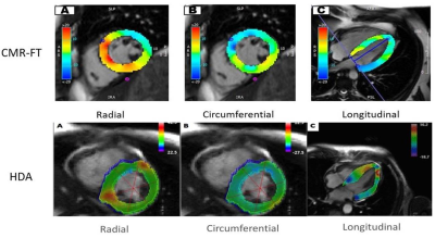
Myocardial strain is commonly performed at transthoracic echocardiography and is a sensitive technique for detecting subclinical disease. Feature tracking (CMR-FT) and heart deformation analysis (HDA) are two techniques that can be applied to balanced steady state free precession cinegraphic images, enabling the assessment of Lagrangian strains at cardiac MRI. We compared myocardial strains derived using these techniques in a cohort of 30 HIV+ patients. CMR-FT and HDA derived myocardial strains were significantly different, with CMR-FT consistently yielding higher strain values than HDA. Our results highlight technique dependence of CMR strain and underscore the need for technique specific normative reference values.
|
|
2971.
 |
Automated Segmentation of the Carotid Bifurcation using Region Growing and Support Vector Machines
Magnus Ziegler, Max Gefvert, Jan Engvall, Ebo de Muinck, Petter Dyverfeldt
Roughly 1 in 40 deaths worldwide are caused by strokes resulting from emboli that reach the brain from ruptured atherosclerotic plaques in the carotid artery. Segmentation of the carotid artery bifurcation in MR is necessary enables further analysis. Unfortunately, this is a slow and difficult task that is often performed manually. Two segmentation methods, one based on Region Growing (RG), and one using Support Vector Machines (SVM), were implemented for segmenting the carotid bifurcation in contrast-enhanced MR Angiograms (CE-MRA). Both methods were tested quantitatively, against ground truth segmentations using the DICE and true-positive ratio (TPR) and were also scored qualitatively using visual inspection. Both methods scored highly (RG 0.890 ± 0.022, SVM 0.890 ± 0.022) using the DICE score and true-positive ratio (RG 0.938 ± 0.026, SVM 0.931 ± 0.285). During qualitative assessments, RG and SVM both scored highly with median score 4/5.
|
|
2972.
 |
Intracranial vessel wall segmentation on 3D black-blood MRI using convolutional neural network
Hao Liu, Dongye Li, Xuesong Li, Qiang Zhang, Guanhua Wang, Yishi Wang, Xihai Zhao, Huijun Chen
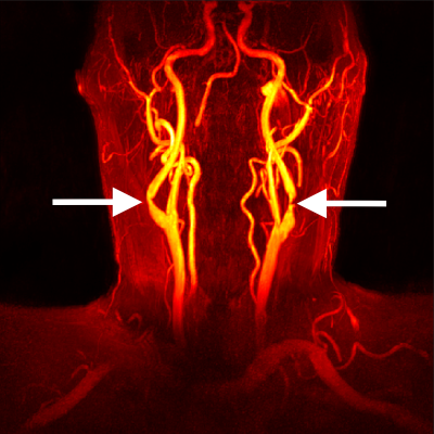
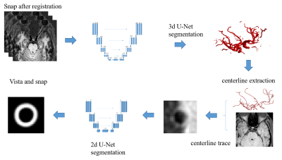
Intracranial artery atherosclerosis is a major cause of stroke. manually segmenting intracranial artery vessel wall is laborious and time-consuming. we proposed an automatic intracranial artery vessel wall segmentation framework to find the centerline of the intracranial artery from SNAP images to segment the final lumen and outer-wall contours on the cross-sectional 2D slices perpendicular to the centerline.
|
|
2973.
 |
ECG Characterization and Correction during Exercise Stress Imaging
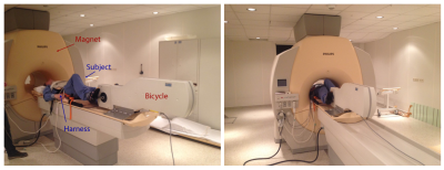
Jacob Macdonald, Grant Roberts, Oliver Wieben
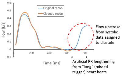
MRI during exercise stress can be a powerful tool in discerning abnormal cardiac behavior not apparent at rest. As a result of increased cardiac and respiratory motion, robust gating is essential for high-quality acquisitions during exercise. Due to increased patient motion, however, missed ECG triggers are more likely during exercise than at rest. For reconstructions with retrospective gating, such missed triggers can result in data attributed to the wrong portion of the cardiac cycle. In this work, we present an algorithm to identify and correct missed ECG triggers, allowing for exercise scans otherwise compromised by poor gating to be salvaged.
|
|
2974.
 |
Phase Unwrapping of 4D Flow Data with Graph Cuts
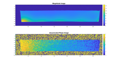
Andrew Justice, Sean Callahan, Jung won Cha, Amir Amini
A common problem with 4D flow magnetic resonance imaging is aliasing that occurs as a result of a low velocity encoding parameter. Consequently, an efficient and robust algorithm is needed to unwrap this data. We propose an iterative graph cuts algorithm to perform the necessary phase unwrapping and attain correct velocity values. The graph cuts algorithm utilizes a global energy minimization framework. This method is shown to accurately unwrap the aliased data more accurately than existing techniques for 4D Flow data. This included unwrapping synthetic data with Vencs down to 20% of the max velocity and SNRs down to 2.
|
|
2975.
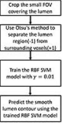 |
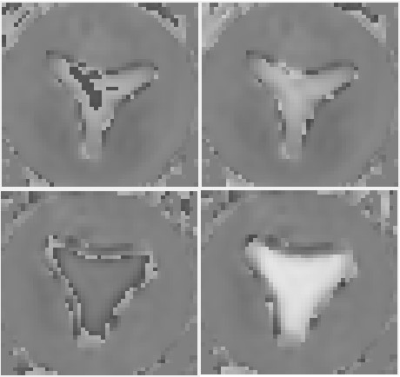
Automatic lumen size measurement in carotid atherosclerosis with phase sensitive magnetic resonance angiography(MRA) using self-trained radial basis function kernel support vector machine
Daniel Hippe, Jie Sun, Chun Yuan, Haining Liu
A self-trained algorithm based on Ostu’s method and a radial basis function (RBF) kernel support vector machine (SVM) model was developed for automatic lumen detection and quantification for the negative polarity map of SNAP magnetic resonance angiography(MRA). Based on an analysis of 15 arteries with carotid stenosis, the proposed automatic lumen segmentation algorithm demonstrated good agreement with manual lumen segmentation of SNAP MRA (intraclass correlation coefficient (ICC=0.95). The automated method also had good agreement with manual segmentation of CE-MRA (ICC = 0.90), which was comparable to the agreement between manually segmented SNAP MRA and CE-MRA (ICC = 0.93).
|
|
2976.
 |
Evaluation of e-prime with cardiac magnetic resonance cine imaging—preliminary feasibility study with comparison to echocardiography
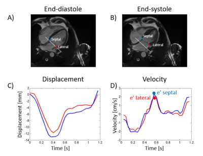
Felicia Seemann, Ricardo Gonzales, Chenxi Hu, Michael Quail, Karl Grunseich, Lauren Baldassarre, Albert Sinusas, Judith Meadows, Hamid Mojibian, Dana Peters
Diastolic dysfunction is commonly assessed by echocardiography, but not by cardiovascular magnetic resonance (CMR). To evaluate diastolic function, t he mitral annular flow (E) and velocity (e’) at the early rapid filling phase are measured. While E can be accurately measured by CMR, methods for measuring e' need to be established. In this study a feature tracking based method for measuring e' is applied to CMR images, and validated against echocardiography. There was an agreement between the methods, but sources of disparities between CMR and echocardiographic e’ measurements need to be further studied in order to improve the accuracy of e’ measurement by CMR.
|
|
2977.
 |
High-resolution Imaging with a priori Knowledge Incorporating the Navier-Stokes equations and the discontinuous Galerkin method (4D flow HIKING): towards flow reconstruction constrained by computational fluid dynamics
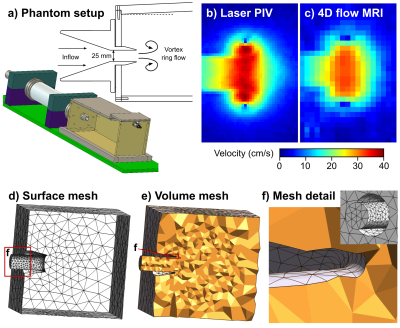
Johannes Töger, Matthew Zahr, Karin Markenroth Bloch, Marcus Carlsson, Per-Olof Persson
Magnetic resonance 4D flow imaging is a promising technique for diagnosis and follow-up of disease. However, 4D flow is limited by long scan times and low resolution. This work presents phantom validation of a new method for 4D flow scan acceleration, called 4D flow high-resolution imaging with a priori knowledge incorporating the Navier-Stokes equations and the discontinuous Galerkin method (4D flow HIKING). Excellent agreement with laser particle image velocimetry (PIV) was found, demonstrating the potential of the framework for scan time reduction and enhanced data quality in 4D flow.
|
|
2978.
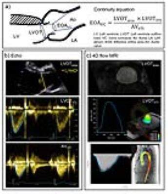 |
Estimation of aortic valve effective orifice area: a same day comparison between Doppler echocardiography and 4D flow MRI
Hyungkyu Huh, Menhel Kinno, James Thomas, Michael Markl, Alex Barker
The purpose of this study was to compare the aortic valve effective orifice area (EOA) estimated between Doppler echocardiography and 4D flow MRI using a consecutive same-day study design to minimize inter-modality variability. Peak velocity and left ventricular outflow tract area were higher for MRI but velocity time integral was higher for echo. These differences were compensatory when computing EOA, which resulted in good agreement despite discrepancies in echo vs MRI. Volumetric 3D velocity information has the potential to better estimate EOA in the presence of eccentric jets. This potential strength will be studied in aortic stenosis patients.
|
|
2979.
 |
Pixel-wise quantitative myocardial perfusion mapping with cloud based non-linear iterative reconstruction using Gadgetron framework
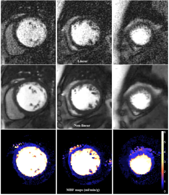
Hui Xue, Sven Plein, Amedeo Chiribiri, Peter Kellman
In this abstract, we present a solution to speed up the non-linear reconstruction for myocardial perfusion imaging and demonstrate its clinical usage through the Gadgetron cloud deployed at Microsoft Azure infrastructure. We also achieved pixel-wise myocardial blood flow mapping on the non-linearly reconstructed images, given the computing power on the cloud. All these processing steps were inline integrated on the clinical MR scanners. As a result, the proposed solution allows us to deploy non-linear perfusion imaging with quantitative flow mapping as a clinical application.
|
|
2980.
 |
Quantitative Classification of Atherosclerotic Plaque Compositions in Carotid Arteries: An in vivo T1 Mapping Study
Huiyu Qiao, Haikun Qi, Dongye Li, Dongxiang Xu, Huijun Chen, Chun Yuan, Xihai Zhao
This study sought to investigate the usefulness of in vivo T1 mapping in quantitative classification of compositions and vulnerability of carotid artery atherosclerotic plaques. We found that it is feasible to quantify the T1 values of atherosclerotic plaque compositions in carotid artery with in vivo T1 mapping. Significant differences in T1 values between fibrous tissue and other plaque compositions indicate that it is possible to classify plaque compositional features using T1 mapping. In addition, our findings of IPH and LRNC with significant different T1 values from other plaque compositions suggest the potential of T1 mapping in classification of plaque vulnerability.
|
|
2981.
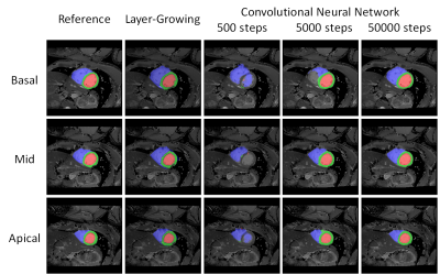 |
Automatic bullseye analysis of myocardial T1 values: a segmentation approach based on deep learning
Yu-Nian Ou, Tsai-Ling Yang, Teng-Yi Huang, Ming-Ting Wu
The study presents an automatic segmentation method for short-axis MOLLI data sets. We used a deep learning method based on convolutional neural network to accurately extract walls and blood pool regions of left and right ventricle. We compared the results with a layer-growing method presented in ISMRM 2017 and found that the accuracy of segmentation was significantly improved when using the deep learning method.
|
|
Vascular
Traditional Poster
Cardiovascular
Thursday, 21 June 2018
| Exhibition Hall 2982-3007 |
13:15 - 15:15 |
|
2982.
 |
Systematic evaluation of contrast-agent related image quality and vascular enhancement in abdominal time-resolved 4DMRA of minipigs
Dariusch Hadizadeh, Gregor Jost, Julian Lütckens, Vera Keil, Christoph Endler, Hubertus Pietsch, Hans Schild, Winfried Willinek
This study systematically evaluated the impact of contrast agent (CA) doses both quantitatively and regarding image quality on time-resolved contrast enhanced MR-angiography (4D-MRA). The intra-individual study-design under highly standardized conditions was realized using an animal model. 5 anesthetized Göttingen minipigs received thoracic-abdominal 4D-MRA at 1.5T at five CA doses from 0.02-0.10 mmol/kgBW. We observed that the further the CA traveled along the circulation, the more a dose reduction resulted in weaker peak signal enhancement and low image quality. We conclude that CA dose reduction has varying effects on image quality in 4D-MRA with respect to vessel types and sizes.
|
|
2983.
 |
Triple Accelerated NCE-MRA with optimised sampling patterns
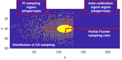
Hao Li, Andrew Priest, Martin Graves, David Lomas
In this study, we developed an acceleration technique combining compressed sensing (CS), parallel imaging (PI) and partial Fourier (PF) for the fresh blood imaging (FBI) sequence. Then, we evaluated the influence of the pattern design parameters and explored the optimal values for these parameters. By using the optimised sampling patterns, the FBI acquisition can be accelerated up to 10 times while the image quality is maintained.
|
|
2984.
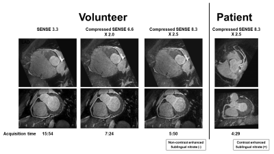 |
Whole-heart coronary MRA at 3.0T: Comparison between conventional method and new acceleration technique by compressed SENSE.
Shinichi Takase, Masaki Ishida, Yoshitaka Goto, Shiho Isoshima, Wakana Makino, Haruno Sakuma, Makoto Obara, Tsunehiro Yamahata, Katsuhiro Inoue, Kakuya Kitagawa, Hajime Sakuma
Compressed SENSE (CSENSE) is a novel method to combine Sensitivity Encoding (SENSE) and compressed sensing for rapid MR imaging. CSENSE can achieve a reduction factor higher than those achieved by SENSE while preserving the image quality by the denoising effect with iterations. In this work, CSENSE was applied to 3.0T free-breathing whole-heart coronary MRA to reduce the acquisition time. As compared to the conventional acquisition method using SENSE, CSENSE allows for up to 2.5-fold reduction of acquisition time without significant degradation of image quality of whole-heart coronary MRA at 3.0T, especially after the contrast injection.
|
|
2985.
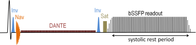 |
Central thoracic vein imaging without Gadolinium: diagnostic confidence of DANTE-based 3D subtractive NCE-MRA and comparison with 2D bSSFP
Andrew Priest, Ilse Patterson, Nadeem Shaida, Nicholas Hilliard, Sarah Hilborne, David Lomas
Imaging of the central thoracic veins is often challenging due to renal failure and/or difficult venous access, which render contrast agent administration problematic. This work assesses a non-contrast-enhanced free-breathing 3D subtractive MR angiography method for thoracic central vein imaging in a group of 18 patients. Evaluation by experienced radiologists demonstrated that angiograms obtained with this new method give high diagnostic confidence, which is significantly better than our standard 2D breath-hold approach in the absence of contrast medium. There is also better agreement between readers for the new sequence.
|
|
2986.
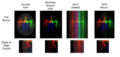 |
Accelerated Acquisition of Vessel-Encoded Arterial Spin Labelling Angiograms with Compressed Sensing
S Schauman, Mark Chiew, Thomas Okell
Vessel-selective ASL angiography provides information about cerebral blood supply not achievable by other non-invasive techniques. It is, however, limited by long acquisition times. Here we demonstrate the benefit of using compressed sensing to reconstruct undersampled vessel-selective angiograms and furthermore, consider how the vessel-encoding process affects the choice of sampling strategy compared to non-selective imaging. We show that vessel-selective angiograms arising from three brain-feeding arteries can be reconstructed with excellent fidelity in the same scan time normally required for non-selective ASL angiography.
|
|
2987.
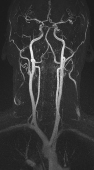 |
Large Field-of-View Nonenhanced Neurovascular MR Angiography Using Ungated Radial Quiescent-Interval Slice-Selective (QISS)
Ioannis Koktzoglou, Ali Serhal, Jianing Pang, Robert Edelman
We report a prototype ungated radial quiescent-interval slice-selective technique for nonenhanced magnetic resonance angiography of the extracranial carotid, vertebrobasilar and proximal intracranial circulations. The proposed method efficiently covers a large field-of-view, provides improved image quality with respect to Cartesian sampling, and provides flexibility to shorten the acquisition time via radial undersampling.
|
|
2988.
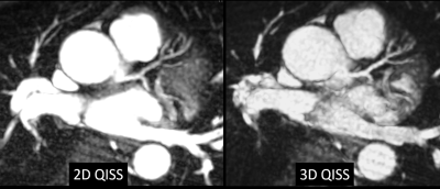 |
Breath-hold Three-dimensional Quiescent-Interval Slice-Selective (QISS) MR Angiography using a Fast-Interrupted Steady-State (FISS) Readout: Application to the Coronary and Renal Arteries
Robert Edelman, Jianing Pang, Ioannis Koktzoglou
Quiescent-interval slice-selective (QISS) is a robust nonenhanced 2D MRA technique, but has potential limitations regarding minimum slice thickness, slice profile, and fat suppression. We therefore implemented a breath-hold prototype 3D version of QISS which uses a thin-slab RF excitation, stack-of-stars k-space trajectory, and fast interrupted steady-state (FISS) readout instead of bSSFP. 2D and 3D QISS were compared for imaging of the coronary and renal arteries. Benefits of 3D QISS included better depiction of small branch vessels and improved quality for multi-planar reconstructions.
|
|
2989.
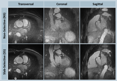 |
Artifact Reduction in 3D Radial Whole-Heart Imaging Using Slab-Selective RF Excitation
Jianing Pang, Davide Piccini, Christoph Forman, Michaela Schmidt
To date, most 3D radial kooshball imaging implementations had used non-selective (NS) radiofrequency pulses for volumetric excitation. However, given the undersampled nature of radial imaging, signal from excited regions in the periphery increases the streaking level in the central area of the field-of-view. In this work, we implemented slab-selective (SS) excitation for 3D radial whole-heart imaging. Results on 10 volunteers showed that SS excitation improved mean apparent signal- and contrast-to-noise ratio by 24% and 40%, respectively, with a mean scan time increase of 26% due to longer TR.
|
|
2990.
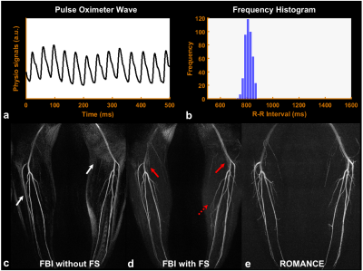 |
Retrospective Multi-Phase Non-Contrast-Enhanced Magnetic Resonance Angiography (ROMANCE MRA) for Robust Angiogram Separation in the Presence of Cardiac Arrhythmia
Hahnsung Kim, Suhyung Park, Eung Yeop Kim, Chul-Ho Sohn, Jaeseok Park
In the proposed ROMANCE MRA, data were continuously acquired over all cardiac phases using retrospective, multi-phase flow-sensitive single-slab 3D fast spin echo (FSE) with variable refocusing flip angles, while an external pulse oximeter was in sync with pulse repetitions in FSE to record real-time information on cardiac cycles. Data were then sorted into k-bin space using the real-time cardiac information. Angiograms were reconstructed directly from k-bin space by solving a constrained optimization problem with both subtraction-induced sparsity and low rank priors. Peripheral MRA was performed in a normal volunteer and a volunteer with cardiac arrhythmia using conventional fresh blood imaging (FBI) and the proposed ROMANCE MRA for comparison.
|
|
2991.
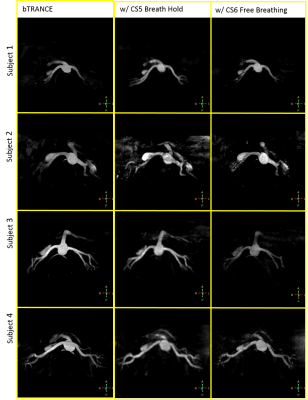 |
Breath Hold Non-contrast Enhanced Angiography of Renal Arteries at 3T using Compressed SENSE Acceleration
Brian Johnson, Ivan Dimitrov, Sandeep Ganji, Yasutomo Katsumata, Mariya Doneva, Ali Pirasteh , Johannes Peeters, Ivan Pedrosa
Non-Contrast Enhanced angiography of the renal arteries is an important technology for patients with chronic kidney disease. Existing techniques, like b-TRANCE, have long acquisition times, which makes them sensitive to motion artifacts. Respiratory triggering or navigation can be used to improve motion robustness. This however results in even longer scan times. Compressed SENSE is an effective way for accelerating 3D acquisitions and can be used to substantially reduce scan times. In this study, we report preliminary results of a breath hold and free breathing approaches for contrast-free renal angiography by combining b-TRANCE with compressed SENSE.
|
|
2992.
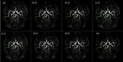 |
A hybrid method combining Keyhole and segmented k-space filling for fast TOF imaging
Zhang Qiong, Chen Shi, Zhao Wuyi, Wei Binyan
In this work, we present a Keyhole method for fast Time Of Flight (TOF) imaging. We compare it with a recently published segmented k-space filling scheme. Moreover, we demonstrate the feasibility of combing the Keyhole and segmented methods for further acceleration.Such a hybrid TOF can be potentially suited for high-resolution angiograms at ultra-high field.
|
|
2993.
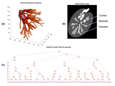 |
Improved Non-Contrast Renal Angiography Using Respiratory and Cardiac Gating with Dynamically Determined Inversion Times: A Simulation Study
Xiaoxuan He, Naoharu Kobayashi, Xiufeng Li, Gregory Metzger
In this simulation study, we aim to demonstrate the feasibility of improving non-contrast enhanced renal MRA by adding a cardiac gate with dynamically determined inversion times. The benefits of the proposal include higher contrast due to better background suppression and improved inflow enhancement, which may be clinically significant for delineating occluded vessels.
|
|
2994.
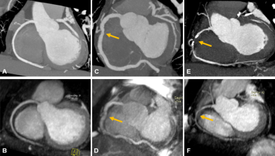 |
Comparison of Whole-Heart Noncontrast-Enhanced 3T MR Angiography and CT Angiography in Detection of Coronary Artery Disease
Jingwen Dai, Jian Cao, Jing An, Lu Lin, Yining Wang, Zhengyu Jin
The aim of this study was to investigate the diagnostic performance of noncontrast-enhanced coronary MR angiography in the detection of clinical significant coronary artery stenosis by using CTA as a reference. The preliminary results indicate that the noncontrast-enhanced coronary MR angiography has an excellent consistency in evaluating coronary artery disease in comparison to CTA. Noncontrast-enhanced coronary MR angiography may be suitable as a screening tool for coronary artery disease.
|
|
2995.
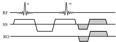 |
An accelerated peripheral MRA based on velocity-selective RF pulse using radial-MAGGULLI
Dongchan Kim, Yeji Han, Jun-Young Chung, HyunWook Park
We recently proposed a new peripheral MRA technique using velocity-selective gradient-echo (VS-GRE) sequence. Despite the high CNR and background suppression of the VS-GRE technique, this technique suffered from the reduced CNR efficiency, which was caused by the reduced sampling efficiency of radial trajectory in the peripheral region with anisotropic FOV. In this work, we propose a combination of the proposed peripheral MRA and the simultaneous multi-slice (SMS) imaging technique in the radial trajectory. In-vivo experiment results show that the proposed method could produce peripheral MRA with the reduced imaging time by radial-MAGGULLI.
|
|
2996.
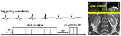 |
Free Breathing Multiple Delays Renal Perfusion MRI using Hadamard encoded pCASL
Naoyuki Takei, Shota Ishida, Nobuyuki Kosaka, R Lebel, Yuki Matta, Hirohiko Kimura, Hiroyuki Kabasawa
Current pCASL renal perfusion imaging is typically restricted to a single post label delay (PLD) time. While multiple PLD (mPLD) times can be achieved with sequential scans with different PLD times, this procedure is time consuming. A rapid acquisition was developed using Hadamard encoding for mPLD pCASL imaging combined with a motion robust timing and readout strategy to permit free breathing renal ASL. The feasibility study explores the application of Hadamard encoding to renal perfusion imaging where spin labeling is affected by pulsatile flow and demonstrated that a cardiac triggered scan provided stable perfusion images achieving ATT corrected renal blood flow with seven PLD acquisition
|
|
2997.
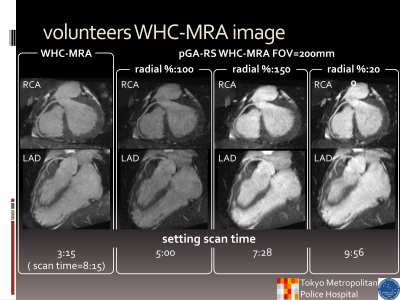 |
Free-breathing zoomed whole heart coronary MRA without respiratory gating using small-FOV 3D stack-of-stars radial sequence with pseudo-golden angle sampling
Takashige Yoshdia, Masami Yoneyama, Kohei Yuda, Takumi Koyano, Yuki Furukawa, Mariko Okura, Nobuo Kawauchi, Haruo Saito
One of the problem of whole heart coronary MRA is the prolongation of acquisition time. It is caused for degrade image quality. However, the radial sampling technique is able to obtain image of inconspicuous artifact such as aliasing and motion; furthermore, the sequence is possible to reduce scan time by understate data sampling. Hence the zoomed whole heart coronary MRA with pseudo golden angle radial sampling was improved image quality without extend scan time.
|
|
2998.
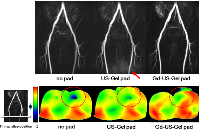 |
The feasibility of a homemade dielectric pad using commercially available ultrasound gel with Gadolinium contrast material to improve B1 homogeneity for non-enhanced peripheral MR angiography
Akiyoshi Yamamoto, Akikazu Harada, Yuji Shintani, Daiji Uchiyama, Seigo Yoshida, Katsumi Nakamura, Mitsue Miyazaki
We investigated an effect of homemade dielectric pads with using commercially available ultrasound (US) gel for improvement of B1 inhomogeneity in the peripheral artery examination using non-contrast fresh blood imaging (FBI) at 3T. We designed the two-bottle phantom mimics the iliac-femoral region, where often observed signal loss in peripheral non-contrast MRA due to B1 inhomogeneity. The result of the phantom study using US gel indicated uniform RF penetration in the B1 map. The US-gel pad improved the RF power penetration under the condition of B1 inhomogeneity and superior visualization of the left superficial femoral artery.
|
|
2999.
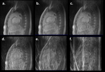 |
Real-time low-field cardiac MRI using an integrated MRI-guided radiotherapy system
H Michael Gach, Sayantan Bhadra, Austen Curcuru, Roger Nana, Clifford Robinson, Phillip Cuculich, Sasa Mutic, Mark Anastasio
The efficacy of stereotactic body radiation therapy (SBRT) cardiac radiosurgery in resolving cardiac arrhythmias was recently reported from a small clinical trial (NCT02919618). However, real-time tracking of the cardiac lesion is challenging using conventional cone-beam CT guided radiotherapy. MRI-guided radiotherapy (MRIgRT) systems integrate real-time MRI for lesion tracking with radiation therapy and can provide excellent cardiac tissue image quality at high frame rates. Real-time cardiac MRI using sparsely-sampled radial acquisitions is demonstrated with iterative reconstruction methods at low-field (0.35 T). The performance goal is to image the heart and track the lesion at 30 Hz with 2.5 mm in-plane resolution.
|
|
3000.
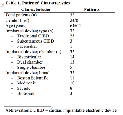 |
Wideband Inversion Recovery Late Gadolinium Enhancement Sequence improves Image Quality in Patients with Cardiac Implanted Electronic Devices
Jadranka Stojanovska, Mason Runge, El-Sayed Ibrahim, Anil Attili, Thomas Chenevert, Maryam Ghadimi-Mahani, Frank Bogun
Late gadolinium enhancement is a gold standard for myocardial scar assessment in patients with ventricular tachycardia before their ablation. The presence of cardiac implantable electronic devices degrade the image quality by producing the hyper signal intensity and make the image non-diagnostic. The modified wideband inversion recovery sequence alleviates these hyper signal intensity artifacts and render diagnostic images.
|
|
3001.
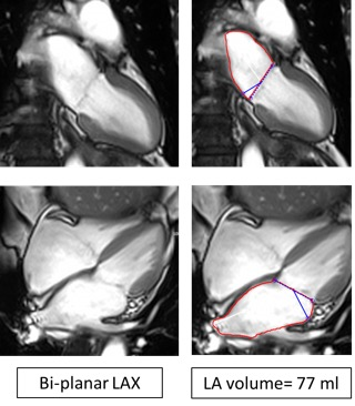 |
The Prevalence of Pulmonary Vein Stenosis Post Radio-Frequency Catheter Ablation in Atrial Fibrillation Patients
Hana Sheitt, Julio Garcia, Andrew Howarth, Stephen Wilton, Carmen Lydell, James White
This study is demonstrating the rule of cardiac MRI in evaluating pulmonary veins (PV) stenosis in atrial fibrillation patients before and after radio-frequency catheter ablation (RFCA).
|
|
3002.
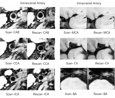 |
Reproducibility of Simultaneous Intracranial and Extracranial Arterial Vessel Wall MR Imaging based on T1 weighted DANTE-SPACE
Liwen Wan, Na Zhang, Lei Zhang, Xiaojing Long, Hairong Zheng, Xin Liu
Intracranial and extracranial atherosclerotic disease are major causes of ischemic stroke. Recently, an improved DANTE-prepared 3D variable-flip-angle turbo spin echo (SPACE) imaging method was developed for high resolution simultaneously imaging of intracranial and extracranial arterial vessel wall with enhanced cerebrospinal fluid suppression. The purpose of this study was to evaluate the scan-rescan, intra-and inter-observer reproducibility when using the method for comprehensive assessment of intracranial and extracranial vessel wall morphology. In conclusion, the improved 3D simultaneous vessel wall imaging technique provided good to excellent reproducibility for intracranial and extracranial arterial vessel wall measurements.
|
|
3003.
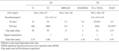 |
Assessment of carotid atherosclerosis: a comparison between 2D and 3D multi-contrast vessel wall magnetic resonance imaging
Yunduo Li, Hanyu Wei, Xihai Zhao, Gador Canton, Jie Sun, Zechen Zhou, Shuo Chen, Rui Li, Chun Yuan
In this study, we compared morphological measurements and identification of plaque components in carotid artery between 2D and 3D multi-contrast vessel wall MRI techniques. 3D multi-contrast vessel wall imaging, with 0.8mm isotropic resolution and 15min total scan time, showed good inter-reader reproducibility and provided comparable morphological information as 2D multi-contrast imaging, and more importantly, has its potential to improve visualization of plaque components.
|
|
3004.
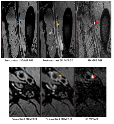 |
High prevalence of intraplaque hemorrhage in peripheral artery disease is indicated by large coverage femoral vessel wall MRI
Niranjan Balu, Jie Sun, Thomas Hatsukami, Daniel Isquith, Susan McKeeth, Chun Yuan, Xue-Qiao Zhao
Intraplaque hemorrhage (IPH) is known to be a high-risk atherosclerotic plaque feature based on carotid imaging but its prevalence is unknown in peripheral artery disease (PAD). Since PAD is a diffuse disease that can occur along a long stretch of the femoral artery, large coverage 3D vessel wall MRI is required to identify IPH prevalence in PAD. This study reports the high prevalence of IPH in patients with an abnormal ankle-brachial index (ABI) using IPH specific large coverage 3D vessel wall MRI.
|
|
3005.
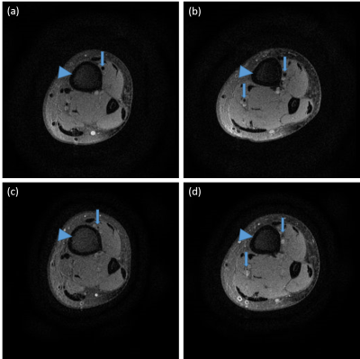 |
Preliminary Investigation of Extravascular Fluid Transport along Arterial Adventitia of Human Lower Extremity
Hongyi Li, Wentao Liu, Yang Fan, Liang Xu, Yupeng Cao, Fang Wang, Dong Han, Min Chen
Extravascular fluid transport have been reported both in human and animal studies during recent decades. Our previous work demonstrated a long-distance extravascular fluid transport which is consisted of oriented fibrous connective tissues in venous adventitia, arterial adventitia and dermis of amputated lower extremities. To further explore the pattern of fluid transport along lower extremity arteries, we implemented contrast enhanced MRI in volunteers and tracked the longitudinal contrast agent transportation. The periarterial regions near tibia showed high signal intensity after contrast agent administration suggest an unexplored extravascular fluid transport. This study may provide a novel diagnosis method of PAD.
|
|
3006.
 |
The Characteristics of Chronic Internal Carotid Artery Occlusion for Successful Endovascular Intervention by 3D MR Vessel Wall Imaging
Jin Zhang, Huilin Zhao, Beibei Sun, Xiaosheng Liu, Jieqing Wan, Weibo Chen, Xihai Zhao, Chun Yuan, Jianrong Xu
Visualization of the extent and components of internal carotid artery chronic total occlusion (CTO) may play an important role in deciding whether patients can undergo the endovascular intervention successfully or not. This study sought to investigate the characteristics of internal carotid artery CTO for successful endovascular intervention by 3D MR Vessel Wall Imaging. We found that patients with lower extent of occlusion and IPH at the proximal occlusion site had a higher success rate of recanalization. The results suggest 3D MR vessel wall imaging might be useful of patient selection for more possibly successful endovascular intervention.
|
|
3007.
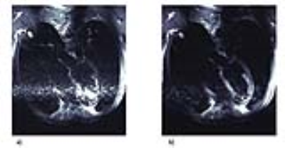 |
Towards Black Blood MRI of the Heart and Large Vessels at 7.0 T: Assessment of Inversion Pulse Quality in Phantom Experiments and In-Vivo Applications
Antonia Barghoorn, Katharina Paul, Till Huelnhagen, Thoralf Niendorf
Inversion recovery prepared cardiac black blood RARE techniques (IR-RARE) are routinely applied at clinical field strengths while still facing numerous challenges at 7.0 T. Realizing the clinical importance of IR-RARE and the benefits of UHF, this study aims at the design of a double inversion recovery prepared imaging technique at 7.0 T. The inversion efficiency and signal suppression efficiency of hyperbolic secant (HS4 and HS8) inversion pulses were analyzed in phantom experiments. First preliminary in-vivo applications using the implemented HSn pulses showed promising results.
|
|
Novel Concepts, Techniques & Methods
Traditional Poster
Cardiovascular
Thursday, 21 June 2018
| Exhibition Hall 3008-3019 |
13:15 - 15:15 |
|
3008.
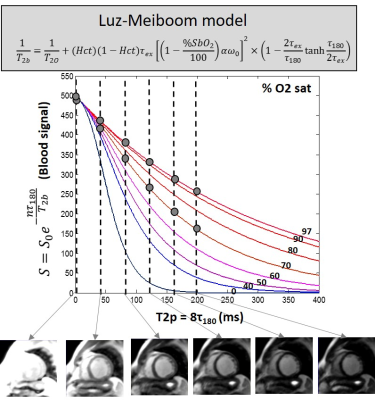 |
Non-invasive quantitative estimation of blood oxygen saturation with MRI: feasibility of machine learning
Juliet Varghese, Rizwan Ahmad, Subha Raman, Lee Potter, Orlando Simonetti
Non-invasive estimation of intra-cardiac blood oxygen (O2) saturation by magnetic resonance (MR) imaging would be useful in evaluating shunt severity in congenital heart disease, and oxygen delivery and consumption energetics in heart failure and pulmonary hypertension. Accurate estimation of blood O2 saturation from MR data may be limited, however, by the lack of an accurate model to characterize the dependence on T2 relaxation of blood on its O2 saturation level. The present study explores the feasibility of machine learning to accurately predict blood O2 saturation; the performance is evaluated in a preliminary cohort of patients against the Luz-Meiboom model.
|
|
3009.
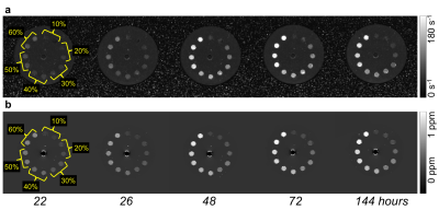 |
Differentiation of blood clot hematocrit and age in vitro using R2* and quantitative susceptibility mapping at 3T
Spencer Christiansen, Junmin Liu, Joy Dunmore-Buyze, Michael Boffa, Maria Drangova
Thrombus composition and age in ischemic occlusion can significantly influence treatment efficacy, yet current MR characterization methods are qualitative and cannot distinguish between the effects of red blood cell age and concentration (hematocrit). We examined the ability of R2* and quantitative susceptibility (QS) maps derived simultaneously from multi-echo GRE acquisition to discriminate between blood clots of varied hematocrit formed in vitro and monitored over a six-day ageing period. Fresh clots (age < 6 hours) of different hematocrit were distinguishable using either R2* or QS values, while aged clots were distinguishable only when both values were considered.
|
|
3010.
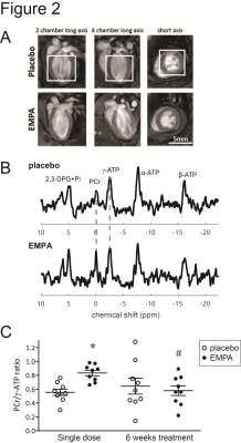 |
Impact of empagliflozin on cardiac energy status and function in diabetic db/db mice
Desiree Abdurrachim, Emmy Manders, Klaas Nicolay, Eric Mayoux, Jeanine Prompers
Diabetes is associated with impaired cardiac energetics and diastolic dysfunction. A substrate shift toward ketones has been proposed to explain the benefits of empagliflozin on cardiovascular outcome in diabetes patients. We investigated the effects of empagliflozin on cardiac energetics and function in diabetic db/db mice using 31P-MRS and MRI. After a single dose of empagliflozin, cardiac PCr/ATP ratio was higher compared with placebo-treated controls, which was associated with increased plasma ketone levels and lower cardiac load. After 6 weeks of treatment, cardiac diastolic function tended to be improved, while plasma ketones and cardiac PCr/ATP ratio were not different from placebo.
|
|
3011.
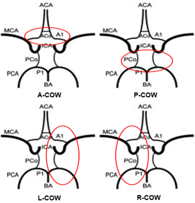 |
Association between Incompleteness of Circle of Willis and Carotid Vulenrable Atherosclerotic Plaques: A CARE-II Study
Changwu Zhou, Chun Yuan, Wei Wang, Cheng Li, Xihai Zhao
The circle of Willis (COW) is an important intracranocervical collateral circulation system. We hypothesized that the integrity of COW may affect the characteristics of carotid plaques by influencing carotid hemodynamics. This study investigated the relationship between incompleteness of COW and the compositional features of atherosclerotic plaques in carotid arteries. We found that the incompleteness of circle of Willis is associated with vulnerability of carotid artery atherosclerotic plaques. Our findings suggest that integrity of circle of Willis may play a role in occurence of high risk plaque features, particularly intraplaque hemorrhage and fibrous cap rupture.
|
|
3012.
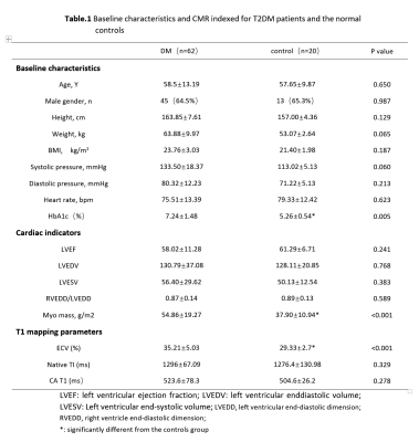 |
Evaluation myocardial fibrosis in diabetes with cardiac magnetic resonance T1-mapping: correlation with the metabolic and diabetic duration
Yue Gao, Zhigang Yang, Xi Liu, Linjun Xie, Li Jiang, Biyue Hu, Yingkun Guo
In order to clarify the relationship among diffuse myocardial fibrosis and abnormal metabolic and duration and diabetes, we compared the T1 mapping parameters on cardiac magnetic resonance (CMR). type 2 diabetes (T2DM) patients and normal controls were enrolled and performed CMR both. Our results showed ECV were higher in T2DM than controls, and positively associated with high level glycosylated hemoglobin and longer duration diabetes. Diabetes myocardial fibrosis could be effectively detected by CMR T1 mapping. The trend of myocardial fibrosis in patients with hyperglycemia and long-term duration is more obvious
|
|
3013.
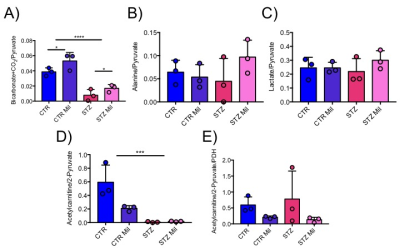 |
In Vivo Hyperpolarized MRI Reveals Metabolic Changes Following Treatment with Mildronate in the Control and Diabetic Heart.
Dragana Savic, Lorenz Holzner, Vicky Ball, M. Curtis, Lisa Heather, Damian Tyler
L-carnitine acts as a buffer of acetyl-CoA units in the mitochondria, as well as facilitating transport of fatty acids. Mildronate can block the biosynthesis of L-carnitine and its uptake by inhibiting CPT-1. The purpose of this study was to investigate the effect of Mildronate treatment on cardiac function and metabolism in the healthy and the diabetic rat heart. We show that daily injections of Mildronate can alter cardiac metabolism in the in-vivo diabetic and healthy rat heart, without any functional changes, and surprisingly Mildronate can increase flux through pyruvate dehydrogenase. Such studies will allow a better understanding of the interactions between metabolism and function in the diabetic heart and may provide new insight into novel therapeutics.
|
|
3014.
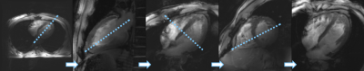 |
Cardiac MRI with the Siemens Terra 7T System: Initial Experience and Optimization of Default Protocols
David Lohr, Maxim Terekhov, Aleksander Kosmala, Maria Stefanescu, Michael Hock, Laura Schreiber
The demand for the application of Ultra-High Field (B0≥7T) MR-scanners in cardiovascular MRI grows permanently despite of technical challenges increasing significantly with the static magnetic field strength. We report initial experience with the new 7T system Siemens Magnetom™ Terra for acquiring MR-images of the human heart. A standard workflow for cardiac assessment has been developed and tested in N=18 healthy volunteers in single transmit mode. Currently CINE scans with 14-17 slices covering up to 35 heart phases are well suited for clinical volumetric heart function characterization. Diagnostic image quality can be provided for subsequent volunteers.
|
|
3015.
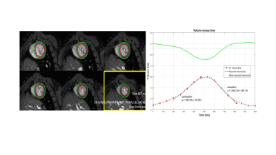 |
Cardiac MRI assessment of the effects of dietary Eicosapentaenoic acid (EPA) on the adverse cardiac consequences of sepsis in rat
Amidou Traore, Thibault Leger, Guilhem Pagès, Lucie Cassagnes, Azarnoush Kasra, Jean-Marie Bonny, Luc Demaison
Severe sepsis is one of the leading cause of death in the intensive care units (ICU) or in short time after discharge from ICU. Developing a rat model of early sepsis involving caecal ligation and puncture, we undertaken this cardiac MRI study to quantitatively assess myocardial function and the protective effect of dietary EPA. Our results showed that, in the exception of the rate of contraction, cardiac functions are less impacted in the early hyperdynamic phase of sepsis with no/or milder modulation of dietary EPA.
|
|
3016.
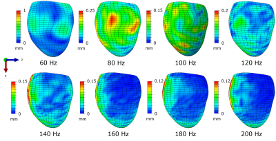 |
Frequency Dependence of Anisotropic Material Properties Estimated form Cardiac Magnetic Resonance Elastography: An In Silico Study
Renee Miller, Arunark Kolipaka, Martyn Nash, Alistair Young
Despite the anisotropy of myocardium, previous cardiac MR elastography studies have investigated isotropic stiffness of heart tissue. Anisotropic material properties could provide a better understanding of structural changes that occur in the heart due to pathologies such as diastolic heart failure. However, optimal imaging parameters to measure anisotropic properties are yet unknown. This study investigates the optimal loading frequency that accurately recovers anisotropic stiffness measurements using simulations of cardiac MR elastography experiments in the presence of Gaussian noise and known fibre orientations. The optimised virtual fields method is used as an inversion method to translate harmonic displacements to stiffness parameters.
|
|
3017.
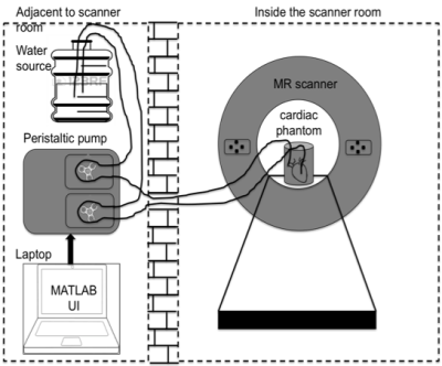 |
Integrated, 3D Printed Cost Effective Phantom solution for MR Imaging of Cardiac Structure and Function
Shivaprasad Chikop, Amaresha Konar, Nithin Vajuvalli, Ramesh Venkatesan, Sairam Geethanath
An integrated cardiac phantom solution was developed to correlate with clinically relevant parameters entered through a user interface (UI). Mimicking of human heart was achieved through integration of a flexible 3D printed heart model and peristaltic pump. Results depict the correlation between the input parameters to output parameters obtained through image processing of the phantom MR images. The work illustrates the structural features and motion measures of the cardiac phantom. The phantom can therefore be employed to assess novel acquisition and reconstruction methods. The utilization of 3D printing enables the use of subject specific phantom to study diverse cardiovascular scenarios.
|
|
3018.
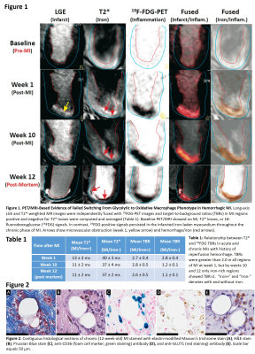 |
18F-FDG PET/MRI Allows Early Detection of Foam Cell Formation and Fat Deposition in Hemorrhagic Myocardial Infarctions
Ivan Cokic, Jane Sykes, John Butler, Michael Kovacs, Hsin-Jung Yang, Damini Dey, Frank Prato, Rohan Dharmakumar
Inability of macrophages (MΦ) to switch from pro-inflammatory (M1, glycolytic) to anti-inflammatory (M2, oxidative) phenotype can lead to increased glucose transporter 1 (GLUT1)-mediated glucose metabolism, decreased fatty acid (FA) beta oxidation, increased intracellular lipid accumulation, and MΦ-to-foam cell transformation. Recent studies in the field of chronic venous leg ulcers have shown that iron-overloaded MΦ fail to switch from M1 to M2 phenotype. In this study we hypothesized that inability of iron-overloaded MΦ to switch from M1 to M2 phenotype underlies fatty degeneration of hemorrhagic myocardial infarction via MΦ lipid accumulation and their transformation into foam cells.
|
|
3019.
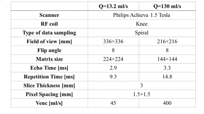 |
A Least Squares Approach for Relative Pressure Measurement from 4D flow PC-MRI
Sina Hooshyar, Sean Callahan, MJ Negahdar, Saeed Kermani, Amir Amini
Noninvasive determination of relative transstenotic pressure drop from 4D flow MRI has been investigated by a fast 3D matrix method based on the Least-Squares strategy. The method was tested with CFD velocity data as input as well as MRI phantom data. While results are comparable to the conventional pressure-poisson equation approach, the method is computationally more efficient.
|
|
| Back |
| The International Society for Magnetic Resonance in Medicine is accredited by the Accreditation Council for Continuing Medical Education to provide continuing medical education for physicians. |
