|
Does Motion Still Matter? New Methods for Handling Motion
Combined Educational & Scientific Session
ORGANIZERS: Edward DiBella, Mary McDougall
Thursday, 21 June 2018
| S01 |
08:00 - 10:00 |
Moderators: Li Feng, Shreyas Vasanawala |
Skill Level: Intermediate to Advanced
Session Number: Th-03
Overview
Motion has long been an issue when obtaining high quality MRI scans, whether the motion is the desired quantity (flow velocity, cardiac function), or it is corrupting images by blurring, ghosting, and other artifacts. A huge amount of research has gone into handling motion for MRI. This session will review hardware and software approaches to handling motion, in order to set the stage for the presentation of selected abstracts of cutting-edge motion methods.
Target Audience
Attendees actively working with MRI would benefit from ideas of handling motion from other areas of MRI, as well as being brought up to speed on basic and current methods for handling motion in MRI, including self-navigation techniques.
Educational Objectives
As a result of attending this course, participants should be able to:
-Describe two methods for hardware-based motion tracking/compensation;
-Identify two methods for software-based motion tracking/compensation, including self-navigated approaches; and
-Recall two new advances in methods for handling motion (from abstracts).
08:00
|
|
 Hardware Methods for Handling Motion: Basic Ideas & Current Methods Hardware Methods for Handling Motion: Basic Ideas & Current Methods
Maxim Zaitsev
Motion during MR image encoding produces inconsistencies in the acquired k-space data, which results in well-known motion artifacts. In clinical settings patient motion during MRI examinations renders a significant fraction of scans non-diagnostic. Recently, due to the increased availability of ultra-high filed imagers of 7T and above capable of sub-millimeter resolution in vivo, it became apparent that even in normal subjects involuntarily motion of about a millimeter may limit significantly the achievable image quality. Motion correction based on additional motion tracking hardware has demonstrated its ability of achieving excellent image quality both in clinical and research settings.
|
08:30
|
|
Software Methods for Handling Motion: Basic Ideas & Current Methods
Debiao Li
|
09:00
 |
1003.
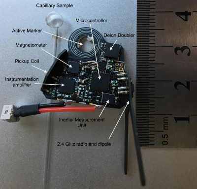 |
Hybrid motion sensing with a wireless device, self-synchronized to the imaging pulse sequence.
Adam van Niekerk, Andre van der Kouwe, Ernesta Meintjes
Here we combine an accelerometer, angular rate sensor, magnetometer, pickup coil and active marker in a (single) small wireless device. The active marker is used to detect radio frequency pulses, enabling synchronisation to the imaging pulse sequence to within 1 μs accuracy. Sinusoidal waveforms inserted into the imaging pulse sequence (lasting 920 μs) are then measured using the onboard pickup coil to allow real-time position and ADC-gradient timing offsets in all 3 axes. Preliminary results suggest that sub-mm accuracy is possible with very low noise (<0.3 mm) in the raw data captured. The new hardware layout has the potential to enable full prospective motion correction from a single small device that fits on the bridge of the subject's nose.
|
09:15
|
1004.
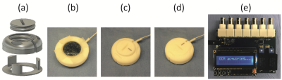 |
Ultrasound-based sensors for physiological motion monitoring
Bruno Madore, Cheng-Chieh Cheng, Pei-Hsin Wu, Frank Preiswerk
A small ultrasound-based sensor was developed for use in MRI. It is about 3x3x1cm in size, can be fixed to the skin, readily fits under imaging coils, and provides up to 20,000 data points in a 0.2ms period. A switching circuit was developed allowing up to four such sensors to be used at a time. Examples are show where a sensor was attached over a carotid artery, over the heart or below the ribs, providing information about blood influx to the brain, cardiac motion or breathing, respectively. These devices and associated processing may prove especially useful at high field, where ECG detection proves ineffective.
|
09:30
|
1005.
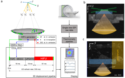 |
3D ultrasound based motion tracking combined with rapid multislice MR-thermometry for MR guided HIFU on mobile organs.
Pierre Bour, Valéry Ozenne, Fabrice Marquet, Baudouin Denis de Senneville, Erik Dumont, Bruno Quesson
MRI-guided High Intensity Focused Ultrasound treatment of mobile organs remain challenging in terms of therapy effectiveness (target tracking) and precision of temperature monitoring (motion and related susceptibility artifacts). In this study, we propose to use some elements of the HIFU transducer in reception for a standalone motion correction of the focus position, allowing full flexibility on the MR-Thermometry sequence. The method is validated in vitro on a mobile gel and in vivo in the liver of pig during breathing, with a 10Hz update of the HIFU focus position under rapid and volumetric MR-thermometry.
|
09:45
|
1006.
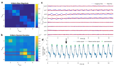 |
Monitoring the Complete Scattering Matrix of a Parallel Transmit Coil during Image Acquisition for Retrospective Cardiac Gating at 7T MRI
Sven Jaeschke, Matthew Robson, Aaron Hess
We propose a new monitoring scheme that enables simultaneous measurements of the complete scattering matrix of a pTx coil during image acquisition by modifying the RF-pulse without prolonging the image sequence and without spin distortion. We show that the use of this monitoring schemes and the measured scattering matrix enable a higher SNR in the estimated cardiac scattering signal, than using RF-reflection of the normal imaging pulse as in previous work. Preliminary results in 7T MRI are shown with successfully, retrospectively gated 2D-CINE images using the proposed method.
|
10:00
|
|
Adjournment & Meet the Teachers |
|
