|
Brain Connectivity & Dynamics
Combined Educational & Scientific Session
ORGANIZERS: Richard Buxton, Hanzhang Lu, Benedikt Poser
Thursday, 21 June 2018
| N01 |
13:15 - 15:15 |
Moderators: Hanzhang Lu, Richard Buxton |
Skill Level: Intermediate
Session Number: Th-05
Overview
This session will provide an overview of the concept of brain dynamics, its analysis methods, neurobiological mechanisms, and potential clinical applications.
Target Audience
This course is designed for scientists and clinicians who are interested brain functional connectivity methods and applications.
Educational Objectives
As a result of attending this course, participants should be able to:
-Describe various methods to compute dynamic functional connectivity from resting-state fMRI data; and
-Explain possible neurobiological underpinnings of dynamic functional connectivity and its clinical applications.
13:15
|
 |
 Dynamic Functional Connectivity Methods Dynamic Functional Connectivity Methods
Thomas Liu
This educational talk will review methods for the assessment of dynamic functional brain connectivity.
|
13:45
|
|
Mechanisms & Clinical Applications of Dynamic Functional Connectivity
Victor Vergara, Vince Calhoun
Dynamic functional network connectivity is a promising technique for clinical applications. It has been successfully applied in the analysis of neurological illnesses and as a source of classification features to separate patients from controls. Advantages and pitfalls of dynamic analysis is discussed and compared to it’s to static connectivity alternative. The two flavors of functional connectivity offer complementary information depending on the specific application. This is exemplified discussing the results obtained from schizophrenia, Huntington’s disease, traumatic brain injury and substance addiction.
|
14:15
|
1122.
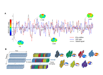 |
Voxel-wise mapping of spontaneous brain network dynamics in the mouse brain
Daniel Gutierrez-Barragan, Stefano Panzeri, Alessandro Gozzi
Resting-state fMRI has been widely used to map brain network dynamics via sliding-window correlations between regional functional signals. Using frame-wise clustering of spontaneous BOLD fMRI, here we map the dynamics of brain-wide network activity in the mouse brain with voxel-resolution, at the subject and group level, without the need to use pre-imposed regional parcellation or correlation windows. This approach revealed a reproducible set of recurrent brain states encompassing previously defined functional connectivity networks, which happen at specific phases of global fMRI signal oscillations. Our results provide a novel interpretative framework for the emergence and organization of brain-wide network dynamics.
|
14:27
|
1123.
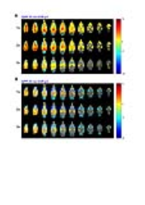 |
Whole brain Quasi-Periodic patterns interact with visual stimulation processing in mice
Michaël Belloy, Behnaz Yousefi, Jacob Billings, Rukun Hinz, Annemie van Der Linden, Shella Keilholz, Georgios A. Keliris, Marleen Verhoye
We show the detection of whole-brain mouse Quasi-periodic patterns (QPPs), highlighting their interaction with the global signal and coincidence with anti-correlation between speculative mouse Default Mode (DMN) and Task-Positive (TPN)-like networks. We further investigated how QPPs interact with sensory information processing. By tracking QPP behavior during an fMRI visual stimulation block design, we illustrate how QPPs can become modulated by visual stimulation, triggering or diminishing their occurrence. We then linearly regressed QPPs from the fMRI images to show how QPPs underlie a substantial fraction of visual response magnitude and variance. These results suggest the relevance of QPPs in sensory processing.
|
14:39
|
1124.
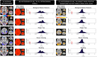 |
Quantitative deconvolution of neuronal-related BOLD events with Multi-Echo Sparse Free Paradigm Mapping
Javier Gonzalez-Castillo, Cesar Caballero-Gaudes, Pater Bandettini
This work proposes a novel formulation of the Sparse Free Paradigm Mapping (SPFM) algorithm for Multi-Echo (ME) fMRI that produces estimates of ΔR2* with interpretable units (s-1) without prior timing information of task events. Here we show how ΔR2* time-series estimated with ME-SPFM have physiologically plausible values, in agreement with previously published estimates of ΔR2* for neuronal events. We also show, how having easily interpretable units permits detection of artefactual events that can be easily removed from subsequent analysis and interpretation. Our results suggest that ME-SPFM may provide a pseudo-quantitative method to study the time-varying nature of brain activity in experimentally unconstrained paradigms (naturalistic, resting-state) or clinical applications (detection of inter-ictal epileptic events).
|
14:51
|
1125.
 |
Efficacy of different dynamic functional connectivity methods to capture cognitively relevant information
Hua Xie, Javier Gonzalez-Castillo, Daniel Handwerker, Peter Molfese, Peter Bandettini, Sunanda Mitra
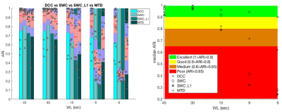
With a multitask dataset (rest, memory, video, and math) serving as ground truth, we evaluated the efficacy of four different methods of estimating dynamic functional connectivity (dFC)—namely sliding window correlation (SWC), sliding window correlation with L1-regularization (SWC_L1), dynamic conditional correlation (DCC), and multiplication of temporal derivatives (MTD)—to capture cognitively relevant information. We used dFC estimates of each method as inputs for k-means, and evaluated how well they segregate scan periods for different tasks. We found that moving average DCC produces best results, especially for short window length (WL ≤ 9sec), suggesting DCC may more reliably reveal dFC linked to mental states.
|
15:03
|
1126.
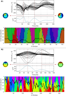 |
EEG microstate and spectral signatures of epileptic patterns found in fMRI dynamic functional connectivity
Rodolfo Abreu, Alberto Leal, Patrícia Figueiredo
We aimed to investigate EEG signatures specifically associated with epileptic patterns of dynamic functional connectivity (dFC) found in BOLD-fMRI data. We estimated dFC using a sliding-window correlation analysis and applied dictionary learning (DL) to identify the most prominent patterns while forcing a certain degree of sparsity in time. Upon the labelling of each time window based on the pattern exhibiting the highest contribution, we investigated pattern-specific microstates (MS) and spectral proprieties in simultaneously recorded EEG data. In contrast with the spectral proprieties, EEG MS revealed robust signatures of epileptic dFC patterns in all patients.
|
15:15
|
|
Adjournment & Meet the Teachers |
|
