|
Faster Imaging, Faster Evaluation
Combined Educational & Scientific Session
ORGANIZERS: Christopher Hess, Alex MacKay
Thursday, 21 June 2018
| N01 |
15:30 - 17:30 |
Moderators: Christopher Hess, Alex MacKay |
Skill Level: Basic to Intermediate
Session Number: Th-07
Overview
In recent years, MR scientists have developed MR techniques (e.g., MR fingerprinting and synthetic MR) which promise to substantially shorten the time required for a clinical exam by MR. This session was organised to 1) explain how these MR techniques work; and 2) describe the potential impact of these fast MR imaging techniques on clinical medicine.
Target Audience
MR scientists, engineers and clinicians who wish to make use of fast imaging techniques at their site.
Educational Objectives
As a result of attending this course, participants should be able to:
-Describe how to develop and implement fast imaging MR protocols at their site;
-List which clinical applications are ideal for implementation of fast imaging techniques; and
-Summarize which clinical applications may not be suitable for use of fast imaging techniques.
15:30
|
|
 Fast Imaging Techniques for Brain Imaging Fast Imaging Techniques for Brain Imaging
Stefan Skare
"Fast Imaging Techniques for Brain Imaging" is a wide topic. This presentation will focus on the most recent advances in fast brain imaging towards comprehensive clinical brain exams by acquiring multiple MR contrasts simultaneously in minimal scan time.
|
16:00
|
|
Medical Applications of Fast MR Imaging of Brain
Kambiz Nael
The target audience of this talk are neuroscientists and clinicians including neurologists and neuroradiologists interested in the development and application of rapid brain MRI imaging.
The objectives are:
1. To be familiar with the latest available methodology for fast brain MR imaging
2. Learn how to obtain routine brain MRI examinations in 5 minutes
3. Learn how to perform and interpret a 6-minute comprehensive stroke MR imaging
4. Accelerate brain vascular imaging using highly under sampled methodology:
- Compressed sensing for brain vessel wall imaging and MRA
- k-t accelerated imaging for clinical brain 4D flow imaging |
16:30
|
1197.
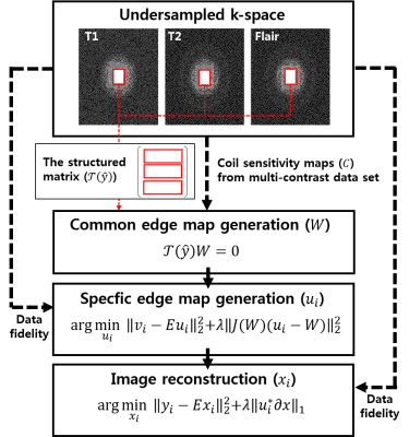 |
Highly Accelerated Multi-Contrast 3D Isotropic MRI in 5 Minutes: A Feasibility Study for Multiple Sclerosis
Xiaoxiao Ma, Xin Lou, Hyunkyung Maeng, Sugil Kim, Suhyung Park, Guobin Li, Chaohong Wang, Jaeseok Park
Multiple sclerosis(MS) is a chronic disease that damages the nerves in the brain and results in multiple areas of scar tissues within the central nervous system. Multi-contrast structural MRI, which includes T1, T2, and FLAIR, has been routinely used in detecting MS lesions. Nevertheless, it is still challenging to accurately detect small MS lesions diffused over the entire brain due to the limitation of spatial resolution and long imaging time. The purpose of this work is to investigate the feasibility of achieving highly accelerated, multi-contrast 3D isotropic (~ 1.0 mm3) MRI (T1, T2, and FLAIR), which exploits sharable information across images, for detection of MS lesions over the whole brain roughly in 5 minutes.
|
16:45
|
1198.
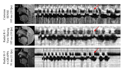 |
Through-time and hybrid radial GRAPPA to improve the visualisation of velic motion in real-time speech MRI
Matthieu Ruthven, Andreia Freitas, Redha Boubertakh, Marc Miquel
Aim: to investigate if radial GRAPPA could be used to: (a) improve velic motion visualisation when compared with standard protocols and reconstructions; (b) enable multislice imaging by sufficiently accelerating real-time MRI data acquisition. Methods: datasets of healthy adult volunteers were acquired at 3T and reconstructed using through-time and hybrid GRAPPA methods.
Results: velic motion visualisation was superior in GRAPPA images than in images acquired using standard protocols and reconstructions. Multislice imaging (two slices) at 8fps per slice was achieved.
Conclusions: radial GRAPPA shows promise as a method for use in clinical imaging of speech.
|
17:00
|
1199.
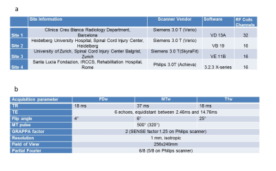 |
A multi-center study on fast full-brain quantitative multi-parameter mapping of R1, MT, and R2*: scan-rescan repeatability and inter-site reproducibility
Video Permission Withheld
Maryam Seif, Tobias Leutritz, Rebecca Samson, Armin Curt, Claudia Gandini Wheeler-kingshott, Patrick Freund, Nikolaus Weiskopf
We present a multi-center, multi-vendor study evaluating repeatability and reproducibility of quantitative MRI data acquired using high resolution (1 mm³) multi-parameter mapping which provides quantitative R1, MT and R2* maps of the whole brain within less than 18 min. The protocol was implemented at four clinical sites with different Siemens and Philips 3T MRI scanners. Scan-rescan measurements of the same five healthy volunteers at all sites showed good intra-site reproducibility in all parameter maps. However, the inter-site comparisons showed higher reproducibility within a single vendor than across vendors.
|
17:15
|
1200.
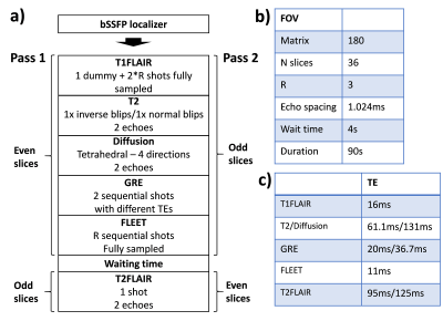 |
Multi-contrast EPI - Towards clinical application
Tim Sprenger, Mathias Engström, Ola Norbeck, Henric Rydén, Enrico Avventi, Johan Berglund, Stefan Skare
We present an enhanced version of our multi-contrast EPI sequence which generates T1-FLAIR, T2-FLAIR, T2*w, T2w, iso-DWI, ADC images of 36 slices in 90 s. We introduce various optimization including tetrahedral diffusion encoding, different partial Fourier factors for the EPI trains and multi-echo acquisition for T2-FLAIR, T2*w, T2w, and DWI. The improved sequence is compared to our previous implementation and superior image quality is shown especially for the diffusion contrast. Finally, multi-contrast EPI patient data is shown and compared to conventional imaging sequences.
|
17:30
|
|
Adjournment & Meet the Teachers |
|
