|
Prostate Cancer: Current Gaps & Future Directions
Combined Educational & Scientific Session
ORGANIZERS: Kathryn Fowler, Kartik Jhaveri, Lorenzo Mannelli, Valeria Panebianco, Scott Reeder, Mustafa Shadi Bashir, Claude Sirlin, Reiko Woodhams
Tuesday, 19 June 2018
| S02 |
08:15 - 10:15 |
Moderators: Daniel Margolis, Alberto Vargas |
Skill Level: Intermediate
Session Number: Tu-03
Overview
This session will focus on state-of-the-art prostate MR acquisition with special attention to current shortcomings of techniques in practice. Additionally, discussion will cover the concepts of clinically significant disease and imaging diagnosis.
Target Audience
Clinicians and physicists who participate in prostate imaging.
Educational Objectives
As a result of attending this course, participants should be able to:
-Summarize current prostate MRI protocols, parameters, and technical limitations;
-Describe cutting-edge and evolving practice applications of prostate MRI; and
-Explain the clinical relevance of MR imaging diagnosis as relates to disease grade, stage, and tumor burden.
08:15
|
|
 Prostate Cancer: Defining Clinically Significant Disease Prostate Cancer: Defining Clinically Significant Disease
Peter Choyke
The goal of performing MRI in prostate cancer is to help distinguish indolent, low risk prostate cancers from those harboring clinically significant features. This presentation reviews the criteria for clinically significant tumors and underscores the benefits that accrue when using MRI as a gateway for the diagnosis of prostate cancer.
|
08:45
|
0342.
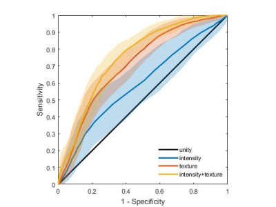 |
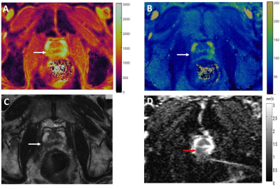
T2-weighted MRI-derived textural features can help the assessment of peripheral zone prostate cancer aggressiveness: results from multi-center data.
Gabriel Nketiah, Mattijs Elschot, Tom Scheenen, Marnix Maas, Tone Bathen, Kirsten Selnæs
The assessment of prostate cancer aggressiveness is currently based on Gleason grading of histological samples obtained by TRUS-guided biopsies, which can lead to substantial underestimations due to sampling errors. We previously showed that textural features derived from T2-weighted MRI could potentially serve as a non-invasive biomarker for prostate cancer aggressiveness. The aim of this work was to validate these preliminary results in a multi-center study. We found that the combination of intensity and textural features could distinguish between low/intermediate and high-grade with an accuracy of 71%, which was significantly higher than intensity (60%) or textural features (68%) alone.
|
08:57
|
0343.
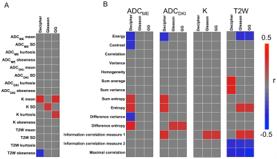 |
Radiomics measured with mpMRI predicts histopathological and genomics markers of prostate cancer aggressiveness.
Stefanie Hectors, Mathew Cherny, Sara Lewis, Kanika Mahajan, Ardeshir Rastinehad, Ashutosh Tewari, Bachir Taouli
The goal of this study was to assess the association of multiparametric MRI (mpMRI) radiomic features with histopathological and genomic markers of prostate cancer (PCa) aggressiveness. mpMRI histogram and texture features showed multiple significant correlations with Gleason score, modified Gleason score Grade Group and genomics Decipher risk score. General linear models showed high accuracy for prediction of the histopathological and genomics features (accuracy range 0.77-0.94). The results indicate that MRI radiomics analysis is promising for noninvasive assessment of PCa aggressiveness on the histopathological and genomics levels.
|
09:09
|
|
Current State-Of-The-Art Imaging Protocol: Where Are The Gaps?
Masoom Haider
In Pi-Rads v2 there are technical recommendations for performance of multiparametric MRI (mpMRI) of the prostate. When using this protocol there are several issues that lead to suboptimal or non-diagnostic images that remain unaddressed. Some of these can be mitigated through altering patient preparation or a changing of pulse sequence parameters. Further optimization and development of robust pulse sequences and imaging systems to improve prostate MRI image quality is an unmet need that can benefit from further research. A second critical area in need of further development is related to value which goes hand in hand with cost reduction. This is also tied to the growing and controversial concerns related to repeated Gd administration for DCE MRI and the unknown long-term effects of Gd deposition in the brain in an otherwise healthy patient population with long life expectancy. The necessity of DCE MRI and the potential of proton spectroscopy to replace DCE MRI are other potential areas of innovation.
|
09:39
|
0344.
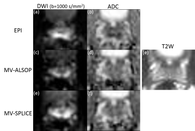 |
Reduced distortion in prostate DWI by using split echo type TSE-DWI (SPLICE) with MultiVane acquisition
Yuta Akamine, Tomoyuki Okuaki, Satoshi Goshima, Kimihiro Kajita, Masatoshi Honda, Masami Yoneyama, Makoto Obara, Marc Van Cauteren
To reduce image distortion in prostate DWI, split-echo type TSE-DWI (SPLICE) was combined with MultiVane (current Philips implementation of PROPELLER) acquisition, named MV-SPLICE. To avoid non-CPMG artifacts in SPLICE, the spin echoes and stimulated echoes were separated by the unbalanced readout gradient and acquired in separate MultiVane k-space for separate reconstruction. ADCs and SNRs in transition zone and peripheral zone, and distortion for DWI and T2W images in the anterior-posterior direction of prostate diameter for MV-SPLICE were compared to conventional EPI and MultiVane TSE-DWI (MV-ALSOP). We demonstrated that MV-SPLICE is insensitive to distortion and can provide comparable ADC measurement.
|
09:51
|
0345.
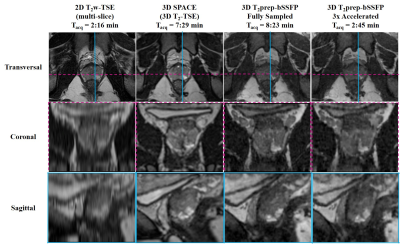 |
Accelerated 3D 1 mm isotropic T2w-Imaging of the Prostate in less than 3 min
Rohini Vidya Shankar, Gastao Cruz, Radhouene Neji, Elisa Roccia, René Botnar, Vicky Goh, Claudia Prieto, Isabel Dregely
Three dimensional (3D), isotropic T2-weighted imaging of the prostate requires a long acquisition time. Here we propose a 3D T2-prepared multi-shot bSSFP acquisition with a variable density undersampled trajectory and TV-SENSE reconstruction. Results from a healthy volunteer study demonstrate that 3D 1 mm isotropic resolution T2-weighted images of the prostate can be acquired in 2 min 45 s, with image quality that is comparable to the clinical standard turbo spin echo sequences but only takes 1/3 of the acquisition time.
|
10:03
|
0346.
 |
Targeted Biopsy Validation of Peripheral Zone Prostate Cancer Characterization with MR Fingerprinting and Diffusion Mapping
Ananya Panda, Gregory O'Connor, Yun Jiang, Alice Yu, Shivani Pahwa, Sara Dastmalchian, Seunghee Margevicius, Mark Schluchter, Robert Abouassaly, Chaitra Badve, Mark Griswold, Lee Ponsky, Vikas Gulani
Targeted biopsy validation is presented for characterization of peripheral zone (PZ) prostate cancer grades and differentiation of prostate cancer from prostatitis, using a quantitative MR protocol comprising of MRF-relaxometry and standard EPI based ADC mapping. Mean T1, T2 and ADC in prostate cancer were significantly lower than in NPZ. Mean T2 and ADC in low-grade cancer were significantly higher than intermediate and high-grade cancer with similar AUCs (0.80) for both for differentiating grades. Mean T2 and ADC in prostate cancer were significantly lower than prostatitis. T2 was a significant predictor for prostate cancer over prostatitis while ADC was not significant.
|
10:15
|
|
Adjournment & Meet the Teachers |
|
