Joint Annual Meeting ISMRM-ESMRMB • 16-21 June 2018 • Paris, France

Educational Course
Peripheral Nerve Imaging: Ryder Cup
Weekday Course

ORGANIZERS: Eric Chang, Garry Gold, Emily McWalter, Edwin Oei, Philip Robinson

 
Wednesday, 20 June 2018
W03/04  08:15 - 10:15 Moderators:  Kimberly Amrami, Richard Hodgson

Skill Level: Intermediate to Advanced

Session Number: W-02

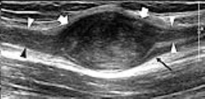
Overview
This course will cover state-of-the-art imaging assessment of peripheral nerves including normal appearance and common pathologies. Four experienced radiologists from different institutions in Europe and North America will discuss various aspects of nerve imaging, including optimizing MR and ultrasound imaging protocols. They will also compare and contrast where these 2 imaging modalities have specific advantages in assessing normal anatomy, nerve function, and abnormality.

Target Audience
Physicians and scientists with a background in peripheral nerve imaging who want to advance their knowledge to the state of the art.

Educational Objectives
As a result of attending this course, participants should be able to:
-Describe the most relevant nerve-related diagnoses and their MR and ultrasound appearances;
-Identify specific diagnoses where one imaging technique may be superior to the other;
-Discuss the differential diagnosis of MR and ultrasound peripheral nerve findings; and
-Implement optimized nerve imaging protocols in their practice.


08:15
 
Upper-Extremity Peripheral Nerve Imaging with US: European Perspective
Carlo Martinoli, Federico Zaottini, Riccardo Picasso, Federico Pistoia
The value of US to assess the most common disorders affecting peripheral nerves in the upper extremity, including compressive neuropathies, polyneuropathies, traumatic injuries, tumours and tumour-like lesions, will be discussed. A careful US approach with thorough understanding of soft-tissue planes and extensive familiarity with anatomy are prerequisites for obtaining reliable information regarding such kind of examination. US can provide a low-cost and noninvasive imaging, speed of performance, and important advantages over MR imaging, including higher spatial resolution and ability to explore long segments of nerve trunks in a single study and to examine tissues in both static and dynamic states with real time scanning.

 
08:40
 
  Upper-Extremity Peripheral Nerve Imaging with MRI: European Perspective
Gustav Andreisek
MR neurography has become the modality of choice to image the peripheral nerves of the upper extremity. This talk will provide a comprehensive overview from a European perspective.

 
09:05
 
  Lower-Extremity Peripheral Nerve Imaging with US: American Perspective
Karen Chen
09:30
 
Lower-Extremity Peripheral Nerve Imaging with MRI: American Perspective
Jenny Bencardino
Entrapment neuropathies present a challenge to clinicians and radiologists alike.  MR neurography has proven a reliable, non invasive, diagnostic method to identify that exact site of nerve entrapment and the associated direct and indirect MR signs of neuropathy.

 
09:55
 
  Panel Discussion
10:20
 
  Adjournment & Meet the Teachers
Back
The International Society for Magnetic Resonance in Medicine is accredited by the Accreditation Council for Continuing Medical Education to provide continuing medical education for physicians.