|
Spinal Cord MR
Combined Educational & Scientific Session
ORGANIZERS: Andre Obenaus, Elna-Marie Larsson
Wednesday, 20 June 2018
| S02 |
08:15 - 10:15 |
Moderators: Andre Obenaus, Elna-Marie Larsson |
Skill Level: Basic to Advanced
Session Number: W-03
Overview
Protocols optimized for brain MR are not necessarily optimized for spinal cord MR; in fact, spinal cord MR is substantially more difficult than brain MR. This session will cover: 1) the important medical applications of spinal cord MR; and 2) the optimal hardware and pulse sequences for spinal cord MR studies.
Target Audience
Clinicians and MR scientists interested in quantitative spinal cord MR.
Educational Objectives
As a result of attending this course, participants should be able to:
-Describe which clinical applications are well served by spinal cord MR;
-Explain which MR sequences and coils produce the best MR images and spectra; and
-Discuss some of the pitfalls of spinal cord imaging.
08:15
|
 |
 How to Optimise Spinal Cord MR Acquisitions How to Optimise Spinal Cord MR Acquisitions
Seth Smith
The goal of this educational presentation is to develop an understanding of where spinal cord MRI is, its optimization, and impediments to future optimization. We will study what spinal cord optimization means and how improvements can have a real, sustained impact on the radiological and scientific community. We will identify needs, both patient and research centered, that could be addressed with improved spinal cord imaging methods. We will close by asking, “if we know what we need to have, and can identify some of the impediments to those needs, how do we optimize a spinal cord examination for maximal impact?”
|
08:45
 |
0668.
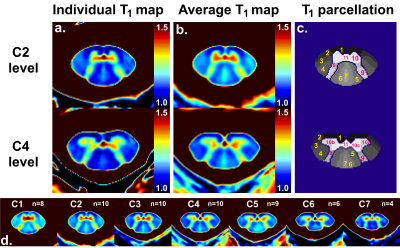 |
In vivo visualization of white and gray matter sub-structures using fast quantitative T1 mapping of the human spinal cord at 7T with 300-µm in-plane resolution
Aurélien Massire, Henitsoa Rasoanandrianina, Manuel Taso, Arnaud Le Troter, Maxime Guye, Jean-Philippe Ranjeva, Virginie Callot
T1 mapping of the cervical spinal cord at 7T with a 300-µm in-plane resolution using the MP2RAGE sequence is presented. A semi-automated post-processing pipeline enabled group studies (n=10 healthy subjects) with quantitative evaluation within small spinal cord sub-structures from C1 to C7, explored for the first time using T1 mapping. This work lays the groundwork for improved characterization of spinal cord degeneration using 7T MRI, by allowing investigation within particular relevant regions such as the anterior/posterior gray matter horns and ventromedial white matter tracts. It also offers new perspectives to build dedicated spinal cord substructures templates.
|
08:57
|
0669.
 |
Spinal cord MRI and fMRI at 16.4 T during spinal cord stimulation in rats: initial experience
Video Permission Withheld
Hanne Laakso, Lauri Lehto, Carlos Cuellar, Igor Lavrov, John Osborn, Shalom Michaeli, Silvia Mangia
This study aims at paving the way for spinal cord MRI and fMRI during spinal cord stimulation (SCS) in rats, with the goal of providing an experimental framework for assessing the impact of SCS on neuroplasticity and functional circuitry. MRI and fMRI of the spinal cord during SCS are extremely challenging due to motion and electrode-induced susceptibility artefacts. Here we demonstrated that high quality MRI and fMRI images of the spinal cord could be obtained at 16.4T during SCS with recently developed MB-SWIFT. This is the first study that attempts spinal cord fMRI during SCS in rats.
|
09:09
|
|
Clinical Applications of Spinal Cord MR
Did Not Present
Majda Thurnher
|
09:39
|
0670.
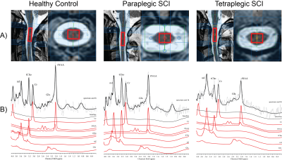 |
Spinal Cord MRS Biomarkers of Clinical Impairment in Chronic Spinal Cord Injury
Patrik Wyss, Eveline Huber, Patrick Freund, Anke Henning
Remote neurodegenerative changes above the level of injury are present following traumatic spinal cord injury (SCI), resulting in atrophy of the cervical cord of up to 30%. This study investigates the underlying biochemical changes at the cellular and molecular level which may subtend the development of atrophy using latest magnetic resonance spectroscopy in the spinal cord in healthy controls and SCI patients. Furthermore, we screen for potential MRS biomarkers investigating the association of biochemical changes and clinical outcome.
|
09:51
|
0671.
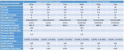 |
Comparison of cervical cord results from a quantitative 3D multi-parameter mapping (MPM) protocol of the whole brain with a dedicated cervical cord protocol
Rebecca Samson, Marco Battiston, Maryam Seif, Julien Cohen-Adad, Patrick Freund, Claudia Gandini Wheeler-Kingshott
We present a comparison of cervical cord metrics obtained using both a whole brain and dedicated cord implementation of a recently introduced quantitative multi-parametric MRI protocol which provides apparent proton density, R1, magnetisation transfer saturation (MTsat) and R2* maps sensitive to microstructural tissue changes in brain and spinal cord. Similar whole cervical cord (levels C1-C5) parameters were obtained using either protocol, and inter-subject variation was low, however in order to investigate tissue-specific cord parameters the dedicated cord protocol with higher in-plane resolution would be desirable.
|
10:03
|
0672.
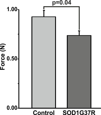 |
1H-[13C]-NMR Investigations of Neuronal and Astroglial Metabolic Activity in Spinal Cord and Brain in Amyotrophic Lateral Sclerosis
Anant Patel, Madhuri Puvvada, TK Sampath Kumar
Amyotrophic lateral sclerosis (ALS) is neurodegenerative disorder resulting from selective loss of both upper and lower motor neurons leading to progressive muscle weakness and paralysis. In the present study, we have used 1H-[13C]-NMR spectroscopy in conjunction with infusion of [1,6-13C2]glucose to evaluate neurometabolic activity in SOD1G37R mouse model of ALS. Our finding suggest reduced levels of glutamate, NAA, NAAG, and increased concentration of myo-inositol in spinal cord. More interestingly, the metabolic activity of glutamatergic and GABAergic neurons is decreased in the spinal cord, while it was increased in the cerebral cortex indicating strikingly different pathology in spinal cord and brain.
|
10:15
|
|
Adjournment & Meet the Teachers |
|
