|
From Molecular Biomarker to Clinical Quantification
Combined Educational & Scientific Session
ORGANIZERS: Arvind Pathak, Kannie WY Chan, Damian Tyler
Wednesday, 20 June 2018
| S01 |
13:45 - 15:45 |
Moderators: Arvind Pathak, Kannie WY Chan |
Skill Level: Intermediate
Session Number: W-06
Overview
This course will provide an overview of the development and use of molecular biomarkers in preclinical or clinical settings. In particular, endogenous/exogenous molecular biomarkers that link the molecular basis of disease with the emergent phenotype and enable clinical readouts that facilitate accurate diagnosis will be discussed. Topics will encompass underlying contrast mechanisms, pharmacokinetics, biomarker quantification and applications of biomarkers for early disease diagnosis, disease staging, and disease- and patient-specific treatment planning and monitoring.
Target Audience
Clinicians, basic and MR scientists, and applications developers interested in the development and translation of molecular biomarkers.
Educational Objectives
As a result of attending this course, participants should be able to:
-Describe the development of current molecular biomarkers;
-Discuss the relationship between the imaging readout and biological correlates; and
-Summarize issues regarding quantification of molecular biomarkers and the clinical utility of molecular imaging.
13:45
|
|
 Technical Aspects of Biomarker Development & Application Technical Aspects of Biomarker Development & Application
Peter Caravan
|
14:20
|
|
Clinical Translation & Utility of Biomarkers
Pek-Lan Khong
Molecular imaging biomarkers hold the promise to provideunparalleled opportunities for elucidating disease phenotypes, understanding pathophysiology, facilitating development of targeted therapies and driving personalised medicine. To realise the full potential of imaging biomarkers, our scientific community is compelled to translate them into clinical research and practice. This talk will present the current and emerging imaging biomarkers in clinical practice, including MRI and hybrid-PET imaging biomarkers, and describe the applications, strategies, opportunities and challenges in clinical translation.
|
14:55
|
0778.
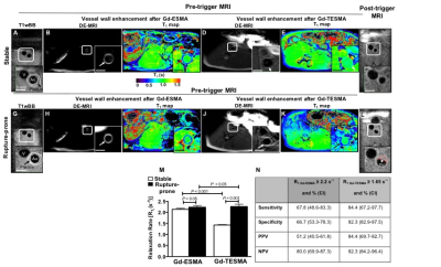 |
Tropoelastin: A novel marker for atherosclerotic plaque instability
Alkystis Phinikaridou, Sara Lacerda, Begoña Lavin, Marcelo Andia, Alberto Smith, Prakash Saha, René Botnar
Elastolysis and ineffective elastogenesis favour the accumulation of tropoelastin, rather than cross-linked elastin, in atherosclerotic plaques and could provide a new marker for plaque progression and instability. We developed a novel tropoelastin-binding gadolinium-based MRI contrast agent and demonstrated the feasibility of molecular imaging of tropoelastin in rabbits. Rupture-prone plaques had significantly higher uptake of the contrast agent compared with stable plaques and quantitive assessment of tropoelastin allowed detection of rupture-prone plaques with high sensitivity, specificity, positive and negative predictive values. Ex vivo analyses confirmed the MRI findings and showed that uptake of the contrast agent was specific for tropoelastin.
|
15:05
|
0779.
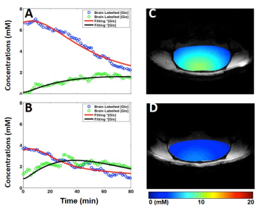 |
Quantitative 3D Deuterium MRS Imaging of Glucose Metabolisms in the Rat Brain
Video Permission Withheld
Ming Lu, Xiao-Hong Zhu, Yi Zhang, Wei Chen
Assessment of metabolic coupling between glycolysis and oxidation is crucial for understanding neuroenergetics. Based on our recently developed in vivo Deuterium (2H) MR (DMR) spectroscopic approach, we further developed a quantitative DMR imaging method for simultaneous measuring the glucose consumption rate (CMRglc) and TCA cycle flux (VTCA) in rat brain at 16.4 T. Regional metabolic rates were quantified and compared between two brain conditions. The metabolite images were also generated. This work demonstrates the feasibility and reliability of in vivo DMR imaging for assessing glucose metabolisms, which makes it possible to image CMRglc and VTCA in brains under different states.
|
15:15
 |
0780.
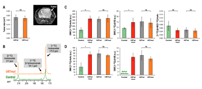 |
Hyperpolarized 13C ß-hydroxybutyrate/acetoacetate as a biomarker for non-invasive monitoring of NAD+/NADH status in glioblastoma
Chloe Najac, Marina Radoul, Lydia Le Page, Georgios Batsios, Pavithra Viswanath, Anne Marie Gillespie, Sabrina Ronen
Conversion of acetoacetate (AcAc) to β-hydroxybutyrate (β-HB) by the mitochondrial enzyme β-hydroxybutyrate dehydrogenase depends upon NADH availability. Previous studies have shown the potential of β-HB-to-AcAc ratio to reflect the redox state in rat hearts and lymphoma cells. Here, we assessed the value of HP [1,3-13C]-AcAc in brain. We demonstrated the potential to probe AcAc to β-HB conversion in normal mice and mice with glioblastoma. Significantly higher levels of [1-13C]-β-AcAc and [1-13C]-β-HB were observed in tumor-bearing mice compared to control mice. Consistent with lower levels of NADH measured in tumors, the [1-13C]-β-HB-to-[1-13C]-β-AcAc ratio trended towards a decrease compared to normal brain.
|
15:25
|
0781.
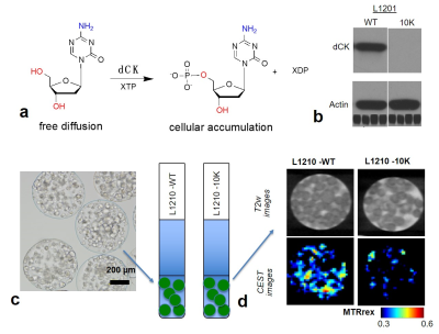 |
CEST MRI detection of Deoxycytidine Kinase activity using natural deoxycytidine
Zheng Han, Jia Zhang, Yuguo Li, Jing Liu, Peter van Zijl, Guanshu Liu
To assess the activity of deoxycytidine kinase (dCK), a drug-resistance-determining enzyme, we developed a new CEST MRI approach, in which the natural substrate, deoxycytidine (dC), is directly used as a CEST MRI reporter. Both in vitro and in vivo results showed that, upon the incubation or injection of dC, dCK(+) tumor cells have significantly higher CEST contrast at 2 ppm than dCK(-) cells, attributed to the accumulation of phosphorylated dC by dCK activity. Because dC is a clinically available agent, this ‘natural” MR molecular imaging method has great potential to be quickly translated to the clinic for patient stratification.
|
15:35
|
0782.
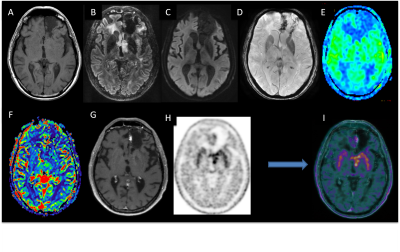 |
PET-MR multiparametric imaging biomarkers for differentiating between progression and radionecrosis of brain tumors
Nadya Pyatigorskaya, Marc Bertaux, Brian Sgard, Lydia Yahia-cherif, Marine Soret, Marie-Odile Habert, Didier Dormont, Damien Galanaud, Aurelie Kas
The aim of this work was evaluating the diagnostic accuracy of PET-MRI in difficult cases of differentiating between tumor progression and radionecrosis in neuro-oncology. For each lesion, PET (SUVmax, SUV mean, SUVpeak) and MRI (ADC, CBV, CBF, pCASL CBF) biomarkers were extracted. The combination of PET and MRI biomarkers allowed to improve the diagnostic accuracy. The logistic regression model has shown that 94% cases were correctly classified using the combination of SUVpeak and pCASL rCBF. Excellent diagnostic accuracy was achieved for both qualitative and quantitative evaluation by means of combined analysis of morphological, functional and metabolic imaging markers.
|
15:45
|
|
Adjournment & Meet the Teachers |
|
