|
Studying the Value of MRI
Combined Educational & Scientific Session
ORGANIZERS: Vikas Gulani, James Pipe
Tuesday, 19 June 2018
| S03 |
08:15 - 10:15 |
Moderators: Vikas Gulani, James Pipe |
Session Number: W-08
Overview
This session continues the work started in the ISMRM Value Initiative, introduced in previous ISMRM meetings. The session will begin with an introduction, followed by two long format talks. One speaker will discuss how the The role of comparative effectiveness and outcomes research in studying the value of MRI. A second speaker will discuss the Value of MR Imaging in the context of the world’s underserved populations, with the focused example of the Indian MRI Mission. After these two talks, the session will continue into proffered papers on studying the value of MRI from multiple perspectives.
Target Audience
Scientists and physicians interested in determining or improving the value of MRI in the healthcare system, either from a technical or a clinical perspective.
Educational Objectives
As a result of attending this course, participants should be able to:
-Describe principles of cost-effectiveness research;
-Identify methods of measuring value and outcomes in cost effectiveness research
-Identify commonly accepted criteria for cost effectiveness and how these can be applied to MRI
-Understand the development and implementation of cutting edge strategies for high value MRI protocols using existing sequences
-Identify successful pathways to technological advances in MRI that lead to rapid comprehensive MRI protocols or MRI exams which provide clinically valuable information that was not previously available.
-Recognize differences in definitions of value of imaging in the between the world’s underserved populations and those in the developed world
-Identify differences in needs from MRI between underserved populations and those in the developed world
-Identify and evaluate the goals of the Indian MRI Mission
08:15
|
|
 Measuring the Value of MRI: Comparative Effectiveness & Outcomes Research Measuring the Value of MRI: Comparative Effectiveness & Outcomes Research
Stella Kang
With growing emphasis on value in health care, there is a need to assess the effects of MRI use on patient health outcomes. The value, or the health outcomes relative to the cost, can be studied using established comparative effectiveness research and outcomes assessment methods. The synthesis of clinical context with test use also allows for analyses of personalized decision making based on test information. The major methods described will include decision analysis, cost effectiveness, and study of intermediate outcomes. These methods can allow for quantification of population level health benefits, comparison with other tests or interventions, and identification of research priorities based on predicted impact.
|
08:37
|
|
The Meaning of MR Value to Underserved Populations: Thoughts & Initial Technical Experience
Sairam Geethanath
|
08:59
|
|
Questions |
09:05
 |
0347.
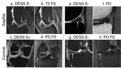 |
Diagnostic Comparison of Two Rapid Knee MRI Protocols for Comprehensive Whole-Joint Assessment: A Multi-Reader Feasibility Study
Akshay Chaudhari, Bragi Sveinsson, Jeff Wood, Kathryn Stevens, Christopher Beaulieu, Edwin Oei, Jarrett Rosenberg, Evan Levine, Feliks Kogan, Marcus Alley, Garry Gold, Brian Hargreaves
There were approximately 1.25 million clinical knee magnetic resonance imaging (MRI) scans performed in the US annually. Most knee MRI protocols require approximately 30 minutes of scan time. However, there is interest in expedited imaging protocols, especially under the ISMRM Value Initiative. Through a study involving 35 patients and 5 readers, we have demonstrated the feasibility of (1) a single 5-minute DESS sequence and (2) a 5-minute DESS sequence paired with a 2-minute coronal PDFS sequence, as two potential methods for accurate, rapid, and comprehensive diagnostic whole-joint knee MRI.
|
09:10
|
0348.
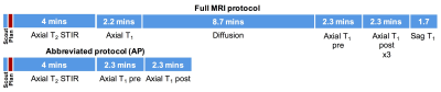 |
How Fast is “Fast MRI” for Breast Cancer Screening?
Emily Conant, Arijitt Borthakur, Mitchell Schnall, Susan Weinstein
We compare the scan and total study times from a newly implemented abbreviated MR protocol (AP) for breast cancer screening consisting of localizer, T2-STIR, and single pair of pre- and post-contrast 3D T1 sequences to similar times from our full MRI screening protocol. A retrospective analysis was performed using scan time data obtained from image dicom files as well as data from the Radiology Information System (RIS) for technologist activity times to determine the variance between the two protocols. The results of this study will help guide operational value improvements and estimates for appropriate pricing for the newly implemented AP for breast cancer screening.
|
09:15
|
0349.
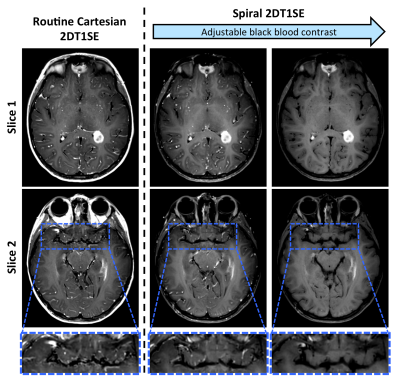 |
Benefits and Challenges of Spiral MRI in Routine Clinical Brain Imaging: Early Results
Melvyn Ooi, Zhiqiang Li, Dinghui Wang, Ryan Robison, Nick Zwart, Ashley Anderson, Akshay Bakhru, Tanya Mathews, Suthambhara Nagaraj, Silke Hey, Jos Koonen, Ad Moerland, Jonathan Chia, Ivan Dimitrov, Harry Friel, Makoto Obara, Indrajit Saha, Yi Wang, Yansong Zhao, Harry Hu, Amber Pokorney, Marco Pinho, Osamu Togao, Tom Chenevert, Ashok Srinivasan, Juan Small, Mara Kunst, Rakesh Gupta, Jalal Andre, Nandor Pinter, Jeffrey Miller, James Pipe
Spiral MRI possesses several advantages vs. Cartesian MRI, due to differences in their k-space trajectories and underlying MR physics, which can be leveraged for added value in routine clinical imaging. A Spiral Neuroimaging Cooperative, consisting of nine clinical sites, was formed for the multi-center evaluation of spiral MRI as an alternative to Cartesian MRI in routine clinical imaging. Post-contrast brain spiral 2DT1SE were compared with Cartesian 2DT1SE or TSE. Spirals demonstrated faster scanning with consistent flow artifact reduction vs. both Cartesian options, and superior overall image quality (T1 contrast, lesion visualization) vs. TSE.
|
09:20
|
0350.
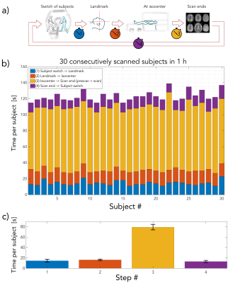 |
30 brain MRI exams in 1 hour using a multi-contrast EPI sequence
Stefan Skare, Tim Sprenger, Ola Norbeck, Henric Rydén, Enrico Avventi, Lars Blomberg, Johan Berglund, Mikael Skorpil, Maria Sandell, Mathias Engström
An in-house developed multi-contrast EPI sequence producing six MR contrasts (T1-FLAIR, T2-w, DWI, ADC, T2*-w, and T2-FLAIR) was used as a fast brain MRI protocol. Seven healthy volunteers were scanned repeatedly to investigate how many brain MRI exams that can be performed over the course of one hour. With a net scan time of one minute and an additional minute for table movements and switch of subjects, it was possible to perform 30 brain MRI examinations in 1 h.
|
09:25
|
0351.
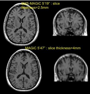 |
Rapid Synthetic MRI of the whole brain using simultaneous multi-slice technique
Suchandrima Banerjee, Graeme McKinnon, Marcel Warntjes
Synthetic MRI enables reconstruction of multiple MRI contrasts from a single scan based on voxel-wise computation of relaxation parameters. This can improvement scan productivity and workflow considerably. The utility of synthetic MRI has recently been demonstrated in clinical settings. However in its current implementation whole brain coverage can be achieved within 5 minutes only if a thick slice prescription (~ 4mm) is used, but this might provide insufficient through-plane resolution for certain clinical protocols and can also lead to partial voluming effects. This work explores the simultaneous multislice approach for more efficient through plane coverage with synthetic MRI.
|
09:30
|
0352.
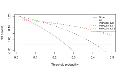 |
Decision Curve Analysis for Prostate Biopsy using MRI with or without Dynamic Contrast Enhancement
Vinay Prabhu, Andrew Rosenkrantz, Stella Kang
Prostate MRI with dynamic contrast enhancement (DCE) is controversial with growing interest in noncontrast (NC) MRI. Decision curve analyses performed on data from two contemporary studies identified ranges of high grade cancer probability thresholds at which DCE had the highest net benefit. DCE MRI has the most benefit in moderate or moderate-high risk threshold ranges, thought to vary based on the study differences in disease prevalence, reference standard, and the presence or absence of additional DCE reconstruction images. The risk ranges in which NC versus DCE MRI optimize net benefit may help inform the best clinical use of prostate MRI.
|
09:35
|
0353.
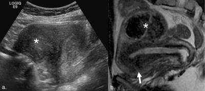 |
Impact of Routine Magnetic Resonance Imaging on Diagnosis and Treatment of Women with Benign Gynecologic Conditions Seen in a Multidisciplinary Fibroid Center
Kim Nhien Vu, Angela Fast, Robyn Shaffer, Deirdre Lum, Berta Chen, David Hovsepian, Pejman Ghanouni
Pelvic ultrasounds often represent the first and only line of imaging for uterine fibroid evaluation. Our study aims to determine the added value of routine pelvic magnetic resonance imaging (MRI) on diagnosis and treatment of women presenting with symptomatic uterine fibroids. A retrospective review was performed on 569 consecutive women referred to our multidisciplinary fibroid center over a three-year period. Compared to ultrasound alone, MRI affected diagnosis in over a third of patients, which also altered treatment options. These findings justify the use of routine pelvic MRI in women with symptomatic fibroids, particularly those presenting with dysmenorrhea.
|
09:40
|
0354.
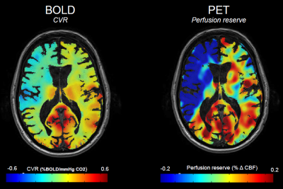 |
Staging hemodynamic failure: BOLD fMRI cerebrovascular reactivity beats [15O]-H2O-PET.
Marco Piccirelli, Christiaan van Niftrik, Geoffrey Warnock, Susanne Wegener, Athina Pangalu, Giuseppe Esposito, Antonios Valavanis, Alfred Buck, Andreas Luft, Oliver Bozinov, Luca Regli, Jorn Fierstra
The actual gold standard stroke risk assessment is cerebral blood flow (CBF) measured with [15O]-H2O-PET, which is available only in few specialized centers and requires ~1mSv radioactive dose. Otherwise, quantitative CVR measurements derived (with our temporal decomposition iterative algorithm) from Blood-Oxygen-Level-Dependent (BOLD) fMRI with CO2 challenge might apply as a surrogate imaging-marker for hemodynamic failure, as the <7 minutes acquisition can easily be clinically implemented, and has – as proved in this study – twice higher specificity*sensitivity for staging hemodynamic failure in chronic cerebrovascular steno-occlusive diseases than PET.
Therefore, BOLD CVR shall be widely implemented for assessing stroke risk.
|
09:45
|
0355.
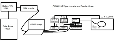 |
A solar powered MR spectrometer system
Martyn Paley
Most MRI systems have very high electrical power consumption and require extensive cryogenic support systems. A spectrometer design has been developed for a low cost, low weight and low power specialised MR system capable of ‘unplugged’ operation for worldwide use in remote locations.
|
09:50
|
0356.
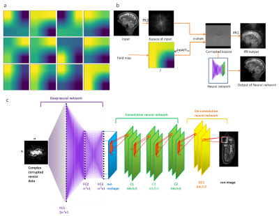 |
REconstruction of MR images acquired in highly inhOmogeneous fields using DEep Learning (REMODEL)
Punith Venkate Gowda, Asha Kumara Swamy, Sachin Jambawalikar, Sairam Geethanath, Thomas Vaughan
The aim of this study was to develop and demonstrate a supervised learning algorithm to reconstruct MR images acquired in highly in-homogeneous magnetic fields. Brain images were used to train a deep neural network. This was performed for image sizes of 32 x 32 and 64 x 64. Results obtained demonstrate REMODEL’s ability to reconstruct the images obtained in in-homogeneous magnetic fields of up to ±50 kHz with high fidelity. The root-mean-square-error for these reconstructions compared to the uncorrupted ground truth was lesser than 0.15 and significantly lesser than the corrupted images.
|
09:55
|
0357.
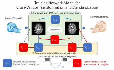 |
Creating Standardized MR Images with Deep Learning to Improve Cross-Vendor Comparability
Enhao Gong, John Pauly, Greg Zaharchuk
A very common task for radiologists is to compare sequential imaging studies that were acquired on different MR hardware systems, which can be difficult and inaccurate because of different designs across scanners and vendors. Cross-vendor standardization and transformation is valuable for more quantitative analysis in clinical exams and trials. With in-vivo multi-vendor datasets, we show that it is possible to achieve accurate cross-vendor transformation using the state-of-art Deep Learning Style-transfer algorithm. The method preserves anatomical information while transferring the vendor specific contrast "style". The usage of unsupervised training enable the method to further train and apply on all existing large scale MRI datasets. This technique can lead to a universal MRI style which benefits patients by improving inter-subject reproducibility, enabling quantifiable comparison and pushing MRI to be more quantitative and standardized.
|
10:00
|
|
Panel Discussion |
10:15
|
|
Adjournment & Meet the Teachers |
|
