|
Weekend Educational Course
Body MRI: Realities & Controversies |
|
Body MRI: Realities & Controversies: Expanding the MRI Clinical Frontier
Weekend Course
ORGANIZERS: Kathryn Fowler, Kartik Jhaveri, Lorenzo Mannelli, Valeria Panebianco, Scott Reeder, Reiko Woodhams
Saturday, 16 June 2018
| S02 |
08:00 - 09:30 |
Moderators: Dow-Mu Koh, Mark Schiebler |
Skill Level: Intermediate
Session Number: WE-03A
Overview
This whole-day course provides an overview of current issues in MRI practice (safety, contrast, state-of-the-art imaging), areas for possible expansion, and educational sessions covering liver, pancreaticobiliary, and GI MRI. The final portion of the session attempts to provide a balanced view of new standardized lexicons that are used in MRI (BI-RADS, LI-RADS, PI-RADS).
Target Audience
This course is aimed at radiologists, imaging scientists and MR technologists who wish to review the state-of-art MRI protocols and indications for assessment of disease in the abdomen and pelvis.
Educational Objectives
As a result of attending this course, participants should be able to:
-Summarize new state-of-the-art equipment, sequences, and contrast agents used in Body MRI;
-Review areas of possible clinical practice expansion: thoracic MRI, emergency indications, and whole body imaging;
-Demonstrate the impact of MRI in diagnostic workup of liver and gastrointestinal disease processes; and
-Illustrate the role of standardized lexicon systems through a point and counterpoint approach.
08:00
|
 |
 Current State of the Art Current State of the Art
Hersh Chandarana
Abdominal or Body MRI suffers from number of limitations such as (1) slow and inefficient acquisition (2) sensitivity to motion related artifacts and (3) limited volumetric coverage and spatial resolution which are constrained by the breath-hold capacity of the patients. Various methods are used clinically and are being investigated to overcome these limitations in order to permit motion robust and rapid imaging of the abdomen. This talk will briefly discuss some of these methods for (1) fast imaging and (2) motion compensated imaging of the abdomen.
|
08:22
|
|
Non-Vascular Thoracic MRI
Did Not Present
Constantine Raptis
Despite advances in imaging unit technology and research in the field, the utilization of thoracic MRI in regular clinical practice for noncardiac and nonangiographic applications has lagged behind expectations. While there are several clinical scenarios in which nonvascular thoracic MRI has a potential primary role, it is at present most frequently utilized as a problem solving modality to answer specific questions that cannot be determined via other imaging modalities, most commonly CT. The purpose of this talk is to explore commonly encountered situations in which thoracic MRI can be utilized to its fullest extent as a problem solving modality.
Substituted presentation:
PET/MRI: Abdominal Indications
Kathryn Fowler, M.D.
|
08:44
|
 |
Body MR in ED
Bobby Kalb
Magnetic resonance imaging is increasingly being used in the emergency department setting, and offers potential advantages related to safety, diagnostic accuracy and efficiency, compared to the more commonly utilized modalities of CT and ultrasound. This presentation will describe methods for advantages of performing MRI in the ED for acute abdominopelvic pain and also PE, in addition to detailing suggested imaging protocols and techniques.
|
09:06
|
|
Whole-Body Imaging
Taro Takahara
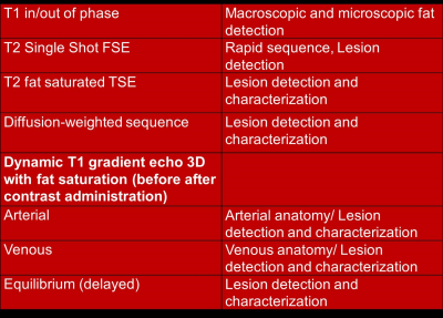
Whole-body Imaging heavily relies on the added value of diffusion-weighted imaging, especially with the use of a STIR pre-pulse (DWIBS). The STIR pre-pulse not only provides fat suppression but also suppression of intestinal contents and improved contrast between cancers and surrounding tissue. It can be used in a similar way as multi-slice CT, since we use the same data set for full/thick/thin MIP reconstructions in any direction. Nevertheless, DWIBS should be used in combination with other sequences such as T2WI (particularly suitable for fusion images), 3D thin fat-suppressed T1WI (differentiation of cancerous lesions and intestinal contents), and in-phase/out-of-phase images (differentiation of red bone marrow from bone metastasis). Furthermore, we can use MRCP-like sequences for visualizing the distribution of ascites. Cine MRI is also a useful tool for the detection of adhesions and MR urography. In the last part of this presentation, quantitative analysis of ADC and tumor volume, and MET-RADS-P will be discussed.
|
09:30
|
|
Break & Meet the Teachers |
|
| |
|
Body MRI: Realities & Controversies: Liver Imaging
Weekend Course
ORGANIZERS: Kathryn Fowler, Kartik Jhaveri, Lorenzo Mannelli, Valeria Panebianco, Scott Reeder, Reiko Woodhams
Saturday, 16 June 2018
| S02 |
10:00 - 11:30 |
Moderators: Shetal Shah, Bachir Taouli |
Skill Level: Intermediate
Session Number: WE-03B
Overview
This whole-day course provides an overview of current issues in MRI practice (safety, contrast, state-of-the-art imaging), areas for possible expansion, and educational sessions covering liver, pancreaticobiliary, and GI MRI. The final portion of the session attempts to provide a balanced view of new standardized lexicons that are used in MRI (BI-RADS, LI-RADS, PI-RADS).
Target Audience
This course is aimed at radiologists, imaging scientists and MR technologists who wish to review the state-of-art MRI protocols and indications for assessment of disease in the abdomen and pelvis.
Educational Objectives
As a result of attending this course, participants should be able to:
-Summarize new state-of-the-art equipment, sequences, and contrast agents used in Body MRI;
-Review areas of possible clinical practice expansion: thoracic MRI, emergency indications, and whole body imaging;
-Demonstrate the impact of MRI in diagnostic workup of liver and gastrointestinal disease processes; and
-Illustrate the role of standardized lexicon systems through a point and counterpoint approach.
10:00
|
 |
Imaging of the Portal Venous System
Reena Jha
Radiologists commonly encounter portal venous abnormalities. Understanding the varying appearances of acute and chronic portal venous thromboses and the associated changes of hepatic enhancement patterns is important for patient management. Furthermore, it is important to recognize that portal venous thrombosis may change the contour of the liver, and simulate cirrhosis. Congenital and acquired chronic portal vein thrombosis may lead to the development of hepatic masses, and may alter the appearance of the bile ducts and mimic malignancy.
|
10:22
|
|
Liver Lesions: Malignant
Thomas Vogl
In summary, liver imaging has found its place in the reality of cross-sectional imaging. The use of contrast agent is still controversial. Here further developments are directed in order to improve the qualitative and quantitative information. Artificial intelligence techniques will integrate multiparametric MRI information in the concept of radionics and radiogenomics.
|
10:44
|
|
Biliary Imaging & Pathology
Mi-Suk Park
In this talk, I will present differential diagnosis of biliary lesions based on imaging phenotypes with pathologic correlation. And then TNM staging with pathologic correlation will be reviewed based on the 8th edition of NCCN guidelines.
|
11:06
|
|
Lunch & Meet the Teachers |
|
| Back |
| The International Society for Magnetic Resonance in Medicine is accredited by the Accreditation Council for Continuing Medical Education to provide continuing medical education for physicians. |
