|
Weekend Educational Course
Advanced fMRI: Connectivity & Cutting Edge |
|
Advanced fMRI: Connectivity & Cutting Edge: Part 1
Weekend Course
ORGANIZERS: Richard Buxton, Benedikt Poser, Joshua Shimony
Saturday, 16 June 2018
| N03 |
13:15 - 14:30 |
Moderators: Shella Keilholz, Benedikt Poser |
Skill Level: Intermediate to Advanced
Session Number: WE-09A
Overview
This is a two-part session that addresses advanced topics in connectivity, from analysis to applications. It also provides an overview on the cutting edge of fMRI acquisition beyond standard 2D BOLD fMRI, including combination with advanced non-fMRI neurotechniques.
Target Audience
The course is tailored to an audience of neuroscientists, neuroradiologists, clinicians and imaging scientists who interested connectivity analysis and its applications, as well as any scientists wanting to learn about the current frontiers in BOLD and non-BOLD acquisition methodology. The course assumes background knowledge on connectivity analysis, as well as basic understanding of BOLD imaging methods.
Educational Objectives
As a result of attending this course, participants should be able to:
-Compare and contrast connectivity analysis strategies;
-Demonstrate advanced connectivity data analysis;
-Describe cortical parcellation methods based on resting state data;
-Describe how connectivity analysis can be applied in disease populations;
-Discuss the latest alternatives to regular 2D EPI based BOLD acquisition;
-Choose the best sampling strategy (2D EPI, 3D EPI, SMS-EPI) for a target application;
-Choose the best fMRI signal mechanism (BOLD, CBF, VASO) for a target application; and
-Explain the challenges related to combining BOLD fMRI with other advanced neurotechniques.
13:15
|
|
 Connectivity: Analysis Connectivity: Analysis
Mark Lowe
An overview of current analysis methods for assessing functional connectivity using resting state fMRI data. A brief review of important preprocessing steps necessary for quality resting state data as well as various complex network analysis methods, including structural equation modeling, clustering methods and graph theoretic methods. Dynamic functional connectivity methods are briefly discussed.
|
13:40
|
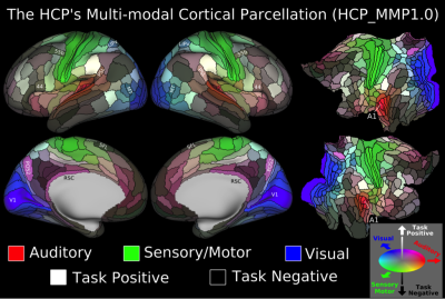 |
A Multi-modal Parcellation of Human Cerebral Cortex
Matthew Glasser
We will discuss the Human Connectome Project’s multi-modal cortical parcellation version 1.0—the data acquisition and analysis requirements, how the parcellation was made, and how it can be applied to individuals. This state of the art map of the cerebral cortex was made possible by using exceptionally high quality MRI data precisely aligned across individuals. Cortical areal boundaries were identified when visible in multiple modalities and areas were painstakingly related to the neuroanatomical literature. A machine learning classifier was then trained to automatically identify each cortical area based on its multi-modal fingerprint in individual subjects, replicating the parcellation.
|
14:05
|
 |
fMRI Connectivity, Depression, and Anhedonia: a Bayesian network analyses in Schizophrenia, Bipolar Disorder, ADHD, and Healthy Controls
Ariana Anderson, Mirella Diaz-Santos, Levon Demirdjian, Spencer Frei, Robert Bilder
Although MRI, fMRI, and genetic biomarkers have been implicated in depression, it is unclear how much these measures illuminate the disorder compared to behavioral and demographic measurements. Using these measurements, we predicted depressive symptoms to compare the effect size of these modalities. In 119 subjects with a diagnosis of Bipolar disorder (n=43), Schizophrenia (n=39), and ADHD (n= 37), random forests models predicted both generalized depression (Hopkin Symptom Checklist) and anhedonia-specific measures (Chapman Scales for Physical and Social Anhedonia) using genetic, structural MRI volumetric measures, resting-state fMRI network connectivity measures, demographic features, and behavioral assessments measures. We found that comorbid behavioral symptoms accounted for nearly 75% of predictive ability. When removing behavioral assessments, only 8.75% of variance in the depression symptom scores were predictable from neuroimaging, demographic, genetic measures. Demographics retained the strongest predictive ability.
|
14:30
|
|
Break & Meet the Teachers |
|
| |
|
Advanced fMRI: Connectivity & Cutting Edge: Part 2
Weekend Course
ORGANIZERS: Richard Buxton, Benedikt Poser, Joshua Shimony
Saturday, 16 June 2018
| N03 |
15:15 - 16:55 |
Moderators: Shella Keilholz, Benedikt Poser |
Skill Level: Intermediate to Advanced
Session Number: WE-09B
Overview
This is a two-part session that addresses advanced topics in connectivity, from analysis to applications. It also provides an overview on the cutting edge of fMRI acquisition beyond standard 2D BOLD fMRI, including combination with advanced non-fMRI neurotechniques.
Target Audience
The course is tailored to an audience of neuroscientists, neuroradiologists, clinicians and imaging scientists who interested connectivity analysis and its applications, as well as any scientists wanting to learn about the current frontiers in BOLD and non-BOLD acquisition methodology. The course assumes background knowledge on connectivity analysis, as well as basic understanding of BOLD imaging methods.
Educational Objectives
As a result of attending this course, participants should be able to:
-Compare and contrast connectivity analysis strategies; -Demonstrate advanced connectivity data analysis;
-Describe cortical parcellation methods based on resting state data;
-Describe how connectivity analysis can be applied in disease populations;
-Discuss the latest alternatives to regular 2D EPI based BOLD acquisition;
-Choose the best sampling strategy (2D EPI, 3D EPI, SMS-EPI) for a target application;
-Choose the best fMRI signal mechanism (BOLD, CBF, VASO) for a target application; and
-Explain the challenges related to combining BOLD fMRI with other advanced neurotechniques.
15:15
|
|
BOLD Acquisition Beyond 2D EPI
Wietske van der Zwaag
Although the vast majority of fMRI studies is still performed with 2D-EPI, there are several other BOLD-sensitive sequences, available on most clinical platforms, that may perform better. The main alternatives to 2D-EPI are 3D-EPI and SMS-EPI, although EVI, MR-encephalography, ME-EPI and SE-EPI have also recently gathered interest. All these sequences will be discussed and compared to one another in terms of their strengths, weaknesses and artifacts. Specific situations in which a specific sequence would be preferred will be used to highlight the relevant strong points.
|
15:40
|
|
fMRI Acquisition Beyond BOLD
J. Jean Chen
Although the BOLD signal has been the workhorse of fMRI, BOLD fMRI is limited by its intrinsic T2/T2* sensitivity and exhibits exaggerated weighting towards large veins. Moreover, the BOLD signal is a relative rather than quantitative measure of brain function that depends on the interplay of perfusion and oxygenation, leaving room for ambiguous interpretation. This talk will summarize recent efforts to explore alternative fMRI methods, including those based on blood flow, blood volume and blood oxygenation. The applications of these methods in both task-based and resting-state studies will be introduced.
|
16:05
|
|
High Resolution Applications: Cortical Layers
Jonathan Polimeni
Laminar fMRI refers to the study of functional activation within the cerebral cortex, with the goal of detecting distinct functional activity within cortical layers, and is an emerging application of high-resolution fMRI. Although individual cortical layers cannot be resolved with current human fMRI techniques, and hemodynamic coupling and variation of fMRI signals across layers is incompletely understood, because of the roles cortical layers play in distributed neuronal processing measuring layer-specific activation is key to understanding brain circuitry, which motivates work towards surmounting these difficulties. This presentation will introduce laminar fMRI, summarize recent advances, and focus on challenges faced when interpreting these data.
|
16:30
|
 |
Combining fMRI with Advanced Neurotechniques
Xin Yu
By combining fMRI with fiber optic calcium recording and optogenetics, as well as two-photon microscopy or convetional optical imaging techniques, the circuit-specific regulatory mechanism of the unique neuro-glial-vascular (NGV) interaction model can be studied at varied brain states in animals. Combine fMRi with advanced neurotechniques provides very powerful methodological platforms to deepnen our understanding how enural circuits mediate specific behavioral outputs, as well as link the cellular mechaism and neural circuit regulation (causality) to the systems level correlation (e.g. fMRI) to behavioral index.
|
16:55
|
|
Adjournment & Meet the Teachers |
|
| Back |
| The International Society for Magnetic Resonance in Medicine is accredited by the Accreditation Council for Continuing Medical Education to provide continuing medical education for physicians. |
