|
Weekend Educational Course
Cardiovascular MRI: Vascular |
|
Cardiovascular MRI: Vascular: Part 1
Weekend Course
ORGANIZERS: James Carr, Tim Leiner
Sunday, 17 June 2018
| S01 |
08:00 - 09:20 |
Moderators: Alex Barker, Christopher François |
Skill Level: Basic to Intermediate
Session Number: WE-15A
Overview
This course highlights the components of a routine vascular exam and provides an overview of widely accepted and cutting-edge techniques for vascular imaging, including clinical applications.
Target Audience
Attendees, both physicians and scientists, who have an interest in evaluating cardiovascular disease with MRI in order to gain an in-depth perspective on the technical foundations, clinical needs, and research promises of both flow imaging, vessel wall assessment and magnetic resonance angiography techniques in order to improve their clinical practice.
Educational Objectives
As a result of attending this course, participants should be able to:
-Develop a working knowledge of a basic vascular exam and in its clinical and research applications;
-Describe clinical applications of vascular imaging techniques; and
-Explain the role of MRI for assessing atherosclerosis.
08:00
|
|
 Contrast Agents: Practical Use & Safety Aspects Contrast Agents: Practical Use & Safety Aspects
Giles Roditi
|
08:20
|
|
Contrast-Enhanced MRA
Jeremy Collins
|
| 08:35 |
|
SAMs: Contrast-Enhanced MRA
|
08:40
|
|
Non-Contrast-Enhanced MRA
Ioannis Koktzoglou
This presentation will review established and emerging methods for non-contrast-enhanced magnetic resonance angiography.
|
09:00
|
|
Basics of Flow Imaging Including Extraction of Quantitative Measurements
Jos Westenberg
Time-of-Flight and Phase-Contrast imaging will be discussed for their role in aniography. Phase contrast imaging is sensitized to flow velocity, affecting the phase signal of flowing spins. This encoding of velocity enables flow velocity quantitation. Quantitative measures derived from velocity encoding will be discussed: valvular flow mapping with regurgitation assessment, transstenotic pressure drop, kinetic energy distribution and wall shear stress. Furthermore, some potential sources of error of phase contrast imaging will be discussed.
|
09:20
|
|
Break & Meet the Teachers |
|
| |
|
Cardiovascular MRI: Vascular: Part 2
Weekend Course
ORGANIZERS: James Carr, Tim Leiner
Sunday, 17 June 2018
| S01 |
10:00 - 11:40 |
Moderators: Alex Barker, Christopher François |
Skill Level: Basic to Intermediate
Session Number: WE-15B
Overview
This course highlights the components of a routine vascular exam and provides an overview of widely accepted and cutting-edge techniques for vascular imaging, including clinical applications.
Target Audience
Attendees, both physicians and scientists, who have an interest in evaluating cardiovascular disease with MRI in order to gain an in-depth perspective on the technical foundations, clinical needs, and research promises of both flow imaging, vessel wall assessment and magnetic resonance angiography techniques in order to improve their clinical practice.
Educational Objectives
As a result of attending this course, participants should be able to:
-Develop a working knowledge of a basic vascular exam and in its clinical and research applications;
-Describe clinical applications of vascular imaging techniques; and
-Explain the role of MRI for assessing atherosclerosis.
10:00
|
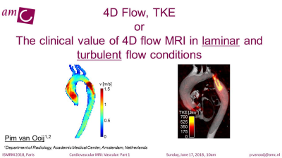 |
Advanced Techniques: 4D Flow, TKE
Pim van Ooij
The first part of the talk will show the derivation of clinically relevant parameters (velocity vectors, peak velcocity, wall shear stress) from 4D flow MRI data in laminar flow conditions. the second part of the talk will show the derivation of clinically relevant parameters (turbulent kinetic energy, turbulent wall shear stress) from 4D flow MRI data in turbulent flow conditions.
|
10:20
|
|
MR Techniques for stroke-related vessel wall imaging
Zhaoyang Fan
Stroke is one of the major causes of morbidity and mortality worldwide and is the number one cause of adult disability. This disease primarily arises from pathogenesis in a large blood vessel such as the aorta, carotid artery, and intracranial artery and venous sinus. Black-blood MR, commonly known as MR vessel wall imaging (VWI), has emerged as a leading noninvasive imaging modality for directly assessing the vessel wall. Many of previous studies have shown the promise of using MR VWI for characterizing different vessel wall pathologies that potentially result in a stroke. The present lecture will focus on recent (within the last decade) technical developments in MR VWI at the carotid artery, intracranial vessels, and aortic arteries.
|
10:40
|
 |
Cerebrovascular Vessel Wall Imaging from a Clinical Perspective
Anja van der Kolk
Vessel wall MRI of the supra-aortic and intracranial vasculature has seen an exponential increase in popularity in the last two decades. It can provide a wealth of information on pathologic processes of the vessel wall associated with cerebrovascular diseases – like atherosclerosis, vasculitis, Moyamoya disease and aneurysms – that may be used in the differential diagnosis of vasculopathies, in (stroke) risk assessment, and for planning individual patient-based treatment strategies. This lecture will discuss the clinical potential of vessel wall MRI in these cerebrovascular diseases, with a specific focus on intracranial vessel wall pathology.
|
11:00
|
 |
Chest & Abdominal
Iacopo Carbone
Cardiovascular MRI: Vascular: Part 2
The target audience of this lecture will be imagers (radiologists or cardiologists), dealing with vascular examinations on regular daily practice. Training doctors will particularly benefit from this lecture.
The presentation will give the basis on how to perform Vascular MRI study of the aorta.The main focus of the presentation will be the acute aortic syndromes, encompassing the whole spectrum of the topic from its pathophysiology and revised classification to different MRI protocols and diagnosis.
At the end of the presentation the audience will be able to recognize and classify the main acute aortic clinical scenarios, differentiating real emergencies from deferrable urgencies and to eventually integrate imaging techniques including CT and echocardiography.
|
11:20
|
|
Peripheral Vascular Disease
Jeremy Collins
|
11:40
|
|
Adjournment & Meet the Teachers |
|
| Back |
| The International Society for Magnetic Resonance in Medicine is accredited by the Accreditation Council for Continuing Medical Education to provide continuing medical education for physicians. |
