|
Weekend Educational Course
Introduction to Diffusion |
|
Introduction to Diffusion: Part 1
Weekend Course
ORGANIZERS: Stephan Maier, Jennifer McNab, Noam Shemesh
Sunday, 17 June 2018
| S04 |
08:00 - 09:30 |
Moderators: Dmitry Novikov, Jennifer McNab |
Skill Level: Intermediate
Session Number: WE-17A
Overview
This course will introduce the basic concepts of diffusion MRI. The course aims to introduce basic diffusion physics and how diffusion can be encoded using different gradient waveforms, the advantages/disadvantages of different diffusion MRI image acquisition techniques, and correction of artifacts. The course then focuses on the different models used to analyze diffusion MRI data and how to interpret the ensuing metrics.
Target Audience
Clinicians and scientists interested in diffusion MRI.
Educational Objectives
As a result of attending this course, participants should be able to:
-Describe basic diffusion imaging concepts;
-Recognize and correct for diffusion MR image artifacts; and
-Select appropriate models for analysis and interpret the ensuing metrics.
08:00
|
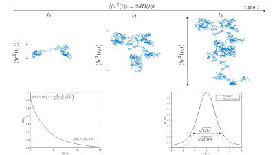 |
 The Physics of Diffusion: What Are We Measuring? The Physics of Diffusion: What Are We Measuring?
Sune Jespersen
This lecture covers the basic physics of diffusion. I cover the random walk as a conceptual model of diffusion as well as a tool for simulations. The relation for the mean square displacement is derived, and scenarios leading to time-dependent diffusivities are described, along with their universal short and long time regimes. A central quantity, the propagator, is introduced, and the diffusion equation describing its evolution derived. Examples of solutions are given, and the cumulant expansion as a general framework to describe diffusion in complex media is presented. The connection to the diffusion MR signal is outlined.
|
08:22
|
|
Diffusion Encoding Using MRI: Single Pulse, Double Pulse, Oscillating Gradients
Andrada Ianus, Ivana Drobnjak
|
08:44
|
|
Image Encoding for Diffusion MRI: EPI, Spiral, Radial, Multi-Shot, Etc.
Robert Frost
Single-shot echo-planar imaging, with parallel imaging and simultaneous multi-slice improvements, remains the most commonly used sequence for diffusion imaging of the brain. Many other sequences have been developed with the goal of increasing the spatial resolution of whole-brain diffusion data and their designs are influenced by several considerations including image artefacts, SNR and efficiency, scan time per image, motion sensitivity, and slice acquisition paradigm. These issues will be discussed and illustrated with examples of recent diffusion sequences.
|
09:06
|
 |
Diffusion MR Signal to Quality Data: Preprocessing & Distortion Correction
Stamatios Sotiropoulos
In this talk, I will present an overview of the issues we encounter in diffusion MRI data and the preprocessing steps needed prior to starting any data analysis. Geometric distortions due to off-resonance effects, distortions due to subject motion, artifacts from physiological noise, detection of outliers and quality control are some of the topics that will be covered. Correcting for artifacts and distortions is key in order to extract the most from the data and allow subsequent analysis in an unbiased and precise manner.
|
09:28
|
|
Break & Meet the Teachers |
|
| |
|
Introduction to Diffusion: Part 2
Weekend Course
ORGANIZERS: Stephan Maier, Jennifer McNab, Noam Shemesh
Sunday, 17 June 2018
| S04 |
10:00 - 11:30 |
Moderators: Chun-Hung Yeh, Jennifer McNab |
Skill Level: Intermediate
Session Number: WE-17B
Overview
This course will introduce the basic concepts of diffusion MRI. The course aims to introduce basic diffusion physics and how diffusion can be encoded using different gradient waveforms, the advantages/disadvantages of different diffusion MRI image acquisition techniques, and correction of artifacts. The course then focuses on the different models used to analyze diffusion MRI data and how to interpret the ensuing metrics.
Target Audience
Clinicians and scientists interested in diffusion MRI.
Educational Objectives
As a result of attending this course, participants should be able to:
-Describe basic diffusion imaging concepts;
-Recognize and correct for diffusion MR image artifacts; and
-Select appropriate models for analysis and interpret the ensuing metrics.
10:00
|
|
Diffusion Data Interpretation: Data-Driven
Jürgen Finsterbusch
The signal of diffusion-weighted MR reflects the tissue structure on a cellular or microstructure scale. Many different approaches have been proposed that aim to characterize diffusion-weighted data or derive diffusion or microstructural tissue properties from it. They could be divided into model- and data-driven approaches. Data-driven approaches either derive diffusion or structural properties directly from the data or, in a broader sense, approximate the data with equations that are “borrowed” from diffusion in simple physical systems or motivated mathematically. The most important of these approaches will be covered in this presentation.
|
10:22
|
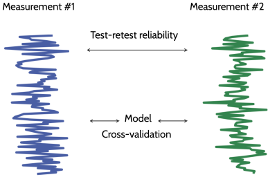 |
Diffusion Data Interpretation: Model-Driven
Ariel Rokem
Models of diffusion MRI are mathematical expressions that describe the data, summarize it and approximate it. The values of the fit parameters are used to interpret the data in light of the structure of the tissue or its physical properties. This presentation will introduce a framework for model comparison using cross-validation: the model is fit to one part of the data and the model parameters are used to predict the signal in another part of the data. Cross-validation is used to assess parameter reliability, as well as accuracy of the model with respect to the data.
|
10:44
|
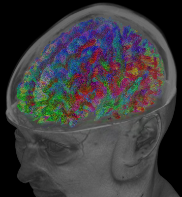 |
Tractography
Thijs Dhollander
In this introductory course on diffusion MRI based tractography, I will introduce the basics of tractography. I will by no means provide a comprehensive overview or review of all possible techniques and literature, but rather focus on the most essential basics of this area and a selection of its applications. As a result of attending this course, participants should be empowered to participate in the ongoing discussions about the strengths and limitations of tractography, as well as make informed and critical decisions on the use of tractography in their own work.
|
11:06
|
|
Microstructure for In-Vivo Human Applications
Susie Huang
This lecture will provide a brief overview of technical considerations involved in in vivo diffusion MR microstructural imaging studies with a focus on human neuroimaging. We will cover biophysical models and signal representations of the diffusion MRI signal as they relate to probing tissue microstructure in the human brain. We will study metrics derived using different diffusion models and acquisition schemes in the setting of normal development and pathological changes in white matter disease. In addition, the specific advantages of high-gradient systems for characterizing tissue microstructure for in vivo human imaging will be explored.
|
11:28
|
|
Adjournment & Meet the Teachers |
|
| Back |
| The International Society for Magnetic Resonance in Medicine is accredited by the Accreditation Council for Continuing Medical Education to provide continuing medical education for physicians. |
