|
Weekend Educational Course
Advanced Topics in Perfusion MRI |
|
Advanced Topics in Perfusion MRI: Part 1
Weekend Course
ORGANIZERS: Fernando Calamante, Hanzhang Lu, Steven Sourbron
Sunday, 17 June 2018
| N03 |
13:15 - 14:55 |
Moderators: Laura Parkes, Thomas Christen |
Skill Level: Intermediate to Advanced
Session Number: WE-22A
Overview
This session is designed to provide an overview of the latest advances in perfusion MRI. The lectures will cover a range of novel acquisition, modeling, and analysis approaches. In particular, it will cover novel aspects of ASL in both brain and body, advanced contrast agent-based acquisition and reconstruction methods, and validation of these quantitative perfusion measures.
Target Audience
This course is designed for scientists and clinicians who are interested in broadening their understanding of perfusion techniques and obtaining an update on current frontiers of perfusion imaging.
Educational Objectives
As a result of attending this course, participants should be able to:
-Describe recent advances in ASL, DSC, and DEC acquisition and reconstruction strategies;
-Discuss the state-of-the-art methods in body ASL MRI;
-Identify the benefits of ASL in functional brain mapping;
-Identify the role of contrast-based methods for measuring physiological parameters beyond perfusion;
-Describe current strategies to validate perfusion-based measures; and
-Identify alternative tracer or contrast agent methods for perfusion imaging.
13:15
|
 |
 Novel Labeling Techniques of ASL Novel Labeling Techniques of ASL
Qin Qin
This lecture will describe in depth the most recent developments of ASL labeling techniques. PCASL has been further optimized to minimize sensitivity to off-resonance and pulsatile velocity at the labeling plane. Multi-delay PCASL aims to reduce the effect of prolonged transit time. In addition to the improvements of spatially selective ASL, advanced velocity-selective pulse trains have been proposed, for more robustness to eddy current effects, or higher perfusion signal with Fourier transform based velocity selective inversion. Acceleration-selective ASL has also been demonstrated. The pros and cons of each labeling method will be compared.
|
13:40
|
|
Technical Advances in Body ASL
Charlotte Buchanan
There have been a number of recent advances in body ASL. ASL has been applied in the heart, liver, kidney and placenta with studies using ASL in diseases such as diabetes, compensated liver cirrhosis and chronic kidney disease. There are however a number of considerations that are required for use of ASL in the body including which labelling and readout schemes to use and how to deal with respiratory and cardiac motion. Efforts are now needed to harmonise techniques and assess variation in sensitivity, specificity and reproducibility in order to make ASL a clinically useable tool for body applications.
|
14:05
|
|
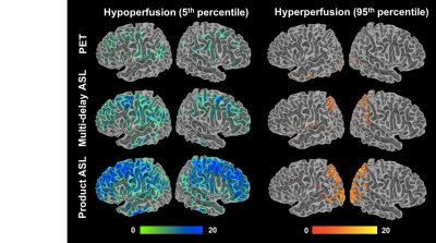
Is ASL Useful for Brain Mapping?
Wen-Chau Wu
Since its introduction in 1990's, ASL has gone through abundant developments that have remarkably improved this contrast-material-free technique in data acquisition, image quality, and quantitative modeling. We now have a variety of methods to choose from and a number of review articles to refer to. However, it still seems an open question when it comes to the usefulness of ASL in brain mapping. In this presentation, the issues that have been keeping us from saying yes to the question will be reviewed. The usefulness and caveats of ASL in brain mapping will be discussed from the viewpoint of clinical application.
|
14:30
|
|
DCE/DSC Beyond Perfusion & Permeability
Ben Dickie
A healthy cerebrovasculature is crucial for meeting the brains ever changing demand for oxygen and nutrients. Aging and many neurodegenerative diseases lead to insidious vascular changes to vessel size, geometry, and blood brain barrier (BBB) integrity. New approaches to the acquisition and analysis of DCE- and DSC-MRI data are providing novel insights into these cerebrovascular abnormalities, aiding understanding of disease mechanisms and helping to identify novel treatment targets.
|
14:55
|
|
Break & Meet the Teachers |
|
| |
|
Advanced Topics in Perfusion MRI: Part 2
Weekend Course
ORGANIZERS: Fernando Calamante, Hanzhang Lu, Steven Sourbron
Sunday, 17 June 2018
| N03 |
15:15 - 16:30 |
Moderators: Thomas Christen, Laura Parkes |
Skill Level: Intermediate to Advanced
Session Number: WE-22B
Overview
This session is designed to provide an overview of the latest advances in perfusion MRI. The lectures will cover a range of novel acquisition, modeling, and analysis approaches. In particular, it will cover novel aspects of ASL in both brain and body, advanced contrast agent-based acquisition and reconstruction methods, and validation of these quantitative perfusion measures.
Target Audience
This course is designed for scientists and clinicians who are interested in broadening their understanding of perfusion techniques and obtaining an update on current frontiers of perfusion imaging.
Educational Objectives
As a result of attending this course, participants should be able to:
-Describe recent advances in ASL, DSC, and DEC acquisition and reconstruction strategies;
-Discuss the state-of-the-art methods in body ASL MRI;
-Identify the benefits of ASL in functional brain mapping;
-Identify the role of contrast-based methods for measuring physiological parameters beyond perfusion;
-Describe current strategies to validate perfusion-based measures; and
-Identify alternative tracer or contrast agent methods for perfusion imaging.
15:15
|
 |
Advanced Reconstruction Techniques for DCE/DSC
Krishna Nayak
This talk will review advanced reconstruction methods for DCE-MRI and DSC-MRI. These methods allow for superior extraction of perfusion, permeability, and other tracer-kinetic parameters of interest (from noisy data), and allow for improvements in spatio-temporal resolution and coverage through the use of sparse data sampling (in k,t space). We will start by reviewing the current workflow in DCE/DSC, and identifying the opportunity for improvement. Then we will go through four classes of reconstruction, which have some overlap: 1) spatial and temporal sparsity constraints, 2) data-driven constraints, 3) tracer-kinetic model-based constraints, 4) arterial input function estimation. Finally, we will discuss the magnitude of the improvement that can be achieved using these techniques.
|
15:40
|
 |
Methods to Validate Quantitative Perfusion Measures
Audrey Fan
Cerebral blood flow (CBF) is a critical physiological biomarker that select appropriate treatments for cerebrovascular patients and is affected in aging and numerous neurological disorders. However, validation of perfusion biomarkers has been challenging, due to (a) inability to compare with a simultaneous reference standard; and (b) insufficient testing in disease cases, where pathology may interact with the imaging mechanism itself to create CBF inaccuracies. This talk describes how to define an appropriate perfusion measure to address a research/clinical question, and design experiments to validate this measurement. It also describes multi-modal imaging and challenge studies (cerebrovascular reactivity) contribute confidence in quantitative perfusion measures.
|
16:05
|
|
New Contrast Agent/Tracer in Perfusion Imaging?
Thomas Christen
The vast majority of perfusion MR approaches require the injection of a gadolinium based contrast agent or the magnetic labeling of arterial spins. Yet, other types of contrasts or tracers have also been proposed to probe the micro or macro vascular network. This lecture will present recent perfusion techniques based on the use of (1) iron oxide particles, (2) spontaneous or challenge-based BOLD contrast fluctuations, and (3) hyperpolarized compounds. These methods can offer high SNR, short acquisition times, and provide access to new biophysical markers such as microvessel geometry, hematocrit and blood oxygenation.
|
16:30
|
|
Adjournment & Meet the Teachers |
|
| Back |
| The International Society for Magnetic Resonance in Medicine is accredited by the Accreditation Council for Continuing Medical Education to provide continuing medical education for physicians. |
