|
Weekend Educational Course
Prostate MRI, MRI in Pregnancy & Genitourinary |
|
Prostate MRI, MRI in Pregnancy & Genitourinary: Part 1
Weekend Course
ORGANIZERS: Kathryn Fowler, Catherine Hines, Kartik Jhaveri, Lorenzo Mannelli, Valeria Panebianco, Scott Reeder, Reiko Woodhams
Sunday, 17 June 2018
| S02 |
13:15 - 14:45 |
Moderators: Masoom Haider, David Karow |
Skill Level: Intermediate
Session Number: WE-24A
Overview
A detailed overview of prostate MRI from diagnosis to surveillance to detection of recurrence. Novel information regarding the role of PET/MRI in prostate cancer will be covered. Placental imaging and imaging in pregnancy will be covered. Genitourinary topics include renal and adrenal pathology and MR urography from technical and clinical applications perspective.
Target Audience
Clinicians, physicists, and radiologists involved in genitourinary imaging, including prostate.
Educational Objectives
As a result of attending this course, participants should be able to:
-Explain the role of prostate MRI in diagnosis (PI-RADS), staging, and detection of recurrence of prostate cancer;
-Describe the MRI approach to placental and fetal pathology;
-Recognize pathologic conditions involving the fetus and placenta;
-Summarize the MRI appearance of adrenal and renal pathology; and
-Explain the technical approach to and diagnosis of urothelial lesions.
| |
Prostate MRI |
13:15
|
|
 Technical Considerations for Prostate MRI Acquisition Technical Considerations for Prostate MRI Acquisition
Alberto Vargas
The aim of this session is to summarize the latest technical developments relevant to optimization of clinical prostate MRI protocols
|
13:37
|
|
Guidelines & Recommendations
Jelle Barentsz
In this talk, a literature review will be shown of PI-RADS v2. The role of experience and training will be discussed. To ensure optimal quality, certification and quality criteria will be provided. Finally, the role of an expertise network will be emphasized. It will be concluded, that it has been shown that PI-RADSv2 is an adequate ‘‘language’’ for assessing the risk of the presence of clinically significant PCa. The sensitivity is significantly better than that of PI-RADSv1. Nonetheless, there is large heterogeneity that could be reduced by an improved PI-RADSv3 and by training and certification of radiologists. Equally important, however, is the training of urologists and other involved physicians in being able to communicate in the same ‘‘language’’.
|
13:59
|
|
Prostate MRI & Recurrence
Jurgen Fütterer
mpMRI is a helpful tool in the evaluation of the treated prostate gland.
|
14:21
|
|
PET/MRI of Prostate Cancer
Joseph Ippolito
This educational talk provides an overview of PET/MRI in the evaluation of prostate cancer with a focus on the strengths and weaknesses of PET/MRI versus PET/CT as well as the individual strengths and weaknesses of MRI versus PET. This talk also discusses methods for evaluating initial staging as well as biochemical recurrence in prostate cancer patients and discusses current progress made in the radiopharmaceutical field and its ability to synergize with emerging methods in MRI. |
| 14:38 |
|
SAMs: PET/MRI of Prostate Cancer
|
14:43
|
|
Break & Meet the Teachers |
|
| |
|
Prostate MRI, MRI in Pregnancy & Genitourinary: Part 2
Weekend Course
ORGANIZERS: Kathryn Fowler, Catherine Hines, Kartik Jhaveri, Lorenzo Mannelli, Valeria Panebianco, Scott Reeder, Reiko Woodhams
Sunday, 17 June 2018
| S02 |
15:15 - 16:45 |
Moderators: Masoom Haider, David Karow |
Skill Level: Intermediate
Session Number: WE-24B
Overview
A detailed overview of prostate MRI from diagnosis to surveillance to detection of recurrence. Novel information regarding the role of PET/MRI in prostate cancer will be covered. Placental imaging and imaging in pregnancy will be covered. Genitourinary topics include renal and adrenal pathology and MR urography from technical and clinical applications perspective.
Target Audience
Clinicians, physicists, and radiologists involved in genitourinary imaging, including prostate.
Educational Objectives
As a result of attending this course, participants should be able to:
-Explain the role of prostate MRI in diagnosis (PI-RADS), staging, and detection of recurrence of prostate cancer;
-Describe the MRI approach to placental and fetal pathology;
-Recognize pathologic conditions involving the fetus and placenta;
-Summarize the MRI appearance of adrenal and renal pathology; and
-Explain the technical approach to and diagnosis of urothelial lesions.
| |
Imaging in Pregnancy |
15:15
|
 |
Placenta & Pregnant Patient
Nathalie Siauve, Marianne Alison, Gihad E Chalouhi, Benjamin Deloison, Laurent J Salomon
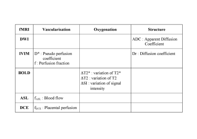
Understanding placental functions remains largely concealed from non-invasive in vivo investigations, although placental insufficiency is responsible for most failures in pregnancy. Functional MRI offers new perspectives for placental imaging. The vascularisation can be studied with IVIM and the pseudo perfusion coefficient (D*) and the perfusion fraction (f), and with ASL and the blood flow (fASL). DCE MRI also provides a definite assessment of perfusion but requires an exogenous contrast agent. The oxygenation can be studied with BOLD. The placental structure can be studied with DWI and the apparent diffusion coefficient (ADC), and with IVIM and the diffusion coefficient (Dr).
|
15:37
|
 |
Fetal MRI
Manjiri Dighe
This presentation will review the technique and common indication of fetal MRI with a review of the newer techniques at the end. Illustrative cases will be presented.
|
| |
GU MRI |
15:59
|
|
Renal & Adrenal Pathologies
Nicolas Grenier
|
16:21
|
|
MRU & Bladder
David Childs
MR urography has proven to be a robust technique for the assessment of the urothelium in both the pediatric (congenital anomalies) and adult (urothelial carcinoma) populations. Technical challenges (motion, contraindication to contrast, etc.) remain, although newer MRI techniques are increasingly providing solutions to these issues. The increasing use of diffusion and perfusion-weighted imaging has also provided the benefit of multi-parametric data. While the detection of urothelial carcinoma can be achieved with various endoscopic and urographic imaging tests, the accurate staging of tumors (both T and N stage) remains a challenge. Advances have been achieved in anatomic MRI, perfusion MRI, and diffusion weighted imaging for the T-staging of bladder carcinoma, although the assessment of nodal status remains a significant challenge. Newly described PET/MRI methods have shown promising results in improving both the specificity of focal urothelial lesions and accurate determination of nodal status, though research is ongoing.
|
16:43
|
|
Adjournment & Meet the Teachers |
|
| Back |
| The International Society for Magnetic Resonance in Medicine is accredited by the Accreditation Council for Continuing Medical Education to provide continuing medical education for physicians. |
