Digital Poster Session
Musculoskeletal Back to Program-at-a-Glance Back to Program-at-a-Glance
|
Monday, 13 May 2019
Digital PosterMusculoskeletal
1259 -1283 Muscle 1
1284 -1308 Bone 1
1309 -1333 Cartilage 1
1334 -1358 Muscle 2 & Other MSK
1359 -1383 Bone 2 & MSK Tumors
1384 -1408 Cartilage 2, Meniscus, Tendon & Ligament
1409 -1433 MSK Techniques & Development: Other MSK |
| |
Muscle 1
Digital Poster
Musculoskeletal
Monday, 13 May 2019
| Exhibition Hall |
08:15 - 09:15 |
| |
|
Computer # |
 |
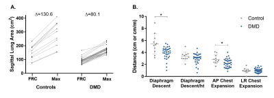
1259.
 |
1 |
Characterizing respiratory muscle composition and function in Duchenne muscular dystrophy using dynamic MRI and chemical shift-encoded imaging

Alison Barnard, Donovan Lott, Abhinandan Batra, William Triplett, Sean Forbes, Samuel Riehl, Rebecca Willcocks, Barbara Smith, Krista Vandenborne, Glenn Walter
In Duchenne muscular dystrophy (DMD), respiratory muscle weakness leads to eventual respiratory failure. For this investigation, dynamic MRI was utilized to characterize diaphragm and chest wall dynamics during breathing, and chemical shift-encoded imaging was utilized to assess fatty infiltration in accessory respiratory muscles in 36 individuals with DMD and 12 unaffected controls. For maximal inspirations, individuals with DMD had significantly reduced anterior-posterior chest expansion, and a subgroup with poor respiratory function had decreased diaphragm descent (normalized to height). The expiratory muscles had high levels of fatty infiltration, and muscle fat fraction was correlated with measures of expiratory muscle strength.
|
|
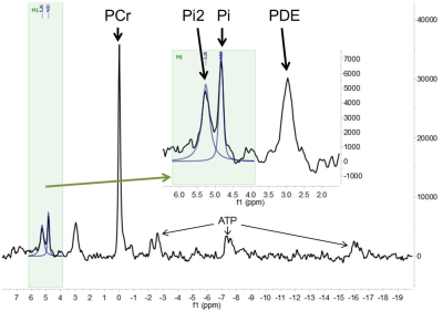
1260.
 |
2 |
Can be Pi2 signal in 31P MR spectra a biomarker of critical limb ischemia in diabetic patients?
Petr Sedivy, Monika Dezortova, Miloslav Drobny, Michal Dubsky, Milan Hajek
A group of 65 diabetic patients with critical limb ischemia was studied by rest and dynamic phosphorous MR spectroscopy. An unknown signal in the position 5.2 – 5.4 ppm (labeled as Pi2) was observed in calf muscle spectra of 14 patients. Two hypotheses of the Pi2 signal explanation were taken into the consideration: a) phosphorus in alkaline phosphate pool or b) phosphorus in an unknown phosphomonoester. Results support hypothesis a) and we think that Pi2 signal at 5.4 ppm in patients with diabetic foot disease could be considered as the biomarker of the most severe muscular damage.
|
|
1261.
 |
3 |
Regional thigh muscle composition based on chemical shift encoding-based water-fat MRI and its association with muscle strength
Maximilian Löffler, Sarah Schlaeger, Stephanie Inhuber, Michael Dieckmeyer, Dominik Weidlich, Ansgar Schwirtz, Ernst Rummeny, Claus Zimmer, Jan Kirschke, Dimitrios Karampinos, Thomas Baum
Chemical shift encoding-based water-fat MRI derived proton density fat fraction (PDFF) of the thigh muscles bears potential as a surrogate marker in subjects with osteoarthritis, sarcopenia, and neuromuscular disorders. Muscle PDFF has shown to correlate with isometric strength at the thigh and spine. However, MR-based muscle fat quantification requires time-consuming segmentation of multiple muscle compartments. Therefore, we investigated if segmentation of single compartment muscles and of different levels of the thigh influences the relation of PDFF to isometric strength. The present study demonstrated that PDFF measurements can be limited to an entire muscle compartment, independent of sampling level.
|
|
1262.
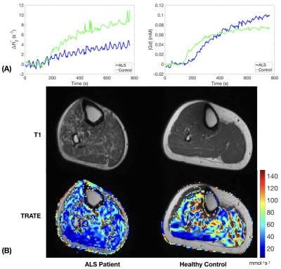 |
4 |
Evaluation of Muscle Degeneration in Amyotrophic Lateral Sclerosis Patients Using MR Cytography
Sudarshan Ragunathan, Laura Bell, Ashley Stokes, Nicole Turcotte, Shafeeq Ladha, C Chad Quarles
Amyotrophic lateral sclerosis (ALS) is a fatal neurodegenerative disease that affects motor neurons resulting in progressive muscle atrophy. The heterogeneous nature of disease progression has limited the reliability and robustness of current clinical indicators used in disease monitoring. To address the need for reproducible, quantitative biomarkers, we propose the applicability of Magnetic Resonance Cytography (MRC) to characterize ALS induced changes to muscle myofiber microstructure. In this clinical study, the role of MRC as a potential biomarker was demonstrated by identifying changes to muscle cytoarchitecture in the lower extremities among ALS patients when compared with healthy muscle.
|
|
1263.
 |
5 |
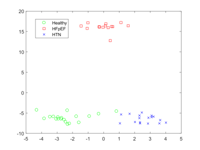
Creatine CEST at 3T following in magnet exercise shows differences in heart failure patients with preserved ejection fraction compared to healthy and hypertensive controls
Neil Wilson, Payman Zamani, Elizabeth Proto, Kevin D'Aquilla, Dushyant Kumar, Deepa Thakuri, Hari Hariharan, Shana McCormack, Julio Chirinos, Ravinder Reddy
Exercise intolerance is a hallmark of heart failure. Here, we have shown for the first time that parameters derived from ROI-based analysis of creatine-weighted CEST signals following exercise can be used to distinguish HFpEF patients from HTN and healthy controls. Identifying these group differences is uniquely possible because CrCEST can measure muscle specific metabolism with high spatial resolution and sensitivity.
|
|
1264.
 |
6 |
Aerobic exercise enhances 31P MRS measured mitochondrial function independent of statin use
Jill Slade, George Abela, David Hurley, Ronald Meyer
Phosphorus MRS is the gold standard measure of in vivo mitochondrial function. In this study, 31P MRS was used to examine exercise-induced adaptations in mitochondrial function in the presence of concurrent statin use. The results show that aerobic exercise training significantly improved muscle oxidative capacity of the plantar flexor muscles in older adults independent of statin use.
|
|
1265.
 |
7 |
The novel visualization technique of fast and slow muscle fibers using q-space imaging: Clinical study
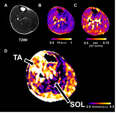
Daisuke Nakashima, Junichi Hata, Yasushi Sera, Takeo Nagura, Morio Matsumoto, Hideyuki Okano, Masaya Nakamura
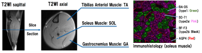
Skeletal muscles include fast and slow muscle fibers. However, a non-invasive approach for appropriately investigating the characteristics of muscles is not available. The present study aimed to determine whether q-space imaging can distinguish between fast fiber dominant tibialis anterior muscle (TA) and slow fiber dominant soleus muscle (SOL). T2WI, FA and ADC maps could not represent the difference between TA and SOL. On the other hand, Kurtosis map could visualize the characteristics of TA and SOL. q-space imaging is a promising method to non-invasively estimate the fiber type ratio in skeletal muscles.
|
|
1266.
 |
8 |
Whole-Body Mapping of Spontaneous Mechanical Activities in Musculature
Martin Schwartz, Petros Martirosian, Thomas Küstner, Günter Steidle, Thorsten Feiweier, Bin Yang, Fritz Schick
Whole-body quantification of spontaneous mechanical activities is of high interest for the assessment of the activity distribution in healthy and non-healthy population. Therefore, a measurement protocol and spatial mapping is investigated for accurate quantification of small subtle spontaneous activities in the human skeletal musculature over the whole-body. This work enables to assess spontaneous activity in muscular regions which are important for potential evaluation and grading in neuromuscular disorders.
|
|
1267.
 |
9 |
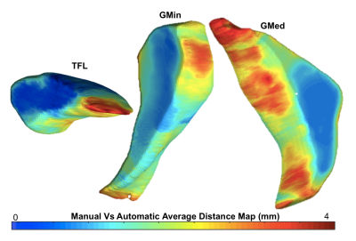
Deep Learning-Based Automatic Estimation of Volume and Fat Fraction in Abductor Muscles and their Associations with T1? and T2 in Hip Osteoarthritis Patients
Radhika Tibrewala, Valentina Pedoia, Carla Kinnunen, Tijana Popovic, Richard Souza, Sharmila Majumdar
In Osteoarthritis, cartilage degeneration can be accompanied by muscle weakness. T1ρ and T2 relaxation times have been used to probe cartilage degeneration. This study aims to develop an automatic machine-learning based segmentation and quantification pipeline to estimate the volumes and fat fractions of the three hip abductor muscles and study their associations with T1ρ and T2 relaxation times. Our results showed fast, reliable segmentations the hip abductor muscles and voxel based correlations between T1ρ and fat fraction and T2 and volumes of the muscles.
|
|
1268
 |
10 |
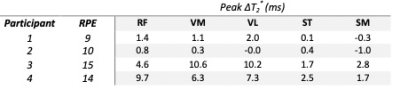
Evaluation of inter- and intramuscular differences using multi-slice T2* measurements after an in-magnet stepping exercise.
Video Permission Withheld
Thom Veeger, Celine Baligand, Andrew Webb, Jurriaan de Groot, Hermien Kan
In this study we explored the feasibility of using an MR-compatible ergometer mimicking stair climbing to study differences in T2* after exercise between and within different thigh muscles. Four healthy subjects performed a 10-minute stepping exercise inside a 3T. Participants exercised at either a high rate of perceived exertion (RPE) or a low RPE. Clear differences between low and high RPE and different muscles, but not within muscles, were found. This shows that it is possible to use stair-climbing using an MR-compatible ergometer to study differences between and within muscles in response to exercise.
|
|
1269.
 |
11 |
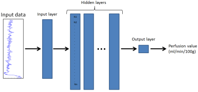
A neural network approach for estimating muscle perfusion from DCE-MRI data
Christopher Conlin, Xiaowan Li, Stephen Decker, Christopher Hanrahan, Gwenael Layec, Nan Hu, Vivian Lee, Jeff Zhang
Perfusion is an important aspect of calf muscle function that can be measured with dynamic contrast-enhanced (DCE) MRI. However, conventional methods for quantifying perfusion from DCE-MRI data require an appropriate tracer-kinetic model, which may not be available clinically. In this study, we examined the feasibility of neural networks (NNs) for quantifying calf-muscle perfusion from DCE-MRI data. We found that NNs estimate perfusion with accuracy comparable to conventional methods, without the need for a tracer-kinetic model. NNs like those developed in this study can be readily incorporated into ordinary MRI scanner software, facilitating routine quantitative perfusion analysis with DCE-MRI.
|
|
1270.
 |
12 |
Correlation between skeletal muscle fat content and insulin resistance in patients with type 2 diabetes mellitus
Fuyao Yu, Huadong Zhou, Fengzhe Wang, Jiazheng Wang, Shinong Pan
To quantitatively investigate various parts of the thigh skeletal muscle fat content in patients with type 2 diabetes to explore its correlation with insulin resistance via MRI.
|
|
1271
 |
13 |
Visualization of Aquaporin 4 using Time-dependent Diffusion MRI in Mouse Skeletal Muscle
Video Permission Withheld
Junichi Hata, Takayuki Obata, Yasuhiko Tachibana, Yawara Haga, Mai Mizumura, Daisuke Nakashima, Yasushi Sera, Masaya Nakamura, Hideyuki Okano
We focused on aquaporin 4 in skeletal muscle and attempted to visualize its function using time-dependent diffusion magnetic resonance imaging (MRI). In addition, the validity of the muscle cell type characteristics was evaluated by immunostaining. The diffusion time was adjusted with the PG-STE method using a 9.4-T MRI scanner. Diffusivity associated with a difference in the diffusion time was found to differ depending on the skeletal muscle type. Thus, it was possible to visualize the water molecule exchange rate of skeletal muscle cell membranes.
|
|
1272.
 |
14 |
Simulation based study of the effect of sub-voxel spatial distribution of permeability of muscle fibres as a function of diffusion time and b-value using a finite element model
Nadia Smith, Jessica Talbott, Chris Clark, Matt Hall
We investigate the effect of the sub-voxel patterning of permeability in muscle tissue on the diffusion signal via a finite element simulation of diffusion MRI on a model of muscle tissue. We observe that permeability with a disordered pattern leads to statistically significant differences in diffusion signal at high b and longer diffusion times.
|
|
1273.
 |
15 |
The relation between fat calibration in multi-echo spin-echo water T2 mapping and STEAM fat T2 relaxation measurements
Martijn Froeling, Eric Hughes, Lara Schlaffke, Hermien Kan, Kieren Hollingsworth
The aim of this study was to quantitatively describe the relation between fat calibration in ME-SE water T2 mapping and STEAM fat T2 relaxation measurements in spectroscopy using j-coupling simulations and investigate its effect on EPG water T2 mapping. Both ME-SE and STEAM T2 mapping methods to estimate the apparent fat T2 relaxation times are heavily influenced by J-coupling. As such the measured T2 relaxation time of fat using STEAM, appears shorter and using ME-SE appears longer, ranging between values of 30 and 140 ms.
|
|
1274.
 |
16 |
Compressed-Sensing 4D Flow MRI of the Skeletal Muscle during Nerve vs Muscle Electrical Stimulation
Francesco Santini, Nicolas Place, Anna Hirschmann, Ning Jin, Oliver Bieri, Xeni Deligianni
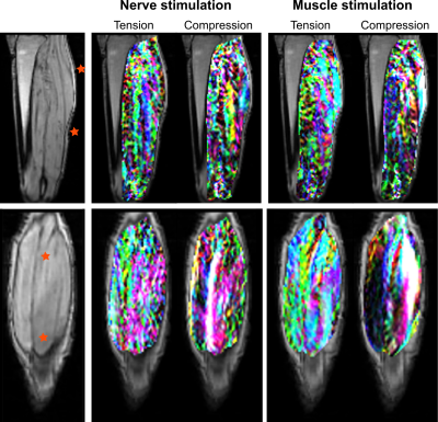
The purpose of this study was to use dynamic 4D phase contrast MR imaging to compare the stimulation patterns of neuromuscular electrical stimulation of the calf muscles when delivered through the muscle belly or through the nerve trunk. Experiments were performed on healthy volunteers and strain maps were obtained for each stimulation modality. A more homogeneous activation of the muscle group was obtained for nerve stimulation, with overall lower strain values with respect to muscle stimulation.
|
|
1275.
 |
17 |
Extended phase graph model based tissue-water T2 estimation from CPMG image data in fat-infiltrated skeletal muscle: application in amyotrophic lateral sclerosis and Kennedy’s disease
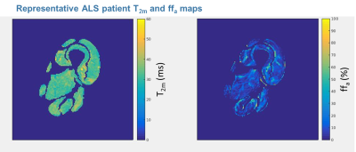
Nick Zafeiropoulos, Uros Klickovic, Luca Zampedri, Stephen Wastling, Christopher Sinclair, Jasper Morrow, Robert Janiczek, Enrico De Vita, Tarek Yousry, Michael Hanna, Linda Greensmith, Pietro Fratta, John Thornton
An MRI CPMG extended phase graph signal model was used to determine muscle-water T2 (T2m) in fat-infiltrated skeletal muscle, using a fixed two-component approximation to the fat signal. Stable estimates of T2m and apparent fat fraction (ffa) in the thigh muscles of amyotrophic lateral sclerosis and Kennedy’s disease patients and healthy controls were obtained. T2m were elevated in both patient groups, as was ffa with a distribution consistent with that obtained by 3-point Dixon MRI.
|
|
1276.
 |
18 |
Fascicle Ellipticity as an Explanation of Transverse Anisotropy in Diffusion MRI Measurements of Skeletal Muscle
Noel Naughton, Anthony Wang, John Georgiadis
Diffusion MRI of skeletal muscle exhibits a transverse anisotropy, the source of which has yet to be conclusively determined. To explore this, histological images were segmented into intracellular and extracellular domains and used to inform a direct numerical simulation of the Bloch-Torrey equation. Histology images were examined at the myocyte and fascicle scale and it was found that results from the fascicle images exhibited increased transverse anisotropy. These results suggest that fascicle organization may pay a hereunto unrecognized role in affecting dMRI in skeletal muscle.
|
|
1277.
 |
19 |
Bidirectional Filtering for Psoas Major Muscle Magnetic Resonance Elastography
Surendra Maharjan, Tomokazu Numano, Tetsushi Habe, Daiki Ito, Takamichi Ueki, Keisuke Igarashi, Toshiki Maeno
The purpose of this present work was to apply bidirectional spatio-temporal image filtering in the preferential direction of shear wave propagation in psoas major muscle Magnetic Resonance Elastography. The results suggested there was improvement in the wave propagation by using combined gaussian bandpass (GBP) and bidirectional filter in compared to GBP only. The calculation of local frequency estimate (LFE) stiffness value of PM muscle was not changed by applying the bidirectional filter.
|
|
1278.
 |
20 |
Gender- and age-related changes in trunk muscle composition using chemical shift encoding-based water-fat MRI
Egon Burian, Daniela Franz, Jan Syväri, Christina Holzapfel, Theresa Drabsch, Jan Kirschke, Ernst Rummeny, Claus Zimmer, Hans Hauner, Dimitrios Karampinos, Thomas Baum
Chemical shift encoding-based water-fat MRI derived proton density fat fraction (PDFF) of muscles has been emerging as a surrogate marker for quantification of ectopic fat accumulation. Increased fatty deposits are a risk factor for metabolic and cardiovascular diseases. With skeletal muscle being the largest body compartment in adults, we are still at the beginning of understanding the functional consequences of these changes. The purpose of the present analysis was to investigate the gender- and age-related changes in PDFF of trunk musculature of healthy adults using chemical shift encoding-based water-fat MRI.
|
|
1279.
 |
21 |
Semi-quantitative MR muscle analysis of VCP inclusion body myopathy
Saya Horiuchi, Hon Yu, Toshimi Tando, Taiki Nozaki, Vincent Caiozzo, Virginia Kimonis, Hiroshi Yoshioka
This study was to demonstrate usefulness of semi-quantitative MR muscle analysis of VCP associated inclusion body myopathy associated with Paget and dementia (IBMPFD). Five-point scoring method was compared with quantification based on fat fraction analysis, and specific patterns of thigh muscle alterations were explored. The semi-quantification provided equivalent accuracy as quantification. The sartorius and adductor magnus were most affected by fatty infiltration, while the adductor longus and rectus femoris were well-preserved. Muscle volume decrease was more frequently seen in the hamstring and adductor muscles. This semi-quantitative method can be widely available in clinical settings and assist noninvasive initial/follow-up IBMPFD diagnosis.
|
|
1280.
 |
22 |
Quantitative evaluation of muscular microvascular permeability by DCE-MRI and texture analysis in diabetic rabbits
Presentation Not Submitted
Bai Yu Liu, Yun Fei Zha, Yang Fan
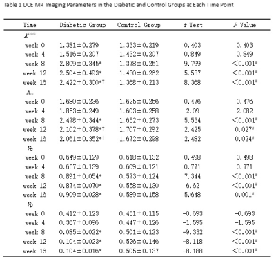
Our purpose is to quantificationally evaluate the microvascular permeability and subtle changes in diabetic skeletal muscle by DCE-MRI and texture analysis. Alloxan-induced diabetic rabbits and normal rabbits were studied at fixed time points (0, 4, 8, 12, and 16 weeks). Permeability parameters Ktrans and Kep increased and then showed a downward trend, Ve increased and Vp decreased in diabetic group. Texture parameters based on Ktrans map showed difference between the two groups. We can draw a conclusion that the microvascular permeability of diabetic skeletal muscle increases while the perfusion decreases and texture analysis based on Ktrans map can detect these subtle changes in early stage.
|
|
1281.
 |
23 |
Multi-parametric MRI analysis of the temporal changes of induced damage and regeneration in dystrophic hind limb muscles
Ravneet Vohra, Joshua Park, Feng Zhang, Guy Odom, Jeffrey Chamberlain, Donghoon Lee
The mdx mouse model is one of the most commonly used animal models for Duchenne muscular dystrophy (DMD). Although the mdx model has a milder phenotype compared to patients with DMD, the model has shown the similarity in some histopathologic events resulting in wide utilizations in preclinical studies for both disease progression and therapeutic intervention. Over the years MRI has been increasingly being utilized to monitor the disease progression in dystrophic mice and DMD patients. We performed MRI to discriminate the time course of damage in regeneration in skeletal muscles if mdx mice.
|
|
1282.
 |
24 |
Mapping of myoglobin oxygen saturation dynamics in the calf during ischemia with a modified slab-selective 2D NMRSI pulse sequence at 3T.
Alfredo Lopez Kolkovsky, Martin Meyerspeer, Pierre Carlier
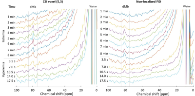
Myoglobin in its deoxygenated form is a 1H NMR visible biomarker of intracellular oxygenation. Its low concentration and very short relaxation times have been major challenges to map deoxy-myoglobin dynamically during a transient state, such as ischemia. Here, we interleaved the acquisition of a full 2D CSI data set at the dMb frequency (~79 ppm), a non-localized dMb spectrum and an anatomical image with radial encoding to track dMb dynamics during an ischemic bout with a temporal resolution of 3.5 seconds. Promising proof-of-concept results are shown. This method suggests a strong potential for energy metabolism studies in vivo.
|
|
1283.
 |
25 |
Simulated effect of diffusion time and skeletal muscle fiber size on the diffusion tensor
David Berry, Erin Englund, Vitaly Galinsky, Chamindra Konersman, Shaochen Chen, Samuel Ward, Lawrence Frank
The sensitivity of diffusion tensor imaging (DTI) to muscle fiber size is dependent upon diffusion time. However, there is no consensus on how to interpret data acquired at different diffusion times. In this study we simulated the relationship between muscle fiber size, diffusion time, and the resulting diffusion tensor in models with simplified and histology informed muscle fiber geometry, using a stimulated echo DTI pulse sequence. Maximum contrast between physiologically relevant fiber sizes was found at 130ms for diffusivity, and 170ms for fractional anisotropy measurements. This data may better inform pulse sequence parameter selection when performing DTI experiments in vivo.
|
|
| Top |
Bone 1
Digital Poster
Musculoskeletal
Monday, 13 May 2019
| Exhibition Hall |
08:15 - 09:15 |
| |
|
Computer # |
 |
1284.
 |
26 |
Mapping collagen and water proton densities in tibial cortical bone using 3D ultrashort echo time cones (3D-UTE-Cones) MR imaging techniques
Saeed Jerban, Yajun Ma, Tan Guo, Lidi Wan, Hyungseok Jang, Liang Li, Eric Chang, Jiang Du
Spatial variations of cortical bone microstructure and mechanics can be described by mapping proton densities as exist in macromolecular, bound, and pore water pools. Comparing bone signal in ultrashort echo time MRI (UTE-MRI) and in inversion recovery UTE MRI against a known external reference signal enabled us to measure total, bound, and pore water proton densities. Measured total water proton density combined with macromolecular fraction from magnetization transfer modelling resulted in macromolecular proton density estimation. We observed strong correlation between proton densities and bone porosity. The developed technique was performed robustly on ten young subjects. This technique may aid diagnosing bone diseases and injuries.
|
|
1285.
 |
27 |
Ultrashort echo time MRI (UTE-MRI) quantifications of cortical bone varied between scans at room temperature and body temperature
Saeed Jerban, Nikolaus Szeverenyi, Yajun Ma, Tan Guo, Sarah To, Eric Chang, Jiang Du
Several quantitative ultrashort echo time MRI (UTE-MRI) techniques have recently been employed to assess cortical bone microstructure. Such techniques were examined mostly ex vivo at room temperature and demonstrated strong correlations with bone microstructure as measured with micro computed tomography (μCT). However, MRI properties of cortical bone may differ in vivo due to higher temperature. We have investigated several UTE-MRI quantifications of cortical bone at body and room temperatures. Significant variations of bone UTE-MRI measures were observed between room and body temperatures. Implementing a linear correction method on UTE-MRI measures based on the presented results here might improve the validity of the techniques for in vivo studies.
|
|
1286.
 |
28 |
1H nuclei compartmentalization, exchange and self-diffusion in cortical bone by one- and two-dimensions NMR in homogeneous and inhomogeneous fields
Presentation Not Submitted
Leonardo Brizi, Marco Barbieri, Claudia Testa, Paola Fantazzini
There is increasing interest in the study of water content, compartmentalization, exchange and its interaction with collagen in cortical bone for the evaluation of bone fracture risk. Here, we present the NMR characterization of 1H nuclei signals of the cortical bone. Different components (collagen, lipid, water) and different water compartments are identified measuring NMR properties and self-diffusion coefficients. The exchange between collagen and water protons is observed and an average residence time in the collagen is estimated. The results can contribute to optimize MRI protocols specifically for bone imaging and to characterize the role of water in this tissue.
|
|
1287.
 |
29 |
Perfect In-Phase Zero TE for Musculoskeletal Imaging
Mathias Engström, Cristina Cozzini, Michael Carl, Graeme McKinnon, Florian Wiesinger
Large FOV Zero Echo-Time (ZTE) has been challenging due to chemical shift artifacts, caused primarily by fat water dephasing, for low readout band-widths (rBW). To correct for this Perfect In-Phase ZTE (pipZTE) is proposed where the chemical shift artifact is removed by acquiring data from multiple rBWs, and then separating the signal into an in-phase and off-resonance compartment in the reconstruction. In this work we explore the performance and properties of the pipZTE approach when scanning large FOV and demanding subjects.
|
|
1288.
 |
30 |
Comparison of Quantitative MRI Fat-Fraction measurement in SIJ joint on different scanner platforms
Alan Bainbridge, Timothy Bray, Naomi Sakai, Sarah Tansley, Nicola Fulstow, Raj Sengupta, Margaret Hall-Craggs
Proton density fat fraction (PDFF) measurements can quantify oedema and fat metaplasia in patients with spondyloarthritis. The reproducibility of PDFF measurement in the bone marrow of the sacroiliac joint was assessed in volunteers on 3 systems from different manufacturers, using a range of specialist and base-level product protocols. PDFF measurements on different platforms correlate well, but there is also a bias between the base-level and advanced methods. Performing an offline reconstruction with a multi-peak fat model and a T2* correction term reduces this bias.
|
|
1289.
 |
31 |
Age Estimation with the Greulich-Pyle Atlas using 3T MR Images of Hand and Wrist
Thomas Widek, Pia Genet, Thomas Ehammer, Eva Scheurer, Thorsten Schwark
Bone age estimation of the hand is very common both in a forensic context and for clinical purposes. Currently, this is done by assessing plain X-rays of the hand. This is a controversial issue, especially in the forensic context, as legal proceedings lack a medical indication for the use of ionizing radiation. The aim of the current study was to validate the use of the X-ray based Greulich-Pyle atlas method on hand MR images in a healthy male cohort. The results show that the application of the Greulich-Pyle method is feasible and that it can be used in daily routine.
|
|
1290.
 |
32 |
Fatty Acid Composition Assessed By 3T MRI in Systemic Lupus Erythematosus
Dimitri MARTEL, Benjamin LEPORQ, Ravinder REGATTE, Stephen HONIG, Gregory CHANG
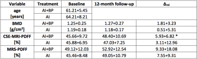
Systemic Lupus Erythematosus (SLE) is a chronic, inflammatory disease. Recent studies demonstrated an increased incidence of osteoporosis (OP) and fractures in SLE patients. Glucocorticoids (Gcs) are part of therapy for SLE and have with long-term intake deleterious effects on bone quality leading ultimately to Glucocorticoido-induced-osteoporosis. Our aim was to assess the marrow composition of a SLE population and compare it to that of OP patients, GCs user and young healthy women using 3T Chemical Shift Encoded- MRI (CSE-MRI).
|
|
1291.
 |
33 |
Diffusion-Weighted MRI in Juvenile Osteochondritis Dissecans (JOCD) at 3T
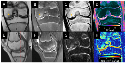
Kai Ludwig, Casey Johnson, Stefan Zbyn, Takashi Takahasi, Shelly Marette, Bradley Nelson, Marc Thompkins, Cathy Carlson, Jutta Ellermann
Diffusion-weighted MRI (DWI) may help elucidate the etiology and progression of juvenile osteochondritis dissecans (JOCD) by probing tissue/cellular characteristics of JOCD lesions and the underlying parent bone. In this study, we observed elevated DWI signal and increased apparent diffusion coefficient (ADC) values within and proximal to OCD lesions compared to surrounding bone marrow and control sites. ADC values within the lesion and the parent bone may help distinguish healing from non-healing lesions, thereby improving prognostication of JOCD and clinical decision making.
|
|
1292.
 |
34 |
An investigation of the relationship between type II collagen degradation products and MRI features of damage in knee osteoarthritis patients
Franklyn Howe, Vivian Ejindu, Christine Heron, Olakunbi Harrison, Soraya Koushesh, Lena Assi, Anasuya Kuttapitiya, Thomas Barrick, Nidhi Sofat
Knee osteoarthritis (OA) is a degenerative disease which produces pain and exhibits damage to cartilage, bone marrow and the development of synovitis. Biomarkers are needed to aid patient stratification for developing improved treatment strategies. We have investigated how type II collagen degradation products (CTX2), which are generated during OA, relate to MRI features of knee damage and patient reported pain. CTX2 was strongly related to synovitis and cartilage damage whereas reported pain was more strongly related to depression and BMI. CTX is thus complementary to pain scores as a marker of OA severity.
|
|
1293.
 |
35 |
Prevalence and Clinical Significance of Residual or Reconverted Red Bone Marrow on Knee MRI in Young Adults
Presentation Not Submitted
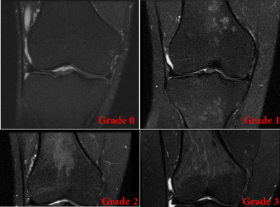
Benny Antony, Jasveen Kaur, Tao Meng, Alison Venn, Flavia Cicuttini, Lyn March, Marita Cross, Terence Dwyer, Andrew Halliday, Graeme Jones, Changhai Ding
An abnormal distribution of residual or reconverted red bone marrow (RBM) has been identified on routine knee MRI. We aimed to identify the prevalence and the association between RBM and symptoms and structural abnormalities in a young population (n=327, aged 31-41 years). The presence of RBM in the distal femoral, proximal tibial and fibular metaphysis was graded based on the percentage of the metaphysis occupied (grade 0 to 3). Reconverted or residual RBM around the knee joint was present only among females and always involved the distal femoral region. RBM was associated with overweight measures and knee joint pain.
|
|
1294.
 |
36 |
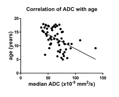
Variation of ADC of skull bone marrow with age and pubertal status in a pediatric population
Erika Pace, Andrew Mackinnon, Nandita deSouza
ADC measurements were possible from the clivus of children. Values showed a significant negative correlation with age. Following puberty, there was a reduction in ADC and a left shift in centile histogram values, likely as a result of fat replacement. This was independent of gender. Bone marrow within the clivus behaves like marrow from tubular bones after puberty, and does not retain a cellular morphology expected from red hematopoietic bone marrow.
|
|
1295.
 |
37 |
Trabecular Bone Imaging Using a 3D Adiabatic Inversion Recovery Prepared Ultrashort Echo Time Cones (3D IR-UTE Cones) Sequence
Ya-Jun Ma, Liang Li, Yanjun Chen, Saeed Jerban, Michael Carl, Eric Chang, Jiang Du
The trabecular bone is both functionally and biomechanically important for vertebrates, including humans. Evaluation of trabecular bone provides important information about risk of both osteoporosis and bone fracture. Direct MR imaging of trabecular bone is difficult due to its ultrashort T2* and low water content, resulting in little or no signal when conventional pulse sequences are used. The purpose of this study was to develop and evaluate a 3D adiabatic inversion recovery prepared UTE Cones (3D IR-UTE-Cones) sequence for volumetric imaging of trabecular bone ex vivo and in vivo on a clinical 3T scanner in clinically acceptable scan times.
|
|
1296.
 |
38 |
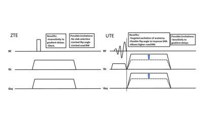
Comparison of ZTE vs UTE for MR Bone Imaging
Michael Carl, Yajun Ma, Ricardo Mello, Jiang Du, Eric Chang
We compared different center-out 3D radial trajectories (ZTE and UTE) and assessed their advantages and disadvantages for bone imaging. We found that while ZTE and UTE show similar results at the same read BW, the higher BWs available with UTE can help reduce undesired background signals in the final bone images.
|
|
1297.
 |
39 |
Estimating the diagnostic value of IDEAL-IQ for vertebral compression fractures caused by osteoporosis and metastasis
Presentation Not Submitted
Zhaolong Zheng, Qingliang Niu, Shasha Wu, Weiqiang Dou
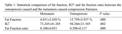
Traditional imaging methods are challenging to diagnose the vertebral compression fractures caused by osteoporosis and metastasis. In this study, the iterative decomposition of water and fat with echo asymmetry and the least squares estimation quantification sequence (IDEAL-IQ)technique, as a novel fat quantification technique, was applied to quantitatively determine the bone marrow fat content for the patients with osteoporosis caused and metastasis caused vertebral compression fractures. We found that the fat fraction (FF) and FF ratio of bone marrow fat in the vertebral body lesions of metastasis were significantly reduced compared with the acute compression fractures due to osteoporosis. Therefore, IDEAL-IQ has been proven an effective method for quantitative diagnosis of vertebral compression fractures.
|
|
1298.
 |
40 |
Discriminating between normal and cam positive hips using MRI texture and gradient-boosted decision trees
Rebecca Thornhill, Taryn Hodgdon, Gerd Melkus, Nick James, Paul Beaulé, Kawan Rakhra
Cam-type femoroacetabular impingement (FAI) results in altered biomechanics and acetabular pathology that has been associated with osteoarthritis of the hip. These early changes can be difficult to detect with routine clinical imaging. Texture analysis offers a more quantitative approach for characterizing gray-level patterns. The purpose of this study was to determine the MRI texture profile of acetabular subchondral bone in normal, asymptomatic cam positive and symptomatic cam-FAI hips with the assistance of gradient-boosted decision trees. This work demonstrates that MRI textural features can be used to generate machine learning models that can identify cam positive hips, regardless of symptom status.
|
|
1299.
 |
41 |
Optimal flip angle for imaging and T2* mapping of the human skull using ultra-short echo-time (UTE) imaging
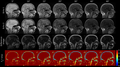
Martin Krämer, Benedikt Herzau, Jürgen Reichenbach
To investigate the influence of the flip angle on imaging and T2* mapping of the human skull, multi-echo 3D ultra-short echo-time (UTE) imaging was performed for multiple flip angles ranging from 5° to 49°. Results based on difference images between two echoes indicate that higher flip angles are better suited for separating the skull from adjacent tissues. In addition, a strong dependency of the skulls T2* values on the flip angle was observed
|
|
1300.
 |
42 |
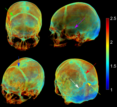
3D visualization of the cranial bone using fully automated segmentation based on ultra-short echo-time (UTE) imaging
Martin Krämer, Benedikt Herzau, Jürgen Reichenbach
To enable 3D-visualization of the cranial bone based on multi-echo ultra-short echo-time (UTE) data, a fully automated segmentation algorithm is presented. The algorithm concatenates several easy to implement processing steps while taking T2* maps calculated from three or more echoes or difference images calculated from the first two echoes of a UTE sequence as input. Comparison between a CT-based segmentation and the UTE-based segmentation showed very good agreement. The 3D visualization allowed easy assessment of the location and the course of cranial sutures as well as of diploic veins.
|
|
1301.
 |
43 |
Quantitative Evaluation of Vertebral Marrow Fat content with Aging in Healthy Human using MRS and Dixon technique
Shao-Chieh Lin, Hou-Ting Yang, Yi-Jui Liu, Hing-Chiu Chang, Xiang-Wei Xie, Wing P. Chang, Yu-Chen Cheng, Yi-Zong Liou
Subjects with osteoporosis have increased fat marrow, and fat content also increases progressively in the normal aging people. Although MRS has shown the potential probing the bone marrow content characteristics of the vertebra body in previous studies, all of them were a ROI analysis and loss local information. The purposes of this study were using MRS and Dixon technique to investigate the change of fatty marrow content with aging in human vertebra body. Our results have demonstrated the water-fat separation could quantitate vertebral marrow adiposity, which will be a potential method to provide local information for osteoporosis-related research fields.
|
|
1302.
 |
44 |
Evaluating Gadolinium Deposition in Rabbit Cortical Bone by Using Ultrashort Echo Time T1 Mapping: Preliminary Results
Kaixuan Zhao, Shisi Li, Yingjie Mei, Qinqin Yu, Keyan Yu, Cuiling Zhu, Jian Wang, Hanwen Deng, Xiaodong Zhang, Jiang Du, Yanqiu Feng
Gadolinium based contrast agents (GBCA) injection for enhanced MRI can induce gadolinium deposition in bones. In this work we investigated the feasibility of evaluating gadolinium deposition in rabbit cortical bone by using ultrashort echo time (UTE) T1 mapping at 7T. Lower T1 values were observed in the GBCA injection group than those in the control group (341±17.6ms vs. 450±10ms). This preliminary result indicates that UTE T1 mapping may be a feasible technique for evaluating bone gadolinium deposition.
|
|
1303.
 |
45 |
HIGH RESOLUTION IMAGING AND QUANTIFICATION OF VASCULATURE WITHIN CORTICAL BONE POROSITY USING DCE-MRI AND HR-pQCT
Matthew Gibbons, Po-hung Wu, Sarah Foreman, Misung Han, Roland Krug, Jing Liu, Thomas Link, Galateia Kazakia
Cortical bone porosity is a major determinant of bone strength. However, the causes of pathological pore growth are not well understood. The prevalence of blood vessels or marrow fat in pores may serve as an indicator for vessel- or marrow-driven processes. We present an algorithm to combine high resolution CT for pore identification and dynamic contrast enhanced MRI for blood vessel identification. Using this algorithm, imaged vessels are associated with specific pores and pore content is quantified.
|
|
1304.
 |
46 |
Vertebral bone marrow fat content changes in postmenopausal women receiving combined aromatase inhibitor and bisphosphonate therapy after one year
Michael Dieckmeyer, Stefan Ruschke, Alexander Rohrmeier, Jan Syväri, Ingo Einspieler, Jan Kirschke, Ernst Rummeny, Claus Zimmer, Dimitrios Karampinos, Thomas Baum
In recent years in-vivo assessment of vertebral bone marrow (VBM) fat composition has been increasingly used in the investigation of osteoporosis and bone metabolism. Postmenopausal women represent a population of particular interest because they are at higher risk for osteoporosis resulting from estrogen deficiency which can be potentially aggravated by aromatase inhibitor (AI) therapy. The present study demonstrated a relative increase in vertebral bone marrow fat content quantified by chemical shift encoding-based water-fat MRI in patients receiving simultaneous AI and bisphosphonate (BP) therapy, contradicting previous findings reported in the literature.
|
|
1305.
 |
47 |
Research on the feasibility of MR imaging of patients receiving anterior cervical surgery using MAVRIC SL-STIR sequence at 3T
Presentation Not Submitted
Renjie Yang, Yunfei Zha, Yu Zhang, Yang Fan
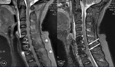
Metal implants are now very common in modern joint and spine surgeries. However, conventional MR images are significantly compromised by implant-induced magnetic susceptibility artifacts. A novel metal artifacts reduction technique, termed MAVRIC SL was proposed. The purpose of this study is to evaluate its clinical feasibility and diagnostic value in patients after anterior cervical surgery compared with routine 2D FSE sequence at 3T. As a result, although the image quality of MAVRIC SL is limited at 3T, it can still provide important additional diagnostic information through substantially reduced metal artifacts.
|
|
1306.
 |
48 |
Value of zero echo time imaging and CT in diagnosis of bone destruction of bone tumors and tumor-like lesions
Presentation Not Submitted
Liping Shi, Nianyun Li, Jie Meng, Han Wang, Yong Zhang, Bing Wu, Yanhong Xu
The purpose of the study was to evaluate the clinical applicability of zero echo time (ZTE) MR and compare the image quality between CT and ZTE imaging of bone tumor and tumor-like lesions. Thirty-six patients including 18 males and 18 females were recruited to undergo ZTE MR and CT. Agreement was assessed between raters and Weight Kappa statistics were performed. The difference of image quality between ZTE and CT imaging were not significant. Our results confirm that ZTE MR imaging provides accurate imaging of bone morphology with CT-like contrast that is not available with standard MR sequences.
|
|
1307.
 |
49 |
Evaluation of the effects of age and gender on water-fat composition of the lumbar vertebral bone marrow with magnetic resonance IDEAL-IQ sequence
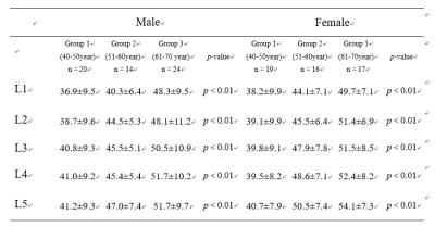
QING FAN, HUIPENG REN, XIAOHU WANG, LINGYUN SUO, Xiaocheng Wei
This study revealed the proton density fat fraction (PDFF) of lumbar vertebral bone marrow in different age groups and gender of normal adults using iterative decomposition of water and fat with echo asymmetry and least squares estimation (IDEAL) technique. We demonstrated significant differences of lumbar vertebral bone marrow PDFF across three age groups. Particularly, the highest PDFF was found in the oldest group. We also found a moderate positive correlation between age and PDFF, while the correlation was higher in female than in male. Taken together, our findings pave the way for exploring age-related lumbar vertebral diseases and metabolic disorders.
|
|
1308.
 |
50 |
7T Arterial Spin Labeling Imaging of Epiphyseal Bone Marrow in Distal Femoral Condyle – A Feasibility Study
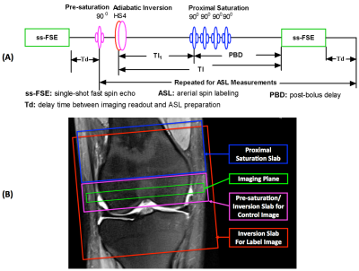
Xiufeng Li, Casey Johnson, Jutta Ellermann
Perfusion imaging of epiphyseal bone marrow in the distal femoral condyle can provide valuable insights into the pathophysiological mechanism of knee injuries or diseases, and has a great potential to facilitate the management of developmental knee diseases. A previous study at 3T indicated that low perfusion signal-to-noise ratio (SNR) imposed a great challenge to achieve high-quality bone marrow arterial spin labeling (ASL) images. 7T can benefit ASL imaging due to greatly increased blood T1 and imaging SNR. The presented study evaluated the feasibility and challenges of epiphyseal bone marrow ASL imaging in the distal femur at 7T.
|
|
| Top |
Cartilage 1
Digital Poster
Musculoskeletal
Monday, 13 May 2019
| Exhibition Hall |
08:15 - 09:15 |
| |
|
Computer # |
|
1309.
 |
51 |
Transfer Learning in Hip MRI Segmentation: Geometry Is More Important Than Contrast
Claudia Iriondo, Michael Girard, Valentina Pedoia, Sharmila Majumdar
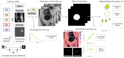
Transfer learning for medical image segmentation tasks is a promising technique that has the potential to overcome the challenges posed by limited training data. In this study we investigate the contribution of geometrically-similar and contrast-similar features for transfer learning to a hip MR segmentation task. We show pretraining with a geometrically similar task leads to more rapid convergence, can stabilize segmentation accuracy as datasets become reduced in size, and leads to more reliable biomarker extraction.
|
|
1310.
 |
52 |
BEES Knees: Bilateral Expedited Exam Supporting Quantitative Imaging of Knees
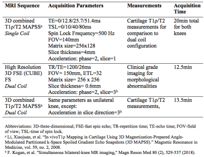
Emma Bahroos, Radhika Tibrewala, Misung Han, Bruno Astuto Arouche Nunes, Valentina Pedoia, Sharmila Majumdar
Osteoarthritis (OA) is a degenerative joint disease that commonly affects bilateral joints. This study combined simultaneous bilateral knee MRI with an automatic image processing for faster acquisition to image biomarker extraction. Simultaneous bilateral knee MRIs of 5 healthy volunteers was compared to singularly acquired knee images. Isotropic 3D CUBE FSE were used to automatically segment cartilage. Voxel based relaxometry from 3D combined T1ρ/T2 was evaluated for both types of acquisition. Our results show the ability of dual coil configuration allows for high resolution isotropic images, while also retaining the accuracy of quantitative data when compared to singly acquired bilateral knees.
|
|
1311.
 |
53 |
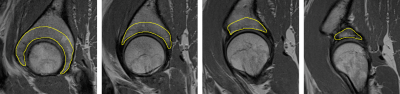
Quantitative Articular Cartilage Assessment in Patients with Juvenile Osteochondritis Dissecans (JOCD) at 3T MRI
Kai Ludwig, Casey Johnson, Stefan Zbyn, Shelly Marette, Takashi Takahasi, Jeffrey Macalena, Bradley Nelson, Marc Thompkins, Cathy Carlson, Jutta Ellermann
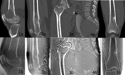
Juvenile osteochondritis dissecans (JOCD) is a disease affecting the knee joint of young active patients that can lead to early osteoarthritic changes. JOCD lesions are formed deep to the articular cartilage with late changes in the overlying articular cartilage. Our study was motivated by clinical observations that the opposing articular cartilage might be affected early. In this study, we observed a significant increase in T2* relaxation times in the articular cartilage of the medial tibia directly opposing the lesions when compared to the control region on the lateral site. These findings might indicate compositional changes in the tibial cartilage matrix due to increased biomechanical loading. Further study of T2* mapping as a potentially clinically realizable method to stage and prognosticate JOCD are warranted.
|
|
1312.
 |
54 |
Diffusion Tractography of the Rat Knee at Microscopic Resolution
Nian Wang, Gary Cofer, Yi Qi, G. Johnson
Application of DTI to map the complex collagen fibril structures in preclinical studies of the knee joint is still challenging, due to the limited spatial resolution previously used, relative low FA values, and relatively low signal-to-noise (SNR). We imaged the rat knees in a preclinical 9.4 T system with powerful gradients (2000 mT/m) to minimize TE. A modified 3D diffusion-weighted spin-echo pulse sequence was used to achieve isotropic spatial resolution at microscopic scale.
|
|
1313.
 |
55 |
Assessment of the Angular Dependence of 3D Ultrashort Echo Time Cones Adiabatic T1? (3D UTE-Cones-AdiabT1?) Imaging
Mei Wu, Yajun Ma, Lidi Wan, Tan Guo, Saeed Jerban, Hyungseok Jang, Eric Chang, Jiang Du
In this study we aimed to evaluate the magic angle sensitivity of the 3D UTE-Cones-AdiabT1ρ sequence in imaging the cadaveric human Achilles tendon and patellar cartilage samples on a clinical 3T scanner. The 3D UTE-Cones-AdiabT1ρ shows much reduced magic angle effect than regular T1ρ and T2*. The superficial layers show reduced magic angle effect compared to the middle and deep layers of articular cartilage. The 3D UTE-Cones-AdiabT1ρ sequence may provide magic angle insensitive evaluation of all the major knee joint tissues, thus providing a truly “whole-organ” approach for more accurate diagnosis of early OA.
|
|
1314.
 |
56 |
T1? at low spin-lock amplitudes is more sensitive to degenerative changes in articular cartilage
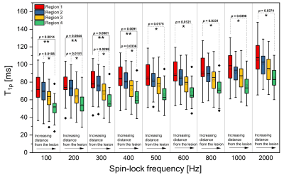
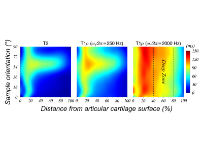
Abdul Wahed Kajabi, Victor Casula, Juuso Ketola, Jaakko Sarin, Irina Mancini, Jetze Visser, Harold Brommer, P. René Weeren, Jos Malda, Juha Töyräs, Mikko Nissi, Miika Nieminen
In this study, continuous wave T1ρ scans at various spin-lock amplitudes (γB1 = 100, 200, 300, 400, 500, 600, 800, 1000 and 2000 Hz) were utilized to evaluate multiple articular cartilage regions at increasing distances from a surgically induced lesion in equine specimens. Significant differences were observed between regions adjacent and distant to the lesion, and the differences between the compared sites were larger at lower spin-lock amplitudes. The variations were in agreement with biomechanical properties (determined via indentation testing) of the regions. The findings suggest that T1ρ at low spin-lock amplitudes is more responsive to progressive alterations in articular cartilage.
|
|
1315.
 |
57 |
UTE-based adiabatic T1? is sensitive to enzymatic proteoglygan degradation in human articular cartilage
Lidi Wan, Adam Searleman, Yajun Ma, Jonathan Wong, Mark Murphy, Jiang Du, Guangyu Tang, Eric Chang
A series of quantitative UTE techniques have been developed to assess articular cartilage. The early stage of osteoarthritis is characterized by proteoglycan (PG) loss in cartilage. This study aimed to determine if quantitative UTE-based biomarkers are sensitive to PG loss induced by chondroitinase ABC in cadaveric human cartilage. Pure cartilage wafers were exposed to sequential enzymatic digestion. MR imaging was performed before and after sequential digestion. PG loss was observed after digestion, with a corresponding increase in UTE adiabatic T1ρ values as compared to controls.
|
|
1316.
 |
58 |
Cartilage T2 Mapping of the Knee Using Fast Spin-Echo Multi-Band Acceleration
Robert Moskwa, Fang Liu, Graeme McKinnon, Rob Peters, Richard Kijowski
A T2 mapping sequence was performed in the axial and sagittal planes with multi-band (MB) acceleration (T2-MB) and without MB acceleration (T2) on the knees of seven healthy volunteers and three subjects with knee osteoarthritis at 3.0T. The use of MB acceleration provided a 50% reduction in scan time. The T2 and T2-MB sequences showed very similar pixel-by-pixel patellar cartilage T2 values in the axial plane and very similar regional knee joint cartilage T2 values in the sagittal plane for all subjects. However, there was a slight bias toward lower cartilage T2 values when using MB acceleration.
|
|
1317.
 |
59 |
Quantitative hip cartilage MRI of patients with hip dysplasia: Evaluation of dGEMRIC, T1? and T2* mapping
Gerd Melkus, Paul Beaulé, Geoffrey Wilkin, Kawan Rakhra
Developmental dysplasia of the hip is a common risk factor of early osteoarthritis. Knowledge of the molecular health of cartilage using quantitative MR methods could diagnose and stage the disease and may also allow for treatment stratification. In this study, we evaluated and compared three different biochemical sensitive MR methods (dGEMRIC, T1ρ and T2*) for cartilage imaging on patients with developmental dysplasia of the hip. Quantitative T1ρ as well as T2* mapping of the hip cartilage correlated significantly with the dGEMRIC method and could possibly replace the contrast-based method.
|
|
1318.
 |
60 |
Longitudinal T1? mapping of contralateral hip in patients with unilateral cam-type femoroacetabular impingement (FAI)
Gerd Melkus, Kawan Rakhra, Paul Beaulé
Cam-type femoroacetabular impingement (FAI) is a major cause of hip osteoarthritis. Quantitative T1ρ MRI has the potential to detect early cartilage degeneration due to its sensitivity to proteoglycan. In this study we performed longitudinally (124 days (average) after surgery and 4.8 years (average) follow up) T1ρ mapping in patients with bilateral (symptomatic and asymptomatic) cam-type FAI on the asymptomatic side after the symptomatic cam-FAI was surgically corrected. The cartilage of the contralateral hip did not show significant proteoglycan depletion and therefore no further degeneration between the initial and the follow up scan was detected. The contralateral hips remained stable.
|
|
1319.
 |
61 |
Multiple-echo steady-state (MESS): Simultaneous water-fat separation, T2T2, T′2T2′,
and T∗2T2∗ mapping
in the knee at 3 tesla
Frank Zijlstra, Peter Seevinck
This study proposes an extension of the double-echo steady-state (DESS) sequence to include multiple readouts. This multiple-echo steady-state (MESS) sequence supports quantification of water, fat, T2T2, T′2T2′,
and T∗2T2∗ in
a single, efficient acquisition. These parameters may provide additional tissue-specific MRI biomarkers for early detection and grading of osteoarthritis (OA), on top of the T2T2 quantification
of cartilage provided by the DESS sequence. In vivo results show that parameter quantification using MESS corresponds well with quantification on water-selective DESS images.
|
|
1320.
 |
62 |
Evaluating the Relationship Between gagCEST MRI and Cartilage Biochemical Composition in Juvenile Bovine Articular Cartilage
Lauren Watkins, Feliks Kogan, Elka Rubin, Marianne Black, Marc Levenston, Garry Gold
Chemical exchange saturation transfer of glycosaminoglycans (gagCEST) is a quantitative MR technique with potential for detecting early changes in cartilage composition. However, its relationship to tissue glycosaminoglycan (GAG) content has not yet been validated using standard biochemical assays. Here, we examine the relationship between gagCEST at 3T and 7T to cartilage biochemical properties using immature bovine femoral cartilage. Comparison of deep and superficial gagCEST asymmetry maps suggest that while gagCEST reflects the laminar differences in biochemical GAG composition, there is a weak correlation between gagCEST asymmetry and GAG content at 7T and 3T.
|
|
1321.
 |
63 |
Assessment of Cartilage pH Using AcidoCEST-UTE MRI at 3T with Histological Correlation
Rachel High, Yajun Ma, Qingbo Tang, Jonathan Wong, Lidi Wan, Jiang Du, Eric Chang
The poor correlation between structural abnormalities of osteoarthritis (OA) and OA pain complicates treatment and pain management. Acidosis is heavily implicated in pain, and thus may be used to identify areas of pain not associated with structural damage. In this study, we used a pH-sensitive imaging method known as chemical exchange saturation transfer (acidoCEST) MRI to assess acidosis in cadaveric cartilage tissue and assess the relationship between pH and osteochondral vascularity as determined on histology. We show that acidoCEST MRI can measure extracellular pH (pHe) in cartilage, enabling further studies into the complex relationship between acidification, osteochondral channels, and pain.
|
|
1322.
 |
64 |
Detecting Early Changes in ACL-Reconstructed Knee Cartilage: Cluster Analysis of T2 Relaxation Times in Superficial and Deep Cartilage and ADC Analysis
Marianne Black, Daehyun Yoon, Kate Young, Akshay Chaudhari, Feliks Kogan, Garry Gold, Marc Levenston, Brian Hargreaves
ACL-injured subjects are at an increased risk of developing osteoarthritis. There is a need to detect early osteoarthritic changes for the development of treatments that can slow or stop osteoarthritis progression. T2 and ADC are considered reflective of the structure and composition of cartilage, and may be valuable for detecting early osteoarthritis. This study used two qDESS acquisitions to obtain T2 and ADC maps in 10 ACL-reconstructed subjects and 10 controls 3-weeks, 3-months and 9-months post-surgery. Our results show that T2 cluster analysis was able to detect changes to the ACL-reconstructed knee as early as 3-months post-surgery.
|
|
1323.
 |
65 |
T1? Relaxation of Human Articular Cartilage Using Time-fractional Order Model
Lixian Zou, Haifeng Wang, Yuanyuan Liu, Weitian Chen, Yanjie Zhu, Dong Liang, Xin Liu
T1ρ imaging is a promising non-invasive diagnostic tool for early detection of articular cartilage degeneration. A mono-exponential model is normally used to describe the T1ρ relaxation process. However, mono-exponentials may not adequately to describe NMR relaxation in complex, heterogeneous, and anisotropic materials, such as articular cartilage. Fractional-order models have been successfully used to describe complex relaxation phenomena in the laboratory frame in cartilage matrix components. In this work, we develop a time-fractional order (T-FACT) model to analyze T1ρ relaxation in human articular cartilage. The results show the proposed method can better represent the T1ρ relaxation in human articular cartilage.
|
|
1324.
 |
66 |
ARCADE: An efficient anisotropic R2R2 relaxation
mapping for human knee cartilage at 3T
Yuxi Pang, Riann Palmieri-Smith, Dariya Malyarenko , Scott Swanson
Water proton R2R2 relaxation
in cartilage at 3T contains both an isotropic and an anisotropic contributions, with the latter being more sensitive to degenerative changes. A composite relaxation (R2−R1ρ)
mapping could be used to separate two contributions; however, its lengthy protocol had prevented it from being adopted in clinical applications. Here, we propose an efficient alternative based on a single T2W sagittal image to isolate an anisotropic R2R2 and
compare it with R2−R1ρ on
five live human knees. The comparable results demonstrate that the developed method could be easily used in clinical studies to characterize anisotropic R2R2 of
articular cartilage.
|
|
1325.
 |
67 |
Toward an orientation-independent MR relaxation metric from R1?R1? dispersion
in articular cartilage
Yuxi Pang
Residual dipolar interaction is the dominant mechanism for R2R2 relaxation
in cartilage, leading to the well-known "magic angle effect" observed in clinical MR imaging that makes reliable diagnostics challenging. Here, we show that the orientation-dependent factor in R2R2 could
be eliminated by a correlation time τbτb derived
from R1ρR1ρ dispersion
in terms of order parameter. This predication was tested on orientated bovine patellar cartilage specimens at 9.4T and on one live human knee at 3T. The preliminary data showed that the derived anisotropic R2R2 and τbτb had
respectively significantly high and moderate positive correlations in good agreement with the predication.
|
|
1326.
 |
68 |
Simultaneous image super-resolution and contrast synthesis techniques applied to routine clinical magnetic resonance images of the knee for advanced automated processing of joint cartilage
Ales Neubert, Pierrick Bourgeat, Jose Manjon, Craig Engstrom, Shekhar Chandra, Stuart Crozier, Jurgen Fripp
While high resolution 3D MR images are well suited for automated cartilage segmentation in the human knee joint, they are not routinely acquired in clinical practice which limits opportunities for reliable segmentation of cartilage using automated algorithms. We propose a neural network for generating synthetic MR images with enhanced contrast and higher spatial resolution from routine, low resolution clinical knee scans. Segmentation results showed that accurate cartilage segmentation can be obtained using the synthesised images.
|
|
1327.
 |
69 |
Localization of the bright ultra-short echo time MRI signal at the osteochondral interface
Olli Nykänen, Henri Leskinen, Mikko Finnilä, Sakari Karhula, Simo Saarakkala, Mikko Nissi
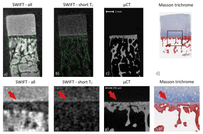
In this study, we investigated the bright signal feature that has been observed at the bone-cartilage interface using ultrashort echo time imaging sequences. We used micro-CT imaging of the same specimens to locate the origin of the signal in SWIFT-MRI images. The results indicated that the bright signal originates from the deep non-calcified cartilage region instead of the calcified cartilage, which has been previously hypothesized to be part of the signal location. The physiological origin of this signal as well as its role in the diagnosis of osteoarthritis remains to be evaluated.
|
|
1328.
 |
70 |
Diagnostic performance of three-dimensional fast-spin echo(3D MATRIX) accelerated with compressed sensing(CS) for internal injury of the knee
Presentation Not Submitted
Yakui Wang, Xiao Jin, Qiang Zhao, Ning Lang, Huishu Yuan
We aimed to evaluate the diagnostic performance of a three-dimensional fast-spin echo(3D MATRIX) accelerated with compressed sensing(CS) for internal injury of the knee joint.Sixty-two knee joints with trauma were examined at 3T MRI system including conventional 2D FSE protocol and 3D MATRIX before arthroscopic operations. Signal-to-noise ratio(SNR), contrast signal-to-noise ratio(CNR) and diagnostic performance were compared between two sequences. We found that 3D MATRIX had significantly higher SNR and CNR, and provided higher sensitivity but lower specificity for diagnosing cartilage injury compared to conventional 2D FSE. And the two sequences had similar diagnostic performance for ACL and meniscus tear.
|
|
1329.
 |
71 |
Comparison of Single-Component and Multi-Component T2 Parameters and Mechanical Parameters of Human Patellar Cartilage at 3.0T
Matthew Grondin, Fang Liu, Michael Vignos, Jiang Du, Corrinne Henak, Richard Kijowski
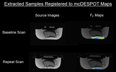
Multi-component Driven Equilibrium Single Pulse Observation of T1 and T2 (mcDESPOT) was used to measure single-component T2 relaxation time (T2Single) and the fraction of the fast relaxing macromolecular bound water component (FF) of 24 human patellar cartilage samples at 3.0T. The cartilage samples underwent unconfined compression testing to measure linear modulus and energy dissipation at 0.01Hz and 10Hz. There were low and marginally statistically significant (p=0.052-0.084) negative correlations between T2Single and linear modulus and energy dissipation. There were moderate and statistically significant (p<0.002) positive correlations between FF and linear modulus and energy dissipation.
|
|
1330.
 |
72 |
MRI texture analysis of vertebral subchondral bone
Presentation Not Submitted
Feifei Zeng, Yunfei Zha, Yang Fan
The purpose of this study is to assess the feasibility of MRI texture analysis as a method of quantifying vertebral subchondral bone (VSB) in early intervertebral disk degeneration (IDD). Sagittal T1WI, T2WI and T2* mapping images of lumbar vertebra were scanned at 3T MRI. Texture parameter values(mean, variance, skewness, correlation and entropy) of VSB (on T1WI) and T2* value of CEP were used for statistical evaluations. It was found that Significant differences in VSB texture parameters (mean ,variance and entropy) and T2* value of CEP were demonstrated between groups at the cephalic and caudal. Accordingly, texture parameter-mean showed significantly higher diagnostic accuracy than other texture parameters (variance and entropy) and T2* value for differentiating early IDD. MRI texture analysis can be used to assess human lumbar early IDD.
|
|
1331
 |
73 |
gagCEST effect strongly depends on GAG molecular composition
Video Permission Withheld
Emma Olsson, Pernilla Peterson, André Struglics, Michael Gottschalk, Patrik Önnerfjord, Jonas Svensson
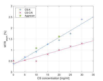
gagCEST has been suggested as a method for in vivo evaluation of cartilage GAG content. The main type of GAG in cartilage is chondroitin sulfate (CS), most commonly CS-A and CS-C. Validation of gagCEST have mostly been performed using CS-A but the main type in mature human articular cartilage is CS-C. In this study we evaluate the gagCEST effect from GAG in different forms. Our results indicate that mainly CS-A is contributing to gagCEST effect in cartilage, while no or little effect is seen from CS-C. gagCEST may therefore not correctly reflect the GAG content of human articular cartilage.
|
|
1332.
 |
74 |
Rapid Quantitative Simultaneous Bilateral Knee Imaging with Fully Automated Femoral Cartilage Analysis: Toward Knee Asymmetry Evaluation.
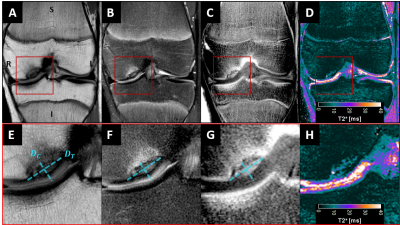
Marco Barbieri, Arjun Desai, Feliks Kogan, Valentina Mazzoli, Elka Rubin, Gastone Castellani, Garry Gold, Brian Hargreaves, Akshay Chaudhari
Quantitative bilateral knee MRI and whole femoral cartilage analysis is currently limited by costs and scan time. We propose a rapid, simultaneous bilateral knee MRI protocol followed by a fully automated pipeline to perform quantitative T2 analysis of the whole femoral cartilage plate of both knees. Five healthy subjects and a subject with an ACL reconstruction were scanned in this study and the results demonstrated high scan-rescan repeatability and a good agreement between manual and automatic segmentation. The proposed acquisition method with automated analysis may make bilateral imaging more feasible and efficient for use in longitudinal and cross-sectional studies.
|
|
1333.
 |
75 |
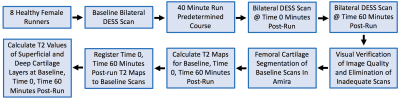
Short-Term Effects of Running on T2 Relaxation Times of Femoral Cartilage in Female Runners
Hollis Crowder, Valentina Mazzoli, Marianne Black, Lauren Watkins, Feliks Kogan, Brian Hargreaves, Marc Levenston, Garry Gold
This study tracks changes in water content in femoral cartilage from running by comparing T2 relaxation times of cartilage at baseline, time 0, and time 60 minutes post-run. Significant decreases in T2 relaxation times between baseline and time 0/time 60 minutes post-run scans occurred in superficial and deep cartilage, and a significant increase in T2 relaxation time occurred between time 0 and time 60 minutes post-run scans in deep cartilage, suggesting a reduction and partial recovery of cartilage water content. This study demonstrates the high sensitivity of T2 to cartilage loading patterns during running and supports the potential of using this setup as a method for identifying early changes in cartilage health.
|
|
| Top |
Muscle 2 & Other MSK
Digital Poster
Musculoskeletal
Monday, 13 May 2019
| Exhibition Hall |
08:15 - 09:15 |
| |
|
Computer # |
|
1334.
 |
76 |
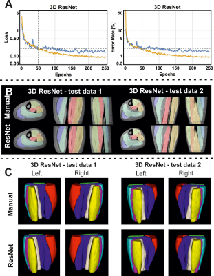
Evaluation of input data and UNet based convolutional network architectures for automated muscle annotation in 2D and 3D
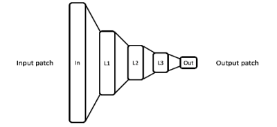
Martijn Froeling , Lara Schlaffke, Marlena Rohm, Ivana Isgum, Hermien Kan, Jelmer Wolterink
Manual annotation of muscle is still one of the most time-consuming steps in skeletal muscle MRI research. In this study we have investigated three aspects of automated muscle annotation using deep convolutional networks. First, we directly compare five different network architectures. Second, we compare the effect of providing various input data all based on Dixon imaging. Third, we investigate the effect of the amount of training data provided to the network. In summary we found that UNet-like convolutional networks allow for accurate and precise annotation of calf muscle in 2D and 3D and that the data provided is the strongest predictor of success.
|
|
1335.
 |
77 |
Assessing diffuse muscle fibrosis by ECV estimation, T1 and T2 relaxometry in a non-dystrophic murine model
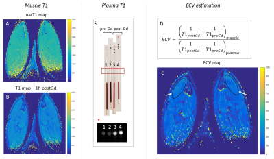
Aurea Martins Bach, Ericky Araujo, Julien Le Louër, Jean Marc Boisserie, Yves Fromes, Pierre Carlier
Skeletal muscle fibrosis, a key pathological feature in muscle disorders, is still inaccessible by NMR. In this study, we investigated the effects of diffuse muscle fibrosis on T2, natT1, and extracellular volume (ECV, estimated from muscle and plasma T1 pre and post-Gd injection). In order to reduce interfering effects with fibrosis, we developed a new non-dystrophic mouse model with increased muscle fibrosis, but limited levels of inflammation and no fat infiltration. A positive correlation between ECV and collagen content could be observed. Collagen content presented a weak positive correlation with T1, and a weak but significant negative correlation with T2.
|
|
1336.
 |
78 |
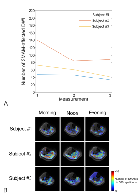
Longitudinal Analysis of Spontaneous Mechanical Activities in Resting Leg Musculature Assessed by Diffusion-Weighted Imaging: Preliminary Results
Martin Schwartz, Günter Steidle, Petros Martirosian, Thomas Küstner, Jürgen Machann, Anja Böhm, Cora Weigert, Bin Yang, Fritz Schick
Accurate quantification and grading of spontaneous mechanical activity of musculature of healthy and non-healthy subjects as measurable by single-shot diffusion-sensitive MRI requires certain long-term stability in order to reflect changes in the underlying muscular condition. Up to now, no longitudinal studies have been conducted, thus short- as well as long-term variation in the same subject under examination is unknown. This work examines the impact of the time of day when the examination takes places as well as long-term changes over 23-62 months in several subjects.
|
|
1337.
 |
79 |
IVIM imaging of muscle following moderate and high-intensity exercise
Erin Englund, David Berry, Samuel Ward, Lawrence Frank, Bahar Shahidi
IVIM imaging provides insight into microvascular blood flow. Here, we investigate IVIM parameters following moderate and high-intensity exercise protocols, activating the plantar flexors and dorsiflexors of the leg. By interleaving b=0 images throughout the diffusion-weighted acquisition, we were additionally able to evaluate T2 changes following exercise. We observed an increase in T2 and diffusion coefficient, D, following high-intensity exercise. Changes were less obvious following moderate-intensity exercise.
|
|
1338.
 |
80 |
Pulse sequence and reconstruction methods for extraction of spatial variation in multicomponent T2 relaxation for diagnosis of fluid and muscle disorders
Ashvin Bashyam, Chris Frangieh, Michael Cima
Significant unmet diagnostic need exists for diseases characterized by changes in T2 relaxation properties of tissue, especially those related to changes in fluid volume status and muscle disorders. Current methods for quantifying these disorders, such as MRI, are resource-intensive limiting widespread adoption. We introduce a novel method of quantitative tissue separation using single-sided magnetic resonance sensors. We explore pulse sequences and fitting techniques to characterize distinct tissue compartments in heterogeneous samples. We then extend these techniques to in vitro and in vivo models, and we finally apply them to measure the progression of acute muscle edema in an animal model.
|
|
1339.
 |
81 |
Fatty infiltration of paraspinal muscles is associated with bone mineral density of lumbar spine
Presentation Not Submitted
xiaodong zhang, Yinxia Zhao, Mingqian Huang, Shaolin Li, Chuan Huang
Paraspinal muscle fatty infiltration (FI) is an important factor affecting spinal function. However, there is no previous study investigating the relationship between paraspinal muscle FF and spinal BMD. Our study demonstrated that fat fractions of erector spinae (ES), multifidus(MF) and psoas(PS) of subjects with normal bone density were all significantly less than those of subjects with osteopenia and those with osteoporosis. There is an inverse correlation between paraspinal muscle FF and vertebral BMD after controlling for age, sex and BMI. Our results show paraspinal muscle FI increases while vertebral BMD decreases.
|
|
1340.
|
82 |
Quantitative evaluation and correlation analysis withperiacetabular muscle MRI in children with Slipped Capital Femoral Epiphysis (SCFE)
Yue Gao, Xiao hong Lv, Qi Li, Jiazheng Wang, Shinong Pan
This work compared theperiacetabular skeletal muscle between the healthy and the affected sides for the slipped capital femoral epiphysis (SCFE) patients using MRI. The correlation was studied between the disease progression and the muscle atrophy or fat infiltration to provide a potential criteria in MR images for the
|
|
1341.
 |
83 |
Permanent and non-permanent changes of skeletal muscle diffusion properties in triathletes and non-athletes detected by diffusion tensor imaging and T2 mapping
Sarah Keller, Enver Tahir, Jitka Starekova, Gunnar Lund, Zhiyue Wang, Jin Yamamura
The combined application of MRI Diffusion tensor imaging (DTI) and T2 mapping in professional triathletes and healthy controls at rest and hours after triathlon enables the detection of changes induced in skeletal muscle diffusion properties, and thus microstructure, caused by daily professional training and intensive exercise.
|
|
1342
 |
84 |
A preliminary study on the correlation between fat infiltration and muscle asymmetry in lumbar intervertebral disc herniation by using IDEAL-IQ
Video Permission Withheld
Hui Hao, Jiayin Tong, Xiaocheng Wei, Jianxin Guo, Xijun Jiao, Xianghui Zhang, Jian Yang
Disc herniation is one of the most common conditions of the lumbar spine.More and more people are suffering from this symptoms.Our study focused on fat infiltration of bilateral lumbar multifidus muscle in patients with lumbar disc herniation. Based on the IDEAI-IQ technology, proton density fat fraction of lumbar multifidus muscle is evaluted. The preliminary results show that the degree of fat infiltration in the lumbar spine protrusion is relatively higher in the herniated side than contralateral side.Our results can reflect the degree of fat infiltration quantitatively, which can be further expanded into the quantitative classification of different populations and different causes.
|
|
1343.
 |
85 |
Intravoxel Incoherent Motion (IVIM) Perfusion Imaging of the Shoulder Muscles Activated by Tennis Playing: Initial Results
Patrick Bosshard, Luciano Pescatore, Sebastian Kozerke, Christian Federau
Playing tennis involves complex simultaneous motion patterns of several muscles of the shoulder. Intravoxel incoherent motion (IVIM) perfusion imaging offers the possibility to map muscle activation by measuring changes in local blood flow. In this preliminary work four healthy right-handed volunteers were examined after tennis exercises using IVIM perfusion imaging. The results indicate a particular use of m. subscapularis and m. pectoralis major during forehand strokes and of the of m. subscapularis during service.
|
|
1344.
 |
86 |
Changes in strain tensor resulting from atrophy induced by Unilateral Limb Suspension of the calf muscle.
Vadim Malis, Usha Sinha, Ryuta Kinugasa, Sinha Shantanu
We quantified 3D strain tensor in the principle and muscle fiber basis along with two invariants (volumetric and octahedral shear strain) from multi-slice velocity encoded phase contrast images of the in-vivo human calf muscle under isometric contractions. Significant decreases in the medial gastrocnemius and soleus contractile strain eigenvalue and in the invariants with suspension may potentially arise from changes in muscle contractility and/or from extracellular remodeling. The significant reduction in shear strain may indicate a decrease in lateral transmission of force that may account for the disproportionate loss of force to loss of mass with atrophy.
|
|
1345.
 |
87 |
Creatine and phosphocreatine mapping of mouse skeletal muscle by a polynomial and Lorentzian line-shape fitting CEST method
Lin Chen, Peter Barker, Robert Weiss, Peter van Zijl, Jiadi Xu
Wild type (WT) mice and Guanidinoacetate N-Methyltransferase deficiency (GAMT-/-) mice that have low Cr and PCr concentrations in muscle were used to assign the Cr and PCr peaks in the skeletal muscle Z-spectrum. A PLOF method was proposed to simultaneously extract and quantify the Cr and PCr CEST signal by assuming two Lorentzian functions for the Cr and PCr peaks and a polynomial function for the background signal. High-resolution PCr and Cr maps of mouse skeletal muscle were obtained by the PLOF CEST method after calibration with in vivo MRS.
|
|
1346.
 |
88 |
Intramuscular magnesium measured by 31P-MRS is more closely associated with age and muscle function than is serum magnesium
Donnie Cameron, Ailsa Welch, Fatemeh Adelnia, Christopher Bergeron, David Reiter, Nicholas Brennan, Kenneth Fishbein, Richard Spencer, Luigi Ferrucci
We evaluate the relationships between muscle strength and intramuscular magnesium, measured by phosphorus magnetic resonance spectroscopy (31P-MRS), or serum magnesium. We further evaluate the relationships between these measures of magnesium status and age, sex, and muscle strength. Data were collected from participants in the Baltimore Longitudinal Study of Aging, a large cohort of normatively aging subjects encompassing a broad age range. Results showed that intramuscular magnesium was more closely associated with age and muscle function than was serum magnesium and may therefore represent a better clinical measure of magnesium status.
|
|
1347.
 |
89 |
Spatial heterogeneities of calf muscle perfusion and its implications
Nan Hu, Xuenchen Wang, Christopher Conlin, Xiaowan Li, Christopher Hanrahan, Gwenael Layec, Lei Zhang, Vivian Lee
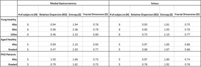
For the study groups of young healthy subjects, aged healthy subjects and peripheral artery disease patients, we measured exercise-stimulated perfusion in calf muscles using both T1-weighted DCE MRI. We found that the heterogeneity and texture complexity of calf muscle (specifically, the medial gastrocnemius and soleus muscle groups) perfusion decreased with exercise load among the young healthy subjects, and decreased with age among all healthy subjects. In addition, the heterogeneity of the calf muscle perfusion is greater among young healthy subjects when compared with peripheral artery disease patients.
|
|
1348.
 |
90 |
How to segment muscle images using qNMRI to obtain the highest discriminant power in natural history studies of muscle diseases in adult patients?
Harmen Reyngoudt, Jean-Marc Boisserie, Julien Le Louër, Cedi Koumako, Benjamin Marty, Pierre G. Carlier
Fat fraction (FF), as calculated from water-fat (Dixon) NMR images, is a largely accepted, if not fully validated, muscle imaging biomarker, which has been proposed as an outcome measure in most neuromuscular disorders these last few years. The question remains, however, as to whether specific muscle or muscle groups should be taken into consideration for longitudinal evaluation in specific neuromuscular diseases. Here, we looked into a cohort of patients suffering from three different neuromuscular disorders: immune-mediated necrotizing myopathy, (sporadic) inclusion body myositis and GNE myopathy. The aim of this work was to compare whole-segment FF with individual muscle and muscle group FF values and identify the most efficient procedure to quantify disease progression, by comparing the standardized response means.
|
|
1349.
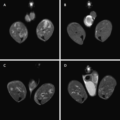 |
91 |
T2, MTR, and diffusion tensor quantitative MRI methods reliably monitor AAV-mediated gene therapy treatment in a murine muscular dystrophy model
Yasser Nazari, Joshua Park, Guy Odom, Ravneet Vohra, Jeff Chamberlain, Donghoon Lee
Herein we investigate the capability of multi-parametric MRI to reliably and decisively monitor both minute qualitative and quantitative changes in the skeletal muscle microenvironment in a Duchenne muscular dystrophy mouse model, namely mdx4Cv. Through the use of an assortment of pulse sequences, including T2, magnetization transfer ratio, and diffusion tensor imaging, the progression of a recombinant adeno-associated viral vector treatment, by the delivery of a truncated micro-dystrophin gene, is monitored in these mice and better understood. This study fundamentally seeks to understand and confirm the practicality of MRI as a diagnostic and prognostic measure in muscular dystrophy models.
|
|
1350.
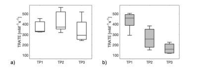 |
92 |
Longitudinal evaluation of myofiber microstructural changes in an ALS model using MR Cytography
Laura Bell, Alberto Fuentes, Deborah Healey, Natenael Semmineh, Ashley Stokes, C. Quarles
Amyotrophic lateral sclerosis (ALS) is a fatal upper and lower motor neuron degradation disease that leads to progressive myofiber abnormalities (e.g. decreased size and distribution). With recently FDA-cleared treatments, it is critical to identify non-invasive biomarkers of early disease onset, progression and therapy response. In this study, we validate the potential of a recently developed MR Cytography approach to detect myofiber microstructural changes in an ALS mouse model over three time points by comparison to relevant histologic markers.
|
|
1351.
 |
93 |
MRI Differentiation of Morel-Lavallee Lesions from Prepatellar Bursitis
Stephen Broski, Justin Friske
Differentiation of prepatellar bursitis from post-traumatic degloving injuries (Morel-Lavallee lesions) about the knee can occasionally present a diagnostic dilemma for clinicians and radiologists, although several features have been described as suggestive of a MLL etiology.
Our study adds to the evidence suggesting that intralesional fat globules and fluid extension beyond the confines of the normal prepatellar bursa are common findings in MLL lesions about the knee and suggestive that a lesion is unlikely to be an isolated prepatellar bursitis.
|
|
1352.
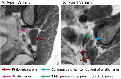 |
94 |
A 1x1x1mm isotropic resolution CUBE-FLEX for imaging piriformis syndrome
Daehyun Yoon, Brian Hargreaves, Christopher Beaulieu, Amelie Lutz
Piriformis syndrome is one of the more common causes for sciatica-type symptoms, defined as impingement of the sciatic nerve by the piriformis muscle. MRI can be very useful in identifying the causative anatomic configuration for presurgical planning, but the conventional 2D sequences can be very challenging for the evaluation of the complex oblique anatomy of the piriformis muscle and sciatic nerve. We present a 5-minute isotropic resolution (1mm3) 3D fast-spin-echo sequence with fat-water separation, allowing arbitrary reformats to specify the causative anatomy as well as T2-weighted contrast to detect abnormal signal of the nerve in the context of associated neuropathy.
|
|
1353.
 |
95 |
High resolution assessment of intramuscular fat content in the calf using T2-weighted MRI
Xiaowan Li, Conlin Christopher, Christopher Hanrahan, Vivian Lee, Jeff Zhang
Intramuscular fat is an important indicator of the health of the lower extremities. It can be difficult with conventional Dixon techniques to acquire high resolution fat-fraction maps, which may be helpful for detailed analysis of the muscle tissue. We propose to use a linear model to generate a high resolution fat-fraction map from high resolution T2-weighted images. The results show that the proposed method and the conventional Dixon method have good agreement. The high resolution fat fraction maps generated with the proposed method reveal anatomic details of intramuscular fat not visible with the conventional method.
|
|
1354
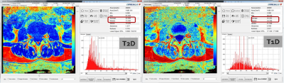 |
96 |
Magnetic resonance (MR) imaging-based fat signal fraction quantification of paraspinal back muscle: Comparison of T1, T2 and gradient-echo m-Dixon method
Video Permission Withheld
Seong Jong Yun, Wook Jin, Na-Young Choi
To compare the T1 and T2 m-dixon sequences with GRE mdixon sequence in FF quantification of paraspinal muscles and to evaluate the association of all sequences in FF quantification. Fat fraction quantification of the paraspinal muscles on three m-Dixon MR sequences were different from one another. But, they have the same tendency and strong correlation.
|
|
1355.
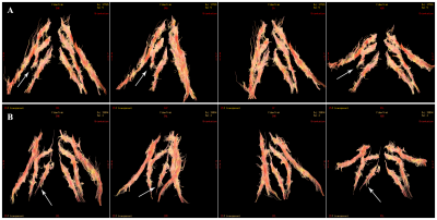 |
97 |
A quantitative and clinical evaluation of nerve root in lumbosacral radiculopathy using diffusion tensor imaging
Presentation Not Submitted
Yin Shi, Weiqiang Dou, Haibin Shi
In this study, we aimed to investigate if diffusion tensor imaging technique can quantitatively evaluate nerve root in lumbosacral radiculopathy by measuring MR fractional anisotropy (FA) values. By measuring 34 patients with unilateral disc related lumbosacral nerve root compression, we found that the mean FA value of nerve root correlate significantly with JOA score that is performed on the clinical function of the patients. Therefore, we can demonstrate that DTI can quantitatively and clinically evaluate nerve root by measuring FA values in disc related lumbosacral radiculopathy.
|
|
1356.
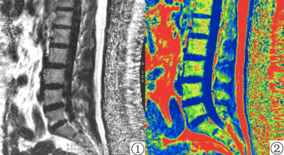 |
98 |
Evaluation of the risk of osteoporosis in diabetic patients by IDEAL-IQ
Yu Song, Qingwei Song, Nan Zhang, Yanwei Miao, Ailian Liu, Lizhi Xie
Osteoporosis is characterized by "decreased bone strength and increased risk of fracture”. Osteoporosis caused by diabetes is metabolized caused by decreased bone mass, bone microstructural destruction, increased bone fragility and prone to fracture. It is found that IDEAL-IQ can monitor bone marrow fat changes, diagnose bone marrow lesions or evaluate their functional status.
|
|
1357.
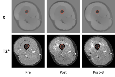 |
99 |
Evaluation of local changes in femoral bone marrow during a mountain ultra-marathon with quantitative MRI
Hoai-Thu Nguyen, Thomas Grenier, Benjamin Leporq, Loïc Bey, Magalie Viallon , Pierre Croisille
This study investigates the local changes in femoral bone marrow in a longitudinal MRI dataset of mountain ultra-marathon runners acquired during the Tor des Géants 2014 based on the MRI quantitative metrics with a rigorous statistical analysis procedure. The results highlight the different characteristics of different quantitative metrics that provides multiple insights into the data and open various perspectives for further analysis.
|
|
1358
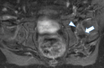 |
100 |
MR evaluation of hips following surgical reduction for developmental hip dysplasia: Are there quantitative and qualitative predictors of avascular necrosis?
Video Permission Withheld
Christian Barrera, Jie Nguyen
The purpose of our study is to investigate whether there are qualitative and quantitative imaging markers on the post reduction MR study that can predict the development of AVN using follow-up pelvic radiographs as reference standards. Post-reduction contrast-enhanced MR studies from thirty-seven infants with unilateral DDH were retrospectively reviewed which showed focally (27%) or globally decreased femoral head enhancement (22%). None of the clinical and imaging markers, including both qualitative and quantitative imaging factors present on the post reduction contrast-enhanced MR, can predict the future development of AVN, which sheds light on our incomplete understanding of the underlying pathophysiology.
|
|
| Top |
Bone 2 & MSK Tumors
Digital Poster
Musculoskeletal
Monday, 13 May 2019
| Exhibition Hall |
08:15 - 09:15 |
| |
|
Computer # |
|
1359.
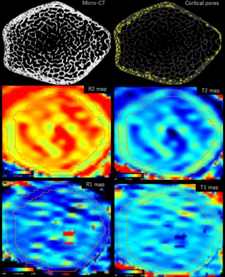 |
101 |
Quantitative comparison of conventional and synthetic MRI for cortical and trabecular bone architecture: ex vivo study of bovine coccyx
Miyuki Takasu, Yuji Akiyama, Akira Nishikori, Yoko Kaichi, Shogo Maeda, Kazuo Awai
Synthetic MRI has recently been shown to offer comparable performance to conventional pulse sequences in the assessment of intracranial abnormalities with greater time efficiency. We assessed the capability to predict bone architecture from MRI by comparing T1 and T2 relaxation times calculated by conventional and synthetic MRI and bone indices determined by micro-CT using bovine coccyx. This experimental study demonstrated that cortical porosity was significantly correlated with the cortical bone T2 relaxation time calculated from synthetic MRI. Synthetic MRI is feasible for assessing bone architecture and bone quality in clinically relevant acquisition time without radiation exposure.
|
|
1360.
 |
102 |
A fully automatic and robust system for quantitative measurement of lumbar curvature
Yao Wang, Fei Gao, Mei Yang, Shui Liu, Xiaodong Zhang, Jue Zhang, Xiaoying Wang
The change of lumbar curvature is used as the intuitionistic reflection for nearly all lumbar spine lesions, such as low back pain. Although several automatic segmentation methods have been proposed for the lumbar spine, those techniques cannot be directly applied to the diagnosis of spinal lesions due to the lack of quantitative estimation in lumbar curvature. In this study, by using a machine learning strategy, we designed an analysis pipeline and developed a fully automated measurement system of lumbar curvature, then validated it against a dataset of 45 subjects with T2w images.
|
|
1361.
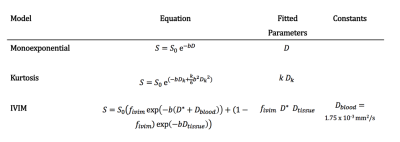 |
103 |
Intravoxel Incoherent Motion Outperforms Monoexponential and Kurtosis Models of Diffusion Attenuation in Normal and Inflamed Bone Marrow
Timothy Bray, Alan Bainbridge, Margaret Hall-Craggs, Hui Zhang
Recently, there has been interest in the use of diffusion-weighted imaging (DWI) for quantifying inflammation of the skeleton. In spondyloarthritis, inflammatory exudates in the bone marrow increase the apparent diffusion coefficient (ADC), likely reflecting increased extracellular water. However, the ADC is a simplistic ‘summary’ measure and fails to disentangle the complex pathophysiological changes occurring at inflamed sites. Here, we show that the intravoxel incoherent motion (IVIM) model captures both the rapid ‘perfusion’ component and the slower ‘tissue’ components of the bone marrow diffusion signal, and thus provides a more accurate description of the signal than monoexponential and kurtosis models.
|
|
1362
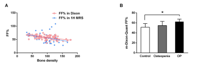 |
104 |
Percentage fat fraction in magnetic resonance imaging: an upgrade of the osteoporosis detecting parameter
Video Permission Withheld
Rong Chang, Xiaowen Ma, Ming Zhang
The diagnosis of osteoporosis (OP) is mainly based on orthopedic imaging approaches. The percentage fat fraction (FF%) in magnetic resonance imaging (MRI) has the potential to be used to predict and diagnose OP. In both 1-H MRS and mDIXON Quant MRI, the FF% exhibited a negative correlation with BMD. Among the three groups, the OP patients had significantly higher FF% compared to healthy subjects. In addition, the FF% according to mDIXON exhibited a positive correlation with age, and BMD showed a negative linear relationship with age. Furthermore, females had a higher FF% level compared to males. Furthermore, height was correlated with BMD but not FF%. MRI investigation (especially the FF% according to the mDIXON Quant imaging system) is useful in OP assessment. Parameters including gender and age, as well as height, are important factors for OP prediction and diagnosis.
|
|
1363.
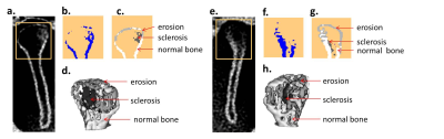 |
105 |
Evaluation of the Condylar Position and Volume in the Temporomandibular Joint Using Zero Echo Time MRI
Wei-Yin Liu, Hung-Ta Wu, Chien-Yuan Eddy Lin, Hsiao-Ling Lee, Wan-You Guo
The first clinical application with zero echo time (ZTE) based MR bone depiction and segmentation in the temporomandibular joint (TMJ) was presented in this study. Our result revealed that ZTE showed computed tomography (CT)-like bone contrast and more detailed demonstration of bony structures, which contributed more reliable structure images and volume measurements. A potential alternative radiation-free diagnostic approach, especially for patients who receive initial workups or serial follow-ups, can be adopted to assess the staging of temporomandibular disorder (TMD) by segmenting cortical bone of the condyle into normal bone, erosion, and sclerosis.
|
|
1364.
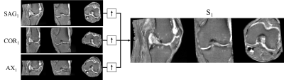 |
106 |
Construction of a population-based anatomical knee atlas and a probabilistic map of the spatial BML distribution
Christoph Arthofer, Yasmine Zedan, Brigitte Scammell, Thomas Kurien, David Walsh, Robert Kerslake, Dorothee Auer
The distribution of bone marrow lesions (BMLs) is an important factor in the assessment of osteoarthritis (OA) in the knee. We present a population-based (PB) anatomical atlas of the knee and a probabilistic map of the spatial distribution of BMLs.
|
|
1365.
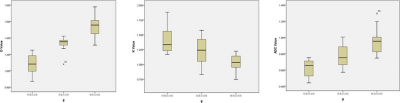 |
107 |
Evaluation of the Activity of Ankylosing Spondylitis by Diffusion Kurtosis Imaging in Sacroiliitis
zhengyang zhou, Jian He, Weibo Chen
Forty-four patients with AS underwent MRI including DKI to evaluate the disease activity of AS. Four disease activity states were chosen: inactive disease and moderate, high, and very high disease activity. The D and ADC values of sacroiliac joints in the active group were significantly higher, while the K value was significantly lower than those of the inactive group. The D value performed best in distinguishing different activity grades. The D and ADC values correlated positively, while the K value correlated negatively, with activity grade significantly. DKI of sacroiliac joints might be useful to evaluate the disease activity of AS.
|
|
1366.
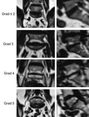 |
108 |
Cardiovascular Risk Factors for degenerative disc diseases of the thoracolumbar Spine in a Healthy General Population: Results from the KORA MRI Study
Sven Walter, Elke Wintermeyer, Roberto Lorbeer, Sergios Gatidis, Konstantin Nikolaou, Fabian Bamberg, Mike Notohamiprodjo
Evidence has grown that disc degeneration may be influenced by cardiovascular risk factors. Thus, the purpose was to assess the association in a healthy sample from the general population. Disc degeneration as well as disc bulging and protrusion of the thoracic and lumbar spine was assessed using the Pfirrmann-score and correlated to risk factors such as obesity, hypertension and diabetes. Hypertension dependently while age and BMI independently associated with disc degeneration. Diabetes increased disc degeneration at Th7/8 and L3/4. Lifestyle conditions had no influence. In conclusion, certain cardiovascular risk factors are associated to disc degeneration and disc bulging and protrusion.
|
|
1367.
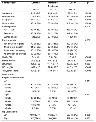 |
109 |
Assessment of the influence of metabolic syndrome on hip osteoarthritis by magnetic resonance imaging in subjects from the general population.
Sven Walter, Elke Wintermeyer, Roberto Lorbeer, Sergios Gatidis, Konstantin Nikolaou, Fabian Bamberg, Mike Notohamiprodjo
With being two of the leading chronic diseases, the purpose was to evaluate the effect of the metabolic syndrome on osteoarthritis of the hip in a healthy sample from the general population. Osteoarthritis was assessed according to the Kellgren-Lawrence classification, while the components of the metabolic syndrome were obtained as part of the clinical study protocol. Blood glucose was found to be a borderline non-dependent factor, while there was no correlation for waist circumference, triglyceride, blood pressure, and HDL. The overall metabolic syndrome was also not significantly correlated. In conclusion, metabolic syndrome and osteoarthritis of the hip are not associated.
|
|
1368.
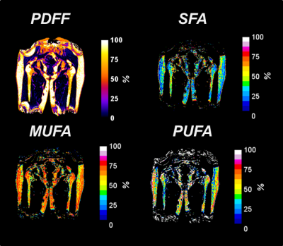 |
110 |
Bone marrow adipose tissue with 3T CSE-MRI in vivo: Preliminary study for long-term repeatibility assessment
Dimitri MARTEL, Benjamin LEPORQ, Ravinder REGATTE, Stephen HONIG, Gregory CHANG
Bone is composed of two tissues: a mineralized cortical and trabecular bone component and a fatty and hematopoietic marrow component. Osteoporosis is a disease of weak bone and increased fracture risk due to low bone mass. Recent studies pointed link between bone marrow adipose tissue (bMAT) and osteoporosis notably in term of bMAT composition. Chemical Shift- Encoded MRI (CSE-MRI) allows detection of fat amount and fat composition. Our aim was to assess long term repeatability of CSE-MRI in proximal femur bMAT.
|
|
1369.
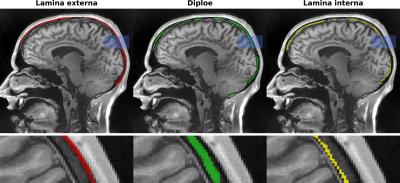 |
111 |
T2* quantification of the cranial bone using ultra-short echo-time (UTE) imaging
Benedikt Herzau, Martin Krämer, Jürgen Reichenbach
Quantification of T2* of the human cranial bone was performed by using high resolution multi-echo ultra-short echo time (UTE) imaging. Due to the extracted different short T2* values across the skull, it became possible to identify and differentiate the three-layer structure of the cranial bone consisting of the two compact bone layers lamina externa and lamina interna as well as the spongy interlayer diploe. Significant differences between the spongy interlayer and the two compact layers were obtained as regards their respective T2* relaxation times.
|
|
1370.
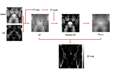 |
112 |
Bone Fraction Mapping with SyNthetic Auto-interpolated in-Phase (SNAP) MRI
Naomi Sakai, Timothy Bray, Stuart Taylor, Margaret Hall-Craggs, Alan Bainbridge
Quantification of bone mineral density (BMD) and/or bone fraction (BF) has long been a goal of MRI research, but thus far no method has successfully translated into clinical practice. Methods including R2* mapping, susceptibility mapping and ultrashort echo time MRI have shown promise but remain difficult to implement. A simpler approach to BMD/BF quantification is to measure the signal loss occurring in bone-containing voxels compared to the signal occurring in a bone-free voxel. We propose a method called SyNthetic Auto-interpolated in-Phase (SNAP) imaging which uses this principle and accounts for spatial variations in B1/coil sensitivity, enabling practical estimation of BMD/BF.
|
|
1371.
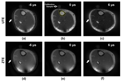 |
113 |
Impact of Hardware Timing Errors on Quantification of Bone Water with UTE and ZTE
Xia Zhao, Hyunyeol Lee, Hee Kwon Song, Felix Wehrli
Radial MR sequences are sensitive to system timing errors, particularly for ultra-short echo time (UTE) and zero echo time (ZTE) techniques. The effects of these timing errors on image quality were systematically evaluated with both simulation and imaging experiments, and their impact on bone water quantification investigated in vivo. Error in bone water concentration in the mid-tibia was found to be up to 35.4% using UTE and 16.6% for ZTE in the presence of timing errors as small as 6 μs. Further, the severity of image artifacts were spatially dependent, with objects further from FOV center more highly distorted.
|
|
1372.
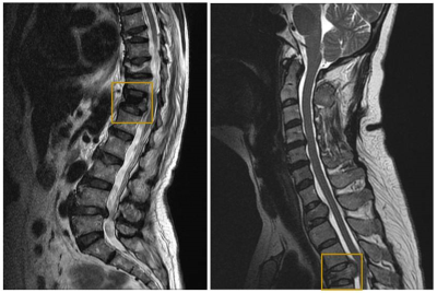 |
114 |
Differentiation of Vertebral Fracture Types using Five Different Convolutional Neural Network Approaches
Presentation Not Submitted
Lee-Ren Yeh, Yang Zhang, Jeon-Hor Chen, Peter Chang, Daniel Chow, Min-Ying Su
Differentiation of benign from malignant vertebral fracture is challenging yet very important for therapeutic planning. In this study, deep learning was conducted to automatically differentiate the fracture types using 5 different convolutional neural networks, including ResNet50, DenseNet, Xception, xceptionResNetV2, and InceptionV3. The final segmentation model was developed using 10-fold cross-validation applied in two different input methods, i.e. single slice or each slice combined with its two neighboring slices. Overall, the prediction accuracy was improved when each slice combined with its two neighboring slices was used as the input. Among the five deep learning approaches, XceptionResnetV2 showed the highest prediction accuracy.
|
|
1373.
|
115 |
Small-sized soft tissue tumor with deep location: Can MRI be used to differentiate benign from malignancy?
Ji Hyun Lee, Hyun Su Kim, Young Cheol Yoon, Wook Jin, Jang Gyu Cha
Small deep soft tissue tumors often results in dilemmatic clinical situation and raise clinical concern that deep location is one of risk factor for malignancy. We conducted this study hypothesizing that DWI with ADC value as well as conventional MRI parameters could help differentiation of benignity and malignancy for small deep soft tissue tumors; univariable and multivariable analyses were performed for all tumors and non-myxoid non-hemosiderin tumors. Although only small tumors were enrolled, lesion size was still important for differentiation of benign and malignant soft tissue tumors, being the most significant parameter in non-myxoid non-hemosiderin group.
|
|
1374.
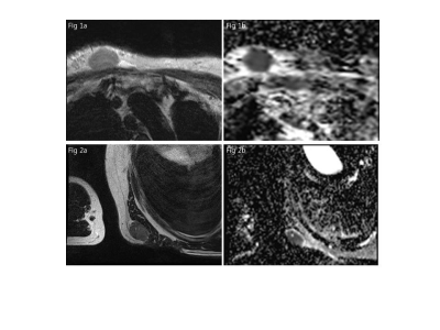 |
116 |
Differentiation of Soft Tissue Lymphoma from Undifferentiated Sarcoma: ADC Histogram Analysis of Whole Tumor Volume and Single-Slice ADC Measurements at 3T
Won-Hee Jee, Sunyoung Whang, Chan Kwon Jung, Joon-Yong Jung, Yohan Son, Mun Young Paek, Yang-Guk Chung, Seok-Goo Cho
Diffusion-weighted MR imaging should be included in differentiating soft tissue lymphoma from undifferentiated sarcoma to avoid unnecessary surgery in soft tissue lymphoma.
|
|
1375.
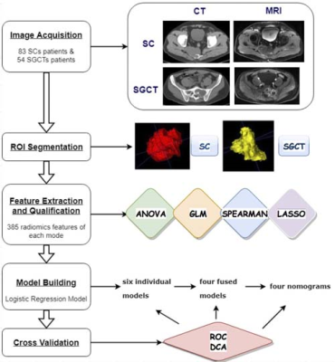 |
117 |
Clinical-Radiomics Nomograms for Preoperative Differentiation of Sacral Chordoma and Sacral Giant Cell Tumor Based on 3D Computed Tomography and Multiparametric Magnetic Resonance Imaging
Presentation Not Submitted
Ping Yin, Sicong Wang, Nan Hong
The purpose of our study was to build and evaluate clinical-radiomics nomograms for the preoperative differentiation of SC and SGCT. We compared individual features and mixed features based on CT and MRI respectively. And we also added clinical data to compare these models in terms of their performance of distinguishing SC and SGCT. Our results reveal that the model based on DWI features had the highest performance among individual scans. Mixed CT features performed better than individual scan, while mixed MR features achieved a low performance. Radiomics model can perform better when combined with clinical data.
|
|
1376.
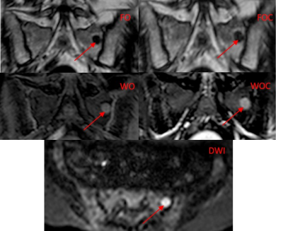 |
118 |
Whole body MRI in myeloma: Optimising scan acquisition and read times.
Saurabh Singh, Elly Pilavachi, Alexandra Dudek, Timonthy Bray, Arash Latifoltojar, Kannan Rajesparan, Shonit Punwani, Margaret Hall-Craggs
The study aim was to identify the highest value sequences for assessment of multiple myeloma by whole body MRI (WBMRI) to optimize scan acquisition and read times. Individual sequences (Dixon -/+ contrast, and diffusion imaging) were assessed in a prospective cohort of 29 patients with newly diagnosed multiple myeloma. Scan sensitivity, positive predictive value and reader confidence in detecting myeloma lesions and lesion conspicuity were assessed. Post-contrast water only Dixon and diffusion weighted images performed best. The results suggest that scan acquisition and read time can be reduced by focussing on WO contrast enhanced and diffusion images.
|
|
1377.
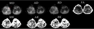 |
119 |
Diffusional kurtosis imaging in the differentiation of benign and malignant bone tumors
Ying Li, Cuiping Ren, Jingliang Cheng, Zhizheng Zhuo
This work investigated and evaluated the role of magnetic resonance (MR) diffusion kurtosis imaging(DKI) in characterizing the bone tumors, and furtherly evaluate the ability of DKI parameters to differentiate benign and malignant tumors by using receiver operating characteristic curve(ROC), which might be helpful for clinical diagnosis and studies.
|
|
1378.
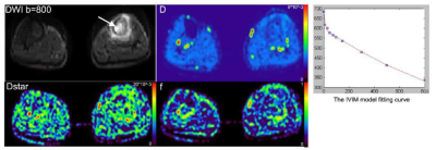 |
120 |
Feasibility study of intra-voxel incoherent motion MR imaging for the differentiation of benign and malignant bone tumors
Ying Li, Cuiping Ren, Jingliang Cheng, Zhizheng Zhuo
This work investigated and evaluated the role of intra-voxel incoherent motion(IVIM) MR imaging in characterizing the bone tumors, which might be helpful for clinical diagnosis and studies.
|
|
1379.
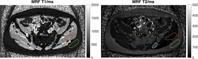 |
121 |
Exploratory evaluation of magnetic resonance fingerprinting confirms increased T1 and T2 in metastatic breast and prostate cancer lesions compared with normal marrow and volunteer values
Matthew Orton, Mihaela Rata, Nina Tunariu, Julie Hughes, Erica Scurr, James d'Arcy, Martin Leach, Wei-Ching Lo, Mark Griswold, Yun Jiang, Vikas Gulani, Dow-Mu Koh
Magnetic resonance fingerprinting (MRF) is a rapid imaging technique that allows generation of multiple, co-registered quantitative maps from a single sequence. This technique has been evaluated in the brain, abdomen, prostate, breast and heart, but has not been applied to metastatic disease in the skeleton. This abstract documents T1 and T2 values obtained with MRF in metastatic bone disease, muscle, fat and bone marrow, compares them with values obtained in volunteers using gold-standard imaging and MRF and demonstrates increased T1 and T2 in lesions compared with normal marrow and values in volunteers.
|
|
1380.
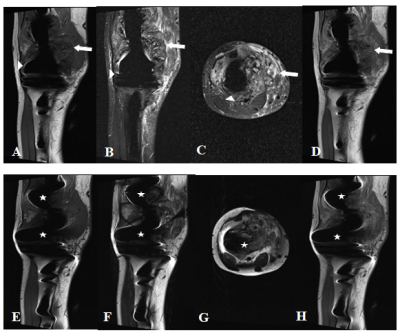 |
122 |
Diagnostic value of MR imaging with metal artifact reduction sequences in local recurrence of malignant bone tumor after joint replacement
Hanqi Wang, Yong Lu
Effective imaging evaluation after joint replacement is important for patients with malignant bone tumor. This study investigated the diagnostic value of MR imaging with metal artifact reduction sequences (MARS) in local recurrence of malignant bone tumor after joint replacement. The sensitivity, specificity, coincidence rate and Kappa value of MARS in the diagnosis of local recurrence were higher than FSE sequences. The ICC was 0.961 between MR images and pathology in measuring recurrent tumor volume. MR imaging with MARS has significant diagnostic value in local recurrence of malignant bone tumor after joint replacement.
|
|
1381
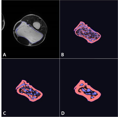 |
123 |
Usefulness of 3-dimensional MR Texture Analysis (3D-MRTA) for Distinguishing Well-differentiated Liposarcoma fom Lipoma
Video Permission Withheld
Seong Jong Yun, Wook Jin, Na-Young Choi, Kyung Nam Ryu
Although magnetic resonance (MR) imaging has been used as standard imaging tool, because of overlapping imaging features, the discrimination between well-differentiated liposarcoma and lipoma is not always clear. Also, no study has evaluated the diagnostic usefulness of 3-dimensional MR texture analysis (3D-MRTA) for distinguishing well-differentiated liposarcoma from lipoma. Therefore, the purpose of this study is to evaluate the usefulness of 3D-MRTA for differentiation between well-differentiated liposarcoma and lipoma, diagnosed on the basis of histopathological features. Regarding mean, MPP, kurtosis, and entropy, values on all sequences were significantly higher in well-differentiated liposarcoma than those in lipoma (p<0.001). The two best performing 3D-MRTA parameters were kurtosis on T2WI (AUC, 0.86; 95 % CI, 0.77-0.95) and entropy on FS-T1CE (AUC, 0.83; 95 % CI, 0.74- 0.93). There were good or excellent interobserver agreements for all measurements (ICC, 0.750–0.885).
|
|
1382.
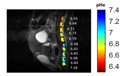 |
124 |
Assessment of Multiple Myeloma pH Using AcidoCEST-FISP MRI at 7T
Rachel High, Adam Searleman, Yajun Ma, Qingbo Tang, Jonathan Wong, Jiang Du, Patrick Frost, Mark Pagel, Eric Chang
Hypoxic conditions in multiple myeloma (MM) lesions drive anaerobic glycolysis and tumor acidosis, which contributes to tumor aggression and metastasis. In vivo imaging of acidosis in these lesions is important for determining the role of extracellular pH (pHe) in the pathology and spread of MM tumors. In this study, we use a novel imaging technique called chemical exchange saturation transfer (CEST) MRI to measure acidosis in vivo. We show that we are able to measure pH in MM lesions located in both vertebral bodies and regions of the knee using CEST MRI.
|
|
1383.
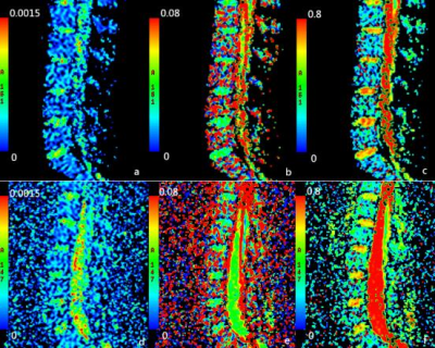 |
125 |
The parameters of Intravoxel Incoherent Motion Diffusion-Weighted Imaging for evaluating microstructure of bone marrow in myeloproliferative diseases: a Pilot Study
jinliang niu, wenqi wu, rong fan, kaiyu wang, weiguo li
Many myeloproliferativediseases,e.g., anemia and acute leukemia, impair microstructure of bone marrow. However, it is unclear the relationship between parameters of IVIM and microstructure of bone marrow (cellullarity, and microvessel density).Texture analysis was performed on D, D* and fmaps of the spinal bone marrow extracted from IVIM MRI on 48 patients (28 patients with acute leukemia and 20 patients with anemia). Bone marrow cellularity and MVD were significantly different between two groups. The f values in group of acute leukemia were significantly higher than that of anemia . The texture parameters of D, D* and f mapping has potential to reveal heterogeneous feature of microstructure in bone marrow.
|
|
| Top |
Cartilage 2, Meniscus, Tendon & Ligament
Digital Poster
Musculoskeletal
Monday, 13 May 2019
| Exhibition Hall |
08:15 - 09:15 |
| |
|
Computer # |
|
1384.
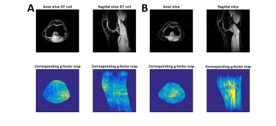 |
126 |
Uncompromised MRI of knee cartilage while incorporating sensitive sodium MRI
Sander Brinkhof, Aidin Haghnejad, Keita Ito, Karin Markenroth Bloch, Dennis Klomp
The goal of this work is to implement a dual tuned sodium-proton coil as transceiver for sodium and transmitter for hydrogen with a high-density proton receive array and compare the proton imaging performance with a state-of-the-art single tuned knee coil. A double-tuned knee coil was constructed as a shielded birdcage and 15 channel proton receiver coil embedded within the double-resonant birdcage. The double-tuned coil was implemented with comparable proton quality and acceleration possibilities compared to the single-tuned alternative while also being able to acquire high SNR sodium images.
|
|
1385.
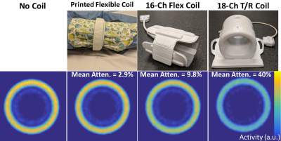 |
127 |
Minimized PET Attenuation in PET-MRI Knee Scanning with Flexible, Screen-Printed MR Coils
Feliks Kogan, Lauren Watkins, Jacob Thoenen, Peder Larson, Garry Gold
Simultaneous PET-MRI imaging is an exciting new technology for quantitative assessment of whole-joint disease in osteoarthritis (OA). Attenuation correction (AC) for MRI hardware is a challenge for PET quantification in hybrid PET-MRI systems. In this work, we tested a new, lightweight, screen-printed, flexible, 12-channel phased-array MR coil which reduces PET photon attenuation from MR hardware in a PET phantom to below 3%, potentially removing the need to correct for it. Further, the close proximity of coil elements to the knee show promise for high SNR MRI knee imaging.
|
|
1386.
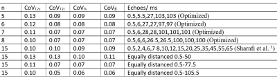 |
128 |
Optimized echo-times for biexponential T2-weighted imaging of the knee cartilage
Nima Gilani
T2-weighted MR signal of the cartilage knee has shown to be better explained by the biexponential relaxation model. The short and long T2 signal components presumably describe tightly bound and loosely bound macromolecular water components of the knee, respectively. More precise estimation of these two parameters might help in the better diagnosis of Osteoarthritis in reasonable scanning times. Here, Cramér-Rao Lower Bound method was used to find optimum echo times that improve estimation of these relaxation components. It was shown that using maximum echo times of twice as much as what is routinely used might substantially improve the biexponential estimates. Echo time optimization might play a role as important as increasing acceleration factors in reducing acquisition times.
|
|
1387.
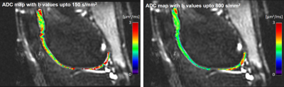 |
129 |
The b-value dependence of apparent diffusion coefficient (ADC) values in the knee cartilage
Daehyun Yoon, Sanghee Choi, Bragi Sveinsson, Brain Hargreaves
Apparent diffusion coefficient (ADC) can be a useful marker to track the degeneration of the knee cartilage during the early stage of osteoarthritis. We recently observed that the estimated ADC values change depending on the adopted b-values used in a DW-EPI sequence. We tested our hypothesis about this b-value dependence of ADC with in-vivo knee scans of 7 healthy subjects. Our statistical test result shows that the ADC estimated with the smaller b-values (0~150 s/mm2) is significantly higher than the ADC estimated with the larger b-values (0~800 s/mm2), suggesting the possibility of multi-component diffusion in the knee cartilage.
|
|
1388.
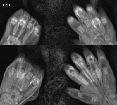 |
130 |
The role of contrast-enhanced whole-body joint MRI in juvenile idiopathic arthritis (JIA)
Lu Yutong, Tamotsu Kamishima
Due to the limitations of clinical description in diagnosis of juvenile idiopathic arthritis(JIA) with subjective factors, this study attempts to explore the role of contrast-enhanced whole-body MRI(WBMRI) in the JIA. We evaluated the WBMRI images of 156 joint regions of 7 patients, including image quality and positive findings, and compared the results with clinical descriptions. The results showed that WBMRI could observe the lesion of joints more objectively and comprehensively than the clinical description, and could show the changes of the lesions with treatment. Therefore, WBMRI may be used to help diagnose JIA and to evaluate the therapeutic effect.
|
|
1389.
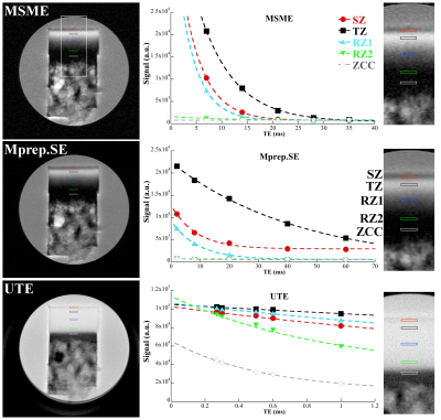 |
131 |
Quantification of Non-calcified and Calcified Zones in Articular Cartilage using MRI and PLM at Microscopic Resolution
Farid Badar, Yang Xia
The zone of calcified cartilage is the least explored region of cartilage in MRI, due to a strong dipolar interaction and a high mineralization of the subchondral region in articular cartilage. T2 weighted images by standard SE and GE sequences show little intensity of ZCC, whereas T2 and T1 weighted at the magic angle can highlight some parts of the region. Quantitative UTE can measure T2 of ZCC down to a fraction of a millisecond. With the aid of PLM, we can correlate the quantitative µMRI data with the optical properties of the ZCC.
|
|
1390.
|
132 |
Quantitative Evaluation of the Relationship Between the Femoral Trochlear Dysplasia and the Degeneration of the Patellofemoral Cartilage by MR T2mapping Technique
Siran Chen, Xinwei Lei, Yingying An, Ying Zhan, Zhizheng Zhuo, Jin Qu
Femoral trochlear dysplasia (FTD) is one of the common causes of early injury of knee cartilage in young people, and most of patients of FTD with the history of patellar dislocation, which seriously affects the quality of patients’ life. We focus on all the lateral, medial, deep and superficial layers of both patella and femoral trochlear cartilage in young people with quantitative T2 mapping. We find the superficial layers of lateral patellofemoral cartilage is mainly dehydration change in FTD, and there is more range of cartilage biochemical changes in age<30 years, compared to in age?30 years, because the controls may appear the cartilage degeneration with increased in age. Compared to the type A of FTD, there are more and wider areas of cartilage dehydration in type B-D, and one of them involve the deep layer and show edema.
|
|
1391.
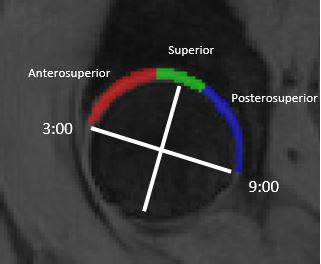 |
133 |
UTE T2* cartilage mapping: Regional differences in asymptomatic hips
Tony Wong, Patrick Quarterman, Diego Jaramillo, Thomas Lynch, Sachin Jambawalikar
UTE T2* images were acquired in 8 asymptomatic hips. The hip cartilage was segmented into anterosuperior, superior, and posterosuperior regions. Differences in mean T2* values between these regions were assessed. Results demonstrated a significant increase in the mean value of the anterosuperior region compared to the posterosuperior region.
|
|
1392.
|
134 |
CAN PREOPERATIVE MR IMAGING FINDINGS PREDICT EARLY FAILURE FOLLOWING ANTERIOR CRUCIATE LIGAMENT RECONSTRUCTION?
Presentation Not Submitted
Michele Mastio, Alejandra Duarte, Jose Raya, Laith Jazrawi, Jenny Bencardino
Despite all the attention given to anterolateral ligament injury as potential outcome predictor in the postoperative ACL knee, we found that MM tears perform best at predicting early ACRL failure. Preserving the integrity of the MM during ACLR procedure may be crucial in minimizing the risk for early ACLR failure.
|
|
1393.
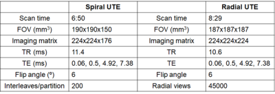 |
135 |
Comparison of Radial and Spiral UTE MRI and T2* quantification of the Knee Joint
Jeehun Kim, Richard Lartey, Kecheng Liu, Tiejun Zhao, Thomas Benkert, Heiko Meyer, Xiaojuan Li
The ultrashort TE (UTE) sequences allow measurement of fast-decaying signal from short T2* tissues in the joint such as tendon, ligament, and meniscus. By acquiring multiple echoes along the decay, T2* map can be generated, which has a potential to be used as a biomarker for diseases such as arthritis and tendinopathy. However, the comparison of T2* maps from different UTE sequences has not been fully explored. In this study, UTE based on stack of spirals and 3D radial acquisition were compared.
|
|
1394.
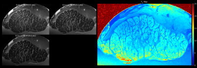 |
136 |
High-resolution ultra-short echo-time (UTE) ex-vivo imaging of Achilles tendons at 9.4T by water content alteration
Martin Krämer, Karl-Heinz Herrmann, Nicholas Brisson, Stefan Zachow, Georg Duda, Jürgen Reichenbach
To enable high-resolution imaging of ex-vivo ovine and porcine Achilles tendons, samples were extracted and stored in distilled water for up to 7 days to induce water intake and prolonged T2. One sample was measured continuously to quantify the change in T2 over time, while the other/second sample was used to acquire high-resolution structural images using a cryogenic measurement coil at 9.4T.
|
|
1395.
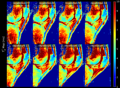 |
137 |
Time-resolved mapping of T2* in the patellar tendon during active knee flexion using multi-echo ultra-short echo-time (UTE) imaging
Martin Krämer, Nicholas Brisson, Stefan Zachow, Georg Duda, Jürgen Reichenbach
To investigate the distribution of T2* in the patellar tendon, and its dependency on factors such as tendon rotation angle and tendon composition, real-time ultra-short echo-time (UTE) imaging was performed during active knee flexion. While T2* is known to depend on the magic angle, it may also be influenced by the underlying tendon structure, which changes its composition when approaching the entheses.
|
|
1396.
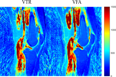 |
138 |
T1 and T2* relaxation parameter mapping of the patellar and quadriceps tendon by using ultra-short echo-time (UTE) imaging at 3T
Marta Maggioni, Martin Krämer, Jürgen Reichenbach
The tendon tissues in the knee are characterized by a short T2 relaxation time due to their fibrous structure and their high collagen content. Conventional MRI technique are unable to pick up such fast decaying signal, thus new methods and sequences such as UTE have been introduced. In this work a 3D UTE sequences was applied to quantify the T2* and T1 relaxation parameters for the patellar and quadriceps tendons.
|
|
1397.
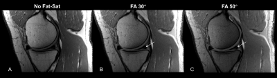 |
139 |
Improving the Conspicuity of Meniscal Tears in Knee MRI at 3T and 7T
Venkata Veerendranadh Chebrolu, Peter Kollasch, Benjamin Howe, Matthew Frick, Suzanne Carlson, Daniel Spence, Kimberly Amrami
In this work we propose the application of a small percentage of fat-saturation (fat-sat) using Spectral Heterogeneity Adaptive RF Pulses (SHARP) to improve the conspicuity of meniscal tears at 3T and 7T. Knee Turbo-spin-echo (TSE) MRI without fat-sat was compared with TSE MRI with a small percentage of fat-sat applied using SHARP to demonstrate the improvement in conspicuity of meniscal tears with SHARP at 3T and 7T.
|
|
1398
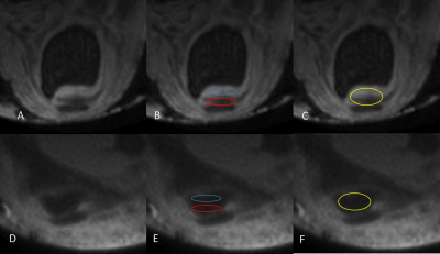 |
140 |
Three-Dimensional Ultrashort Echo Time(3D UTE) MRI of Achilles Tendon at 4.7TMRI With Comparison to ConventionalSequences in an Experimental MurineModel of Spondyloarthropathy
Video Permission Withheld
benjamin dallaudiere, aurelien trotier, emeline ribot, sebastien lepreux, sylvain miraux, olivier hauger
Unlike bSSFP T2 sequences, 3D-UTE sequences enable visualization of normal enthesis anatomy and early detection of abnormalities in pathological conditions.
|
|
1399.
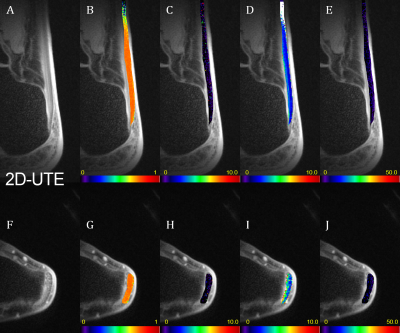 |
141 |
The comparison of two ultra-short echo time methods for T2* mapping in Achilles tendon and enthesis
Vladimir Juras, Peter Latta, Martin Kojan, Zenon Starcuk, Xeni Deligianni, Oliver Bieri, Siegfried Trattnig
We compared the performance of two MR techniques, ultra-short TE (UTE) and variable-echo time (vTE), to estimate mono- and bi-exponential T2* in Achilles tendon and enthesis at 3T. In axial slice orientation, the mean T2* values for UTE were (mono, short, long) 4.1 ± 1.1 ms, 0.9 ± 0.3 ms and 19.6 ± 7 ms, for vTE we found the values 10.5 ± 3.9 ms, 1.4 ± 0.4 ms and 25 ± 3 ms. In sagittal slice orientation, the mean T2* values for UTE were (mono, short, long) 2.8 ± 0.5 ms, 0.4 ± 0.1 ms and 10.5 ± 1.7 ms, for vTE we found the values 5.3 ± 1.5 ms, 1.6 ± 0.3 ms and 22.3 ± 5.6 ms. T2* values calculated with vTE are overestimated in comparison to UTE, however, the clinical value of vTE might be higher, since it provides more slices with reduced blurring and lower scan times.
|
|
1400
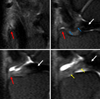 |
142 |
Utility of radial reformatted images created from 3T high-resolution 3D wrist MRI in evaluation of the radioulnar ligaments.
Video Permission Withheld
Taiki Nozaki, Saya Horiuchi, Akimoto Nimura, Atsushi Tasaki, Nobuto Kitamura, Keiichi Akita, Yasuyuki Kurihara, Hiroshi Yoshioka
The radioulnar ligament (RUL) has been thought as the major stabilizer of the distal radioulnar joint (DRUJ) under dynamic loading. A detailed depiction of the dorsal and palmar RULs on MRI is very important. However, identification of the RULs including their ulnar styloid attachment on MRI is very difficult on the routine coronal plane, due to not only their small size with complexity of structures but the partial volume effect. The purpose of this study was to investigate the value of radial-slice MPR images sourced from 3D isotropic high-resolution MR images for the depiction of the RULs.
|
|
1401.
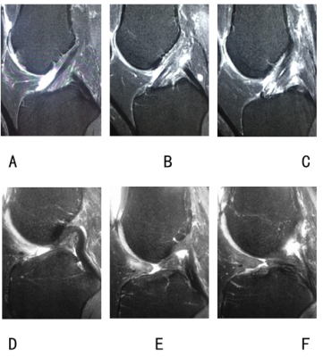 |
143 |
The value of 30°flexed knee MRI in evaluating anterior cruciate ligament tears
Xiao Jin, Yakui Wang, Qiang Zhao, Ning Lang, Huishu Yuan
The study aimed to investigate the value of 30°flexed knee MRI in evaluating anterior cruciate ligament (ACL) tears. 64 patients with knee trauma and hospitalized for arthroscopy were included and performed knee MRI in slightly flexed(about 17°) and 30° flexed positions successively. Images of both positions have high sensitivity, specificity and accuracy in diagnosing ACL tears.30°flexion images were superior to 17°in delineating ACL full length, torn ACL’s disrupted sites and ligament remnants, while no significant difference was found in delineating ACL double-bundle structure. Thus 30°flexed knee MRI was recommended in patients suspecting ACL tears.
|
|
1402.
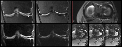 |
144 |
Could the 3D CAIPIRINHA accelerated SPACE imaging replace the conventional 2D MRI in routine knee examination?
Ruyi Xie, Dong Sun, Xiaoyue Zhou, Qiong Zhang, Xiaoming Li
3D CAIPIRINHA accelerated SPACE is a newly developed technic that can achieve faster acquisition and high resolution isotropic 3D imaging of the knee. However, its clinical diagnostic performance in knee joint hasn’t been proved yet. In this study, we compared the clinical diagnostic performance of 3D CAIPIRINHA accelerated SPACE and the conventional routine knee MRI in patients with knee injuries. We found that 3D CAIPIRINHA accelerated SPACE was able to detect more cartilage lesions and meniscus tear than the conventional 2D knee protocol. Whereas, the clinical diagnostic performance of the other lesions, such as bone marrow edema and ligaments tear were similar of the two protocols.
|
|
1403.
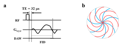 |
145 |
Effects of sampling window on quantitative imaging of major tissues in the knee joint using 3D ultrashort echo time (UTE) Cones sequences
Lidi Wan, Yajun Ma, Saeed Jerban, Micheal Carl, Eric Chang, Guangyu Tang, Jiang Du
We have developed a series of 3D UTE-Cones MRI techniques for quantitative assessment of all major tissues in the knee joint. Continuing improvements in scan time will facilitate their clinical use, but it remains unclear how acceleration affects quantification. This study focused on the effects of stretching sampling windows on quantitative UTE biomarkers in cadaveric human knee joint tissues. We found that extending sampling windows results in vastly reduce scan time without adverse effects on quantitative accuracy of T1, AdiabaticT1ρ, macromolecular fraction and MTR of articular cartilage, menisci, tendons and ligaments in the knee joint.
|
|
1404.
 |
146 |
Assessing the performance of knee meniscus segmentation with deep convolutional neural networks in 3D ultrashort echo time (UTE) Cones MR imaging
Michal Byra, Mei Wu, Xiaodong Zhang, Hyungseok Jang, Yajun Ma, Eric Chang, Sameer Shah, Jiang Du
Automatic segmentation of the knee menisci would facilitate quantitative and morphological evaluation in diseases such as osteoarthritis. We propose a deep convolutional neural network for the segmentation of 3D UTE-Cones Adiabatic T1ρ-weighted volumes of the meniscus. To show the usefulness of the proposed method, we developed the models using regions of interests provided by two radiologists. The method produced strong Dice scores and consistent results with respect to meniscus volume measurement. The inter-observer agreement between the models and the radiologists was very similar to that of the radiologists alone.
|
|
1405.
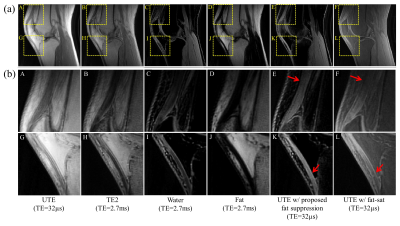 |
147 |
Fat suppression based on single point Dixon for morphological and quantitative UTE imaging
Hyungseok Jang, Yajun Ma, Michael Carl, Saeed Jerban, Eric Chang, Jiang Du
Use of fat saturation pulses with UTE sequences can improve short T2 contrast and parameter estimation, but can reduce the short T2 signal due to the broad spectrum. Moreover, conventional two-/three-point Dixon-based methods can misestimate fat contents due to the short T2* decay in UTE imaging. In this study, we demonstrate feasibility and efficacy of a single point Dixon method to suppress fat for morphological and quantitative UTE imaging. We evaluate the proposed framework in in vivo experiments in a clinical 3T MR system: morphological UTE imaging and UTE-T1 mapping in knee joints and UTE-bone imaging in a leg.
|
|
1406.
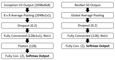 |
148 |
Meniscal Tear Detection with Machine Learning: Initial Experience
Eric Bultman, Akshay Chaudhari, Arjun Desai, Garry Gold
Despite rapid recent advances in convolutional neural networks used for image classification, generalizability of these networks to medical image data has not been thoroughly investigated. In this work, we utilize two networks designed to classify ImageNet natural-image data – Inception-v3 and ResNet-50 – and investigate their performance in classifying meniscal tears on MR examinations of the knee. Using limited segmentation and manual tear identification, slice-wise sensitivity of 0.68 and 0.58 is achieved for the respective networks. Applying the “two-slice-touch” rule, sensitivity is significantly increased, but with concomitant decrease in specificity. Our results support the feasibility of utilizing CNNs for meniscal tear identification.
|
|
1407.
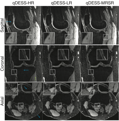 |
149 |
Accurate 3D T2 Relaxometry with Simultaneous High-Resolution Structural Imaging using Deep Learning
Akshay Chaudhari, Arjun Desai, Zhongnan Fang, Eric Bultman, Jin Lee, Garry Gold, Brian Hargreaves
Rapidly obtaining high-resolution structural magnetic resonance images (MRI) and generating quantitative biomarkers, such as the T2 relaxation time, using a single sequence is useful for musculoskeletal imaging. However, high-resolution is at odds with high signal-to-noise ratio (SNR) in MRI, which makes it challenging to simultaneously optimize for image quality and quantitative accuracy. In this study, we demonstrate how deep-learning-based super-resolution can create high-resolution images with accurate T2 values using a prospectively-sampled 5-minute quantitative double-echo steady-state sequence. We validate this method using high-SNR reference sequences for T2 accuracy and high-resolution reference sequences and a reader study for image quality assessment.
|
|
1408.
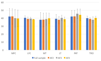 |
150 |
Prospective Fast T1? Mapping of Knee Articular Cartilage using Compressed Sensing
Mingrui Yang, Jeehun Kim, Chaoyi Zhang, Leslie Ying, Xiaojuan Li
Quantitative magnetic resonance (MR) mapping is known to provide additional information compared to the conventional qualitative weighted MR images. Relaxation times such as spin-spin relaxation time (T2) and spin-lattice relaxation time (T1ρ) have been shown enable early detection of human knee cartilage degeneration. Obtaining these relaxation parameter maps, however, requires long acquisition time. Compressive sensing (CS) has been extensively studied over the last decade as one of the possible ways to accelerate MR acquisition. These studies, however, are retrospective. In this study, we perform a prospective downsampling study on T1ρ mapping of knee cartilage using CS, which implements the downsampling pattern into the in vivo MR scan sequence.
|
|
| Top |
MSK Techniques & Development: Other MSK
Digital Poster
Musculoskeletal
Monday, 13 May 2019
| Exhibition Hall |
08:15 - 09:15 |
| |
|
Computer # |
|
1409.
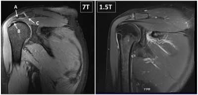 |
151 |
Clinical shoulder MR imaging at 7 Tesla in comparison to arthroscopy and 1.5 Tesla MRI
Oliver Kraff, Andrea Lazik-Palm, Stefan Rietsch, Sascha Beck, Konrad Koersmeier, Michael Kamminga, Jens Theysohn, Harald Quick
An 8-channel transmit/15-channel receive radiofrequency coil setup for 7T shoulder imaging has been evaluated in eight patients presenting with shoulder pain. In total, ten gradient echo and turbo spin echo sequences were included in the imaging protocol. Image quality was assessed in consensus by two radiologists. A structured report focusing on the rotator cuff was generated based on the 7T images and subsequently compared with the arthroscopical report taken as the gold standard. Finally, pathologies found at 7T were visually compared with preexisting 1.5T MR images from the same patients.
|
|
1410.
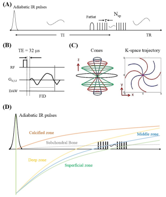 |
152 |
Imaging of the Region of the Osteochondral Junction (OCJ) Using a 3D Adiabatic Inversion Recovery Prepared Ultrashort Echo Time Cones (3D IR-UTE Cones) Sequence at 3T
Ya-Jun Ma, Saeed Jerban, Michael Carl, Lidi Wan, Tan Guo, Hyungseok Jang, Graeme Bydder , Eric Chang, Jiang Du
The osteochondral junction (OCJ) region serves as a functional barrier between the synovial joint space and subchondral bone marrow, and may play an important role in the pathogenesis of osteoarthritis (OA). MRI of the OCJ region is difficult due to the short T2 of deep radial uncalcified cartilage and calcified cartilage, which results in little or no signal when conventional pulse sequences are used. The purpose of this study was to use a 3D adiabatic inversion recovery prepared UTE Cones (3D IR-UTE-Cones) sequence for volumetric imaging of OCJ region with high spatial resolution and contrast in clinically acceptable scan times.
|
|
1411.
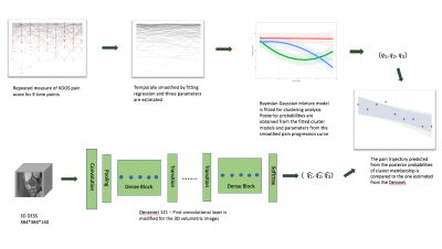 |
153 |
Predicting Pain Trajectories in Knee Osteoarthritis Subjects by Learning Image Biomarkers from Structural MRI
Jinhee Lee, Felix Liu, Sharmila Majumdar, Valentina Pedoia
The relationship between image biomarkers in structural MRI and knee osteoarthritis pain progression is investigated. A Bayesian Gaussian mixture model is selected to identify the distinct knee pain trajectories among subjects in the dataset from the Osteoarthritis Initiative. Deep learning is employed to predict the probability of an individual’s pain curve cluster membership using the 3D structural MRI. Utilizing the strength of the model-based approach, the pain curves are simulated from the GMM posterior probabilities and the weights learned to evaluate the 3D DenseNet’s performance.
|
|
1412
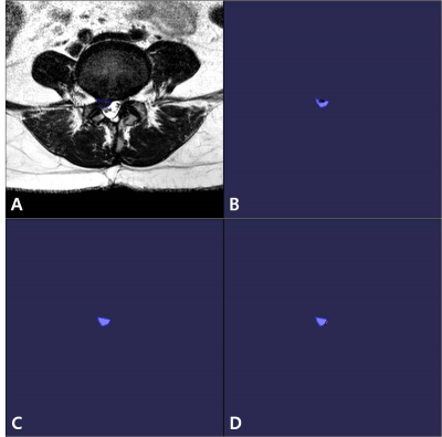 |
154 |
Usefulness of Magnetic Resonance Texture Analysis for Differentiation between Recurrent Disc Herniation and Postoperative Epidural Fibrosis
Video Permission Withheld
Seong Jong Yun, Wook Jin, Na-Young Choi, Kyung Nam Ryu
Although magnetic resonance (MR) with contrast-enhancement has been used as standard imaging tool for distinguishing recurrent disc herniation from postoperative epidural fibrosis, it is relatively invasive and vulnerable to contrast material-related complication. Also, the differentiation between recurrent disc herniation and postoperative epidural fibrosis on non-enhanced MR is not always clear. Until now, no study has evaluated the diagnostic usefulness of MR texture analysis (MRTA) for differentiation between recurrent disc herniation from postoperative epidural fibrosis. Therefore, the purpose of this study is to evaluate the usefulness of MRTA for differentiation between recurrent disc herniation from postoperative epidural fibrosis. Regarding mean, skewness, MPP, and entropy, values on all sequences were significantly lower in recurrent disc herniation than those in postoperative epidural fibrosis (p<0.001). The best performing MRTA parameters were MPP on T2WI (AUC, 0.81; 95 % CI, 0.72-0.90) and on T1WI (AUC, 0.80; 95 % CI, 0.70- 0.88). There were excellent interobserver agreements for all measurements (ICC, 0.838–0.905).
|
|
1413.
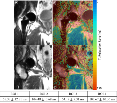 |
155 |
3D Multi-spectral T2 Mapping Near Metal Implants
Sampada Bhave, S. Sivaram Kaushik, Matthew Koff, Hollis Potter, Kevin Koch
The need for examining tissue integrity near metal implants in post-operative arthroplasty is rapidly increasing. Quantitative T2 mapping can be beneficial for characterization of tissues and early diagnosis of tissue pathology. Current T2 mapping techniques lack the capability to image near metal hardware. A novel 3D fast spin echo based two-echo multi-spectral T2 mapping technique is proposed to address this need. This technique is compared to the commercially available approach for accuracy of relaxometry measurements both in phantom and human experiments.
|
|
1414.
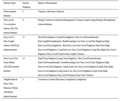 |
156 |
Texture analysis based on T2WI, DWI and delayed T1-enhanced imaging for the differentiation of benign and malignant bone tumors
Ying Li, Cuiping Ren, Jingliang Cheng, Zhizheng Zhuo
With the development of MR techniques, the T2WI, DWI and delayed T1-enhanced imaging techniques have applied as the necessary and general clinical scanning sequences for the bone tumors. And the tumors would present a specific intensity distribution for a specific modality of these high resolution images. Therefore, this work investigated and evaluated the role of textures extracted from T2WI, DWI and delayed T1-enhanced imaging in characterizing the subtypes of bone tumors, and furtherly evaluate the ability of these textures to differentiate benign and malignant tumorsby using support vector machine classifier1 (SVM), which might be helpful for clinical diagnosis and studies.Results showed that the texture parameters extracted from these images have the ability to distinguish benign from malignant bone tumors based on SVM.
|
|
1415.
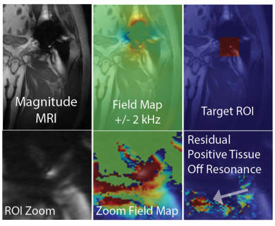 |
157 |
MRI Off-Resonance Analysis Correlates with Histology Necrosis Observations in Failed Total Hip Arthroplasty
Kevin Koch, Juan Liu, Matthew Koff, Hollis Potter
This study presents the analysis of 78 total hip arthroplasty revision subjects that underwent advanced MRI off-resonance analysis and optical histology necrosis grading of tissue biopsies. The off-resonance analysis, which leverages multi-spectral MRI methods, sought to quantify metal particle deposition in tissues or “metallosis”. Direct measurements of off-resonance signatures correlated strongly with histology necrosis grade (p < 0.013). The results of this study provide encouragement that quantitative MRI may be useful as a non-invasive biomarker of tissue destruction in symptomatic total hip arthroplasty.
|
|
1416.
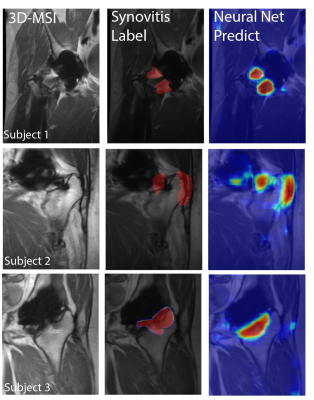 |
158 |
Computer Aided Detection of Synovial Abnormalities Near Total Hip Replacements on 3D-MSI MRI using Deep Neural Networks
Kevin Koch, Ruben Stern, Robin Karr, Andrew Nencka, Matthew Koff, Hollis Potter
3D-MSI increases the visibility of a large number of important pathologies commonly found near implanted orthopaedic hardware, including: host-mediated adverse local tissue reactions, infection, osteolysis, and osteonecrosis. MRI identification of these pathologies aids in planning for surgical revision and has been shown capable of predicting tissue destruction in symptomatic hip replacements. Identification of these features is difficult, even for the interpreting physicians with substantial specialized training and experience . To address this current challenge, a deep-learning based pattern classification approach using 3D-MSI MRI is proposed and demonstrated to predict patterns of adverse synovial responses near hip replacements.
|
|
1417.
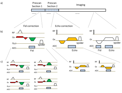 |
159 |
A Comprehensive Gradient Trajectory Correction Strategy for Clinical 3D UTE Imaging
Qi Liu, Bo Li, Xiaomao Gong, Chunjing Tang, Jian Xu, Weiguo Zhang
A comprehensive gradient trajectory correction strategy for non-selective 3D radial UTE imaging was proposed that consistently produces images free of gradient related artifacts by incorporating the actual gradient waveform, and features user friendliness and time-saving by removing the need for separate calibration scan. Application of this innovative design on phantom and volunteer imaging indicates it is a robust and promising technique.
|
|
1418.
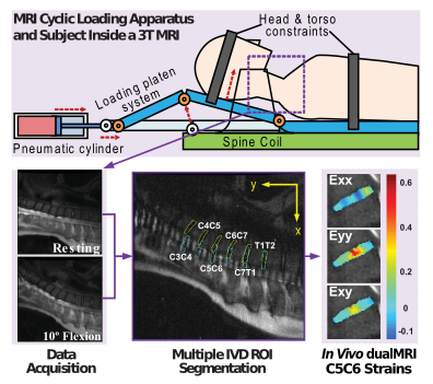 |
160 |
Intervertebral Disc Mechanics Measured by dualMRI In Vivo
Robert Wilson, Leah Bowen, Woong Kim, Luyao Cai, Corey Neu
Functional imaging of intratissue strain in intervertebral discs (IVD) provides an opportunity to investigate tissue in vivo. For the first time, we utilized displacement under applied load MRI (dualMRI) to document principal strains of healthy cervical IVDs in vivo. The transverse and shear average and maximum strains showed position-dependent responses. Additionally, we compared dualMRI results to conventional T2 and T1ρ MRI metrics. No qualitative trend was observed in the conventional MRI data, highlighting the potentially increased sensitivity of dualMRI. In vivo dualMRI may prove a valuable tool to noninvasively investigate IVD biomechanics through strain characterization, leading to better tissue evaluation.
|
|
1419.
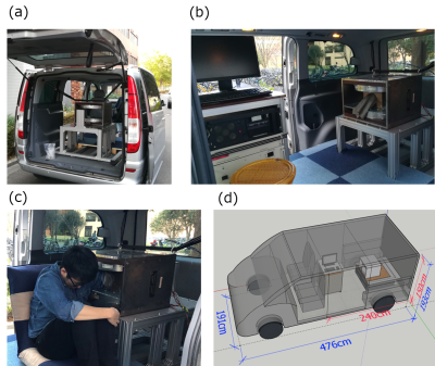 |
161 |
Image denoising with deep transfer learning for screening baseball elbow injuries using portable scanners
Mayu Nakagomi, Sodai Hoshiai, Yoshikazu Okamoto, Yasuhiko Terada
Portable MRI scanners have the advantages of maximizing clinical availability in remote environments. We have recently developed a portable, elbow scanner installed in a standard-size car. This system allows us to detect early symptoms of baseball elbow in remote places, but it often suffers from the low signal-to-noise ratio in noisy, outdoor environments. Here we proposed a deep-learning based approach, a denoising convolutional neural network with transfer learning, for denoising images of the potable scanner. We verified that the proposed denoising technique improved the quality of noisy images and increased the clinical feasibility of the portable scanner.
|
|
1420.
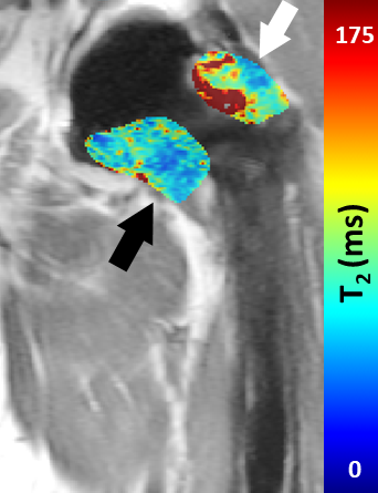 |
162 |
Clinical Feasibility of MAVRIC Based T2 Mapping Near Total Hip Arthroplasty
Jacky Cheung, Mauro Miranda, Kelly Zochowski, Sampada Bhave, Hollis Potter, Kevin Koch, Matthew Koff
Morphologic MRI is commonly performed to assess the presence and extent of abnormal synovial reaction in total hip arthroplasty (THA) but relies on the reader’s training for an accurate diagnosis. Newly developed quantitative methods, including MAVRIC based T2 mapping, may provide a quantitative means for assessing synovial reactions. This study performed MAVRIC based T2 mapping on individuals with THA. Prolonged T2 values were found in subjects with synovitis. This study displays the feasibility of MAVRIC based T2 mapping near THA.
|
|
1421.
 |
163 |
Automated Cartilage and Meniscus Segmentation of Knee MRI with Conditional Generative Adversarial Nets
Sibaji Gaj, Mingrui Yang, Kunio Nakamura, Xiaojuan Li
Clinical translation of quantitative MRI techniques requires accurate cartilage and tissue segmentation. In this work, we have developed and tested a fully automated cartilage and meniscus segmentation model for knee joint using deep learning. To improve segmentation performance by incorporating multi-scale spatial constraints in objective function, the proposed model combines CGAN and U-Net and the dice and cross-entropy loss are added to the CGAN’s objective function. The segmentation performance has been improved for all six compartments and the average dice coefficient for segmentation during testing is 0.88 compared to 0.79 of existing U-Net based segmentation.
|
|
1422.
 |
164 |
Effects of trajectory and k-space undersampling in Compressed Sensing-Parallel Imaging 3D-GRASE
Alexandra Cristobal-Huerta, Dirk Poot, Mika Vogel, Juan Hernandez-Tamames
Compressed sensing parallel imaging (CSPI) 3D-GRASE can reduce the acquisition time compared to CSPI 3D-FSE. Image quality of 3D-GRASE strongly depends on the sampling pattern used, since gradient-echoes (GREs) and spin-echo (SE) are combined in the same k-space. Moreover, successful CSPI relies on appropiate incoherent sampling patterns. In this work we propose and investigate the influence of several sampling patterns on coherence and in-vivo image quality of PD-weighted
knee CSPI 3D-GRASE. With the best sampling pattern CSPI 3D-GRASE obtain high image quality with significantly reduced acquisition time (57%) and SAR (66%) compared to CSPI 3D-FSE acquisitions.
|
|
1423.
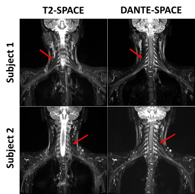 |
165 |
DANTE-SPACE-based imaging of the brachial plexus
Presentation Not Submitted
Xiaoyong Zhang, Liqi Yang, Guijin Li, Yan Li, Bei Li, Yi Sun, Chenhui Li, John Grinstead , Guoxi Xie, Xin Liu
Magnetic resonance neurography (MRN) has been increasingly used to evaluate brachial plexopathy. However, MRN is limited by the lack of relative contrast between nerves and their surrounding tissues. As an alternative to MRN, T2-SPACE has been proposed as a black-blood technique that permits more direct visualization of the brachial plexus; however, it has similar limitations. A preliminary study demonstrated that T2-weighted DANTE-SPACE outperformed SPACE and might be a potential alternative for the visualization of the brachial plexus. In this study, we hypothesized that T2-weighted DANTE-SPACE would address the above issue due to its superior blood flow suppression.
|
|
1424.
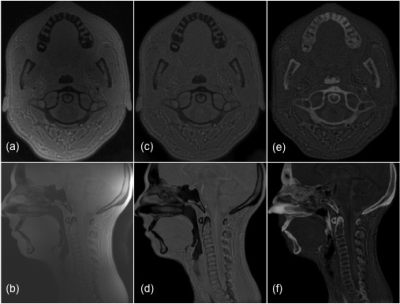 |
166 |
Feasibility of bright-bone cervical spine MRI using Zero TE sequence at 3T
Aiming Lu, Joel Felmlee, Krzysztof Gorny, David Kallmes, Peter Kollasch, Carrie Carr
Direct visualization of solid cortical bone structures with MRI is gaining increased interest. “Bright-bone” techniques based on ultrashort echo time (UTE) and Zero TE (ZTE) sequences enable obtaining signals from the cortical bone and are thus desirable for visualizing cortical bone. This work demonstrated that high quality cortical bone imaging of the human skull and c-spine can be achieved on a clinical 3T scanner in a reasonable acquisition time (<6 minutes). This can potentially widen the usage of MRI for C-spine imaging.
|
|
1425.
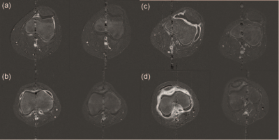 |
167 |
Quantitative MRI to assess disease activity change in children with juvenile idiopathic arthritis
Joshua Bennett, Amanda Wood, Nicola Smith, Ravi Mistry, Karen Allen, Sharmila Jandial, John Tuckett, Claire Gowdy, Helen Foster, Flora McErlane, Kieren Hollingsworth
Assessment of disease activity in children with juvenile idiopathic arthritis (JIA) is central to avoiding permanent joint damage. Affected joints are characterised by synovial proliferation resulting in synovial effusion and hypertrophy. Long-standing poorly controlled synovitis is associated with a high frequency of joint damage. 11 children with JIA were examined with quantitative MRI to measure synovial volume and rate of contrast uptake before and 12 weeks after routine intra-articular corticosteroid injection. All children showed marked reductions in synovial volumes, and in rates of uptake of gadolinium. These reductions were more consistent than changes in clinical assessments.
|
|
1426.
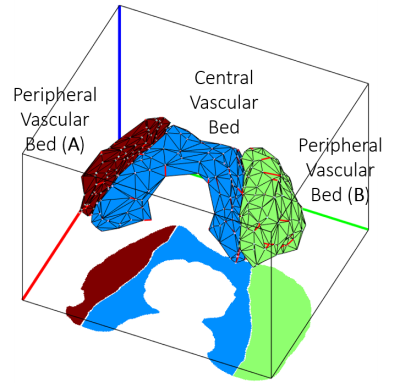 |
168 |
Visualization and quantification of epiphyseal cartilage vasculature using quantitative susceptibility maps of pediatric knee specimens
Kai Ludwig, John Strupp, Casey Johnson, Stefan Zbyn, Mikko Nissi, Ferenc Tóth, Kevin Shea, Cathy Carlson, Jutta Ellermann
Visualization of the vasculature within the epiphyseal cartilage of the distal femur of the skeletally immature knee joint is possible with susceptibility-weighted MRI. A post-processing pipeline is described to segment and quantify the vascular density within two distinct vascular beds of the distal femoral epiphyseal cartilage using quantitative susceptibility maps. The described post-processing may allow identification of vascular abnormalities at early stages of development and also may improve assessment of therapeutic interventions.
|
|
1427.
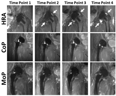 |
169 |
A Prospective, Longitudinal Assessment of Adverse Local Tissue Reactions in Resurfacing Hip Arthroplasty Versus Primary Total Hip Arthroplasty in Asymptomatic Subjects
Matthew Koff, Mauro Miranda, Jacky Cheung, Kelly Zochowski, Bin Lin, Edwin Su, Douglas Padgett, Hollis Potter
This prospective study compares the prevalence of MRI detected adverse local tissue reactions (ALTRs) in metal-on-polyethylene (MoP), metal-on-metal hip resurfacing arthroplasty (HRA), and ceramic-on-polyethylene (CoP) total hip arthroplasty subjects. Images acquired at four time points with a 1-year interval showed a higher prevalence of ALTRs in the HRA than CoP or MoP subjects. Self-assessed symptomatology scores did not significantly differ by implant type at follow-up, indicating that ATLRs can be clinically silent. This study permits better understanding of the natural history of ALTRs complicating hip arthroplasty.
|
|
1428.
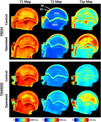 |
170 |
T1? and T2 Relaxation Times are Sensitive to Ischemic Injury in Femoral Head Specimens from a Piglet Model of Avascular Necrosis Independent of a Freeze/Thaw Cycle
Casey Johnson, Ferenc Toth, Cathy Carlson, Stefan Zbyn, Kai Ludwig, Harry Kim, Jutta Ellermann
We investigated whether T1, T2, and T1ρ mapping can detect early ischemic injury to bone, marrow, and cartilage in an animal model of femoral head avascular necrosis. We imaged and compared six pairs of freshly-harvested ischemic and control femoral head specimens. We then imaged the specimens a second time after a freeze/thaw cycle. We found that T1ρ and T2 mapping were sensitive to ischemic injury to the femoral heads 48 hours after onset of ischemia. Furthermore, this sensitivity was maintained after the freeze/thaw cycle. This work indicates that T1ρ and T2 mapping may help assess ischemic bone and joint disorders.
|
|
1429
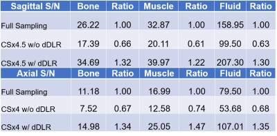 |
171 |
Scan Time Reduction in High Resolution Knee Imaging using Compressed Sensing and Denoising Deep Learning Reconstruction
Video Permission Withheld
Mitsue Miyazaki, Masaaki Umeda, Shinichi Kitane, Cheng Ouyang, Sheronda Statum, Won Bae, Christine Chung
Compressed sensing (CS) uses undersampling at the expense of image blurring and increased noise. We have developed denoising deep learning reconstruction (dDLR) to reduce noise and regain signal-to-noise ratio (SNR) in highly undersampled (4-4.5x) CS images. Feasibility study was performed in fat-suppressed T2 and proton density knee images, by evaluating SNR and image quality (sharpness, blurring, and artifact scores). Compared to reference (no CS or dDLR), images obtained with CS had lower SNR (by 25 to 40%) and image scores due to sharpness and blurring. After processing with dDLR, SNR and image scores were restored the reference levels.
|
|
1430.
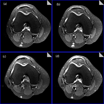 |
172 |
Use of passive B1+ shimming via dielectric pads for uniformity improvements in 7T clinical knee imaging
Andrew Fagan, Kimberly Amrami, Matthew Frick, Benjamin Howe, Peter Kollasch, Venkata Chebrolu, Joel Felmlee
Passive B1+ shimming was performed using custom dielectric pads to improve image uniformity for clinical knee imaging at 7T. Image non-uniformity is problematic for diagnosing knee pathologies, and post-processing solutions are limited at 7T due to the lack of a uniform body transmit coil. A range of pads made from high permittivity perovskite materials were fabricated and tested on 6 subjects. An optimal configuration was determined, comprising the pad dimensions and composition, commensurate with producing optimal image uniformity while fitting all subjects scanned. Dielectric pads represent an easy-to-use solution for improving clinical image uniformity at 7T.
|
|
1431.
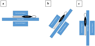 |
173 |
An approach to validate MRI Compatible axial Knee joint Loading Device with various standing posture in Standing MRI
Sandeep Jogi, Rafeek T, Sriram Rajan, Dharmesh Singh, Vidur Mahajan, Vasantha Venugopal, Amit Mehndiratta, Anup Singh
Study of knee joint behaviour under load have a significant potential for Osteoarthritis (OA) diagnosis and monitoring. There are few reported studies on monitoring changes in knee cartilage under load using MRI. Reported process of validation of such loading device are in term repeatability and reproducibility. In this study, an approach is proposed to validate the loading-device using MRI data acquired in standing posture. OpenMRI(0.25T) was used to acquire data in various postures and results were compared with 3T-MRI data with and without load reliability and accuracy of various measurements such as bone-gap, cartilage-thickness and T2-values were evaluated.
|
|
1432.
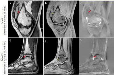 |
174 |
Evaluating Intraarticular Bleeding of Haemophilic Arthropathy Using Quantitative Susceptibility Mapping
Presentation Not Submitted
Fang Zhou, Yihao Guo, Caixia Li, Jing Sun, Yingjie Mei, Yanqiu Feng, Yikai Xu
This work aims to explore the clinical application of quantitative susceptibility mapping (QSM) in evaluating the haemarrhrosis of heamophilic arthropathy with large heamosiderin. Our results showed that there was strong correlation between QSM and bleeding time, demonstrating that QSM has the potential to be a biomarker in evaluating the intraarticular bleeding of heamophilic arthropathy with large heamosiderin.
|
|
1433.
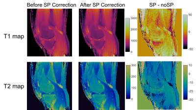 |
175 |
Feasibility and Repeatability of MR Fingerprinting in Knee Joints
Mingrui Yang, Jeehun Kim, Vikas Gulani, Mark Griswold, Xiaojuan Li, Dan Ma
There is an increasing clinical need for improving early diagnosis and evaluation of early response after interventions and treatment in musculoskeletal disorders. Quantitative MRI is a promising tool towards these goals. However, quantifying relaxation times normally requires long acquisition time and is prone to B0 and B1 inhomogeneity and other imperfections. Moreover, it is difficult to optimize the acquisition parameters of conventional relaxation time imaging sequences to provide accurate T1 and T2 quantifications with a large range. MR fingerprinting (MRF) is a recently developed technique, which enables fast imaging and quantification of multiple tissue parameters simultaneously, and shows promising results in neural, body and cardiac imaging. However, the application of MRF to MSK imaging is very limited. The goal of this study was to demonstrate the feasibility and quantification repeatability of MRF in knee joints.
|
|
Back to Program-at-a-Glance |  Back to Top Back to Top |
| The International Society for Magnetic Resonance in Medicine is accredited by the Accreditation Council for Continuing Medical Education to provide continuing medical education for physicians. |
