|
Digital Poster: Young Investigator Awards
Digital YIA Poster
Monday, 13 May 2019
|
ISMRM Booth, located on the Exhibition Floor |
13:45 - 15:45 |
13:45
|
0106.
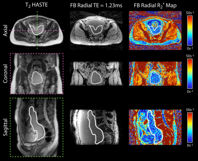 |
3D R2* Mapping of the Placenta During Early Gestation Using Free-Breathing Multiecho Stack-of-Radial MRI at 3 T

Tess Armstrong, Dapeng Liu, Thomas Martin, Rinat Masamed, Carla Janzen, Cass Wong, Teresa Chanlaw, Sherin Devaskar, Kyunghyun Sung, Holden Wu
Ischemic placental disease can lead to hypoxia and abnormal pregnancy outcomes. R2* mapping using MRI can characterize placental hypoxia. However, conventional Cartesian MRI requires breath-holding which limits volumetric coverage, resolution, and signal-to-noise ratio. In addition, little is known about the nominal range of placental R2* at 3T and during early gestation. Therefore, we developed and evaluated a new free-breathing 3D stack-of-radial (free-breathing radial) technique for full volume placental R2* mapping at 3T. Free-breathing radial demonstrated good repeatability and established a nominal range of placental R2* in pregnant subjects during early gestation at 3T.
|
14:05
|
0107.
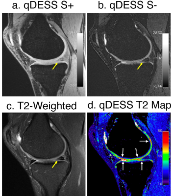 |
5-Minute Double-Echo in Steady-State with Separated Echoes for Comprehensive Whole-Joint Knee MRI Assessment with and without a Proton-Density-Weighted Sequence
Akshay Chaudhari, Zhongnan Fang, Murray Grissom, Bragi Sveinsson, Jeff Wood, Christopher Beaulieu, Edwin Oei, Jarrett Rosenberg, Feliks Kogan, Jin Hyung Lee, Marcus Alley, Garry Gold, Kathryn Stevens, Brian Hargreaves
Most knee MRI protocols require 20+ minutes of scan time, leading to interest in expedited protocols. Here, we first demonstrate in a study with 35 patients and 5 readers that for diagnostic knee MRI, a 3D 5-minute quantitative double-echo steady-state (qDESS) sequence has high agreement with the conventional sequences, where the addition of a proton-density-weighted sequence engenders near-perfect agreement. In a second study with 51 patients and 2 readers, we demonstrate that qDESS with two-fold enhanced slice resolution using deep-learning-based super-resolution and T2 maps has high agreement with the conventional sequences, where both methods have similar agreement with arthroscopic findings.
|
14:25
|
0108.
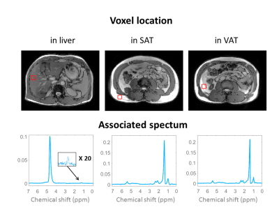 |
3D chemical shift-encoded MRI for volume and composition quantification of abdominal adipose tissue during an overfeeding protocol in healthy volunteers
Angéline Nemeth, Bérénice Segrestin, Benjamin Leporq, Kevin Seyssel, Khuram Faraz, Valérie Sauvinet, Emmanuel Disse, Pierre-Jean Valette, Martine Laville, Hélène Ratiney, Olivier Beuf
The aim of this work was to assess chemical shift-encoded MRI (CSE-MRI) method to quantify content and composition changes of fat storage in healthy volunteers during a 31 days overfeeding protocol while comparing CSE-MRI results with DEXA, MRS and Gas chromatography measurements. A total of 21 volunteers underwent a NMR protocol at 3T with an axial 3D CSE-MRI on abdominal region and spectroscopy acquired on subcutaneous adipose tissue, visceral adipose tissue and liver. The NMR protocol was used to analyze the volume and the fatty acid composition of abdominal adipose tissues, and the fat content in the liver.
|
14:45
|
0109.
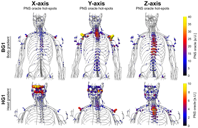 |
Prediction of Peripheral Nerve Stimulation Thresholds of MRI Gradient Coils using Coupled Electromagnetic and Neurodynamic Simulations
Mathias Davids, Bastien Guérin, Axel vom Endt, Lothar Schad, Lawrence Wald
Peripheral Nerve Stimulation (PNS) has become the major limitation in many fast MRI sequences for state-of-the-art gradient systems. We present the first (to our knowledge) full PNS model for assessing magnetostimulation thresholds and a method to incorporate these thresholds as constraints in the coil-winding design phase. Our model consists of comprehensive female and male body models for EM simulations, co-registered atlases of peripheral nerves, and a neurodynamic model describing the nerve responses to induced electric fields. We validated our framework based on three commercial MR gradient systems and found close resemblance between simulated thresholds and experimentally obtained group PNS thresholds.
|
15:05
|
0110.
 |
SPARKLING: variable-density k-space filling curves for accelerated MRI
Carole Lazarus, Pierre Weiss, Nicolas Chauffert, Franck Mauconduit, Loubna El Gueddari, Christophe Destrieux, Ilyess Zemmoura, Alexandre Vignaud, Philippe Ciuciu
This work reports the use of new non-Cartesian k-space trajectories whose improved efficiency allows to significantly reduce MR scan time with minimum deterioration of image quality. Instead of using simple geometrical patterns, we introduce an approach inspired from stippling techniques, which automatically designs optimized sampling patterns along any desired density by taking full advantage of the hardware capabilities. We use our strategy to accelerate the acquisition time of T2*-weighted scans acquired at 7T on in vivo human brains. We compare our method to standard non-Cartesian trajectories (spiral, radial) and demonstrate its superiority regarding image quality and robustness to system imperfections.
|
15:25
|
0111.
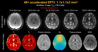 |
Echo Planar Time-resolved Imaging (EPTI)
Fuyixue Wang, Zijing Dong, Timothy Reese, Berkin Bilgic, Mary Manhard, Jingyuan Chen, Jonathan Polimeni, Lawrence Wald, Kawin Setsompop
A new technique, termed Echo Planar Time-resolved Imaging (EPTI), was developed to address EPI’s geometric distortion and blurring, and to provide temporal signal evolution information across the EPI readout window. Using a small number of EPTI-shots, a time-series of multi-contrast images can be created free of distortion and blurring (up to 100 T2- and T2*-weighted images). This should make EPTI useful for numerous applications. Here, we demonstrated EPTI in brain to provide i) rapid simultaneous quantitative mapping of T2, T2*, proton density and tissue phase, as well as ii) multi-echo and quantitative T2* fMRI.
|
|
