Digital Poster Session
Cardiovascular Back to Program-at-a-Glance Back to Program-at-a-Glance
|
Monday, 13 May 2019
Digital PosterCardiovascular
1951 -1975 Technical Advances in Flow Imaging
1976 -2000 Clinical Applications of Flow Imaging
2001 -2025 Tissue Characterization 1
2026 -2050 Tissue Characterization 2
2051 -2075 Contrast-Enhanced & Non-Contrast MR Angiography
2076 -2100 Atherosclerosis & MR Angiography
2101 -2124 Myocardial Function & Deformation 1
2125 -2149 Cardiovascular Image Processing & Reconstruction
2150 -2174 Myocardial Function & Deformation 2
2175 -2199 Cardiovascular Emerging Methods
2200 -2224 Cardiovascular Miscellaneous |
| |
Technical Advances in Flow Imaging
Digital Poster
Cardiovascular
Monday, 13 May 2019
| Exhibition Hall |
16:00 - 17:00 |
| |
|
Computer # |
|
1951.
 |
1 |
Phase contrast coronary blood velocity mapping with both high temporal and spatial resolution using Golden Angle rotated Spiral k-t Sparse Parallel imaging (GASSP)

Dan Zhu, Gabriele Bonanno, Robert Weiss, Michael Schär
Coronary phase contrast MRI for accurate measurement of coronary blood flow requires high spatial resolution due to the small vessel size, and high temporal resolution due to cardiac motion, especially of the right coronary artery (RCA). This study uses golden angle rotated spiral k-t sparse parallel imaging (GASSP) to accelerate the acquisition to achieve both high spatial and high temporal resolution in a breath-hold. GASSP was validated in the stationary popliteal artery (knee) and then implemented in the RCA. GASSP achieved high image quality throughout the cardiac cycle and popliteal peak velocity and mean-square error compared favorably to gold standard.
|
|
1952.
 |
2 |
High spatiotemporal resolution cones 4D flow using memory-efficient iterative reconstruction
Christopher Sandino, Frank Ong, Joseph Cheng, Michael Lustig, Marcus Alley, Shreyas Vasanawala
4D flow MRI enables comprehensive cardiovascular assessment, but is limited by long acquisition times and motion corruption. Non-Cartesian sampling strategies, such as radial and cones, exhibit excellent aliasing properties that allow reduced scan times and improve motion robustness. However, trade-offs between spatial and temporal resolution are necessary due to computational burden of iterative 4D non-Cartesian reconstruction. This has restricted cones 4D flow to low temporal resolution venous applications. Here we present a memory-efficient iterative reconstruction utilizing batch processing to enable arbitrary spatiotemporal resolution. We demonstrate feasibility of sub-millimeter, 30 cardiac phase cones 4D flow for coronary artery and valvular assessment.
|
|
1953.
 |
3 |
Dual-Venc 4D Spiral Imaging of Aortic Flows
Sean Callahan, Alex Henn, MJ Negahdar, Michael Kendrick, Hui wang, Narayana Singam, Marcus Stoddard, Amir Amini
Dual-Venc is a technique for MR flow imaging which uses two Vencs to acquire a cardiac cycle, improving diastolic data. Dual Venc 4D flow with spiral readouts was used to image the outflow tract and through the aortic valve in both phantom and patients with severe aortic stenosis. In-vitro model of the aortic arch included a calcific polymeric valve which behaved physiologically. The results of in-vitro and in-vivo scans show that 4D Spiral Dual-Venc Flow is comparable in results to 4D Cartesian Flow in systole, while improving diastolic data, and reducing scan time by 30% to 50%.
|
|
1954.
 |
4 |
Pseudo spiral compressed sensing accelerated whole-heart 4D flow MRI: validation against EPI readout
Carmen Blanken, Lukas Gottwald, Jos Westenberg, Eva Peper, Bram Coolen, Gustav Strijkers, Aart Nederveen, R Planken, Pim van Ooij
4D flow MRI facilitates detailed evaluation of cardiac hemodynamics in patients with cardiovascular disease. In this study, we investigated the performance of pseudo spiral compressed sensing (CS) accelerated whole-heart 4D flow MRI in a comparison with a clinically used EPI readout. CS-accelerated 4D flow MRI yielded similar results to EPI-accelerated 4D flow MRI in terms of velocity vector fields during ventricular ejection and filling and led to consistent blood flow measurements across heart valves. Our data suggest that CS 4D flow MRI has the potential to be accelerated even further for quantitative whole-heart hemodynamic imaging.
|
|
1955.
 |
5 |
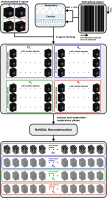
Self-gated 5-minute whole-heart 4D flow imaging
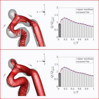
Aaron Pruitt, Adam Rich, Yingmin Liu, Ning Jin, Lee Potter, Orlando Simonetti, Rizwan Ahmad
4D flow imaging can provide comprehensive hemodynamical analysis of blood flow through the heart and great vessels; however, acquiring 4D flow images with whole-heart coverage is prohibitively time-consuming. In this work we describe a highly accelerated and fully self-gated whole-heart 4D flow acquisition and reconstruction methodology. Additionally, we show proof-of-concept of a fully self-gated 5-minute whole-heart 4D flow exam and demonstrate good agreement in aortic flow quantification compared to conventional 2D phase contrast.
|
|
1956.
 |
6 |
A NOVEL MATLAB TOOLBOX FOR PROCESSING 4D FLOW MRI DATA
Julio Sotelo, Joaquin Mura, Daniel Hurtado, Sergio Uribe
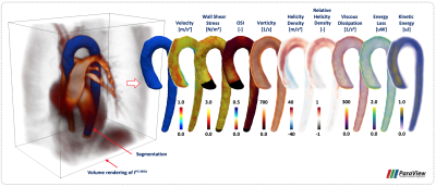
Current software used to process 4D Flow MRI data only allow to quantify a few hemodynamics parameters. Furthermore, the information of these parameters is normally given in a few 2D locations. In this work, we show the development of a novel, free and editable MATLAB toolbox (MathWorks, Natick, MA, USA) called FEMQ-4D that allows the quantification of several hemodynamic parameters in 3D using finite elements (FE) methods. As a complement to this tool, the output of FEMQ-4D, i.e 3D maps of hemodynamic parameters, can be visualized using the open source software PARAVIEW (KitwareInc., Clifton Park, New York, USA).
|
|
1957.
 |
7 |
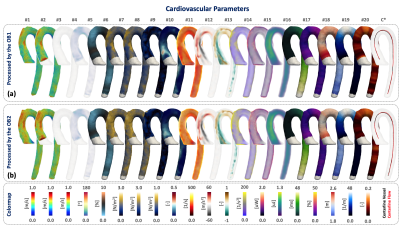
REPRODUCIBILITY OF A SINGLE FINITE ELEMENT METHODOLOGY USED TO CALCULATE SEVERAL CARDIOVASCULAR PARAMETERS FROM 4D FLOW MRI DATA.
Julio Sotelo, Rodrigo Herrera, Cristian Montalba, Juan Urbina, Joaquin Mura, Daniel Hurtado, Sergio Uribe
Several cardiovascular parameters can be calculated using multiple approaches from 4D flow MRI data. Usually the calculation of these parameters is time consuming, which may affect the reproducibility of the results. Recently, we proposed a novel methodology based on finite elements that allows obtaining several quantitative cardiovascular parameters along the entire aorta from a single segmentation of 4D flow data. Using this methodology, our results are accurate, and reproducible. We showed that we did not find statistical differences between manual and automatic measurements for geometrical analysis. Furthermore, a 93.7% and 81.2% of cardiovascular parameter shown strong and excellent correlation for intra and for inter-observer analysis respectively, providing a robust and consistent results.
|
|
1958.
 |
8 |
Accelerated 4D flow MRI using a Low-Rank Tensor reconstruction
Bobby Runderkamp, Eva Peper, Jasper Schoormans, Qinwei Zhang, Bram Coolen, Gustav Strijkers, Aart Nederveen
4D flow MRI provides visualization and quantification of complex blood flow. However, the inherent high dimensionality leads to long acquisition times. In this work, 4D flow MRI was accelerated using the novel Low-Rank Tensor framework. To reduce the amount of unknowns, the 4D flow dataset is approximated by a Tucker decomposition, whose components are obtained from navigator and sparse data with iterative optimization exploiting sparsity after variable k-space undersampling. Using this technique, 4D flow MRI acquisition could be accelerated up to 20 times (flow phantom) and 8 times (in-vivo), while preserving measurement accuracy of high velocity magnitudes and cardiac variability.
|
|
1959.
 |
9 |
Wearable Seismocardiography as a Quick Screen for Thoracic Aorta Flow Abnormalities Necessitating 4D Flow MRI
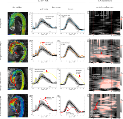
Ethan Johnson, J. Alex Heller, Daniel Gordon, Flori Garcia Vicente, Alex Barker, Mozziyar Etemadi, Michael Markl
Seismocardiography, the measurement and analysis of forces and accelerations from the beating heart that propagate through the chest wall, can yield insights about cardiac and hemodynamic health. In subjects with pathological or deranged blood flow in the thoracic aorta, the characteristics of SCG accelerations change as a result of the altered flow. Here, we use 4D flow MRI to investigate the specific SCG changes that are associated with abnormal aortic flow, and we consider the potential to use SCG as a quick screen for need of a comprehensive 4D flow MR examination.
|
|
1960.
 |
10 |
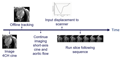
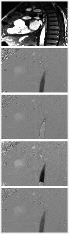
Valvular flow imaging in the era of feature-tracking: Pilot study to measure mitral flow
Felicia Seemann, Einar Heiberg, Marcus Carlsson, Lauren Baldassarre, Maolin Qiu, Dana Peters
Magnetic resonance imaging of the mitral valve is challenging since the valve moves in and out of the image plane during the cardiac cycle. To more accurately measure mitral flow, a phase contrast sequence that uses offline feature-tracking of the valve in the long-axis view is proposed. The tracking result is exported to the scanner, allowing the slice position to change in real-time based on the cardiac phase. The slice-following sequence outperformed the conventional sequence when quantifying regurgitant volumes. Hence, the new sequence is a promising method for improving the accuracy of trans-valvular flow.
|
|
1961.
 |
11 |
Time Resolved 4D Flow MRI Quantification without Segmentation
Carson Hoffman, Oliver Wieben
4D flow sequences enable the acquisition of time resolved velocity fields over an averaged cardiac cycle. Flow quantification and velocity profile analysis typically requires manual segmentation and plane placement, which can lead to inaccuracies with lack of reproducibility and large post-processing times. Improving upon the semi-automated, 4D flow post-processing techniques with the application of centerline labeling and k-means based segmentation, here we propose a fully automated, time-resolved flow quantification method which utilizes flow as a function of distance instead of segmentation. This method may further decrease the time involved with 4D flow processing and increase the agreement with manual segmentation.
|
|
1962.
 |
12 |
Deep Spatiotemporal Phase Unwrapping of Phase-Contrast MRI Data
Jiacheng Jason He, Christopher Sandino, David Zeng, Shreyas Vasanawala, Joseph Cheng
This work demonstrates the advantage temporal information provides for deep phase unwrapping of phase-contrast MRI data. Using a patch-based, three-dimensional ResNet architecture, our model performs better than state-of-the-art single-step algorithms. Our deep spatiotemporal phase unwrapping model continues the quest to lower Venc values to increase dynamic range and velocity-to-noise ratio (VNR) of 4D flow data by providing a robust method for phase unwrapping.
|
|
1963.
 |
13 |
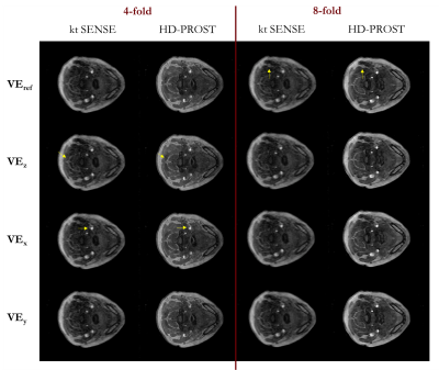
Accelerated Carotid 4D flow MRI with Multicontrast HD-PROST Reconstruction
Andreia Gaspar, Aurelien Bustin, Karl Kunze, Radhouene Neji, Teresa Correia, Nuno Silva, Rita Nunes, René Botnar, Claudia Prieto
4D flow MRI is time consuming, since it requires the acquisition of time-resolved images with three-directional velocity encoding. Undersampled reconstruction techniques have been proposed to accelerate 4D flow carotid imaging, however scan time remains lengthily for high-resolution acquisitions. In this work, we propose to further accelerate 4D flow carotid MR imaging by exploiting patch-based similarities in local, non-local and multi-contrast dimensions with high-dimensional patch-based undersampled reconstruction (HD-PROST). The results show similar velocities for both k-t SENSE and the proposed HD-PROST, however higher precision was obtained with HD-PROST.
|
|
1964.
 |
14 |
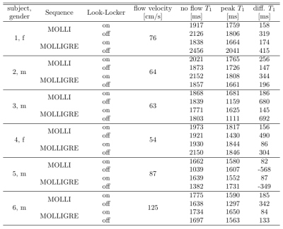
Effect of inflow and in-plane saturation in SASHA and MOLLI T1 and T1* maps in a perfusion phantom and in-vivo
Ingo Hermann, Tanja Uhrig, Jorge Chacon-Caldera, Mehmet Akçakaya, Lothar Schad, Sebastian Weingärtner
Aim of this work was to evaluate the effect of flow on blood T1 measurement considering inflow of non-inverted spins. Experiments using a flow phantom demonstrate shorter T1 for increasing flow velocity, with high reproducibility. In-vivo measurements show major variations throughout the cardiac cycle, validating the flow sensitivity observed in phantom measurements.
|
|
1965.
 |
15 |
Fetal whole-heart 4D blood flow visualisation using motion-corrected multi-planar real-time PC-bSSFP MRI
Thomas Roberts, Joshua van Amerom, Anthony Price, Maria Deprez, David Lloyd, Laurence Jackson, Milou van Poppel, Kuberan Pushparajah, Mary Rutherford, Reza Rezavi, Joseph Hajnal
Measurement of blood flow in the fetal heart and the great vessels is challenging due to fetal motion and small vessel sizes. 2D methods for fetal flow imaging require significant slice piloting to locate the vessels, and small changes in fetal position can often necessitate reacquisition. In this work, we demonstrate the potential for motion-corrected whole-heart 4D flow imaging in the fetus using stacks of highly accelerated 2D bSSFP slices, which are inherently sensitive to velocity. Real-time acquired images were aligned in space and cardiac phase, and vectorially combined to yield time-resolved flow information.
|
|
1966.
 |
16 |
Fully dimensional vessel segmentation in non-contrast-enhanced 3D PC-MRI image data.
Judith Zimmermann, Lennart Tautz, Christian Meierhofer, Heiko Stern, Bjoern Menze, Anja Hennemuth
Fully dimensional (spatial + temporal) segmentation of blood vessels is crucial to perform 3D PC-MRI based quantitative characterization of hemodynamics. However, most prior works neglect the temporal movement of vessels, making it a 3D-only segmentation problem. Therefore, the objective of this work was to show feasibility of a deformable-registration-based algorithm for 4D segmentation of the aorta. Performance of the proposed algorithm proved to be acceptable, with overall Dice index and Hausdorff distance of 0.86±0.04 and 3.63±0.75 mm, respectively.
|
|
1967.
 |
17 |
Computational metrics for quantitative characterization of vortical flow patterns based on 3D PC-MRI data.
Judith Zimmermann, Johann Drexl, Sarah Nordmeyer, Anja Hennemuth
Vortices and helices are crucial features of hemodynamic flow. Such structures may define new clinically relevant biomarkers when assessing cardiovascular pathologies mediated by abnormal flow patterns (e.g. aneurysm formation). Thus, retrieving such structures in time-resolved and velocity-encoded 3D PC-MRI image data is of tremendous interest. However, prior studies only focused on a voxel-wise identification, and are lacking meaningful quantitative metrics which characterize the full vortical flow pattern. The objective of this work is to propose metrics for fully automated detection and quantitative characterization of vortical flow patterns in the aorta.
|
|
1968.
 |
18 |
An application of the myocardial strain analysis method based on the deformation registration algorithm (DRA) in heart transplantation
Presentation Not Submitted
Xuehua Shen, Yating Yuan, Mingxing Xie, Li Zhang, Wei Sun, Jia Liu, Xiaoyue Zhou, Jing Song, Bo Liang
With the development of cardiac magnetic resonance(CMR), there have been various methods to quantify myocardium. As a novel method, deformation registration algorithm (DRA) has been confirmed in many cardiac diseases. In this article, we use this method to analyze the wall deformation of orthotopic heart transplantation(HTx). We found that though left ventricular ejection fraction (LVEF) have no significant difference between the HTx patients and the healthy volunteers, the left ventricular (LV) peak longitudinal strain (LV Ell) and LV peak systolic longitudinal strain rate have significant decrease in HTx patients, indicating that the ventricular deformation is more sensitive than LVEF in response to the myocardial diseases.
|
|
1969.
 |
19 |
How gating affects 2D phase contrast flow in the ascending aorta at 7.0T MRI
Maria Stefanescu, Jan-Peter Grunz, David Lohr, Stefan Herz, Aleksander Kosmala, Laura Schreiber
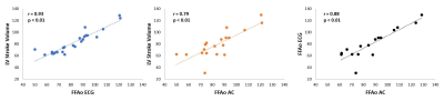
2D phase contrast MRI offers a fast method for blood flow evaluation in the ascending aorta which can be used for estimation of left ventricular function. With proper ECG or acoustic triggering, forward flow in the aortic root (FFAo) should resemble left ventricular stroke volume (LVSV) in absence of valve disease. However, for ultra-high field strengths (≥7.0T) flow parameters in the ascending aorta derived from phase contrast have not yet been validated. Our results suggest that accurate gating reduces cardiac motion artifacts for 7.0T to an extent where LVSV can be estimated reliably based on FFAo in phase contrast sequences.
|
|
1970.
 |
20 |
Objective extraction of the temporal evolution of the mitral valve vortex ring from 4D flow MRI
Corina Kräuter, Ursula Reiter, Clemens Reiter, Albrecht Schmidt, Andreas Greiser, Marc Masana, Michael Fuchsjäger, Rudolf Stollberger, Gert Reiter
The mitral valve vortex ring is a promising flow structure for analysis of diastolic function, however, methods for objective extraction of its formation to dissolution are lacking. We present a novel algorithm for objective extraction of the temporal evolution of the mitral valve vortex ring from magnetic resonance 4D flow data and validated the method against visual analysis. The algorithm successfully extracted mitral valve vortex rings during both early- and late-diastolic filling and agreed substantially with visual assessment. Early-diastolic mitral valve vortex ring properties differed between healthy subjects and patients with ischemic heart disease.
|
|
1971.
 |
21 |
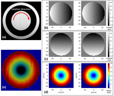
Phantom for Phase-Contrast MRI Sequence Validation and Quality Control
Alireza Vali, Sebastian Schmitter, Liliana Ma, Xiaoke Huang, Sebastian Flassbeck, Simon Schmidt, Michael Markl, Susanne Schnell
Systematic assessment and optimization of 2D Phase-Contrast (PC) MRI as well as 4D flow MRI sequences require reliable phantoms that can create known velocity fields with large velocity ranges corresponding to different cardiovascular regions. An air-driven rotation phantom was constructed and its performance in establishing well-defined velocity fields at different rotational speeds was examined using 3-directional 2D PC MRI acquisitions. Furthermore, the reproducibility of the phantom was examined with a test-retest experiment on two different days. It was demonstrated that the phantom could create reproducible linear velocity fields to be used as a reference for in-vitro validation of PC MRI sequence.
|
|
1972.
 |
22 |
Experimental Validation of 4D Flow MRI for the Assessment of Flow Dynamics within a Patient-Specific Intracranial Aneurysm Model using Tomographic Particle Image Velocimetry
Rafael Medero, Katrina Ruedinger, Alejandro Roldán-Alzate
4D flow MRI has shown to be a feasible tool for assessing hemodynamics in different vascular territories with high spatial resolution. This study aimed to compare velocity components and magnitudes within a patient-specific intracranial aneurysm in-vitro model using 4D Flow MRI and a ground truth experimental technique, tomographic particle image velocimetry (tomo-PIV). Tomo-PIV offers higher temporal and spatial resolution allowing the assessment of intra-cycle differences caused by pulsatile flow. This analysis was done to assess the ability of 4D Flow MRI to capture the complex flows within an intracranial aneurysm considering the cardiac cycle averaging of the data.
|
|
1973.
 |
23 |
Influence of respiration-induced B0 variations in real-time phase contrast MRI of the cerebrospinal fluid
Kristina Peters, Kilian Weiss, David Maintz, Daniel Giese
The error induced by susceptibility changes due to respiration in the measurement of CSF flow was investigated. Real-time dynamic B0 measurements and PCMRI images of 10 healthy subjects were acquired. A good agreement was found between both acquisitions. B0 amplitudes and temporal shifts with respect to respiration signals showed dependencies on echo times, temporal distances between phase contrast images and subjects. Resulting errors between 0.4 and 41 % in PCMRI images were shown in simulations. In conclusion, the present work demonstrates that B0 variations during respiration may have a confounding effect when estimating respiration dependent flow in CSF.
|
|
1974.
 |
24 |
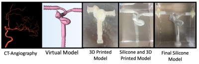
Analysis of Velocity Component Distribution and Cardiac Phase in Aortic Dissection False Lumen 3D Printed Models Using In Vitro 4D Flow MRI
Sylvana García-Rodríguez, Philip Corrado, Alejandro Roldán-Alzate, Christopher Francois
False lumen hemodynamics is an important factor in aortic dissection progression. As a methodology to further characterize false lumen velocities, two patient-specific 3D printed models underwent 4D Flow MRI, from which histograms of velocity components were generated at several locations along the lumen. Two VENC settings were used and the data was grouped in diastole and systole. Histograms of normal and tangential components serve as descriptors of flow regimes and offer the possibility to correlate with thrombus formation and clinical progression. VENC is important especially for the assessment of tangential components.
|
|
1975.
 |
25 |
Voxel-wise comparison of CFD and 4DMR results in the cerebral venous outflow tract of a pulsatile tinnitus patient
Evan Kao, Henrik Haraldsson, Megan Ballweber, Matthew Amans, David Saloner
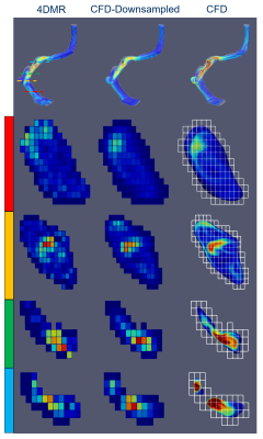
To investigate abnormal hemodynamics in pulsatile tinnitus patients in vivo, we use 4DMR. We also use computational fluid dynamics (CFD) to overcome 4DMR’s limited spatial and temporal resolution. To ensure the simulation actually reflects in vivo blood flow, we systematically adjust the simulation boundary conditions to match the CFD and 4DMR data. This requires downsampling CFD data to 4DMR resolution and a quantitative voxel-wise comparison. Our results suggest 4DMR underestimates velocities in vivo due to its resolution. This effect is confirmed by phantom 4DMR data taken at multiple resolutions.
|
|
| Top |
Clinical Applications of Flow Imaging
Digital Poster
Cardiovascular
Monday, 13 May 2019
| Exhibition Hall |
16:00 - 17:00 |
| |
|
Computer # |
|
1976.
 |
26 |
Velocity transfer function in the right pulmonary artery correlates with right ventricular remodeling and pulmonary functional impairments in COPD
Oleg Sharifov, Thomas Denney, Jr, J. Michael Wells, Gregory Payne, Swati Gulati, Himanshu Gupta, Mark Dransfield, Steven Lloyd
Chronic obstructive pulmonary disease (COPD) is often associated with pulmonary artery (PA) hypertension (PH), however a mild to moderate PH is frequently not identified on non-invasive testing in COPD patients. The novel non-invasive cardiovascular magnetic resonance (CMR) derived parameter, velocity transfer function (VTF), has been recently shown to correlate with invasive PA impedance. Here, we tested the VTF to evaluate its association with clinical/pulmonary functional indices and RV remodeling in patients in early COPD. We found that elevated VTF mean high frequency modulus was associated with major clinical and functional criteria indicating cardiovascular/respiratory dysfunction, which may link to PH.
|
|
1977.
 |
27 |
Left and Right Heart Ventricular-Vascular Coupling in Pulmonary Venous Hypertension
Mohammed S.M. Elbaz, Vamsi Reddy, Muhannad Abbasi, Roberto Sarnari, Daniel Gordon, Michael Cuttica, Benjamin Freed, Michael Markl, James Carr
Pulmonary Venous hypertension (PVH) is a life-threatening disease with a complex etiology that involves both the left and right heart. However, the mechanism of left heart dysfunction as a precursor to changes in right heart hemodynamics and dysfunction remains unclear. Here, we assessed the complex functional-hemodynamic coupling between the left and right heart and pulmonary arteries. We investigated the association of LV and RV function with advanced 4D Flow MRI hemodynamic metrics of volumetric viscous energy loss, kinetic energy and vorticity in the right heart and pulmonary arteries in PVH patients.
|
|
1978.
 |
28 |
Is Right Ventricular Kinetic Energy Correlated with Repaired Tetralogy of Fallot Outcome?
Jacob Macdonald, Kathan Amin, Philip Corrado, Christopher Francois, Oliver Wieben
Many patients with repaired Tetralogy of Fallot require additional pulmonary valve replacement surgery later in life. Previous 4D flow MRI studies have suggested that right ventricular kinetic energy may be a useful biomarker in this patient population. In this long-term follow-up study, kinetic energy measurements derived from 4D flow MRI were compared with each patient’s need for pulmonary valve replacement surgery in the next decade. Patients who did not need surgery showed significantly higher stroke volume index normalized by kinetic energy than those who required surgery, suggesting this parameter may have some prognostic value in this application.
|
|
1979.
 |
29 |
Blood Flow Alterations in Repaired Tetralogy of Fallot Adult Patients: Preserved blood flow distribution and altered flow biomarkers.
Daniel Yakimenka, Ahmed Abdelhaleem, Alireza Sojoudi, An Le, David Patton, James White, Julio Garcia
Tetralogy of Fallot (TOF) is a common congenital disorder which is treated by surgical repair. This study aimed to investigate alterations in blood flow in adults with repaired TOF. We hypothesized that abnormal remodeling of the right heart and the PA following surgical repair leads to impaired blood flow distribution, increased wall shear stress (WSS), greater energy loss (EL) and greater pressure drop (PD) in rTOF adults. This study contributes towards the understanding of blood flow hemodynamics after surgical repair.
|
|
1980.
 |
30 |
Comparing Regional Left Ventricular Flow and Myocardial Strain after Myocardial Infarction
Philip Corrado, Gregory Barton, Niti Aggarwal, Jonathan Weinsaft, Christopher Francois, Oliver Wieben
This work employed time-resolved (4D) flow cardiac MRI and feature-tracking myocardial strain to characterize the relationship between left ventricular (LV) strain and kinetic energy after myocardial infarction. Kinetic energy indexed to end diastolic volume in the LV apex varied directly with peak radial strain in the LV apex, and with global LV ejection fraction. This method of regional analysis may be of clinical use in characterizing LV contractile and hemodynamic function in the post MI population.
|
|
1981.
 |
31 |
Quantitative MRI detects impaired vascular reactivity in women after preeclamptic pregnancy
Michael Langham, Felix Wehrli, Nadav Schwartz
Large body of evidence suggests maternal endothelial dysfunction (EDF) has a central role in the development of preeclampsia, the most serious hypertensive pregnancy disorder that significantly increases risk for future cardiovascular diseases. Because pathophysiology of preeclampsia remains within 72 hrs of delivery of the placenta, the quantification of surrogate MRI markers of EDF was performed after birth in women with and without hypertensive pregnancy. The quantitative MRI protocol evaluates peripheral micro- and macrovascular reactivity and central arterial stiffness in a single scan session. Preliminary results show a trend of impaired vascular reactivity after hypertensive pregnancy relative to normotensive pregnancy.
|
|
1982.
 |
32 |
Pulmonary Artery 3D Wall Shear Stress is Lower in Patients with WHO Group 2 Pulmonary Hypertension
Michael Scott, Daniel Gordon, Mohammed Elbaz, Vamsi Reddy, Jeremy Collins, Benjamin Freed, Sanjiv Shah, Michael Cuttica, Michael Markl, James Carr
Patients with pulmonary hypertension (PHTN) are known to have altered pulmonary artery (PA) hemodynamics in addition to differing PA pressures measured using invasive right heart catherization. 4D flow MRI can provide information about PA hemodynamics, such as 3D wall shear stress that might be useful in diagnosis or grading of PHTN. Previous work on WSS used manually placed planes for evaluating regional WSS, we derive WSS metrics over the entire vessel. 3D WSS measurements in the PA were significantly different between a cohort of patients with WHO group 2 PHTN and healthy controls and were correlated with catheter-based pressure measurements.
|
|
1983.
 |
33 |
Displaced aortic flow and increased circumferential wall shear stress associate with ascending aortic dilatation in tricuspid aortic valve patients – a prospective clinical study
S. Petteri Kauhanen, Marja Hedman, Pekka Jaakkola, Ritva Vanninen, Petri Saari, Timo Liimatainen
Four-dimensional (4D) flow was measured in 20 patients with dilated ascending aorta (AA) and in 20 controls. Aortic flow was displaced from the center line of the AA in patients with AA dilatation. Flow displacement was present in the proximal and tubular parts of AA. Total wall shear stress (WSS) was higher on the displaced side compared to the opposite side of the aorta. The circumferential WSS (WSSC) ratio to total WSS was higher in the inner curvature of dilated AA in the proximal part and WSSc was elevated in the whole aortic ring in the distal part of AA.
|
|
1984.
 |
34 |
Association Between Aortic Flow and Myocardial Motion Velocity in Patients with Marfan Syndrome
Wen-Xin Ye, Hsu-Hsia Peng, Hsin-Hui Chiu, Wen-Yih Isaac Tseng
We aimed to explore the possible correlation between aortic flow and myocardial motion in Marfan syndrome (MFS) patients. MFS group presented lower mean velocity and higher retrograde flow in the ascending aorta (AAo). MFS group also exhibited lower basal systolic Vz and diastolic Vr, higher diastolic Vz, and prolonged diastolic TTPz. The mean velocity showed positive correlation with basal systolic Vz, diastolic Vz, and diastolic Vr. The retrograde flow was correlated with basal diastolic TTPz. In conclusion, the correlation between aortic flow and myocardial motion in MFS patients might provide helpful information in long-term surveillance in MFS patients.
|
|
1985.
|
35 |
Non-Invasive Evaluation of Inhaled Nitric Oxide on Pulmonary Blood Flow Dynamics using 4D Flow MRI in Pediatric Pulmonary Arterial Hypertension Patients
Nivedita Naresh, Michal Schafer, Lorna Browne, Dunbar Ivy, Dayna Zimmerman, Vitaly Kheyfets, Alexander Barker, Uyen Truong
Pediatric pulmonary arterial hypertension (PAH) is a significant cause of morbidity and mortality. Acute vasodilatory reactivity (AVR) using right heart catheterization is a necessary component of a diagnostic PAH evaluation. The aim of this study was to non-invasively evaluate AVR hemodynamic changes in pediatric patients with PAH using 4D flow MRI. We have shown that post-iNO treatment in pediatric PAH patients, peak velocity and peak wall shear stress were increased. Future studies in larger cohorts of PAH pediatric patients are needed to comprehensively evaluate the effectiveness of non-invasive AVR in comparison to gold standard right heart catheterization.
|
|
1986.
 |
36 |
Interaction between pulmonary flow and myocardial motion in patients with repaired tetralogy of Fallot
Jung-Hsiu Liu, Meng-Chu Chang, Ming-Ting Wu, Ken-Pen Weng, Hsu-Hsia Peng
We aimed to investigate the interaction between pulmonary flow and myocardial motion in patients with repaired tetralogy of Fallot (rTOF). The 4D flow MRI and tissue phase mapping were employed to quantify the pulmonary flow and myocardial motion velocity, respectively. The pulmonary retrograde flow was correlated with left ventricular (LV) peak diastolic longitudinal velocity and time-to-peak (Vz and TTPz) in rTOF patients. Pulmonary pulsatility index was correlated with diastolic RV TTPr and LV TTPz. In conclusion, the correlation between abnormal pulmonary artery flow and altered myocardial motion may provide helpful information in evaluating the cardiac function in rTOF patients.
|
|
1987.
 |
37 |
Potential impact of pulmonary area on retrograde flow and pressure difference in patients with repaired tetralogy of Fallot
Jung-Hsiu Liu, Meng-Chu Chang, Ming-Ting Wu, Ken-Pen Weng, Hsu-Hsia Peng
The residual pulmonary regurgitation can lead to heart failure in patients with repaired tetralogy of Fallot (rTOF). We aimed to explore the possible impact of pulmonary area on pulmonary retrograde flow and pressure. Patients with rTOF presented higher retrograde flow than normal controls. The area ratio correlated with with retrograde flow in rTOF patients. Negative correlations between area ratio and pressure difference was also presented in patients. In conclusion, pulmonary area was correlated with retrograde flow and pressure difference in rTOF patients. The information of appropriate pulmonary area ratio might be helpful for treating the pulmonary stenosis in patients with rTOF.
|
|
1988.
 |
38 |
Turbulent Kinetic energy assessment of HOCM by using Multi-VENC 4D Flow MRI
Kotomi Iwata, Tetsuro Sekine, Masaki Tachi, Yoichi Imori, Junya Matsuda, Yasuo Amano, Makoto Obara, Masahi Ogawa, Hitoshi Takano, Shinichiro Kumita
The purpose of this study was to clarify the relationship between TKE derived from 4D Flow MRI and LVOT gradient measured by US in the patients with HOCM. We recruited 3 volunteers, and 10 patients who were diagnosed as HOCM by cardiac ultrasound examination. The TKEsum and TKEpeak of HOCM group were higher than those of volunteers (107.0±25.0mJ vs. 39.2±8.7mJ, p=0.025; 15.5±3.8mJ vs. 4.1±0.6mJ, p=0.025). There was no significant correlation between each two TKE value and LOVT gradient (p=0.203, p=0.556, respectively). The TKE can clearly reveal the flow characteristics of HOCM and may provide different value form established US measurement.
|
|
1989.
 |
39 |
Toward a new Aneurysm Classification Framework: Initial Report on Stationary Spatial Blood Flow Pattern in Small Intracranial Aneurysms based on Submillimetric 4D Flow MRA at 7Tesla
Ang Zhou, Omid Amili, Sean Moen, Mostafa Toloui, Andrew Grande, Filippo Coletti, Bharathi Jagadeesan, Pierre-Francois Van de Moortele
Asymptomatic sub 7mm intracranial aneurysms pose a difficult therapeutic challenge: left untreated these sub 7mm aneurysms may remain stable, but they also have the potential to grow in size or rupture causing a devastating subarachnoid hemorrhage (SAH). Pre-emptive treatment (surgical or endovascular) however carries non-negligible morbidity and mortality risks, which often are greater than the risk of rupture if left untreated. Here introduce the use of stationary flow patterns for a robust analysis of sub millimetric 4D Flow MRI .
|
|
1990.
 |
40 |
Hemodynamic Evaluation for Idiopathic Pulmonary Artery Hypertension Patients Using 4D Flow MRI
Presentation Not Submitted
Jiachen Ji, Xiaole Wang, Shuo Chen, Yin Guo, Yunduo Li, Yunlong Yue, Rui Li, Chun Yuan
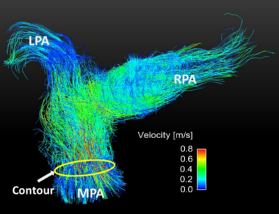
We collected 4D flow MRI data and right heart catheterization (RHC) data from 20 idiopathic pulmonary artery hypertension (IPAH) patients and applied parameter analysis. The results indicate the difference of the flow characteristics in the main pulmonary artery (MPA) between the patients with right ventricle (RV) function loss and the patients with normal RV function. To conclude, 4D flow MRI is a good tool to detect hemodynamics and has the potential to assist in evaluating the state and the curative effect of IPAH.
|
|
1991
 |
41 |
Non-invasive estimations of turbulence driven relative pressure drops – applying the concept of virtual fields on 4D flow MRI
Video Permission Withheld
David Marlevi, Hojin Ha, Joao Fernandes, Tino Ebbers, Pablo Lamata, David Nordsletten
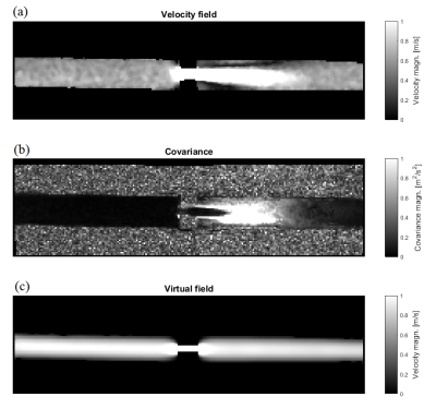
4D flow MRI with six-directional flow encoding has enabled the assessment of turbulent flows, including mapping of incoherent flow variance. Using such, non-invasive estimation of turbulence-driven pressure drops can be computed. Here, we present an extension of the virtual-Work-Energy-Relative-Pressure method8 for the assessment of turbulence-driven pressure drops. Using the concept of virtual fields, the method accurately assesses pressure drops over a range of stenotic valve phantoms, being validated against catheter-based measurements. With virtual probing enabling the assessment of pressure drops through complex, narrow vasculatures, the incorporation of turbulence enhances the utility of the method, enabling for refined clinical hemodynamic analysis.
|
|
1992.
 |
42 |
Hemodynamics of Severely Stenosed Carotid Artery before and after Endarterectomy by 4D Flow MRI with a High Spatiotemporal Resolution
Don-Gwan An, Seungbin Ko, Jeesoo Lee, Jee-Hyun Cho, Doosang Kim, Sang Hyung Lee, Simon Song
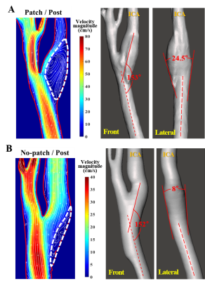
In vitro 4D flow MRI measurements were performed for patient-specific carotid artery phantoms subject to a pulsatile flow before and after carotid endarterectomy (CEA) to secure a high-spatiotemporal resolution of 0.35 mm and 0.025 sec. As a result, we observed that the flow rate ratio of internal carotid artery (ICA) to common carotid artery (CCA) was not recovered after CEA of a patch repair due to a large recirculation motion in the ICA bulb which blocks the flow into ICA. Detailed hemodynamics are presented along with normalized time-averaged wall shear stress (NTA|WSS|), oscillatory shear index (OSI), abnormal regions, etc.
|
|
1993
 |
43 |
Vortex Formation Time in Chinese Children: a CMR Study
Video Permission Withheld
Liwei Hu, Rongzhen Ouyang, Yafeng Peng, Chen Guo, Yong Zhang, Xiaofen Yao, Yumin Zhong, Christopher François
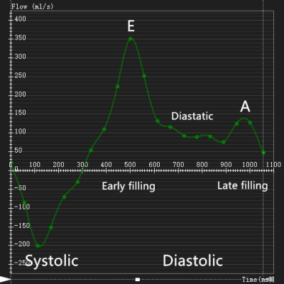
Vortex formation time (VFT) is an index of left ventricular (LV) systolic and diastolic performance, with normal values, based on echocardiography, from 3.3 to 5.5 ms in adults [1]. With the increasing intensity of vortex, the vortex ring is pinched off; this instant is defined as the vortex ring formation time. With echocardiography VFT is measured from trans-mitral inflow velocities. Although cardiovascular magnetic resonance (CMR) is the gold standard for assessing LV systolic function, its use in evaluating LV diastolic function is more limited [2]. Through-plane motion of the mitral valve results in underestimations of the peak mitral inflow velocities with standard 2D flow CMR acquisitions. 4D flow CMR analysis improves the accuracy of peak mitral inflow velocities because of its ability to track mitral valve plane motion [3]. However, vortex ring quantification with 4D flow CMR has not been widely used [4]. The VFT has not been evaluated in children and could be useful tools for assessment of diastolic function using CMR.
|
|
1994.
 |
44 |
4D Flow Assessment of Aortic Valve Stenosis in a Single Breath-Hold
Adam Rich, Yingmin Liu, Lee Potter, Ning Jin, Orlando Simonetti, Rizwan Ahmad
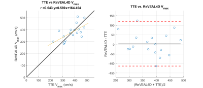
MRI-based 4D flow imaging is capable of yielding spatially and temporally resolved mapping of the blood velocity vector. Long acquisition times associated with 4D flow imaging limits its clinical utility. In this work, we apply a recently proposed technique, called ReVEAL4D, to perform 4D flow imaging in 19 patients with aortic valve stenosis. The peak velocity obtained using ReVEAL4D shows good agreement with both transthoracic echocardiography (TTE) and traditional GRAPPA-based 4D flow imaging.
|
|
1995.
 |
45 |
Highly accelerated 4D flow with compressed sensing for efficient evaluation of whole-heart hemodynamics
Liliana Ma, Ning Jin, Kelvin Chow, Christoph Forman, Andreas Greiser, James Carr, Michael Markl
Recently, a highly accelerated compressed sensing (CS) 4D flow framework with navigator gating and retrospective ECG-gating was developed and tested in healthy volunteers and patients. This study aimed to determine the optimal protocol for high resolution (2-3 mm3) whole-heart 4D flow MRI in <8 minutes without respiratory gating, and minimization of respiratory artifacts while improving scan efficiency.
|
|
1996
 |
46 |
The assessment of aortic regurgitation using phase contrast MRI is influenced by complex flow
Video Permission Withheld
Frida Truedsson, Christian L Polte, Odd Bech-Hanssen, Åse A Johnsson, Kerstin M Lagerstrand
Assessment of aortic regurgitation (AR) severity by cardiovascular MRI can be obtained directly by phase-contrast MRI. For some AR-patients, especially those with aortic dilatation and bicuspid aortic valve, the flow profile can be highly complex displaying asymmetric outflow jets, helical vortex flow and systolic backward flow. This study showed that such complex flow influences the accuracy of the AR assessment and needs to be taken into account in clinical practice as it may compromise the decision-making and timing of surgery.
|
|
1997.
 |
47 |
Highly-accelerated real-time phase-contrast and cine MRI using radial k-space sampling and compressed sensing for imaging blood flow and function in the left atrium: inter-scan reproducibility analysis
Suvai Gunasekaran, Hassan Haji-Valizadeh, Liliana Ma, Rishi Arora, Philip Greenland, Daniel Lee, Rod Passman, Michael Markl, Daniel Kim
Standard ECG-gated phase-contrast (PC) and cine MRI methods are likely to produce non-diagnostic image quality and/or poor reproducibility in patients with atrial fibrillation due to irregular heart rhythm. One approach to address this problem is to develop highly-accelerated real-time PC and cine MRI acquisitions which are insensitive to arrhythmia. In response, we developed such methods using radial k-space sampling and compressed sensing. In this study, we sought to evaluate the inter-scan reproducibility of highly accelerated real-time PC and cine MRI methods for imaging blood flow and function in the left atrium.
|
|
1998.
 |
48 |
Auto-calibrated Simultaneous Multi-Slice Pulse-Wave Velocity Imaging
Sebastian Schmitter, Giulio Ferrazzi, Bernd Ittermann, Tobias Schaeffter, Susanne Schnell
Pulse Wave Velocity (PWV) MR imaging is an established technique to derive aortic stiffness. The underlying phase-contrast velocity data is typically acquired during multiple breath-holds within multiple 2D planes placed perpendicularly to the aorta. In this work we investigate the application of an auto-calibrated multiband approach to simultaneously excite and acquire three slices. With this technique all data is obtained in a single breath-hold and without the need of external reference scans. Blood velocities and PWV for different MB acquisitions are compared to results obtained with a singleband approach that excites each slice separately.
|
|
1999.
 |
49 |
Assessment of Pulmonary Hypertension using 4D flow and SSFP MRI
Daniel Gordon, Muhannad Abbasi, Carson Herman, Michael Markl, Pascale Aouad, Jeremy Collins, Roberto Sarnari, Benjamin Freed, Michael Cuttica, Sanjiv Shah, James Carr
Right heart catheterization (RHC) is the current gold standard for the diagnosis of pulmonary hypertension (PHTN). However, the use of invasive and ionizing procedures during RHC has driven research to find alternative ways for PHTN assessment using MRI. We propose time resolved 3D imaging (4D flow) and SSFP cardiac MRI as an alternative method for assessing hemodynamics for PHTN. Using One-way Analysis of Variance (ANOVA), inter-observer variability, and post hoc analysis our findings indicate the possibility of MRI to detect hemodynamic changes among various groups of PHTN and healthy controls which could lead to successful diagnostic distinctions between groups.
|
|
2000
 |
50 |
Inter-Scanner, Inter-Software, and Inter-Reader Cross-over Interindividual Comparison of Quantitative Parameters in 4D Flow MRI
Video Permission Withheld
Malte Sieren, Andre Nowak, Nicolas Kirschke, Joachim Graessner, Hendrik Kooijman, Joerg Barkhausen, Alex Frydrychowicz, Thekla Oechtering
Before introduction into clinical routine 4D Flow MRI has to be tested in larger scale studies. For data to meet quality standards for these studies various potential error sources have to be addressed. The aim of this study was to provide a comprehensive inter-scanner, inter-vendor, inter-individual cross-over evaluation of a 4D Flow sequence. The thoracic aorta of eight volunteers was examined on two 3T MRI-Scanners of different vendors and analyzed by two readers using three different software. While there was no significant difference between readers and MRI scanners, differences between analysis-software where beyond clinically acceptable limits.
|
|
| Top |
Tissue Characterization 1
Digital Poster
Cardiovascular
Monday, 13 May 2019
| Exhibition Hall |
16:00 - 17:00 |
| |
|
Computer # |
|
2001.
 |
51 |
Structural and functional myocardial impairments in Becker muscular dystrophy using quantitative cardiac magnetic resonance imaging
Benjamin Marty, Raymond Gilles, Karim Wahbi, Pierre Carlier
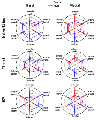
The management of cardiac involvement is central for Becker muscular dystrophy (BMD) patients since heart failure represents the most frequent cause of death in this population. We performed a comprehensive CMR evaluation of functional and structural myocardial alterations encountered in a cohort of 88 BMD patients. A total of 26% of the BMD patients had a reduced ejection fraction (EF). Globally, native T1, T2 and ECV values were significantly higher in BMD patients than in healthy volunteers, even in sub-clinical phenotypes and correlated with EF. Our results encourage a more systematic inclusion of CMR in the standard of care applied to BMD patients.
|
|
2002.
 |
52 |
3D whole-heart free-breathing BOOST-T2 mapping
Giorgia Milotta, Giulia Ginami, Aurelien Bustin, Radhouene Neji, Claudia Prieto, Rene Botnar
Cardiac MRI enables the assessment of whole-heart anatomy with both bright-and-black-blood contrasts. Additionally, quantitative myocardial T2 mapping is an emerging technique that enables non-contrast tissue characterization. However, conventional T2 mapping is performed under breath-hold with limited spatial resolution and coverage. Moreover, anatomic and quantitative images are acquired sequentially with different geometries and at different motion states. Here, we propose a novel quantitative 3D whole-heart sequence (qBOOST-T2) which provides co-registered 3D high-resolution bright-blood, black-blood and T2 map volumes from a single free-breathing scan. qBOOST-T2 was evaluated in a standardized T1/T2 phantom and healthy subjects and compared to current gold standard techniques.
|
|
2003.
 |
53 |
3D Whole-heart High-resolution Motion Compensated Joint T1/T2 Mapping
Giorgia Milotta, Giulia Ginami, Aurelien Bustin, Radhouene Neji, Claudia Prieto, Rene Botnar
Tissue characterization including identification and quantification of fibrosis and oedema plays an important role in many myocardial diseases. Conventionally T1 and T2maps are acquired sequentially under several breath-holds. These approaches however achieve limited spatial resolution and coverage. Furthermore, partial volume effects at water-fat interfaces may affect the T1 and T2 quantification. In this work, we propose a free-breathing high-resolution whole-heart joint T1 and T2 mapping sequence with Dixon encoding which provides co-registered 3D T1 and T2 maps and complementary 3D fat images.
|
|
2004.
 |
54 |
Imaging myocardial reperfusion injury using cardiac quantitative susceptibility mapping
Brianna Moon, Srikant Kamesh Iyer, PhD, Eileen Hwuang, Michael Solomon, Anya Hall, Rishabh Kumar, Elizabeth Higbee-Dempsey, Andrew Tsourkas, PhD, Yoshiaki Saito, MD, Akito Imai, MD, Keitaro Okamoto, MD, Avanti Gulhane, MD, Harold Litt, MD-PhD, William Matthai, MD, James Pilla, PhD, Joseph Gorman III, MD, Robert Gorman, MD, Samuel Keeney, Victor Ferrari, MD, Giovanni Ferrari, PhD, Walter Witschey, PhD
Hemorrhage is a frequent complication of reperfusion therapy for acute myocardial infarction (MI). This study investigated reperfusion injury with respect to the duration of myocardial ischemia by analyzing magnetic susceptibility, an endogenous imaging biomarker of tissue iron, in a large animal model. We demonstrate with cardiac quantitative susceptibility mapping (QSM), there is a significant shift in infarct tissue magnetic susceptibility with longer time-to-reperfusion and non-reperfused infarcts compared to remote myocardium which correlates with iron content and infarct pathophysiology.
|
|
2005.
 |
55 |
T1 is significantly higher in myocardial T1 when measured during free breathing than at inspiration
Laura Saunders, Andy Swift, David Capener, James Wild
Release of breath hold results in misalignment of the myocardium during cardiac T1 mapping. Several registration methods have been developed to overcome this [1][2][3], however, it has not been established whether the process of respiration effects myocardial T1 when mapped with dynamic MOLLI sequences. 10 healthy volunteers underwent 1.5T MOLLI T1 mapping during both breath hold and free breathing acquisition. Images were registered using synthetic images created via a combined inversion recovery and respiratory signal modulation model, which was verified using Dice Similarity Coefficient. Myocardial T1 was found to be higher in healthy volunteers when acquired during free breathing, when compared to inspiration.
|
|
2006.
 |
56 |
Impact of a ten-year national Italian networking on cardiac complications in patients with thalassemia major
Antonella Meloni, Laura Pistoia, Riccardo Righi, Nicolò Schicchi, Stefania Renne, Antonino Vallone, Emanuele Grassedonio, Gennaro Restaino, Saveria Campisi, Sabrina Armari, Vincenzo Positano, Alessia Pepe
Over a period of 10 years, the continuous monitoring of cardiac iron levels and a tailored chelation therapy allowed a reduction of myocardial iron overload (MIO) in the 70% of patients with thalassemia major (TM) enrolled in the MIOT (Myocardial Iron Overload in Thalassemia) Network. A consequent improvement of cardiac function and a reduction of heart failure were detected. So, a national networking was effective in improving the care and reducing cardiac outcomes of TM patients.
|
|
2007.
 |
57 |
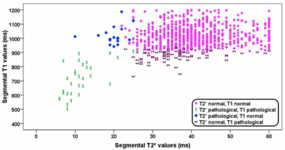
Detection of myocardial iron overload with magnetic resonance by native T1 and T2* mapping using a segmental approach
Antonella Meloni, Nicola Martini, Daniele De Marchi, Andrea Barison, Laura Pistoia, Massimo Allò, Silvia Macchi, Roberta Renni, Mauro Murgia, Gerardi Calogera, Vincenzo Positano, Alessia Pepe
T2* and T1 values assessed in the 16 myocardial segments showed a good agreement (90% concordance) in the identification of myocardial iron overload (MIO) in patients with hemoglobinopathies.
|
|
2008.
 |
58 |
Genotypic Groups as Risk Factor for Cardiac MR Abnormalities and Complications in Thalassemia Major
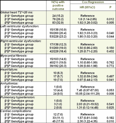
Laura Pistoia, Antonella Meloni, Massimiliano Missere, Paolo Preziosi, Giuseppe Peritore, Ada Riva, Valentina Vinci, Giovanni Palazzi, Alessandra Spiga, Alessandra Quota, Vincenzo Positano, Alessia Pepe
On the basis of the type of gene mutation, three groups of patients with thalassemia major (TM) were identified: homozygotes β+, compound heterozygotes β+/β° and homozygotes β°. Compared to the milder genotype group homozygotes β+, the other two groups showed a significantly higher risk of myocardial iron overload (MIO) and left ventricular dysfunction. Moreover, homozygotes β° showed a significantly higher risk of CC than homozygotes β+ patients. These data support the knowledge of the different genotypic groups in the clinical management of β-TM patients.
|
|
2009.
 |
59 |
Automatic Detection and Quantification of Myocardial Scar in Patients with Prior Myocardial Infarction at 3T without Contrast Agents
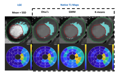
Xinheng Zhang, Hsin-Jung Yang, Guan Wang, Ivan Cokic, Qi Yang, Sotirios Tsaftaris, Rohan Dharmakumar
Native T1 maps at 3T has the capacity to accurately characterize chronic myocardial infarction (MI) territories, however it requires accurate identification of remote myocardium which in some cases is limited by image contrast between infarcted and remote myocardial territories. To overcome this limitation, we evaluated multiple automatic segmentation algorithms. Native T1 maps acquired in chronic MI patients were segmented using Gaussian Mixture Model, Otsu’s and K-means methods. K-means approach showed the best performance when compared to LGE. We conclude that K-means approach can accurately delineate MI territories.
|
|
2010.
 |
60 |
Assessing myocardial infarct size by novel TRAFFn relaxation time method in lymphatic insufficient mice
Elias Yla-Herttuala, Taina Vuorio, Svetlana Laidinen, Seppo Yla-Herttuala, Timo Liimatainen
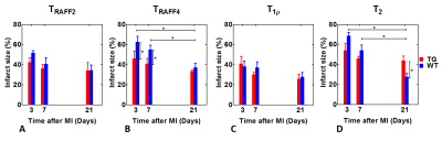
We applied novel RAFFn relaxation times (TRAFF2 and TRAFF4), T1ρ and T2 to study effects of cardiac lymphatic system in myocardial infarct (MI). Infarct size based on the TRAFF4 relaxation time maps was significantly larger in earlier time points post MI in wild-type compared to lymphatic insufficient mice groups. No differences were found in relaxation times between groups. Area-of-overestimation (AOE) values remained stable in lymphatic insufficient group while in wild-type group a decreasing trend of AOE was observed. We conclude that effects of lymphatics after MI can be detected based on infarct size difference measured with different relaxation times.
|
|
2011.
 |
61 |
Myocardial T1 mapping using inversion recovery with radial simultaneous multi-slice readout and model-based reconstruction
Ye Tian, Jason Mendes, Edward DiBella, Ganesh Adluru
Here we propose an inversion recovery based radial simultaneous multi-slice sequence for myocardial T1 mapping. 3 slices of T1 maps were acquired simultaneously within one breath hold spanning 11 heartbeats. Model based reconstruction was used to jointly reconstruct images at different inversion times and estimate T1 maps. Native T1, post-contrast T1 and ECV maps agree with results from the slice-by-slice Cartesian MOLLI sequence.
|
|
2012
 |
62 |
Early Detection of Myocardial Fibrosis by CMR Extracellular Volume Fraction Quantitation in a Hypertensive Swine Model
Video Permission Withheld
Baiyan Zhuang, Chen Cui, Arlene Sirajuddin, Andrew Arai, Shihua Zhao, Minjie Lu
At present, hypertensive left ventricular hypertrophy (LVH) is generally considered to be an adaptive hypertrophy of cardiomyocytes due to an increase in left ventricular afterload. However, recent studies have shown that myocardial interstitial fibrosis may play an important role in cardiac hypertrophy. This study aims to investigate the relationship between extracellular volume (ECV) fraction and left ventricular remodeling in hypertension by prospective randomized controlled swine model of hypertension, and explain its internal mechanism based on pathology.
|
|
2013.
 |
63 |
Comparison of cardiac function, morphology and tissue characteristics between two subtypes of primary aldosteronism: a 3 Tesla MR study
Satoshi Higuchi, Hideki Ota, Kazumasa Seiji, Yuta Tezuka, Ryo Morimoto, Tatsuya Nishii, Tetsuya Fukuda, Kei Takase
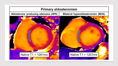
The purpose of this study is to compare cardiac morphology, function and tissue characteristics between patients with two subtypes of primary aldosteronism (PA), aldosterone-producing adenoma (APA) and bilateral hyperaldosteronism (BHA). One-hundred-and-forty-three consecutive PA patients underwent 3T MR examinations including cine, late gadolinium enhancement and pre- and post-contrast T1 mapping. APA group demonstrated higher myocardial native T1 and left-ventricular end-diastolic volume index than BHA after controlling for patients’ demographic data. The results indicate that APA group, with higher hormonal activity than BHA, may be suffered from LV volume overload and myocardial fibrosis or edema as compared with BHA group.
|
|
2014.
 |
64 |
Myocardial T1 mapping with Shortened Acquisition Window using a two-layer Sparsifying Transform
Yanjie Zhu, Dong Liang, Maryam Nezafat, Chong Duan, Reza Nezafat
Long acquisition window in myocardial T1 mapping degrades the image quality. Physiological motion induced fluctuations along the parametric direction corrupt the similarity among T1-weighted images and adversely impact the performance of the compressed sensing-based methods. In this study, we propose a two-layer sparsifying transform (MO2) combined with motion correction to shorten the acquisition window of myocardial T1 mapping. Results showed that the proposed method enabled higher acceleration factor. The resulting shorter acquisition window and fewer RF saturation pulses increased the accuracy of the native T1 measurement.
|
|
2015.
 |
65 |
Accelerating modified Look-Locker inversion recovery (MOLLI): Shortened inversion-recovery-based myocardial T1 mapping schemes
Li Huang, Radhouene Neji, Muhammad Nazir, John Whitaker, Filippo Bosio, Amedeo Chiribiri, Reza Razavi, Sébastien Roujol
Abnormal native myocardial T1 times are associated with a variety of cardiomyopathies, and are widely measured by inversion-recovery-based myocardial T1 mapping techniques such as modified Look-Locker inversion recovery (MOLLI). These sequences are limited in patients with severe breathholding difficulties for relatively long duration of breathholds. In this work, we sought to develop and characterize shortened schemes using less amount of T1-weighted images to reduce their sensitivity to imperfect breathholds.
|
|
2016.
 |
66 |
Tuning Blipped CAIPIRINHA for simultaneous multi-slice (SMS) balanced SSFP cardiac imaging
Zakarya Bentatou, Stanislas Rapacchi, Thomas Troalen, Maxime Guye, Monique Bernard, Alexis Jacquier, Frank Kober
In this study, we used blipped-CAIPIRINHA simultaneous multi-slice (SMS) technique to 1) extend bSSFP coverage to 3 simultaneous slices and 2) apply it to cardiac T1 mapping. Tests were conducted on phantom and in six healthy volunteers. SMS blipped-CAIPIRINHA-bSSFP sensitivity to slice gap, slice thickness and pixel bandwidth was successfully established in terms of SNR. Native T1 quantification values over three levels of the heart (base, mid and apex) were reliable, accurate, and in line with the ones obtained with a regular single slice acquisition.
|
|
2017.
 |
67 |
Hybrid PET/MR in Cardiac Sarcoidosis: A segmental analysis.
Emily Aherne, Ali Serhal, Ryan Avery, Alexander Ruh, Louise Collins, Hatice Savas, Gary Dillehay, James Carr
Cardiac sarcoidosis (CS) is difficult to clinically diagnose but associated with high morbidity and mortality. Hybrid PET/MR has been shown to provide clinicians with complementary data regarding both the pattern and activity of myocardial disease. We performed a segmental quantitative analysis of MR parameters on 30 patients including T1 and T2 mapping, ECV, scar and strain acquired as part of a hybrid PET/MR study to evaluate characteristic patterns of imaging in this heterogeneous disease process. Scar quantification had a significant positive correlation with T1 and ECV and was negatively correlated with strain.
|
|
2018.
 |
68 |
Validation of Fully Automatic SegmenTal Relaxometry (FASTR) in patients post myocardial infarction
Venkat Ramanan, Idan Roifman, Xiuling Qi, LaBonny Biswas, Graham Wright, Nilesh Ghugre
T1 mapping has started to be used for both diffuse and focal diseases of the heart and recently in multi-slice mode to investigate the regional (segmental) variations in T1 and ECV. The processing usually involves contouring the endo and epicardial boundaries manually, which could be cumbersome especially for high-volume studies. Recently, we presented a fully automatic approach (FASTR), to calculate T1 and ECV values segment-wise. Here we present the initial validation of our technique in patients post myocardial infarction. FASTR is comparable to an expert-driven contour and has low bias and similar variability in comparison to inter-observer analysis.
|
|
2019.
 |
69 |
Myocardial T1 and T2 Mapping and Tissue-tracking Strain Analysis in Hemodialysis Patients with Preserved Left Ventricular Ejection Fraction by Cardiac Magnetic Resonance Imaging
Presentation Not Submitted
xiaoyu han, heshui shi
Myocardial T1 and T2 Mapping and Tissue-tracking Strain Analysis in Hemodialysis Patients with Preserved Left Ventricular Ejection Fraction by Cardiac Magnetic Resonance Imaging
|
|
2020.
 |
70 |
Simultaneous analysis of heart and kidney oxygenation using T2* BOLD MRI: investigation of cardiorenal relationship
Michinobu Nagao, Umiko Ishizaki, Kiyoe Ando, Eri Watanabe, Akiko Sakai, Yasuhiro Goto, Masami Yoneyama, Takashi Namiki, Shuji Sakai
Tissue hypoxia plays a key role in the development and progression of cardiac and renal diseases. Blood oxygenation level dependent magnetic resonance imaging (BOLD-MRI) is the most promising imaging technique to monitor tissue oxygenation in humans. Cardiorenal syndrome is widely accepted as a complex clinical problem routinely faced by clinicians. However, the mechanism from the viewpoint of oxygenation is not understood. We analyses simultaneously myocardial and renal oxygenation using T2* cardiac MRI (T2*-CMR) and investigates the cardiorenal relationship.
|
|
2021.
 |
71 |
Optimizing native T1-based detection of chronic myocardial infarctions at 3T: Influence of MOLLI flip angle on the relative myocardium-to-blood T1 contrast
John Van Dyke, Ivan Cokic, Rohan Dharmakumar, Behzad Sharif
Prior studies have shown that native T1 mapping at 3T provides an effective non-contrast approach for characterizing the presence and transmurality of chronic myocardial infarctions. The underlying mechanism is likely a combination of T1/T2 and magnetization transfer (MT) effects. The choice of flip angle is known to significantly affect the degree to which MOLLI-based T1 maps are confounded by MT and T2 effects. With the motivation to optimize the performance of MOLLI T1 mapping for detection of subendocardial infarcts, we hypothesized that increasing the flip angle beyond the conventionally-used 35° will provide a higher relative contrast and contrast-to-noise ratio between the infarcted myocardium and adjacent bloodpool.
|
|
2022.
 |
72 |
Quantitative Multiparametric Myocardial Evaluation in Hypertrophic Cardiomyopathy using Cardiac Magnetic Resonance Fingerprinting: Comparison to Conventional Cardiac Relaxometry.
Bernd Wintersperger, Jesse Hamilton, Christian Houbois, Yuchi Liu, Kate Hanneman, Nicole Seiberlich, Marshall Sussman
Multiparametric quantitative myocardial tissue characterization has demonstrated promising results in the differential diagnosis of non-ischemic cardiomyopathies. While conventional cardiac relaxometry techniques employ various different sequence approaches with subsequent fitting data fitting, library based cardiac magnetic resonance fingerprinting (cMRF) enables single breath-hold multi-contrast acquisitions. This investigation into the comparison of cMRF with standard modified Look-Locker inversion recovery (MOLLI) T1 mapping and its derived parameters and T2-prep FLASH T2 mapping demonstrated promising results in the assessment of patients with hypertrophic cardiomyopathies.
|
|
2023.
 |
73 |
Combined SAturation recovery and Variable flip Angle (SAVA) for free-breathing high-resolution three-dimensional cardiovascular magnetic resonance T1 mapping at 3T
Rui Guo, zhensen chen, Dongyue Si, Daniel A. Herzka, jianwen luo, Hanyan Ding
Cardiac Magnetic Resonance T1 mapping enables quantitative characterization of myocardium, which appeals huge attention from clinic. In this work, we proposed a free-breathing high-resolution three-dimensional (3D) T1 mapping sequence. By using variable flip Angle technique for readouts and saturation-recovery preparation for T1weightings, the proposed sequence is highly efficient in acquisition of all volumes, including the one at the equilibrium of the longitudinal magnetization. Whole-heart pre- and post-contrast homogeneous T1 maps at an imaging resolution 1.25×1.25×8 mm3 were successfully obtained within 10 minutes in healthy volunteers. After rigid registration, 3D Extracellular Volume maps were eventually achieved.
|
|
2024.
 |
74 |
Association of Myocardial Tissue Characterization between Cardiovascular MR and 11C-acetate PET imaging
Shuai Liu, Ximin Shi, Xue Lin, Li Huo, Ligang Fang, Fei Shang, Xiaomeng Wu, Shengji He, Rui Guo, Haiyan Ding, Huimin Duan, Xihai Zhao
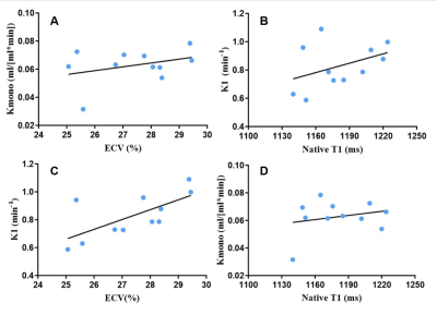
The metabolic alteration in myocardium always accompanied with the structural remodeling. Therefore, it is possible to bridge the imaging markers in CMR with those in 11C-acetate PET imaging. This study investigated the correlation of measurements of interventricular septum between CMR and 11C-acetate in healthy male adults. We found that there was a significant association between ECV measured by quantitative CMR and K1 measured by PET imaging. Our results may indicate a dynamic balance between myocardial blood flow and ECV physiologically. The clinical significance of this relationship needs to be further investigated in patients with cardiac diseases.
|
|
2025.
 |
75 |
Single breath-hold saturation-recovery 3D cardiac T1 mapping via compressed SENSE at 3T
Tiago Ferreira da Silva, Paula Montesinos, Robert Austin Benn, Carlos Galan-Arriola, Manuel Lobo-González, Gonzalo López-Martín, Borja Ibañez, Javier Sánchez-González
We propose a new 3D T1 mapping technique (CS 3D T1 mapping) to acquire the entire left ventricle in a single breath-hold. The technique combines flexible saturation time sampling, sharing the saturation pulses between different RR intervals and compressed SENSE. The proposed sequence successfully acquired a 3D-T1 map of 12 slices in a single breath-hold (14 heart beats) at 3T, achieving T1 values in good agreement with the T1 values estimated by the IR-SE sequence. Additionally, myocardium T1 values obtained with the proposed technique are similar to those already published in literature for 3T. |
|
| Top |
Tissue Characterization 2
Digital Poster
Cardiovascular
Monday, 13 May 2019
| Exhibition Hall |
16:00 - 17:00 |
| |
|
Computer # |
|
2026.
 |
76 |
The structural basis for haemodynamically compromising VT assessed using high-resolution late gadolinium enhanced cardiovascular magnetic resonance imaging under contrast steady state
John Whitaker, Steven Kim, Adam Connolly, Radhouene Neji, Rashed Karim, Steven Williams, Louisa O'Neill, Rahul Mukherjee, Henry Chubb, Srijoy Mahapatra, Luigi Camporota, Matthew Wright, John Silberbauer, Sébastien Roujol, Martin Bishop, Mark O'Neill, Reza Razavi
Using a translational porcine model, the structural basis for post myocardial infarction ventricular tachycardia was assessed using in-vivo cardiac magnetic resonance imaging. High-resolution LGE imaging was acquired under contrast steady state in order to allow detailed tissue characterisation. Arrhythmia was induced and assessed under haemodynamic support to allow the unambiguous identification of arrhythmogenic tissue involved in these scar mediated VT circuits. The electrophysiological and imaging data was then registered to establish the structural features of tissue involved in these rhythms. It was identified that tissue participating in the diastolic phase of post-MI VT was thinner, had non-transmual scar or intermediate signal intensity and had higher gradients in tissue thickness.
|
|
2027.
 |
77 |
Intramyocardial and Pericardial Fat Quantification in Boys with Duchenne Muscular Dystrophy and Healthy Controls at 3T.
Nyasha Maforo, Holden Wu, Patrick Magrath, Pierangelo Renella, Nancy Halnon, Daniel Ennis
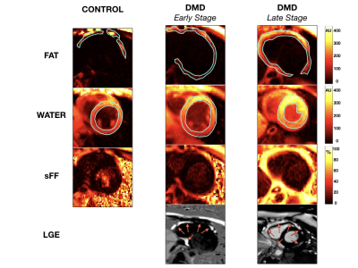
Emerging cardiac MRI biomarkers of Duchenne muscular dystrophy (DMD), a fatal X-linked genetic disorder, include intra-myocardial fibro-fatty infiltration, to identify the onset of microstructural remodeling in boys with DMD. The study aims were to: 1) characterize and compare intra-myocardial signal fat fraction (sFF) between boys with DMD and healthy controls; and 2) report and compare pericardial fat volume and sFF estimates in boys with DMD and healthy controls. We detected no intra-myocardial and pericardial sFF differences between DMD boys and healthy controls respectively. Boys with DMD presented with significantly more pericardial fat volume compared to healthy boys.
|
|
2028.
 |
78 |
A Single-Image Super-Resolution Method for Late Gadolinium Enhancement CMR
Jin Zhu, Guang Yang, Tom Wong, Raad Mohiaddin , David Firmin, Jennifer Keegan , Pietro Lio
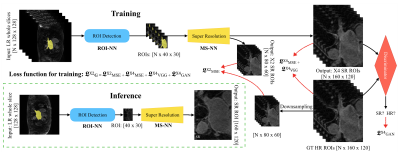
3D late gadolinium enhanced (LGE) CMR is a useful imaging modality for detecting scar tissue in patients with atrial fibrillation. In order to visualize the thin-walled left atrium and scar tissue, high spatial resolution and contiguous coverage are required. However, increased spatial resolution requires markedly prolonged scanning time. In this paper, we propose a ROI focused single-image super-resolution (SISR) method based on the generative adversarial networks architecture to increase the apparent spatial resolution of 3D LGE data without increasing scan time. The proposed SISR method can boost the spatial resolution of the LGE CMR images while maintaining the perceptual quality.
|
|
2029.
 |
79 |
3D whole-heart dark-blood late gadolinium enhancement without additional magnetization preparation for simultaneous detection of both atrial and ventricular fibrosis
Robert Holtackers, Suzanne Gommers, Joachim Wildberger
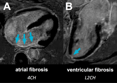
Accurately acquiring the location and extent of thin atrial fibrosis patterns demands for improved imaging techniques compared to conventional ventricular LGE. Recently, we developed a novel 2D dark-blood LGE approach that significantly increases scar-to-blood contrast without using additional magnetization preparation. In this feasibility study, we sought to translate this novel approach towards 3D fibrosis imaging for simultaneous detection of both atrial and ventricular scar. 3D dark-blood PSIR LGE proves to be a readily available approach for detection of both atrial and ventricular fibrosis, with improved robustness for arrhythmias, increased scar-to-blood contrast, improved patient comfort, and enabling a more versatile analysis.
|
|
2030.
 |
80 |
Novel phase-sensitive late gadolinium enhancement MRI for assessment of myocardial infarction in large animals
Anna Naumova, Niranjan Balu, Hiroshi Tsuchida, Lauren Neidig, RS Thies, Charles Murry, Chun Yuan
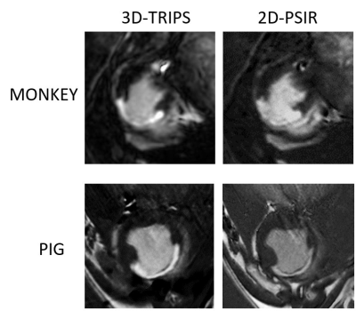
Novel phase sensitive reconstruction method of the gadolinium delayed hyperenhancement, 3D-TRIPS, was used for the first time in the infarct visualization and quantitation in the hearts of non-human primates and Yucatan mini-pigs. Advantages of using 3D-TRIPS reconstruction vs. standard 2D-PSIR method for MR imaging of infarct include scan time shortening due to elimination the background phase-reference scan and improvement of the contrast ratio between normal and infarcted myocardium as well as between scar and blood. The novel cardiac MRI 3D-TRIPS method provides robust infarct size measurements while preserving scan time.
|
|
2031.
 |
81 |
A 3D high resolution MRI method for visualization of fibro-fatty infiltration in arrhythmogenic right ventricular cardiomyopathy (ARVC) in human heart.
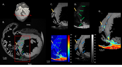
Kylian Haliot, Valéry Ozenne, Richard Walton, Olivier Bernus, David Benoist, Michel Haissaguerre, Julie Magat, Bruno Quesson
The goal of this study is to present 3D high resolution MR-acquisition methods for ex vivo imaging of the myocardial substrate to identify fibro-fatty infiltration. For this purpose IDEAL and Magnetization Transfer acquisitions were acquired in 3D to visualized and identify fibrosis, fat infiltration from arrhythmogenic right ventricular cardiomyopathy (ARVC) and healthy human hearts.
|
|
2032.
 |
82 |
A real-time myocardial velocity encoding sequence to optimize trigger delay in motion-compensated cardiac DWI
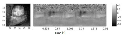
Anne-Lise Le Bars, Kévin Moulin, Daniel Ennis, Jesús Dos Reis, Laurent Bonnemains, Jacques Felblinger, Chen Bailiang, Odille Freddy
Motion-compensated spin echo sequences have been developed to obtain diffusion sensitivity in the presence of bulk cardiac motion. First and second order motion can be compensated but higher order motion can occur in certain cardiac phases. Here, we propose a real-time velocity-encoded sequence (6ms time resolution) to optimize and adapt the trigger delay on a subject-specific basis. In one volunteer, the variability in diffusion signal was analyzed as a function of trigger delay and compared to myocardial velocity profiles. Mid-systole was found to provide the most reliable ADC and conversely the worst ADC was found in early diastole.
|
|
2033.
 |
83 |
Analysis and Correction of Off-Resonance Artefacts in Free-Breathing In-Vivo Motion-Compensated Spin-Echo Cardiac Diffusion Tensor Imaging at 3T
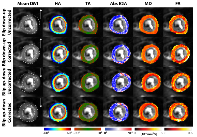
Robbert van Gorkum, Constantin von Deuster, Christian Stoeck, Sebastian Kozerke
Cardiac diffusion tensor imaging (cDTI) sequences inherently suffer from low signal-to-noise (SNR) ratios. Although high field strength systems improve SNR, long single-shot readout trains such as echo-planar imaging experience detrimental effects due to changes in magnetic susceptibility at tissue boundaries. Using synthetic and in vivo free-breathing cDTI data, an iterative time-segmented off-resonance correction methodology was implemented and evaluated. Using this approach, the cDTI data was geometrically restored to the original shape, and underlying tensors metrics were corrected. The framework holds potential to aid geometrically accurate in vivo cDTI for multi-contrast and multi-modal imaging studies.
|
|
2034
 |
84 |
Optimisation of diffusion encoding schemes for in vivo cardiac DTI
Video Permission Withheld
Irvin Teh, Christopher Nguyen, Christopher Kelly, Erica Dall'Armellina, Debiao Li, Sven Plein, Jürgen E Schneider
Cardiac diffusion tensor imaging (CDTI) measurements are sensitive to a range of imaging parameters including the number of signal averages (NSA) and the number of unique diffusion-weighting directions (ND). However, there is no clear guidance on their specifications for clinical imaging. We evaluated the impact of ND and NSA on the accuracy and precision of the mean diffusivity, fractional anisotropy and helix angle in 10 healthy volunteers. Our findings support the need for standardisation of CDTI protocols to facilitate inter-study and inter-site comparison of data, and definition of clinically relevant thresholds for catalysing the clinical adoption of CDTI.
|
|
2035
 |
85 |
Rapid cardiac diffusion-weighted imaging with novel motion-compensated spherical tensor encoding
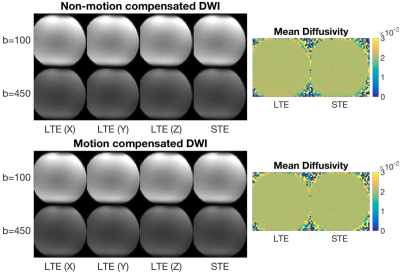
Video Permission Withheld
Irvin Teh, Filip Szczepankiewicz, Erica Dall’Armellina, Sven Plein, Markus Nilsson, Jürgen E. Schneider
Cardiac diffusion-weighted imaging (CDWI) is an emerging method for non-invasive assessment of cardiac microstructure. In CDWI, averaging and triggering are routinely used to mitigate motion artefacts, which results in long acquisition times. We have developed a novel motion-compensated spherical tensor encoding technique that reduces the acquisition time by a factor of up to three. Quantitatively, the method yields similar mean diffusivity as conventional methods, but enables accurate measurements in less than a minute.
|
|
2036.
 |
86 |
Fully automated in-vivo DT-CMR analysis with deep learning
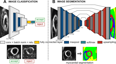
Pedro Ferreira, Andrew Scott, Zohya Khalique, Guang Yang, Sonia Nielles-Vallespin, Dudley Pennell, David Firmin
Currently post-processing of in-vivo DT-CMR data is done off-line as it requires manually input. Two convolutional neural networks (CNN) were trained to classify and segment the LV in order to automate and enable on-the-fly post-processing of DT-CMR data while scanning. The fully automated DT-CMR analysis with deep learning performed effectively with high levels of accuracy when compared to an experienced user.
|
|
2037.
 |
87 |
Different Myocardial Perfusion Status in Acute Myocardial Infarction and Infarct-like Myocarditis: A Novel Intravoxel Incoherent Motion Diffusion-Weighted Imaging based MRI Study.
Presentation Not Submitted
Dong-Aolei An, Bing-Hua Chen, Tong-Tong Han, Jian-Rong Xu, Lian-Ming Wu
This study was to evaluate the diagnostic value of myocardial perfusion in patients with acute myocardial infarction (AMI) and “infarct-like myocarditis” using IVIM-DWI imaging. And the results showed: 1. Insufficient oxygen in the necrotic or infarted myocardium might affect myocardial perfusion and function. 2. IVIM-DWI may be a reliable sequence to evaluate different myocardial perfusion patterns in acute myocardial infarction and infarct-like myocarditis. 3. Acute myocardial infarction may exhibit lower myocardial perfusion status compared with infarct-like myocarditis due to different pathophysiological process.
|
|
2038.
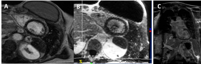 |
88 |
Towards higher spatial resolution late gadolinium enhancement using 3D Turbo Spin Echo (3D TSE LGE)
Dana Peters, Chenxi Hu, Gigi Galiana, Steffen Huber
3D inversion recovery (IR) turbo spin echo (TSE) was investigated for its value in higher spatial resolution 3D late gadolinium enhancement (LGE). Studies in simulations, phantoms and patients found that the SNR is higher with 3D IR TSE, potentially permiting increased spatial resolution, and LGE is feasible.
|
|
2039.
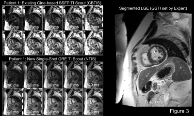 |
89 |
A Magnetization Recovery Matched TI-Scout for Exact Determination of Inversion Time in Segmented Inversion Recovery Gradient Echo Imaging for Late Gadolinium Enhancement
Wolfgang Rehwald, Igor Klem, Marshall Ronningen, Elizabeth Jenista, Nestor Mena, George Gamoneda, Rafael Rojas, Stephen Darty, Enn-Ling Chen, Clerio De Azevedo Filho, Brenda Hayes, Shawn McCarthy, Fawaz Alenezi, David Wendell, Han Kim, Raymond Kim
We present a new TI-scout sequence based on multiple single-shot GRE acquisitions with timing and readout parameters matched to segmented inversion recovery imaging for which optimal TI is to be determined. Temporal magnetic evolution of the scout is identical to that of the segmented GRE sequence for LGE. The new technique substantially improves upon the existing SSFP cine-based TI scout that typically underestimates TI and provides mediocre image quality. We show accurate TI determination by comparison with gold standard TI found by an expert and compare image quality and signal ratios of existing, new TI scout, and segmented LGE.
|
|
2040.
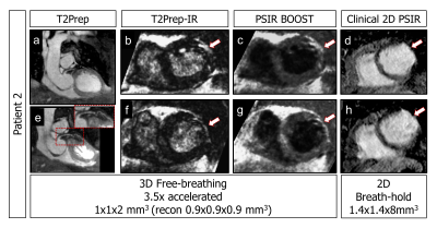 |
90 |
Simultaneous High-resolution 3D Whole-heart Bright-Blood Coronary Angiography and Black-Blood Late Gadolinium Enhancement (LGE)
Teresa Correia, Imran Rashid, Giulia Ginami, Tevfik Ismail, Radhouene Neji, Rene Botnar, Claudia Prieto
The T2-prepared BOOST sequence was recently introduced for simultaneous post-contrast 3D whole-heart bright-blood coronary angiography and black-blood late gadolinium enhancement (LGE) imaging. This sequence enables simultaneous visualization of cardiac anatomy, the coronary arteries and scar tissue. However, high-resolution fully-sampled BOOST requires long acquisition times of ~20min. Here, we propose to accelerate the T2-prepared BOOST sequence and achieve high spatial resolution 3D whole-heart black-blood LGE and coronary MR angiography. This is accomplished by extending XD-ORCCA, a highly efficient respiratory-resolved motion-corrected framework, to BOOST imaging. The proposed framework enables simultaneous black-blood LGE and bright-blood coronary angiography within clinically feasible acquisition times.
|
|
2041.
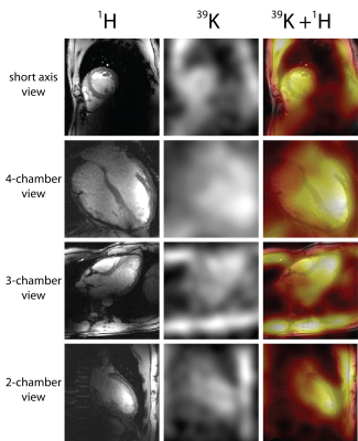 |
91 |
In Vivo Potassium MRI of the Human Heart at 7.0 Tesla
Daniel Wenz, Armin Nagel, Andre Kuehne, Thoralf Niendorf
Potassium ions (K+) play a critical role in cardiac electrophysiology; changes in their concentration might reflect ongoing pathophysiological processes related to cardiovascular diseases. In vivo potassium (39K) MRI, due to its extremely low sensitivity remains a major challenge. Here we investigated, for the first time, the feasibility of in vivo 39K MRI of human heart in a healthy subject. To achieve this goal, we developed a custom-built 39K/1H RF coil, which is tailored for 39K MRI of human heart at 7.0 Tesla. This approach facilitated 39K MRI of human heart with an isotropic spatial resolution of 14.5 mm3 within a total scan time of 30 minutes.
|
|
2042.
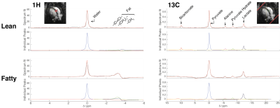 |
92 |
Hyperpolarized Magnetic Resonance Reveals Cardiac and Hepatic Metabolic Dysfunction in the ZDF Model of Type 2 Diabetes
Jack Miller, Kerstin Timm, Vicky Ball, Cher-Rin Chong, Justin Lau, Jürgen Schneider, Günaj Rakipovski, Birgitte Andersen, Damian Tyler
The Zucker Diabetic Fatty (ZDF) rat is a well-known hyperinsulinaemic and hyperlipidaemic model of severe uncompensated Type 2 diabetes (T2DM) that displays systolic cardiac dysfunction late in life at the 40 week timepoint. Hyperpolarized Magnetic Resonance (MR) is a new medical imaging technique that can provide a novel way to probe metabolism in vivo and has been widely used to demonstrate physiological and pathological changes in pyruvate metabolism in the rodent heart. This work presents a magnetic resonance characterisation of the ZDF rat with hyperpolarized MR that detects metabolic alterations prior to the development of cardiac dysfunction.
|
|
2043.
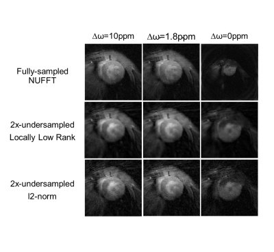 |
93 |
Compressed sensing of under-sampled cardiac-CEST images may bias subsequent contrasts measured from endogenous metabolites
Bonnie Lam, Michael Wendland, Moriel Vandsburger
While accelerated imaging via compressed sensing reconstruction has been explored in several dynamic contrast settings, incorporation of compressed sensing for accelerated image acquisition has not been fully explored in the setting of CEST. In this study we probed how reconstruction of increasingly under-sampled CEST-MRI data using various compressed sensing methods inserted bias into resultant z-spectra and measured CEST contrasts.
|
|
2044
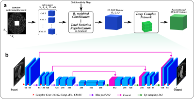 |
94 |
Ultrafast Reconstruction of UnderSampled (URUS) 3D Late Gadolinium Enhancement Imaging using Deep Complex Network
Video Permission Withheld
Hossam El-Rewaidy, Warren Manning, Reza Nezafat
A new framework based on deep complex network is introduced for fast reconstruction of undersampled 3D CMR data. In this work, the complex nature of the acquired data was maintained throughout the network to achieve optimal performance. High resolution 3D LGE dataset of 217 subjects was acquired at prospective acceleration rates ranging from 3 to 5 and used to train and validate (7:3) the proposed methods. Further retrospective undersampling was performed to test the capacity of the proposed methods to perform at higher acceleration rates. LGE scar quantification showed high correlation with the ground-truth.
|
|
2045.
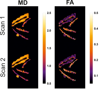 |
95 |
Characterization of tissue properties of tissue engineered cardiac patches using DTI at 22.3T
Jeanine Prompers, Alain van Mil, Miguel Castilho, Madison Ainsworth, Corina Metz, Vitaly Khlebnikov, Jos Malda, Joost Sluijter, Martijn Froeling
Engineering three-dimensional (3D) tissues with similar properties to native myocardium offers a promising approach to restore cardiac function after myocardial infarction. However, visualizing the orientation of the tissue in tissue engineered cardiac patches using immunofluorescent imaging has proven difficult due to the 3D and dense tissue structure. In this study, we have applied diffusion tensor imaging at 22.3T at ultra-high resolution (62.5μm isotropic) to characterize the tissue properties of cardiac patches. We show that adding fibroblasts induces cellular organization of stem cell-derived cardiomyocytes in the patch, resulting in more diffusion restriction and higher anisotropy, better mimicking native myocardial tissue properties.
|
|
2046
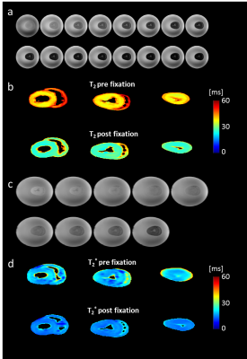 |
96 |
Ex-vivo Diffusion Tensor Imaging of the Porcine Heart at Ultra High Field Strength - 7T: Impacts of Tissue Fixation Duration and Stimulated Echo Mixing Time
Video Permission Withheld
David Lohr, Maxim Terekhov, Sebastian Kress, Franziska Veit, Heike Walles, Laura Schreiber
Diffusion time dependent changes of ADC, FA, HA were analyzed in two fixed, excised porcine hearts using a stimulated echo approach at 7T with mixing times 50-1000ms. A standard SE-method was used as a reference and SNR differences were evaluated using the multiple image method. The data will allow better comparison of in-vivomeasurements using SE- and STE-approaches. Ex-vivo measurements, which can provide high fidelity data at resolutions unachievable in-vivo, are usually performed on fixed tissue. We therefore assessed the impact of tissue fixation on T2 and T2* at 7T, which will allow optimization of future acquisition protocols.
|
|
2047.
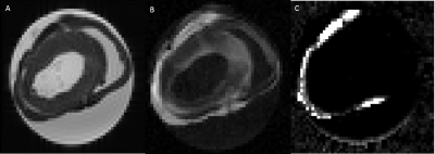 |
97 |
Epicardial fat imaging with diffusion weighted MRI
Cyril Tous, Alistair Young, Beau Pontré
Epicardial fat is associated with the development of metabolic syndrome and higher cardiovascular risk profile. Excessive fat can cause ventricular arrhythmias and alter repolarization. Fat is typically quantified with the Dixon technique, adding extra scan time. The microstructure of fat can also be differentiated with non-Gaussian diffusion weighting because the size of adipocytes differ from myocytes. Our automatic segmentation of the fat has 0.99 in accuracy and specificity compared with the Dixon technique. Further studies are required to associate myofibre misalignment with fat response in pathological hearts. Our technique can address both of these measures.
|
 |
2048.
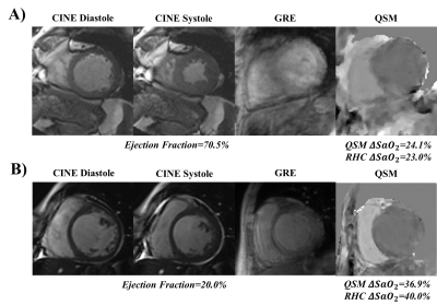 |
98 |
Navigator Gated Cardiac Quantitative Susceptibility Mapping for Differential Oxygen Saturation Quantification: Initial Validation via Invasive Catheterization
Yan Wen, Thanh Nguyen, Zhe Liu, Sarah Eskreis-Winkler, Kofi Deh, Evelyn Horn, Harsimran Singh, Jiwon Kim, Yi Wang, Jonathan Weinsaft, Pascal Spincemaille
The feasibility of measuring differential RV-to-LV oxygen saturation with cardiac quantitative susceptibility mapping (QSM) has been demonstrated in healthy volunteers; here, we present our initial validation of cardiac QSM based oxygenation measurement in patients through comparison with gold standard right heart catheter measurements.
|
|
2049.
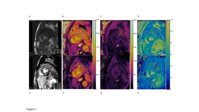 |
99 |
Initial Utility of Cardiac Magnetic Resonance Fingerprinting for Quantitative T1/T2 Parametric Mapping in Hypertrophic Cardiomyopathy
Andrew Patterson, Yuchi Li, Armando Ugo Cavallo, Jesse Hamilton, Vikas Gulani, Nicole Seilberlich, Sanjay Rajagopalan
The performance of cardiac Magnetic Resonance Fingerprinting (cMRF) in tissue characterization in patients with hypertrophic cardiomyopathy (HCM) is not known. In this study, we report our experience in a small cohort of patients with HCM who have undergone cMRF and standard techniques for imagine acquisition of T1/T2 Mapping and extracellular volume (ECV) estimation.
|
|
2050.
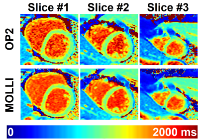 |
100 |
Clinical evaluation of a two-heartbeat inversion-recovery-based myocardial T1 mapping at 1.5T
Li Huang, Radhouene Neji, Muhammad Nazir, John Whitaker, Filippo Bosio, Amedeo Chiribiri, Reza Razavi, Sébastien Roujol
Cardiomyopathies can be revealed in the presence of abnormal native myocardial T1 times. Conventional myocardial T1 mapping techniques often require relatively long breathholds, thus limiting their usages in patients with severe breathholding difficulties. In this work, we evaluated the potential of a two-heartbeat inversion-recovery-based myocardial T1 mapping in patients.
|
|
| Top |
Contrast-Enhanced & Non-Contrast MR Angiography
Digital Poster
Cardiovascular
Monday, 13 May 2019
| Exhibition Hall |
16:00 - 17:00 |
| |
|
Computer # |
|
2051.
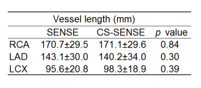 |
101 |
Clinical Performance of Non-contrast Whole-heart Magnetic Resonance Coronary Angiography with Compressed Sensing: Comparison with Conventional Sensitivity Encoding Imaging
Yuki Ohmoto-Sekine, Junji Takahashi, Norihito Miura, Saori Amemiya, Kei Fukuzawa, Makiko Ishihara, Hiroshi Tsuji, Yasuji Arase
Non-contrast whole-heart magnetic resonance coronary angiography (WHMRA) has proven value for noninvasive assessment of coronary arteries. However, there are major technical challenges associated with this technique, such as image quality degradation due to long scan times. Compressed sensing (CS) reconstruction might be a powerful solution for shortening scan time. We conducted a feasibility study for WHMRA with conventional parallel imaging and CS reconstruction. WHMRA with CS reconstruction is a promising method that can shorten the scan time while maintaining mostly acceptable images, although there is still room for improvement, especially for stenotic vessel evaluation.
|
|
2052.
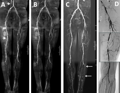 |
102 |
Evaluation of quiescent-interval single-shot (QISS) magnetic resonance angiography (MRA) in diabetic patients with clinical limb ischemia in comparison with contrast-enhanced MRA with calf-compression at 3 Tesla
Presentation Not Submitted
Liming Wei, Xiaoyue Zhou, Jungong Zhao
Early diagnosis and aggressive management are critical to mitigate the devastating natural history of Critical limb ischemia (CLI). Computed tomography angiography (CTA) and contrast-enhanced magnetic resonance angiography (CE-MRA) are established methods in the diagnostic workup of PAD. However, the high incidence of infrapopliteal disease in CLI and the calcified nature of these vessels in patients suffering from diabetes mellitus are problems even for the dual-energy CT. In addition, diabetic patients with CLI frequently experience several comorbidities, which can hamper administration of iodinated contrast media and lead to contrast-induced nephropathy. What’s more, the high prevalence of chronic renal impairment in diabetic patients with CLI and CE-MRA studies often place such patients at an increased risk of nephrogenic systemic fibrosis. Quiescent-interval single-shot (QISS)-MRA is a robust non-enhanced MRA method that has shown promising results at 1.5 T and 3 T. Here, we wished to evaluate the image quality and diagnostic accuracy of QISS-MRA at 3 T in diabetic patients with CLI compared with CE-MRA with calf compression, with digital subtraction angiography (DSA) serving as the standard reference.
|
|
2053.
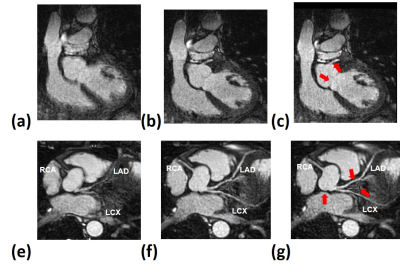 |
103 |
Free-breathing whole-heart coronary MRA with Radial SSFP and fully automated 3D rigid body motion corrected reconstruction
Guruprasad Krishnamoorthy, Joao Tourais, Jouke Smink, Marc Kouwenhoven, Suthambhara Nagaraj, Marcel Breeuwer
Prolonged acquisition time and susceptibility to respiratory motion remain to be the major challenge in 3D free-breathing whole-heart MR angiography. Recently, respiratory self-navigated 3D-Radial CMRA technique has been proposed to address the said limitations. In this technique, a 1-D projection oriented in Superior-Inferior direction are acquired at every heartbeat before each interleave of 3D-Radial imaging data for 1D motion correction of the heart with 100% respiratory gating efficiency. In this work, an extension of this method is proposed to estimate and correct for the 3D motion of the heart along with a robust navigator processing and automatic coil selection algorithm (3D-PRONAV). The proposed method is validated in five volunteers.
|
|
2054.
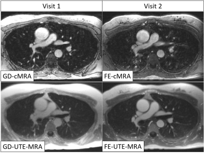 |
104 |
Comparison of Gadolinium- and Ferumoxytol- enhanced Conventional and ultra-short echo time MR-Angiography for the Depiction of the Pulmonary Vasculature
Gesine Knobloch, Timothy Colgan, Mark Schiebler, Kevin Johnson, Tilman Schubert, Scott Reeder, Scott Nagle
Pulmonary MRA is a promising technique for detection of pulmonary embolism without ionizing radiation. Unfortunately, a subset of patients fail breath-holding during conventional MRA (cMRA). Free-breathing ultra-short echo time (UTE)-MRA could overcome this limitation but would require a blood pool contrast agent due to longer scan times. Ferumoxytol (FE) can be used as an off-label MR-contrast agent with a blood half-life of 10-14 hours. This study compared gadobenate dimeglumine (GD)-enhanced-UTE-MRA to FE-cMRA and FE-UTE-MRA, using GD-cMRA as reference standard. Results suggest that FE-UTE-MRA has improved performance compared to GD-cMRA for depicting non-vascular structures while offering excellent angiographic image quality.
|
|
2055.
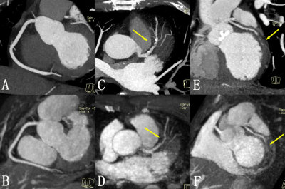 |
105 |
Contrast-enhanced compressed sensing whole-heart 3T MR angiography in detection of coronary artery stenosis: A preliminary comparative study with computed tomography angiography
Jingwen Dai, Jian Cao, Lu Lin, Xiao Li, Jing An, Michaela Schmidt, Christoph Forman, Yining Wang, Zhengyu Jin
The aim of this study was to investigate the diagnostic performance of contrast-enhanced compressed sensing (CS) whole-heart 3T MR angiography in the detection of clinical significant coronary artery stenosis by using CTA as a reference. The preliminary results indicate that the contrast-enhanced CS coronary MR angiography has a good consistency in evaluating coronary artery disease in comparison to CTA and a short enough acquisition time that can be applied in the waiting time between contrast injection and late gadolinium enhancement imaging.
|
|
2056.
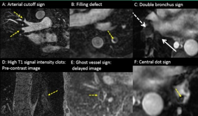 |
106 |
Direct and Indirect Signs of Pulmonary Embolism at MRA: Inter and Intra Reader Agreement
Nanae Tsuchiya, Donald Benson, Chris Francois, Mark Schiebler
Direct and indirect finding of pulmonary embolism are important for making the correct diagnosis on pulmonary MRA. We have assessed the prevalence of both expected direct and indirect findings of PE and also evaluated the inter and intra reader agreement of these indirect and direct findings. Our results demonstrate that experienced and inexperienced observers can reproducibly observe direct and indirect findings of PE on CEMRA exams.
|
|
2057.
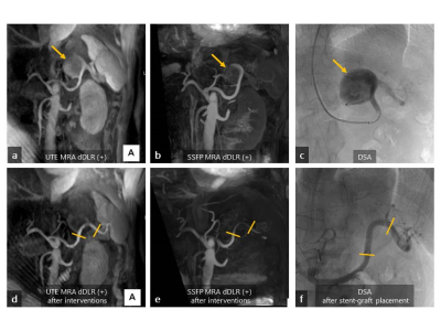 |
107 |
Ultrashort TE Time-Spatial Labeling Inversion Pulse MR Angiography Denoised with Deep Learning Reconstruction for Abdominal Visceral Arteries: A Feasibility Study
Ryuichi Mori, Hideki Ota, Atsuro Masuda, Tomoyoshi Kimura, Tatsuo Nagasaka, Takashi Nishina, Sho Tanaka, Yoshimori kassai, Kei Takase
We have developed ultrashort TE (UTE) time-SLIP MRA for the evaluation of abdominal visceral arteries. In this volunteer study, although image quality and number of visualized renal arteries on UTE time-SLIP MRA were inferior to those on steady-state free precession (SSFP) time-SLIP MRA, deep learning reconstruction enabled significant improvement of signal-to-noise ratio and contrast to the background tissue. In patients with visceral artery aneurysms treated by endovascular intervention, contrast ratio of treated segment adjacent to metal devices to the aorta were higher on UTE than SSFP time-SLIP MRA. This proposed technique is feasible for visceral artery disease after endovascular interventions.
|
|
2058.
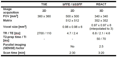 |
108 |
REACT - Relaxation-Enhanced MR Angiography without Contrast and Triggering in comparison with other non-contrast MR angiography techniques for imaging of the pelvic vessels.
Mark Terwolbeck, Maike Bode, Lea Hitpass, Shuo Zhang, Christiane Kuhl, Nils Kraemer
Conventional MRI and CE-MRA have limitations in venous imaging. Recently introduced Relaxation-Enhanced MR Angiography without Contrast and Triggering (REACT) is based on 3D dual-echo generalized DIXON in combination of magnetization preparation, and has the potential to better characterize both arterial and venous signals with robust blood-tissue contrast. Comparing REACT with conventional non-contrast sequences regarding vessel depiction, REACT revealed similar results for large vessels and was superior for the assessment of small peripheral vessels.
|
|
2059.
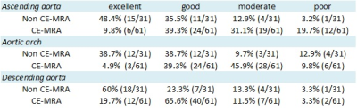 |
109 |
Radial self-navigated non contrast-enhanced (CE-) MRA in comparison to conventional navigator-gated CE-MRA of the thoracic aorta in a vascular patient collective
Martina Correa Londono, Verena Obmann, Nino Trussardi, Davide Piccini, Michael Ith, Hendrik von Tengg-Kobligk, Bernd Jung
This study compares a conventional Cartesian contrast-enhanced MRA sequence with a native 3D radial MRA sequence for imaging the aortic root and the ascending aorta. The difference in image quality between those two MRA sequences has been shown to be highly significant at the ascending aorta and the aortic arch with higher quality for the radial self-navigated non-contrast enhanced MRA. The results of our study support the potential of a native MRA protocol as a diagnostic tool in monitoring the geometry or size of the aortic root and the ascending aorta.
|
|
2060
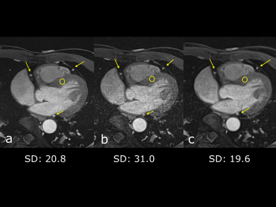 |
110 |
Improved vessel delineation in whole-heart coronary MRA with sub-millimeter isotropic resolution using deep learning reconstruction compared with routine whole-heart coronary MRA
Video Permission Withheld
Sanae Takahashi, Haruhiko Machida, Toshiya Kariyasu, Rieko Niitsu, Isao Miyazaki, Tatsuya Yoshioka, Keita Fukushima, Saori Yuda, Yuta Shimizu, Takayuki Yonaha, Akihito Nakanishi, Hiroshi Kusahara, Youhei Matsuoka, Miho Kitamura, Takao Yamamoto, Kenichi Yokoyama
Whole-heart coronary MRA (WHCMRA) with sub-millimeter isotropic resolution is expected to improve spatial resolution and overcome limitations of routine WHCMRA, whereas increased image noise is the drawback. A state-of-the-art 3T MR scanner with the maximal gradient magnetic field of 100 mT/m and capability of deep learning reconstruction (DLR) that aims to reduce image noise is useful for improving vessel sharpness and delineation of the coronary arteries, even in the distal segments, on WHCMRA with sub-millimeter isotropic resolution with use of the DLR with preserving image noise compared with routine WHCMRA.
|
|
2061.
 |
111 |
The feasibility of high resolution vessel wall imaging (HR VWI) based minimum intensity projection (mIP) MRA for evaluating steno-occlusive MCA
Presentation Not Submitted
Mi Shen, Shuo Chen, Yuxin Wang, Peiyi Gao
Stenosis, distal branches and collateral circulation were of great importance in assessing vulnerable vessel beds in stroke patients. Minimum intensity projection (mIP) MRA was derived from high resolution vessel wall imaging (HR VWI), which can be used as evaluating vessel wall, as well as vessel lumen. In this study, we evaluated the feasibility of HR VWI based mIP MRA for evaluating MCA stenosis, occlusion, accompanying microvascular (AMV) and leptomeningeal branches. Using CTA as a standard, mIP MRA outperformed TOF MRA in assessing MCA stenosis, occlusion, AMV and leptomeningeal branches. Consistent with CTA, mIP MRA is feasible for clinical application, and may help in detecting collaterals.
|
|
2062.
 |
112 |
Non-Contrast-Enhanced Abdominal MRA at 3T using Velocity-Selective Pulse Trains
Dan Zhu, Wenbo Li, Dapeng Liu, Guanshu Liu, Yigang Pei, Taehoon Shin, Qin Qin
Non-contrast-enhanced (NCE) abdominal MR angiography (MRA) with large spatial coverage is clinically desired. The current method using spatially selective inversion (SSI) pulse to null static tissue and blood is only limited to small field of view (FOV) due to its sensitivity to slow arterial inflow. Velocity-selective inversion (VSI) based approach was shown to preserve the arterial blood inside the imaging volume at 1.5T. In this study, a novel strategy of VSI + VSS was proposed for abdominal MRA of large FOV, and its advantage over SSI based approach was demonstrated among healthy subjects at 3T.
|
|
2063.
 |
113 |
Acceleration of whole-heart coronary MR angiography using 3D non-selective bSSFP with Compressed SENSE
Kazuo Kodaira, Nagao Michinobu, Masami Yoneyama, Isao Shiina, Yasuhiro Goto, Yoshihiro Ikeda, Yutaka Hamatani, Mamoru Takeyama, Isao Tanaka, Shuji Sakai
Whole heart coronary magnetic resonance angiography (WHC-MRA) has a limitation of long scan time, which often causes degradation of image quality. 3D non-selective balanced TFE (bTFE) with shortening TR can reduce the scan time. However, it is yet not applied for WHC-MRA. Compressed SENSE (C-SENSE) is suitable for subjects with a high sparse such as vessels and biliary trees, and can accelerate the scan time of WHC-MRA while ensuring the image quality. We propose a new combination of 3D non-selective bTFE with C-SENSE for WHC-MRA, and examine the image quality and the scan time in comparison to the conventional methods.
|
|
2064.
 |
114 |
Non-Contrast-Enhanced zTE MR Angiography of Intracranial Atherosclerotic Disease
Presentation Not Submitted
Weiqiang Dou, Yun Ji, Yong Shen, Kai Xu
To investigate if zero-echo-time magnetic resonance angiography (zTE-MRA) is feasible in the characterization of intracranial atherosclerotic disease, we measured in this study a large population with atherosclerotic disease confirmed clinically. The acquired zTE-MRA images were thus compared systematically with those using time-of-flight (TOF)-MRA technique. Serving digital subtraction angiography (DSA) as a reference, zTE-MRA showed comparable imaging quality with DSA in the aspect of flow signal in the parent artery and higher signal intensity with less artifact than TOF-MRA. We therefore demonstrated that zTE-MRA could be a promising technique and further applied in the clinic routinely for patients with atherosclerotic disease.
|
|
2065.
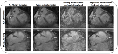 |
115 |
High Resolution, Respiratory Motion-Resolved Coronary MRA Using a Reordered Variable-Density 3D Cones Trajectory
Srivathsan Koundinyan, Frank Ong, Mario Malavé, Bob Hu, Dwight Nishimura
High resolution MR angiography is important to properly visualize fine lesions in coronary arteries. In this work, we employ a time-efficient and motion-robust variable-density 3D cones trajectory for sub-millimeter data acquisition. Although the imaging data is severely undersampled, we capitalize on the diffuse aliasing properties of the non-Cartesian cones trajectory to separate the data into several respiratory phases for motion compensation. For improved image quality, we present and analyze a modified cones reordering strategy, mindful of balanced SSFP imaging and unwanted eddy current effects, for distributed k-space coverage irrespective of how the readouts are retrospectively sorted.
|
|
2066.
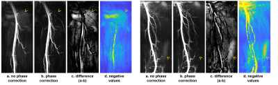 |
116 |
Highly accelerated NCE-MRA: Phase correction to remove background artefacts for complex subtraction
Hao Li, Andrew Priest, Martin Graves, David Lomas
For subtractive Non-Contrast-Enhanced MR Angiography (NCE-MRA), complex subtraction of k-space data (prior to reconstruction) can exploit the sparsity of the difference images for under-sampled dataset reconstruction, but it causes background artefacts. This study proposed a phase correction method to restore the polarity of negativesignal and null background artefacts.
|
|
2067.
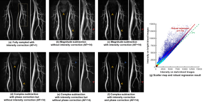 |
117 |
Highly Accelerated NCE-MRA Using Complex Subtraction with Intensity Correction: Improved Reconstruction Accuracy and Background Tissue Suppression
Hao Li, Andrew Priest, Martin Graves, David Lomas
We propose a compressed sensing reconstruction method for highly accelerated subtractive non-contrast-enhanced MRA, based on complex subtraction in k-space. This method applies intensity difference correction between bright- and dark-blood image sets before reconstruction and uses a phase correction procedure to remove background artefacts caused by complex subtraction. Experimental results demonstrate improved background tissue suppression. Compared with magnitude subtraction approaches, the proposed method achieved higher reconstruction accuracy at large acceleration factors, permitting the use of higher acceleration factors without unacceptable loss of image quality.
|
|
2068.
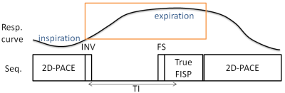 |
118 |
Free-breathing renal MR angiography using NATIVE TrueFISP: A comparison of different triggering techniques
Fang Dong
Non-contrast renal magnetic resonance angiography (MRA) is particularly valuable since contrast agents increase the risk of the Nephrogenic Systemic Fibrosis for the patients with renal failure. NATIVE TrueFISP together with triggering technique is commonly used for free breathing renal MRA. Here, we use 2D-PACE (Prospective Acquisition CorrEction) for respiratory triggering, therefore no external gating devices are needed and the preparation time is saved. The result shows that the image quality achieved by 2D PACE is comparable or superior to the conventional triggering techniques, which promises to be an alternative triggering method for renal MRA in clinical routine.
|
|
2069.
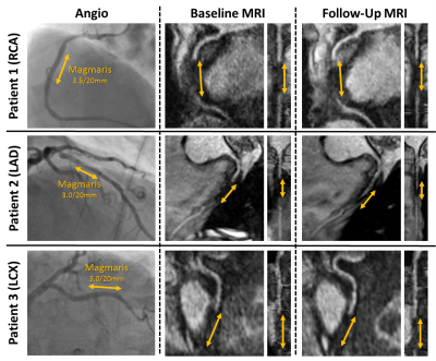 |
119 |
Initial Study on Coronary MRI at 3T in the Presence of Resorbable Magnesium Scaffolds
Simon Reiss, Axel Krafft, Sarah Schmidt, Timo Heidt, Constantin von zur Mühlen, Michael Bock
Coronary MRI allows for the assessment of vascular patency and thus is a desirable imaging tool for the follow-up of vascular patency after percutaneous coronary interventions in which stenosis are treated with stents. We show that, coronary MR in the presence of recently introduced resorbable magnesium scaffold seems not feasible for the detection of in-stent restenosis within the first year post-intervention as signal voids caused by the scaffold may be miss-interpreted as stenosis. However, coronary MRI provides a valuable tool to further study the resorption process of magnesium scaffolds and to visualize the artery after complete resorption of the scaffold.
|
|
2070.
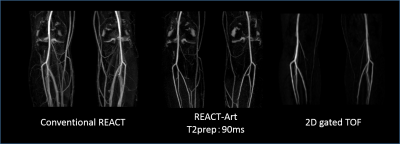 |
120 |
Non-triggered, Non-Contrast-Enhanced Lower Extremity MR Angiography using Artery-Weighted REACT
Hiroshi Hamano, Masami Yoneyama, Yasutomo Katsumata, Ryuji Baba, Kenji Iinuma
Non-contrast enhanced lower extremity MR angiography, such as TOF, PC and TRANCE, is commonly used to diagnose peripheral arterial disease, but these techniques are occasionally unstable due to presence of arrhythmia, atrial fibrillation or tachycardia. Relaxation-Enhanced MR Angiography without Contrast and Triggering (REACT) derives high intravascular signal from T2prep pulse followed by STIR pulse and dual-echo gradient echo DIXON readout. However conventional REACT cannot distinguish arterial and venous signals clearly. We validate optimum T2prep time and TFE factor to improve the contrast between artery and vein. We demonstrate that REACT-Art provided more artery-dominant contrast compared to conventional REACT.
|
|
2071.
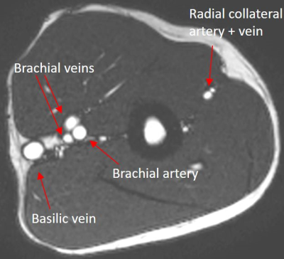 |
121 |
Dynamic MRI visualization of the brachial artery during upper arm air cuff inflations
Jouke Smink, Laura Bogatu, Jens Muehlsteff, Erik Bresch
We investigated the feasibility of imaging the upper arm using real-time and cine MRI during air cuff inflations in order to characterize the elastic properties of the brachial artery. Both the cine, as well as the real-time MR image data allow the direct observation of the occlusion and re-opening of veins and arteries as a function of cuff pressure. They could therefore be potentialy well suited to estimate the elastic properties of the brachial artery, e.g. the area-pressure curve, in vivo. Furthermore, the MR data allow observing the compression and displacement of arm tissue with the future aim to validate and extend existing models of the auscultatory and oscillometric blood pressure measurements.
|
|
2072.
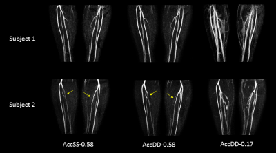 |
122 |
Investigation of Non-Contrast Enhanced Peripheral Artery MR Angiography using AccASL with Double Labeling and Dual AENC Scheme for Realizing Consistent Visualization in Various Blood Flow Conditions
Obara Makoto, Takuya Hino, Yasuo Yamashita, Takeshi Kamitani, Shuhei Shibukawa, Natsuo Konta, Osamu Togao, Masami Yoneyama, Yuta Akamine, Yu Ueda, Marc Van Cauteren
Acceleration-Selective Arterial Spin Labeling (AccASL) with double labeling and dual acceleration encoding (AENC) scheme (AccDD) was developed for consistent non-triggered peripheral non-contrast enhanced MR angiography. AccDD requires only 50% longer scan time than conventional single labeling and single AENC scheme and can provide better artery visualization. In addition, veins, with an expected flow slower than arteries, were more visualized using lower AENC acquisition of AccDD, indicating AccDD is sensitive to a wide flow velocity range. Consistent flow visualization in various flow conditions with patients can be expected.
|
|
2073.
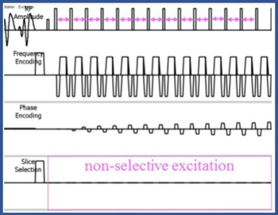 |
123 |
Whole heart coronary MRA using non-selective balanced SSFP sequence at 3.0T: comparison of image quality
Isao Shiina, Michinobu Nagao, Masami Yoneyama, Kazuo Kodaira, Yasuhiro Goto, Yoshihiro Ikeda, Yutaka Hamatani, Mamoru Takeyama, Isao Tanaka, Shuji Sakai
3 tesla derived MRCA has a limitation of low contrast for coronary arteries because of setting lower flip angles due to high SAR. Therefore, the signal contrast of for coronary arteries becomes low. 3D non-selective balanced TFE (3D nsbTFE) has the possibility to solve this problem. We examine image quality of MRCA obtained from 3 tesla and 3D nsbTFE.
|
|
2074.
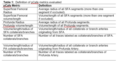 |
124 |
Quantitative measurements of decreased arterial collateralization and branching in peripheral artery disease.
Niranjan Balu, Li Chen, Thoetphum Benyakorn, Daniel Hippe, Henrik Haraldsson, Warren Gasper, David Saloner, Chun Yuan, Thomas Hatsukami
The extent of collateralization/branching (CB) of lower limb vessels in peripheral artery disease (PAD) can inform risk of ischemia and response to revascularization. However quantitative imaging metrics of CB have not been assessed in the setting of severe PAD. We developed automated quantitative measurements of lower limb vascular morphology (pCafe) and compared CB in patients undergoing revascularization for PAD. Assessment of pCafe metrics in severe PAD suggests CB is increased with occlusion compared to stenosis indicating compensatory CB development.
|
|
2075.
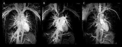 |
125 |
Non-Contrast Enhanced MR Angiography for Selective Pulmonary Artery and Aorta imaging at 1.5T
Yajing Zhang, Qian Jiang, Jiazheng Wang
Non-contrast enhanced pulmonary MRA has been increasingly used for the diagnosis of abnormality of vessels such as pulmonary arteriovenous malformation (PAVM). This study aims to validate the vessel-selective MRA using time-spatial labeling inversion pulse. The pulmonary vessel or aorta could be intendedly selected with a high intensity against the surrounding anatomies. The contrast between vessels were evaluated using an ROI approach.
|
|
| Top |
Atherosclerosis & MR Angiography
Digital Poster
Cardiovascular
Monday, 13 May 2019
| Exhibition Hall |
16:00 - 17:00 |
| |
|
Computer # |
|
2076.
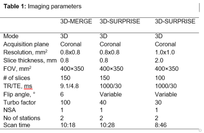 |
126 |
Large coverage peripheral artery 3D vessel wall MRI using SUper-Resolution for Psf-Restored Isotropic turbo Spin Echo (3D SURPRISE)
Niranjan Balu, Zechen Zhou, Thomas Hatsukami, Chun Yuan
Vessel wall MRI for peripheral artery disease (PAD) atherosclerotic lesions requires large coverage due to diffuse distribution of lesions, and high-resolution due to smaller plaque components. However current large coverage PAD approaches uses gradient echo sequences that may not be optimal for highly calcified PAD lesions. Therefore we developed and evaluated a large coverage 3D femoral MRI using SUper-Resolution for Psf-Restored Isotropic turbo Spin Echo (3D-SURPRISE) that provides shorter scan times with higher SNR and CNR than an existing large coverage gradient echo femoral vessel wall MRI (3D-MERGE).
|
|
2077.
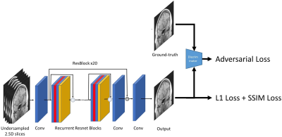 |
127 |
Generative Adversarial Networks based Compressed Sensing for Multi-contrast Intracranial Vessel Wall Imaging Acceleration
Niranjan Balu, Long Wang, Tao Zhang, Zechen Zhou, Enhao Gong, Kristi Pimentel, Mahmud Mossa-basha, Thomas Hatsukami, Chun Yuan
Multi-contrast isotropic high resolution intracranial vessel wall imaging (VWI) can enable direct detection and follow-up surveillance of intracranial vessel wall pathologies. The overall scan time can be largely reduced by proper random undersampling. However, the image reconstruction process can be time consuming causing deployment difficulties of accelerated intracranial VWI for clinical use. In this study, a generative adversarial networks based compressed sensing method was developed for multi-contrast intracranial image reconstruction. The preliminary results demonstrate comparable/improved image quality for vessel wall delineation in comparison to the traditional image reconstruction method, while providing a significant reduction in reconstruction time.
|
|
2078.
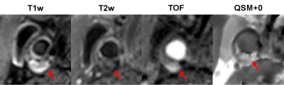 |
128 |
QSM identifies calcification and intraplaque hemorrhage in patients with significant carotid stenosis
Thanh Nguyen, Yan Wen, Kelly Gillen, Philip Stieg, Darren Schneider, Qi Yang, Yi Wang
We developed and evaluated carotid QSM for detecting calcification and intraplaque hemorrhage in carotid plaques. Preliminary results in patients with significant carotid stenosis showed QSM was able to detect both calcification and intraplaque hemorrhage in good agreement with findings on CT and multi-contrast MRI.
|
|
2079.
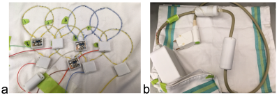 |
129 |
A lightweight and ultra-flexible “blanket” coil design for carotid artery wall imaging
Thanh Nguyen, Bei Zhang, Yan Wen, Yi Wang, Ryan Brown
We aimed to build an ultra-flexible blanket coil for carotid wall imaging. Results in healthy volunteers showed that the coil improves comfort and allows easy positioning in the neck while providing 32% higher SNR at the carotid bifurcation compared to the product 64-channel neurovascular coil.
|
|
2080.
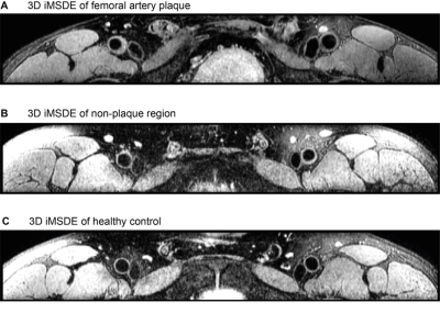 |
130 |
Plaque permeability assessed with dynamic contrast-enhanced MRI predicts ferumoxytol nanoparticle delivery in patients with peripheral artery disease
Kang Zheng, Jasper Schoormans, Lotte Stiekema, Claudia Calcagno, Iwona Cicha, Aart Nederveen, Gustav Strijkers, Erik Stroes, Bram Coolen
We investigated whether dynamic-contrast enhanced (DCE-)MRI could be used to predict accumulation of ferumoxytol nanoparticles in femoral plaques. To this end, we implemented USPIO enhanced quantitative T2* imaging, as well as 3D black-blood DCE-MRI sequences for femoral artery vessel wall imaging. Patients with peripheral artery disease (PAD) and healthy volunteers were included in this study. We show that ferumoxytol nanoparticle delivery to atherosclerotic plaques is associated with plaque permeability as assessed with DCE-MRI in patients with PAD.
|
|
2081.
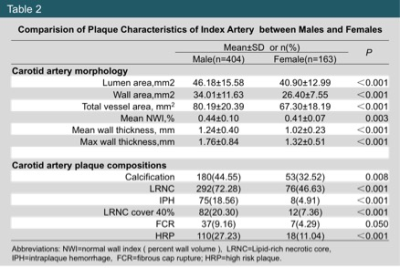 |
131 |
Sex Differences in Symptomatic Carotid Artery Atherosclerosis: A Chinese Atherosclerosis Risk Evaluation (CARE-II) Study
Lina Zhu, Feiyu Li, Xihai Zhao, Chun Yuan, Xiaoying Wang
The results of this multicenter study ( including 13 hospitals and medical centres across China) demonstrated that, in Chinese patients with recent ischemic cerebrovascular events, males developed higher risk of atherosclerotic plaques carotid arteries than females. The different constituent ratios of traditional risk factors, especially smoking, may contribute to the sex differences in carotid high-risk plaques.
|
|
2082.
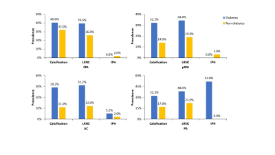 |
132 |
Comparison of Femoral Artery Atherosclerotic Plaque Characteristics between Patients with and without Diabetes Mellitus: A 3D MR Vessel Wall Imaging Study
Yongjun Han, Zhu Zhu, Maobin Guan, Dandan Yang, Xihai Zhao
Diabetes mellitus is a documented high risk factor for the development of atherosclerosis. This study investigated the characteristics of atherosclerotic plaque in diabetic patients in comparison with non-diabetic patients using 3D multi-contrast MR vessel wall imaging. We found that patients in diabetic group had higher plaque burden, prevalence of plaque and more complex composition than those in non-diabetic group.
|
|
2083.
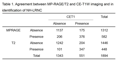 |
133 |
Identification of Carotid Non-Hemorrhagic Lipid-Rich Necrotic Core by Magnetization-Prepared Rapid Acquisition Gradient-Echo Imaging: Validation by Contrast-Enhanced T1W Imaging
Xihai Zhao, Huiyu Qiao, Hualu Han, Dongxiang Xu, Gaifen Liu, Chun Yuan
Lipid-rich necrotic core (LRNC) plays a key role in the vulnerability of atherosclerotic plaques and is associated with ischemic cerebrovascular events. Previously, LRNC was identified using contrast-enhanced T1W (CE-T1W) imaging which needs administration of contrast agent. This study determined the capability of MP-RAGE imaging in identification of non-hemorrhagic LRNC (NH-LRNC) validated by CE-T1W. We found that moderate to good agreements between MP-RAGE and CE-T1W imaging in identification and quantification of NH-LRNC. MP-RAGE showed smaller bias in measuring NH-LRNC than T2W imaging. Our results suggest that MP-RAGE might be a better non-CE imaging technique for assessing NH-LRNC in carotid arteries.
|
|
2084
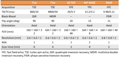 |
134 |
Identification of Intrapalque Hemorrhage in Carotid Artery by Simultaneous Non-contrast Angiography and intraPlaque hemorrhage (SNAP) Imaging: Validation by Histology
Video Permission Withheld
Dongye Li, Huiyu Qiao, Yongjun Han, Hualu Han, Dandan Yang, Jingli Cao, Huimin Xu, Tao Wang, Xihai Zhao
It is important to accurately characterize carotid intraplaque hemorrhage (IPH) due to its strong association with plaque progression and ischemic stroke. Previous study proved that MP-RAGE is the best T1-weighted sequence in detecting carotid IPH. Most recently, investigators found that SNAP imaging can detect more IPHs compared with MP-RAGE. This study sought to investigate the performance of SNAP imaging in evaluating carotid IPH validated by histology. We found that SNAP sequence showed better agreement with histology compared with MP-RAGE in assessing IPH, suggesting that SNAP imaging might be a more sensitive and accurate imaging approach in characterizing carotid IPH.
|
|
2085.
|
135 |
Volumetric assessment of arteriosclerotic plaque burden in ApoE-KO mice using high resolution MR microscopy
Courtney Whalen, Alexandra Caamano, Floyd Mattie, Lukas Neuberger, Neil Huang, Catharine Ross, Rita Castro, Thomas Neuberger
There is a need to address current knowledge gaps in atherosclerosis' etiology to develop successful strategies to reduce mortality due to this disease. In this study, we investigated the feasibility of using 14T MR microscopy to measure the extent of atherosclerosis in apoE-KOmice on diet studies. Results from the ex-vivo 3D MR microimaging of the aortic atherosclerotic plaques with a resolution of 10x10x15µm3 are presented. A contrast agent was used to increase the sensitivity of the 14-tesla system and provide the resolution necessary to assess in detail the wall vessel changes associated with the atherosclerotic process.
|
|
2086.
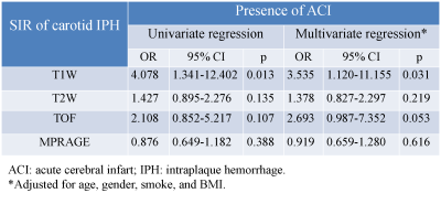 |
136 |
Signal-Intensity-Ratio of Carotid Intraplaque Hemorrhage Stratifies Risk of Acute Cerebral Infarct: A Chinese Atherosclerosis Risk Evaluation Study
Presentation Not Submitted
Yang Liu, Dandan Yang, Yongjun Han, Chun Yuan, Wei Wang, Xihai Zhao
Previous studies have shown that the presence of carotid IPH is a strong predictor for future ischemic events. The present study investigated the usefulness of the signal-intensity-ratio of IPH in carotid arteries for stratification of acute cerebral infarct (ACI) risk using multicontrast MR vessel wall imaging. We found that the signal-intensity-ratio of carotid IPH on T1W images was independently associated with the presence of acute cerebral infarct. Our results suggest that the age of carotid IPH might be an independent indicator for the risk of acute ischemic cerebrovascular events.
|
|
2087.
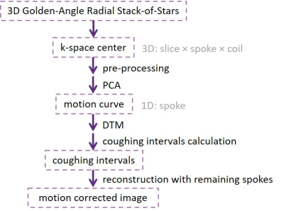 |
137 |
Carotid Artery Self-navigation Coughing Motion Correction Using Principle Component Analysis Jointed with Differential Threshold Method
Yu Wang, Haikun Qi, Yajie Wang, Guanhua Wang, Huijun Chen
Black-blood MRI is an important tool to evaluate atherosclerosis for carotid artery, however, it is susceptible to motion and no former motion correction method for carotid artery could achieve 100% acquisition efficiency. Therefore, a 100% acquisition efficiency self-navigation carotid artery motion correction method based on Principle Component Analysis (PCA) jointed with Differential Threshold Method (DTM) was proposed for carotid artery imaging using radial sampling. Experimental results demonstrated that the proposed method can detect acute motions such as coughing, compute the motion-corrupted interval, and achieve higher reconstruction quality after removing the motion-corrupted spokes.
|
|
2088.
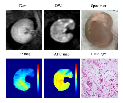 |
138 |
Multiparametric evaluation of Atheroembolic renal disease in a microsphere-induced animal model
Hanjing Kong, Fei Gao, Chengyan Wang, Xiaodong Zhang, Min Yang, Jue Zhang, Xiaoying Wang
Atheroembolic renal disease (AERD) is the major cause of renal insufficiency in the elderly, and cholesterol embolism is a manifestation of this disease. AERD is often an underdiagnosed clinical illness because of the lack of sensitive examination methods. Recently, multiparameter MRI showed its potential in enhancing the efficiency of diagnosing renal diseases. In this study, we examine the value of multiparameter MRI in AERD detection and further compared with histology.
|
|
2089.
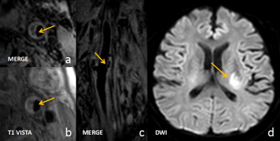 |
139 |
Irregularity of Carotid Atherosclerotic Plaque Determined by Multidimensional Magnetic Resonance Imaging Stratifies the Risk of Acute Cerebral Infarction
Presentation Not Submitted
Dan Zhou, Dong Liu, Jin Li, Xihai Zhao
Carotid plaque surface features including surface irregularities and ulceration represent an important indicator for plaque vulnerability and are associated with the occurrence of neurological symptoms1-2. This study aimed to determine the association between surface irregularity of carotid plaque on MR images at multi-dimensions and acute ischemic stroke. Ninety-seven symptomatic patients with atherosclerotic plaque in carotid artery determined by ultrasound imaging were enrolled and underwent MR imaging for carotid artery and brain. We found that the irregularity information at multi-dimensions which combined the axial and oblique images had stronger predictive value for acute ischemic lesion than axial images alone.
|
|
2090.
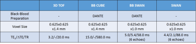 |
140 |
Calcification and USPIO detection in Carotid Artery Plaque using QSM and SWI
Pascal Ruetten, Ammara Usman, Andrew Priest, Jonathan Gillard, Martin Graves
In this work we demonstrated the feasibility and potential benefits of using Susceptibility Weighted Imaging and Quantitative Susceptibility Mapping for application in carotid artery plaque imaging for detection, quantification, and distinction of areas of calcification and USPIO uptake.
|
|
2091.
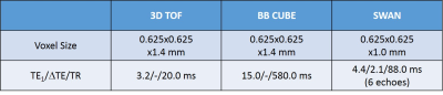 |
141 |
Plaque feature analysis using a joint method of QSM and water-fat separation
Pascal Ruetten, Alison Cluroe, Ammara Usman, Jonathan Gillard, Martin Graves
In this work we investigated the feasibility of using a joint method of water-fat separation and Quantitative Susceptibility Mapping to characterize carotid artery plaques. The combination of both methodologies was able to detect strong changes in susceptibility, to detect plaque calcification, and also detect changes in the local fat fraction of the plaques in order to identify a lipid core.
|
|
2092.
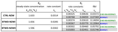 |
142 |
Evaluation of the endothelial (dys)function in a rabbit model of atherosclerosis by DCE-MRI and pharmacokinetic modelling
Peter Opriessnig, Gunter Almer, Claudia Cabella, Stefan Ropele, Seth Hallstroem, Gerd Hoerl
Endothelial dysfunction as a consequence of cardiovascular risk factors and increased endothelial permeability are involved in remodeling processes of the arterial wall and progression of AS early on. DCE-MRI in combination with pharmacokinetic modelling and a special nitric oxide donor compound together with dynamic vasomotor patterns could provide novel information related to changes of the endothelial function during the progression and pathogenesis of atherosclerosis and may contribute to a better mechanistic understanding of AS pathophysiology. In this work, we demonstrate the feasibility to investigate endothelial (dys)function and NO induced vasomotor function of the abdominal aorta in AS induced rabbits simultaneously.
|
|
2093.
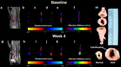 |
143 |
In-Vivo Quantification of Aortic Stiffness in Abdominal Aortic Aneurysm Porcine Model using Magnetic Resonance Elastography: A Comparison to Histopathology and Burst Testing
Huiming Dong, Duncan Russell, Xiaokui Mo, Matthew Joseph, Prateek Kalra, Richard White, Arunark Kolipaka
Aortic stiffness is an important biomarker that can provide more accurate rupture potential of abdominal aortic aneurysm (AAA) than diameter. Aortic MR elastography (AMRE) is a non-invasive technique to estimate aortic stiffness and has not been validated against gold standard methods. The aim of the study is to estimate aortic stiffness in AAA-induced porcine model using in-vivo AMRE, and compare it with histopathology and burst testing. Results demonstrated that AAA stiffness was significantly higher than normal aorta (p<0.002), while %elastin and burst pressure was significantly lower in AAA (p<0.0001). Aortic stiffness showed significant negative correlation to %elastin and burst pressure.
|
|
2094.
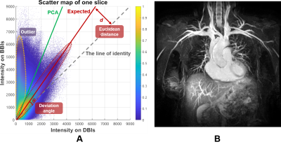 |
144 |
Background tissue suppression for subtractive NCE-MRA techniques based on robust regression using the deviation angle
Hao Li, Shuo Wang, Andrew Priest, Martin Graves, David Lomas
Intensity differences of static tissues between bright- and dark-blood images commonly exist in subtractive non-contrast-enhanced MRA techniques, resulting in residual background signal in the subtracted images. Some linear regression models can correct the intensity difference but are sensitive to the large number of blood pixels in thoracic MRA. A robust regression model is developed in this study and is demonstrated to have an effective performance in background suppression for different subtractive NCE-MRA techniques.
|
|
2095.
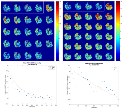 |
145 |
CrCEST reveals characteristics of skeletal muscle creatine metabolism in patients with Peripheral Arterial Disease
Helen Sporkin, Roshin Mathew, Christopher Kramer, Craig Meyer
Peripheral arterial disease (PAD) is an atherosclerotic disease of the limbs. Creatine chemical exchange saturation transfer (CrCEST) MRI can be used to visualize creatine metabolism as it occurs in the muscles of the calf. We have successfully applied CrCEST in 13 healthy subjects and 7 PAD patients in order to characterize the effect of ischemia on skeletal muscle energetics. Creatine concentration decay was assessed by CrCEST over a period of 8 minutes after plantarflexion exercise. Time course CEST asymmetry plots show features of PAD decays that help reveal mechanisms of metabolic dysfunction.
|
|
2096.
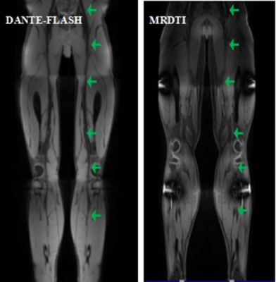 |
146 |
A fast and contrast-free MR approach to the diagnosis of deep vein thrombosis based on DANTE-prepared gradient echo
Huan Mao, Kewen Peng, Xueping He, Hanwei Chen, Xiaoyong Zhang, Xiaoming Bi, Xin Liu, Debiao Li, Zhaoyang Fan, Guoxi Xie
Black-blood magnetic resonance thrombus imaging (BTI) has proved successful for the diagnosis of deep vein thrombosis (DVT) with high sensitivity, specificity, and accuracy. Previous BTI method is based on a 3D variable flip angle fast spin echo (3D-vFSE) sequence, which has high specific absorption rate (SAR). DANTE-FLASH is another black-blood MR technique previously proposed for vessel wall imaging at lower extremities1. We hypothesized that DANTE-FLASH may suffice the purpose of diagnosing DVT while avoiding high SAR. This work was aimed to investigate the feasibility of using DANTE-FLASH to diagnose DVT.
|
|
2097.
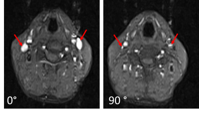 |
147 |
Visualization of jugular veins from sitting upright to supine position using low field MRI
Helena Ajo Asensio, Frank Simonis, Bennie ten Haken
Cerebral venous blood takes different pathways depending on body position. Hence, 3D MR imaging of the cervical region at different body inclination angles might give valuable information in patients whose diagnosis relies on imaging of venous pathways. In this study was shown that using a time of flight method the internal jugular veins could be segmented by applying a threshold followed by manual cropping. Subsequently, it was shown that their diameter decreases as the rotational angle is increased towards the upright position. The amount of decrease might give clinically relevant information about the state of the veins and cranial drainage.
|
|
2098
 |
148 |
Multi-class segmentation of the Carotid Arteries using Deep Learning
Video Permission Withheld
Magnus Ziegler, Jesper Alfraeus, Elin Good, Jan Engvall, Ebo de Muinck, Petter Dyverfeldt
The rupture of atherosclerotic plaques in the carotid arteries can lead to strokes, which is one of the most common causes of death worldwide. MRI can provide geometric, compositional, and hemodynamic information about the carotid arteries, but in order to access this information, the images must first be segmented to delineate the regions of interest. This work proposes a state-of-the-art convolutional neural network, developed from the DeepMedic architecture, that performs automated, multi-class segmentation of the carotid arteries. Results show high quantitative and qualitative scores, with DICE = 0.8750, Sensitivity = 0.9374, Specificity = 0.9942, and F2 = 0.9067.
|
|
2099.
 |
149 |
Large-scale image quality assessment using deep learning: impact of physiological factors and acquisition settings in whole-heart coronary MRA
Aurélien Maillot, John Heerfordt, Robin Demesmaeker, Jonas Richiardi, Dimitri Van De Ville, Tobias Kober, Juerg Schwitter, Matthias Stuber, Davide Piccini
Understanding which factors affect image quality is essential in order to perform high quality MRI acquisitions. Using a deep convolutional neural network, we performed automated Image Quality Assessment of 1102 heterogeneous whole-heart coronary MRA volumes acquired with a respiratory self-navigated ECG-triggered bSSFP sequence. A non-parametric multivariate rank regression was performed to predict image quality from available physiological and acquisition parameters. A large agreement between the Image Quality Scores (IQSs) estimated by the neural network and the fitted IQSs from the regression model was found (Spearman correlation 0.57). Gender, age, BMI, average RR interval, voxel size, trigger time and flip angle were found to be significant predictors of IQSs.
|
|
2100.
 |
150 |
Quantitative Multi-contrast Vessel Wall Imaging in the Carotid Arteries: Preliminary Results on Reproducibility in Healthy Volunteers
Axel Krafft, Christian Licht, Lilli Kaufhold, Christoph Strecker, Markus Huellebrand, Jürgen Hennig, Andreas Harloff, Anja Hennemuth, Ute Ludwig
Internal carotid artery stenosis is a major source of ischemic stroke. Assessment of plaque formation and longitudinal progression necessitates reproducibility of imaging and post-processing methods. In this study, preliminary results on the reproducibility of wall thickness measurements using 3D multi-contrast vessel wall imaging at two different time points in healthy volunteers are presented. Between the two exams, relative deviations of the vessel wall thickness were mostly below 10% and below 15% when comparing different sequences. In conclusion, reproducible assessment of the carotid arteries with multi-contrast vessel wall imaging is feasible.
|
|
| Top |
Myocardial Function & Deformation 1
Digital Poster
Cardiovascular
Monday, 13 May 2019
| Exhibition Hall |
16:00 - 17:00 |
| |
|
Computer # |
|
2101.
 |
151 |
Fetal cardiac MRI using rapid gradient-spoiled spiral imaging at 1.5 and 3T
Eric Schrauben, Christopher Roy, Adrienne Campbell-Washburn, Davide Marini, Michael Seed, Christopher Macgowan
Recent advances in image acquisition and reconstruction techniques have allowed for dynamic imaging of the fetal heart using balanced SSFP (bSSFP). Here we demonstrate the use of a center-out spiral trajectory that is gradient spoiled to maintain temporal resolution and decrease banding from bSSFP. This technique is applied in adult volunteers for quantification of contrast and image sharpness, and is further demonstrated in two fetal subjects to display real-time and CINE recons of the fetal heart.
|
|
2102.
 |
152 |
3D cardiac MR elastography can monitor therapy for heart failure with preserved ejection fraction: feasibility in a porcine model
Shivaram Poigai Arunachalam, Arvin Arani, Dawn Pedrotty, Alan Sugrue, Suganti Shivaram, Phillip Rossman, Joshua Trzasko, Kevin Glaser, Yi Sui, Armando Manduca, Kiaran McGee, Richard Ehman, Barry Borlaug, Philip Araoz
Heart failure with preserved ejection fraction (HFpEF) is an increasingly common clinical syndrome with diagnostic challenges and no effective treatment which is characterized by increased cardiac filling pressures resulting in increased myocardial stiffness. The purpose of this study was to assess changes in myocardial stiffness after pericardiectomy in a pig model of HFpEF, and to demonstrate the feasibility of Cardiac MR Elastography (CMRE) as a noninvasive tool to monitor treatment in HFpEF. Results show a significant decrease in myocardial stiffness aster pericardiectomy implying the potential of CMRE for treatment design, planning and monitoring for HFpEF in humans
|
|
2103.
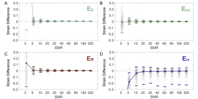 |
153 |
Cardiac Strains from DENSE MRI: Evaluation of SNR Requirements and Strain Error using a Computational Phantom
Ilya Verzhbinsky, Luigi Perotti, Kévin Moulin, Tyler Cork, Michael Loecher, Daniel Ennis
Displacement Encoding with Stimulated Echoes (DENSE) MRI encodes high-resolution cardiac tissue displacements into the phase of the complex MR signal. However, due to the inherent difficulty of obtaining ground truth strain measurements in the beating heart, it remains unclear how image SNR impacts the bias and range of error for computed cardiac strains. In this work, we present a computational deforming heart-like phantom to evaluate cardiac strains computed using a widely available, open-source DENSE Image Analysis Tool. We show that a strain error range within 0.05 and near-zero median error bias can be achieved with SNR>20.
|
|
2104.
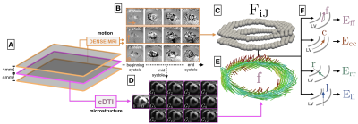 |
154 |
Estimating Aggregate Cardiomyocyte Shortening Using Diffusion and Displacement Encoded MRI
Ilya Verzhbinsky, Luigi Perotti, Kévin Moulin, Michael Loecher, Daniel Ennis
Orientation and deformation changes of left ventricular cardiomyocyte aggregates (“myofibers”) underlie many forms of cardiovascular disease. In vivo cardiomyocyte aggregate shortening (Eff) has mechanistic significance, but there exists no established technique to measure in vivo Eff. We present a pipeline to compute Eff by combining multi-slice Displacement Encoded with Stimulated Echoes (DENSE) MRI and in vivo cardiac Diffusion Tensor Imaging (cDTI) data. We show that Eff computed in healthy swine has decreased transmural variability compared to radial and circumferential strains. The spatial uniformity and mechanistic significance of Eff make it a compelling candidate for early detection of cardiac dysfunction.
|
|
2105.
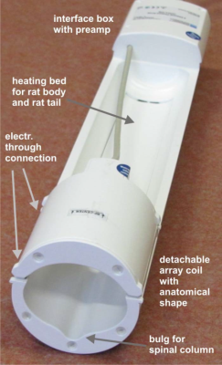 |
155 |
Tiny Golden Angle Real-time Cardiac MRI in the Mouse Model
Hao Li, Alireza Abaei, Qinghua Lu, Volker Rasche
Even though an established imaging method, small animal cardiac MRI is a time-consuming technique. Where self-gating techniques enabled wider-spread applications, acquisition times in the minute range for a single slice still limit its application for physiological stress or first-pass perfusion imaging. We translated the tiny golden angle sparse sense approach to small animal application. The technique was tested in ten animals and the resulting functional parameters compared with the self-gating approach. Further, initial application for pharmacological stress imaging was tested in three animals.
|
|
2106.
 |
156 |
Fully Self-Gated Cardiac and Respiratory Motion-Resolved 5D MRI for Rapid Assessment of Left Ventricular Function
Jérôme Yerly, Lorenzo Di Sopra, Gabriella Vincenti, Davide Piccini, Juerg Schwitter, Matthias Stuber
Cardiac magnetic resonance is the gold standard for left ventricular (LV) function assessment; however, current clinical protocols follow a rather complex workflow to plan and to acquire standard short-axis cine MR images. To simplify the workflow and remove these hurdles, we implemented and validated a time efficient free-running, free-breathing, fully self-gated cardiac and respiratory motion-resolved 5D MRI framework with isotropic spatial resolution. Assessment of LV function using the proposed framework and conventional 2D cines demonstrated good agreement between both techniques. When compared to the conventional approach, the proposed framework improves time efficiency and facilitates workflow.
|
|
2107.
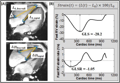 |
157 |
Fast and reproducible right ventricular longitudinal strain is a sensitive marker of pulmonary arterial hypertension
Shuang Leng, Yang Dong, Xiaoke Shang, Xiaodan Zhao, Ru-San Tan, Wen Ruan, Gangcheng Zhang, John C. Allen, Angela S. Koh, Bryant Jennifer, Lynette Teo, Ping Chai, James W. Yip, Jonathan Yap, Soo Teik Lim, Marielle V Fortier, Teng Hong Tan, Ju Le Tan, Yucheng Chen, Liang Zhong
Survival of patients with pulmonary arterial hypertension (PAH) is closely related to right ventricular (RV) function. This study aims to introduce a fast global longitudinal strain (GLS) measurement for RV function assessment by automatically tracking the distance variation between tricuspid annular insertion points and the RV apical epicardium. The intra- and inter-observer variation of coefficients range from 3.9% to 5.7% with processing time averaging 32 ± 8 seconds per subject. Results demonstrate that lower fast GLS measurements are closely related to impaired RV function and higher risks in PAH.
|
|
2108.
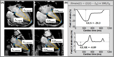 |
158 |
A fast and semi-automated approach for assessing left ventricular longitudinal strain with cine cardiovascular magnetic resonance imaging in heart failure with preserved and mid-range ejection fraction
Shuang Leng, Xiaodan Zhao, Ru-San Tan, Angela S. Koh, Bryant Jennifer, Jun-Mei Zhang, Ris Low, David Sim, John C. Allen, Hak Chiaw Tang, Liang Zhong
This study aimed to study the left ventricular (LV) longitudinal deformation by a semi-automated and rapid assessable strain parameter (i.e. global longitudinal strain, GLS) with standard cardiovascular magnetic resonance images. Study population consisted of 50 normal controls, 60 patients with heart failure (10 HFpEF, 10 HFmrEF and 40 HFrEF) and 10 patients with hypertrophic cardiomyopathy. Average processing time of the fast strain measurement method was 78 ± 8 seconds per case with intra- and inter-observer variations ranging from 3.3% to 5.8%. Results demonstrated that the fast and reproducible GLS is a powerful independent predictor in patients with HFpEF and HFmrEF.
|
|
2109.
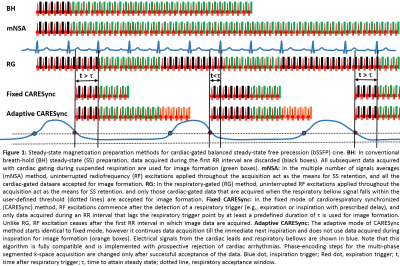 |
159 |
Elimination or Minimization in Depth of Sedation in Pediatric Cardiac Patients Considered High-Risk for Anesthesia: Using CArdioREspiratory Synchronized (CARESync) Balanced Steady-State Free Precession Cine Imaging
Amol Pednekar, Premal Trivedi, Siddharth Jadhav, Cory Noel, Prakash Masand
General anesthesia (GA), while not always required, is frequently necessary in infants and children undergoing cardiac magnetic resonance imaging (CMR) based on risk-benefit of breath-hold under GA v/s diagnostic value of the images. Primarily, requirement of breath-hold for cine imaging to evaluate ventricular volumes and function, a key prognostic measure in spectrum of congenital heart diseases, governs the administration of anesthesia. Our experience demonstrates that completely free-breathing-CMR studies including CArdio-REspiratory Synchronized cine imaging allow elimination of anesthesia or minimization in depth of sedation while providing diagnostic morphologic, functional, and pathophysiologic evaluation in young children, and adolescents considered high-risk for anesthesia.
|
|
2110.
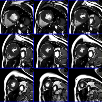 |
160 |
Compressed Sensing CMR Cine Imaging for Assessment of Left Ventricular Function and Mass
wang jiajia, Ge yinghui, Lin Qin, Pan Yukun
The purpose of the study was to assess the accuracy of left ventricular (LV) volumes and mass with innovative single breath-hold compression sensing (CS) cine CMR. 154 patients were enrolled for two techniques CMR cine. Two radiologists evaluated the image quality. LV volumes and mass were quantificationally analyzed. The image quality score of CS cine was slightly worse (4.7 ± 0.5 vs. 4.2 ± 0.7, Z = -5.934, p < 0.0001). All LV volumes and mass with two techniques were no significant differences (p > 0.05). CS cine CMR may replace the multiple breath-holds cine CMR.
|
|
2111.
 |
161 |
Four-dimensional whole-heart cine imaging with isotropic resolution in one single breath-hold: initial results
Davide Piccini, Lorenzo Di Sopra, Jérôme Yerly, Gabriella Vincenti, Tobias Rutz, Juerg Schwitter, Matthias Stuber
In cardiac MRI, the evaluation of left ventricular ejection fraction (LVEF) is based on well-defined protocols and involves a stack of 2D cine images acquired during multiple breath-holds. This procedure is time-consuming and may result in suboptimal coverage and inter-slice misregistration. Here, a novel acquisition protocol for 3D cine imaging with whole-heart coverage in one single breath-hold with isotropic spatial resolution is described and tested in 10 subjects in comparison to the 2D gold-standard. All 2D datasets and 9/10 3D datasets were considered adequate for LVEF calculation. Bland-Altman analysis of LVEF shows good agreement and no bias between the two measurements.
|
|
2112.
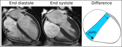 |
162 |
Left ventricular longitudinal contribution to stroke volume in pregnancy complicated by preeclampsia and normal pregnancy compared to non-pregnant controls
Katarina Steding-Ehrenborg, Daniel Salehi, Grigorios Kalapotharakos, Marcus Carlsson, Stefan Hansson, Håkan Arheden, Erik Hedström
We hypothesized that cardiac pumping in healthy pregnancy and pregnancy complicated by preeclampsia would be affected by the increased volume load on the heart during normal pregnancy and increased pressure load in pregnancy complicated by preeclampsia. The aim of this study was therefore to compare healthy pregnant women and women with preclampsia to healthy non-pregnant controls matched for age and cardiac index. The results were unexpected. Women with preeclampsia did not differ compared to healthy non-pregnant controls, whereas healthy pregnant women had significantly lower atrioventricular plane displacement and consequently a lower longitudinal contribution to stroke volume.
|
|
2113.
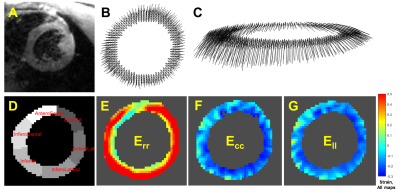 |
163 |
Highly Accelerated Balanced SSFP Cardiac Cine 3D-DENSE Quantifies Human 3D Myocardial Strain
Ronald Beyers, Nouha Salibi, Thomas Denney
Quantification of 2D and 3D myocardial strain at cine frame-rates has been previously demonstrated with echo-planar and spiral sequence versions of Displacement Encoding with Stimulated Echoes (DENSE). However, these non-conventional acquisition methods, with their sensitivities to off-resonance, has hindered their integration into mainstream cardiac MRI application. Here we present a more conventional, but highly accelerated, balanced SSFP (bSSFP) version of Cardiac Cine 3D-DENSE for application to human subjects. In vivo human scans at 3T demonstrated good agreement of myocardial radial (Err), circumferential (Ecc) and longitudinal Ell) strain values between bSSFP 3D-DENSE to those reported in previous literature.
|
|
2114.
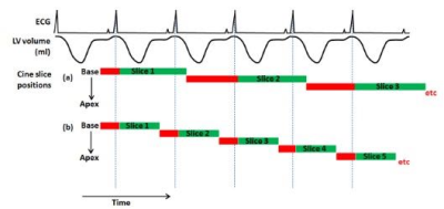 |
164 |
Reliability and accuracy of pediatric ventricular function analysis by short axis 'single-cycle-stack-advance' single-shot compressed sensing cines in minimal breath hold time
Peter Gatehouse, Suzan Hatipoglu, Sylvia Krupickova, Sandrine Foldvari, Piers Daubeney, Carmel Hayes, David Firmin
Cardiac cine SSFP by compressed sensing for ventricular volumetric analysis is promising for patients unable to tolerate long scanning sessions, as previously reported. We investigate a method requiring shorter breath-hold duration, robust to free-breathing if necessary, and assess its reliability and measurement accuracy for biventricular volume analysis in pediatric patients.
|
|
2115.
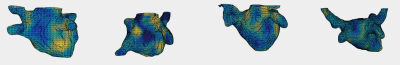 |
165 |
Free-breathing left atrial volumetric cine imaging and strain mapping with interleaved radial, multi-band self-gating, UNFOLD, and parallel imaging
Chenxi Hu, Xenophon Papademetris, James Duncan, Dana Peters
We developed a multi-slice free-breathing volumetric GRE cine imaging sequence for generating a 3D peak strain map for the left atrium. The sequence uses interleaved radial acquisition and a novel multi-band self-gating technique, accelerated with UNFOLD, and parallel imaging. The method was validated in 8 healthy subjects against alternative motion-correction methods. The results showed a stable performance using multi-band self-gating, and a sufficient image quality to generate a 3D strain map using feature tracking. This method provides a novel approach to interrogate the relationship between left atrial fibrosis, quantified by 3D left atrial gadolinium enhancement, and left atrial strain.
|
|
2116.
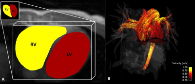 |
166 |
Sex Differences in Cardiac Biomechanics of Healthy Volunteers
David Rutkowski, Gregory Barton, Christopher François, Niti Aggarwal, Alejandro Roldán-Alzate
The prevalence and prognoses of cardiac diseases differ between men and women due to a number of physiological factors. However, the implications of these differences on cardiac flow dynamics are not yet fully understood. In this study, healthy volunteers were imaged with four-dimensional flow and bSSFP MRI. Differences were observed between metrics of ventricular kinetic energy, ventricular flow vorticity, myocardial strain, and efficiency indices. Results may, in part, explain the hypothesized physiological protective effects of the pre-menopausal female heart, and their relevance to efficient cardiac flow dynamics.
|
|
2117.
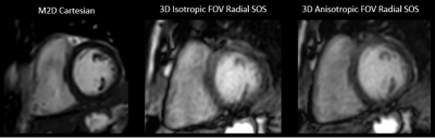 |
167 |
Quantitative comparison between Multislice 2D Breath-hold Cartesian and Free-breathing Anisotropic FOV 3D Golden Angle Radial Stack-of-Stars for functional cardiac MRI
Joao Tourais, Guruprasad Krishnamoorthy, Marc Kouwenhoven, Jouke Smink, Marcel Breeuwer
Radial trajectories are ideal in dynamic imaging due to its robustness to flow and motion. Multislice 2D Breath-hold Cartesian acquisition is still the most used approach to acquire functional cardiac MRI. In this work, we demonstrate the feasibility of Free-breathing 3D Radial stack-of-stars imaging with in-plane anisotropic FOV. The prosed approach can reduce imaging times in acquisitions where the object dimensions are anisotropic, while still enabling a reliable Ejection Fraction estimation.
|
|
2118.
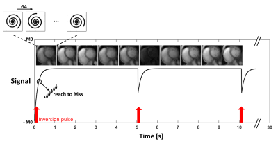 |
168 |
Inversion Recovery Cardiac and Respiratory Self-gated Simultaneous Acquisition of Cine and LGE Images Using a Golden-angle Spiral Pulse Sequence
Ruixi Zhou, Yang Yang, Daniel Weller, Abdul Ahmed, John Mugler, Mathews Jacob, Michael Salerno
We proposed an inversion recovery respiratory and cardiac self-gated continuous spiral acquisition pulse sequence to acquire cine and LGE images simultaneously. Data is acquired using a single spiral interleaf, rotated by the golden-angle in time, with an inversion pulse applied every 5 seconds. Cine images were reconstructed from the steady state portion of the signal using L+S, while LGE images were reconstructed from the data at a specific TI after each inversion pulse using SPIRiT. This strategy will enable whole heart cine and LGE imaging in less than 5 minutes without the need for breath holding or ECG gating.
|
|
2119.
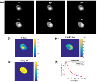 |
169 |
Optimization of cardiac functional magnetic resonance imaging at 7T
El-Sayed Ibrahim, V Emre Arpinar, L Tugan Muftuler, Andrew Nencka, Kevin Koch
In this study, we provide preliminary data from phantom and in vivo scans for optimizing 7T cardiac functional imaging. The results show that high-quality cardiac functional imaging can be achieved at 7T by optimizing the scan settings and imaging parameters, especially to mitigate B1 inhomogeneity effects. Adjusting the imaging flip angle and adding dielectric pads to the imaged region-of-interest could help improve B1 homogeneity and reduce signal nulling effects. High spatial and temporal resolutions and improved tagging persistence achieved at 7T allows for accurate global and regional cardiac function measurements and access to information not available at lower field strengths.
|
|
2120.
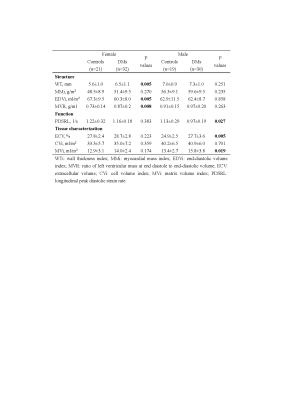 |
170 |
Sex Differences in Type 2 Diabetes Mellitus-Related Left Ventricular Remodeling
Presentation Not Submitted
Lan Lan, Yongning Shang, Xiaochun Zhang, Haibo Xu, Xiaoyue Zhou
This study aimed at demonstrate the influence of gender difference on T2DM related LV structural, functional, as well as interstitial remodeling. Short-axis cine imaging and pre- and post-contrast T1 mappings were analyzed in 62 T2DM patients and 40 healthy controls. Significant LV concentric remodeling was noted in female T2DM population while male patients exhibited extracellular matrix remodeling and diastolic dysfunction.
|
|
2121
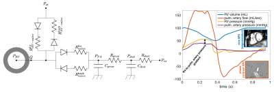 |
171 |
Longitudinal study of ventricular remodeling in tetralogy of Fallot patients using CMR coupled with biomechanical modeling
Video Permission Withheld
Radomir Chabiniok, Maria Gusseva, Animesh Tandon, Matthew Jones, Shakeel Qureshi, Kuberan Pushparajah, Dominique Chapelle, Tarique Hussain
We employ a biophysical modeling framework to augment the information obtained from cardiovascular MRI data of patients with chronic pulmonary valve regurgitation in order to assist in optimal timing of pulmonary valve replacement therapy (PVR). The longitudinal follow-up of patients post-PVR shows no significant change in ventricular ejection fraction. However, the model is able to detect a decreasing request on generation of active myocardial stress while not decreasing the cardiac output – presumably favorable with the long-term prognosis of the heart. Coupling biophysical modeling with MRI data has the potential to further augment the diagnostic value of MRI.
|
|
2122.
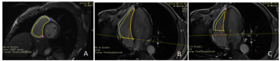 |
172 |
Right ventricular strain and smoking-related parameters: a correlational research
Shuangchun Ma, Xin LI, Shilan LI, Lizhi Xie, Qingwei Song, Ailian Liu, Zhiyong Li
Smoking is associated with an increased risk of coronary artery disease and other cardiovascular diseases. In this study, we analyzed the correlation between right ventricular strain and smoking-related parameters of 22 smoking volunteers and found that there was a positive statistical correlation between right ventricular radial strain, circumferential strain and smoking-related parameters respectively when the right ventricular ejection fraction was normal. Therefore, it is concluded that the right ventricle has a compensatory capacity for smoking on the heart for a certain period of time.
|
|
2123.
 |
173 |
PyMRStrain: A Python Library for the Generation of SPAMM, C-SPAMM, and DENSE Synthetic Images
Hernán Mella, Joaquín Mura, Julio Sotelo, Sergio Uribe
The generation of synthetic MR images allows the testing of several for postprocessing methodologies under controlled conditions. In this work, we developed an open source Python/C++ library for the generation of synthetic SPAMM, C-SPAMM and DENSE images, based on known magnetization expressions and using finite element geometries.
|
|
2124.
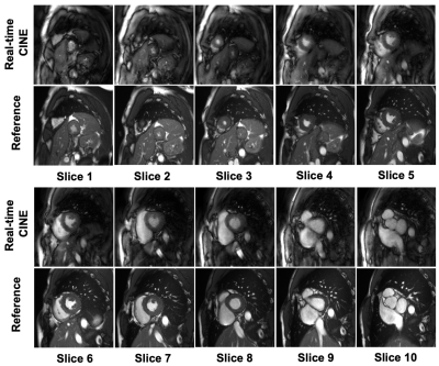 |
174 |
High Resolution Real-time Spiral Cine with Whole Heart Coverage in Under 30 Seconds
Zhixing Wang, Xue Feng, Yang Yang, Ruixi Zhou, Michael Salerno, Craig Meyer
This study develops a spiral-in/out bSSFP pulse sequence combined with a low rank plus sparse (L+S) reconstruction method for free-breathing ungated high resolution real-time cine with whole heart coverage. Within 30 seconds, this proposed method achieves total ten slices spiral cardiac movies with an in-plane 1.37 x 1.37 mm2 spatial resolution and 40 ms temporal resolution.
|
|
| Top |
Cardiovascular Image Processing & Reconstruction
Digital Poster
Cardiovascular
Monday, 13 May 2019
| Exhibition Hall |
17:00 - 18:00 |
| |
|
Computer # |
|
2125.
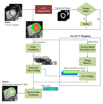 |
1 |
Transfer Learning Based Automated Myocardial T2 and Extracellular Volume Quantification
Yanjie Zhu, Ahmed S. Fahmy, Chong Duan, Reza Nezafat
Manual analysis of myocardial tissue mapping is time consuming. Deep learning has a potential to facilitate the analysis but requires big training datasets. In this study, a deep fully convolutional neural network, trained using native T1 mapping dataset, is used for T2 and extracellular volume (ECV) quantification based on transfer learning. We prospectively acquired T2 (401 patients) and ECV maps (381 patients) to access the network performance. Compared with the manually analyzed reference values, the transfer learning-based automated analysis platform shows good performance for myocardial T2 and ECV mapping. The platform has potential to fully automate myocardial tissue mapping.
|
|
2126.
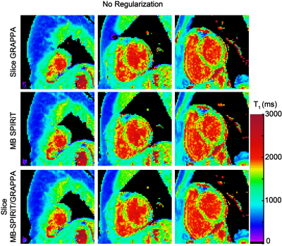 |
2 |
Multi-Band SPIRiT Strategies for Improved Simultaneous Multi-slice Myocardial T1T1 Mapping
Omer Demirel, Sebastian Weingärtner, Steen Moeller, Mehmet Akçakaya
Myocardial T1 mapping is typically performed with three or more breath-holds, each covering one slice. This leads to patient discomfort and long exam times. Recently, simultaneous multi-slice/multi-band (SMS/MB) imaging was proposed to improve coverage and reduce scan time. It was shown that k-space interpolation outperformed SENSE-type reconstructions in terms of T1 precision. However, the slice-GRAPPA approach is a kernel estimation approach and does not allow for additional regularization. In this work, we sought to use a SPIRiT approach for improving SMS/MB myocardial T1 mapping by performing regularized multi-band reconstruction.
|
|
2127.
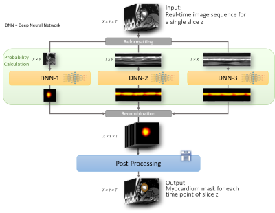 |
3 |
Automatic Analysis of Multicycle Real-time MRI for the Assessment of Variable Cardiac Function based on Multi-orientation U-net Segmentation
Anja Hennemuth, Jan-Martin Kuhnigk, Michael Steinmetz, Sebastian Kelle, Teodora Chitiboi, Jens Frahm, Markus Huellebrand
Real time MRI is a promising modality for the measurement or myocardial function without the need for breath-holding or ECG triggering. To enable the quantitative assessment of non-temporally aligned image slices representing multiple heartcycles we present an automatic image analysis approach based on a segmentation using the U-net convolutional neural network model. The comparison of segmentation masks with reference data show a very good DICE coefficient of 0.94. The comparison of quantitative results achieved based on the expert-corrected conventional segmentation shows promising results and suggests that further improvement can be achieved through parameter adaptation.
|
|
2128.
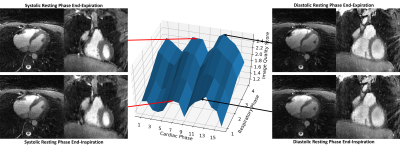 |
4 |
Automated Image Quality Assessment in 5D Whole-Heart MRI Aimed at Guiding Readers of High-Dimensional Dynamic Imaging
John Heerfordt, Lorenzo Di Sopra, Robin Demesmaeker, Jérôme Yerly, Tobias Kober, Matthias Stuber, Davide Piccini
A neural network trained to assess the quality of whole-heart coronary MRA images acquired with a respiratory self-navigated ECG-triggered bSSFP sequence was tested on images from a similar, but continuous non-ECG-triggered counterpart. Since cardiac and respiratory motion-resolved reconstructions of such acquisitions oftentimes consist of up to 150 individual 3D volumes, it is desirable to be able to automatically identify the volume with highest image quality for initial display to the reader. We found that the best image quality according to the neural network agreed with human visual assessment and was found in volumes corresponding to cardiac resting phases at end-expiration.
|
|
2129.
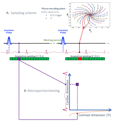 |
5 |
Free-Breathing, Self-Navigated and Dynamic 3-D Multi-Contrast Cardiac CINE Imaging Using Cartesian Sampling and Compressed Sensing
Elisabeth Hoppe, Jens Wetzl, Christoph Forman, Gregor Körzdörfer, Manuel Schneider, Peter Speier, Michaela Schmidt, Andreas Maier
We present a free-breathing multi-contrast 3-D cardiac CINE acquisition and reconstruction technique based on Compressed Sensing. Inversion pulses were repeatedly applied during a continuous acquisition to sample contrast- and cardiac-resolved 3-D data, while a self-navigation method was applied for respiratory gating. Validation was performed in a phantom, showing recovery curves and T1* maps in good correlation to known T1 values for the phantom as well as a MOLLI reference measurement. Feasibility for in-vivo application was demonstrated in a healthy volunteer.
|
|
2130.
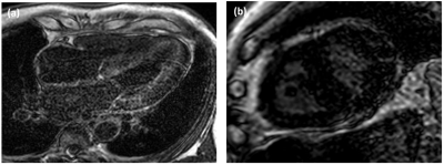 |
6 |
Left-atrial global longitudinal strain as an imaging biomarker of cardiac amyloidosis
Jadranka Stojanovska, Behnaz Khazai, Mason Runge, Maryam Ghadimi, Anil Attili, Thomas Chenevert, El-Sayed Ibrahim
Cardiac amyloidosis (CA) is a clinical disorder that could result in early death. MRI is a valuable modality for diagnosis of CA, where late-gadolinium enhancement is typically used for assessing adverse outcomes. However, in the era of unknown long-term effects of the gadolinium, there is a need to investigate alternative imaging biomarkers associated with CA, which is the primary aim of this study. The secondary aim is to evaluate the association between MRI variables and mortality in CA. The results demonstrated that depressed left-atrial global longitudinal strain is significantly associated with biopsy-proven CA, independent of other MRI and clinical measures.
|
|
2131.
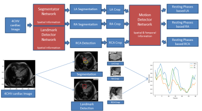 |
7 |
Automatic Cardiac Resting Phase Detection for Static Cardiac Imaging Using Deep Neural Networks
Seung Su Yoon, Elisabeth Hoppe, Michaela Schmidt, Christoph Forman, Puneet Sharma, Christoph Tillmanns, Andreas Maier, Jens Wetzl
To perform static cardiac imaging, manual inspection of CINE images is currently necessary to detect a quiescent window within the cardiac cycle. We propose an automated system using two chained Deep Neural Networks to determine localized end-systolic and end-diastolic resting phases. The first network finds a region of interest (e.g. RCA, right or left atrium) and the second determines a quantitative motion curve for this region. Training and evaluation was performed on data from volunteers and patients acquired on different scanners and field strengths and a comparison to manually annotated resting phases showed accuracy in the range of 35ms.
|
|
2132.
 |
8 |
Fully Automated Cardiac Bounding Box Detection for Localized Higher-Order Shimming Using Deep Learning
Asha K. Kumara Swamy, Chandrashekar M. Patil, Punith B. Venkategowda, Vikram Nagalli, Michael Wangler, Michaela Schmidt, Jens Wetzl
Localized higher-order shimming is a common method for improving image quality in phase sensitive sequences used in cardiac imaging, but requires the manual placement of a three-dimensional bounding box around the heart in which the localized shimming is performed. We present an automated method for detecting such a bounding box from the localizer images using Deep Learning. Two-dimensional bounding boxes are first detected in each localizer slice and then combined to one three-dimensional bounding box. We compare two approaches, either training individual models for each localizer orientation or a joint model for all orientations.
|
|
2133.
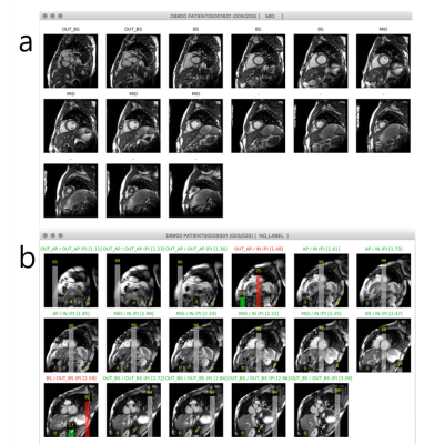 |
9 |
Cardiac short-axis slice range classification via transfer learning: Evaluation of seven popular deep CNNs
Namgyu Ho, Yoon-Chul Kim, Yeon Hyeon Choe
In cardiac MRI, left ventricle (LV) segmentation typically follows the identification of a short-axis slice range, which may require a manual procedure. The standard cardiac image processing guidelines indicate the importance of correct identification of the slice range. In this study, we investigate the feasibility of deep learning in automatically classifying the slice range. Images were classified into one of three categories: out-of-apical (OAP), apical-to-basal (IN), and out-of-basal (OBS). We developed our in-house user interface to label image slices into one of the three categories for learning. We evaluated the performance of the models, fine-tuned from seven popular deep CNNs.
|
|
2134.
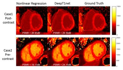 |
10 |
Fast and Robust T1-mapping using Convolutional Neural Networks
Haris Jeelani, Yang Yang, Roshin Mathew, Michael Salerno, Daniel Weller
The pixel-wise nonlinear regression method for T1-mapping is susceptible to noise. We propose a convolutional neural network framework for fast and robust cardiac MRI T1-mapping. A dense type of architecture is used for producing denoised T1-maps. Results show the proposed framework improves PSNR by 6dB compared to the pixel-wise nonlinear regression. The Wilcoxon signed rank test shows a significant reduction in the standard deviation of T1-values produced by the proposed method as compared to nonlinear regression. After training, the time required for producing one T1-map from the undersampled images is 6.45 seconds.
|
|
2135.
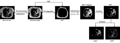 |
11 |
Automated measurement of peri-organ visceral adipose tissue in MRI as a powerful biomarker of metabolic profile
Mohamed ZARAI, Karine CLEMENT, Judith ARON, Nadjia KACHENOURA, Khaoula BOUAZIZI, Alban REDHEUIL
The aim of this work is to develop an automatic segmentation algorithm to classify truncular adipose tissue into different compartments. MRI acquisitions including cine-SSFP and DIXON imaging were performed at 1.5 T in 117 individuals (metabolic patients and healthy controls). Fat maps were filtered with a top-hat filter to correct intensity inhomogeneities. An active contour and a k-means algorithms were used to discriminate the SAT and the VAT. Accurate and reproducible quantification of the adipose tissue is crucial for metabolic studies since they serve as good indicators of metabolic and associated cardiovascular risks.
|
|
2136.
 |
12 |
Application of Deep Learning Reconstruction for Denoising of Compressed Sensing non-contrast coronary MRA images to achieve improved Diagnostic Confidence.
Yoko Kato, Bharath Ambale-Venkatesh, Yoshimori Kassai, John Pitts, Larry Kasuboski, Jason Ortman, Shelton Caruthers, Joao Lima
Non-contrast Magnetic resonance coronary artery (MRCA) image acquisition has technical limitations of long acquisition time or reduced image resolution. We explore the use of a denoising approach with deep learning image reconstruction (dDLR) from k-space data. We investigate the effect of various levels of dDLR on Compressed Sensing non-contrast MRCA (CS-MRCA) images and optimize dDLR algorithms that achieve the best diagnostic confidence (DC) and a high signal-to-noise-ratio (SNR).
|
|
2137.
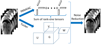 |
13 |
High-Resolution Dynamic Myocardial T1 Mapping Using Low Rank Tensor Regularization Methods
Burhaneddin Yaman, Sebastian Weingärtner, Steen Moeller, Nikolaos Kargas, Nicholas Sidiropoulos, Mehmet Akçakaya
Recently a new method called TOPAZ was developed for cardiac phase-resolved myocardial T1 mapping, which is performed in breath-hold duration that subsequently limits its spatial resolution. These datasets are multidimensional which makes tensor regularization a natural fit for regularization. In this work, we sought to compare different tensor regularization techniques to enable high-resolution TOPAZ acquisitions.
|
|
2138.
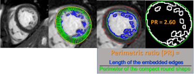 |
14 |
Evaluation of Cumulative Perimetric Ratio as Quantitative Index for Degree of Left Ventricular Myocardial Trabeculations in Adolescents and Young Adults: Potential Biomarker for Left Ventricular Non-compaction
Amol Pednekar, Siddharth Jadhav, Cory Noel, Prakash Masand
Balanced steady-state free precession imaging distinguishes the non-compacted left ventricular (LV) myocardial trabeculation (NC) from compacted myocardium (C). However, current diagnostic criterion (NC/C length ratio > 2.3) for left ventricular non-compaction (LVNC) suffers from subjective variability and tends to over-diagnose. Cumulative perimetric ratio (CPR) derived from epi and endocardial contours routinely drawn for LV functional analysis provides comprehensive measure of irregularity and extent of the LV trabeculations overcoming the inherent morphologic variability from base to apex. CPR has a potential to serve as a valuable biomarker for prominent trabeculations, complementary to NC/C length and mass ratios.
|
|
2139.
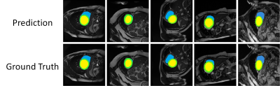 |
15 |
Fully automatic segmentation of cine cardiac MR images based on deep learning: an open-source toolbox with a failure alarm mechanism
Zheng-Sheng Lin, Teng-Yi Huang
In this study, we implemented a open-source toolbox for heart segmentation and analysis based on a deep-learning algorithm. We evaluated the accuracy of the system using the ACDC datasets provided in STACOM 2017. Also, we developed an alarm mechanism in this system to report the potential failure of the analysis procedures.
|
|
2140.
 |
16 |
View-independent cardiac MRI segmentation with rotation-based training and testing augmentation using a dilated convolutional neural network
Xue Feng, Chirstopher Kramer, Craig Meyer
Left and right ventricle segmentation is an important step in quantitative analysis of cardiac MR images. Convolutional neural networks (CNN) have shown great improvement and are quickly becoming the mainstream methods. One challenge in cardiac MRI segmentation from short-axis images is the variability of the imaging views and the fact that CNN is not rotation-invariant. To address this issue, we trained a view-independent network and further improved its performance with a rotation-based testing augmentation. Consistent improvement in performance was obtained as measured by Dice scores and visual contour quality.
|
|
2141.
 |
17 |
Highly accelerated cardiac imaging using a high-density 72 channel local receiver array at 3 Tesla
Mark Gosselink, Hugo Klarenberg, Hildo Lamb, Gustav Strijkers, Tim Leiner, Dennis Klomp, Martijn Froeling
Cardiac Magnetic Resonance Imaging (CMR) protocols can be extremely time consuming, expensive, and uncomfortable for the patient. The purpose of this study is to evaluated the acceleration performance for CINE CMR in-vivo scans using a 72 channel high-density coil array at 3 Tesla. We demonstrate highly accelerated CINE imaging within a single breath hold with acceptable SNR and minimal SENSE artifacts.
|
|
2142.
 |
18 |
Manifold recovery in cardiac dynamic MRI (dMRI) with different reduction factors
Ziling Jiao, Yichen Zheng, Hanyu Wei, Shuo Chen, Leslie Ying, Chun Yuan, Rui Li
Manifold models have been developed as an effective tool in dynamic MRI recently. With the abilities of characterizing the relationship between dMRI images series, they could be used to reconstruct high-quality image series from MRI data sampled below the Nyquist rate. However, the amount of MRI data required by manifold recovery is not clear yet. In this study, we did some simulation experiments to investigate the relationship between image quality of cardiac dynamic MRI and reduction factor, and proposed an optimized reduction factor considering both image quality and acquisition efficiency.
|
|
2143.
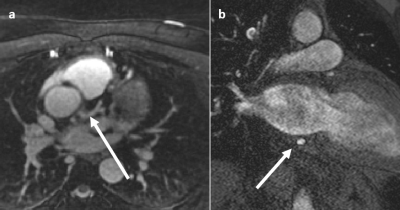 |
19 |
MRI-Derived Virtual Fractional Flow Reserve (vFFR): Preliminary Results
Jackson Hair, John Oshinski
Preliminary results are presented for calculation of virtual fractional flow reserve (vFFR) using magnetic resonance coronary angiography and phase-contrast magnetic resonance to define the boundary conditions for a computational fluid dynamics (CFD) model.
|
|
2144.
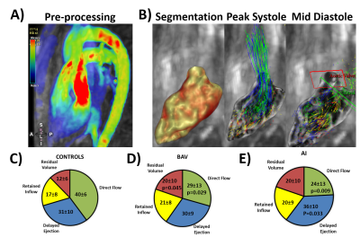 |
20 |
Usefulness of Left Ventricular Flow Component Analysis in Patients with Bicuspid Aortic Valve Disease Insufficiency
Julio Garcia, Alireza Sojoudi, An Le, Michael Bristow, Carmen Lydell, Andrew Howarth, Paul Fedak, James White
Acute aortic insufficiency (AI) is often developed in patients with bicuspid aortic valve (BAV). Flow component analysis (FCA) derived from 4D flow MRI can be used as a novel marker of LV function worsening. This study aimed to explore the value of FCA in the assessment of AI severity in BAV patients.
|
|
2145.
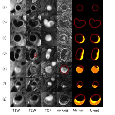 |
21 |
Carotid plaque composition segmentation in multi-contrast MRI with U-net
Presentation Not Submitted
Yin Guo, Jifan Li, Dingkun Liu, JIachen Ji, Shuo Chen, Xihai Zhao, Dongxiang Xu, Chun Yuan, Rui Li
Carotid plaques may cause strokes, the composition of which is crucial for assessing the risk. While multi-contrast plaque magnetic resonance imaging (MRI) is a powerful technology, it is both tedious and error-prone for a radiologist to review these images, and traditional analytic algorithm relying on manually crafted features perform poorly as well. We propose a novel approach with deep convolutional neural network (CNN), to be specific, U-net, to segment these plaque tissues. Some modifications on loss functions, convolution patterns and training protocols help our model perform well. On a dataset of 1098 subjects, we show that we achieve significantly better accuracy than previous models.
|
|
2146.
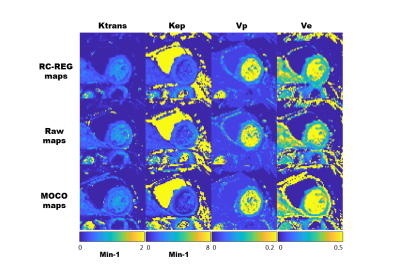 |
22 |
Myocardial permeability mapping analysis framework with an optimized Residual Complexity based registration
Jean Sebastien Louis, Freddy Odille, Damien Mandry, Jacques Felblinger, Clément Venner, Marine Beaumont
Myocardial perfusion data registration is challenging because of the inherent contrast variation in addition to cardiac and respiratory motions. Residual Complexity (RC) has been proposed as a new intensity based similarity measure for registration and has been reported to be more robust to contrast variation compared to other minimization indicators. In this work, we proposed a myocardial permeability maps framework with an optimization of a RC-based registration algorithm. We evaluated the performance of this algorithm, in comparison with MOCO and with no correction, on image quality and permeability maps.
|
|
2147.
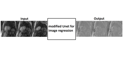 |
23 |
Deep Learning for Radial SMS Myocardial Perfusion Reconstruction
Johnathan Le, Ye Tian, Jason Mendes, Brent Wilson, Edward DiBella, Ganesh Adluru
Although dynamic contrast enhanced MRI has been successfully applied for characterizing coronary artery diseases, an acquisition scheme limited to 2-4 short axis slices restricts coverage of the left ventricle. Radial simultaneous multi-slice has been shown to improve DCE cardiac perfusion by providing complete coverage of the left ventricle but also requires an increase in reconstruction time. Here we propose using a modified Unet with a residual artifact learning framework to improve reconstruction time and image quality of spatio-temporal constrained reconstruction methods for radial SMS datasets. Results demonstrate promising improvements with a speed up in reconstruction by a factor of ~150.
|
|
2148.
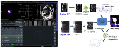 |
24 |
Inline Myocardial Perfusion flow mapping and Analysis: Powered by Gadgetron Inline AI
Hui Xue, Ethan Tseng, Marianna Fontana, James Moon, Peter Kellman
This abstract presents an AI powered system to perform automated quantitative perfusion flow mapping and analysis on the MR scanner. The key components consist of deep neural network models to a) detect LV on AIF image series and b) segment myocardium to generate AHA bull's eye plot. This solution was implemented in Gadgetron framework and has been deployed to clinical MR scanners. As a result, pixel-wise perfusion flow maps with segmentation of myocardium is automatically generated and available on the MR scanner shortly after the end of data acquisition.
|
|
2149.
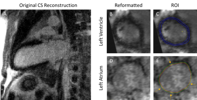 |
25 |
Self-Navigated, Free-Breathing 3D Left Atrial Late Gadolinium Enhancement MRI with Stack-of-Stars k-space Sampling and GRASP Reconstruction: A Preliminary Study for Quantification of Atrial Fibrosis
Suvai Gunasekaran, Hassan Haji-Valizadeh, Rod Passman, Daniel Lee, Daniel Kim
Given the thin nature of the left atrial (LA) wall (1-3 mm) and the need to perform respiratory gating with navigator echoes, the clinical translation of LA late gadolinium-enhanced (LGE) MRI has proven difficult. This study describes a self-navigated, free-breathing 3D LA LGE pulse sequence with stack-of-stars k-space sampling and GRASP reconstruction. This study shows initial experience in quantifying LA fibrosis in patients with atrial fibrillation undergoing ablation therapy.
|
|
| Top |
Myocardial Function & Deformation 2
Digital Poster
Cardiovascular
Monday, 13 May 2019
| Exhibition Hall |
17:00 - 18:00 |
| |
|
Computer # |
|
2150.
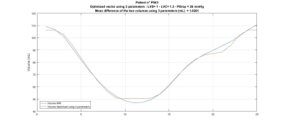 |
26 |
Doxorubicin induced cardiotoxicity highlighted from the combination of CMR, exercise tests data and CircAdapt model.
Tanguy Artz, Daniel Curnier, Delphine Perie
This study aims to discriminate three risk groups of leukemia survivors investigating subtle changes in their myocardium mechanical properties. Volumes extracted from cine-MRI images coupled to incremental exercise data allowed the quantification of the mechanical properties of the left ventricle using a reverse material identification approach based on the CircAdapt model. While both passive and active properties did not show significant changes in ALL survivors between risk groups, the increased arteriovenous pressure drop suggested that the cardioprotective agent entailed a compensation of the cardiac function through the peripheral components of the oxygen delivery chain.
|
|
2151.
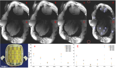 |
27 |
Acceleration Factor Analysis of Different Dedicated 8Tx/16Rx Coil Arrays for Cardiac MRI at 7T
Maria Stefanescu, Maxim Terekhov, Ibrahim Elabyad, David Lohr, Michael Hock, Laura Schreiber
Signal-to-noise ratio (SNR) in cardiac MRI (cMRI) is a parameter associated with the field strength, the parallel imaging (PI) acceleration factor and the geometry of receive array element arrangement (g-factor). The purpose of this work was to evaluate four different arrays with symmetrical and asymmetrical distribution of 16RX and 8TX elements ex-vivo in swine at 7T MRI. The asymmetrical design proved to be superior with regard to SNR and image homogeneity.
|
|
2152.
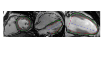 |
28 |
The Prognostic Role of Syncope and Cardiac Magnetic Resonance Based Left Ventricular Myocardium Deformation in Arrhythmogenic Right Ventricular Cardiomyopathy(ARVC) Patients
Presentation Not Submitted
Meng-ting Shen, Zhi-gang Yang, Kai-yue Diao, Li Jiang, Shan Huang, Xiao-yue Zhou, Ying-kun Guo
To explore prognostic role of clinical characteristics and CMR based strains in ARVC patients. CMR was performed in 45 ARVC patients. LV global strains in ARVC patients was significantly reduced than controls. Patients with syncope had statistically lower LV global stains and greater LGE incidence than without syncope. At 1037.51 ± 603.38 days mean follow-up, 14 patients reached end point. By univariate and multivariate regression analysis, syncope (OR= 48.66, [4.20-564.46]; P=0.002) and GLS> -12.72% (OR= 16.96, [1.79-160.66]; P=0.014) remained significantly associated with end point. Therefore, our study found syncope and GLS> -12.72% could predict adverse events in patients with ARVC
|
|
2153.
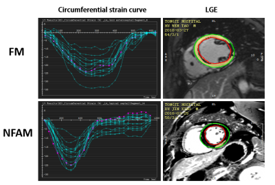 |
29 |
Incremental value of cardiac deformation analysis in Fulminant Myocarditis: a cardiovascular magnetic resonance imaging study
Haojie Li, Hui Zhu
Newer quantitative myocardial T1 and T2 mapping and feature-tracking (FT) myocardial strain analysis might add additional diagnostic information and further broaden the diagnostic targets. This study demonstrate that the assessment of cardiac strains applying FT on standard cine images is feasible in patients with fulminant myocarditis. And Cardiac strains parameters, especially, global peak systolic circumferential and longitudinal strain are significantly impaired in patients with FM. Myocardial strain metrics can sufficiently discriminate between FM and NFAM patients and show basic associations with the extent of myocardial inflammation.
|
|
2154.
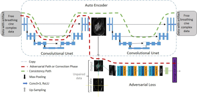 |
30 |
Free-breathing Multi-Phase MRI using Deep Learning-based Respiratory Motion Compensation
Vahid Ghodrati, Jiaxin Shao, Mark Bydder, Kim-Lien Nguyen, Xiaodong Zhong, Yingli Yang, Peng Hu
To minimize respiratory motion-induced image blurring and artifacts, conventional cardiothoracic and abdominal MRI techniques rely mostly on breath-holding. These approaches result in limited time window for data acquisition, especially in many ill patients who are unable to breath-hold for an extended period of time. In this study, we employed deep learning as a promising tool for detection and correction of complex respiratory motion during free-breathing MRI scanning. On average, our proposed network increased the sharpness of the images 20 percent.
|
|
2155.
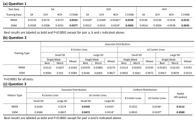 |
31 |
Analysis of Sampling Strategies for Convolutional Neural Network Based Cardiac Magnetic Resonance Image Reconstruction
Junyu Wang, Yang Yang, Xue Feng, Daniel Weller, Michael Salerno
Recently, convolutional neural network (CNN) based fast cardiac image reconstruction techniques have shown the potential to produce rapid, high quality reconstructions from under-sampled data. However, the relationship between the k-space sampling strategy, image content, training process and reconstruction performance has not been extensively studied. To address this, our study trained different CNN based cardiac image reconstruction models for different image content and various sampling patterns. We showed that better reconstruction results were achieved when using mixed image content as training data and distributing more energy at the center of k-space. Radial acquisition showed the lowest RMSE suggesting potential improvement of CNN performance with non-Cartesian acquisition.
|
|
2156.
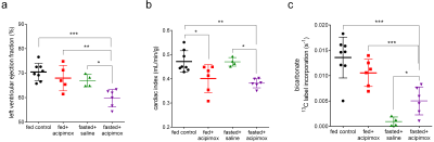 |
32 |
Assessing the effects of substrate modulation on cardiac function and metabolism in the rodent heart with CINE MRI and hyperpolarized [1-13C]pyruvate
Kerstin Timm, William Watson, Vicky Ball, Oliver Rider, Damian Tyler
Heart failure is associated with reduced cardiac energetics and this has been linked to impaired substrate utilization. We have assessed in rats how cardiac function and pyruvate dehydrogenase (PDH) flux change upon substrate manipulation, by varying plasma non-esterified fatty acid (NEFA) levels with acipimox injections in both fed and fasted rats. We found that NEFA depletion in fasted rats led to cardiac systolic dysfunction which may be explained by an insufficient compensatory increase in PDH flux boosting glucose oxidation. This model of substrate-manipulation heart failure could be used to assess potential heart-failure drugs in the future.
|
|
2157.
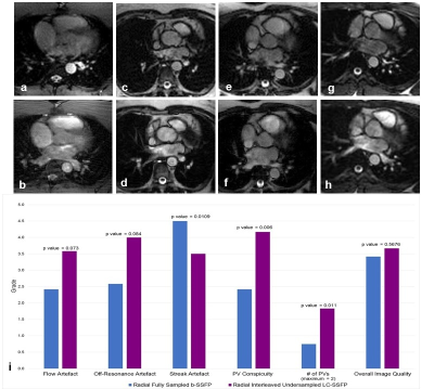 |
33 |
Radial LC-SSFP for Left Atrial Cine Imaging
Jaime Robb, Chenxi Hu, Dana Peters
Balanced steady-state free precession (b-SSFP) imaging of left atrial cine is severely affected by off-resonance artefacts, particularly in the pulmonary veins. Acquisition and combination of data sets with different phase-cyclings has been shown to remove banding artefacts with a trade-off in scan time. The purpose of this work is to present an approach to banding artefact removal in b-SSFP imaging for left atrial cine. We propose the use of interleaved, undersampled radial projections with LC-SSFP. This method provides increased pulmonary vein conspicuity and image quality with only minor increase in scan time and streaking artefact.
|
|
2158.
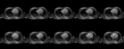 |
34 |
3D stack of spirals MRI with extra motion-state dimensions: a feasibility study
Javier Montalt-Tordera, Grzegorz Kowalik, Jennifer Steeden, Jyh-Miin Lin, Vivek Muthurangu
It has been previously shown that a 3D golden ratio stack of spirals sequence is suitable for fast thoracic MRI, including cardiac gated and elapsed time reconstructions. This work examines the feasibility of including a respiratory motion-state dimension. 5D thoracic datasets have been reconstructed using ESPIRiT to resolve along the cardiac and respiratory cycles. Although challenges exist, preliminary testing indicates that this is an effective method to increase data efficiency, thereby improving image quality and/or reducing the time of acquisition.
|
|
2159.
 |
35 |
Demonstration of Circumferential Heterogeneity in Displacement and Strain in the Abdominal Aortic Wall by Spiral Cine DENSE MRI
Elizabeth Iffrig, John Wilson, W. Taylor, Xiaodong Zhong, John Oshinski
Herein, we describe a method for implementation of cine DENSE imaging in the abdominal aortic wall. The method uses in house software to post-process and track displacement of intermural pixels and applies a quadrilateral based solution to measure strain. This new method permits the measurement of circumferential heterogeneity of both displacement and strain.
|
|
2160.
 |
36 |
A preliminary study on the mechanism of left ventricular myocardial strain in patients with pulmonary hypertension
Presentation Not Submitted
Jing SONG, Yousan CHEN, Xiangchuang KONG, Yue CUI, Yukun CAO, Jia LIU, Xiaoyue ZHOU, Heshui SHI, JING SONG
There is little understanding about the mechanism of left ventricular strain in pulmonary hypertension (PH) patients at present. In this study, we compared left ventricular strain between PH patients and the healthy controls. For PH patients with preserved or mildly improved left ventricular ejection fraction (LVEF), the ventricular strain parameters decreased compared to the healthy controls. We found that left ventricular myocardial strain impairment in HP patients is associated with right ventricular overload of volume or pressure and decreased ejection capacity.
|
|
2161.
 |
37 |
Robust free-breathing cardiac cine-MRI using a combination of serial motion averaging and Compressed SENSE
Yasuhiro Goto, Michinobu Nagao, Masami Yoneyama, Isao Shiina, Kazuo Kodaira, Mamoru Takeyama, Isao Tanaka, Shuji Sakai
The present study investigates the clinical utility of cardiac cine-MRI using free-breathing scan with serial motion averaging (SMART) and Compressed-SENSE (C-SENSE). Left ventricular functional parameters derived from free-breathing SMART with C-SENSE showed good correlations to those from conventional breath-hold technique. Cine-MRI with C-SENSE can reduce both scan time (around 40% of the scan time) and burden of multiple breath-holdings without any penalty for the image quality; therefore, it might contribute to great advantages in routine clinical work.
|
|
2162.
 |
38 |
Real-time strain encoding using echo planar imaging with centric reordering and parallel imaging
Yingmin Liu, Ning Jin, Yue Pan, Rizwan Ahmad, Orlando Simonetti
We developed a realtime strain encoding technique using EPI readout with centric reordering and parallel imaging acceleration. This technique was tested in a strain phantom and normal volunteers to measure through-plane left ventricular strain. Quantified strains correlated well with fully sampled k-space acquisitions. The proposed technique allows larger field-of-view (FOV) and provides superior coverage of both left (LV) and right ventricles (RV) while maintaining equivalent temporal and spatial resolution as the fully sampled sequence.
|
|
2163.
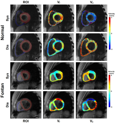 |
39 |
Evaluation of Myocardial Motion in Patients with Fontan Circulation by Using Tissue Phase Mapping Cardiac Magnetic Resonance
Xue-Jhe Lu, Meng-Chu Chang, Ming-Ting Wu, Ken-Pen Weng, Hsu-Hsia Peng
Patients with Fontan circulation nowadays have a well long-term survival with the improvement of post-surgery care and follow-up examination techniques. However, ventricular dysfunction and heart failure may occur in a couple years after surgery. To evaluate the risk of cardiac events in an early disease stage is important for patient managements. In this study, we measured myocardial motion velocity by tissue phase mapping. Fontan patients presented significantly decreased systolic and diastolic longitudinal velocity. In conclusion, evaluation of myocardial motion velocity could provide helpful information in detecting myocardial remodeling of functional ventricle in Fontan patients.
|
|
2164.
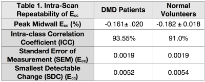 |
40 |
Intra-Exam Repeatability of CINE DENSE Strain in Duchenne Muscular Dystrophy
Patrick Magrath, Nyasha Maforo, Mike Loecher , Holden Wu, Pierangelo Renella , Nancy Halnon , Daniel Ennis
Heart failure leads the causes of mortality in Duchenne Muscular Dystrophy (DMD), the most common fatal genetic disorder. Circumferential strain (Ecc) is a promising biomarker for improved diagnosis and evaluation of therapy, but its repeatability has not been evaluated in a DMD cohort. The objective was to quantify the intra-exam repeatability of peak mid-wall circumferential (Ecc) strain derived from CINE DENSE in boys with DMD (N=11) compared to age-matched controls (N=10). Intra-scan repeatability was high, with a smallest detectable change in strain of 0.005, well under the previously reported 0.013±0.015 decrease in Ecc/year reported in boys with DMD.
|
|
2165.
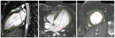 |
41 |
Strain and Torsion Analysis of Left Ventricular Using Tissue Tracking to Evaluate Deterioration of Ventricular Function as MR Marker in Patients with Repaired Fontan
Liwei Hu, Rongzhen Ouyang, Aimin Sun, Qian Wang, Chen Guo, Xiaofen Yao, Yumin Zhong
At present, cardiovascular magnetic resonance (CMR) has emerged as the gold standard for the quantification of ventricular volume and ejection fraction in repaired Fontan patients [1]. However, left ventricle ejection fraction (LVEF) reflects the geometric change of LV, but not the contractile function of the myocardium [2]. Strain and torsion are new MRI markers to detect myocardial deformation using balanced steady state free precession (b-SSFP) cine imaging in CMR. Myocardial Strain has been shown as an earlier marker of contractile dysfunction than EF and a subclinical sign of heart muscle cells [3,4]. Torsion as a measurement of myocardial afterload, wall and fiber stresses is an important determinant of ventricular function and hypertrophy. Ventricular twist mechanics hold promise for better understanding mechanisms of ventricular dysfunction [5]. To best our acknowledge, There were much fewer data about the comparison between the prognostic value of perioperative NT-proBNP levels and MRI quantitative indexes in children after surgical repair of CHD. The study is to compare cardiac strains and torsion among the repaired Fontan patients with normal and abnormal NT- proBNP levels as well as healthy controls. We hypothesized that the global ventricular strain and torsion could be as MRI quantitative indexes for evaluation of deterioration of ventricular function in patients with repaired Fontan.
|
|
2166.
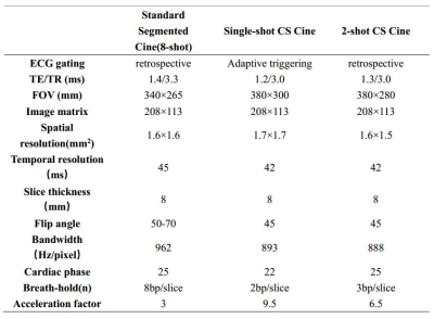 |
42 |
Multi-shot compressed sensing techniques accelerate cine sequence acquisition: an evaluation of diagnostic efficacy
Presentation Not Submitted
Jian Wang, Xiao Li, Lu Lin, Jian Cao, Jing An, Michaela Schmidt, Christoph Forman, Yi-ning Wang, Zheng-yu Jin
An evaluation of diagnostic efficacy study about compressed sensing techniques accelerates cine sequence acquisition, which quantitatively and qualitatively compares 3 kinds of cine sequences in CMR.
|
|
2167.
 |
43 |
Comparison of Different Real-Time Sequences for Cardiac MRI
Patrick Metze, Tobias Speidel, Claudia Lumpp, Hao Li, Volker Rasche
In this work we compare different real-time protocols for cardiac functional imaging. Image quality is assessed qualitatively and quantitatively and functional parameters are calculated from three short axis slices. All imaging methods are compared to the current ECG-gated, breathhold reference standard (BH-CINE). All real-time methods show acceptable visual image quality with only slight differences, which is supported by the quantitative analysis. The functional analysis shows slight systematic differences, but not on a statistically significant level.
|
|
2168.
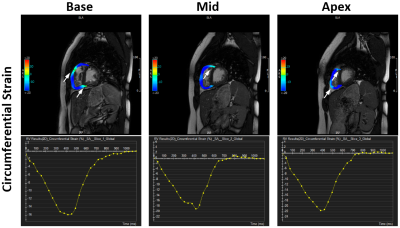 |
44 |
Assessment of regional ventricular function in tetralogy of Fallot using strain analysis. Comparison of techniques
El-Sayed Ibrahim, Prachi Agarwal, Maryam Ghadimi-Mahani, Jadranka Stojanovska
Assessment of ventricular function, especially in the right ventricle (RV), is important for cardiac evaluation in Tetralogy of Fallot (ToF). MRI tissue tracking (TT) is a recently introduced technique for analyzing myocardial strain directly from the cine images. In this study, we evaluate the feasibility of using TT strain analysis for measuring myocardial contractility in ToF and compare the results to measurements from the more established strain-encoding (SENC) MRI technique. The results showed that TT strain analysis allows for fast assessment of heart contractility in ToF with global strain measurements that are insignificantly different from those from SENC analysis.
|
|
2169.
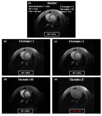 |
45 |
Optimization of cardiac functional MR imaging in rats. Image quality versus scan tolerance in sick animals
El-Sayed Ibrahim, Matthew Budde, Dhiraj Baruah, Anne Frei, Rachel Schlaak, Michael Flister, Carmen Bergom
Using MRI for studying cardiac function in rat models of radiation-therapy is important for better understanding of cardiotoxicity. In this study, we developed an optimized cardiac functional MRI protocol for rat imaging, and investigated the effects of changing imaging parameters on cardiac measurements. The results showed that blood-to-myocardium contrast-to-noise ratio in the cine images significantly affects ejection-fraction measurements, whereas reduced resolution has less effect on ejection-fraction. However, reduced resolution in the tagged images has significant effect on strain measurements, whereas tagline density has less effect, as long as sufficient resolution is maintained and more than one tagline intersects the myocardium.
|
|
2170.
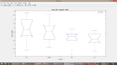 |
46 |
CMR coupled to optical flow and virtual fields methods for the quantification of myocardial mechanical properties in cancer survivors.
Hadi Begdouri, Farida Cheriet, Delphine Perie
Subtle changes in strains and mechanical proprieties of the myocardial tissue were assessed in childhood acute lymphoblastic leukemia survivors using the optical flow and virtual field methods on CMR images. Significant changes between risk groups were observed on the strain in the 2 chambers view only. However, significant changes between risk groups were observed on the stresses, shear and Young’s moduli in all views. The dynamic elastic and shear moduli obtained along the cardiac cycle might be indicators of doxorubicin induced cardiotoxicity.
|
|
2171.
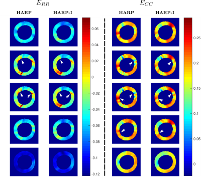 |
47 |
An Harmonic Phase Interpolation Method for the Estimation of Motion from Tagged MR Images
Hernán Mella, Joaquín Mura, Julio Sotelo, Sergio Uribe
Tagging MR is still considered the gold standard for motion quantification in the heart walls. Although there are several methods for postprocessing this kind of images, harmonic phase methods such as HARP and SinMod has been widely used because of its simplicity and reliable results. In this work, we present a novel Harmonic Phase Interpolation (HARP-I) for the estimation of motion, which has low noise sensitivity, performs better than HARP and SinMod, and does not depend on temporal resolution.
|
|
2172.
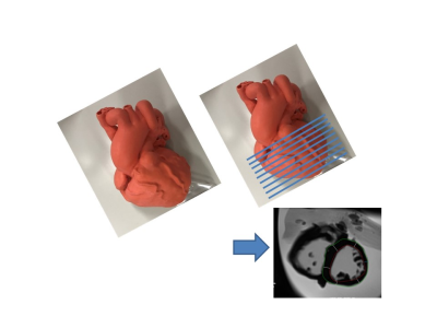 |
48 |
MR-derived both ventricular volumetric analyses with a semi-automatic threshold-based segmentation algorithm in congenital heart diseases: Head-to-head validation in human heart model.
Yoshiaki Morita, Wataru Ueki, Yoskue Hayama, Naoaki Yamada, Hideki Ota, Tetsuya Fukuda, Kei Takase
This study aimed to validate cine MRI-derived ventricular volume measurements with a semi-automatic threshold-based segmentation algorithm in a model of congenital heart disease, by comparing it with the standard method of manual tracing MRI and angiocardiography. This novel method allowed more accurate and objective ventricular volumetry in both ventricles of congenital heart diseases and will be of great help for practical use.
|
|
2173.
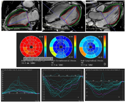 |
49 |
Role of Cardiac magnetic resonance imaging in evaluation of left ventricular function and myocardial stress difference in patients with hypertrophic cardiomyopathy
Presentation Not Submitted
Xiaochun Zhang, Li Zhu
Cardiac magnetic resonance multi-parameter imaging was used to evaluate left ventricular function and myocardial stress in patients with hypertrophic cardiomyopathy. A series of multiparametric magnetic resonance imaging was performed on 34 patients with asymmetrical hypertrophic cardiomyopathy diagnosed by MRI, using CVI software. Cardiac function was detected, and 322 hypertrophic myocardial segments and 222 non-hypertrophic segments were characterized for post-tracking. According to the American Heart Association 17-segment rule, the strain parameters and left ventricle of 16 segments except the apex were measured. The overall strain parameters preliminarily concluded that the left ventricular global stress of patients with hypertrophic cardiomyopathy was lower than that of normal people; the peak radial strain, peak long axis strain, peak circumferential strain and peak diameter of hypertrophic myocardial segments in patients with hypertrophic cardiomyopathy The strain rate and peak circumferential strain rate were significantly lower than the non-hypertrophic myocardial segments.
|
|
2174.
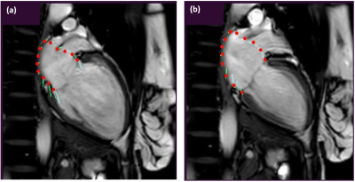 |
50 |
Association between myocardial strain and mortality in restrictive cardiomyopathy
Jadranka Stojanovska, Behnaz Khazai, Mason Runge, Maryam Ghadimi, Anil Attili, Thomas Chenevert, El-Sayed Ibrahim
Restrictive cardiomyopathy (RCM) is a rare form of heart muscle disease that is characterized by restrictive ventricular filling. The overall prognosis of RCM is poor with progression to heart failure and mortality. The primary aim of this study is to evaluate the association between myocardial strain and major RCM adverse events, such as death. The secondary aim is to assess the difference of MRI variables in RCM between women and men. The results demonstrated that decreased left-atrial longitudinal strain is significantly associated with mortality in RCM, independent of other MRI and clinical measures and beyond the need for gadolinium administration.
|
|
| Top |
Cardiovascular Emerging Methods
Digital Poster
Cardiovascular
Monday, 13 May 2019
| Exhibition Hall |
17:00 - 18:00 |
| |
|
Computer # |
|
2175.
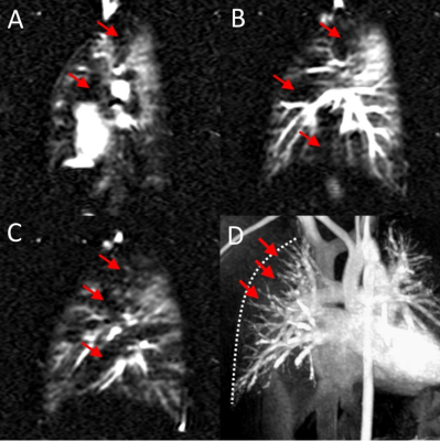 |
51 |
Non-contrast, free-breathing pulmonary perfusion imaging in pediatric patients with congenital heart disease using multi-slice FAIR at 1.5T
Joshua Greer, Vasu Gooty, Animesh Tandon, Gerald Greil, Tarique Hussain, Ananth Madhuranthakam
Congenital heart disease (CHD) is associated with anatomical abnormalities in the pulmonary arteries (PAs) which can result in asymmetric flow to the lungs, as well as an overall reduction in lung perfusion. Non-contrast pulmonary perfusion imaging using arterial spin labeling has the potential to provide both quantitative perfusion maps as well as a measure of asymmetric pulmonary blood flow. In this study, multi-slice, free-breathing pulmonary perfusion imaging is demonstrated using the FAIR technique for full coverage of the lungs, and flow measurements are compared with the standard phase contrast measurement approach.
|
|
2176.
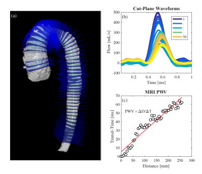 |
52 |
Sex Differences in Local and Regional Aortic Stiffness Assessed by 4D Flow MRI
Timothy Ruesink, Gregory Barton, Alejandro Roldán-Alzate
Differences in occurrence of cardiovascular disease between men and women is thought to be attributable to sex differences in arterial stiffness. However, to understand, diagnose and treat CVD, analysis of how and where arterial stiffness affects men and women differently is required. 4D Flow MRI was used to estimate regional and local aortic stiffness in 19 volunteers (age 20-32, 10 men, 9 women). Women had lower regional stiffness, suggesting they experience a reduced afterload on the heart, lowering disease risk. This study provides non-invasive comprehensive stiffness data, previously inaccessible, that may shed light on sex-specific cardiovascular differences.
|
|
2177.
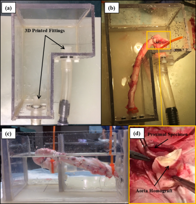 |
53 |
Ex Vivo Cardiovascular Model Design and Construction: Preliminary Results
Timothy Ruesink, Ryan Pewowaruk, Joshua Hermsen, Petros Anagnostopoulos, Alejandro Roldán-Alzate
Cardiovascular fluid dynamics (CFD) has clinical application and is often studied in vitro, in vivo or using computational simulation. However, inherent assumptions and limitations of these methods limit their clinical translation. Ex vivo modeling, coupled with 4D Flow MRI, can provide physiological time-resolved velocity data, while overcoming limitations of other experimental methods. An ex vivo MRI model of a porcine aorta was designed and assessed for its use in cardiovascular modeling and pulse wave velocity (PWV) measurement validation. Physiologically reasonable flow, produced at the specimen inlet, and preliminary PWV data show potential for future CFD and validation studies.
|
|
2178.
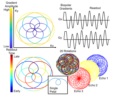 |
54 |
Multi-Echo Flow-encoded Rosette (MELROSE) enables velocity and T2* assessment of both extravascular tissue and intravascular blood for motion robust, quantitative cardiovascular blood flow and oxygenation mapping
Adam Bush, Christopher Sandino, Shreya Ramachandran, Marcus Alley, Shreyas Vasanawala
Cardiac catheterization is an invasive albeit common procedure performed in children with congenital heart disease for intrathoracic oxygen saturation assessment, exposing patients to anesthesia and risk of infection and complication. Prior MRI based intrathoracic oximetry methods have been limited due to partial volume contamination of the blood pool with surrounding tissue and motion corruption. Recently, subtractive MRI oximetry methods have demonstrated reliability and robustness but are limited to Cartesian strategies in the brain[1, 2]. In this work we use a subtractive, velocity encoded, non-Cartesian rosette trajectory for quantitative, motion robust, extra and intravascular flow and T2* mapping entitled Multi Echo fLowencoded ROSEtte (MELROSE). We validate flow and T2* values in a flow phantom and present preliminary results in a healthy subject.
|
|
2179.
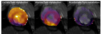 |
55 |
Parametric measurements of acute myocardial infarction in a large preclinical model
Jakob Lindhardt, Esben Hansen, Rasmus Tougaard, Nikolaj Bøgh, Rolf Schulte, Christoffer Laustsen
Hyperpolarized [1-13C]pyruvate was used to evaluate metabolic and perfusion changes in myocardial infarction in the critical acute reperfusion phase. Regional changes was observed by mapping of metabolic ratios of lactate, alanine and bicarbonate. Metabolic exchange rates from hyperpolarized fumarate was calculated using Michael-Menten kinetics. Hyperpolarized measures were compared with standard perfusion MRI and tissue staining evaluating infarction and salvage. The results of this study, draws use of hyperpolarized [1-13C]pyruvate closer to evaluation of myocardial infarction in a human study.
|
|
2180
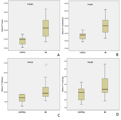 |
56 |
Diagnostic value of native T1/T2 map in patients with cardiac involvement in idiopathic inflammatory myopathy
Video Permission Withheld
Lu Huang, Peijun Zhao, Rob J.van.der Geest, Liming Xia, Qiao Tao
Idiopathic inflammatory myopathy (IIM) is a chronic autoimmune systemic disease that involves myocardium. The aim of this study is to evaluate the diagnostic value of histogram analysis of native T1 and T2 maps to identify patients with cardiac involvement in IIM. Fifteen patients with cardiac involvement in IIM and 11 normal controls were enrolled in this study. Native T1 and T2 maps were assessed by histogram analysis. Compared to the controls, the native T1 mean, median, SD and entropy were elevated significantly in IIM patients, while T2 histogram parameters showed no significant difference. A bi-parametric model including native T1 median and SD demonstrated the highest accuracy (AUC=0.93) for detecting cardiac involvement in IIM.
|
|
2181.
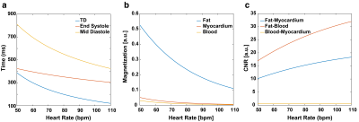 |
57 |
Dual magnetization prepared epicardial fat imaging
Haiyan Ding, Rui Guo, Dongyue Si, Jianwen Luo
Epicardial fat has been more recognized as an endocrine organ and potentially associated with cardiovascular disease and metabolic disorders. High spatial imaging resolution is crucial to identify and quantify epicardial fat. This work proposed an efficient dual magnetization prepared imaging method. Combined with saturation and inversion preparation pulses, desired contrast between fat and surrounding tissues with high spatial resolution in a single breath hold was generated. Both simulation and in vivo experiment were conducted to validate the sequence. In vivo imaging resolution 1.0 by 1.0 by 5 mm3 was achieved, which enabled clearly delineation of epicardial fat successfully.
|
|
2182.
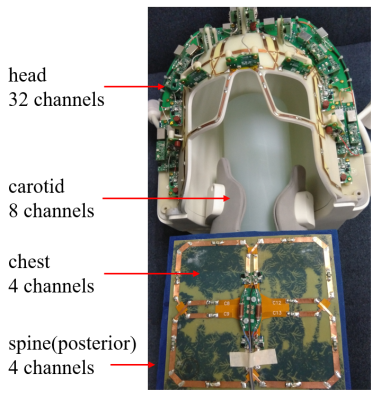 |
58 |
One-stop scanning of cerebrovascular, carotid and cardiovascular MRI with a 48-channel coil system at 3 T
Qiaoyan Chen, Zidong Wei, Lei Zhang, Changjun Tie, Qiang He, Xiaoliang Zhang, Xin Liu, Hairong Zheng, Ye Li
One-stop magnetic resonance (MR) vessel wall imaging that includes cardiovascular magnetic resonance (CMR), intracranial and cervical carotid arteries imaging is of considerable interest for clinical diagnosis of vascular diseases, such as ischemic stroke. Due to the small cross-sectional size of the vessel wall and susceptibility effects, one-stop MR vessel wall imaging remains challenging. In this work, intracranial and cervical carotid arteries, and CMR imaging was performed simultaneously by using a dedicated 48-channel coil system. Moreover, intracranial and carotid arterial wall images with an isotropic spatial resolution of 0.63 mm can be acquired by using the head and carotid coil system within 7.7 minutes.
|
|
2183.
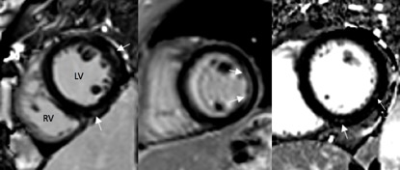 |
59 |
Early Assessment of Subclinical Cardiac Involvement in Immune-Mediated Myopathy: a 3T Cardiac Magnetic Resonance Study
Presentation Not Submitted
peijun zhao, Lu Huang, Lingping Ran, Dazhong Tang, Xiaoyue Zhou, Liming Xia
Cardiac involvement in immune-mediated myopathy often leads to a poor prognosis. We studied 32 patients with immune-mediated myopathies to investigate the role of cardiac magnetic resonance in the early detection of cardiac involvement. A comprehensive CMR examination using a 3T MR scanner, including the sequences for cardiac cine, T1 mapping (before and after contrast), T2 mapping, and late gadolinium enhancement (LGE), was performed on all the subjects. Focal myocardial fibrosis and diffusely elevated myocardial T1, T2, and ECV values were observed. The sequence of LGE combined with mapping technologies could improve sensitivity for the diagnosis of subclinical cardiac involvement.
|
|
2184.
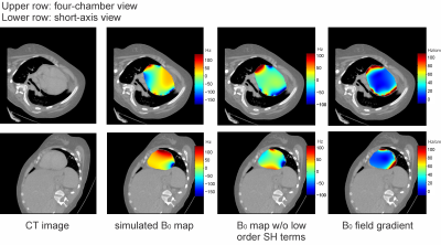 |
60 |
High Resolution Simulation of B0 Field Conditions in the Human Heart Based on Segmented CT Images
Yun Shang, Sebastian Theilenberg, Wolfram Mattar, Maxim Terekhov, Sachin Jambawalikar, Laura Schreiber, Christoph Juchem
B0 inhomogeneity leads to dark band artifacts in cardiac MRI, in particular with the use of steady-state free precession (SSFP) pulse sequences. Limited spatial resolution of MRI-derived in vivo B0 maps and, moreover, the lack of population data prevents the systematic analysis of the problem at hand and the development of optimized B0 shim strategies. We used readily available CT images to derive the B0 conditions in the human heart at very high resolution. Calculated cardiac B0 fields showed consistency with MRI-based B0 measurements and local field artifacts concur with typical dark band locations. The approach is expected to enable the development of population-specific B0shim strategies for the human heart.
|
|
2185
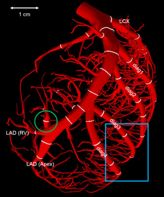 |
61 |
Regional Heterogeneity of Errors in Myocardial Perfusion Quantification Using Bolus-Based MRI
Video Permission Withheld
Johannes Martens, Sabine Panzer, Jeroen van den Wijngaard, Maria Siebes, Laura Schreiber
Aim of the project is the computational modeling of contrast agent dispersion in coronary arteries down to pre-arteriolar level during contrast-enhanced MRI myocardial perfusion measurements. From a high resolution imaging cryomicrotome dataset a vascular 3D model of the left main coronary artery is extracted and furnished with a computational grid. Using an advanced boundary condition, Navier-Stokes equations for blood flow and the advection-diffusion equation for CA transport are solved to obtain CA bolus dispersion values on this model of unprecedented detail, and to analyze myocardial blood flow quantification errors. The analysis of the obtained results shows strong variability on the cm-scale.
|
|
2186.
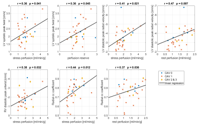 |
62 |
Altered myocardial velocities and stress perfusion in heart transplant patients affected by coronary allograft vasculopathy
Roberto Sarnari, Muhannad Abbasi , Arif Jivan , Rahim Gulamali, Alexander Ruh, Julie Blaisdell, Brandon Benefield, Ryan Dolan, Kambiz Ghafourian, Jane Wilcox, Sadiya Khan, Esther Vorovich, Jonathan Rich, Allen Anderson, Clyde Yancy, James Carr, Daniel Lee, Michael Markl
Coronary allograft vasculopathy (CAV) is responsible for long term mortality after heart transplant (HTx). Myocardial perfusion impairment resulting from CAV can lead to early graft dysfunction. Our study aimed to quantify perfusion and 3-directional myocardial velocities by cardiac magnetic resonance perfusion and tissue phase mapping (TPM) sequences and describe the relationship at global and segmental level. Thirty two HTx patients affected by CAV were analyzed. Myocardial perfusion reserve was reduced in CAV patients and associated with reduced left ventricular twist during contraction. LV diastolic radial velocities and interventricular synchrony were associated as to rest and to stress myocardial perfusion variations
|
|
2187.
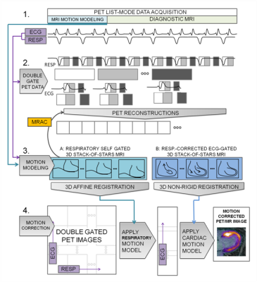 |
63 |
Initial Experience with MR-Based Respiratory and Cardiac Motion-Corrected PET/MR Imaging of the Coronary Arteries
Philip Robson, MariaGiovanna Trivieri, Nicolas Karakatsanis, Georgios Soultanidis , Ronan Abgral, Marc Dweck, Pedro Moreno, Gianluca Torregrossa, John Puskas, Zahi Fayad
A major advantage of hybrid PET/MR systems is the radiation-free high spatial and temporal resolution of MR imaging that can be used to estimate cardio-respiratory motion present during PET data acquisition. This information can be incorporated into reconstruction algorithms to correct for motion in the PET data to reduce blurring and increase target-to-background ratios (TBR) of PET hotspots. This may be of particular importance in cardiac imaging where the heart is in constant motion. In this work, we report our initial experience applying cardio-respiratory motion-corrected PET/MR to 18F-sodium fluoride PET/MR imaging of the coronary arteries.
|
|
2188.
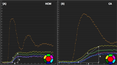 |
64 |
Comparison of regional microvascular dysfunction of hypertrophied myocardium and its relation with myocardial strains in cardiac amyloidosis and hypertrophic cardiomyopathy
Presentation Not Submitted
Shan Huang, Zhi-gang Yang, Kai-yue Diao, Ke Shi, Yue Gao, Yi Zhang, Meng-ting Shen, Yong He, Sen He, Ying-kun Guo
Cardiac amyloidosis and hypertrophic cardiomyopathy can be both characterized with increased wall thickness. We intended to explore the microvascular function of the two different phenotypes of hypertrophy and its relation with regional myocardial function. Amyloid infiltration caused more severe myocardial perfusion disorder and systolic dysfunction. Significant correlations were observed between microcirculation and myocardial strain.
|
|
2189.
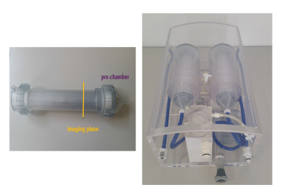 |
65 |
Evaluation of the performance of voxel-wise myocardial perfusion quantification methods using a traceable phantom
Loïc Coquelin, Géraldine Ebrard, Judith Lehnert, Nadia Smith, Xenios Milidonis, Tobias Schaeffter, Amedeo Chiribiri
Assessing and comparing myocardial imaging methodologies require the use of a physical standard. A novel 3D printed cardiac phantom mimicking transmural myocardial perfusion gradients is used in this study, first to evaluate the repeatability of phase contrast MRI (PC-MRI) measurements and then, to compare two common methods for perfusion quantification using dynamic contrast enhanced MRI (DCE-MRI) measurements: the dual bolus and the model-based approach.
|
|
2190.
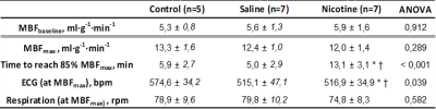 |
66 |
Effect of nicotine administration on myocardial perfusion under adenosine stress: A dynamic investigation using cine-ASL in mice.
Anne Tonson, Amira Trabelsi , Monique Bernard, Frank Kober
We have monitored myocardial perfusion (MBF) dynamically under pharmacologically-induced stress in mice using a time-optimized cine-ASL MRI approach with about 2.5min temporal resolution. One main advantage of the repeatable cine-ASL in this context is to monitor MBF during successive stress episodes in the same animal allowing for internal control. In this study we clearly demonstrated that single bolus nicotine administration (1mg·kg-1) significantly reduced perfusion by 35% under adenosine stress (tail vein continuous infusion, 90µg·kg-1·min-1) in healthy mice.
|
|
2191.
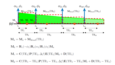 |
67 |
Improved Quantification for Steady-Pulsed ASL Perfusion Imaging
NAM LEE, Ahsan Javed, Terrence Jao, Krishna Nayak
Steady-pulsed ASL (spASL) provides high labeling efficiency and is appropriate for organs with highly pulsatile blood supply (e.g. myocardium), but quantification remains a major challenge. Here, we describe a method for quantifying tissue blood flow in spASL based on a numerical solution of the Bloch equations with flow effects. The exact acquisition timing, including irregularities generated by heart rate variability, is incorporated in this formalism. Dictionary-based quantification of blood flow is demonstrated using myocardial spASL data from healthy swine at rest.
|
|
2192.
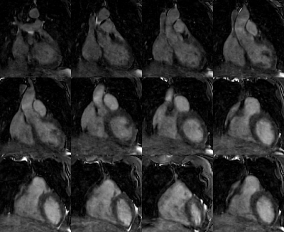 |
68 |
3D Cartesian Free-running Cardiac and Respiratory Resolved Whole-heart MRI
Thomas Kuestner, Aurelien Bustin, Gastao Cruz, Teresa Correia, Giulia Ginami, Radhouene Neji, René Botnar, Claudia Prieto
Free-breathing continuous acquisitions, so called free-running, enable 3D whole-heart coverage for motion-resolved functional cardiac MRI. In prior work approaches based on 3D radial imaging were proposed for this task. However, free-running 3D radial image reconstruction is computationally demanding. In this work, we propose a novel 3D Cartesian free-running bSSFP sequence. Data is acquired continuously with a variable-density Cartesian trajectory with spiral profile order and retrospectively binned based on respiratory self-navigation and cardiac ECG signal synchronization. A multi-bin-PROST reconstruction is proposed to exploit spatial, cardiac and respiratory temporal redundancies to reconstruct high-resolution images.
|
|
2193.
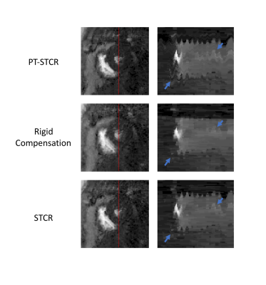 |
69 |
3D motion compensated reconstruction using pixel tracking for cardiac perfusion MRI: comparison with rigid shift correction
Ye Tian, Jason Mendes, Ganesh Adluru, Edward DiBella
We developed a pixel tracking motion compensation reconstruction framework for 3D acquisitions. The method was compared to a rigid motion compensation reconstruction method and to reconstruction without motion compensation using a cardiac perfusion dataset acquired with a 3D stack-of-stars sequence. The pixel tracking motion compensation method improved reconstruction, while the rigid motion compensation led to blurring.
|
|
2194.
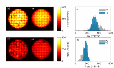 |
70 |
Dynamic T1 Mapping for Quantitative Myocardial Perfusion Imaging
Christoph Kolbitsch, Kirsten Becker, Judith Lehnert, Jean Pierre Bassenge, Xenios Milidonis, Myles Capstick, Amedeo Chiribiri, Tobias Schaeffter
Myocardial perfusion imaging is a valuable diagnostic tool to assess cardiac viability. Quantitative perfusion imaging promises to provide absolute values of myocardial perfusion allowing for objective disease assessment and monitoring. Here we present a phantom study evaluating accuracy and reproducibility of a high-resolution dynamic T1 mapping approach to directly measure changes in contrast agent concentration and yield quantitative myocardial perfusion without the need of signal-intensity calibration. Our proposed technique provides one high-resolution T1 map for each cardiac cycle and leads to accurate myocardial perfusion values verified with standard 2D phase-contrast MRI (difference of estimated flow 10 – 20%).
|
|
2195.
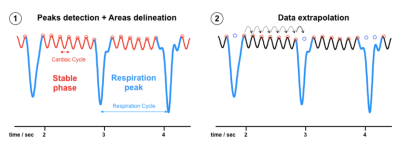 |
71 |
Self-gated 5D cardiac MRI in mice : a detailed study
Colleen Cardiet, Aurélien Trotier, Emeline Ribot, Baudouin Denis de Senneville, Sylvain Miraux
Until now, mouse respiratory movement has never been deeply studied and the data acquired during breathing period are not used to obtain 4D cardiac images in mice. First, this study aims at evaluating the respiration movement and its impact on the heart region. First, motion field maps of the thorax during breathing demonstrated motion of 1mm in the anteroposterior direction. Second, 4D cardiac images were reconstructed using the data acquired during the breathing periods and a technic based on sliding window. This improvement allows a considerable acquisition time shortening (about 30%) for 3D cine whole heart imaging on small animals.
|
|
2196.
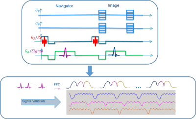 |
72 |
PolyGate: Benefits of a spatially resolved navigator for self-gating cardiac imaging in multiple rodents.
Willy Gsell, Cesar Molinos, Carlos Correcher, Michael Heidenreich, Joren Vandengaer, Wouter Oosterlinck, Uwe Himmelreich, Christophe Deroose, Arno Nauerth
We implemented a spatially resolved navigator based on the self-gated IntraGate-FLASH method. The new navigator offers many advantages such as the possibility to scan several animals at the same time, improve the quality of the motion correction and to distinguish between the different types of motion from a single acquisition. Moreover, we have been able to synchronize MRI with PET for simultaneous acquisition and to retrospectively reconstruct gated PET-images.
|
|
2197.
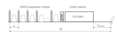 |
73 |
DANTE-Prepared FLASH: Three-Dimensional Vessel Wall Imaging Of ApoE-/- Mouse at 7 Tesla
Yuanbo Yang, Zhonghao Li, Yingjie Mei, Ming Zhao, Guoxi Xie, Yanqiu Feng
There is a growing preclinical demand for methods to characterize atherosclerotic plaque in vivo. Recently proposed DANTE for dark-blood preparation successfully in vessel wall imaging. However, previous studies were performed at 3T and used for vessel wall imaging of human. It remains unclear that if DANTE module could be used for apoE-/- mouse at 7T. Here we demonstrated a sequence using DANTE dark-blood preparation combined with FLASH readout (DANTE-FLASH) for three-dimensional (3D) vessel wall imaging at 7 Tesla (T).
|
|
2198.
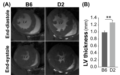 |
74 |
Cardiac MRI for quantifying myocardial perfusion deficits in a mouse model of hypertrophic cardiomyopathy
Min-Chi Ku, Frank Kober, Andreas Pohlmann, Qadri Fatimunnisa, Michael Bader, Thoralf Niendorf
Measuring the myocardial morphological and functional changes is not sufficient to assess the underlying subclinical myocardial microstructural changes in hypertrophic cardiomyopathy (HCM). Despite cardiac MRI (CMR) is advanced in characterizing the changes in myocardial microstructure, in vivo assessment of the kinetics of microstructural changes including microvasculature deficits and the mechanism underlying disease progression is still missing. We hypothesize that the impairment of myocardial perfusion may contribute to the microstructural changes in HCM. To test this, we used state-of-the-art arterial spin labeling (ASL) based CMR method to quantify the myocardial perfusion changes in a HCM mouse model.
|
|
2199.
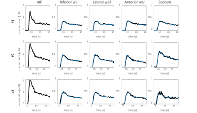 |
75 |
Extended Quantitative Cardiac First-Pass Perfusion Imaging in Mice Using Accelerated Data Acquisition and Spatially Distributed, Two-Compartment Exchange Modelling
Grzegorz Kwiatkowski, Sebastian Kozerke
The objective of the present work was to improve data acquisition and quantification of first pass perfusion imaging in the in-vivo murine heart. Undersampled data were acquired and reconstructed using k-t SPARSE. A two-compartment exchange model was employed to provide an additional characterization of myocardial tissue based on compartment volumes and the permeability surface area product. The feasibility of the proposed method was tested using perfusions analysis of first-pass perfusion data acquired with extracellular and intravascular contrast agents in mice. The results presented here show that two-compartment exchange modelling in the in-vivo murine heart is feasible and gives access to tissue parameters beyond myocardial blood flow.
|
|
| Top |
Cardiovascular Miscellaneous
Digital Poster
Cardiovascular
Monday, 13 May 2019
| Exhibition Hall |
17:00 - 18:00 |
| |
|
Computer # |
|
2200.
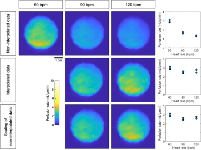 |
76 |
Impact of temporal resolution on the quantification of myocardial perfusion in dynamic-contrast enhanced MRI
Xenios Milidonis, Muhummad Nazir, Russell Franks, Amedeo Chiribiri
Quantification of myocardial perfusion using dynamic contrast enhanced imaging provides an objective and sensitive assessment of coronary artery disease in the clinic. While many factors were found to affect the accuracy of perfusion estimates, there has been little evidence on the impact of temporal resolution. This study sheds light on this using simulated, phantom and clinical data. Simulations and phantom experiments demonstrate large deviations between measurements when the same data are sampled with different temporal resolutions, while interpolation of clinical data to uniform temporal resolution leads to significantly different perfusion rates.
|
|
2201.
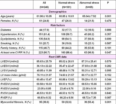 |
77 |
PROGNOSTIC VALUE OF DIPYRIDAMOLE STRESS CMR IN PATIENTS WITH KNOWN OR SUSPECTED CORONARY ARTERY DISEASE: A LONG TERM FOLLOW-UP STUDY
Antonella Meloni, Cinzia Nugara, Maria Vaccaro, Chrysanthos Grigoratos, Giancarlo Todiere, Andrea Barison, Daniele De Marchi, Giuseppina Novo, Giovanni Aquaro, Alessia Pepe
We determined the long-term prognostic value of dipyridamole stress-CMR in patients with known or suspected coronary artery disease (CAD). Reversible perfusion deficit and diabetes identified patients at high risk of fatal and non fatal cardiac events. Myocardial fibrosis was a strong predictor for death.
|
|
2202.
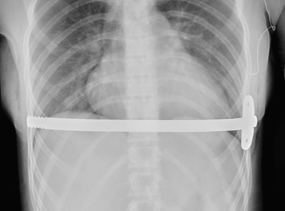 |
78 |
Correction of B0 Field Distortion Induced by Stainless Steel Nuss Bar in Cardiac MRI Using Permanent Magnets
Zhiyue Wang, Tarique Hussain, Trung Nguyen, Lorrie Burkhalter, Adam Alder
Pectus excavatum is often corrected by the Nuss procedure, in which a metal bar (usually made of stainless steel) is inserted horizontally into the chest cavity. MRI examination is important to assess the cardiac function with a focus on the right heart. The metal bar causes susceptibility artifacts in cine MRI. We explore the feasibility to improve the B0 field homogeneity using a field correction device to be placed on the surface of the chest utilizing permanent magnets. A device was constructed based on computer simulations. The device generates magnetic multipole fields and effectively improved B0 homogeneity in a phantom.
|
|
2203.
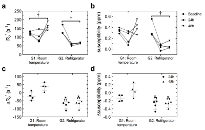 |
79 |
Comparison of room temperature, refrigerator and cryogenic storage for preservation of blood clot R2* and quantitative susceptibility values in vitro at 3T
Spencer Christiansen, Junmin Liu, Joy Dunmore-Buyze, Mike Boffa, Maria Drangova
Mechanical thrombectomy therapy allows valuable stroke thrombus MR imaging studies to be completed without patient burden, but the effects of delay between retrieval and imaging on MR characteristics need to be considered. We tested the ability of room temperature, refrigerator and cryogenic storage to preserve blood clot R2* and quantitative susceptibility (QS) values up to 48 hours in vitro. Room temperature storage provided adequate preservation up to 24 hours while refrigerator storage significantly decreased R2* and QS; cryogenic storage led to non-significant decreases in R2* and QS and shows promise for complete, long-term preservation if cell lysis can be mitigated.
|
|
2204.
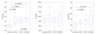 |
80 |
Structural Changes in Cardiac Allograft Vasculopathy Patients after One Year Post Heart Transplantation
Muhannad Abbasi, Roberto Sarnari , Arif Jivan , Alexander Ruh, Julie Blaisdell, Brandon Benefield , Ryan Dolan, Kai Lin, Daniel Lee, Kambiz Ghafourian, Jane Wilcox, Sadiya Khan , Esther Vorovich, Jonathan Rich, Allen Anderson, Clyde Yancy , Michael Markl, James Carr
Cardiac MRI demonstrates differences between heart transplant recipients more than one year post-HTx and controls using T2, T1 and ECV. Our study demonstrated elevated global T2 and ECV, and segmental T1 in HTx patients when compared to controls. This may provide further insight into the complex pathophysiology of this disease process, which includes a chronic and subacute component. CMR could potentially be implemented as an adjunct to the current methods of detecting CAV (intravascular ultrasound and invasive coronary cineangiography)
|
|
2205
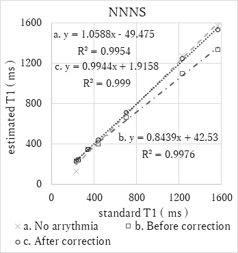 |
81 |
Correction of the effects of heart rate changes on MOLLI
Video Permission Withheld
Rei Ikegawa, Akira Ichikawa, Nobuyoshi Oomi, Kuninori Kobayashi, Mika Mori, Shigehide Kuhara
In fast cardiac T1 mapping implementing ECG-gating, the recovery time of the longitudinal magnetization will change with heart rate variation, which will affect T1 measurement accuracy. Several studies have investigated the influence of heart rate variability on modified look-locker inversion recovery; however, no analytical approach for correcting the T1 value after scanning has been reported to date. Here, we investigated the effect of heart rate change on measurement of T1 values and report a correction method. This proposed method can reduce the effect of arrhythmia in fast cardiac T1 mapping.
|
|
2206.
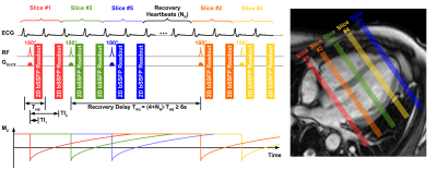 |
82 |
Improved fast multi-slice myocardial T1 mapping (FAST1) for full left ventricular coverage in three breathholds
Li Huang, Radhouene Neji, Muhammad Nazir, John Whitaker, Filippo Bosio, Amedeo Chiribiri, Reza Razavi, Sébastien Roujol
Modified Look-Locker inversion recovery (MOLLI) is a commonly used approach for myocardial T1 mapping, and yields high precision as well as reproducibility. This technique requires one breathhold per slice, resulting in prolonged examination time when full left ventricular coverage is required. Our previously developed fast multi-slice myocardial T1 mapping (FAST1) technique showed potential for time-efficient full left ventricular coverage. In this work, the FAST1 sequence is further improved using Bloch-equation-based fitting for higher T1 accuracy and heart-rate independence. Compared to MOLLI, the proposed new FAST1 can yield higher T1 accuracy, enhanced tolerance to T2 and heart-rate variations, as well as similar repeatability and T1 map quality at 1.5T.
|
|
2207.
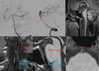 |
83 |
Diagnostic value of 3D T1-SPACE technique for basilar artery stenosis: Comparison with DSA
zhenjia wang, wei yu, zhaoyang fan
This is the first attempt to evaluate the accuracy and application value of Three-Dimensional Variable-Flip-Angle Turbo Spin-Echo (3D T1-SPACE) for the diagnosis of basilar atherosclerotic disease.
|
|
2208
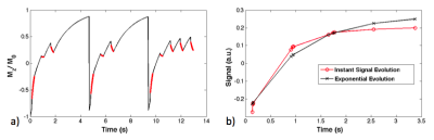 |
84 |
Simultaneous T1 and T2 Mapping with MOLLI Acquisition: Proof of Concept
Video Permission Withheld
Jing Liu, Peng Lai, Yan Wang, David Saloner, Karen Ordovas
MOLLI is known to underestimate T1 values. Instead of fitting MOLLI data to an ideal but unrealistic exponential inversion recovery model, we simulated signal evolution incorporating actual scan parameters based on which we improved T1 mapping as well as demonstrated the feasibility of deriving T2 mapping simultaneously.
|
|
2209.
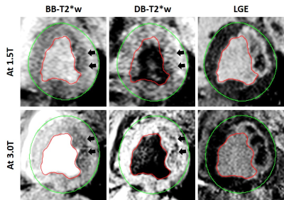 |
85 |
Detecting Hemorrhagic Myocardial Infarction with Dark Blood T2*: Experimental Findings
Xingmin Guan, Hsin-Jung Yang, Jane Sykes, John Butler, Guan Wang, Frank Prato, Rohan Dharmakumar
Dark-blood prepared T2* MRI has become the preferred method for examining global myocardial iron overloading. However, whether dark-blood prepared T2* MRI is superior to bright-blood T2* MRI for the detection of hemorrhagic myocardial infarctions is not known. Here we show that, compared to bright-blood T2* MRI, dark-blood prepared T2* MRI yields significantly lower image contrast for detecting hemorrhagic myocardial infarction at 1.5T and 3.0T. We conclude that unless this loss in image contrast is mitigated, dark-blood T2* MRI may not provide adequate sensitivity for detecting hemorrhagic myocardial infarctions.
|
|
2210.
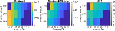 |
86 |
Optimization of Steady-Pulsed Arterial Spin Labeling for Myocardial Perfusion Imaging
Ahsan Javed, Nam Lee, Hung Do, Nilesh Ghugre, Graham Wright, Eric Wong, Krishna Nayak
Arterial spin labeling (ASL) is an attractive non-contrast option for myocardial perfusion imaging. Steady pulsed labeling has been shown to provide the highest signal efficiency in-vivo. In this work, we determine the flip-angle and imaging interval that provide maximum signal efficiency and temporal SNR using Bloch simulation and experiments in healthy swine. We demonstrate that an imaging FA of 40-50ºand imaging interval of 1-RR is optimal.
|
|
2211.
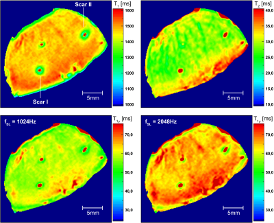 |
87 |
Improved infarct scar imaging and scar classification using native T1?-mapping
Maximilian Gram, Daniel Gensler, Michael Seethaler, Anton Xu, Peter Michael Jakob, Peter Nordbeck
Myocardial mapping techniques are known to provide new diagnostic possibilities for morphological and functional information including accurate tissue classification for several diseases. However, without the use of contrast agents, the differentiation between healthy and diseased tissue is hardly possible. In the present study, a scar/infarct model has been employed to investigate the contrast ratio performance of different native MRI mapping techniques (T1, T2, and T1ρ) under controlled conditions. Here, T1ρ provides the best results with an up to 12-times increased contrast ratio. Hence, T1ρ-mapping might be a very promising technique for scar imaging without the use of contrast agents.
|
|
2212.
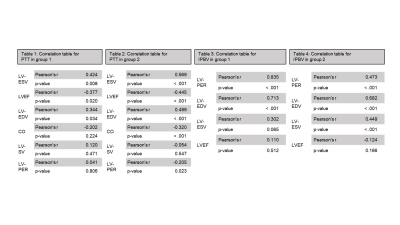 |
88 |
Retrospective analysis of pulmonary transit time in different patient samples and recording conditions.
RAYMOND GILLES, Marie-Anne Labaisse, Bernard Pilet, Karim Wahbi, Benjamin Marty, Pierre Carlier
A retrospective analysis of pulmonary transit time and intra-pulmonary blood volume in Becker dystrophic myopaths at 3T and unselected patients / cardiopaths and normals at 1,5 T.
|
|
2213.
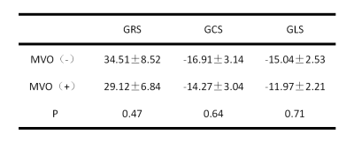 |
89 |
Changes of left ventricular myocardial strain in acute STEMI patients with microvascular obstruction: A CMR feature tracking study
Presentation Not Submitted
Xiaonan Wang, Yang Hou
Changes of left ventricular myocardial strain in acute ST-elevation myocardial infarction patients before left ventricular remodeling is obvious, while the relation to microvascular obstruction still need to explore.
|
|
2214.
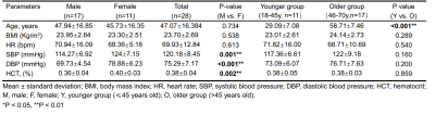 |
90 |
The Effects of Age and Gender on Cardiac Structure and Function in Healthy Chinese Adult Population: A Multiparameter Cardiac Magnetic Resonance Imaging Study
Presentation Not Submitted
Bang Jun Guo, Long Jiang Zhang, Wei Qiang Dou, Yong Shen
With gender difference and age growing, the cardiac structure and function maychange in healthy population. The present study aimed to for the first time systematically investigate the impact of age and gender on cardiac structure and function by using cine MR, T1 mapping and intravoxel incoherent motion diffusion weighted imaging (IVIM DWI). In the present study, we have found cardiac structure and function significant difference between male and female healthy volunteers.Further studies are ongoing to examine a large cohort.
|
|
2215.
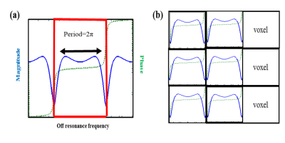 |
91 |
Off-resonance error suppression in MOLLI T1 mapping using a specific CE-FAST
Mostafa Berangi, Vahid Ghodrati, Fereshteh Hasanzadeh, Puria Rafsanjani, Zahra Alizade Sani, Abbas Nasiraei Moghaddam
MOLLI is a well-known method for cardiac T1 mapping. Data sampling in MOLLI is based on SSFP readout. It is therefore susceptible to off-resonance which is considered an error source for T1 mapping. We have presented a method to suppress off-resonance variation error in T1 estimation by means of a specific CE-FAST in combination with MOLLI. We have evaluated our method in simulation and Phantom experiment with promising results.
|
|
2216.
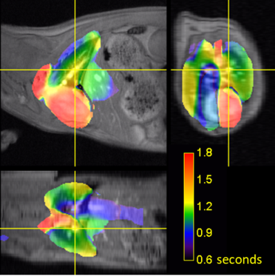 |
92 |
Pulmonary circulation time measurements and lung blood volume estimation in mice by magnetic particle imaging and magnetic resonance imaging
Michael Kaul, Johannes Salamon, Caroline Jung, Matthias Graeser, Harald Ittrich, Gerhard Adam, Kersten Peldschus
Pulmonary circulation time and/or the blood volume of the lungs can be used to depict pathologic changes. The purpose of our study was to determine the circulation time of a bolus of superparamagnetic iron oxide nanoparticles through the lungs by MPI and to estimate the blood volume of the lungs supplemented by cardiac MRI. The pulmonary circulation times were 907 ± 141 ms. The determination of the blood volume revealed a volume of 233 ± 48 µL.
|
|
2217.
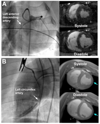 |
93 |
A swine model of selective coronary stenosis using transcatheter delivery of a 3D printed implant for MR imaging: Feasibility and Efficacy
Caroline Colbert, Jiaxin Shao, Jesse Currier, Olujimi Ajijola, Rohan Dharmakumar, Sandra Duarte-Vogel, Peng Hu, Kim-Lien Nguyen
This study aims to investigate the feasibility and efficacy of a closed-chest method for coronary stenosis creation in swine models of ischemic heart disease (IHD). We successfully deployed high-resolution 3D printed intracoronary implants in three healthy swine subjects using a transcatheter angioplasty delivery approach. All subjects underwent angiography and MR cine imaging for assessment of ventricular regional wall motion. At post-mortem dissection, we successfully retrieved the implants. These results support early feasibility and efficacy of our proposed approach for applications including stress testing, infarct creation, and evaluation of diagnostic techniques or therapeutics in swine models of IHD.
|
|
2218.
 |
94 |
Cardioprotective Effects of Mild Therapeutic Hypothermia in Hemorrhagic Myocardial Infarctions: Early Findings in a Swine Model of Acute Reperfused Myocardial Infarction
Eric Johnson, Guan Wang, Richard Tang, Behzad Sharif, Ivan Cokic, Andreas Kumar, Rohan Dharmakumar
Patients who experience hemorrhagic myocardial infarctions (hMIs) have increased risk of adverse outcomes, yet a protective therapy remains to be developed. We hypothesized that mild hypothermia to the heart can reduce the effects of hemorrhage and improve left ventricular functional recovery in the setting of reperfused infarctions. This was studied using a swine model of hemorrhagic infarction exposed to cooled pericardial saline infusion to confer hypothermia. Results show a significant decrease in hemorrhagic remnants and increase in LVEF for the hypothermia group relative to controls at 1 month post-MI. Therapeutic cooling has the potential to offer cardioprotective benefits for hMI.
|
|
2219.
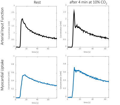 |
95 |
Quantitative High-Resolution Myocardial First-Pass Perfusion Imaging of CO2-Induced Vasodilation in Rat
Grzegorz Kwiatkowski, Sebastian Kozerke
Respiratory-induced acidosis is an emerging alternative to induce increased coronary blood flow necessary to facilitate the quantification of the stress myocardial blood flow and hence myocardial perfusion reserve. The aim of this study was to quantify the pharmacokinetic effect of the CO2 gas challenge on myocardial perfusion in rats using a high-resolution first pass perfusion CMR.
|
|
2220
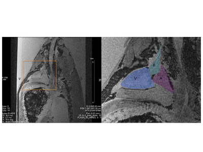 |
96 |
Successfully Overcoming the Challenge: Cardiac Cine of Zebrafish in MRI.
Video Permission Withheld
Astrid Wietelmann, Clemens Müller, Arno Nauerth, Didier Stainier, Anabela Bensimon-Brito
Zebrafish has been a widely used model to study cardiovascular development and diseases. Among the multiple advantages, zebrafish has the remarkable ability to regenerate lost or damaged organs including the heart. In vivo visualization of the cardiovascular system is particularly easy during early stages of development due to zebrafish transparency, which is lost in the adult stages. Therefore, it is of great interest to refine and adapt non-invasive imaging methods that allow a functional assessment of the zebrafish adult heart. The application of high-field functional MRI of the beating zebrafish heart will be presented and discussed.
|
|
2221.
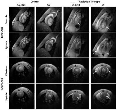 |
97 |
Cardiotoxic effects of radiation therapy detected by MRI in genetically modified rats
El-Sayed Ibrahim, Dhiraj Baruah, Matthew Budde, Anne Frei, Rachel Schlaak, Michael Flister, Carmen Bergom
In this study, we present preliminary results detecting radiation therapy (RT) effects on cardiac function in pre-clinical models using high-field 9.4T MRI. Salt-sensitive (SS) and Brown-Norway (BN) rats were selectively bred to produce SS.BN3 consomic rats, which are genetically identical to SS rats except for chromosome 3 which is inherited from the BN rat. Adult female SS and SS.BN3 rats (both control and RT animals) were scanned to acquire cine and tagging images, from which different cardiovascular parameters were measured. The results show differences in heart mechanics and ventricular remodeling between the two rat strains in response to RT, despite maintained global cardiac function.
|
|
2222.
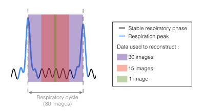 |
98 |
USPIO-enhanced highly-resolved pulmonary MR angiography in mice with a 3D UTE sequence.
Colleen Cardiet, Aurélien Trotier, Emeline Ribot, Sylvain Miraux
Pulmonary imaging has always been challenging due to many constraints : parenchyma heterogeneity, respiratory and cardiac motion. Until now, pulmonary arteries imaging is commonly done with micro CT scanner, but this imaging method is not convenient for longitudinal evaluations. In this study, we present a magnetic resonance angiography (MRA) technique of the lungs with high spatial resolution combining an UTE 3D SG sequence and the injection of iron nanoparticles. To prevent motion artifact, images acquired during respiration were neglected.
|
|
2223.
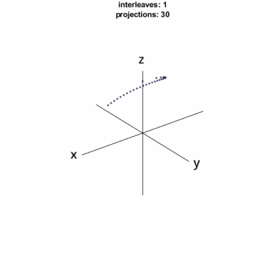 |
99 |
Free-breathing Whole-heart Magnetic Resonance Angiography with Self-gated Radial Phyllotaxis Trajectory
Wai-Yan Ryana Fok, Andrew J. Powell, Mehdi H. Moghari
Whole-heart magnetic resonance angiography is commonly performed with diaphragmatic navigator gating to compensate for respiratory motion, but this approach is inefficient as data must be reacquired when it is outside the acceptance window. We therefore developed and validated a self-gated respiratory motion compensation technique based on a radial phyllotaxis k-space trajectory in which all data are accepted throughout the respiratory cycle.
|
|
2224.
 |
100 |
Left Ventricle Segmentation with Densely Connected Full Convolutional Network
Zhanli Hu, Yin Wu, Siyue Li, Wenjian Qin, Dong Liang, Xin Liu, Yongfeng Yang, Hairong Zheng
Cardiac functional analysis is important in heart disease diagnosis. Conventional manual segmentation of left ventricle is time consuming and observer dependent. Our proposed Densely Connected Full Convolutional Network (DenseV-Net) enables automatically process medical images. Its densely connected convolutional block consists of residual calculation with Elu used as active function. The results show that the proposed DenseV-Net can efficiently segment left ventricle from cardiac cines with mean DSC of 0.90±0.12, more accurate compared to V-Net (0.85±0.13, P<0.05). The method offers a feasible way for efficient analysis of cardiac function.
|
|
Back to Program-at-a-Glance |  Back to Top Back to Top |
| The International Society for Magnetic Resonance in Medicine is accredited by the Accreditation Council for Continuing Medical Education to provide continuing medical education for physicians. |
