Digital Poster Session
General Cancer Imaging Back to Program-at-a-Glance Back to Program-at-a-Glance
|
Tuesday, 14 May 2019
Digital PosterGeneral Cancer Imaging
2299 -2323 Cancer Metabolism, pH & Oxygenation
2324 -2348 Cancer Therapy Response Assessment
2349 -2373 Cancer Perfusion, Diffusion & Relaxometry
2374 -2398 Cancer Detection & Monitoring |
| |
Cancer Metabolism, pH & Oxygenation
Digital Poster
General Cancer Imaging
Tuesday, 14 May 2019
| Exhibition Hall |
08:15 - 09:15 |
| |
|
Computer # |
|
2299.
 |
1 |
Magnetic Resonance Imaging of Oxygen Carriers in Treatment of Hypoxic Tumours

Emma Bluemke, Joshua Owen, Elinor Thompson, Paul Kinchesh, Sean Smart, Eleanor Stride, Daniel Bulte
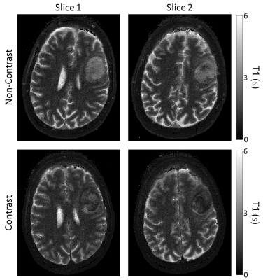
It is established that tumour hypoxia is a predictor of cancer disease progression, treatment failures, and metastatic potential. There remains a need for oxygen delivery mechanisms for hypoxia reduction. The ideal method for measuring oxygen in tissue is noninvasive and quantitative, allowing tumour pO2 measurements to be obtained before, during, and after treatment. We investigated effects of oxygen nano-carriers on the longitudinal relaxation times of tumour tissues in vivo and a phantom. T1 decreased with increased oxygen concentration in phantom. The injection of the oxygenated nanobubbles resulted in a statistically significant decrease in T1-weighted signal when measured 6-8 minutes post-injection.
|
|
2300.
 |
2 |
Investigating the effects of hypoxia on fibroblast invasion and metabolism
Jesus Pacheco-Torres, Tariq Shah, Flonne Wildes, Dimitry Artemov, Zaver Bhujwalla
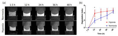
Fibroblast are considered as a major source of Collagen 1 fiber in the tumor stroma and to play a fundamental role in extracellular matrix (ECM) modification. Thus, cancer associated fibroblast has been related with increased tumor proliferation, invasion and metastasis. In the present study, we want to characterize the effect of different tumor microenvironments, as hypoxia and acidic extracellular pH, in the ability of prostate fibroblast cells to invade and degrade the extracellular matrix (ECM), as well with changes in their metabolome. We used our MR compatible cell perfusion system to assess this.
|
|
2301.
 |
3 |
Choline kinase-a downregulation decreases prostate cancer associated fibroblast viability
Jesus Pacheco-Torres, Flonne Wildes, Tariq Shah, Balaji Krishnamachary , Zaver Bhujwalla
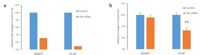
Cancer associated fibroblasts (CAFs) significantly influence the proliferation, invasion, and metastasis of cancer cells. CAFs are detected in most tumors and provide a ubiquitous target that is being actively investigated in cancer treatment. In prostate cancer, fibroblasts have been shown to induce growth, confer castration-resistance, and increase metastatic potential. Choline kinase (Chk)-α downregulation has been previously shown to significantly decrease cancer cell viability but its effect on CAFs has not been investigated before. Here we found, for the first time, a significant decrease of prostate cancer fibroblast (PCAF) viability with Chk-α downregulation.
|
|
2302
 |
4 |
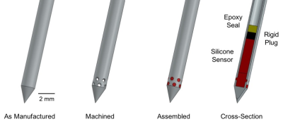
Design and validation of an MRI-based oxygen sensor for a cervical cancer clinical trial
Video Permission Withheld
Gregory Ekchian, Junichi Tokuda, Brian Barnes, Robert Cormack, Larissa Lee, Michael Cima
Many cancer patients experience lower survival rates if they have less well oxygenated tumors. Lower tumor oxygen levels can lead to a reduced effectiveness of radiation therapy. The ability to overcome this radiotherapy resistance has been severely limited by the lack of a clinically compatible quantitative oxygen sensing technology. We report the design and validation of a silicone-based oxygen sensor measured with MRI for an approved human clinical trial in patients with cervical cancer. The sensor has been validated for compatibility with the clinical workflow and is specifically designed to achieve the endpoints of the trial.
|
|
2303.
 |
5 |
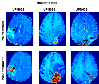
Multiparamter MRI Investigation of High-Grade Glioma Response to CAR T Cell Immunotherapy
Harshan Ravi, Olya Stringfield, Gustavo De Leon, Sandra Johnston, Christine Brown, Kristin Swanson, Robert Gatenby, Natarajan Raghunand
Immunotherapy is gaining interest for treatment of even poorly immunogenic cancers like gliomas. Pseudo-progression and pseudo-response are frequently noted on standard of care MRI in glioma patients receiving immunotherapy. We present a "Habitat" imaging approach to objectively follow response to CAR T cell therapy of glioma.
|
|
2304.
 |
6 |
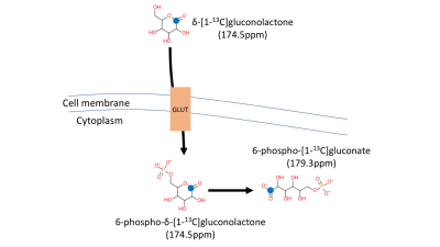
In vivo evaluation of pentose phosphate pathway activity in orthotopic glioma using hyperpolarized d-[1-13C]gluconolactone
Georgios Batsios, Pavithra Viswanath, Peng Cao, Celine Taglang, Elavarasan Subramani, Robert Flavell, Peder Larson, Sabrina Ronen
Flux via the pentose phosphate pathway (PPP) is typically upregulated in tumor cells. Imaging this upregulation could therefore help in monitoring tumor development and response to treatment. A previous study presented the use of hyperpolarized δ-[1-13C]gluconolactone to detect flux through PPP by monitoring its conversion to 6-phospho-[1-13C]gluconate in isolated perfused livers. Here, we demonstrate that HP δ-[1-13C]gluconolactone can also be used to monitor PPP activity in healthy brain and in gliomas, and that the ratio of HP 6-phospho-[1-13C]gluconate to 6-phospho-δ-[1-13C]gluconolactone is significantly higher in tumor regions compared to healthy brain.
|
|
2305.
 |
7 |
Tracking adoptive cell transfer of primary human and mouse T-cells in naïve NSG and Balb/c mice, respectively, using PET and MRI methods
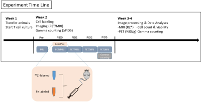
Naomi Sta Maria, Leslie Khawli, Sharon Lin, Vyshnavi Pachipulusu, Long Zhen, Daniel Cohrs, Alan Epstein, Russell Jacobs
We determined the biodistribution of adoptively transferred primary T-cells in naïve mice to create a basis for quantifying adoptively transferred modified T-cells in mice cancer models, using PET and MRI cell labeling techniques simultaneously. Separate populations of T-cells were labeled with either a PET (89Zr) or an MRI label (ferumoxytol), and were injected intravenously into mice together. Animals underwent simultaneous PET/MRI imaging up to 5 days following cell injection in an MR Solutions 7 Tesla scanner with a PET camera. We looked at two models: primary mouse T-cells in naïve Balb/c mice and primary human T-cells in naïve NSG.
|
|
2306.
 |
8 |
A Pre-Clinical PET/MRI/MRS Study on Lactate Transport Inhibition by Bitter Melon Juice in Pancreatic Cancer Models
Deepanshi Dhar, Aaron Safadi, Jenna Steiner, Raina Komal, Chapla Agarwal, Rajesh Agarwal, Natalie Serkova
Pancreatic cancer (PDAC) is a highly aggressive malignancy, displaying poor response to the frontline chemotherapeutics. PDAC cells undergo cellular reprograming to meet their bioenergetic and biosynthetic demands, with glycolytic shift emerging as the primary metabolic hallmark of carcinogenesis. Bitter melon juice (BMJ) is a widely consumed vegetable in Asia; recent studies have reported an increased AMPK phosphorylation and activation in BMJ-treated tumor xenografts. Here, we report on an inhibition of lactate export in PANC1 cells upon BMJ treatment, leading to acidification and cell death mediated by decreased transporter expression of GLUT1 and MCT4, both in vitro and in vivo.
|
|
2307.
 |
9 |
13C Metabolomic and Fluxomic Study of Human Melanoma Metabolic Network in vivo
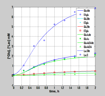
Alexander Shestov, Seung-Cheol Lee, Kavindra Nath, Jeff Roman, Clementina Mesaros, David Nelson, Dennis Leeper, Ian Blair, Jerry Glickson
Information from 13C isotopomers, which appear as multiplets in 13C spectra can be measured and quantified in vivo. Using this additional information alone with extended melanoma tumor bionetwork model has enable simultaneous fitting of experimental dynamic isotopomer turnover curves and evaluation of metabolic parameters and fluxes
|
|
2308
 |
10 |
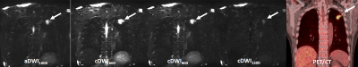
Comparison of the Capability for Quantitative Distinguishing Malignant from Benign Solitary Pulmonary Nodules among actual DWI, computed DWIs with different b values and FDG-PET/CT
Video Permission Withheld
Yuji Kishida, Masao Yui, Yoshimori Kassai, Shinichiro Seki, Takeshi Yoshikawa, Katsusuke Kyotani, Takamichi Murakami, Yoshiharu Ohno
There are no major papers that compared differentiating capability of SPNs among actual DWI (aDWI), computed DWIs (cDWIs)with different b values and FDG-PET/CT in patients with SPN. We hypothesize that cDWI obtained appropriate b value can improve the capability for differentiating malignant from benign SPNs as compared with aDWI and FDG-PET/CT. The purpose of this study is to directly compare the capability for differentiating malignant from benign pulmonary nodules among aDWI, cDWIs with different b values and FDG-PET/CT.
|
|
2309.
 |
11 |
Towards a Small Molecule GDPD6 Inhibitor: Investigating Dipyridamole via 1H HRMRS and Computational Studies
Caitlin Tressler, Kanchan Sonkar, Kristine Glunde
We are studying the role of GDPD6 in breast cancer, as well as its potential as a therapeutic target. GDPD6 silencing experiments showed decreased invasion and migration in breast cancer cells. There is currently no small molecule inhibitor for GDPD6. We have identified dipyridamole as potential GDPD6 inhibitor, which can be used both in the lab and potentially in the clinic. We are using a combination of 1H MRS and computational studies to determine how dipyridamole inhibits GDPD6 to evaluate its potential as an inhibitor and identify other potential small molecule inhibitors of GDPD6.
|
|
2310.
 |
12 |
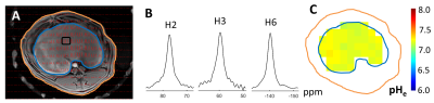
Time evolution of extracellular pH with BIRDS in a rabbit model of human liver cancer
Daniel Coman, Lynn Savic, Isabel Schobert, John Walsh, Lucas Adam, Nina Tritz, MingDe Lin, Julius Chapiro, Albert Sinusas, Todd Constable, Douglas Rothman, James Duncan, Fahmeed Hyder, Dana Peters
Here we report extracellular pH (pHe) mapping with BIRDS using TmDOTP5- in normal and VX2 tumors in rabbit liver tissue. Transarterial chemoembolization (TACE) was performed and the rabbits were scanned without TACE, or at 1 day and 2 weeks post TACE. The pHe maps show lower pHe in tumor and tumor edge compared to normal liver. Tumor acidity prior to TACE remain at 1 day post TACE, but it is almost normalized at 2 weeks post TACE. The ability to measure pHe in a translational model and compare it with “normal” tissue improves tumor detection and monitoring of tumor treatment.
|
|
2311.
 |
13 |
Toward Quantitative MRI Parameter Mapping of Triple Negative Breast Cancer Patient-Derived Xenographs: The Challenge of Tumor Heterogeneity
Xia Ge, John Engelbach, James Quirk, Larry Bretthorst, Joel Garbow, Joseph Ackerman
Triple negative breast cancer patient-derived xenographs were implanted in the 4th abdominal mammary fat pads of mice enrolled in a ~60 minute, multi-contrast, same-day (morning vs. afternoon), test-retest MRI protocol. Quantitative T1, T2, and ADC maps were acquired. Parameter distributions were characterized by standard statistical measures (mean, median, standard deviation, skewness, and kurtosis) and a Bayesian implementation of the maximum-entropy method-of-moments density function. TNBC PDX T2 maps were found to be markedly more robust to test-retest assessment compared to T1 and ADC maps. These results will inform studies employing MRI assessment of TNBC PDX response to docetaxel/carboplatin therapy.
|
|
2312.
 |
14 |
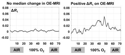
OE-MRI, DCE-MRI and DWI provide complementary response evaluation in patients with rectal cancer treated with chemoradiotherapy
Ross Little, Anubhav Datta, Adam Featherstone, Yvonne Watson, Susan Cheung, Lucy Buckley, Mark Saunders, Geoff Parker, James O'Connor
Biomarkers derived from functional MRI have potential to monitor response to therapy and stratify patient care. In this study of 22 patients with rectal cancer we evaluated the relative merits of using OE-MRI, DCE-MRI and DWI biomarkers to assess response to chemoradiotherapy. We show that OE-MRI is feasible in rectal cancer tumours and provides complementary information to that measured by DWI and DCE-MRI. Data suggests that OE-MRI may be useful as a pharmacodynamic tool to identify hypoxia modification as this was present by day 14, but not at day 7 into therapy.
|
|
2313.
 |
15 |
Association between Metabolism Measured by PET/CT and Vascular Parameters Measured by Dynamic Contrast Enhanced MRI in Spinal Lesions
Jiahui Zhang, Enlong Zhang, Yanyan Zhang, Yongye Chen, Ning Lang, Hon Yu, Huishu Yuan, Min-Ying Su
A total of 49 patients with spinal lesions receiving DCE-MRI and 18F-FDG PET/CT were analyzed. The ROI was manually placed on strongly enhanced area on MRI to measure DCE enhancement kinetics, and from which the wash-in and maximum enhancement ratio, wash-out slope, Ktrans and kep were extracted. SUVmax was measured from the corresponding lesion on FDG uptake map. The results showed that vascular parameters measured from DCE-MRI were not correlated with glucose metabolism measured by PET/CT; therefore, they represent different aspects of lesion, and may be combined for better staging or predicting prognosis rather than being used for diagnosis.
|
|
2314
 |
16 |
Weekly T2’ and rOEF-mapping monitoring tumor oxygenation in patients with recurrent glioblastoma undergoing antiangiogenetic therapy
Video Permission Withheld
Katharina J. Wenger, Elke Hattingen, Joachim P. Steinbach, Oliver Bähr, Marlies Wagner, Ralf Deichmann, Jan-Rüdiger Schüre
For tumor treatment with bevacizumab (BEV), a VEGF-specific antibody, some preclinical reports describe a partial normalization of vessels resulting in a transient improvement in tumor oxygenation, while others observed a decrease in neovascularization, with induction of intratumoral hypoxia. By weekly monitoring rOEF with MRI in six glioblastoma patients until tumor progression according to RANO, we were able to discriminate between two tumor phenotypes with different biological behavior.
|
|
2315
 |
17 |
Quantification of Cerebral Blood Flow using arterial spin labeling in glioblastoma multiforme; challenges of calibration in the presence of oedema.
Video Permission Withheld
Paula Croal, Flora Kennedy-McConnell, Benjamin Harris, Ruichong Ma, Stasya Ng, Puneet Plaha, Simon Lord, Nicola Sibson, Michael Chappell
Arterial spin labeling (ASL) offers a non-invasive and repeatable method for quantifying CBF, a promising biomarker in cancer imaging. However, the consensus for voxelwise calibration may not be appropriate for application in tumours. We hypothesise that voxelwise calibration in the presence of oedema will decrease sensitivity to alterations in CBF, and test this by measuring CBF with pseudocontinurous ASL in seven patients with glioblastoma multiforme, comparing the impact of voxelwise, white matter, and CSF calibration on tumour CBF. Calibration choice significantly affects absolute CBF; with a loss of CBF contrast in tumours when using voxelwise calibration, which may have clinical implication.
|
|
2316.
 |
18 |
The effect of sunitinib treatment assessed by intravital microscopy and DCE-MRI in pancreatic ductal adenocarcinoma xenografts
Jon-Vidar Gaustad, Catherine Wegner, Anette Hauge, Trude Simonsen, Einar Rofstad
The effect of sunitinib treatment was evaluated by DCE-MRI, intravital microscopy, and immunohistochemistry in pancreatic ductal adenocarcinoma (PDAC) xenografts growing in dorsal window chambers or intramuscularly in the hind leg of mice. Sunitinib selectively removed small-diameter vessels and increased blood flow velocity. The increased blood flow velocity was not sufficient to compensate for the loss of tumor vessels, and, consequently, sunitinib-treated PDAC xenografts showed increased fractions of hypoxic tissue. Ktrans derived by pharmacokinetic analysis of DCE-MRI data was sensitive to microvascular density and hypoxia in both untreated and sunitinib-treated PDAC xenografts.
|
|
2317.
 |
19 |
Correlation of Multiparametric MRI with extracellular pH mapping in a Rabbit Model for Liver Cancer
Dana Peters, Lynn Savic, Steffen Huber, John Walsh, Isabel Schobert, Lucas Adam, Nina Tritz, Fahmeed Hyder, Mingde Lin, James Duncan, Douglas Rothman, Albert Sinusas, Julius Chapiro, R. Todd Constable, Daniel Coman
Hepatocellular Carcinoma (HCC) was studied using multiparametric MRI in a rabbit liver tumor model, comparing native T1 and T2* mapping, ADC, and dynamic contrast enhanced parameters, with extracellular pH maps. Tumor heterogeneity was well characterized by parametric mapping.
|
|
2318.
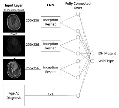 |
20 |
Implementation of A Novel Deep Learning Network on Predicting Isocitrate Dehydrogenase (IDH) Mutation in Patients with Gliomas
Haonan Xiao, Zheng Chang
The purpose of this study is to investigate the feasibility of the Inception-ResNet to reduce image pre-processing and improve the prediction accuracy of the IDH status of gliomas. The T1w-post contrast, T2, and FLAIR images of 91 glioma patients after intensity normalization are fed to the network as training and validation set, and another group of 12 patients is randomly selected as the test set. The prediction accuracies of two repeated experiments are consistent, both greater than 90%. The result shows that with Inception-Resnet, IDH status could be predicted at a high accuracy with minimal image pre-processing.
|
|
2319.
 |
21 |
Interrogations of Human Lung Cancer Metabolomics Measured from Intact Tissue Magnetic Resonance Spectroscopy with Mass Spectrometry Imaging
Stephen Eyles, Mari Mino-Kenudson, Igor Kaltashov, Richard Vachet, Yiying Zhang, Kristen Sikora, Cedric Bobst, David Christiani, Leo Cheng
Current radiology can detect small lung cancer (LuCa) lesions. However, their high costs coupled with their unproven efficacies as screening tools have prevented their use in annual screening protocols to detect LuCa at early and clinically asymptomatic stages. A simple and non-invasive screening technique, preferably a blood test, is needed to control the disease. Here we present results from mass spectrometry imaging that can produce localized “microscopic” maps of cancer metabolomic distributions revealed by high-resolution magic angle spinning magnetic resonance spectroscopy (HRMAS MRS), and can further assist establish blood serum LuCa biomarkers from analyses of human LuCa tissue-serum paired samples.
|
|
2320.
 |
22 |
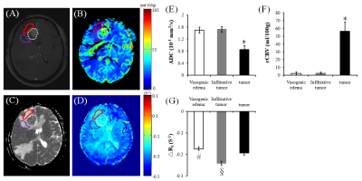
Differentiating infiltrative tumor from vasogenic edema in glioblastom using oxygen-enhanced MR imaging
Junchao Qian, Xian Zhang, Xiang Yu, Qi Chen, Junjun Li, Le Zhang, Huajing Xie, Mei Zhu, Jun Yang, Hongzhi Wang
Glioblastoma (GBM) induces both vasogenic edema and extensive tumor cells infiltration, both of which present with similar appearance and not be differentiated on conventional MRI. To distinguish between these infiltrative tumor and vasogenic edema components within the nonenhancing lesion area using novel techniques thus holds great clinical importance. Oxygen-enhanced MRI may directly reflect tissue oxygenation, has shown promising applications in the measurement of hypoxia or radiation-induced necrosis. Therefore, in this study we explored the possibility to differentiate vasogenic edema from infiltrative tumor in patients with GBM using oxygen-enhanced MRI. The results showed significant more negative ΔR1 levels (p < 0.05) were observed in the infiltrative tumor area compared to those in the vasogenic edema and tumor site. Oxygen-enhanced MR imaging has thus the potential to differentiate infiltrative tumor from vasogenic edema in glioblastoma.
|
|
2321.
 |
23 |
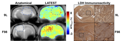
Lactate chemical exchange saturation transfer MRI as a biomarker for differentiating lactate dehydrogenase activity in 9L and F98 glioma
Puneet Bagga, Mohammad Haris, Hari Hariharan, Ravinder Reddy
Lactate chemical exchange saturation transfer (LATEST) MRI method has been shown to be applicable in detecting and imaging changes in the lactate level in human subjects post heavy exercise and to measure the lactate in a mouse model of lymphoma. In this study, LATEST was implemented to differentiate the lactate dehydrogenase (LDH) activity in vivo in two different preclinical glioma models. The two gliomas studied are widely used 9L (highly immunogenic, gliosarcoma) and F98 (weakly immunogenic, glioblastoma). The LATEST contrast was found to be higher in the tumor region of F98 rats compared to the 9L glioma carrying rats.
|
|
2322.
 |
24 |
PET/MRI as a sensitive diagnostic tool for peritoneal carcinomatosis: early results from a single center prospective study
Amy Law, Naik Vietti Violi, Stefanie Hectors, Eliahu Bekhor, Somali Gavane, Daniel Labow, Bachir Taouli
The goal of our study was to assess the diagnostic value of PET/MRI for diagnosis and spatial localization of peritoneal carcinomatosis (PC) in patients prior to potential cytoreductive surgery (CRS) and hyperthermic intraperitoneal chemotherapy (HIPEC). We found that PET/MRI is sensitive and accurate at predicting PC at the patient level as compared to surgery, the reference standard. However, PET/MRI was not to as accurate in localizing PC in the abdominopelvic cavity. Findings need to be validated in a larger study.
|
|
2323
 |
25 |
PET/MRI versus PET/CT in Oncology: A Prospective Single-center Study Focusing on Implications for Patient Management and Cost Considerations
Video Permission Withheld
Marius Mayerhoefer, Helmut Prosch, Lucian Beer, Dietmar Tamandl, Thomas Beyer, Ivo Rausch, Dominik Berzaczy, Markus Raderer, Christoph Hoeller, Matthias Preusser, Ahmed Ba-Ssalamah, Georgios Karanikas, Julia Kesselbacher, Gerald Prager, Michael Weber, Bernhard Brauner, Markus Mitterhauser, Harald Eidherr, Alexander Haug
To prospectively investigate the clinical impact of PET/MRI, compared to PET/CT, in a mixed population of cancer patients, and to perform an economic evaluation of PET/MRI. 263 patients (330 same-day PET/CT and PET/MRI examinations) were analyzed. PET/MRI was accurate in 319/330 examinations, and PET/CT in 277/330 examinations; respective accuracies (97.3% vs. 83.9%) differed significantly (P<0.001). Additional findings on PET/MRI had implications for clinical management in 21/263 patients (8.0%). Incremental cost-effectiveness ratios for PET/MRI were 22.47 EUR (~26.28 USD) per percent of diagnostic accuracy, and 37.64 EUR (~44.06 USD) per percent of correctly managed patients.
|
|
| Top |
Cancer Therapy Response Assessment
Digital Poster
General Cancer Imaging
Tuesday, 14 May 2019
| Exhibition Hall |
08:15 - 09:15 |
| |
|
Computer # |
|
2324.
 |
26 |
Golden angle radial undersampling to accelerate synthetic CT generation with generative adversarial networks for prostate MR-guided Radiotherapy
Matteo Maspero, Tom Bruijnen, Mark Savenije, Linda Kerkmeijer, Peter Seevinck, Cornelis van den Berg
Synthetic-computed tomography (sCT) is crucial to enable MR-only radiotherapy and accurate MR-based dose calculations. In this work, we assessed the feasibility of using undersampled golden angle radial acquisition in combination with a conditional adversarial network to accelerate both acquisition and sCT generation for patients with prostate cancer. Golden angle radial acquisitions were simulated for several undersampling factors in a retrospective manner on 3D Cartesian spoiled gradient-echo data that were clinically acquired on fourty patients with prostate cancer. Dose planning demonstrated that accurate MR-based dose calculations were possible up to an undersampling factor 30 resulting in fast (<20s) sCT generation, which is of particular interest for online MR-guided radiotherapy.
|
|
2325.
 |
27 |
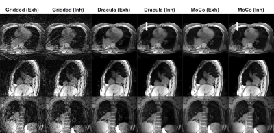
Rapid 4D-MRI reconstruction using a Deep RAdial ConvoLutionAl neural network: Dracula
Joshua Freedman, Oliver Gurney-Champion, Hannah Bainbridge, Jennifer Kieselmann, Michael Dubec, Henry Mandeville, Simeon Nill, Marc Kachelrieß, Uwe Oelfke, Martin Leach, Andreas Wetscherek
4D-MRI could inform online treatment plan adaptation on MRI guided radiotherapy systems, but long iterative reconstruction times (> 10 minutes) limit its use. A deep convolutional neural network was trained to learn the joint MoCo-HDTV algorithm and high-quality 4D-MRI (1.25x1.25x3.3 mm3, 16 respiratory phases) were reconstructed from gridded raw data in 27 seconds. Calculated 4D-MRI exhibited a high structural similarity index (0.97 ± 0.013) with the iteratively reconstructed test images and only a minor loss of fine details. Despite exclusively training the network on data from a diagnostic scanner, 4D-MRI were successfully reconstructed from raw data acquired on an MR-linac.
|
|
2326.
 |
28 |
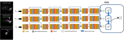
A deep neural network based model for treatment response prediction using longitudinal diffusion MRI
Yu Gao, Vahid Ghodrati, Anusha Kalbasi, Jie Fu, Dan Ruan, Minsong Cao, Chenyang Wang, Fritz Eilber, Nicholas Bernthal, Susan Bukata, Sarah Dry, Scott Nelson, Mitchell Kamrava, John Lewis, Daniel Low, Michael Steinberg, Peng Hu, Yingli Yang
A deep neural network based model was proposed to predict post-radiotherapy treatment effect score for localized soft tissue sarcoma patient using longitudinal diffusion MRI. Diffusion images were acquired three times throughout patient’s hypofractionated radiotherapy treatment. A convolutional neural network was constructed to learn the most useful spatial features from the tumor ADC maps at each time point, which is then fed into a recurrent neural network to exploit the temporal information between the extracted features. Excellent prediction performance of 97.4% accuracy on slice-based classification, and 95% accuracy on patient-based classification were achieved on independent test sets.
|
|
2327.
 |
29 |
Investigation of Abdominal Organ Respiratory Motion Probability Distribution Function and its Inter-Fractional Reproducibility Assessment Using Fast Volumetric 4D-MRI for Probability-Based Radiotherapy Planning
Yihang Zhou, Jing Yuan, Oi Lei Wong, Kin Yin Cheung, Siu Ki Yu
Respiratory motion is a major concern in radiotherapy (RT) in liver cancer patients. Probability-based treatment planning is an evolving approach for tumor motion management. A major hurdle of this approach is that the dosimetric error is tightly linked to the reproducibility of the tumor motion probability density function (PDF). Previous studies in lung used single-slice dynamic MRI for PDF reproducibility evaluation that could only captured the 2D respiratory motion restricted by the MRI acquisition speed. Moreover, inter-subject and inter-fractional variability of time evolved PDF might be underestimated by using just two fractions. In this study, we aim to investigate the inter-fractional and inter-subject abdominal motion PDF and its reproducibility using an ultrafast volumetric 4D- MRI.
|
|
2328.
 |
30 |
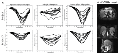
Respiratory motion characterization and motion uncertainty estimation using a fast 3D+t MRI and bootstrapping for abdominal radiotherapy
Oi Lei Wong, Jing Yuan, Yihang Zhou, Siu Ki Yu, Kin Yin Cheung
Respiratory motion characterization and uncertainty estimation is important in radiotherapy treatment planning, and is hindered by the imaging capability of 4DCT or time-resolved single/multi-slice MRI (2D+t). In this study, characterization of the positional and orientation uncertainty of abdominal organ respiratory motion, extracted from the time-resolved volumetric 3D+t MRI, was done using bootstrapping and tensor analysis.
|
|
2329.
 |
31 |
Baseline Tumor Apparent Diffusion Coefficient Value Can Predict First-line Sunitinib Therapy Response of Stage IV Clear Cell Renal Cell Carcinoma
Liqiang Cui, Yichen Wang, Yan Chen
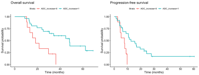
Our single-center retrospective study focused on Stage IV Clear Cell Renal Cell Carcinoma patients who received first-line sunitinib therapy and found that baseline tumor apparent diffusion coefficient value derived from 3T diffusion weighted imaging showed different level in different response group. Baseline tumor ADC value also had significant correlation with progression-free survival. Patients with higher tumor ADC value had significantly longer progression-free survival. Basline tumor ADC can be a potential predictor in assessing targeted therapy response of Stage IV ccRCC.
|
|
2330.
 |
32 |
Treatment response and recurrence prediction on MR during radiotherapy in patients with head and neck squamous cell carcinoma
Tim Schakel, Boris Peltenburg, Remco de Bree, Chris Terhaard, Marielle Philippens
Weekly MR imaging allows for tumor monitoring during treatment. T2 weighted imaging and distortion-free DW-TSE SPLICE were acquired weekly in 20 patients. Changes in volume and ADC could be followed over the course of (chemo)radiotherapy: volume decreases and ADC increases. Tumor delineation is crucial and becomes increasingly difficult during treatment. For the current patient population, 4 patients developed recurrent disease. However, volume changes measured on T2 weighted imaging and ADC changes did not yet show to be prognostic of tumor recurrence.
|
|
2331.
 |
33 |
Respiratory motion variability in 4D-MRI for MR-guided radiotherapy
Bjorn Stemkens, Max van Riel, Tom Bruijnen, Jan Lagendijk, Cornelis van den Berg, Rob Tijssen
Respiratory-induced motion of abdominal tumors can lead to displacements up to five centimeter, making radiotherapy treatments very challenging. The respiratory motion can be characterized by a 4D-MRI, acquired prior to treatment. In this study we investigate how long the 4D-MRI is valid for after acquisition. Additionally, the longitudinal validity of a motion model, derived from the 4D-MRI is assessed.
|
|
2332.
 |
34 |
Radiomic analysis to determine glioma’s IDH1 gene status based on multi-MR sequences
Yi Liu, Tong Han, Xiaoling Yan, Xuebin Zhang, Zhengting Cai
The purpose of this retrospective study was to demonstrate the feasibility of radiomic methods to determine glioma’s IDH1 gene status based on MR imaging. We used a training set (99 patients)with a test set (29 patients), and extracted 1029 radiomic features from each sequence of T2WI, ADC, FLAIR, T1WI-CE and the combined, then reduced by Least absolute shrinkage and selection operator. Five logistic regression classifiers were built based on training set, evaluated using test set and compared by DeLong test. The results indicated the radiomics of combined four sequences had the best performance in distinguishing IDH1 gene status.
|
|
2333.
 |
35 |
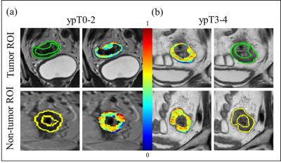
Reproducible radiomic features from post-chemoradiation T2-weighted MRIs can more accurately discriminate pathologic T stage in rectal cancer patients
Amrish Selvam, Jacob Antunes, Justin Brady, Joseph Willis, Rajmohan Paspulati, Anant Madabhushi, Satish Viswanath
We present initial results for identifying radiomic features to effectively characterize pathologic chemoradiation response in rectal cancers, by ensuring feature reproducibility within reference non-tumor regions on post-chemoradiation T2w MRIs. Our approach used a new feature reproducibility measure to prune radiomicfeatures and thus minimize variability due to acquisition or imaging differences. Microscale gradient radiomic features were found to be most reproducible and discriminable in segregating rectal cancer patients by post-chemoradiation pathologic stage independent of disease metastasis, across both training (AUC=0.75) and hold-out validation (accuracy = 84.6%) cohorts.
|
|
2334.
 |
36 |
Directional-gradient based radiomic descriptors from pre-treatment perfusion DSC-MRI to differentiate long-term from short-term survivors in Glioblastoma: Preliminary findings
Bolin Song, Ramon correa, Prateek Prasanna, Niha Beig, Anant Madabhushi, Pallavi Tiwari
We explored the utility of radiomic analysis to identify radiomic features (computer extracted features from MRI) that distinguish long-term survival patients from their short-term survival counterparts based on the pre-treatment perfusion DSC-MRI. Initial results indicate that dynamically extracted radiomic features from enhancing tumor and infiltrative edges on perfusion scans can segregate the 2 survival groups. A non-invasive means of predicting survival based on perfusion imaging may help clinicians to determine prognosis, and inform treatment strategy.
|
|
2335.
 |
37 |
Early non-invasive prediction of response to Temozolomide in low-grade glioma
Marina Radoul, Elavarasan Subramani, Chloe Najac, Georgios Batsios, Anne Marie Gillespie, Sabrina Ronen
Newly diagnosed low-grade glioma (LGG) patients have a relatively long survival, but nonetheless ultimately recur. New therapies are therefore being considered for LGG. One approach is the use of Temozolomide, previously reserved for treating high grade glioblastoma. However, early indicators of response are still needed. Here, we investigated response to Temozolomide treatment in an orthotopic LGG mouse model. Using 1H MRS we detected an early decrease in total choline and a surprising increase in both glutamine and glutamine plus glutamate that were associated with ultimate tumor shrinkage. This identifies potential early metabolic biomarkers of response to Temozolomide treatment in LGG.
|
|
2336
 |
38 |
4DMRI-based abdominal corset study for radiotherapy purposes
Video Permission Withheld
Kai Dolde, Christian Dávid, Gernot Echner, Ralf Floca, Clemens Hentschke, Nina Niebuhr, Kai Ohmstedt, Nami Saito, Merkur Alimusaj, Beate Flügel, Asja Pfaffenberger
Abdominal organ motion provides challenges for radiotherapy treatments, leading to inhomogeneous dose distributions with over- and underdosage regions in the target volume. Repeated 4D-MRI acquisitions, allow to analyze inter- and intrafractional spatial motion. The aim of this study was to investigate the impact of abdominal corsets for motion reduction purposes, based on repeated 4D-MRI data sets. We found pronounced reductions in cranio-caudal and anterior-posterior direction using corsets, which additionally lead to more reproducible motion amplitudes. Lower amplitudes and better reproducibility are beneficial for radiotherapy and could lead to smaller irradiation margins and dose reductions to healthy tissue.
|
|
2337.
 |
39 |
Response monitoring by DCE-MRI in an experimental prostate tumor after single dose 12C-ion and photon radiotherapy
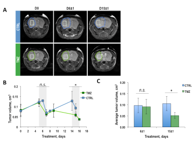
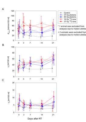
Alina Bendinger, Lisa Seyler, Maria Saager, Charlotte Debus, Peter Peschke, Dorde Komljenovic, Jürgen Debus, Jörg Peter, Ralf Floca, Christian Karger, Christin Glowa
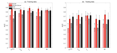
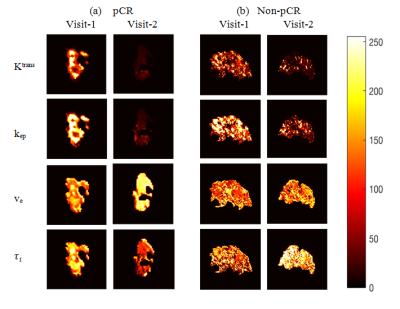
A series of DCE-MRI measurements was used to quantify the vascular changes after therapeutic and subtherapeutic doses of photon and 12C-ion irradiation of the anaplastic rat prostate tumor Dunning R3327-AT1. DCE-MRI data were analyzed by pharmacokinetic modelling employing the Extended Tofts model. Independent of dose, 12C-ions led to stronger and earlier treatment response than photons within the observation period indicated by increased Ktrans and ve parameters. Results were correlated to histological analyses for microvascular density, vessel maturity, tumor hypoxia, and proliferation that further underlined the faster, stronger, and more homogeneous treatment response after 12C-ion irradiation.
|
|
2338.
 |
40 |
Change of Radiotherapy Planning Target Volume Delineated on Pre-Treatment and mid-RT Follow-up MRI After 3-4 Weeks of Treatment
Yang Zhang, Liming Shi, Xiaonan Sun, Tianye Niu, Ning Yue, Jeon-Hor Chen, Tiffany Kwong, Min-Ying Su, Ke Nie
As tumor shows substantial shrinkage over the course of treatment, should radiation treatment volume be adjusted? A quantitative method using “radial distance”- the distance from the outer boundary of the tumor to the center of the rectum, was developed to evaluate the gross tumor volume (GTV) delineated on MRI acquired before treatment and after 3-4 weeks of radiation. In 35 patients, the mean tumor volume decreased from 19.1 to 10.5 cm3 but the mean radial distance only decreased slightly from 16.3 to 15.6 mm. When the remaining tumor was close to the rectal wall, the PTV should not be adjusted.
|
|
2339.
 |
41 |
Quantifying Information Content of Multiparametric MRI Data for Automatic Tumor Segmentation using CNNs
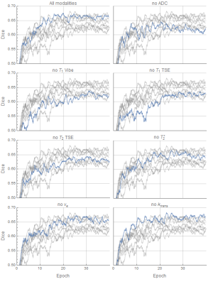
Lars Bielak, Nicole Wiedenmann, Thomas Lottner, Hatice Bunea, Anca-Ligia Grosu, Michael Bock
Multimodality imaging with CT, PET, and MRI is the basis for precise tumor segmentation in radiation therapy. We analyze which MR imaging contrasts mainly improve the segmentation performance of a CNN by training multiple networks using different input channels. The predictive value of 7 different contrasts is compared for two tumor regions, gross tumor volume and lymph node metastasis, in head and neck tumor patients.
|
|
2340.
 |
42 |
Integration of DCE-MRI Texture Features with Clinical Data for Improved Early Prediction of Breast Cancer Therapy Response
Archana Machireddy, Guillaume Thibault, Alina Tudorica, Aneela Afzal, May Mishal, Kathleen Kemmer, Arpana Naik, Megan Troxell, Eric Goranson, Karen Oh, Nicole Roy, Neda Jafarian, Megan Holtorf, Wei Huang, Xubo Song
This study investigated the effect of integrating clinical data with DCE-MRI texture features in early prediction of breast cancer therapy response. DCE-MRI data collected from 55 breast cancer patients before and after the first cycle of neoadjuvant chemotherapy were subjected to pharmacokinetic analysis. Texture features were extracted from voxel-based DCE-MRI parametric maps. Predictive performances with imaging features alone and in combination with clinical features were assessed and compared. Addition of clinical features to image texture features increased predictive capability in discriminating pathologic complete response (pCR) vs. non-pCR compared to using imaging features alone.
|
|
2341.
 |
43 |
Comparison of DCE-MRI Parametric Map-Based Features for Early Prediction of Breast Cancer Therapy Response
Archana Machireddy, Guillaume Thibault, Alina Tudorica, Aneela Afzal, May Mishal, Kathleen Kemmer, Arpana Naik, Megan Troxell, Eric Goranson, Karen Oh, Nicole Roy, Neda Jafarian, Megan Holtorf, Wei Huang, Xubo Song
DCE-MRI data from 55 breast cancer patients collected before and after the first cycle of neoadjuvant chemotherapy were subjected to pharmacokinetic analysis. Four texture features, GLCM, RLM, single- and multi-resolution fractals extracted from DCE-MRI parametric maps, were analyzed for early prediction of therapy response. Generally, the multi-resolution fractal features from individual maps or the concatenated features from all parametric maps showed better predictive performance. The results suggest that multi-resolution analysis, which decomposes the texture at various spatial-frequency scales, may more accurately capture changes in tumor vascular heterogeneity as measured by DCE-MRI, and thus provide better early prediction of therapy response.
|
|
2342.
 |
44 |
SLICs Algorithm for Non-Invasive Response Evaluation in Osteosarcoma with Multiparametric MR Imaging
Amit Mehndiratta, Esha Kayal, Sneha Patil , Sameer Bakhshi, Raju Sharma, Devasenathipathy Kandasamy
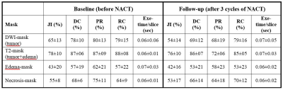
Osteosarcoma is a highly morbid bone-tumor with poor prognosis. Neoadjuvant-chemotherapy(NACT) is the current standard of care. The response of NACT is judged on Histopathology-examination(HPE) after surgical resection of tumor. However, a non-invasive and accurate methods for evaluation of treatment response during the course of therapy is highly desirable. In this research, a Simple-linear-iterative-clustering supervoxels(SLICs) algorithm based methodology using multiparametric MRI (T2,DWI and ADC) has been developed for identification of sub-parts of tumor (active-tumor, necrosis). The volume of active-tumor and necrosis were estimated using this novel approach in patients with OS, before NACT(baseline) and after 3 cycles of NACT(follow-up). The level of necrosis estimated using SLICs and measure with HPE showed a close match. SLICs based estimation of necrosis level is a non-invassive technique that can be useful in response evaluation of cancer imaging.
|
|
2343.
 |
45 |
Diffusion-weighted MRI for assessing longitudinal effect of radiation (photon beam) versus proton beam therapy on cranial bone marrow in children treated for brain tumors
Erika Pace, Enrico Clarke, Henry Mandeville, Andrew Mackinnon, Nandita deSouza
Bone marrow ADC measurements were feasible from the clivus in children. Measurements were reproducible (95% confidence intervals -5.5% to +11%). Following radiation (photon) treatment or proton beam therapy, there was an early rise in ADC at 2 months consistent with bone marrow edema, followed by a fall. The level of early ADC increase (39% for radiation therapy, 42% for proton beam therapy) and pattern of change was similar in both treatment regimens.
|
|
2344.
 |
46 |
Extended texture analysis of unenhanced T1 and T2 sequences on whole body MRI for evaluation of response to chemotherapy in patients with multiple myeloma
Kaspar Ekert, Christopher Kloth, Marius Horger
Extended texture analysis of unenhanced T1 and T2 sequences on whole body MRI for evaluation of response to chemotherapy in patients with multiple myeloma. Patients in a pre-treatment and post-treatment setting using a standardized imaging protocol and a standardized hematological and clinical surveillance were included. 107 features, based on the pyradiomics library, were analyzed for the main medullary lesion in myeloma patients. Extracted texture features were able to discriminate between responders and non-responders at follow-up in particular when using T2-imaga data.
|
|
2345.
 |
47 |
Genomically and Radiographically Adjusted Dose (GRAD) Framework for Biologically Adaptive MR-guided Radiotherapy
Zachary Boyd, William Hall, Douglas Prah, Eric Paulson
Tumor burden, tumor proliferation, and tumor hypoxia, all of which vary in space and time, are evidence-based contributors of radiotherapy failure. In addition, it has been demonstrated that gene expression can influence radiosensitivity. We demonstrate here the initial feasibility of a framework to incorporate genomic and radiographic information to derive patient-specific, voxelwise radiation dose prescription maps for use in a biologically adaptive MR-guided radiotherapy (BAMRgRT) strategy.
|
|
2346
 |
48 |
MRI Independent Predictors of Pathological Complete Response to Neoadjuvant Chemoradiotherapy in Locally Advanced Rectal Cancer
Video Permission Withheld
Lijuan Wan, Hongmei Zhang, Chongda Zhang
In order to tapped the potential of pCR prediction on T2WI comprehensively, both quantitative and qualitative parameters were evaluated in patients with locally advanced rectal cancer. A development group were enrolled to assess these parameters and an external validation group to verify the diagnostic performance. Post-nCRT CATV (CATVpost) and the reduction rate of SIT (SITRR) were proved that were independently associated with pCR and can help for pCR prediction.
|
|
2347
 |
49 |
The Evaluation of Signal Intensity Related Predictors on T2-weighted for Pathological Complete Response to Neoadjuvant Chemoradiotherapy in Locally Advanced Rectal Cancer
Video Permission Withheld
Lijuan Wan, Chongda Zhang, Hongmei Zhang
In order to evaluate the value of tumor signal intensity related parameters on T2-weighted magnetic resonance imaging (MRI) for pathological complete response (pCR) prediction, the signal intensity of tumor(SIT) and the muscle(SIM) were both measured automatically, SIT was defined as an absolute T2W signal intensity of tumor, and SIM was used to correct SIT, resulting in the relative T2W signal intensity(SIT/M), the reduction rate of SIT and SIT/M were calculated.Post-nCRT SIT(SITpost), post-nCRT SIT/M(SIT/Mpost), SITRR and SIT/MRR were proved to be significantly different between pCR and non-pCR, and The diagnositic efficiency is better in non-mucinous adenocarcinoma than mucinous adenocarcinoma.
|
|
2348.
 |
50 |
Application of 3D_NerveVIEW to neurography of nasopharyngeal carcinoma patients in radiotherapy treatment planning
Presentation Not Submitted
Biaoshui Liu, Yimei Liu, Wenchao Diao, Along Chen, Yingjie Mei, Qinping Gu, Queenie Chan, Jianhua Wu, Chengguang Lin, Ying Sun
In radiotherapy, CT/MR images are used to delineate the region of targets and normal structures. In this study, 3D_NerveVIEW sequence was performed on a volunteer, and then the image of cranial nerve was rigidly registered and fused to CT image. In the fused image, the cranial nerve had better visualization than CT and T1w images, and the contour of nerve was easily identified. Which could improve the accuracy of nerve contour and reduce the radiotherapy-induced nerve palsy.
|
|
| Top |
Cancer Perfusion, Diffusion & Relaxometry
Digital Poster
General Cancer Imaging
Tuesday, 14 May 2019
| Exhibition Hall |
08:15 - 09:15 |
| |
|
Computer # |
|
2349.
 |
51 |
An analysis of post-processing steps for residue function dependent DSC-MRI biomarkers through their clinical impact on glioma diagnosis for both 1.5 and 3T
Laura Bell, C. Quarles
Several recent initiatives have focused on optimizing and standardizing DSC-MRI imaging protocols and post-processing steps. With the availability of public imaging databases that include clinical outcomes, various post-processing steps can be carefully assessed for their impact on the clinical outcomes. Here we evaluated post-processing steps for advanced perfusion biomarkers that relay on determining the residue function by examining the clinical impact of each step. In summary we determined that updating the current deconvolution steps is beneficial, and that normalization allows for tumor grading across clinical field strengths.
|
|
2350.
 |
52 |
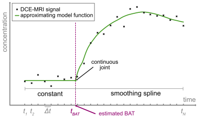
Bolus arrival time estimation for DCE-MRI signals without fast up-slope
Alina Bendinger, Charlotte Debus, Christin Glowa, Christian Karger, Jörg Peter, Martin Storath
Accuracy in pharmacokinetic modelling of DCE-MRI data can be impaired due to a delay between the contrast agent arrival in the tissue of interest and an artery further upstream. To correct the delay, bolus arrival times (BATs) are estimated from the concentration curves. However, the state-of-the-art method for estimating BATs may give unsatisfactory results if the curves do not exhibit a fast up-slope. We propose a spline-based method for BAT estimation for concentration curves without fast up-slopes which are often observed in small animal data. The proposed method gives accurate results on simulated and in vivo acquired rat data.
|
|
2351.
 |
53 |
IDH Genotypes Differentiation in Glioblastomas Using DWI and DSC-PWI in the Enhancing and Peri-Enhancing Region
Hua Zhang, Dairong Cao, Zhen Xing, Dejun She, Yu Lin, Zhongshuai Zhang, Mengxiao Liu
The purpose of this study is to evaluate the contribution of DWI and DSC-PWI in the peri-enhancing region for discriminating glioblastomas IDH genotypes. Further, the diagnostic value of this two MR techniques were compared with those in the enhancing portion. Features of conventional MRI, rADCmin-t, rADCmin-p, rCBVmax-t and rCBVmax-p were compared between IDH-m and IDH-w glioblastomas. IDH-mutated glioblastomas tended to present in frontal lobes and younger patients. Both rCBVmax-t and rCBVmax-p show significant difference between two subgroups, while rADCmin-t and rADCmin-p do not. The results showed that the accuracy of rCBVmax-p is higher than that of rCBVmax-t in the diagnosis of IDH-m glioblastomas. rCBVmax-p may have a better diagnostic value than rCBVmax-t in predicting IDH glioblastomas genotypes.
|
|
2352.
 |
54 |
Evaluating the effectiveness of preload in mitigating the leakage effect of dynamic contrast susceptibility MRI
Xin Li, Seymur Gahramanov, Ramon Barajas, Edward Neuwelt
Dynamic Susceptibility Contrast (DSC) Magnetic Resonance Imaging (MRI) with low molecular weight Gadolinium based contrast agent (GBCA) is often confounded by GBCA’s leakage into intersitium space. Thus, pre-DSC injection of GBCA (preload) is often used to mitigate the underestimation of relative cerebral blood volume (rCBV) due to the leakage of GBCA. Here, we present results to demonstrate that preload is generally effective. However, small dose effect could still be expected in the process.
|
|
2353.
 |
55 |
Bayesian Pharmacokinetic Modeling of Dynamic Contrast-Enhanced Magnetic Resonance Imaging: Validation and Application
Andreas Mittermeier, Birgit Ertl-Wagner, Jens Ricke, Olaf Dietrich, Michael Ingrisch
We implemented a tracer-kinetic model within a Bayesian framework which infers full posterior probability distributions for parameter estimates. We validate our Bayesian model using a digital reference object and compare it to a standard non-linear least squares approach. Furthermore, we use this approach to obtain pharmacokinetic parameter distributions during the course of a therapy for breast cancer DCE-MRI data, and we demonstrate how Bayesian posterior distributions can be utilized to assess treatment response.
|
|
2354.
 |
56 |
Investigating How to Optimally Combine Multimodal MRI Data to Better Identify Glioblastoma Infiltration.
Haitham Al-Mubarak, Antoine Vallatos, Joanna Birch, Lindsay Gallagher, Lesley Glmour, Anthony Chalmers, William Holmes
The infiltration of glioblastoma tumour cells into normal tissue presents a major obstacle to effective treatment, may then be responsible for tumour recurrence after surgery. Clinical MRI failed to detect the invasion of tumour cells. The purpose of this study is to investigate how the information contained in the individual MR images and multi-regression analysis can be used to probe of invasion, applying a mouse model of an infiltrative brain tumour.
|
|
2355.
 |
57 |
Accuracy and precision of DCE-MRI acquired with golden-angle radial k-space under-sampling
Andrew Fagan, Silvin Knight, Matthew Clemence, James Meaney
The effects of using a continuous golden-angle radial k-space sampling trajectory, with varying degrees of under-sampling and compressed sensing image reconstruction, on the accuracy and precision of pharmacokinetic modeling of DCE data, were quantitatively investigated. DCE image temporal resolutions (Tres) ranging from 1.85s to 0.09s (corresponding to radial sampling densities of 100% to 4.68%) produced absolute accuracy/precision errors in all Ktrans, ve and kep values of ≤ 2%/4% (for Tres =1.85s) to ≤ 12%/11% (for Tres =0.09s), respectively. These results demonstrate that DCE image acquisition protocols can be designed which constrain pharmacokinetic parameter value errors within prescribed thresholds.
|
|
2356.
 |
58 |
Optimized tumor volumes by dynamic contrast enhanced MR imaging for assessing response to neoadjuvant chemotherapy in triple negative breast cancer
Benjamin Musall, Beatriz Adrada, Abeer Abdelhafez, Hagar Mahmoud, Ken-Pin Hwang, Jong Bum Son, Lumarie Santiago, Gary Whitman, Huong Le-Petross, Tanya Moseley, Rosalind Candelaria, Bora Lim, Senthil Damodaran, Jennifer Litton, Stacy Moulder, Wei Yang, Jingfei Ma, Mark Pagel, Gaiane Rauch
We evaluated several methods of measuring tumor volumes on DCE MRI for assessment of treatment response in triple negative breast cancer (TNBC) undergoing neoadjuvant chemotherapy (NAC), including functional tumor volume (FTV), enhanced tumor volume(ETV), and clinical tumor volume (CTV). We compared different parameters for measurement of functional tumor volume at baseline as well as its changes during therapy, and established optimal parameters for FTV measurements. We found that optimized FTV and ETV have potential to serve as an imaging biomarker for evaluation of NAC treatment response in TNBC patients
|
|
2357.
 |
59 |
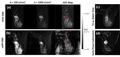
Quantitative ADC measurement of breast cancer with ssEPI and reduced FOV diffusion weighted imaging techniques
Benjamin Musall, Beatriz Adrada, Abeer Abdelhafez, Hagar Mahmoud, Ken-Pin Hwang, Jong Bum Son, Lumarie Santiago, Gary Whitman, Huong Le-Petross, Tanya Moseley, Rosalind Candelaria, Thorunn Helgason, Elizabeth Ravenburg, Jennifer Litton, Stacy Moulder, Wei Yang, Jingfei Ma, Mark Pagel, Gaiane Rauch
The goal of this study was to assess differences in quantitative ADC of breast cancer between ssEPI and rFOV DWI techniques. The two techniques were used to acquire breast DWI images in 27 patients at three different time points during their neoadjuvant chemotherapy. Tumor ADC from the two techniques at baseline and mid-treatment scans show strong correlation and minimal bias. However, tumor ADC from the two techniques at pre-surgery correlated more moderately and showed a slight bias. The relative and absolute changes in ADC at mid-treatment or pre-surgery from baseline showed only moderately-strong non-parametric correlation between the two techniques.
|
|
2358.
 |
60 |
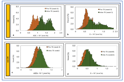
Non-gaussian IVIM-DWI for HPV-related oropharyngeal cancer patients who received marked dose de-escalation in chemo-radiotherapy: Intra-treatment imaging response evaluation
Ramesh Paudyal, Nadeem Riaz, Vaios Hatzoglou, Nancy Lee, Amita Shukla-Dave
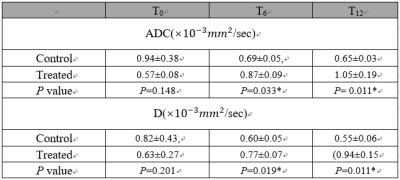
This study aims to evaluate treatment response in human papillomavirus-related (HPV+) oropharyngeal squamous cell carcinoma using pre- treatment (TX), intra- TX week 1, 2, 3, and post-TX week 4 quantitative imaging metrics derived from non-Gaussian IVIM DWI. ADC and D showed a significant increase between pre- and post-TX week 4 in complete response (CR) group, who were treated with dose de-escalation to 30Gy chemo-radiation therapy.
|
|
2359.
 |
61 |
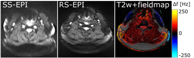
Readout-segmented vs. Single Shot Diffusion MRI for Radiation Therapy Planning in Head and Neck Tumor
Lars Bielak, Nicole Wiedenmann, Thomas Lottner, Hatice Bunea, Anca-Ligia Grosu, Michael Bock
Image distortion is a major limitation in radiation therapy (RT) planning, especially for diffusion weighed imaging in regions with strong B0-inhomogeneity. In this study we analyze the improvement of Readout-segmented-EPI over conventional single shot EPI in the geometrically challenging anatomical region of the neck. RS-EPI effectively increases geometric accuracy in head and neck tumor DWI and significantly reduces ghosting artifacts at the cost of a slightly prolonged acquisition time. Therefore it has proven a clear clinical benefit compared to standard SS-EPI.
|
|
2360.
 |
62 |
Combined diffusion and perfusion MRI in Glioblastoma predicts glial stem cells proliferation and aggressiveness
Tanguy Duval, Jean-Albert Lotterie, Anthony Lemarié, Caroline Delmas, Christine Toulas, Elizabeth Moyal, Vincent Lubrano
Interpretation of diffusion and perfusion MRI in the hyper-FLAIR is challenging. In this work, biopsies were extracted from 16 subjects and infiltrative tumorous stem cells were counted and cultivated intraoperatively to measure their aggressiveness. Diffusion was found to be a good predictor of the time to form tumorous neurospheres. Glioblastoma stem cells were found preferably in regions with strong perfusion, that is to say near vascular niches.
|
|
2361.
 |
63 |
Getting more from less: a morphological model of diffusion in the prostate for improving the predictive power of DWI in identifying tumors.
David Willis, Donnie Cameron, Paul Malcolm, Glyn Johnson
We constructed a morphological model of diffusion in the prostate from a limited number of diffusion-weighted images to increase the sensitivity of such diffusion imaging to the presence of prostate cancer. Estimating the measurement error (9.9%) and characterizing the prostate from a large public dataset (n=206) has shown morphological relationships (|r|>0.5) and provided distributions and relationships within the available ADC measures. A model can then be used to give expected values to test against, and enable much larger datasets to be synthesized with the aim of testing various machine learning approaches.
|
|
2362.
 |
64 |
Serial ADC measurements in the T2 hyperintense, but otherwise normal-appearing white matter of glioblastoma patients correlates with survival
Aaron Rulseh, Jan Sroubek, Jan Klener, Josef Vymazal
Glioblastoma is the most common malignant primary intracranial tumor and, despite multi-modal treatment, the prognosis remains poor. Additional tools to improve early detection or evaluate treatment response are highly desirable. We evaluated serial ADC measurements in the T2-hyperintense, but otherwise normal-appearing white matter at 1.5 T in thirty-five subjects diagnosed with glioblastoma and treated by surgical resection, radiotherapy, temozolomide and tumor-treating fields. We found that serially increasing ADC in the T2-hyperintense, but otherwise normal-appearing white matter in glioblastoma patients is prognostically favorable, with significantly greater overall survival and progression-free survival in patients with increasing ADC.
|
|
2363.
 |
65 |
Characterising early stage cervical cancer using radiomic features derived from T2- and diffusion-weighted images: a potential prognostic tool in surgical management?
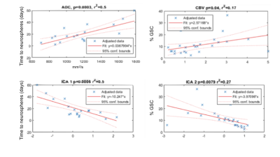
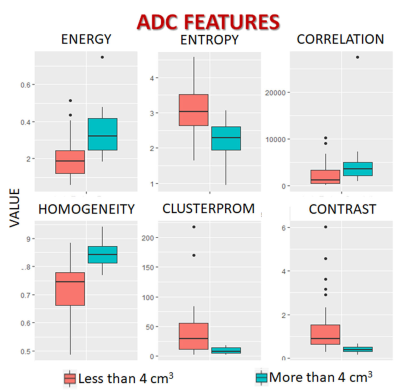
Ben Wormald, Simon Doran, James D'Arcy, James Petts, Thomas Ind, Nandita deSouza
Radiomic features were compared between cervical tumors below and above the volume threshold of eligibility for trachelectomy (< or >4 cm3) to determine their potential prognostic value. Textural feature differences between smaller and larger tumors were similar for both the T2-W and the ADC data. Homogeneity and Energy were increased and Entropy, Contrast and Cluster Prominence decreased in larger tumors. This may reflect the transition from a mixed morphology (tumor elements interspersed with normal glands and stroma) in smaller tumors to more homogenous sheets of malignant cells as tumors increase in size and de-differentiate.
|
|
2364.
 |
66 |
Cluster analysis of IVIM parameter maps reveals tumor subregions of different proliferative status
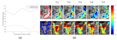
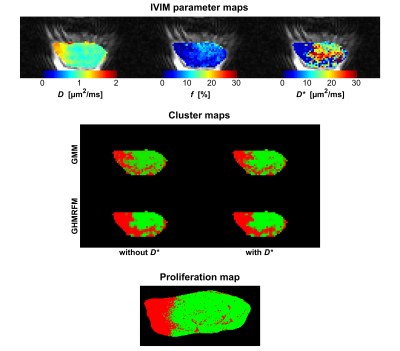
Oscar Jalnefjord, Mikael Montelius, Jonathan Arvidsson, Eva Forssell-Aronsson, Göran Starck, Maria Ljungberg
Tumors are often heterogeneous, which can be seen with various imaging techniques. Even so, analysis based on quantitative imaging is often restrained to average tumor parameter values. In this study we used cluster analysis to identify tumor subregions based on IVIM parameter maps. The tumor subregions showed strong agreement with proliferative status as derived from histological analysis.
|
|
2365.
 |
67 |
Longitudinal diffusion kurtosis MRI in an intracranial rat glioblastoma model
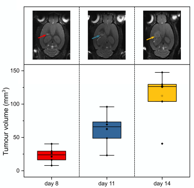
Clementine Lesbats, Claire Kelly, Gabriela Czanner, Harish Poptani
Diffusion kurtosis MRI was used to evaluate the longitudinal changes in a tumor microstructure of a rat model of glioblastoma. F98 tumor cells were injected into six rat brains and imaged longitudinally 8, 11 and 14 days post-implantation. For DKI, an EPI-DTI sequence was used with 2 b-values (1000-2000 s/mm2) and 15 directions. Diffusional kurtosis parameters increased in the tumor compared to the contralateral healthy brain. No significant change with time in the tumor was observed for any diffusion or kurtosis parameters.
|
|
2366.
 |
68 |
Probing the combined effects of collagen concentration and cell density on MR diffusion and relaxivity using a model system
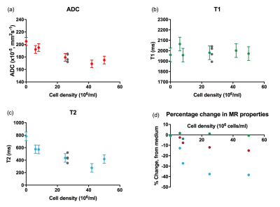
Hannah MacDonald, David Collins, Nandita deSouza
Cell-encapsulating collagen-based models can be used to investigate the relative contributions of the intra and extracellular compartments to ADC, T1 and T2. ADC is mostly affected by cell density, while T2 is influenced primarily by the collagen density; a 120% reduction in T2 was seen when collagen density was increased seven-fold, but this reduction was only 80% in cell containing collagen gels. ADC was not altered by increasing collagen density, unless cell density was also increased.
|
|
2367.
 |
69 |
Diffusion Weighted Imaging at 7T for Differentiation by Grade and Cellularity of Murine Endogenous Pancreatic Ductal Adenocarcinoma
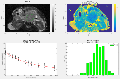
Irina Heid, Geoffrey Topping, Florian Englert, Katja Steiger, Ernst Rummeny, Markus Schwaiger, Franz Schilling, Rickmer Braren
DWI of mice with endogenous PDAC tumours were acquired in a 7T MRI system without breath gating.
Inclusion of DWI with b-values above 800 s/mm2 in fits substantially improves the qualitative appearance and reduces variance of uniform region fit ADC.
Tumours were grouped (based on histology) by cellularity (amounts of neoplastic cells and stroma, and clustering) and separately by grade. ADC reliably distinguishes tumours of different cellularity (PDAClow 1.58±0.08; PDACmed 1.35±0.07; PDAChigh 1.17 ± 0.11; P<0.0001). Grades G2 and G3 were not distinguishable via ADC (1.43±0.15 vs. 1.43±0.16 10-3 mm2/s), however G4 had significantly lower ADC (1.16±0.10 10-3 mm2/s, P<0.0001).
|
|
2368.
 |
70 |
Detection of pre-metastatic niches in perfused mouse livers by diffusion-weighted imaging at ultra-high field
Rui Simões, Sergio Caja-Galán, Rafael Henriques, Bruno Costa-Silva, Noam Shemesh
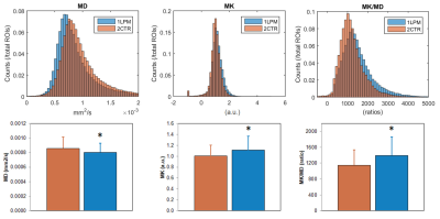
Cancer cells can induce phenotypic modifications at future sites of dissemination (pre-metastatic niches), which support tumor growth and metastasis. Here we evaluated whether diffusion-weighted imaging (DWI) could detect mouse liver pre-metastatic niches (LPM) ex vivo using ultrahigh magnetic field MRI. Our results show that mean diffusivity (MD) and mean kurtosis (MK) can depict microstructural changes associated with LPM formation, consistent with a more fibrotic and cellular microenvironment revealed by histologic analysis of the same samples. These results represent a solid step toward the development of a non-invasive imaging tool for pre-metastatic niche diagnosis.
|
|
2369.
 |
71 |
T1 relaxivity in the bone marrow to monitor response to therapy in acute myeloid leukemia xenografts
Ana Gomes, Diana Passaro, Dominique Bonnet, Bernard Siow
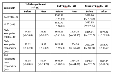
Mouse models of cancer are extensively used to better understand the pathobiology of the disease, to test potential novel therapies, and for the development of diagnostic and prognostic imaging tools. Currently, diagnosis of acute myeloid leukemia (AML) is quite invasive. It is based predominantly on blast quantification in the blood and bone marrow (BM) analysis, and imaging is not part of the clinical follow-up of the patients. T1 relaxivity of the mouse BM has not been reported previously. . BM of mice injected with AML cells from patients present higher T1 relaxation values than normal BM, and it further increased after chemotherapy treatment. Although analysis of bigger cohorts of patients would be required, our data suggest a good potential for BM T1 monitoring as a non-invasive marker for AML resistance to therapy.
|
|
2370.
 |
72 |
Quantitative Imaging of Pharmacodynamics in a Phase 1 Clinical Study of the Vascular Disrupting Agent Crolibulin (EPC2407)
Andres Arias Lorza, William Read, Raoul Tibes, Ronald Korn, Natarajan Raghunand
Diffusion and DCE-MRI were performed at baseline and 2-3 days following Crolibulin (EPC2407) treatment in a phase 1 clinical study of this vascular disrupting agent. ADCw, Ktrans, Ve, and Vp parameter maps were computed and co-registered across scan dates. Over 10 subjects there was an average of 44% decrease in mean tumor Ktrans 2-3 days after initiation of therapy relative to baseline Ktrans values. The decrease in whole-tumor Ktrans was significantly greater in subjects who received 24 mg/m2 drug relative to those who received 13 mg/m2 Crolibulin. Voxel-wise analysis of changes in ADCw, Ktrans, Ve, and Vp will be presented.
|
|
2371.
 |
73 |
Optimisation of luminal water imaging for classification of prostate cancer
Fiona Gong, William Devine, Francesco Giganti, Edward Johnston, David Atkinson, Shonit Punwani
Luminal Water Imaging (LWI) using a multi-echo T2 sequence with 64 echoes has been proposed for microstructural assessment of prostate cancer. We have previously demonstrated that LWI could be simplified and performed using 32 echoes. The purpose of this study was to investigate whether further reduction in echo train length is possible. Reducing echo train length reduces SAR and provides the opportunity to improve LWI resolution and/or volume coverage without exceeding maximum SAR requirements for imaging patients.
|
|
2372.
 |
74 |
The influence of different ROI delineation strategies for relaxation measurements in nasopharyngeal carcinoma using Synthetic MR imaging
Liangru Ke, Tie-bao MENG, Hui-ming LIU, Long Qian, Bing Wu, Hui Li, Yun He, Hao-qiang HE, Chuan-miao XIE
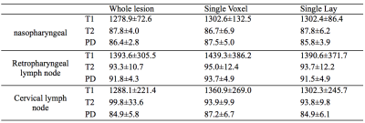
Recently, a novel quantification method named synthetic MRI have attracted more and more attention in the field of clinical research, such as neural disorders and tumor since the first study in 2008. However, this quantitative assessment of diseases based on relaxation times requires regions of interest (ROI), the delineation of which can impact the accuracy of estimated values. To evaluate how the distinct methods of ROI delineation would impact the relaxation value estimation, in current study, 30 patients with nasopharyngeal carcinoma (NPC) were acquired using synthetic MRI.
|
|
2373.
 |
75 |
Intravoxel Incoherent Motion Diffusion-weighted Imaging for Monitoring the Immune Response to Cyclophosphamide in C57BL/6 Mice with GL261 gliomas
Junjiao Hu, Long Qian, Xiangran Cai
It is known that the intravoxel incoherent motion (IVIM) diffusion-weighted imaging (DWI) has been widely applied to the detection and characterization of tumors. However, there were no studies to investigate the use of IVIM-DWI in the evaluation of anti-neoplastic agents induced immune response. In this work, to assess whether IVIM-DWI can predict the immune response to anti-neoplastic agents, six C57BL/6 mice with GL261 mouse gliomas were applied using Metronomic cyclophosphamide. Our results indicated that IVIM-DWI is sensitive to detect the Cyclophosphamide-induced Immune Response.
|
|
| Top |
Cancer Detection & Monitoring
Digital Poster
General Cancer Imaging
Tuesday, 14 May 2019
| Exhibition Hall |
08:15 - 09:15 |
| |
|
Computer # |
|
2374.
 |
76 |
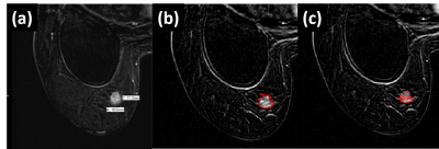
Correlation of breast tumor grade and lymphovascular invasion with biomechanical properties: first results from a breast cancer trial
Presentation Not Submitted
Sweta Sethi, Daniel Fovargue, Stefan Heinz Hoelzl, Ayse Sila Dokumaci, Emma Burnhope, Jurgen Runge, Sanjay Mistry, Keshthra Satchithananda, Arnie Purushotham, Ralph Sinkus
Magnetic Resonance Elastography (MRE) has been considered a promising novel imaging modality in the quantification of viscoelastic properties of breast tumours. The purpose of this study was to evaluate reproducibility and repeatability of a newly developed MRE breast system and investigate whether aberrant biomechanical properties correlate with tumour histopathology. MRE was conducted on 20 healthy volunteers and 15 breast cancer patients. Malignant lesions demonstrated an increase in viscoelasticity when compared to adipose or fibroglandular tissue. While lesions with lymphovascular invasion demonstrated a tendency towards more elevated viscoelasticity than those without lymphovascular invasion, histological grades clearly did not correlate with biomechanics.
|
|
2375.
 |
77 |
MRI-based radiomic to predict lipomatous soft tissue tumors malignancy
Benjamin Leporq, Amine Bouhamama, Fabrice Lame, Catherine Bihane, Michael Sdika, Jean-Yves Blay, Frank Pilleul, Olivier Beuf
In this study a MRI-based radiomic method was developed to predict lipomatous soft tissue tumors malignancy. 81 subjects with lipomatous soft tissue tumors whose histology was known and with fat-suppressed T1w contrast enhanced MR images available were retrospectively enrolled to constitute a database. A linear support vector machine was used after learning base dimension reduction to develop the model. Results demonstrate that the evaluation of lipomatous tumor malignancy is feasible with good diagnosis performances using a routinely used MRI acquisition in clinical practice.
|
|
2376.
 |
78 |
Quality assurance of quantitative MRI for biomarker discovery in locally advanced cervical cancer
Petra van Houdt, Kari Tanderup, Jesper Kallehauge, Remi Nout, Robert Hudej, Supriya Chopra, Jamema Swamidas, Zdenko van Kesteren, Cornelis van den Berg, Michaela Daniel, Dietmar Georg, Eirik Malinen, Jean-Charles Côté, Ives Levesque, Uulke van der Heide
Large multi-center studies are needed to realize the utilization of quantitative MRI (qMRI) as a biomarker for cervical cancer. In this study we created a framework for a multi-center imaging biomarker study, maximizing the consistency between quantitative results in the presence of a large variety of MRI systems. This way, large deviations in qMRI values can be detected and corrected before enrolment of patients in a study. Furthermore, these results can be used to determine the statistical power of the study.
|
|
2377.
 |
79 |
The value of 1.5T contrast-enhanced T1 SPACE sequence in the simulation and planning for Cyberknife stereotactic radiosurgery of brain metastases: patient positioning accuracy, lesion detectability, and target delineation reliability
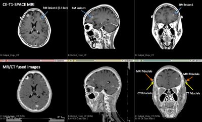
Jing Yuan, Oilei Wong, Winky Fung, Gladys Lo, Franky Cheng, Yihang Zhou, George Chiu, Kin Yin Cheung, Siu Ki Yu
Stereotactic radiosurgery (SRS) is increasingly used for multiple brain metastases (BM) treatment and imposes critical requirements on the accuracy of BM detection, localization and definition in the treatment planning. SPACE sequence is valuable in BM detection for diagnosis, while its value in the BM SRS planning has rarely been explored. We prospectively and quantitatively assessed CE-T1-SPACE in the treatment simulation and planning of Cyberknife-guided BM SRS on a 1.5T MRI-simulator. The results showed that CE-T1-SPACE facilitated high patient positioning accuracy, superior BM detectability and reliable GTV delineation, showing great value in the treatment planning of BM SRS.
|
|
2378
 |
80 |
Measuring eye deformation between planning and proton beam therapy position using MRI.
Video Permission Withheld
Myriam Jaarsma-Coes, Megan Schuurmans, Kilany Hassan, Eleftheria Astreinidou, Marina Marinkovic, Femke Peters, Jan-Willem Beenakker
MRI is becoming a new important imaging modality for ocular tumours. The acquisition of the MR-images for therapy planning are acquired in supine position, but proton beam radiotherapy is performed with the patient in sitting position. By performing scans in supine and in flexed position, we found that this change in gravity direction produces no substantial changes (<0.3mm) in eye and tumour shape. Our results indicate that supinely acquired MR images can be used to accurately plan proton beam radiotherapy of ocular tumours.
|
|
2379.
 |
81 |
Understanding the Biomechanical Signature of Pressurised Tumour on the Surrounding Tissue: a Modelling Study
Marco Fiorito, Jack Lee, Daniel Fovargue, Adela Capilnasiu, David Nordsletten, Ralph Sinkus
Solid tumour growth is often associated with the accumulation of mechanical stresses acting on the surrounding host tissue. These forces alter the biomechanics of the adjacent soft tissue, generating a variation in stiffness resulting in a signature pattern that can be probed through MR-Elastography. The probed stiffness, however, is strongly dependent on the direction of propagation of the employed shear waves, leading to the reconstruction of anisotropic mechanical properties of the peri-tumoural tissue. Here we present, using theoretical and experimental means, a closed theoretical understanding of the observed alteration of tangent stiffness of soft tissue generated by pressurised tumour expansion.
|
|
2380.
 |
82 |
Molecular MRI differentiation between thyroid papillary carcinoma and thyroid adenoma without cystic degeneration using endogenous protein-based Amide Proton Transfer Signals
guomin li, yingjie mei, jianhao yan
To identify thyroid papillary carcinoma from thyroid adenoma, we acquired amide proton transfer(APT) value of the both by using the 3T MRI. The differences of APT value of the both were statistically compared by means of nonparametric methods and receiver operating characteristic (ROC) curve analyses were used. The results showed statistical differences among the two nodules, suggesting that APTw imaging can be considered for differentiation of thyroid carcinoma from benign thyroid carcinoma.
|
|
2381.
 |
83 |
Detailed MRI Report Findings Play Important Role in Establishing Predictive Machine Learning Models For Recurrence in Nasopharyngeal Carcinoma
Presentation Not Submitted
Weijing Zhang, Chunyan Cui, Huali Ma, Li Tian, Annan Dong, Zhiqiang Tian, Xinlei Deng, Xucheng Zhang, Nian Lu, Haojiang Li, Lizhi Liu
To compare different machine-learning approaches, develop the best predictive model for recurrence, and explore interactions between different types of data in non-metastatic nasopharyngeal carcinoma (NPC). Auto Machine Learning (AutoML) classifier plus the minimum redundancy and maximum correlation (mRMR) method achieved the best predictive accuracy to build prediction model for recurrence in NPC. The model incorporating databases including T/N stage data, clinical data, or detailed MRI report findings showed the best performance. Detailed MRI report findings have potential as useful biomarkers in predicting NPC recurrence, which may help develop more individualized multidisciplinary treatment and follow-up strategies.
|
|
2382.
 |
84 |
In vivo MR imaging of pelvic lymph nodes at ultra-high magnetic field (7T)
Tom Scheenen, Bart Philips, Rutger Stijns, Ansje Fortuin, Marloes Van Der Leest, Mark Ladd, Harald Quick, Jelle Barentsz, Stefan Rietsch, Sacha Brunheim, Stephan Orzada, Marnix Maas
The presence of metastases in pelvic lymph nodes marks the transition from local to systemic disease in many primary cancers in the lower abdomen. This crucial step in disease progression determines prognosis and the choice of treatment. Detection of metastatic lymph nodes is currently done with invasive diagnostic surgery, but could profit from USPIO-enhanced MRI. In this 7T study we present an in vivo anatomical baseline of number, size and location of visible lymph nodes in healthy volunteers, as well as the feasibility of using USPIO-enhanced MRI to detect suspicious lymph nodes in patients with prostate and rectal cancer.
|
|
2383.
 |
85 |
Detection and Risk-stratification of Prostate Cancer with MR Molecular Imaging using Extradomain-B Fibronectin as a Biomarker
Zheng-Rong Lu, Amita Vaidya, Nadia Ayat, Jing-Can Qin, Sarah Roelle
Early detection and differential diagnosis of high-risk prostate cancer is imperative, so as to enable risk-stratification and decision-making in disease management. This research shows that the ECM oncoprotein Extradomain-B Fibronectin (EDB-FN) is strongly associated with high-risk prostate tumors and with low-risk prostate tumors that evolve into high-risk tumors, highlighting the potential of EDB-FN as a promising diagnostic biomarker for prostate cancer imaging. In addition, we have developed EDB-FN-specific peptide targeted MRI contrast agents that facilitate accurate differential detection and risk-stratification of prostate cancers.
|
|
2384.
 |
86 |
The Effects of Ground Truth Variance on Radio-Pathomic Mapping in Prostate Cancer
Sean McGarry, John Bukowy, Kenneth Iczkowski, Wei Huang, Tatjana Antic, Gladell Paner, Allison Lowman, Tucker Keuter, Anjishnu Banerjee, Alex Barrington, Samuel Bobholz, Petar Duvnjak, Michael Griffin, Mark Hohenwalter, Kenneth Jacobsohn, Peter LaViolette
achine learning provides a framework for non-invasively extracting more information from a clinical prostate scan by leveraging aligned post-surgical tissue samples with in-vivo imaging to create predictive models of histological characteristics. Many of these algorithms rely on a pathological diagnosis as the ground truth for the classification or regression task. This study aims to investigate the effects of varying the ground truth label in generating voxel-wise radio-pathomic maps of epithelium and lumen density in prostate cancer.
|
|
2385.
 |
87 |
Feasibility of Magnetic Resonance Fingerprinting of Glioblastoma Multiforme
Joshua Kaggie, Fulvio Zaccagna, Mary McLean, Dimitri Kessler, Guido Buonincontri, Rolf Schulte, Amy Frary, Martin Graves, Ferdia Gallagher, Tomasz Matys
Magnetic Resonance Imaging (MRI) research seeks to establish fast, sensitive, repeatable, and quantitative methods. The reduction of MRI acquisition times is important for patients who have significant disease, such as glioblastoma multiforme (GBM), and therewith difficulties with lengthy scan sessions. Magnetic Resonance Fingerprinting (MRF) is a method that can enable fast quantitative T1 and T2 mapping by exploiting transient signals caused by the variation of pseudorandom sequence parameters. This work demonstrates the feasibility of MRF in three patients with GBM, showing two before and after gadolinium contrast.
|
|
2386.
 |
88 |
Zero TE based pseudo CT conversion: impact of different HU value assignment methods for bones in the Head.
Cristina Cozzini, Mikael Bylund, Sandeep Kaushik, Joakim Jonsson, Josef Lundman, Mathias Engström, Tufve Nyholm, Florian Wiesinger
Patient specific and accurate pseudo CT are needed for the adoption of MR-only in the radiation therapy workflow. Zero TE (ZTE) acquisition has proven to be very robust and reliable for bone segmentation, with the additional advantage of showing a reproducible inverse linear correlation with corresponding CT HU for ZTE intensity values in the bone range. Here we specifically investigate the impact on the dose accuracy of continuous versus single HU value assignment for bones and the strength of the Zero TE inverse linear correlation to CT values for accurate pseudo CT conversion.
|
|
2387.
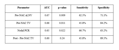 |
89 |
Statistical prediction of recurrence-free survival at 10 years in breast cancer patients
Isha Punn, Renee Cattell, Pauline Huang, James Kang, Thomas Ren, Varsha Talanki, Ashima Muttreja, Sarah Dacosta, Jules Cohen, Haifang Li, Lea Baer, Cliff Bernstein, Sean Clouston, Roxanne Palermo, Timothy Duong
This study examined whether axillary lymph-node size accurately predicts or improves prediction of 10-year recurrence-free survival. We found that for single-variable analysis, the top predictor of recurrence-free survival was pre-neoadjuvant chemotherapy (NAC) nodal volume (AUC=0.67), followed by pre-NAC tumor volume (AUC=0.66). In 4-variable analysis, the top set of predictors was pre-NAC nodal volume, human epidermal growth factor receptor 2 (HER2) positivity, pre-NAC tumor volume, and tumor volume difference between pre- and early-NAC time points (AUC=0.79). This is the first study of investigating prediction performance of recurrence-free survival using longitudinal volume change of axillary lymph-node volume in breast cancer patients.
|
|
2388.
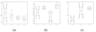 |
90 |
Gradient-entropy based radiomic features to predict molecular sub-types of pediatric Medulloblastoma on Gadolinium-enhanced T1w MRI
Sukanya Iyer, Marwa Ismail, Benita Tamrazi, Ashley Margol, Ramon Correa, Prateek Prasanna, Niha Beig, Ruchika Verma, Volodymyr Statsevyc, Anant Madabhushi, Pallavi Tiwari
Genomic Characterization of Medulloblastoma (MB) has recently identified 4 distinct molecular subgroups: Sonic Hedgehog (SHH), wingless (WNT), Group 3, and Group 4. These subgroups have shown different clinical behaviours and benefits to subgroup-specific treatments. We explored the feasibility of a new gradient-entropy radiomic feature, CoLlAGe, to distinguish molecular sub-types of MB on Gd-T1w MRI. Our results using multi-class comparison via one way ANOVA and post-hoc comparison showed significant differences in CoLlAGe features obtained across molecular sub-types. Our feasibility results suggest that the CoLlAGe features in different tumor regions observed on routine Gd-T1w MRI may potentially serve as surrogate markers to non-invasively characterize molecular sub-types of pediatric MB.
|
|
2389.
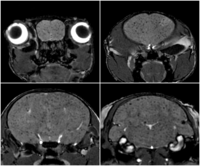 |
91 |
Characterization of the arrest and retention of iron-labeled breast cancer cells and the growth and progression of brain metastases in NSG mice
Natasha Knier, Amanda Hamilton, Ashley Makela, Paula Foster
Patient-derived xenografts in NSG mice provide a novel and more clinically relevant model of studying breast cancer brain metastasis in comparison to traditional cell lines in nude mice. NSG and nude mice both received brain-seeking breast cancer cells and were imaged with MRI to assess cell arrest, retention, and growth. Images revealed significantly more brain metastases and overall whole-body tumour burden in NSG mice than nudes. These results provide characterization of the NSG mouse as a preclinical platform for PDX models and demonstrates the importance of imaging to establish this model for future advancements in drug development and personalized medicine.
|
|
2390.
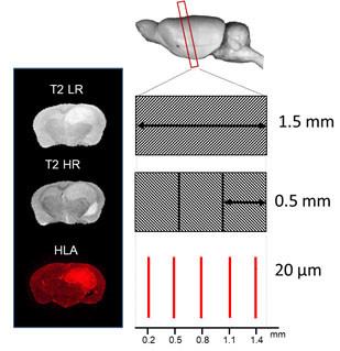 |
92 |
Stacked In-plane Histology for Quantitative Validation of Non-invasive Imaging Biomarkers: Application to an Infiltrative Brain Tumour Model
Haitham Al-Mubarak, Antoine Vallatos, Joanna Birch, Lindsay Gallagher, James Mullin, Lesley Glmour, John Foster, Anthony Chalmers, William Holmes
Despite the advance in medical imaging, it is important to validate new imaging biomarkers for a particular disease against histopathology, which is considered the ground truth. Here we propose a methodology for the quantitative validation of MRI biomarkers by the co-registration of histological probability maps with MR images. Using a mouse model of infiltrative brain tumours, we show this approach is far more robust than those currently applied.
|
|
2391.
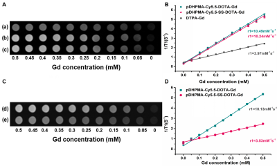 |
93 |
Reductive microenvironment responsive gadolinium-based polymers as potential safe MRI contrast agents
Xueyang Xiao, Shiwei Guo, Kui Luo, Qiyong Gong
The abstract provided an alternative strategy to develop highly efficient and safe gadolinium-based MRI macromolecular contrast agents (Gd-mCAs) via conjugation of small molecular DOTA-Gd to a stimuli-responsive biodegradable and amphiphilic block DHPMA copolymer through a ROX-sensitive biocleavable disulfide bond. Also, its potential as efficient and safe MRI mCAs for cancer diagnosis have been investigated.
|
|
2392.
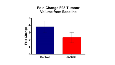 |
94 |
MR spectroscopy to assess decreased tumor choline as a marker of response to choline kinase inhibitors
Claire Kelly, Clementine Lesbats, Sofya Osharovich , Arthur Taylor, Violaine See, Edward Delikatny, Harish Poptani
Tumor volume and single voxel in vivo ¹H MRS were used to assess the effects of the choline kinase inhibitor JAS239 in the F98 rat glioblastoma (GBM) model. Five F344 rats were inoculated with GBM cells and subsequently treated for 5 consecutive days with 4 mg/kg JAS239 or saline. A reduction in total choline (tCho) in tumors treated with JAS239, along with tumor growth arrest was noted in comparison to saline treated rats. JAS239 preferentially inhibited choline metabolism in tumors as no changes were observed in tCho levels from the contralateral brain.
|
|
2393.
 |
95 |
High-Throughput Automatic Tumor Detection and Segmentation in Small-Animal MR Imaging of Patient-Derived Tumor Xenografts
Presentation Not Submitted
Sudipta Roy, Kooresh Shoghi
Computer aided tumor detection and segmentation of small animal MR images are prone to spurious lesion, false detection, under segmentation, over segmentation, incompatibles of huge number of images for small animal MR imaging. We propose computer aided method using the combination of fast C-means, morphology and single-phase level set to detect and segment tumor lesions from T2 weighted MR images. Proposed method gives over 90% accuracy when applied to homogeneous tumors.
|
|
2394.
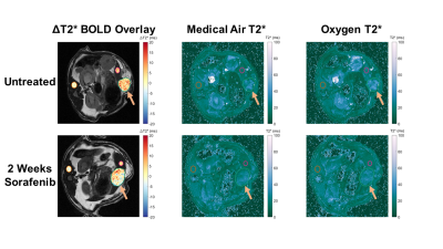 |
96 |
Blood oxygen level dependent MRI detects changes in hepatocellular carcinoma induced by sorafenib treatment
Keith Michel, Nina Munoz, Kiersten Maldonado, James Bankson, Jia Sun, Aliya Qayyum, Rony Avritscher
Imaging biomarkers are needed for assessing treatment response in hepatocellular carcinoma (HCC). We evaluated BOLD MRI with hyperoxic challenge in an orthotopic rodent model of HCC for animals treated with the widely used kinase inhibitor sorafenib. A reduction in ΔT2* in tumor and background liver was exhibited for rats treated with sorafenib relative to untreated controls, while no significant change was observed in skeletal muscle. These results demonstrate that BOLD MRI could be a useful tool for detecting treatment effects in HCC.
|
|
2395.
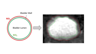 |
97 |
TrueFISP MRI assay for fast & reliable tumor stage classification in carcinogen induced orthotopic bladder cancer
Daniele Procissi, Alexander Glaser , Yanni Yu, Joshua Meeks
Using a TrueFISP sequence we implemented a non-invasive assay for stage classification of bladder tumors using a orthotopic murine bladder cancer model. Because generation of this tumor model is achieved by administration of carcinogenic agents it is naturally heterogeneous. The imaging method proposed allows accurate assessment of tumor burden and provides a tool for randomization to experimental treatments.
|
|
2396.
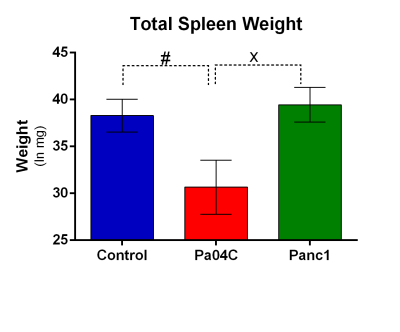 |
98 |
Spleen metabolism altered by human pancreatic cancer xenografts
Santosh Kumar Bharti, Paul Winnard, Raj Kumar Sharma, Yelena Mironchik, Marie-France Penet, Zaver Bhujwalla
Cachexia is a poorly understood metabolic syndrome characterized by cancer-induced tissue wasting and weight loss. Cachexia occurs with the highest frequency and severity in pancreatic ductal adenocarcinoma (PDAC). To further understand this syndrome, here we used 1H MRS to analyze spleen metabolites in normal mice and mice with and without cachexia-inducing PDAC. We detected profound spleen weight loss in cachectic mice. 1H MR spectra identified significant depletion of amino acids, cholines, creatine in cachectic mice that provide new insights into the syndrome that may present novel strategies to prevent or reduce cachexia-induced weight loss and the morbidity and mortality associated with the syndrome
|
|
2397.
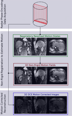 |
99 |
3D non-rigid motion correction for quantitative assessment of hepatic lesions with dynamic contrast enhanced MRI
Matteo Ippoliti, Tobias Schaeffter, Marcus Makowski, Christoph Kolbitsch
Dynamic Contrast Enhanced (DCE) MRI of the liver is a powerful qualitative and quantitative clinical methodology used to identify lesions and characterize their functional behavior. Respiratory motion can lead to artefacts, impairing the calculation of contrast uptake curves and quantitative functional information. Here we present an approach, which provides non-rigid motion-corrected endothelial permeability (Ktrans) maps obtained from DCE images with temporal resolution of 6s and isotropic spatial resolution of 1.5mm3 acquired during free-breathing. The proposed framework was evaluated in 10 patients and led to an improved visualization of hepatic lesions and subsequent derivation of Ktrans maps.
|
|
2398.
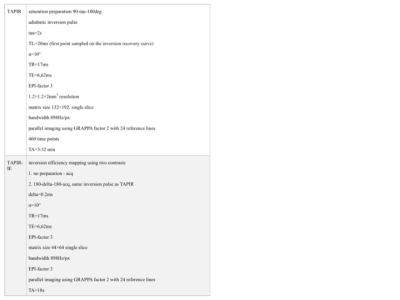 |
100 |
A novel imaging biomarker for cancer from multicomponent T1 relaxometry
Ana-Maria Oros, Anna Weglage, N. Jon Shah
We hypothesised that T1 relaxation curves sampled with very high temporal resolution reflect the existence of several environments in healthy and brain tumour tissue. Relaxation properties of healthy as well as tumour tissue, identified by FET-PET in a hybrid MR-PET environment, were investigated using a Look-Locker inversion recovery sequence sampled with a 17ms time resolution and 460 time points. The properties of normal appearing tissue were very similar in patients and healthy volunteers. In addition, a novel component was identified in brain tumour patients, which seems characteristic of the presence of tumour and oedema.
|
|
Back to Program-at-a-Glance |  Back to Top Back to Top |
| The International Society for Magnetic Resonance in Medicine is accredited by the Accreditation Council for Continuing Medical Education to provide continuing medical education for physicians. |
