Digital Poster Session
Acquisition, Reconstruction & Analysis Back to Program-at-a-Glance Back to Program-at-a-Glance
|
Tuesday, 14 May 2019
Digital PosterAcquisition, Reconstruction & Analysis
2399 -2423 Optimizing Reconstruction
2424 -2448 Image Reconstruction
2449 -2473 Image Processing & Analysis |
| |
Optimizing Reconstruction
Digital Poster
Acquisition, Reconstruction & Analysis
Tuesday, 14 May 2019
| Exhibition Hall |
08:15 - 09:15 |
| |
|
Computer # |
|
2399.
 |
101 |
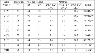
Optimization of a 3-channel gradient waveform for FRONSAC encoding

E. Bhuiyan, Nadine Dispenza, R. Constable, Gigi Galiana
This work reports the performance of various gradient waveforms for Fast Rotary Nonlinear Spatial Acquisition (FRONSAC) encoding, varying the amplitude, frequency and phase of the oscillation on different channels. Waveforms using three NLG channels were used to image an American College of Radiology (ACR) phantom, and root-mean-square error (RMSE) relative to a fully sampled reference was used to evaluate performance. Experimentally observed trends support those reported in previous work, which was based on theory and simulations. For the given hardware, the results suggest that the best combination for C3, S3 and Z2 are 64, 64 and 32 cycles per readout and 1.74×10⁶mTm-3, 1.74×10⁶mTm-3 and 3.05×10⁶mTm-2 respectively.
|
|
2400.
 |
102 |
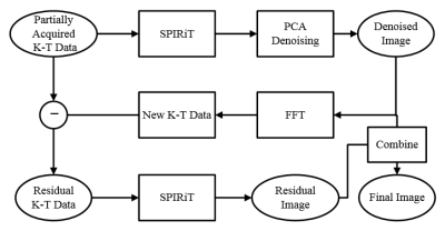
Feasibility Study of Improving SPIRiT by Exploiting Artificial Sparsity in Dynamic MRI
Presentation Not Submitted
Jucheng Zhang, Zhikang Wang, Wenhong Ding, Xia Kong, Zhifeng Chen
Improving spatiotemporal resolution is of great importance for dynamic MRI in clinical circumstances. An improved SPIRiT method using artificial sparsity and PCA denoising is proposed in this work. Simulated cardiac perfusion phantom and in-vivo cardiac cine experiments were conducted. The proposed method showed better image quality compared with GRAPPA and the frame-by-frame SPIRiT method.
|
|
2401.
 |
103 |
PHASE OFFSET CORRECTION METHODS FOR 7T MRI
Shaeez Abdulla, David Reutens, Steffen Bollmann, Viktor Vegh
At the 7T MRI field, the absence of a volume reference coil results in inter channel phase offsets. It is therefore important to understand the impact of using different phase offset correction methods for producing combined phase images. We quantitatively analysed multi-channel offset corrected 7T GRE-MRI phase images of a phantom obtained using five established methods. Magnetic susceptibility images of a brain were assessed qualitatively in addition. We found that methods which phase offset correct using echo time dependent signal phases contain systematic errors, whereas single echo time methods produce more accurate results.
|
|
2402.
 |
104 |
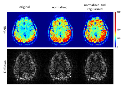
System conditioning during GRAPPA kernel training improves temporal SNR in accelerated EPI-based functional, diffusion, and perfusion MRI applications
W Hoge, Jonathan Polimeni
This work examines methods to improve the conditioning of the linear system of equations used to compute GRAPPA and Dual-Polarity GRAPPA reconstruction coefficients, and it's effect on temporal SNR in applications that employ accelerated EPI data. We test three methods: (i) system normalization, (ii) simple Tikhonov regularization, and (iii) 2D k-space filters applied to the calibration data prior to the linear system formation. Examples of tSNR improvement are shown, drawing from EPI-based in-vivo functional, diffusion, and perfusion imaging data acquired at 3T and 7T.
|
|
2403.
 |
105 |
Low-Latency Reconstruction for Real-Time Speech MRI
Sagar Sudhakara, Yongwan Lim, Weiyi Chen, Shrikanth Narayanan, Krishna S. Nayak
Real-time MRI provides the ability to visualize dynamic processes as they occur. This may require low latency, defined as the total time between when a pose occurs and when a digital representation appears on a screen for interpretation and/or use by the scan operator. We explore the tradeoff between image quality and latency for speech production imaging, where high-latency constrained reconstruction is the current state-of-the-art. We demonstrate that image quality adequate for a) confirmation of stimuli compliance and b) identification of subject motion can be provided to the scan operator with a latency less than 70ms.
|
|
2404.
 |
106 |
Simultaneous multislice reconstruction for spiral MRI using slice-SPIRiT
Changyu Sun, Yang Yang, Craig Meyer, Xiaoying Cai, Michael Salerno, Daniel Weller, Frederick Epstein
Simultaneous multislice (SMS) imaging provides through-plane acceleration. While current reconstruction methods for non-Cartesian imaging (and also for Cartesian imaging) utilize either in-plane or through-plane coil information, we reasoned that a slice-SPIRiT model could utilize both in-plane and through-plane kernel calibration information, and potentially outperform methods like conjugate-gradient SENSE (CG-SENSE). We developed a slice-SPIRiT method and compared it to CG-SENSE for spiral cardiac cine imaging. Slice leakage artifacts using slice-SPIRiT were 52.9% lower than using CG-SENSE in phantoms, and the artifact power of slice-SPIRiT was 24.2% less than CG-SENSE in five volunteers. Slice-SPIRiT is a promising method for spiral SMS imaging.
|
|
2405.
 |
107 |
Self-Estimated Subspace Reconstruction for Highly-Accelerated Dynamic Golden-Angle Radial MRI
Li Feng, Qiuting Wen, Hersh Chandarana, Ricardo Otazo
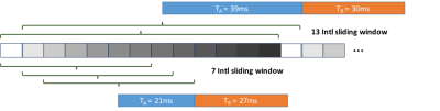
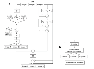
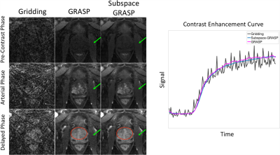
Subspace-constrained reconstruction is a powerful technique to accelerate dynamic MRI. However, its performance is relatively limited for applications where a robust temporal model is not available. This work proposes to estimate temporal basis from undersampled dynamic golden-angle radial data without the need of a model or additional navigators, and to apply the estimated temporal basis for subspace-constrained reconstruction of undersampled dynamic images. The reconstruction algorithm also enforces an additional low-rank constraint on the resulting low dimensional dynamic images in the subspace. The proposed self-estimated subspace-constrained reconstruction technique was demonstrated for DCE-MRI of the prostate.
|
|
2406.
 |
108 |
Refined-subspaces for two iteration single shot T2-Shuffling using dictionary matching
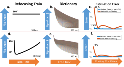
Yamin Arefeen, Nick Arango, Siddharth Iyer, Borjan Gagoski, Kawin Setsompop, Jacob White, Elfar Adalsteinsson
Half-Fourier-acquisition-single-shot-turbo-spin-echo (HASTE) serves as a valuable tool for fetal MRI as it is robust to fetal motion and produces images with T2-weighted contrast. However, due to T2-decay and T1-recovery during the acquisition, clinically applied HASTE with sub-180° refocusing pulses and partial-fourier readouts, often yield images with compromised diagnostic quality compared to multi-shot T2-weighted imaging. T2-shuffling exploits a forward model of signal evolution to mitigate blurring and improve contrast in image reconstruction. We propose single-shot imaging with a refined-subspace, iterative application of T2-shuffling, with demonstration in numerical models, that reduces blurring artifacts and improves image contrast in comparison to conventional HASTE.
|
|
2407
 |
109 |
Improving the Performance of Accelerated Image Reconstruction in K-Space: The Importance of Kernel Shape
Video Permission Withheld
Rodrigo Lobos, Justin Haldar
A variety of popular k-space reconstruction methods (e.g., GRAPPA, SPIRiT, SAKE, LORAKS) assume that missing k-space data can be interpolated by convolving the k-space data with appropriate filters. In most of these methods, the kernel shape is usually chosen to be rectangular. However, when these filters are interpreted in the spatial domain, the use of rectangular kernels implies that the filters will have anisotropic resolution. In this work, we investigate the use of elliptical kernels that have more isotropic resolution. Results demonstrate that elliptical kernels have better reconstruction performance, lower computational complexity, and lower memory usage than rectangular kernels.
|
|
2408.
 |
110 |
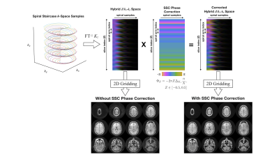
Improved Parallel Imaging with a 3D Spiral Staircase Trajectory
Ashley Anderson III, Dinghui Wang, James Pipe
A flexible 3D “Spiral Staircase” (SSC) trajectory is introduced that reduces g-factor losses from through-plane parallel imaging acceleration, regardless of coil geometry. Results demonstrate up to a 5x g-factor improvement over Cartesian SENSE for through-plane acceleration in axial brain acquisition with R = 3.
|
|
2409.
 |
111 |
Making Reconstruction WORK (Weighted Optimized Reconstruction of K-Space): Improving CNR/SNR via non-FFT Weighted Reconstruction
Vincent Schmithorst, Ashok Panigrahy
FFT-based reconstruction is suboptimal in the presence of signal decay during acquisition and between-excitation (shot-to-shot) variance in relaxation parameters. We present a novel reconstruction algorithm, Weighted Optimized Reconstruction of K-space (WORK), which weights each k-space point differently, optimizing for all sources of variance. Simulation results demonstrate the potential for 2X temporal SNR improvement in gradient-echo EPI acquisitions compared to standard FFT reconstruction while preserving spatial information. Substantial SNR improvement is also demonstrated for a pCASL 2D GE-EPI-SMS acquisition.
|
|
2410.
 |
112 |
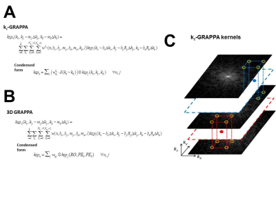
kz-GRAPPA for 3D parallel imaging with localized estimation of interpolation kernels
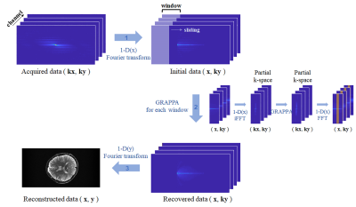
Steen Moeller, Sudhir Ramanna, Essa Yacoub, Kamil Ugurbil, Mehmet Akcakaya
A kz-dependent shift-variant 3D GRAPPA approach for reconstructing 2D under-sampled k-space is proposed. The method results in equal or lower g-factors compared to a conventional shift-invariant 3D kernel. In turn, this permits higher 2D ky-kz accelerations, and promises significant advantages for functional and diffusion imaging. The method is demonstrated with anatomical and diffusion imaging using thin slabs.
|
|
2411.
 |
113 |
Full 3D ky-kz-kx GRAPPA reconstruction of SMS MB EPI
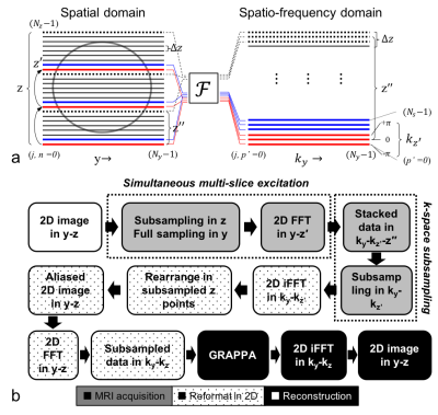
Chan Hong Moon, Hoby P. Hetherington , Jullie W. Pan
EPI with sub-second sampling rates is essential for fMRI to increase tSNR and filter out physiological noise. Simultaneous multiple slice (SMS)-EPI using multi-band (MB) slice excitation has been successfully applied to acquire whole brain fMRI data in < 1s. However, the reconstruction of SMS-EPI remains in separate 2D/1D GRAPPA or partial 3D GRAPPA in ky-kz’-kx domains. To further increase acquisition speed, TR< 500ms, higher k-space dimensional GRAPPA can be used to improve reconstruction performance, e.g. increase SNR and decrease aliasing artifacts. To meet this need we developed a full 3D ky-kz-kx GRAPPA reconstruction for SMS-EPI validated it by simulation and experiment at 7T.
|
|
2412.
 |
114 |
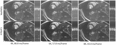
Reducing g-factor for TGRAPPA accelerated real-time cardiac cine imaging
Sen Jia, Haifeng Wang, Xin Liu, Hairong Zheng, Dong Liang
TGRAPPA acceleration alleviates the intense tradeoff between spatial and temporal resolutions for real-time cardiac cine imaging. However, it suffers from significant noise amplification due to ill-conditioned inverse reconstruction at high acceleration factors. A quadruple extended TGRAPPA reconstruction model is established to jointly utilize the additional spatial encoding capability of background phase and the high-order noise model by nonlinear kernel method. Prospective real-time cine experiments showed superior noise suppression of this non-iterative technique at 6-8X acceleration.
|
|
2413.
 |
115 |
Virtual Slice Concept for Improved Simultaneous Multi-Slice MRI Employing an Extended Leakage Constraint
Suhyung Park, Liyong Chen, Alexander Beckett, David Feinberg
Simultaneous multi-slice (SMS) MRI has recently drawn attentions in its use by acquiring linearly combined signals contributed from all excited slices. In this work, we introduce a novel, SMS reconstruction that extends an inter-slice leakage constraint to intra-slice aliasing with virtual slice concept by generalizing parallel MRI as a special case, thus directly estimating the individual slices from undersampled SMS data. Motivated by the leakage block, we generate virtual slices from intra-slice aliasing signals and then penalize these virtual slices as well as real slices simultaneously by keeping only aliasing-free slice of interest while enforcing inplane aliasing and neighboring slices to zeros, respectively.
|
|
2414.
 |
116 |
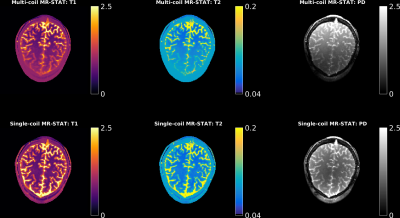
Enhanced MR-STAT by a multi-coil reconstruction framework
Oscar van der Heide, Mike Eijbersen, Cornelis van den Berg, Peter Luijten, Alessandro Sbrizzi
MR-STAT is a framework for obtaining multi-parametric quantitative MR maps using data from single short scans. A large-scale optimization problem is solved in which spatial localisation of signal and estimation of tissue parameters are performed simultaneously by directly fitting a Bloch-based volumetric signal model to the time domain data. Previously, only data from a single receive channel could be incorporated into the reconstruction. In this work we extend the MR-STAT framework to allow parameter maps to be reconstructed from multi-coil data resulting in a more robust reconstruction process that has higher scan-efficiency and is less prone to coil shading artefacts.
|
|
2415.
 |
117 |
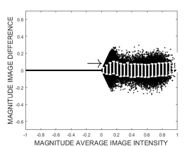
Correcting MRI-specific biases introduced when Bland-Altman plots are used to compare the performance of reconstruction algorithms
Michael Smith, Ishani DasGupta, Elise Fear
The Bland-Altman plot is a commonly-used graphical method to compare two measurement techniques and look for systematic biases or outliers. We have identified that the Bland-Altman approach of plotting the differences between the two techniques against their average will introduce false biases in an MRI context. These biases are introduced by the magnitude operation necessary to display or analyze MRI images. We demonstrate a modified Bland-Altman approach that corrects these biases.
|
|
2416.
 |
118 |
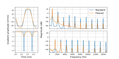
Acoustic Noise Reduction Using Digital Filters
JP Jordaan, Andre Van der Kouwe, Ali Alhamud, Ernesta Meintjes
We explored the use of digital low-pass filters to reduce the acoustic noise produced during DTI acquisitions, focusing mainly on the EPI readout. The filters attenuate the high-frequency harmonics of the gradient waveforms which results in reduced spectral content of the mechanical vibrations induced in the gradient coil assembly. Results showed a reduction of 2.9 dBA for peak sound pressure level (SPL) and 2 dBA for equivalent continuous sound level (Leq) with no acquisition time penalty or reduction in image quality. The proposed method has the potential to be generalized to most gradient waveforms.
|
|
2417.
 |
119 |
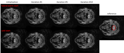
Method for Reduction of Reconstruction Time for Compressed Sensing of Multi-Image Series
Andrew Wheaton, Samir Sharma, Antonios Matakos
This proof-of-concept study demonstrates a method to reduce CS reconstruction time for multi-image series (e.g. relaxometry mapping, multi-echo, or dynamic) by leveraging the similarity of data across the image series. The method consists of two components: a) re-using auto-calibrated coil sensitivity maps computed from data of the first image[0] and b) warm starting the iterative reconstruction of each image[i] using the final output from the reconstruction of the previous image[i-1] in the series. One insight is a ‘hybrid warm start’ created by combining the magnitude from the previous image[i-1] reconstruction and the phase of the back-projection of the current image[i].
|
|
2418.
 |
120 |
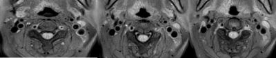
Optimization of increasing signal-to-noise ratio (SNR) and contrast of black-blood T1-weighted images in carotid artery
Yuhki Hamada, Daisuke Yoshimaru, Ayumi Ueno, Ayumu Funaki, Chifumi Maruyama, Tatsunori Sone, Masataka Sato
We have developed a new black-blood imaging with radial scan (multi vane) method and improved motion sensitized driven equilibrium (iMSDE). In this study, we changed the following parameters [refocusing flip angle (RFA), TE prep, flow velocity encoding (VENC)]. In addition, we measured SNR and Contrast ratio (CR) to optimize image quality. As RFA increased in radial scan with iMSDE, SNR of muscle rose gently and CR increased. With the extension of TE prep, SNR of muscle declined and CR also declined. As VENC decreased, CR rose gently. There was a significant difference compared with the conventional method.
|
|
2419.
 |
121 |
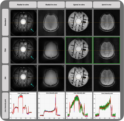
Rapid prOtotyping of 2D non-Cartesian K-space trajEcTories (ROCKET)
Presentation Not Submitted
Pavan Poojar, Sairam Geethanath, Ashok Reddy, Ramesh Venkatesan
Rapid prOtotyping of 2D non-CartesIan K-space trajEcTories (ROCKET) aims to aid researchers interested in rapid development and testing of new MR methods starting from pulse sequence design to image analysis. This was achieved by utilizing Pulseq for pulse sequence design and graphical programming interface for image reconstruction and analysis. ROCKET was demonstrated on two non-Cartesian k-space trajectories – FID based radial and spiral. Each trajectory was tailored into three different trajectories based on rotating angle – standard, golden angle and tiny golden angle. All studies were performed on Siemens scanner demonstrated on in-vitro phantom and in-vivo healthy brain acquisitions and SNRs were computed.
|
|
2420.
 |
122 |
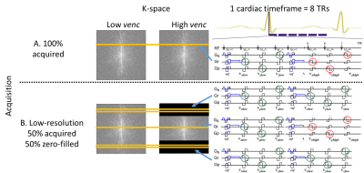
Dual-venc phase contrast MRI with increased flow encoding efficiency
Maria Aristova, Alireza Vali, Hassan Haji-Valizadeh, Liliana Ma, Suvai Gunasekaran, Daniel Kim, Michael Markl, Susanne Schnell
To decrease scan time while maintaining SNR and velocity dynamic range of dual-venc 2D phase-contrast MRI with 3-directional velocity encoding, the sequence was modified to zero-fill the extremes of k-space in the high-venc (HV) acquisition while collecting fully sampled low-venc (LV). In vitro sensitivity analysis shows antialiasing success over 95% with up to 50% zero-filled HV scans. Preliminary data from a healthy control aorta indicate that antialiasing success approaching 100% can be maintained using HV scans. This promising approach may be extended to further improve flow encoding efficiency in volumetric scans.
|
|
2421.
 |
123 |
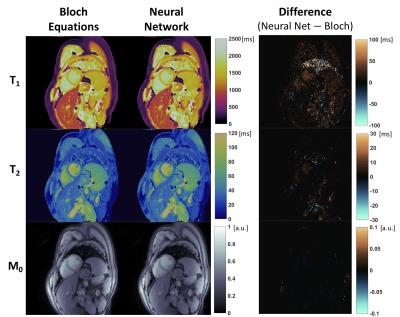
A Neural Network for Rapid Generation of Cardiac MR Fingerprinting Dictionaries with Arbitrary Heart Rhythms
Jesse Hamilton, Danielle Currey, Mark Griswold, Nicole Seiberlich
Cardiac MR Fingerprinting with ECG gating typically requires that a new Bloch equation simulation be performed after each scan so that the subject’s cardiac rhythm is incorporated in the dictionary. However, this may be challenging for clinical translation and online reconstruction. This study proposes to use a neural network to rapidly generate the dictionary when given input ranges for T1 and T2, as well as the cardiac rhythm (RR intervals). The network produces dictionaries for arbitrary cardiac rhythms and is more than 100 times faster than performing a Bloch equation simulation.
|
|
2422.
 |
124 |
THE IMPACT OF MR IMAGES ACQUISITION PROCESS ON RADIOMIC FEATURES: PHANTOM STUDIES TO SUPPORT CLINICAL RESEARCH
Linda Bianchini, Francesca Botta, Daniela Origgi, Marta Cremonesi, Paolo Arosio, Alessandro Lascialfari
Radiomic analysis of radiological images allows the extraction of quantitative features that can represent a support tool for clinical decision. The investigation of these features stability during the MR image acquisition process represents the aim of this study. The features short- and long-term repeatability was tested on a common MR phantom, imaged with the clinical protocol for gynecological imaging. The non-repeatable features were identified and can be excluded a priori in potential clinical studies. Simultaneously, a dedicated phantom was designed to mimic the pelvis and to investigate the stable features, especially the ones characterizing the texture of the imaged tissue.
|
|
2423.
 |
125 |
On the Selection of Slice Profile for Through-Plane Resolution in Multi-Slice MR Imaging
Eric Stinson, Soudabeh Kargar, Roger Grimm, Stephen Riederer
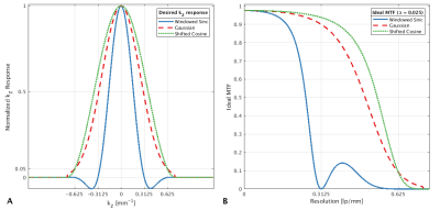
Multislice imaging is a mainstay of clinical MR exams, but image reformatting is limited by through-plane resolution. Some methods aim to overcome this by acquiring overlapping slices and deconvolving the slice profile. However, slice profiles which have zero crossings in the Fourier (spatial frequency) domain preclude the recovery of those spatial frequencies. Here, we describe the problem and provide a solution in the form of slice profiles without zero crossings in kZkZ-space.
|
|
| Top |
Image Reconstruction
Digital Poster
Acquisition, Reconstruction & Analysis
Tuesday, 14 May 2019
| Exhibition Hall |
08:15 - 09:15 |
| |
|
Computer # |
|
2424.
 |
126 |
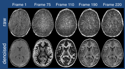
90 min in 12 min? Accurate Surface Reconstruction from Short Ultra-High (0.5mm iso) Resolution T1-Weighted Image
Giovana Cover, Andre van der Kouwe, Reza Farivar
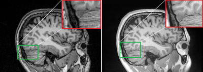
Gray matter (GM) thickness is a marker of injury and is detected using magnetic resonance imaging (MRI). Even though it possible to acquire images that has substantiallyhigher resolution, they suffer from high noise, requiring multiple acquisitions and averaging to yield suitable quality. In this work, we aimed to improve the sensitivity of surface-based GM estimation from 0.5mm resolution MRI acquired over a clinically-normal time. We evaluated four denoising filters and compared the GM thickness estimates using surface reconstruction from the results, showing that it is possible to reduce MRI acquisition time and maintain relevant features for the GM thickness.
|
|
2425.
 |
127 |
Compressed Sensing Velocity Encoded Phase Contrast Imaging: Monitoring Skeletal Muscle kinematics
Vadim Malis, Usha Sinha, Sinha Shantanu
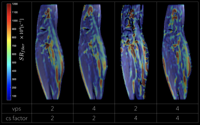
Velocity Encoded Phase Contrast (VE-PC) imaging is an established technique for monitoring muscle kinematics. Dynamic studies require consistent repeated execution of motion paradigms to map skeletal motion. The high number of repeated contractions limits studies to low % maximum voluntary contraction and limits participation of cohorts with compromised muscle function. We explore combining multi-coil data with compressed sensing and reconstruction to reduce acquisition times. VE-PC images acquired with different compressed sensing factors are assessed for accuracy of velocities and strain rate tensor during isometric contractions. Our results show that CS undersampling by 4 yields accurate velocity and strain tensor values.
|
|
2426.
 |
128 |
A GPU-based Modified Conjugate Gradient Method for Accelerating Wave-CAIPI Reconstruction
Haifeng Wang, Shi Su, Xin Liu, Yuchou Chang, Dong Liang
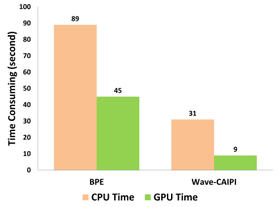
Wave-CAIPI is a novel 3D imaging method with corkscrew trajectory in k-space to speed up MRI acquisition. However, the 3D data acquisitions of Wave-CAIPI are also tremendous for reconstruction calculations. In order to accelerate the reconstruction procedure, we realized a Wave-CAIPI reconstruction using a modified GPU-based conjugate gradient (CG) algorithm to reduce time cost of reconstructions. The experiments of in vivo human brain dataset show that using our GPU-based Wave-CAIPI reconstruction can achieve similar image results as the conventional CPU-based Wave-CAIPI reconstruction with less time cost than the conventional CPU-based Wave-CAIPI reconstruction.
|
|
2427.
 |
129 |
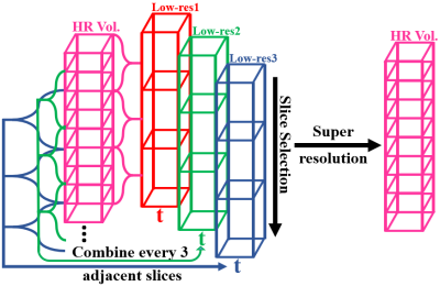
Spatial-Temporal Super-Resolution Technique on Complex-Valued T2*-Weighted Dynamic MRI
Duohua Sun, Chidi Ugonna, Marc Lindley, Nan-kuei Chen
We present an approach for improving spatial and temporal resolution of complex-valued T2*-weighted dynamic MRI. Compared with traditional magnitude-valued spatial super-resolution method, our technique can better recover signal loss caused by the susceptibility dephasing effect. We propose that phase information can be utilized in spatial super-resolution to reduce the dephasing artifact. The feasibility of temporal super-resolution using complex-valued data is also separately evaluated for time-signal variation recovery. One limitation of our temporal super-resolution approach, which will be addressed in our future work, is the presence of leakage artifacts in the recovered time-signal due to linear interpolation bias.
|
|
2428.
 |
130 |
Robust 3D UTE T2* Mapping in MSK Using Fractional Order Bloch Equation
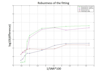
Dorottya Papp, Gyula Kotek, Stephan Breda, Dirk Poot, Edwin Oei, Juan Hernandez-Tamames
Determination of the T2* relaxation times with biexponential and monoexponential models has limitations, especially in case of complex, heterogeneous materials1. Using fractional order fitting model we can overcome these limitations8,9. In this model the introduced α parameter is the measure of the deviation from the monoexponential decay, and it accounts for micro-structural complexity. To evaluate the fractional order fitting, it was performed in patella tendon from UTE measurements, and we could demonstrate that compared to biexponential and monoexponential models it is less sensitive to variations to SNR.
|
|
2429.
 |
131 |
Native Space Outlier Rejection (NaSOR) for Arterial Spin Labelling
Courtney Bishop, Mari Lambrechts, James O'Callaghan, Adam Connolly, Roger Gunn
ASL suffers from relatively low signal-to-noise, so data cleaning strategies are required to optimise its utility. A previous method for outlier rejection of 2D-PASL data required time-consuming spatial normalization to standard space, degrading the original ASL data, and was limited to single inversion-time (TI) 2D-PASL. We therefore developed two native-space processing workflows, termed Native Space Outlier Rejection (NaSOR) and Native Space Perfusion-weighted Outlier Rejection (NaSPOR). The two native-space workflows performed comparably to an implementation of the previous standard-space method, in terms of both percentage of outliers rejected and coefficients of variation (CV) for test-retest CBF values, suggesting clinical utility.
|
|
2430.
 |
132 |
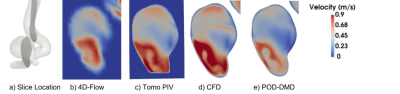
Verification and Validation of Merging Patient-Specific Computational Fluid Dynamics and 4D-Flow MRI
Alexandria Miller, Ali Bakhshinejad, Mojtaba Firoozabad, Ahmadreza Baghaei, Raphael Sacho, Kevin Koch, Christoff Roloff, Philipp Berg, Roshan D'Souza
In this work, we present the result of verification and validation of our previously developed method that enabled merging of patient-specific Computational Fluid Dynamics (CFD) and 4D-Flow MRI using Proper Orthogonal Decomposition (POD) to address limitations of both modalities. A constant fluid flow boundary condition was applied on a transparent in vitro aneurysm phantom geometry and the volumetric velocity field was scanned using 4D-Flow MRI, and tomographic Particle Image Velocimetry (tPIV). The latter has much higher spatial resolution and can be used to verify the accuracy of the results of merging CFD and volumetric 4D-Flow MRI. Results show that the POD-based merging algorithm enables reconstruction of fine flow details not seen in 4D-Flow MRI due to limited spatial resolution.
|
|
2431.
 |
133 |
Single-lung dynamics assessed using XD-GRASP MRI and automatic segmentation
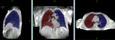
Artem Mikheev, Nicole Wake, Henry Rusinek, Hersh Chandarana
MRI is an attractive modality for monitoring dynamic lung function since it does not expose patients to ionizing radiation. In this study, a method to automatically segment the right and left lungs using XD-GRASP MRI was developed. The accuracy of our segmentation algorithm was assessed by comparing the automated segmentation results to manual segmentations outlined by expert observers. Excellent agreement was seen between the automated technique and the ground truth, suggesting clinical applicability of the method.
|
|
2432.
 |
134 |
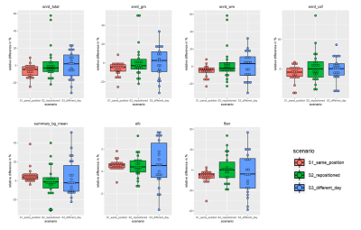
Intra-Session, Intra-Day and Inter-Day Reproducibility of MRI Image Quality Metrics in a Controlled Scan Setup
Till Huelnhagen, Ricardo Corredor-Jerez, Michael Amann, Emmanuelle Brès, Pavel Falkovskiy, Philippe Cattin, Tobias Heye, Oliver Bieri, Till Sprenger, Christoph Stippich, Jens Wuerfel, Ernst-Wilhelm Radue, Tobias Kober
Image quality plays a vital role in automated pipelines for medical image processing. Automated tools have thus been developed to detect low-quality images and ensure reliability of downstream results. These tools, however, often rely on image processing algorithms that can be sensitive to certain image features. In this study, we investigate the reproducibility of image quality measures provided by the open source image quality control tool MRIQC with respect to different scan setups. Results show that the reproducibility of some IQ measures is linked to the variation in the scan setup while for others it is less dependent on it.
|
|
2433.
 |
135 |
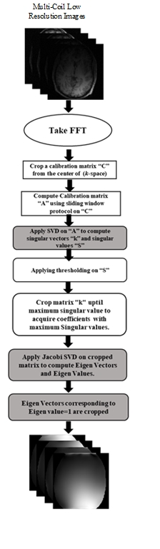
GPU Based Parallel Framework for the Estimation of Receiver Coil Sensitivities in SENSE Reconstruction
Presentation Not Submitted
Afaq Ashfaq, Muhammad Khalil, Hassan Shahzad, Sohaib Ayyaz, Hammad Omer
The estimation of receiver coil sensitivity information is important for SENSE (Sensitivity Encoding) reconstruction. Inaccurate sensitivity profiles degrade the reconstructed image quality. However, the methods to estimate the receiver coils sensitivity information are computationally intensive. This work proposes a parallel framework (for GPU implementation) for a recently proposed method of sensitivity estimation (which uses Eigen value decomposition of the multi coil low resolution images). The results show that the proposed method provides a 3.5x speed in our experiments while maintaining the reconstructed image quality.
|
|
2434.
 |
136 |
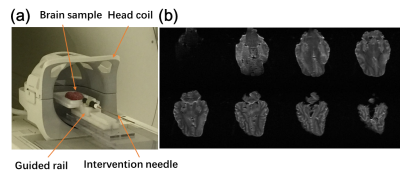
A novel feature based image reconstruction for neuro-interventional MRI
Kang Yan, Blanca Zufiria, Alexa Singer, Xudong Chen, Zhiyu Yang, Shuo Li, Suhao Qiu, Huajun She, Bomin Sun, Yiping Du, Zhipei Liang, Yuan Feng
Interventional MRI (I-MRI) provides exceptional advantages to other imaging modalities in image-guided neurosurgery. However, real-time imaging presents great challenges for temporal/spatial resolution, image contrast, and SNR. We presented a novel feature based image reconstruction algorithm using golden-angle sampling and compressed sensing. Images were decomposed into the reference part and the novel feature reflecting the interventional process. Experiments of using porcine brain for biopsy showed the proposed method had better performance in terms of SNR and computational time. It demonstrated that the proposed method have potentials in applications of MR-guided intervention such as image-guided epilepsy treatment.
|
|
2435.
 |
137 |
Efficient Motion-Corrected, Model-Driven Reconstruction for Simultaneous Multi-Contrast MPnRAGE
Steven Kecskemeti, Andrew Alexander
A model-based denoising algorithm is developed for efficient reconstruction of a large number of images with different T1 contrasts using MPnRAGE. The method takes only 50% the time of a single iteration of traditional constrained reconstruction methods.
|
|
2436.
 |
138 |
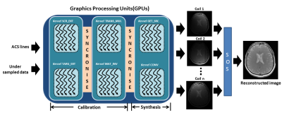
High performance GPU enabled GRAPPA reconstruction using CUDA
Omair Inam, Hamza Akram, Zoia Laraib, Mahmood Qureshi, Hammad Omer
GRAPPA is a parallel MRI technique that enables accelerated data acquisition using multi-channel receiver coils. However, processing a large data limits the performance of GRAPPA in terms of reconstruction time. This work presents a new GPU-enabled-GRAPPA reconstruction method using optimized CUDA kernels, where multiple threads simultaneously communicate and cooperate to perform: (i) parallel fittings of GRAPPA kernel on auto-calibration signals; (ii) parallel estimations of reconstruction coefficients; (iii) parallel interpolations in under-sampled k-space. In-vivo results of 8-channel, 1.5T human head dataset show that the proposed method speeds up the GRAPPA reconstruction time up to 15x without compromising the image quality.
|
|
2437.
 |
139 |
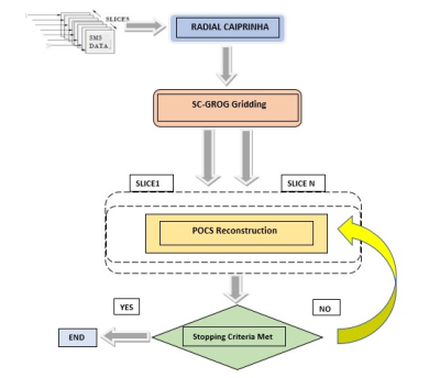
Self-Calibrated GRAPPA Operator Gridding (SC-GROG) for radially encoded Multi-Slice (SMS) Imaging
ZOONA JAVED, IBTISAM ASLAM, HAMMAD OMER
This work proposes a novel approach using Self-calibrating GRAPPA operator gridding (SC-GROG) for radially encoded simultaneous multi-slice imaging (SMS). The proposed method is implemented by combining non-Cartesian (NC) under-sampling (radial) with CAIPRINHA phase manipulation to accelerate data acquisition in SMS. Radial datasets are gridded using SC-GROG and reconstructed iteratively using Projection onto convex sets (POCS) algorithm. The results are compared with conventional NUFFT with POCS at increasing accelerations factors and quantified in terms of SSIM, PSNR and Artifact Power. It can be inferred from the results that the proposed method produces accurate reconstructions of SMS datasets.
|
|
2438.
 |
140 |
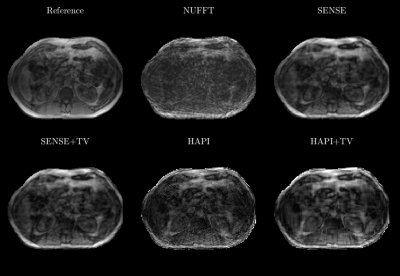
Rapid Abdominal Imaging Using Highly Accelerated Projection Imaging (HAPI
L. Tugan Muftuler, Nikolai Mickevicius, Andrew Nencka, Eric Paulson
Our group recently reported a new fast radial imaging method called Highly Accelerated Projection Imaging (HAPI) with coil sensitivity encoding. We demonstrated that radial projections acquired at specific angles and at high resolution resulted in a well-conditioned matrix equation. In the previous work, the performance of HAPI was demonstrated with simulations and a simple phantom scan using an 8-channel receive array. In the study presented here, the HAPI method was tested in vivo with volunteers.
|
|
2439
 |
141 |
Sparse-SENSE Reconstruction of GROG gridded Radial MRI
Video Permission Withheld
Khan Afsar, Ibtisam Aslam, Hammad Omer
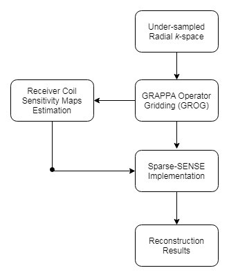
Under-sampled non-Cartesian trajectories play a vital role in accelerating MRI scan time; however, the solution image may have aliasing artifacts. In this work, a GROG gridding based Sparse-SENSE reconstruction is presented to get the solution image from the non-Cartesian under-sampled radial MR data. The proposed method is tested on 1.5T human head data at different acceleration factors (i.e. 4, 6 and 9) and compared with pseudo-Cartesian GRAPPA scheme. The results show that the proposed method provides significant improvement (e.g. 87% improvement in AP at AF=4) in the reconstructed images as compared to conventional pseudo-Cartesian GRAPPA reconstruction.
|
|
2440.
 |
142 |
A novel reconstruction method using regional constraints, designed for the dual-band EPI scanned with four-channel receiver coil elements
Presentation Not Submitted
Hiroshi Toyoda, Sosuke Yoshinaga, Mitsuhiro Takeda, Hiroaki Terasawa
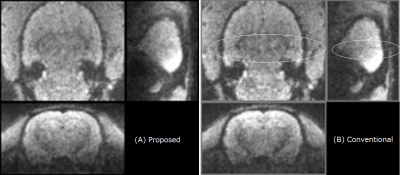
A novel reconstruction method was proposed for dual-band EPI in an MRI system equipped with four-channel receiver coils. This method was based on a conventional kernel method utilizing an iterative calculation with regional constraints in the image domain. The method significantly improves the quality of the reconstructed images, even in the regions with less coil sensitivity. The results showed higher signal-to-noise ratio, less signal leakage, and better long-term stability in repetitions in comparison to the conventional method. The proposed method can be applied to clinical systems that have relatively few receiver coils, as well as animal systems.
|
|
2441.
 |
143 |
Rapid Parallel MRI with Convolution-based Reconstruction (CORE) and Deblurring by Compressed Sensing
Efrat Shimron, Andrew Webb, Haim Azhari
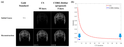
Methods combining Compressed Sensing (CS) and Parallel MRI (PI) for accelerated MRI have shown great promise, yet they are commonly hindered by heavy iterative computations. This work introduces the novel CORE-Deblur method for accelerated MRI, which integrates CS and PI and offers fast computations with very few iterations. CORE-Deblur utilizes the recently introduced CORE-PI technique and introduces the novel concept of using CS for image deblurring. Experiments with in-vivo data show that for highly subsampled k-space (R=5) CORE-Deblur reduces the number of CS iterations by 10-fold (from 95 to about 5-7) and improves the reconstruction accuracy by 5%-8%.
|
|
2442.
 |
144 |
Navigated Steady-State Free Precession with Water-Excitation for Real-Time Cardiac Imaging at 3 Tesla
Xi Peng, Sutton Brad
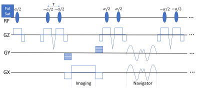
Real-time imaging offers the opportunity to be performed without the need for ECG synchronization and breath holding while requiring good contrast and high spatiotemporal resolution to resolve the myocardium dynamics. We propose a novel method employing bSSFP acquisition with partial separable model for high quality real-time cardiac imaging. For acquisition, the new method interleaves a self balanced spiral-in and spiral-out navigator with Cartesian acquisition for temporal basis estimation. A (1-1) binomial water excitation pulse is adopted to suppress lipid signal and achieve steady-state of the water. For reconstruction, the new method exploits the partial separable model with "soft" SENSE and sparsity constraints. In vivo experiments have been conducted and results show that the proposed method is able to produce high quality dynamic cardiac images.
|
|
2443.
 |
145 |
A localized reconstruction method for parallel imaging in MRI
Seohee So, Hyunseok Seo, HyunWook Park
Conventional parallel imaging methods reconstruct MR images from subsampled data, which utilizes spatial sensitivity information of multi-channel RF coil. In this study, localization of receiving coil sensitivity along readout direction (x) is introduced to efficiently utilize the coil sensitivity for parallel imaging. In the x-ky space, localization window is applied for estimation of missing data. Sensitivity localization in the readout direction makes near channels more weighted than distant channels for calculating estimation kernel. The proposed reconstruction method for parallel imaging considers the correlation between spatial sensitivities along the readout direction of receiving channels and region to be reconstructed.
|
|
2444.
 |
146 |
Robust Cardiac and Respiratory Self-Gating Using an Adapted Singular Spectrum Analysis (SSA-FARI): Application to Simultaneous-Multi-Slice Imaging
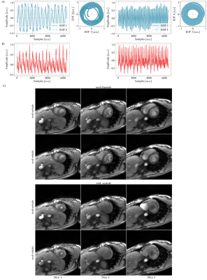
Sebastian Rosenzweig, Nick Scholand, H. Christian M. Holme, Martin Uecker
In cardiac MRI we have to deal with both cardiac and respiratory motion. Nowadays, breath holds and the use of external devices such as ECG are clinical practice to deal with this motion. However, not all patients can hold their breath and external devices are error-prone. Therefore, self-gating techniques have been developed to extract the respiration and cardiac signal from the k-space data itself. Many of those require various pre- and post-processing steps like filtering or averaging and lack robustness. Here, we present a novel and robust, yet easy to implement self-gating approach based on Singular Spectrum Analysis.
|
|
2445.
 |
147 |
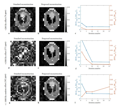
Joint Iterative Image Reconstruction and Field Map Estimation In Low Field MRI
Kirsten Koolstra, Merel de Leeuw den Bouter, Thomas O'Reilly, Peter Börnert, Rob Remis, Martin van Gijzen, Andrew Webb
Inaccuracies and temporal fluctuations in field map measurements form a major problem in image reconstruction for permanent magnet based low field MRI systems. These inaccuracies can potentially be corrected by using a joint image reconstruction and field map estimation algorithm. Simulation results show improved image quality when using a new updating scheme compared to standard iterative reconstructions.
|
|
2446.
 |
148 |
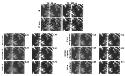
Nonlocal multispectral MRI upsampling for enhanced quality of high-resolution imaging reconstruction
Nikkita Khattar, Richard Spencer, Mustapha Bouhrara
High-resolution (HR) image reconstruction, or upsampling, is used widely in MRI post-processing analyses including image segmentation and registration. The nonlocal means (NLM) upsampling approach is simple to implement and has demonstrated excellent performance for HR image reconstruction from low-resolution images. Here, we extend this to incorporate multispectral (MS) data sets in which multiple images are acquired over a variable acquisition parameter. Further, we show that the use of our recently introduced nonlocal estimation of multispectral magnitudes filter for upsampling further enhances the quality of the reconstructed HR images as compared with use of the NLM filter or its MS version.
|
|
2447.
 |
149 |
A Partial-Fourier Method Recovering Signal Loss from Off-resonance
Seul Lee, Haisam Islam, Gary Glover
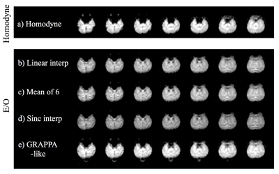
Functional MRI (fMRI) can have signal dropout due to off-resonance at susceptibility interfaces between air and tissue. Partial Fourier reconstruction is used for fMRI since it reduces scan time, however, existing partial Fourier reconstruction is vulnerable to off-resonance. In a previous study, we introduced a new partial Fourier reconstruction (even/odd (E/O)) and showed the new method was more robust to off-resonance compared to homodyne through simulation from fully sampled data. In this study, we acquired subsampled hypercapnia task fMRI data using both homodyne and E/O and showed there is less signal dropout and higher activation with E/O.
|
|
2448.
 |
150 |
A fully automated method for concealing patient identity in 3D multi-contrast brain MR images
Ke Gan, Weitian Chen
In addition to personally identifying information (PII) commonly found in metadata of medical images, superficial anatomical features contained in 3D brain MR images pose a unique challenge to medical privacy, and this place a serious obstacle for data sharing in large-scale collaborative efforts. A fully automated method for concealing patient identity in 3D multi-contrast brain MR images is presented. The proposed method is training-free and can be applied to automatically conceal patient’s identity information in the 3D brain MR images, which makes this approach particularly useful for handling brain MR images in large neuroimaging databases.
|
|
| Top |
Image Processing & Analysis
Digital Poster
Acquisition, Reconstruction & Analysis
Tuesday, 14 May 2019
| Exhibition Hall |
08:15 - 09:15 |
| |
|
Computer # |
|
2449.
 |
151 |
Image fusion of multiple independent MRI brain slabs to cover a whole Dugong brain in a small-bore, high-field pre-clinical scanner
Simone Zanoni, Kenneth Ashwell, Andre Bongers
This study we report about MRI of a fixed Dugong brain in a small bore pre-clinical MRI scanner. To enable the scanning of the brain that exceeds the sensitive scanner dimensions we propose a multi-slab imaging approach that uses an optimized image post-processing pipeline to merge independent slabs into a continuous high-resolution high-contrast 3D volume. We demonstrate that using this imaging and post-processing approach it is feasible to investigate relatively large objects in a pre-clinical scanner and retain full 3D information with the full benefit from the superior high resolution imaging capabilities of a high field pre-clinical scanner.
|
|
2450.
 |
152 |
Integrated image-space and Fourier-space analyses for unwrapping phase images of low signal-to-noise ratio
Nan-kuei Chen, Pei-Hsin Wu
We report a new post-processing procedure that integrates image-space and Fourier-space data analyses to improve the accuracy and reliability of phase unwrapping for MRI data of low signal-to-noise ratio (SNR). Our data demonstrate that the new phase-unwrapping method outperforms the conventional procedures in critical brain regions (e.g., near the air-tissue interfaces), and should prove valuable for studies that require accurate measurements of MRI phase values, such as quantitative susceptibility mapping (QSM), B0 field mapping, and temperature mapping.
|
|
2451.
 |
153 |
Bayesian Hierarchical Modelling for Analyzing Neuroimaging Results
Christopher Hammill, Benjamin Darwin, Darren Fernandes, Jason Lerch
The most common analysis of structural brain MRIs involves massively univariate modelling. Such analyses separately approach different levels of resolution (whole brain, regional, and voxel) and do not provide an easy solution to understanding whether some areas of the brain are more or less affected than others. Here we explore applying hierarchical bayesian modelling to simultaneously analyze brain MRI studies at multiple levels of resolution while allowing for the explicit interrogation of whether brain areas are differentially affected. In addition, we show that hierarchical modelling provides improved parameter recapture, sign error rate, and model fit.
|
|
2452.
 |
154 |
TemplateFlow: Standardizing standard 3D spaces in neuroimaging
Oscar Esteban, Rastko Ciric, Christopher Markiewicz, Yaroslav Halchenko, Mathias Goncalves, Satrajit Ghosh, Russell Poldrack, Krzysztof Gorgolewski
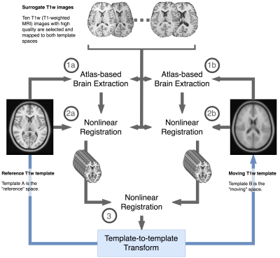
New community templates are generated to improve the resolution of data, to offer better alignment of brain structures across individuals incorporated into the template, and to ensure a better correspondence between the study cohort and the template. The resource is modular, thereby allowing researchers to easily use templates "off-the-shelf" or add new templates to the repository. Spatial mappings are distributed with the templates to allow transferring brain landmarks, masks, surfaces, segmentations, and parcellations between templates.
|
|
2453.
 |
155 |
SEGUE Unwraps MRI Phase Images Acquired in Mouse Brains at 9.4 Tesla Faster than PRELUDE
Anita Karsa, Rosie Goodburn, Karin Shmueli
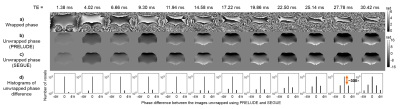
MRI phase images are increasingly used for Susceptibility Mapping or distortion correction. Spatial phase unwrapping is crucial but challenging: the computation time of PRELUDE, the current gold-standard method for robust, 3D unwrapping, increases rapidly with higher field strengths and longer echo times. A new method we have developed, SEGUE, produces similar results to PRELUDE in multi-echo brain and head-and-neck images, 1.6 to 83 times faster, but SEGUE has not been tested in pre-clinical high-field-strength phase images. Here, we show that SEGUE is similarly accurate and up to 4 times faster than PRELUDE in mouse brain images at 9.4 Tesla.
|
|
2454.
 |
156 |
Automated perfusion lesion delineation in stroke: comparison with experts and alternative automated strategies
Dave Saraswat, Jiun-Yiing Hu, Ivana Galinovic, Jochen Fiebach, Ahmed Khalil
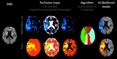
This study aimed to validate an in-house script that detects perfusion lesions in dynamic susceptibility contrast magnetic resonance images of acute stroke patients and compare its performance with commercially available software. Perfusion lesions were estimated from time-to-maximum and mean transit time maps of 94 stroke patients using our algorithm, Perfscape/Neuroscape, PMA, and Stroketool. These automatically delineated lesions were volumetrically and spatially compared with those delineated by a trained expert. Our algorithm performs comparably to other programs on the market and overestimates lesion volumes to a lesser extent; however, it is currently limited by its reliance on manual input.
|
|
2455.
 |
157 |
Knowledge-based definition of morphological alterations in brain MRI through the angle-based thresholding approach
Yusuke Tomogane, Jill Chotiyanonta , Can Ceritoglu, Kumiko Oishi, Michael Miller, Susumu Mori, Kenichi Oishi, for the Pediatric Imaging, Neurocognition and Genetic study
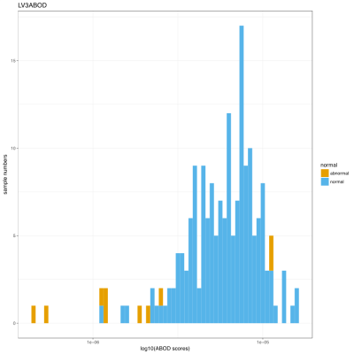
An automated method to detect brain morphological alterations was developed, which was designed for clinical pediatric brain MRIs with heterogeneous clinical conditions. Numerous image-feature-recognition algorithms have successfully defined abnormalities related to specific diseases, but there has been little research into a method that could identify a wide-range of radiological findings that could vary depending on the type and severity of different pathologies. A proposed approach—structural image parcellation followed by an angle-based outlier detection (ABOD) algorithm—could identify mild morphological alterations with high sensitivity and excellent specificity, when applied to clinical pediatric brain MRIs.
|
|
2456.
 |
158 |
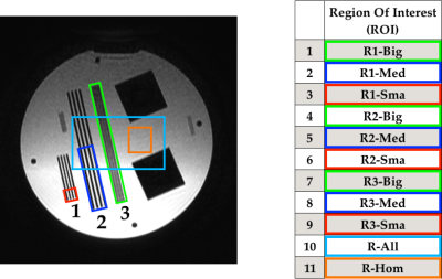
Scanner Variability of MR Based Radiomics Features
Peter Gibbs, Eun Sook Ko, Meredith Sadinski, Elizabeth Morris
This work utilizes an MR phantom to determine the repeatability, quartile coefficient of dispersion and potential efficacy of textural parameters calculated from gray level co-occurrence matrices, run length matrices, size zone matrices and neighborhood gray tone difference matrices. Images were obtained at 3 different field strengths, across 3 different manufacturers. Parameters based on gray level co-occurrence matrices showed excellent repeatability and low dispersion, whilst still demonstrating excellent discrimination between contrasting regions of interest.
|
|
2457.
 |
159 |
Segmenting Brain Metastases Using Deep Learning on Multi-Modal MRI
Presentation Not Submitted
Darvin Yi, Endre Grøvik, Michael Iv, Elizabeth Tong, Greg Zaharchuk, Daniel Rubin
Detecting and segmenting brain metastases is a tedious and time-consuming task for many radiologists, particularly with the growing use of multi-modal 3D imaging. Using deep learning to learn from the comprehensive pixel-wise labeled MRI-data, this work aims to train a fully convolution neural network for automatic detection and segmentation of brain metastases using multi-modal MRI. By training and testing on over 100 and 50 patients, respectively, including a variety of size and number of brain metastases from several primary cancers, this work provides a comprehensive investigation on the value and potential use of machine learning in a clinically relevant setting.
|
|
2458.
 |
160 |
Signal Enhancement and Optimum Receiver Arrays For Human Hyperpolarized 13C MR Spectroscopic Imaging
Hsin-Yu Chen, Adam Autry, Jeffrey Brender, Shun Kishimoto, Murali Krishna, Maryam Vareth, Robert Bok, Galen Reed, Albert Chen, Lucas Carvajal, Jeremy Gordon, Mark van Criekinge, David Korenchan, Duan Xu, Yan Li, John Kurhanewicz, Peder Larson, Daniel Vigneron
A data-driven processing framework was proposed for dynamic hyperpolarized 13C-MR Spectroscopic Imaging to maximally extract diagnostic information from existing datasets and techniques that utilized whitened-SVD2 to optimally combine array data, and tensor-low-rank denoising3,4 to enhance SNR. The framework was applied and evaluated on brain, abdomen, and pelvic datasets acquired using multi-channel arrays or single-element receivers. Substantial improvement in quality of low-SNR lactate and alanine was observed with 30+ fold apparent SNR gain, whereas high-SNR pyruvate remained largely artifact-free. Correlation of high kPL with biopsy-confirmed cancer strongly indicated that this recovered important pathological information.
|
|
2459.
 |
161 |
Reduction of Gibbs artifacts with the Domain Decomposition Fourier Continuation Method
Ruonan Shi, Jae-Hun Jung, Ferdinand Schweser
Magnetic Resonance (MR) images are obtained from the measured k-space Fourier coefficients. Due to acquisition time limitations and noise typically only a limited part of the k-space is acquired, resulting in Gibbs ringing on the images. We propose an efficient and accurate local reconstruction method that removes the Gibbs ringing and yields sharp image profiles near local edges.
|
|
2460.
 |
162 |
Association of geometric features with genetic markers in glioblastoma multiforme
Adam Hasse, Mark Dapash, Yong Jeong, Star Su, Abrianna Cummings, Sameer Ansari, Daniel Ginat, Timothy Carroll
A data analysis pipeline was developed to study relationships between geometric imaging features and the underlying tumor phenotype. This pipeline was run on the tumors of 203 patients clinically diagnosed with glioblastoma multiforme. For each tumor, the tumor bulk and percent non-enhancing volume was calculated, along with the surface regularity of any tumor with a 3D T1-weighted post-contrast MR scan. These features were compared to the expression of P53 and Ki67 sampled from these tumors. Although there were significant differences between multiple features for both genes, only the surface regularity was a significant predictor of Ki-67 proliferative index.
|
|
2461.
 |
163 |
Spherical harmonics coefficients estimation using deep neural network
Zhangxuan Hu, Zhe Zhang, Yuhui Xiong, Chun Yuan, Hua Guo
Diffusion-weighted imaging can be used to detect orientations of fibers to study human brain connectivity using tractography techniques. Spherical deconvolution based techniques have been widely used for the estimation of fiber orientation distribution (FOD), in which FODs are represented using spherical harmonics coefficients. However, high quality FOD estimation still requires large number of measurements. In this study, a deep neural network based method is proposed to estimate high quality FODs using highly q-space undersampled measurements thus to improve the acquisition efficiency.
|
|
2462.
 |
164 |
A Simple Fully Automated Method for Skull-Stripping Quality Control in Brain MR Image Processing Pipelines Evaluated Using Multicenter Data
Till Huelnhagen, Ricardo Corredor-Jerez, Claudia Bigoni, Veronica Ravano, Mário João Fartaria, Adrian Tsang, Rodrigo D. Perea, Sara Makaretz, Maria Laura Blefari, Yuchuan Zhuang, Bénédicte Maréchal, Elizabeth Fisher, Tobias Kober
Automated brain segmentation approaches are increasingly being used for decision support in routine clinical settings. While segmentation may be considered a “solved problem” in research, it is still challenging to assure reliable performance of automated tools in clinical settings, which is a crucial requirement for diagnostic tools. To ensure correct results, automated quality control procedures are of vital importance, but they are often difficult to implement or time-consuming to run. We propose a simple and fast fully automated method to detect segmentation errors, and we evaluate its performance to detect skull-stripping-errors using results of two different brain segmentation algorithms on a large multicenter dataset. Results show that the method is able to detect skull-stripping-errors with high specificity.
|
|
2463.
 |
165 |
A brain extraction algorithm for infant T2-weighted images based on the fuzzy c-means thresholding
Inyoung Bae, Dongchan Kim, Jun-Young Chung, Yeji Han
Brain extraction is an important step in image processing for research and diagnostic assessments using brain MR images. In this work, we proposed a brain extraction algorithm optimized for both 2D and 3D infant T2-weighted images based on the fuzzy c-means thresholding and spatial information of the neighboring slices. Quantitative analysis using the dice ratio was performed to compare the results of brain extraction using the proposed method, BET, iBEAT, and the manual segmentation.
|
|
2464.
 |
166 |
A 3D Fully Convolutional Network with various input dimension for brain extraction in MRI
Presentation Not Submitted
Xibo Zhang, Zhe Liu, Pascal Spincemaille, Yi Wang
A 3D Fully Convolutional Network is proposed using cascade architecture and combining two different channels to overcome the low accuracy of traditional methods. The network is applied to do the brain extraction.
|
|
2465.
 |
167 |
Are Root Mean Squared Error (RMSE) and Structural Similarity Index (SSIM) the Most Appropriate Metrics for Assessment of MR Image Quality?
Allister Mason, James Rioux, Sharon Clarke, Andreu Costa, Matthias Schmidt, Valerie Keough, Thien Huynh, Steven Beyea
Quantifying MR image quality is important for the evaluation of new image acquisition and reconstruction techniques. Automated objective image quality metrics (IQMs) such as root mean squared error (RMSE) and the structural similarity index (SSIM) are commonly used surrogates for radiologists’ perception of image quality, which can be considered the gold standard. By calculating the correlation between radiologists’ subjective grading and various IQM scores on degraded MR images, we demonstrate that RMSE and SSIM do not correlate as well as other IQMs and are potentially not the most appropriate metrics for assessment of MR image quality.
|
|
2466.
 |
168 |
Signal intensity form of Tofts model for quantitative analysis of dynamic contrast enhanced MRI data
Xiaobing Fan, Aritrick Chatterjee, Aytekin Oto, Gregory Karczmar
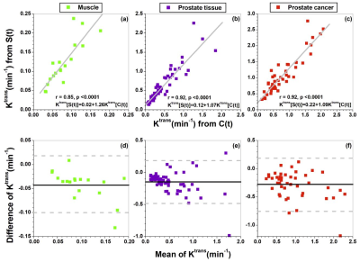
The Tofts pharmacokinetic model requires contrast agent concentration as function of time (C(t)), which is normally calculated using the non-linear model that could contribute some errors. Here, we present signal intensity (S(t)) form of standard Tofts pharmacokinetic model without calculating C(t). Human prostate DCE-MRI data were analyzed to compare physiological parameters calculated from the Tofts model using S(t) and C(t). The Ktrans and ve calculated from S(t) were correlated strongly with the values calculated from C(t). Bland–Altman analysis showed moderate to good agreement between for the Ktrans and ve calculated from Tofts model with S(t) and C(t).
|
|
2467.
 |
169 |
Test-retest reliability of resting-state brain small-world network properties across different data processing and modeling strategies
Qianying Wu, Ya Chai, Hui Lei, Fan Yang, Jieqiong Wang, Xue Zhong, John Detre, Hengyi Rao
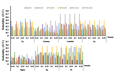
Resting-state fMRI assessed with graph theoretical modeling provides a noninvasive approach for measuring brain network topological organization properties, yet their reproducibility remains uncertain. Here we examined the test-retest reliability of seven brain small-world network metrics from well-controlled resting-state scans of 16 healthy adults using different data processing and modeling strategies. Among the seven network metrics, Lambda exhibited highest reliability whereas Sigma performed the worst. Weighted network metrics provided better reliability than binary network metrics, while reliability from the AAL90 atlas outweighed those from the Power264 parcellation. Global signal regression had no consistent effect on reliability of these network metrics.
|
|
2468.
 |
170 |
Development of a Quantitative Assessment tool for Peripheral Artery Feature Extraction (pCafe)
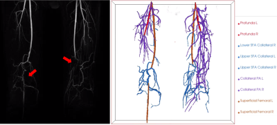
Li Chen, Thoetphum Benyakorn , Gador Canton, Niranjan Balu , Thomas Hatsukami , Jenq-Neng Hwang , Chun Yuan
Peripheral artery disease is a relatively common disease, normally caused by reduced blood flow to the limbs due to atherosclerosis in the arteries supplying them. Peripheral arteries’ anatomy, including collateral circulation, and flow information enable disease status assessment. We developed pCafe to semi-automatically trace peripheral arteries from 3D magnetic resonance angiography and measure both morphometry (anatomy) and intensity features (velocity). pCafe was validated on subjects with, and without peripheral artery occlusion, showing excellent agreement with human reviewer’s measurement (intra-class coefficient of 0.998). pCafe may be a useful tool to quantitatively characterize peripheral vascular structures in peripheral artery disease research.
|
|
2469
 |
171 |
Simultaneous Voxel-based Magnetic Susceptibility and Morphometry Analysis in Patients with Alzheimer’s Disease
Video Permission Withheld
Hirohito Kan, Yuto Uchida, Nobuyuki Arai, Yoshino Ueki, Yoshihiro Akagawa, Harumasa Kasai, Yasujiro Hirose, Noriyuki Matsukawa, Yuta Shibamoto
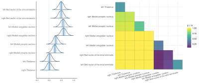
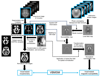
This abstract introduces an analysis pipeline of voxel-based magnetic susceptibility and morphometry (VBMSM) on single MR scan. To validate the proposed pipeline, we conducted VBMSM in control and Alzheimer’s disease (AD) groups. VBM was performed using the magnitude image. The susceptibility map was estimated by new dipole inversion utilized segmentation result. For whole brain susceptibility comparison, the susceptibility map was spatially normalized by the same transformation parameter for VBM. Significant susceptibility increases could be detected in regions associated withβ-amyloid deposition in AD. Brain atrophy also could be detected in AD. VBMSM is adaptable to neurodegenerative diseases including AD.
|
|
2470.
 |
172 |
Automated cloud-based workflow for quantification of MRI signal intensity – initial real-world clinical validation
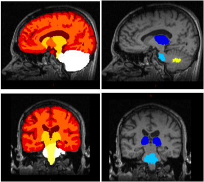
Marc Ramos, Vesna Prckovska, Paulo Rodrigues, Jinnan Wang, Franklin Moser, Markus Blank, Sheela Agarwal, Jacob Agris, David Moreno-Dominguez
One-third of brain MRI scans performed worldwide make use of gadolinium-based contrast agent injections to enable detection of the breakdown in the blood brain barrier by the resulting enhancement. Observations have shown increased signal intensity particularly in the globus pallidus and thalamus after multiple doses of linear contrast agents but there is no standard procedure to measure this contrast intensity. Current quantitative methods are manual, labor intensive, time-consuming and provide variable results.
We present a fully automatic workflow which accelerates the investigation of signal intensity in these nuclei after multiple doses of contrast agents by extracting the T1-weighted modal intensity value and applying appropriate corrections and normalizations to allow comparison across acquisitions and protocols. Automatic results matched up to 94% correlation with manual results and reduced the time by 90%.
|
|
2471
 |
173 |
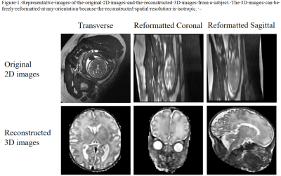
Accurate Prenatal Diagnosis of Cleft Palate Using Magnetic Resonance Imaging with 3D Super-resolution Reconstruction
Video Permission Withheld
Wei Yuan, Na Lu, Zhixian Deng, Xuhong Peng, Yingwei Qiu, Ting Song, Tianjing Zhang, Zhongping Zhang, Guoxi Xie
The routine clinical technique for prenatal diagnosis of cleft palate (CP) is ultrasound (US). However, the technique has difficulties on definitive diagnosis of fetal CP especially cleft posterior palate because of its technical limitations. Previous studies have demonstrated that magnetic resonance (MR) imaging can be a useful adjunct to the diagnosis of fetal CP and provides better diagnosis performance than US. However, these studies are mainly based on 2D MR imaging which has limited resolution along slice selective direction and cannot freely visualize the fetal palate from any orientation. To address this issue, we sought to use a 3D super-resolution reconstruction method to reconstruct 3D isotropic volumetric images from 2D images stacks and evaluate its feasibility of CP diagnosis.
|
|
2472.
 |
174 |
Analysis of Coil Combination for bSSFP Elliptical Signal Model
Nicholas McKibben, Grayson Tarbox, Michael Mendoza, Neal Bangerter
The elliptical model method for removing residual banding in balanced steady state free precession images requires accurate phase information to operate. Most datasets have separate data for each coil channel with different sensitivity, requiring combination either before or after processing using the elliptical model to eliminate differences in coil sensitivity. We demonstrate that the order in which these steps are taken matters, requiring coil combination after processing with the elliptical model depending on the method.
|
|
2473.
 |
175 |
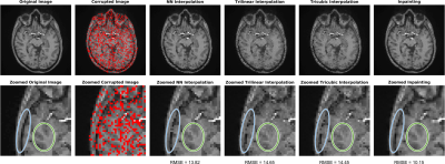
Comparison of Spatial Interpolation and Inpainting Methods for Estimation of Bad Fittings in Chemical Shift Imaging Data
Angel Torrado-Carvajal, Daniel Albrecht, Ovidiu Andronesi, Eva-Maria Ratai, Vitaly Napadow, Marco Loggia
Chemical Shift Imaging (CSI) allows for the quantification of brain metabolite concentrations across multiple voxels/slices. However, issues with model fit (e.g., suboptimal standard deviation, line width/full width at half-maximum, and/or signal-to-noise ratio) can result in the significant loss of usable voxels. Here, we show that an image restoration method called “inpainting” can be successfully used to restore poorly fitted CSI voxels. This method exhibits superior performance (lowest root-mean-square errors) compared to more traditional methods. Inpainting and similar techniques can prove particularly useful as a means of minimizing voxel loss in group voxelwise analyses in standard space.
|
|
Back to Program-at-a-Glance |  Back to Top Back to Top |
| The International Society for Magnetic Resonance in Medicine is accredited by the Accreditation Council for Continuing Medical Education to provide continuing medical education for physicians. |
