Digital Poster Session
Spectroscopy & Non-Proton MR Back to Program-at-a-Glance Back to Program-at-a-Glance
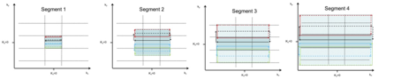
|
Tuesday, 14 May 2019
Digital PosterSpectroscopy & Non-Proton MR
2474 -2498 MRSI Acquisition & Reconstruction
2499 -2523 Other Nuclei MR
2524 -2548 MRS Clinical Application |
| |
MRSI Acquisition & Reconstruction
Digital Poster
Spectroscopy & Non-Proton MR
Tuesday, 14 May 2019
| Exhibition Hall |
09:15 - 10:15 |
| |
|
Computer # |
|
2474.
 |
1 |
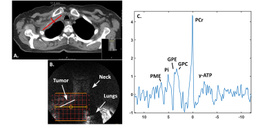
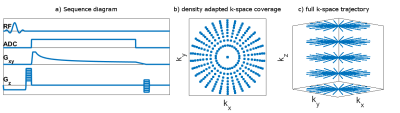
Feasibility of 7T 31P MRSI in lung tumors

Quincy van Houtum, Catalina Arteaga de Castro, Wybe van der Kemp, Joost Verhoeff, Jochem van der Voort van Zyp, Dennis Klomp
In this study we show the feasibility of 31P MRSI acquisition from a lung carcinoma tumor in a patient using a 31P whole body birdcage coil at 7T. We showed that even without B0 shimming, 31P spectra could be aligned and averaged to differentiate several metabolites, related to membrane metabolism, in the lung tumor. 31P MRSI has great potential for the detection of therapy response in lung tumor cancer, as often the tumor is still relatively large to obtain sufficient spectral signal.
|
|
2475.
 |
2 |
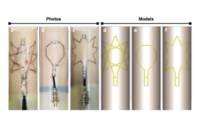
Reducing Signal Spreading with Non-Cartesian Encoding Methods for abdominal 31P 3D-MRSI with Focus on the Gallbladder
Lorenz Pfleger, Lukas Hingerl, Albrecht Schmid, Philipp Moser, Wolfgang Bogner, Yvonne Winhofer, Siegfried Trattnig, Martin Krššák
This study focuses on 31P 3D MR spectroscopic imaging (MRSI) of the gallbladder and the reduction of voxel bleeding by Non-Cartesian encoded data sampling. Our results demonstrate on a phantom that the contamination due to point-spread-function (PSF) can be decreased compared to conventional Cartesian phase encoding. Qualitative improvements were investigated by metabolic mapping of biliary phosphatidylcholine (PtdC) originating from the gallbladder.
|
|
2476.
 |
3 |
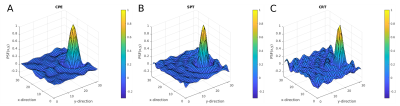
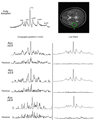
Comparison of Reconstruction Methods for Compressed Sensing phosphorus 31P-MRSI
Alejandro Santos Diaz, Michael Noseworthy
Phosphorus MR spectroscopy and spectroscopic imaging (31P-MRS/MRSI) provide information about energy metabolism, membrane degradation and pH in vivo. However, these methods are not often used primarily because of excessive scan time. Recently, compressed sensing has awakened interest as an acceleration method for MR signal acquisition. In this work we present a 31P-MRSI sequence that combines a flyback-EPSI trajectory with compressed sensing, and we compared two reconstruction methods, conjugate gradient L1-norm minimization and low-rank Hankel matrix completion. Overall, our results showed good preservation of spectral quality for low acceleration factors and an improved performance with the low-rank approach.
|
|
2477.
 |
4 |
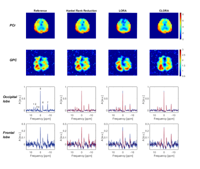
31P MRSI of the human brain at 9.4 T: Metabolic imaging applying low-rank denoising
Loreen Ruhm, Johanna Dorst, Nikolai Avdievich, Andrew Wright, Christian Mirkes, Jonas Bause, Klaus Scheffler, Anke Henning
31P MRSI enables the imaging of important components of the energy and cell membrane metabolism but suffers from low intrinsic sensitivity. In this work, we tested the LORA and CLORA low-rank noise reduction approaches to improve the quality of 3D 31P MRSI data and present first in vivo results for 31P MRSI acquired from the human brain at ultrahigh field strength of 9.4 T.
|
|
2478.
 |
5 |
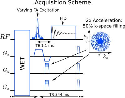
Accelerating High-Resolution Semi-LASER 1H-MRSI Using SPICE
Yibo Zhao, Yudu Li, Rong Guo, Bryan Clifford, Xin Yu, Zhi-Pei Liang
Most of the current 1H-MRSI techniques have several major practical limitations, including long data acquisition time, low spatial resolution and poor SNR. To overcome these limitations, we accelerate the semi-LASER technique by incorporating subspace modeling. With this improvement, semi-LASER is cable of achieving 1.9×1.6 mm2resolution in a 1.5 minutes scan, which is a significant improvement over the conventional semi-LASER. This imaging capability has been validated with in vivo experiments, and it may significantly enhance the practical utility of 1H-MRSI.
|
|
2479.
 |
6 |
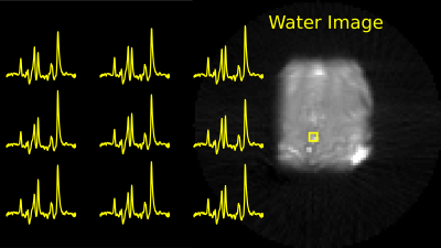
Non-Water suppressed High-Resolution 1H-MRSI of the Brain Using Short-TE SPICE with semi-LASER Concentric Ring Trajectory Acquisition
Uzay Emir, Pingyu Xia, Ulrike Dydak, Xiaopeng Zhou, Albert Thomas, Mark Chiew, Rong Guo, Yudu Li, Yibo Zhao, Zhi-Pei Liang
Magnetic resonance spectroscopic imaging (MRSI) is an appealing technique in both research and clinical settings. However, the utility of MRSI has been hampered by long acquisition times and artifacts caused by lipid contamination and poor water suppression. Recent advances in MRSI acquisition and preprocessing, like concentric rings (CRT) trajectories and SPICE (SPectroscopic Imaging by exploiting spatiospectral CorrElation) (REF3), have overcome some of these issues. This work reports our success in integrating SPICE with CRT acquisitions to address the challenges of sensitivity, spectral quality, speed, and spatial resolution.
|
|
2480.
 |
7 |
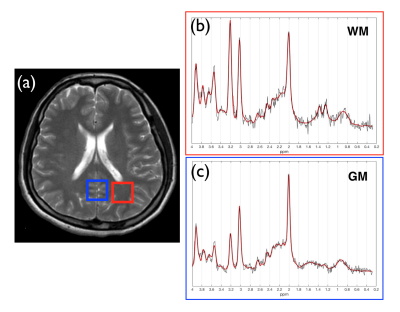
Parallel Imaging for Concentric Circle Readouts with GRAPPA Reconstruction for Full-Brain 3D-FID-MRSI at 7T
Lukas Hingerl, Bernhard Strasser, Philipp Moser, Gilbert Hangel, Stanislav Motyka, Eva Heckova, Stephan Gruber, Siegfried Trattnig, Wolfgang Bogner
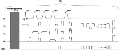
Non-Cartesian sampling methods for MRSI such as concentric ring trajectories (CRT) are highly suitable at field strenghts ≥7T while being SNR-efficient due to its self-rewinding property and low-pass k-space-weighting. However slewrate constraints enforce the CRT to sample the k-space periphery with 2-fold or 3-fold the time of the inner circles via more temporal interleaves (TI). The combination of variable density parallel imaging (PI) and CRT for MRSI allows high acceleration factors since the undesired but necessary variable TIs can be easily undersampled and allowing therefore higher accelerations in the k-Space periphery.
|
|
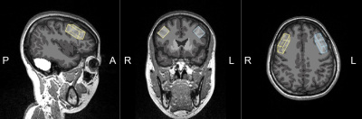
2481.
 |
8 |
In vivo echo-planar spectroscopic imaging (EPSI) at 7 tesla with readout segmentation for improved spectral bandwidth
Graeme Keith, Marco Vicari, Rosemary Woodward, David Porter
The use of echo-planar spectroscopic imaging at ultra-high field strengths is attractive due to its suitability for high spatial and spectral resolution (HiSS) acquisitions. The drawback of the method at 7T and above is the decreasing spectral bandwidth as field strength increases. This work seeks to decouple the spectral bandwidth from the spatial resolution by the use of readout segmentation to achieve shorter echo spacing. Readout segmented EPSI spectra collected in vivo at 7T and comparable to a standard SVS method are presented. This allowed the calculation of metabolite maps for NAA, creatine and choline.
|
|
2482.
 |
9 |
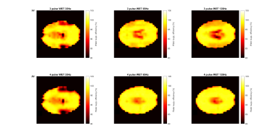
Improved whole brain water suppression efficiency with four-pulse WET in echo-planar spectroscopic imaging (EPSI) at 7 tesla
Graeme Keith, David Porter
Water suppression in MR spectroscopic imaging can be sensitive to variations in B1 in the sample, such as are present at 7 tesla. This work compares two versions of the WET water suppression method, the standard 3-pulse method and the extended 4-pulse method which is expected to be less sensitive to B1 variation. It is found that the 4-pulse method provides a greater consistency of water suppression efficiency across a range of B1 in both phantoms and the brain at 7T.
|
|
2483.
 |
10 |
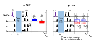
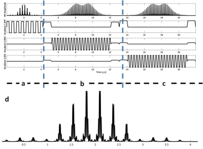
Fast spectral imaging at 7T using COKE (COherent K-t-space Epsi) with a spatially selective IR pulse (achieved by controlling pTX coil phases)
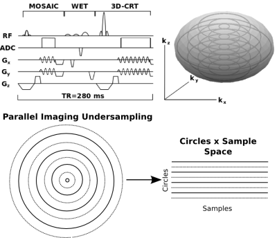
Rita Schmidt
In a recent study, we demonstrated the feasibility of the COKE (Coherent K-t-space EPSI) sequence at 3T MRI to halve acquisition time or double the spectral width (SW) compared to EPSI. In this study, we explored, on a phantom with mimicking brain properties, the benefits of COKE at 7T for fast spectroscopic imaging; using an SW of 2500Hz to better cover the metabolites' frequency range. We combined it with spatially selective inversion recovery (IR) using B1 phases optimization in 8-channels transmit coil to minimize the signal drop in the center and to optimize the IR in the lipid region.
|
|
2484.
 |
11 |
MR-SASSI-Accelerated, B1-Insensitive, Magnetic Resonance Spectroscopic Imaging at 7T: first in vivo results
Rebecca Feldman, Gaurav Verma, Priti Balchandani
Magnetic Resonance Spectroscopic Imaging (MRSI) is a signal-starved technique compared to conventional magnetic resonance imaging. At ultra-high fields, such as 7 Tesla the increased signal to noise permits the acquisition of improved spectra. Spatial localization performed the volume of interest is can be time consuming. We created a multi-region excitation pulse embedded within a B1-insensitive MRSI sequence and demonstrated its use in vivo to excite two distinct spectroscopic grids which are simultaneously acquired and disentangle using a low-resolution reference scan, thus accelerating the acquisition of MRSI data.
|
|
2485.
 |
12 |
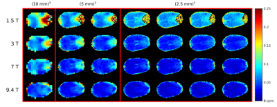
How does spatial resolution affect the spectral quality and quantification accuracy of whole-brain MRSI? A simulation study at 1.5T, 3T, 7T and 9.4T
Stanislav Motyka, Philipp Moser, Lukas Hingerl, Gilbert Hangel, Eva Heckova, Bernhard Strasser, Korbinian Eckstein, Simon Robinson, Benedikt Poser, Stephan Gruber, Siegfried Trattnig, Wolfgang Bogner
The quality of MRSI data depends strongly on B0 inhomogeneities, which cause broadening of metabolite resonances and decrease signal-to-noise ratios (SNR). B0 inhomogeneity is more severe at higher B0 field, diminishing the expected SNR and spectral resolution improvements. We have created simulation models which allow us to investigate how the spectral quality and quantification accuracy of MRSI changes with increasing spatial resolution and B0 field strength, using experimentally acquired data from 1.5T, 3T, 7T, and 9.4T. These simulations show not only that accurate MRSI quantification generally benefits from smaller voxels, but it does so particularly at UHF.
|
|
2486.
 |
13 |
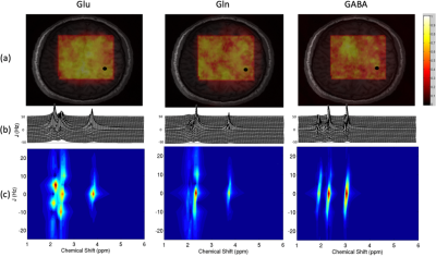
Accelerated J-Resolved 1H-MRSI with Limited and Sparse Sampling of (k, tJ)-Space
Lihong Tang, Yibo Zhao, Yudu Li, Rong Guo, Bryan Clifford, Chao Ma, Zhi-Pei Liang, Jie Luo
J-resolved 1H-MRSI is a powerful tool for mapping brain molecules, especially those with large spectral overlaps (e.g., glutamate, glutamine and GABA), yet it requires very long data acquisition times. Building on work in accelerated subspace-based imaging, this work proposes an accelerated acquisition scheme with limited and sparse sampling of (k, tJ)-space, based on a physics-based spectral model and Cramer-Rao lower bound analysis. A model-based processing scheme is also proposed, which performs model-based reconstruction and spectral quantification directly from the measurement data. The proposed method has been evaluated with simulated and in vivo experiments which have yielded very encouraging results.
|
|
2487.
 |
14 |
Detection of Choline, Glycine and Myo-inositol in Malignant Breast Cancer In-vivo Using Multi-dimensional Spectroscopic Imaging
Andres Saucedo, Manoj Sarma, Sumit Kumar, Kavya Umachandran, Melissa Joines, Stephanie Lee-Felker, Maggie DiNome, Michael Thomas
Multi-parametric MR techniques have been used to diagnose and monitor the therapeutic outcome of cancer in the breast and other tissues and organs. One-dimensional MRSI has shown significantly elevated choline and higher water-to-fat ratio in malignant tumors as compared to healthy controls. Two-dimensional MRS resolves peaks along an additional spectral dimension which overcomes the overlap limitation of 1D MRSI, thereby providing more discriminatory information for developing non-invasive methods for cancer grade determination. This study presents the first application of an accelerated, echo-planar based technique that acquires correlated (2D) spectroscopy data for each voxel of 1.5ml resolution within a 3D volume (5D EP-COSI) in breast cancer. Our preliminary results in a pilot cohort of malignant and breast cancer patients demonstrate changes in unsaturated fatty acids and increased choline in malignant group compared to benign and healthy women. These pilot results indicate the potential application of the 5D EP-COSI technique which may be useful in improving the specificity of breast cancer.
|
|
2488.
 |
15 |
High-Resolution T1 maps of Brain Metabolites
Antoine Klauser, Frédéric Grouiller, Sebastien Courvoisier, Francois Lazeyras
High-resolution T1 maps are measured over the whole human brain with proton FID-MRSI with incremental flip-angles. MRSI datasets with multiple flip-angles are reconstructed simultaneously through a low-rank-total generalized variation model and T1 values are determined by fit of the steady state magnetization with B1inhomogeneity correction. Twofold compressed-sensing acceleration enables acquisition of a single flip-angle in 5 min and resulting in acquisition of 3 flip-angles in 15 min. Precise determination of T1 would enables an accurate correction of signal loss and might provide important information on brain micro-structure.
|
|
2489
 |
16 |
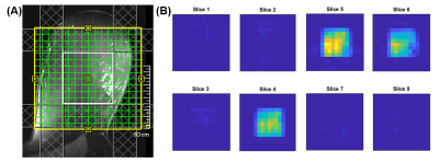
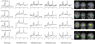
Comparison of ROI averaging and Spectral Localization by Imaging (SLIM) Using High Resolution 3D Echo Planar Spectroscopic Imaging (EPSI)
Video Permission Withheld
Sean Ellis, Peter Adany, Phil Lee, In-Young Choi
Conventional spectroscopic imaging methods have limitations that make acquiring metabolic information for complexly-shaped brain regions a challenge. The following study compares two methods for acquiring the regional metabolic spectra for a complex compartment shape: spectral estimation via the spatial-averaging of voxels, and Spectral Localization by Imaging (SLIM). Both techniques used the original data sets acquired from 3D Echo Planar Spectroscopic Imaging sequences. The two methods were compared, with the results showing that SLIM could provide comparable compartment spectra using fewer voxel acquisitions without a significant drop in spectral quality.
|
|
2490
 |
17 |
Validation of B0-adjusted and sensitivity-encoded spectral localization by imaging (BASE-SLIM) using High Resolution 3D EPSI
Video Permission Withheld
Peter Adany, In-Young Choi, Sean Ellis, Phil Lee
B0-adjusted and sensitivity-encoded spectral localization by imaging (BASE-SLIM) provides non-Fourier based localization for arbitrarily shaped compartments. We have extended BASE-SLIM to 3D and compared the outcome of BASE-SLIM reconstruction with that of voxel averaged high resolution 1H MRSI.
|
|
2491.
 |
18 |
B0 Drift Correction in Proton Chemical Shift Imaging
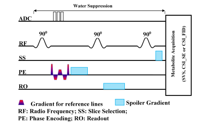
XIANFENG SHI, Young-Hoon Sung, Douglas Kondo, Perry Renshaw
This study aims to improve 1H CSI data quality, by presenting a novel method for correction of B0 instability (0.9 Hz/min drift) due to gradient system heating produced by application of DTI and fMRI sequences. The method tracks magnetic field drift using three reference lines in the 1H CSI data, which allow misaligned spectral data to be corrected in post-processing. This novel method may be combined with any spectroscopic technique that employs water suppression. Both phantom and in vivo data are presented, to demonstrate improved SNR and spectrum quality, with minimal influence on metabolite data acquisition or added cost.
|
|
2492.
 |
19 |
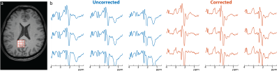
Retrospective motion compensation for edited MRSI data
Kimberly Chan, Peter Barker
A retrospective motion compensation method for edited MR spectroscopic imaging (MRSI) data of the human brain is presented. The algorithm identifies movement-affected data by comparing the residual water and lipid peaks at the same k-space point, and either phase corrects, replaces or removes motion-affected FIDs. The method was applied to in vivo GABA-edited MRSI data: relative to uncorrected spectra, the corrected spectra had significantly less subtraction artifacts. The method was also demonstrated for correction of glutathione-edited MRSI data.
|
|
2493.
 |
20 |
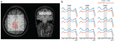
Spectroscopic imaging of macromolecule-suppressed J-difference editing of GABA at 3 Tesla
Kimberly Chan, Peter Barker
In this study, the feasibility of macromolecule-suppressed MR spectroscopic imaging (MRSI) of GABA in the human brain at 3T was investigated. PRESS-localized MRSI was performed for both macromolecule (MM)) suppressed GABA and non-suppressed (GABA+) editing. GABA concentrations and MM fractions were assessed and compared. Data quality metrics (B0 homogeneity, and GABA and water fit errors) were also calculated. A significant linear correlation of GABA+ with GABA concentrations was found. MM-suppressed GABA and GABA+ concentrations agreed with previously reported single-voxel values. Data quality metrics were also similar to those of prior single-voxel acquisitions.
|
|
2494.
 |
21 |
Ultra high-field, high-resolution semi-LASER MRSI of the brain
Yan Li, Artan Kaso, Ralph Noeske, Angela Jakary, Rolf Schulte, Christopher Hess, Janine Lupo, Peder Larson
The purpose of this study was to implement and optimize multi-voxel semi-LASER MRSI in brain regions that are frequently used in clinical studies, such as deep gray structures and motor cortex, within a clinically feasible time.
|
|
2495.
 |
22 |
Superresolution MRSI: a desirable acquisition trajectory.
Claudiu Schirda
The smoothly varying waveform and sampling that starts at k=0 and the innate property of rewinding periodically to k=0, makes the rosette trajectory achieve the same spatial resolution and spectral bandwidth as other trajectories (EPSI, SSI, CONCEPT) using less than twice gradient strength and slew rate. This makes it an ideal candidate for superresolution MRSI and ultra-high field SI acquisitions.
|
|
2496.
 |
23 |
Effects of Point Spread Function and Regularization Information on the MRSI with Compressed Sensing
Jung-Hsiang Chang, Yi-Hsun Yang, Tzu-Cheng Chao, Cheng-Wen Ko
Compressed Sensing can be very useful in accelerating Phase-encoded Proton MRSI. The sampling functions and the reconstruction settings have been known as critical factors in recovering the data of the accelerated acquisition. The present work compared the choices of sampling functions and the regularization information in the reconstruction in a hope to optimize the framework of Compressed Sensing based MRSI. The results suggest that the spectral quality can be retained for as high as five-fold acceleration with an appropriate undersampling and reconstruction setting.
|
|
2497.
 |
24 |
Undersampling strategies for compressed sensing accelerated MR spectroscopic imaging
Nutandev Bikkamane Jayadev, Christopher Lawhorn, John Chang, Vikram Kodibagkar
Magnetic Resonance Spectroscopic Imaging (MRSI) is an invaluable tool in cancer diagnosis due to its specificity but long scan times makes it less popular for routine clinical use. Clinical integration of accelerating methods such as compressed sensing (CS) can improve patient throughput. Two different undersampling strategies were implemented and statistical fidelity was tested to obtain a five-fold reduction is acquisition time; 1) a conventional variable density pseudorandom undersampling and 2) an a priori information based strategy that exploits anatomical scans. Statistical results from in vivo studies show the feasibility of using CS accelerated MRSI without loss of data fidelity.
|
|
2498.
 |
25 |
Compensation of Spectral Line Broadening in Proton-Echo-Planar-Spectroscopic-Imaging (PEPSI) using Dynamic Expansion of K-Space and Parallel Imaging
Stefan Posse, Bruno Sa De La Rocque Guimaraes
This study introduces a novel MRSI approach using dynamically expanding k-space acquisition during spectral encoding to compensate B0 inhomogeneity related signal losses and spectral line broadening, taking advantage of the sparsity of the spectral signal in the time domain. 2D PEPSI with segmented increases in k-space to a maximum 6x6-fold expansion in kx and ky using spectral time domain and ky undersampling was implemented on a clinical scanner. The study characterizes signal gains and resulting spectral line narrowing in regions with B0 inhomogeneity in phantoms and in vivo. This approach complements emerging hardware solutions for improving higher-order shimming.
|
|
| Top |
Other Nuclei MR
Digital Poster
Spectroscopy & Non-Proton MR
Tuesday, 14 May 2019
| Exhibition Hall |
09:15 - 10:15 |
| |
|
Computer # |
|
2499.
 |
26 |
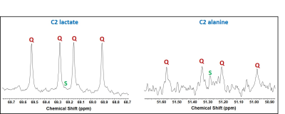
Spectral Editing of NAD+/NADH in 31P NMR spectra of Human Brain
Jimin Ren, A Dean Sherry, Craig Malloy
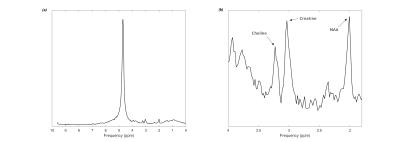
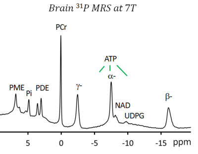
Nicotinamide adenine dinucleotides (NAD+/NADH) play an essential role in cellular redox reactions and many biological processes. Altered NAD+/NADH levels and redox state may be associated with development of neurodegenerative diseases and psychotic disorders. 31P MRS is currently the only non-invasive technique to measure NAD+/NADH levels and redox state in human brain in vivo. However, the present technique suffers two major drawbacks: (1) the severe overlapping of the NAD+/NADH signals with the α-ATP resonance, and (2) the distorted baseline underneath these signals. Here we present a novel spectral editing method that allows resolution of NAD+/NADH from α-ATP at baseline.
|
|
2500.
 |
27 |
Fast Quantification of Creatine Kinase Reaction Rate in Mouse Skeletal Muscle Using 31P Magnetic Resonance Fingerprinting
Kihwan Kim, Charlie Wang , Yuning Gu, Shuying Huang, Bryan Clifford, Zhi-Pei Liang, Xin Yu
We evaluated the accuracy and efficiency of a 31P magnetic resonance fingerprinting (MRF) method for fast measurement of the creatine kinase (CK) rate constant in mouse skeletal muscle. Our results showed consistent measurement of CK rate constant with less than 10% variations with only 6 signal averages, corresponding to 2 min acquisition.
|
|
2501.
 |
28 |
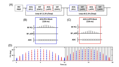
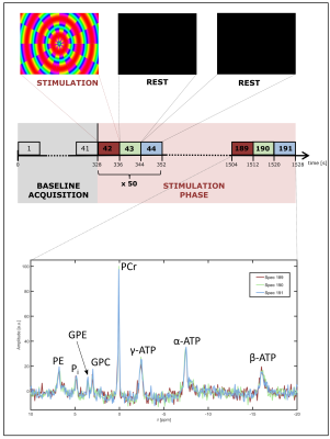
Functional 31P Magnetic Resonance Spectroscopy of the Human Visual Cortex using Repeated Short-Term Stimulation at 7T
Vanessa Franke, Ferdinand Zimmermann, Arthur W. Magill, Mark E. Ladd, Peter Bachert, Andreas Korzowski
Functional 31P Magnetic Resonance Spectroscopy allows the noninvasive observation of high-energy phosphate turnover in vivo, and might enable insight into the energy metabolism of activated brain areas. The purpose of this study was to investigate possible changes in 31P spectra of the human brain at B0 = 7T in response to repeated short-term visual stimulation. No significant changes in signal intensities and frequencies of 31P-containing brain metabolites were observed, which agrees with results from recent studies at ultra-high fields using long-term stimuli.
|
|
2502.
 |
29 |
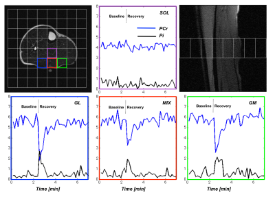
Dynamic 31P-MRSI of Human Calf Muscles using Compressed Sensing and a Low Rank Reconstruction
Alejandro Santos Diaz, Diana Harasym, Michael Noseworthy
Dynamic 31P-MRSI experiments require temporal resolution on the order of seconds to concurrently assess metabolic change in different muscles. In this study we developed a pulse sequence using a flyback-EPSI readout combined with compressed sensing (CS) to achieve a 9 second temporal resolution and tested it in 11 healthy volunteers during an exercise-recovery challenge of the lower leg muscles. Our results showed that the sequence was capable of assessing PCr depletion/recovery and intracellular pH at rest and following exercise, of multiple muscle groups simultaneously, using a clinical 3T MR system.
|
|
2503.
 |
30 |
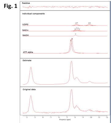
Dependence of 31P MRS Redox Ratio on T1 Saturation
Shizhe Li, Jan Willem van der Veen, Li An, JoEllyn Stolinski, Maria Ferraris-Araneta, Christopher Johnson, Jun Shen
31P measurement of the oxidized (NAD+) and reduce form (NADH) of nicotinamide adenine dinucleotide has been applied to in vivo assessment of the redox state of the brain. Since uridine diphosphate glucose (UDPG) overlaps with the nicotinamide adenine dinucleotide peaks we examined the effect of changes in the relative intensities of the UDPG basis components on quantification of the redox ratio. We found that the fitted redox ratio is significantly dependent on the UDPG basis whose chemically distinct components are subject to different T1 saturation effect in vivo.
|
|
2504.
 |
31 |
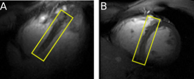
Human cardiac pH and Pi concentration during dobutamine stress measured by 7T 31P-MRS
Albrecht Schmid, Andrew Apps, Ladislav Valkovic, Elisabeth Tunnicliffe, William Clarke, Mark Peterzen, Stefan Neubauer, Oliver Rider, Christopher Rodgers
The hydrolysis of adenosine triphosphate (ATP) to adenosine diphosphate (ADP) in the reaction ATP + H20 ? ADP + Pi (inorganic phosphate) is used to drive all cellular processes, including those involved in ventricular contraction and relaxation. When spectral quality is sufficient to quantify the Pi peak, it is possible to assess the ratio of Pito phosphocreatine (Pi/PCr), which is an established measure of the muscle control of energy production. It is also possible to assess cardiac intracellular pH from the Pi to PCr frequency offset. Most human cardiac 31P-MRS studies report only the PCr/ATP ratio, and are typically unable to quantify cardiac Pi because of partially overlapping resonances from 2,3-diphosphoglycerate in blood. We aimed to use ultra-high (7T) field strength and a novel 31P STEAM sequence to 1) non-invasively measure myocardial Pi/PCr and pH at rest and 2) for the first time record these parameters during catecholamine stress.
|
|
2505.
 |
32 |
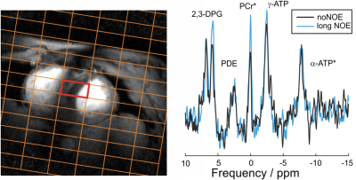
NOE enhancement for 31P-MRS of skeletal and cardiac muscle at 7T
Chang-Hoon Choi, Ladislav Valkovic, William T. Clarke, Albrecht I. Schmid, N. Jon Shah, Christopher T. Rodgers
Phosphorus MR spectroscopy (31P-MRS) is a proven method for probing tissue energetics and membrane metabolism. Nuclear Overhauser effect (NOE) enhancement can considerably improve the quality of 31P spectra. This has been demonstrated in the brain and prostate at 7T, but NOE has not yet been applied elsewhere in the human body at 7T. In this study, we evaluated NOE enhancement for 31P-MRS in human skeletal muscle and in the heart in vivo at 7T. We observe significant enhancements (e.g. for PCr/γ-ATP: 25%/16% in muscle, 31%/11% in heart), within regulatory SAR limits and without increasing the scan duration.
|
|
2506.
 |
33 |
Nutritional ketosis increases NAD+/NADH ratio in healthy human brain: an in vivo study by 31P-MRS
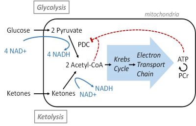
Lijing Xin, Özlem Ipek, Maurice Beaumont, Maya Shevlyakova, Nicolas Christinat, Mojgan Masoodi, Norman Greenberg, Rolf Gruetter, Bernard Cuenoud
Ketones represent an important alternative fuel for the brain under glucose hypo-metabolic conditions induced by neurological diseases or aging, however their metabolic consequences in healthy brain remain unclear. Here we report that ketones can increase the redox NAD+/NADH ratio in the resting brain of healthy young adults. As NAD is an important energetic and signaling metabolic modulator, these results provide mechanistic clues on how nutritional ketosis might contribute to the preservation of brain health.
|
|
2507.
 |
34 |
Metabolic fate of glucose in atypical meningioma studied by 13C NMR isotopomer analysis
Kumar Pichumani, Omkar Ijare, David Baskin
Meningiomas are tumors arising from meninges, the membranes surrounding the brain and the spinal cord. Majority of meningiomas (grade-1) are benign and grow slowly. However, atypical meningiomas (grade-2) exhibit increased cellular abnormalities, and grow at a faster rate than benign meningiomas. Moreover, atypical meningiomas prone to recurrence and show resistance to radiotherapy. Atypical meningiomas show higher 18F-FDG uptake in PET scans. No prior reports are available on investigating metabolic fate of glucose in atypical meningiomas. The goal of this study is to probe the metabolic fate of glucose using NMR based [U-13C]glucose isotopic tracing methods in patient-derived atypical meningioma cells.
|
|
2508.
 |
35 |
An Optimized PRESS Sequence for the Detection of 13C4-Glutamate at 9.4 T
Brennen Dobberthien, Anthony Tessier, Atiyah Yahya
Glutamate (Glu) incorporates 13C label on its C4 carbon (13C4-Glu) following a 13C-labelled glucose (Glc) infusion, resulting in a ≈2.51ppm proton “satellite” peak that provides an indirect measure of 13C4-Glu. Quantification of the satellite peak is complicated at short echo time (TE) due to overlap with the ≈2.49ppm N-acetylaspartate (NAA) peak. A PRESS, point resolved spectroscopy, (TE1, TE2) combination of (20ms, 106ms) was found to be suitable for resolving the ≈2.51ppm 13C4-Glu proton peak from that of NAA at 9.4T by exploiting differences in J-coupling evolution. The efficacy of the technique is verified on rat brain during a [U-13C6]-Glc infusion.
|
|
2509.
 |
36 |
Cardiac mechanical function and metabolism during hyperpolarized 13C experiments
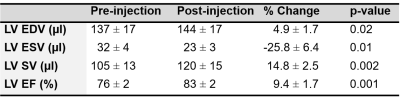
Gregory Barton, Erin Macdonald, Kara Goss, Marlowe Eldridge, Sean Fain
We investigated the relationship between contractile function and hyperpolarized (HP) [1-13C]pyruvate metabolism in a small animal model. We demonstrated significant functional changes in cardiac contractile function between pre- and post-infusion of [1-13C]pyruvate. The combined effect of infusion volume and pyruvate substrate likely explains most of the augmentation in myocardial mechanical function seen in these experiments. These data indicate the most appropriate time to image myocardial contractile function is soon as possible after HP 13C pyruvate infusion.
|
|
2510.
 |
37 |
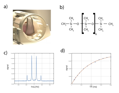
Protocol for multi-site quantitative evaluation of 13C radio frequency coils
Galen Reed, Rolf Schulte, Jae Park, Craig Malloy, Rie Hansen, Albert Chen
We present a protocol for measurement of SNR profile of 13C RF coils for clinical imaging systems. This protocol makes use of standard, vendor-provided pulse sequences as well as the natural abundance 13CH3 resonance of the dimethyl silicone (DMS) phantoms which are widely distributed. We also provide an open source code for processing and analysis.
|
|
2511.
 |
38 |
Enhanced sodium quantification accuracy in a 3T clinical 23Na MR stroke study
Nadia Paschke, Roberta Egoriti, Manuel Winkler, Eva Neumaier-Probst, Sherif Mohamed, Melina Samartzi, Marc Fatar, Lothar Schad
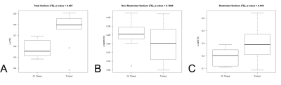
Tissue sodium quantification from sodium magnetic resonance acquisitions is a promising biomarker in ischemic stroke diagnostics and can be incorporated in clinical magnetic resonance protocols. Since no gold standard of protocol design and post-processing exists yet, we investigated three research questions for enhanced sodium quantification in 20 stroke patients: Are transmission field corrections necessary? Should the relaxation behavior be included in the quantification? Should manual evaluations of region of interests be replaced by automatic whole brain segmentation analyses? Based on this study, we propose to include user-independent segmentation and relaxation correction in 23Na whole brain analysis, while omitting transmission corrections.
|
|
2512.
 |
39 |
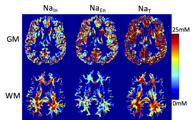
Combined 23Na/39K MRI for the quantification of Na+ and K+ concentrations in human skeletal muscle at 7 T
Lena V. Gast, Max Müller, Bernhard Hensel, Michael Uder, Armin M. Nagel
A non-invasive determination of Na+ and K+ concentrations in skeletal muscle tissue is desirable to gain insights into pathological processes connected to various diseases. In this work, the feasibility of combined quantitative 23Na/39K MRI at 7 T using a double-tuned 23Na/39K birdcage calf coil was examined. In phantom measurements, a 23Na/39K SNR ratio of 46.8 was found. Moreover, Na+ and K+ concentrations close to the real concentrations were determined. In skeletal muscle tissue, fast transverse relaxation of 39K leads to underestimation of K+ concentrations if no relaxation correction is applied.
|
|
2513.
 |
40 |
Whole-brain 23Na multi-parametric mapping at 7 Tesla
Lisa Leroi, Jacques Stout, Arthur Coste, Ludovic de Rochefort, Mathieu Santin, Romain Valabrègue, Franck Mauconduit, Cécile Rabrait-Lerman, Fawzi Boumezbeur, Alexandre Vignaud
Quantifying physical properties of sodium could be of benefit to assess more specifically changes in cellular homeostasis accompanying neuroinflammatory or neurodegenerative processes. Here, we performed whole-brain Quantitative Imaging using Configuration States (QuICS) in vivo at 7 Tesla to assess simultaneously Total Sodium Concentration (TSC) and relaxation times (T1 and T2). An acquisition time of 20 minutes was sufficient for a 10 mm3 isotropic resolution. In the future, the use of non-Cartesian trajectories could further reduce the overall acquisition time, opening the way to the additional estimation of the trace apparent diffusion coefficient.
|
|
2514.
 |
41 |
Optimizing Compressed Sensing for quantitative Sodium MRI of the human brain
Yasmin Blunck, Scott Kolbe, Bradford Moffat, Roger Ordidge, Jon Cleary, Leigh Johnston
The clinical application of Sodium MRI is hampered due to relatively low image quality and associated long acquisition times (TA). Compressed Sensing (CS) aims at a reduction of TA, but has been found to encompass quantitative estimation bias when used in low SNR x-Nuclei imaging. This work analyses CS in human brain Sodium MRI from both angles, acquisition speed-up and quantification, and presents an optimized setup allowing an up to four-fold TA reduction with recommendations for quantitative assessments. The demonstrated global optima of CS weighting parameters and achievable reduction in TA greatly support the transition of Sodium MRI into clinical routine.
|
|
2515.
 |
42 |
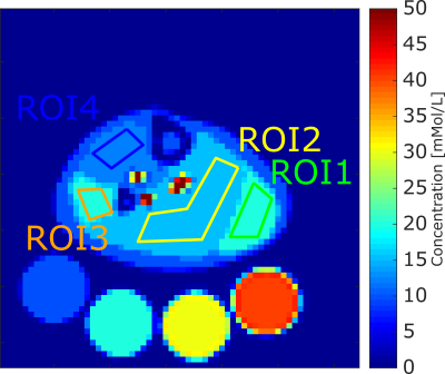
Measurement of 23Na MRI point-spread function (PSF) using a 3D printing resolution phantom
Paul Polak, Rolf Schulte, Michael Noseworthy
Acquisition of in vivo 23Na MRI suffers from many inherent technical challenges, including low signal, short T2 relaxation times, and the necessity of dedicated hardware for transmitting and receiving. Despite these issues, research remains attractive because of sodium’s essential role in cellular homeostasis, pH regulation and action potentials in neurons. Quantification of data acquisition and reconstruction techniques are essential in order to overcome 23Na MRI’s difficulties, and we present measurement of the point-spread functions of 3D radial pulse sequences in resolution phantoms with differing sodium concentrations.
|
|
2516.
 |
43 |
Comparison of Relaxometry-Weighted Sodium MRI and IDH Status in Cerebral Gliomas
Aliaksandra Shymanskaya, Wieland Worthoff, Karl-Josef Langen, N. Shah
Patients with cerebral gliomas were investigated using an enhanced SISTINA sequence to estimate sodium relaxation and its correlation to the IDH mutational status. Sodium MRI is used for the indirect assessment of sodium relaxation parameters through the relative change at two echo times in tumour and contralateral tissue. Sodium signals attributed to mostly restricted and non-restricted sodium are differentiated through the development of multiple quantum coherences. Changes in relaxation rates in total sodium correlate to the IDH mutational status on short time scale. This correlation could be caused by the change in sodium distribution in different compartments in diseased tissue.
|
|
2517.
 |
44 |
Measurement of Intraneurite Sodium Concentration from NODDI-based Partial Volume Correction of in vivo 23Na MRI
Iris Asllani, Guillaume Madelin, Marco Bozzali, Mara Cercignani
The sodium ion plays a crucial role in maintaining healthy brain function and metabolism. Changes in sodium concentration measured with 23Na MRI have been implicated in several diseases in the brain and other organs. However, due to SNR and scanning time requirements, 23Na MRI remains a low resolution technique hampered by partial volume effects. Here, we combined a partial volume correction (PVC) algorithm, previously used in Arterial Spin Labeling perfusion MRI, with Neurite Orientation Dispersion and Density Imaging (NODDI) to extract sodium concentration values from intra- and extra-neurite compartments in the brain in vivo using 23Na MRI at 3T.
|
|
2518.
 |
45 |
Optimized 3D Dictionary-Learning Compressed-Sensing Reconstruction for Quantitative Sodium Imaging in the Skeletal Muscle
Matthias Utzschneider, Sebastian Lachner, Nicolas Behl, Lena Gast, Andreas Maier, Michael Uder, Armin Nagel
Quantitative sodium MRI could be a sensitive tool for therapy monitoring in muscular diseases. However, sodium MRI suffers a low signal-to-noise ratio (SNR). 3D dictionary-learning compressed-sensing (3D-DLCS) enables SNR improvement and acceleration of sodium MRI, but it is dependent on parameterization. In this work a simulation based optimization method for 3D-DLCS is presented, which finds the most suitable parameters for 3D-DLCS in the context of sodium quantification. The method is applied in an in vivo study to quantify sodium in the skeletal muscle. The optimized 3D-DLCS yields a lower quantification error than the reference reconstruction method (Nonuniform FFT).
|
|
2519.
 |
46 |
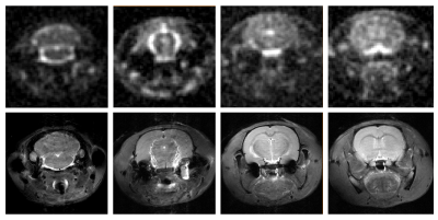
Ouabain Inhibition Reverses Sodium Fluxes in a Preclinical Model of Migraine: a 23Na MRI Study at 21.1 T
Nastaren Abad, David Hike, Jens Rosenberg, Michael Harrington, Samuel Grant
Increased sodium potassium ATPase(Na,K-ATPase) pump activity raises cerebrospinal fluid and brain sodium, resulting in the onset and progression of central sensitization [1-4]. With the goal of testing whether impaired Na,K-ATPase activity is implicit in the onset of migraine, this study makes use of ouabain to inhibit Na,K-ATPase activity and delineate sodium changes that may lie at the heart of migraine. At high spatial and temporal resolution, 23Na multislice CSI scans were acquired from a rodent migraine model, at 21.1 T following the onset or potential inhibition of central sensitization to identify localized sodium changes over 3-h after induction.
|
|
2520.
 |
47 |
Multi-layered radiofrequency coil design for X-nuclei Imaging
Tony Zhou, Justin Lau, Andrew Tyler, Chris Randell, Jack Miller, Damian Tyler
A novel multi-layered radiofrequency coil for X-nuclei imaging is presented which implements stacked layers for improved B1+ and SNR. The multi-layer design increased B1+ by 27% in 23Na phantom experiments and 19% in electromagnetic simulations compared to a single layer coil. Transmit-receive efficiency for a 13C multi-layer coil was double that of a quadrature coil, requiring half the power to achieve a 90° flip. An averaged SNR map from CSI indicated receive sensitivity gain of 33% from the quadrature to multi-layer design.
|
|
2521.
 |
48 |
Density Adapted Stack of Stars Sequence for 23Na using Dictionary Learning Compressed Sensing Reconstruction
Fabian Kratzer, Sebastian Flassbeck, Armin Nagel, Peter Bachert, Mark Ladd, Nicolas Behl
Sodium plays important roles in many cellular processes, which motivates imaging of the 23Na distribution. Short relaxation times and low in-vivo signal result in the need of sequences with short echo times and techniques to improve the SNR. Therefore, we present a stack of stars (SOS) sequence with density adapted readout gradients to increase SNR. We combine this sequence with an anisotropic dictionary learning compressed sensing reconstruction to further reduce noise in the images.
|
|
2522.
 |
49 |
Quantitative susceptibility mapping and sodium imaging based analysis of susceptibility and sodium concentrations in the basal ganglia
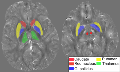
Till Schneider, Nicolas Behl, Martin Bendszus, Sina Straub
23Na concentrations and iron deposition in cerebral gray matter have both shown to be increased in degenerative and inflammatory cerebral diseases. This study employs sodium imaging and quantitative susceptibility mapping to assess differences in sodium concentrations and susceptibility within the basal ganglia in healthy volunteers at 7T. Results indicate a fundamentally different distribution of 23Na concentrations compared to the distribution of susceptibility within the nuclei of the basal ganglia and suggest that not only susceptibility but also 23Na concentrations may be physiologically distributed in a characteristic manner.
|
|
2523.
 |
50 |
Using dynamic sodium imaging to assess furosemide action in the porcine kidney
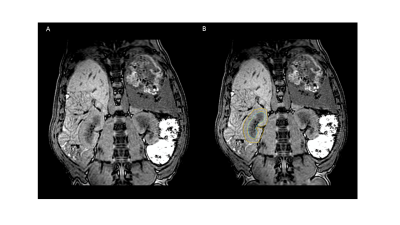
James Grist, Esben Hansen, Rasmus Tougaard, Frank Riemer, Christoffer Laustsen, Ferdia Gallagher
The ability to image sodium is believed to be a powerful tool to understand physiological processes, however have yet to be effectivity translated to clinical realistic usage. Through the use of an efficient k-space trajectory (3D cones) we have undertaken dynamic imaging of the renal sodium distribution pre and post- a furosemide injection. This work shows a decrease in the cortico-medullary sodium gradient over time, as well as being the first renal study on a clinical system with dynamic sodium imaging.
|
|
| Top |
MRS Clinical Application
Digital Poster
Spectroscopy & Non-Proton MR
Tuesday, 14 May 2019
| Exhibition Hall |
09:15 - 10:15 |
| |
|
Computer # |
|
2524.
 |
51 |
Cerebellar GABA+/Glx ratios in essential tremor patients are correlated with tremor severity
Sofie Tapper, Nathanael Göransson, Peter Lundberg, Peter Zsigmond, Anders Tisell
The aims of this study were to investigate whether GABA+ and Glx concentrations, and the relation between them, were altered in patients with severe essential tremor (ET) compared to healthy controls, and to investigate if the GABA and Glx concentrations were associated with the tremor severity. We observed an increasing GABA+/Glx ratio with tremor severity in the ET patients. Our conclusion was that this increasing cerebellar GABA+/Glx ratio mainly was driven by the decrease in Glx rather than an increase in GABA+, which suggests that an increasing tremor severity is partly due to a disturbance in the Glx concentration.
|
|
2525.
 |
52 |
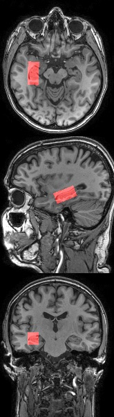
The Neurotransmitter NAAG in Different Phases of Relapsing-Remitting Multiple Sclerosis Patients
Patrik Wyss, Nikolai Pfender, Roland Martin, Andreas Lutterotti, Anke Henning
This study explores the alterations of N-Acetyl aspartate (NAA) and the neurotransmitter N-Acetyl aspartyl glutamate (NAAG) in the cerebral normal-appearing white tissue of multiple sclerosis patients with a relapsing-remitting course. A two-dimensional J-resolved single voxel spectroscopy sequence and two-dimensional prior-knowledge fitting is used to disentangle the resonance lines of NAA and NAAG.
|
|
2526.
 |
53 |
High intrahepatic lipid content is associated with low choline status in humans-a 1H-MRS study at 3 Tesla
Pandichelvam Veeraiah, Kay H M Roumans, Joachim E Wildberger, Patrick Schrauwen, Lucas Lindeboom, Vera B Schrauwen-Hinderling
Non-alcoholic fatty liver (NAFL) has become a major threat to metabolic health. Animal studies have suggested that disturbances in choline metabolism may be linked to the development of NAFL. However, to date, human data on the link of choline and NAFL is scarce. The trimethylammonium (TMA) group of choline can be detected with 1H-MRS at 3.20 ppm. Here, we investigated the relationship between intrahepatic choline levels and hepatic lipid content on healthy overweight/obese subjects using 1H-MRS at 3 Tesla. Our results showed that high hepatic lipid content is associated with low choline content in the liver.
|
|
2527.
 |
54 |
Association of hepatic histologic features with magnetic resonance spectroscopy derived hepatic fat and water T1 and T2 estimates in adults with nonalcoholic fatty liver disease
Alexandra Schlein, Claude Sirlin, Yingzhen Zhang, Guilherme Cunha, Rohit Loomba, Gavin Hamilton
The purpose of this study is to assess possible associations between hepatic histologic features of NAFLD and the T1 and T2 of water and fat, measured by a multi TR, multi TE 1H MRS STEAM sequence that acquires 32 spectra for a range of TRs and TEs in a single breath-hold. In evaluation of 51 adults Water T1 showed a positive association with fibrosis. Water T1 and T2 and Fat T1 and T2 all showed associations with steatosis grade; no other statistically significant associations were observed. This may contribute to noninvasive methods of detection and monitoring of NALFD.
|
|
2528.
 |
55 |
J-edited Cerebral MR Spectroscopy in Patients with Hepatic Encephalopathy
Helge Zöllner, Georg Oeltzschner, Markus Butz, Markus Jördens, Nur-Deniz Füllenbach, Dieter Häussinger, Hans-Jörg Wittsack, Alfons Schnitzler
Hepatic encephalopathy (HE) associated with elevated brain ammonia levels. The detoxification of ammonia leads to regionally selective alterations in several brain metabolites. The present study investigates these neurotransmitter changes in HE patients in the GABAergic and glutamatergic system. Using MEGA-PRESS, MR spectroscopy was performed in cerebellum, thalamus, and motor cortex. Preliminary results unravel increased GABA levels accompanied by elevated glutamine and reduced myo-Inositol in the cerebellum, but an absence of GABA level changes in the two other regions. These initial findings may lead to further explanation of cognitive and motor deficits in HE, but need to be substantiated further.
|
|
2529.
 |
56 |
1 H MRS Analysis of Pancreas Metabolites altered by Cachexia
Raj Kumar Sharma, Santosh K. Bharti, Paul T. Winnard Jr, Yelena Mironchik, Marie-France Penet, Zaver M. Bhujwalla
Cancer induced cachexia is a syndrome characterized by tissue wasting and weight loss. Cachexia occurs with the highest frequency and severity in pancreatic ductal adenocarcinoma (PDAC). To further understand this syndrome, we used 1H MRS to analyze pancreas metabolites in mice with and without cachexia-inducing PDAC xenografts. We detected significant weight loss in cachectic mice. 1H MR spectra identified significant differences in amino acids, BCAA, alanine, pyruvate, phosphocholine, niacinamide and NAD in cachectic mice that provide new insights into the morbidity and mortality associated with the syndrome that may lead to novel strategies to arrest this syndrome.
|
|
2530.
 |
57 |
Chemical deregulation in breast tissue of women at familial high risk of breast cancer correlates with the IBIS Breast Cancer Risk Evaluator Tool using 2D COSY at 3 Tesla
Natali Naude, Gorane Santamaria, Graham Galloway, Chris Foster, Aaron Urquhart, Thomas Gaass, Saadallah Ramadan, Scott Quadrelli, Lisa Rich, Ian Bennett, Jessica Buck, Peter Malycha, Carolyn Mountford
Evaluation of breast tissue of women with a high familial risk of developing breast cancer using in vivo 2D COSY as part of a standard 3T breast MRI showed statistically significant alterations in the type of double bonds in fatty acyl chains compared with controls. Further, when separating the familial cohort using the IBIS risk evaluatior tool into a group above and below 20% lifetime risk, statistically significant differences in both cholesterol and metabolites were recorded.
|
|
2531.
 |
58 |
Neurochemical profile of the human hippocampus at 3T after traumatic brain injury
Maria Yanez Lopez, David Sharp
The aim of this work was to investigate the metabolic profile of the hippocampus in a clinical population (moderate/severe traumatic brain injury patients in the acute phase), using an MR Spectroscopy LASER sequence at 3T. With ongoing data acquisition, preliminary results show reduced levels of total choline (tCho), metabolite reflecting membrane turnover.
|
|
2532.
 |
59 |
Whole-brain high resolution 3D MRSI for measuring 2HG and tumor metabolism in mutant IDH glioma patients
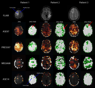
Bernhard Strasser, Borjan Gagoski, Bijaya Thapa, Xianqi Li, Wolfgang Bogner, Julia Small, Jorg Dietrich, Daniel Cahill, Tracy Batchelor, Ovidiu Andronesi
2-Hydroxyglutarate (2HG) detection using MRSI is a very promising, but challenging technique. Although high-resolution MRSI reduces the already small SNR of 2HG, it also reduces the spectral linewidth and provides more voxels for quantification. This study compares two high-resolution spiral MRSI sequences with a low-resolution MEGA-edited sequence, and one with a short echo time for 2HG detection in brain tumor patients. Three patients and three volunteers were measured with all four sequences. The two high-resolution sequences perform better with less false-positive 2HG detection in volunteers, and a more reliable 2HG quantification in IDH-mutated tumors.
|
|
2533.
 |
60 |
Comparison of MEGA-PRESS and Short Echo Time PRESS on Classification of IDH Mutation Using Machine Learning at 3T
Ayhan Gursan, Gokce Hale Hatay, Cengiz Yakicier, M. Necmettin Pamir, Koray Ozduman, Alp Dincer, Esin Ozturk-Isik
Isocitrate dehydrogenase (IDH) mutation is common in grade II and grade III gliomas, and results in better patient prognosis IDH mutant (IDH-mut) gliomas. Magnetic resonance spectroscopy (MRS) studies indicated an increase in 2-hydroxyglutarate (2HG) and decrease in glutamate (Glu) and glutathione (GSH) as a result of IDH mutation. The goal of this study is to compare IDH mutation classification performances of short echo-time (TE) PRESS and MEGA-PRESS by using machine learning in 60 glioma patients. Highest average classification accuracy was 75% with coarse decision trees for short TE PRESS, and 74% with ensemble of bagged of trees for MEGA-PRESS.
|
|
2534.
 |
61 |
Metabolic Markers of Disease Progression in Pediatric Diffuse Intrinsic Pontine Gliomas
Stefan Bluml, Brenda Kurland, Marvin Nelson, Benita Tamrazi, Rafael Ceschin, Ian Pollack, Ashok Panigrahy
Diffuse intrinsic pontine gliomas (DIPG) are inoperable and highly resistant to chemo- and radiation therapy. DIPG carry the worst prognosis among pediatric brain tumors with progress in the development of therapies compromised by low patient numbers and unavailability of tissue samples to characterize disease status. In this work we present evidence, that non-invasive MR spectroscopy can provide robust early indicators that can assess the effectiveness (or ineffectiveness) of potential new therapeutic approach at an early stage and accelerate the completion of clinical trials in small cohorts of patients.
|
|
2535
 |
62 |
Evaluation of the elevated signal at 3.55 ppm in 1H MRS spectra of certain glioma patients
Video Permission Withheld
David Hundertmark, Benjamin Bender, Uwe Klose
In MR-spectroscopy of gliomas, sometimes an elevation of the signal at 3.55 ppm at an echo time of 135 ms is found, which can be interpreted as myo-inositol or as glycine. Due to coupling effects, the signal of inositol should be reduced at an echo time of 90 ms, while the glycine signal should be larger than at TE 135 ms. In measurements of glioma patients, which show an enhanced signal at 3.55 ppm at TE 135ms, we found a decreased signal at TE 90. Therefore, we saw no indication of elevated glycine concentration in gliomas.
|
|
2536.
 |
63 |
Neurometabolic consequences of perinatal HIV infection and exposure are still observed in children at 11 years
Amy Graham, Martha Holmes, Francesca Little, Els Dobbels, Mark Cotton, Barbara Laughton, Andre van der Kouwe, Ernesta Meintjes, Frances Robertson
HIV establishes reservoirs within the brain, causing damage despite individuals adhering to antiretroviral therapy. The long-term consequences of perinatal HIV infection and early treatment in children remain unclear. Proton magnetic resonance spectroscopy was carried out to assess the effects of HIV on neurodevelopment, at a metabolic level, comparing HIV-positive, HIV-exposed-uninfected (HEU) and HIV-unexposed children at 11 years old. Absolute metabolite concentrations were compared between these groups, through linear regression analysis. Elevated choline levels within two regions of interest suggest putative inflammation in HIV-positive children. A reduction of N-acetyl-acetate in a white-matter region of HIV-positive and HEU children implies axonal damage.
|
|
2537.
 |
64 |
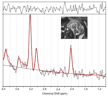
MR Spectroscopic Changes in Infants Exposed to Prenatal Opioids: A Pilot Study
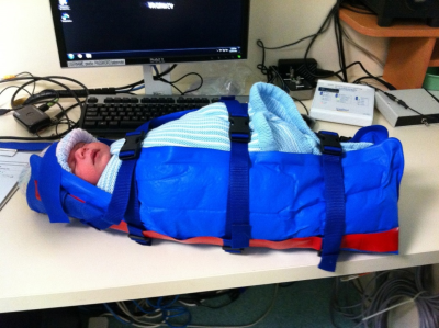
Caroline Rae, Danielle Christmas, Ian Wright, John Feller, Mohamed Abdel-Latif, Sara Clews, Janet Falconer, Julee Oei
Proton spectra were obtained from the left caudate, left hippocampus and subventricular zone of the brains of one week old babies born to mothers who were opioid users. The study suggests that decreased brain volumes after prenatal opioid-exposure are associated with hippocampal spectral abnormalities with increased severity related to multiple opioid use.
|
|
2538
 |
65 |
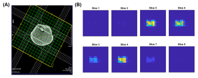
Altered biochemical profiles in fetuses with congenital heart disease
Video Permission Withheld
Subechhya Pradhan, Kushal Kapse, Gilbert Vezina, Mary Donofrio, Jessica Quistorff, Catherine Lopez, Nicole Simard, Catherine Limperopoulos
Brain injury is a major complication in infants with complex congenital heart disease (CHD). There is growing evidence that impaired brain development has its origins in the fetal period. We prospectively characterized in vivo fetal brain metabolic profiles in 307 fetuses (210 health fetuses and 97 with CHD). Findings from measurements of metabolite concentrations of NAA, Cr, and Cho increased with advancing GA in healthy and CHD fetuses. In CHD fetuses, tNAA/tCh ratios were significantly lower while lactate concentrations were significantly higher compared to healthy fetuses, suggesting early-life disturbances in fetal brain biochemistry.
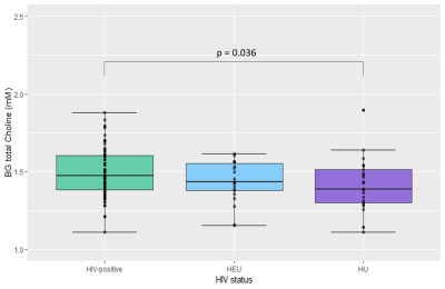
|
|
2539.
 |
66 |
Accelerated Five-Dimensional Echo-Planar Correlated Spectroscopic Imaging to assess Lipids and Metabolite differences between Type-2 Diabetic and Healthy Calf Muscle
Andres Saucedo, Manoj Sarma, Christine Darwin, Sumit Kumar, Kavya Umachandran, Rajakumar Nagarajan, Michael Thomas
Obesity-related diseases such as Type 2 Diabetes have become increasingly widespread. This condition can be characterized in part by changes in the fat composition of muscle, specifically in the relative concentrations of extra-myocellular (EMCL) and intra-myocellular (IMCL) lipids. Although 1D MRS techniques have been applied to assess skeletal muscle metabolite composition, they are hindered by lipid contamination from EMCL and spectral overlap which can complicate quantitation and differentiation from IMCL. 2D MRS improves spectral dispersion, allowing clear separation of both EMCL and IMCL and determination of the unsaturation index of muscle lipids. In this study, we apply a 5D (3D spatial + 2 spectral) echo-planer correlated spectroscopic imaging (EP-COSI) technique to assess the lipid and metabolic differences within the calf muscle among three groups of subjects – diabetic, age-matched healthy, and young healthy controls.
|
|
2540.
 |
67 |
Metabolic abnormalities in cingulate gyrus in HIV infection by 3D rosette spectroscopic imaging
Victor Yushmanov, Jullie Pan, Claudiu Schirda, Hoby Hetherington, Jeffry Alger, Peter Barker, Todd Parrish, Ned Sacktor, Michal Povazan, James Becker
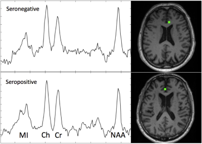
The purpose of this study was to assess metabolic abnormalities in HIV infection by MRSI covering brain cortex and parts of striatum. Sixteen HIV-seropositive subjects (younger and older than 60) and age-matched 30 seronegative controls were evaluated using a fast rosette 3D MRSI sequence at 3T, segmentation and parcellation into 13 brain regions, and fGM regression statistics to evaluate abnormalities. MI/NAA, Cr/NAA, Ch/NAA and Ch/Cr increase in anterior and posterior cingulate as a function of HIV serostatus and age. Fast MRSI enables the detection of subtle metabolic abnormalities in HIV infection at clinically acceptable scan times (<10 min) at 3T.
|
|
2541.
|
68 |
Metabolite markers of glutamatergic activity and neuro-inflammation in the superior temporal gyrus in patients with schizophrenia.
Sai Merugumala, M. Niznikiewicz, E. Del Re, K. Spencer, H. Liao, P. Nestor, R. McCarley, N. Bolo, A. Lin
Many studies have shown that the superior temporal gyrus undergoes many changes in schizophrenia. Magnetic resonance spectroscopy studies of the brain have also shown brain metabolite levels are altered in schizophrenia however the superior temporal gyrus has not been examined in detail. The aim of this study was to compare brain metabolite levels in patients with schizophrenia and controls as well as examine their correlation with electrophysiology measures.
|
|
2542.
 |
69 |
The Potential Impact of Multiparametric MRS in the Early Detection of Neurodegeneration in Multiple Sclerosis
Assaf Tal, Ivan Kirov
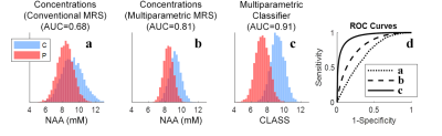
Clinical proton MRS conventionally measures metabolites' concentrations, but neglects to acquire their relaxation constants, despite the fact that these are known to vary in many pathologies. Using computer simulations and literature values for n-acetyl-aspartate, we show that incorporating this additional information can greatly facilitate the detection of neurodegeneration in early stage multiple sclerosis (MS), increasing the area under the corresponding receiver operating characteristic curves from 0.68 to 0.91. These results strongly motivate the need for developing robust sequences for clinical multiparametric magnetic resonance spectroscopy .
|
|
2543.
 |
70 |
Mapping of Regional Distributions of Brain Metabolites in Healthy Young Adults using Three-dimensional Echoplanar Spectroscopic Imaging
Eric Petersen, David Roalf, Ruben Gur, Raquel Gur, Andrew Crow, Ravinder Reddy, Sumei Wang, Suyash Mohan, Andrew Maudsley, Sanjeev Chawla
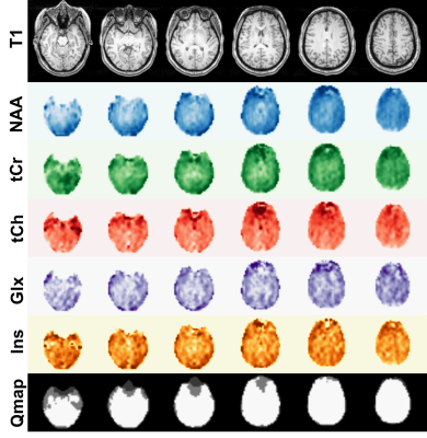
To determine the regional distributions of metabolites from different lobar regions of brain in a cohort of healthy individuals in late adolescence and early adulthood, a total of 19 subjects (mean age=22) underwent 3D-echoplanar spectroscopic imaging. The parametric maps of N-acetylaspartate (NAA), creatine (tCr), choline (tCho), myoinositol (mI) and glutamate/glutamine (Glx) were generated using sophisticated post-processing steps. These maps were normalized to MNI atlas. Significant spatial variations in metabolite ratios of NAA/tCr, tCho/tCr, mI/tCr and Glx/tCr were observed across different lobar regions of brain. These findings will undergird future efforts to understand metabolite distributions in neurodevelopmental disorders.
|
|
2544.
 |
71 |
7T 1H-MRS of the anterior cingulate in patients with psychosis spectrum and mood symptoms
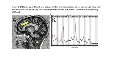
Eric Petersen, Sanjeev Chawla, Andrew Crow, Theodore Satterthwaite, Raquel Gur, Ruben Gur, Ravi Reddy Nanga, Hari Hariharan, Mark Elliott, Ravinder Reddy, David Roalf
Data from 12 typically developing (TD), 10 clinical high risk for psychosis (CHR), 6 psychosis (PSY) and 4 mood disorder (MD) participants who underwent 1HMRS at 7T. Short TE single voxel spectra (SVS) were obtained using a custom-modified PRESS sequence from the anterior cingulate gyrus. Data quality was high and tissue contribution within the acquisition voxel was similar across diagnostic groups. NAA, Creatine, Choline, GSH and Glu were significantly lower in PSY as compared to TD. CHR showed an intermediate pattern for all brain neurometabolites, except GSH, which was elevated as compared to TD. MD patients, in general, showed higher concentrations of NAA, Cho, GSH and Glu as compared to TD.
|
|
2545.
 |
72 |
Metabolic Alterations in the left dorsolateral prefrontal cortex in Sleep-Related Hypermotor Epilepsy: A Proton Magnetic Resonance Spectroscopy Study
Weina Wang, Xiaorui Su, Simin Zhang, Qiang Yue, Qiyong Gong
Sleep-Related Hypermotor Epilepsy (SHE) is characterized by bizarre motor behavior during sleep. The aim of this present study was to investigate metabolic alterations in the bilateral DLPFC using 1H-MRS to understand the underlying pathophysiology of SHE. Thirty-nine subjects with SHE and 60 controls were studied. The left DLPFC NAA and mI concentrations of SHE patients were significantly lower than controls. There was an asymmetry of NAA in the control group. These findings may present executive function decline in SHE and further verify the left frontal lobe is more vulnerable in right-handed SHE patients.
|
|
2546.
 |
73 |
Longitudinal cerebral metabolic changes in delayed neurologic sequelae after carbon monoxide intoxication using 1H MR spectroscopy.
Hui-ru Tsai, Jung-Hsiang Chang, Ping-Hong Lai, Jie-Yuan Li, Cheng-Wen Ko
In this study, we explored the metabolic changes in WM and GM of patients with and without DNS using 1H MRS longitudinally at onset, 1, 3, and 9 months after CO intoxication. Decreased tNAA/Cr and increased Cho/Cr were observed in WM of patients with DNS as literatures have reported. The longitudinal change of Glx/Cr and Ins/Cr in WM and GM of patients with DNS implies themselves that may provide valuable information for monitoring DNS development.
|
|
2547.
 |
74 |
Evidence for increasing hippocampal metabolite concentrations during healthy aging
Leo Sporn, Erin MacMillan, Ruiyang Ge, Kyle Greenway, Cornelia Laule, Fidel Vila-Rodriguez
Previous magnetic resonance spectroscopy (MRS) studies have concluded that hippocampal metabolite concentrations remain stable during healthy adult aging. However, these studies used short repetition times (TR ≤ 2s), which leads to heavy T1-weighting. We used a longer TR (4s) to reduce T1-weighting and found hippocampal metabolite concentrations increase with age for N-acetyl-aspartate, creatine, choline and myo-inositol. Our findings illustrate the importance of using sufficiently long TR in MRS to avoid T1-relaxation effects influencing the measurement of metabolite concentrations.
|
|
2548.
 |
75 |
Effects of Transcranial Direct Current Stimulation on Metabolite Concentrations in Children
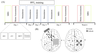
Chidera Nwaroh, Lauran Cole, Adrianna Giuffre, Helen Carlson, Frank MacMaster, Adam Kirton, Ashley Harris
Transcranial direct current stimulation (tDCS) is a form of non-invasive brain stimulation that safely modulates brain activity. Several studies have shown that tDCS of the motor cortex facilitates motor learning and plasticity but there is little information on the underlying mechanisms. This analysis of metabolite changes in response to 1mA tDCS using typical PRESS and MEGA-PRESS is important in developing a complete understanding of the effects of stimulation. In this pediatric study, we did not detect the same GABA and glutamate changes in response to tDCS that have been seen in the adult literature.
|
|
Back to Program-at-a-Glance |  Back to Top Back to Top |
| The International Society for Magnetic Resonance in Medicine is accredited by the Accreditation Council for Continuing Medical Education to provide continuing medical education for physicians. |
