Digital Poster Session
Neuro Back to Program-at-a-Glance Back to Program-at-a-Glance
|
Tuesday, 14 May 2019
Digital PosterNeuro
2549 -2573 Flow in the Brain
2574 -2598 Parkinson's Disease
2599 -2623 Treasure Chest of Neuro Gems
2624 -2648 Neuroanatomy: Seeing Is Believing |
| |
Flow in the Brain
Digital Poster
Neuro
Tuesday, 14 May 2019
| Exhibition Hall |
09:15 - 10:15 |
| |
|
Computer # |
|
2549.
 |
76 |
CSF Flow and Aging: An Early Marker of Pathology?

Arun Venkataraman, Rashid Deane, Jianhui Zhong
Phase Contrast-Magnetic resonance imaging (PC-MRI) provides detailed information on flow of spins, and has been applied to blood and cerebrospinal fluid (CSF) flow. In the field of CSF imaging, PC-MRI is mostly used as a clinical tool to look for frank CSF changes. However, subtle CSF changes are thought to occur in neurovascular pathologies, as well as neurodegenerative disorders such as AD. In addition, it has been shown that CSF flow may change during the aging process. In this abstract, we seek to select the optimal imaging parameters to investigate aging-related CSF changes.
|
|
2550.
 |
77 |
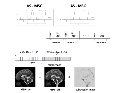
Visualization of irregular CSF flow by dynamic iMSDE SSFP using acceleration- selective motion - sensitized gradient (AS-MSG)
Tomohiko Horie, Nao Kajihara, Haruo Saito, Shuhei Shibukawa, Susumu Takano, Natsuo Konta, Makoto Obara, Tetsuo Ogino, Tetsu Niwa, Kagayaki Kuroda, Mitsunori Matsumae
We reported a technique to visualize the irregular flow of cerebrospinal fluid (CSF) by using dynamic improved motion-sensitized driven-equilibrium steady-state free precession (dynamic iMSDE SSFP). The purpose of this study was to investigate the characteristics of intracranial CSF visualization with dynamic SSFP using acceleration selective motion sensitized gradient (AS-MSG). The dynamic SSFP using AS-MSG distinguished acceleration flow in CSF from constant flow. This technique is suggested to contribute to the diagnosis of various diseases in the CSF space.
|
|
2551
 |
78 |
Microgravity-induced changes in pituitary morphology, brain volumetry, and cerebral spinal fluid hydrodynamics: relationship to spaceflight associated neuro-ocular syndrome
Video Permission Withheld
Larry Kramer, Khader Hasan, Michael Stenger, Ashot Sargsyan, Brandon Macias
A longitudinal study of astronauts with long duration exposure to microgravity showed intracranial volumetric expansion which did not return to baseline after a 1 year of post-flight recovery. These findings were associated with increased cerebral spinal fluid pulsatility through the cerebral aqueduct suggesting diminished intracranial compliance. Additionally, there was development of pituitary gland deformity similar to that seen in idiopathic intracranial hypertension implicating the presence of elevated intracranial pressure during spaceflight.
|
|
2552.
 |
79 |
Evaluation of Intracranial Pressure-Regulation by MRI-measured Cerebrospinal Fluid Pulsation
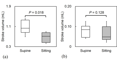
Masatomo Uehara, Tosiaki Miyati, Naoki Ohno, Riho Okamoto, Moemi Tachimoto, Mitsuhito Mase, Hiroshi Furusho, Satoshi Kobayashi, Toshifumi Gabata
We conducted this study to determine the cerebrospinal fluid pulsation in the supine and sitting positions using multiposture MRI. The stroke volume of the aqueduct is not affected by intracranial pressure change. Cerebrospinal fluid pulsation measurements to evaluate the intracranial pressure-regulation function should be taken at the boundary between cranial and spinal cavities rather than in the aqueduct.
|
|
2553.
 |
80 |
Low b-value diffusion weighted imaging to evaluate cerebrospinal fluid dynamics
Toshiaki Taoka, Shinji Naganawa, Hisashi Kawai, Toshiki Nakane, Katsutoshi Murata
We evaluated the signal intensity of the CSF on low b-value (b=500 s/mm2) diffusion weighted images (DWI) in cases with ventricular dilatation versus controls by a scoring method. Although low b-value DWI cannot quantify the absolute flow speed, it may be possible to evaluate the distribution of altered CSF dynamics within the cranium in the cases of ventricular dilatation. We also evaluated the characteristic signal void findings adjacent to the septum pellucidum in the cases with ventricular dilatation, which was speculated to be due to a standing wave in a thinned septum pellucidum.
|
|
2554.
 |
81 |
Automated CSF Detection for Post-hemorrhagic Hydrocephalus in Preterm Infants Using 3D U-Net
Li Zhao, Xue Feng, Craig Meyer, Kushal Kapse, Matthew Whitehead, Adre du Plessis, Catherine Limperopoulos
Post-hemorrhagic hydrocephalus is a prevalent and severe neurological complication in very premature infants. Converging evidence suggests that increased ventricular size is an important and potentially modifiable risk factor for adverse neurological outcomes. MRI measures of CSF volume often rely on manual measurements to quantify ventricular size because automatic neonatal brain segmentation methods often fail in the setting of severe brain injury. In this pilot study, we proposed and validated a deep convolutional neural network method, 3D U-Net, to automatically identify the lateral ventricular system and the external cerebrospinal fluid regions. The proposed method showed superior accuracy in a preliminary cohort of 19 scans of very preterm infants compared to a conventional method.
|
|
2555.
 |
82 |
Assessment of Hemodynamic and Hydrodynamic alternations in Spontaneous Intracranial Hypotension by using MR-based Intracranial Pressure Method
JyhWen Chai, Yi-Hsin Tsai, Yi-Ying Wu, Yi-Jhe Huang, Hung-Chieh Chen, Clayton Chi-Chang Chen
Epidural venous dilatation is commonly seen in patients with spontaneous intracranial hypotension, and its presence often indicates a distinctly altered cerebrospinal hemodynamic/CSF dynamic. With the MR-ICP technique, significant statistical differences were found in various hemodynamic and CSF dynamic parameters. The result suggests that EVD is a representative feature of hemodynamic/CSF-dynamic change in SIH, and also highlights the potential of MR-ICP as a reliable method of assessment for SIH.
|
|
2556.
 |
83 |
MRI assessment of glymphatic function in the non-human primate brain
Ian Tagge, Steven Kohama, Theodore Hobbs, Jeffrey Pollock, Thierno Bah, Jeffrey Iliff, William Rooney
The astrocyte mediated exchange of cerebrospinal fluid and interstitial fluid comprise the glymphatic system, a physiology that facilitates waste removal in the brain parenchyma. Impaired solute and waste clearance may contribute to neurodegenerative conditions, and may also be associated with age. Here, we present preliminary measurements of glymphatic function in healthy adult and aged rhesus macaque brain via intrathecal injection and DCE-MRI. We demonstrate that kinetics of GBCA distribution in the CNS occur on timescales amenable to study using DCE-MRI techniques. Our preliminary results indicate that impairment in glymphatic physiology occurs with age in the rhesus macaque.
|
|
2557.
 |
84 |
Enhanced perivascular space contrast using T1-T2 fusion and adaptive spatial filtering
Farshid Sepehrband, Giuseppe Barisano, Nasim Sheikh-Bahaei, Meng Law, Arthur Toga
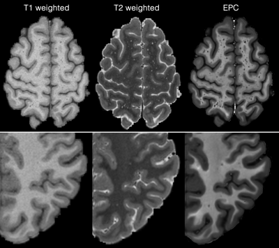
Imaging the perivascular space (PVS), also known as Virchow-Robin space, has shown to be of significant clinical value. Its role in glymphatic system1 and reported pathological changes of the PVS in neurological disorders2–10 highlight the need for methodological development specific to this compartment. Here we propose a fusion framework that enhances PVS contrast, allowing robust clinical rating. The Enhanced PVS Contrast (EPC) was achieved by combining T1- and T2-weighted images that were adaptively filtered to remove non-structural high frequency spatial noise.
|
|
2558.
 |
85 |
Assessment of Cerebral Hemodynamics Change of Hypertension using Multi-TI Arterial Spin-Labeling
Presentation Not Submitted
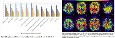
Qiuju Fan, Zhen Yang, Nan Yu, Qi Yang, Yong Yu, Yue Li, Shaoyu Wang
In our study, we evaluate the diagnostic value of mTI-ASL as a noninvasive method to detect subtle hemodynamic abnormalities in hypertension at different stage.
|
|
2559.
 |
86 |
Study of the cerebral blood flow metabolism in patients with Parkinson’s disease using arterial spin labeling MRI
Presentation Not Submitted
Hong-Ying Zhang, Weiqiang Dou
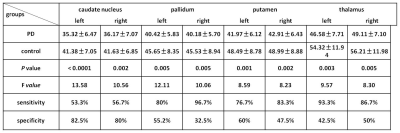
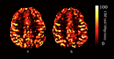
There are still debates on the alterations of subcortical metabolism in Parkinson’s disease. We performed 3D pseudo-continuous pulse ASL on PD and control groups. The absolute cerebral blood flow (CBF) values and relative CBF (rCBF) in subcortical regions were automatically extracted and calculated. We found widespread decreased absolute CBF in PD patients. However, the subcortical rCBF increased significantly. We conclude that widespread blood hypoperfusion in PD brain is absolute, and hyperperfusion in the subcortical brain regions is only relative to the whole brain level of patients themselves.
|
|
2560.
 |
87 |
Using Perfusion Weighted Imaging to Aid in Drawing Prominent Veins on Quantitative Susceptibility Mapping
Miller Fawaz, Min-Gyu Park, Luo Yu, E. Mark Haacke
Current literature references Asymmetrically Prominent Cortical Veins as a valid marker, but identification is user dependent. We aim to quantify APCV using PWI, greatly reducing the reliance on observer input. This method is a stepping stone for automatic APCV segmentation and has the potential to play a role in establishing a reliable identifier for ischemic penumbra from SWI data.
|
|
2561.
 |
88 |
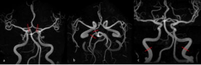
Correlation between internal carotid artery flow and circle of Willis anatomy
Te-Chang Wu, Jeon Hor Chen
Measurement of proximal cerebral inflow volume and individual cerebral angio-architecture are crucial for patient-specific analysis of hemodynamic effects of cerebrovascular disease. However, the detail of bilateral ICA flow in individuals of complete but asymmetric COW is lacking. This retrospective study included total 210 healthy adult for delineation of the relationship between detailed COW variations and bilateral ICA flow volume in healthy adults. Furthermore, the correlation of ICA diameter to ICA flow in the setting of circle of Willis variants was also proposed.
|
|
2562.
 |
89 |
Perivascular spaces in healthy young subjects
Giuseppe Barisano, Farshid Sepehrband, Nasim Sheikh-Bahaei, Meng Law, Arthur Toga
Enlargement of perivascular spaces has been associated with a number of diseases. However, morphological features of perivascular spaces in healthy subjects and their clinical role are still not completely understood. We analyzed on MRI perivascular spaces in a large sample of healthy young subjects. Our results showed a high inter-subjects variability of perivascular spaces. Twins presented similar amount of perivascular spaces. Perivascular spaces in basal ganglia were significantly correlated with subjects’ height, brain volume, and brainstem volume. These findings are relevant for all future studies investigating the role of perivascular spaces.
|
|
2563.
 |
90 |
Computational assessment of enlarged perivascular spaces on brain magnetic resonance images in Vascular Dementia patients.
Martha Singh, Anuja Pradhan, Mustafa Salimeen, Habib Tafawa, Xianjun Li, Miaomiao Wang, Congcong Liu, Guanyu Yang, Qu Qiumin, Jian Yang
Enlarged perivascular spaces (EPVS) are common in Vascular Dementia (VaD) patients, associated with aging, inflammation, etc. Many studies address EPVS as it is related to count and volume but very few on the density using 3T MRI. Our aim in this study is to describe an effective and user-friendly computational method to aid in the perivascular spaces segmentation to yield EPVS count, volume and density in VaD patients. EPVS count, volume and density are significantly greater than in the control group (P<0.05). The results suggest that computational assessment of EPVS can further aid in an early diagnostic of VaD.
|
|
2564.
|
91 |
Dynamic Contrast-Enhanced Imaging Reveals Region-Specific Blood-Brain Barrier Damage in Bipolar Depression
Lyn Kamintsky, Kathleen Cairns, Ronel Veksler, Chris Bowen, Steven Beyea, Alon Friedman, Cynthia Calkin
This study addresses the need for mechanism-based understanding and diagnosis of bipolar depression. Using dynamic contrast-enhanced MRI we identified extensive blood-brain barrier (BBB) leakage in 28% of bipolar patients (and zero controls). All bipolar patients with extensive BBB leakage also had insulin resistance and worse metabolic, psychiatric and cognitive symptoms. We found depression to be associated with region-specific BBB leakage, with the nucleus accumbens best predicting depression severity. Our findings highlight BBB damage as a mechanism contributing to the dysfunction of depression-associated brain regions, and suggest that insulin resistance increases the risk of extensive BBB leakage.
|
|
2565.
 |
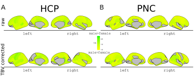
92 |
Chronic anemic patients have impaired cerebral oxygen delivery using Pseudo Continuous Arterial Spin Labeling MRI
Yaqiong Chai, Adam Bush, Chau Vu, Natasha Lepore, Thomas Coates, John Wood
We quantified and compared cerebral blood flow (CBF) and oxygen delivery in patients with thalassemia intermedia and other chronic anemic patients and healthy controls, using arterial spin labeling MRI. Anemic patients exhibited elevated CBF globally and in grey matter (GM). While global and GM O2 delivery was preserved in anemic patients, white matter (WM) O2 delivery was 20% lower in this cohort compared to healthy controls. Age was the strongest predictor for both CBF and O2 delivery, but the mechanisms of decreased WM O2 delivery needs further study, given the inadequate neovascularization in response to chronic hypoxia and other factors.
|
|
2566.
 |
93 |
Sodium T2* Heterogeneity of Cerebrospinal Fluid in Healthy Brains and Neurological Disorders
Yongxian Qian, Tiejun Zhao, Kathik Lakshmanan, Timothy Shepherd, Yulin Ge, Yvonne Lui, Fernando Boada
The literature reports a wide variance in CSF T2* values (46-64ms). This variance may suggest T2* heterogeneity of CSF. Here we explore the possibility of CSF T2* heterogeneity among healthy and neurologically-disordered brains.
|
|
2567.
 |
94 |
The influence of draining veins on apparent grey matter volume changes caused by hypercapnia
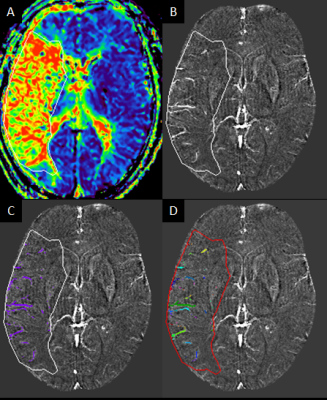
Julia Huck, Christopher Steele, Anna-Thekla Jäger, Audrey Fan, Sophia Grahl, Christine Tardif, Uta Schneider, Arno Villringer, Pierre-Louis Bazin, Claudine Gauthier
Past studies have shown that T1-weighted measures of grey matter volume (GMV) can be biased by differences in blood volume. Here, we investigate the vascular compartments associated with this bias by quantifying the spatial relationship between t-values for the apparent GMV increase observed during hypercapnia and the location of draining veins. Draining veins were identified using the VENAT atlas. Overall, the results of this analysis demonstrate that while proximity to veins is related to the presence of higher t-values (larger apparent GMV change during hypercapnia), large veins themselves are unlikely to be the main cause of this bias; suggesting that smaller veins or arteries may have a larger role in the observed bias.
|
|
2568.
 |
95 |
Hyperoxia Challenge in Healthy and Anemic Subjects using BOLD MRI
Chau Vu, Julie Coloigner, John Wood
This study explores the brain’s response to 100% oxygen inhalation in anemic subjects using BOLD MRI. Hyperoxic challenge has previously been used to identify brain regions with increased oxygen extraction fraction from inadequate perfusion. After controlling for changes in peripheral oxygen saturation, hyperoxic BOLD response was not significantly different between sickle cell disease patients, non-sickle anemic patients and healthy controls. Therefore, our results suggest that chronically anemic patients do not have increased oxygen extraction fraction from inadequate resting oxygen delivery under resting conditions.
|
|
2569.
 |
96 |
Imaging Cerebellum Venous Oxygenation: a T2-based approach
SHENGWEN DENG, Dengrong Jiang, Juan Vasquez, , Hanzhang Lu, Peter Fox
Cerebellum has been used for normalization in fMRI and blood flow studies, and yet little for its oxygen metabolism. This abstract aims to explore T2-Relaxation-Under-Phase-Contrast (TRUPC) MRI to reliably image cerebellar venous oxygenation. We try to explore the regional small vein Yv and it adjacent sinus signals.
|
|
2570.
 |
97 |
Extracranial space in healthy infants: an age-related study based on MRI anatomical images
XIAOHU WANG, LEI ZHANG, ZHUANQIN REN, XIAOCHENG WEI, QING FAN
To date, there is no consensus exists on diagnostic criteria for pathological external hydrocephalus in infants and young children. In this study, extracerebral space of 212 healthy subjects were measured on different anatomical slices and their age correlation were analyzed. The results demonstrated that extra cerebral space measured both on axial and coronal plane features similar age-related change. The results of this study may be a valuable reference in diagnosis of external hydrocephalus.
|
|
2571
 |
98 |
Quantitative Measurement of Glymphatic Flow in Man with Contrast-Enhanced MRI
Video Permission Withheld
Christopher Filippi, Jared Steinklein, Leah Waldman, Xiangzhi Zhou, George Pinder, Richard Watts
Impairment in glial lymphatic "glymphatic" flow is hypothesized to be an etiologic factor in the development of Alzheimer’s disease (AD). We report a quantitative study of glymphatic flow in man, combining intrathecal administration of gadobutrol (macrocylic gadolinium-based contrast agent) with serial T1-mapping to produce contrast concentration maps up to 3 days post-injection. This demonstrates proof-of-concept feasibility and offers data on the pharmacokinetics of glymhatic flow.
|
|
2572.
 |
99 |
Could Diffusion MRI monitor the brain glymphatic system? A proof-of-concept study using an aquaporin-4 channel inhibitor pharmacological challenge
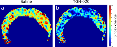
Clement Debacker, Tomokazu Tsurugizawa , Boucif Djemai, Luisa Ciobanu, Denis Le Bihan
Dysfunction of the Glymphatic System (GS), which clears brain tissue from waste, has been proposed as a mechanism to several brain pathologies, including the Alzheimer’s disease. GS has been investigated with preclinical imaging through the invasive intracisternal injection of Gadolinium. In this study, we have show, using a pharmacological mouse brain model, that diffusion MRI, through the Sindex approach could reveal noninvasively changes in brain cortex tissue following injection of an Aquaporin 4 channel inhibitor known to interfere with the GS via the inhibition of astrocyte swelling.
|
|
2573.
|
100 |
Intra-Voxel Incoherent Motion imaging as a potential marker of parenchymal glymphatic flow
Swati Rane, Elaine Peskind, Rebecca Hendrickson, Murray Raskind, Jalal Andre
We tested the applicability of IVIM to detect increased glymphatic flow using the pseudo-diffusion coefficient (D*). Using known effects of prazosin, to increase interstitial fluid volume and glymphatic flow, we showed that D* increased significantly in 6 individuals before and 9-12 weeks after prazosin. This increase was greater than conventional diffusion. It was also larger than the inter-scan variability of D*, measured in 3 individuals 10 weeks apart.
|
|
| Top |
Parkinson's Disease
Digital Poster
Neuro
Tuesday, 14 May 2019
| Exhibition Hall |
09:15 - 10:15 |
| |
|
Computer # |
 |
2574.
 |
101 |
MRI Evidence for the Ascending Spread Hypothesis of Parkinson’s Disease
Jamie Blair, Matthew Barrett, James Patrie, T Druzgal
Ascending spread models of Parkinson’s disease neurodegeneration remain controversial despite being the dominant model of disease progression in the literature. This study conducted an in vivo evaluation of the ascending spread hypothesis for PD in early and late-stage Parkinson’s disease using measurements of regional grey matter density (GMD) obtained from T1-weighted MRI. Results of this study provide in vivo evidence that regions implicated in stages three and four of the ascending spread model are degenerating ahead of regions implicated in stages five and six. These results further support the proposed ascending pattern of pathological spread in PD.
|
|
2575.
 |
102 |
Cortical thinning pattern according to the differential nigrosome involvement in patients with Parkinson’s disease
Eung Yeop Kim, Na-Young Shin, Uicheul Yoon, Young Hee Sung
The dopaminergic neurons within the substantia nigra pars compacta form five clusters (nigrosomes 1-5 [N1-N5]), and N1 has been considered to be the most affected, followed by N2, N4, N3, and N5 in Parkinson’s disease (PD). Recently, N4 was defined on 3T MRI and found to be involved in late-stage PD compared to N1, suggesting sequential involvement from N1 to N4. We found wider cortical thinning in patients with N4 loss compared to those with N1 loss, similar to the cortical thinning propagation pattern seen with PD progression, which supports the sequential progression hypothesis.
|
|
2576.
 |
103 |
Olfactory bulb atrophy and smell deficits in H&Y stage-1 Parkinson’s disease
Rachel Stanford, Lauren Spreen, Thyagarajan Subramanian, Qing Yang, Jianli Wang
The olfactory bulb (OB) is highly affected by Lewy bodies, the hallmark pathology of Parkinson's disease (PD). Hyposmia has been reported to occur in the majority of early-stage PD patients. We investigated whether there is olfactory bulb atrophy in early-stage PD patients. Our data demonstrated significantly reduced olfactory bulb volumes as well as lower psychophysical smell test scores in H&Y stage-1 PD patients compared to healthy controls.
|
|
2577.
 |
104 |
Meta-analysis of the diagnostic effect size of neuromelanin MRI in Parkinson’s disease
Abdul Halim Sapuan, Saadnah Naidu, Stefan Schwarz, Yue Xing, Dorothee P Auer
The clinical diagnosis and monitoring of Parkinson’s disease (PD) remain challenging which prompted substantial research efforts to develop pathophysiological meaningful biomarkers. Depigmentation of the substantia nigra (SN), pars compacta, is a pathological hallmark of PD that can be detected by neuromelanin-sensitive MRI (NM-MRI). We undertook a meta-analysis on the pooled diagnostic accuracy of NM-MRI in 14 case-control studies including 755 subjects (427 PD). We show a consistent decrease of SN NM signal in PD vs controls independent of the acquisition and analysis methods with a pooled standardized mean difference of SMD=1.06, 95% CI, 0.84, 1.28, p<0.00001. In conclusion, this meta-analysis supports NM-MRI metrics as a diagnostic biomarker of PD.
|
|
2578.
 |
105 |
Utility of Quantitative Susceptibility Mapping & Diffusion Kurtosis Imaging in the Diagnosis of Early Idiopathic Parkinson’s Disease
Septian Hartono, Samantha Tan, Ann Chu Ning, Soo Lee Lim, Tong San Koh, Ming-Ching Wen, Huihua Li, Fiona Setiawan, Samuel Ng, Nicole Chia, Saifeng Liu, Mark Haacke, Eng King Tan, Louis Tan, Ling Ling Chan
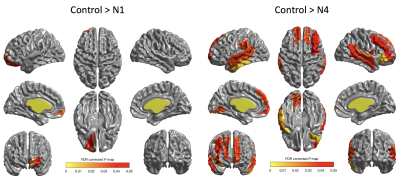
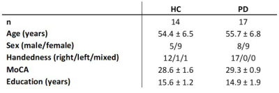
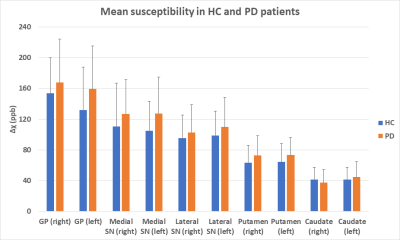
Parkinson's disease (PD) is characterised by dopaminergic neuronal loss and iron overload in the nigrostriatum. Diffusion Kurtosis Imaging (DKI) and Quantitative Susceptibility Mapping (QSM) allow quantification of alterations in tissue microstructure based on water diffusion and iron deposition respectively. Our case-control study in PD using DKI revealed greater cellular loss in the lateral SN and complex microstructural degradation in the putamen. QSM showed spatially variant iron deposition (Δχ) in the grey nuclei congruent with histochemical reports, and multivariate analysis showed that putaminal and lateral nigral Δχ significantly predicted UPDRS. Significant correlations between Δχ and DKI indices were found in the putamen.
|
|
2579.
 |
106 |
Longitudinal Changes in Cerebral Blood Flow Calculated by Arterial Spin Labeling MRI in Parkinson’s Disease
Dilek Arslan, Sevim Cengiz, Kardelen Eryurek, Ozan Genc, Ani Kicik, Emel Erdogdu, Zeynep Tufekcioglu, Basar Bilgic, Hasmet Hanagasi, Aziz Ulug, Ibrahim Gurvit, Tamer Demiralp, Esin Ozturk-Isik
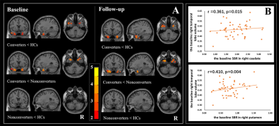
The aim of this study is to monitor perfusion changes over one and a half years in Parkinson’s disease with mild cognitive impairment (PD-MCI). Cerebral blood flow (CBF) maps were created for baseline and follow-up scans by fitting a kinetic curve model for each pixel of arterial spin labeling MR images. The CBF maps were registered to MNI152 brain atlas, and perfusion changes were assessed at 119 distinct brain regions. The CBF of PD-MCI patients decreased at occipital fusiform gyrus, right occipital fusiform gyrus, anterior part of left supramarginal gyrus, and anterior part of right middle temporal gyrus over time.
|
|
2580.
 |
107 |
Studying the effect of medication status on resting state network metrics in Parkinson’s disease
Saadnah Naidu, Yue Xing, Dorothee Auer
Current drug therapies for Parkinson’s disease (PD) offer symptom control without the capability for disease modification. Furthermore, unpredictable on/off fluctuations and dyskinesias present challenges in titrating appropriate doses. Our study aims to utilise resting state functional MRI (fMRI) to determine the effect of PD medication, as preliminary step to future work to address these limitations.
|
|
2581.
 |
108 |
Prognosis of body function in Parkinson’s disease using diffusion magnetic resonance imaging
Chih-Chien Tsai, Po-Yuan Chen, Sung-han Lin, Shu-Hang Ng, Yao-Liang Chen, Chin-Song Lu, Jur-Shan Cheng, Yi-Hsin Weng, Wey-Yil Lin, Yi-Ming Wu, Jiun-Jie Wang
Parkinson’s disease (PD) is a common progressive neurodegenerative disorder characterized by resting tremor, bradykinesia, restricted mobility, and postural instability. PD has a progressive course and is associated with increased mortality, with physical disability and non-motor symptoms exerting a significant negative impact on quality of life. Robust early prediction of clinical outcomes in Parkinson’s disease would be paramount for implementing appropriate management interventions. The predictive power varied according to the clinical measures used and was highest in the prediction of UPDRS. This finding was further confirmed by using bootstrap approach and leave-one-out cross-validation analysis.
|
|
2582.
 |
109 |
Analysis of changes in brain structure in patients with Parkinson's disease and their correlation with the Hoehn-Yahr stage using the MPRAGE sequence
Chunyan Zhang, Ruichen Zhao, Jinxia Zhu, Tobias Kober, Chen Chen, Hong Lu, Hongcan Zhu, Bing Xue, Hong Liang, Jingliang Cheng
In this study, the volume changes of brain structure in Parkinson's disease (PD) patients were analyzed. We found extensive structural brain changes in PD patients, and most of these changes were correlated with the Hoehn-Yahr stage. The results showed that volume changes in some brain regions may be a potential imaging marker for early diagnosis of PD, and the MPRAGE sequence may be a suitable and quick method to provide a reference for clinicians to diagnose PD.
|
|
2583.
 |
110 |
Arteriovenous Structure and Blood Flow Abnormalities in Parkinson’s Disease
Chunyan Zhang, Bo Wu, Xiao Wang, Chen Chen, Ruichen Zhao, Hong Lu, Hongcan Zhu, Bing Xue, Hong Liang, Sean Sethi, E. Mark Haacke, Jingliang Cheng
Few researchers have paid attention to the vascular supply and venous outflow in Parkinson’s disease (PD). In this work, we evaluated arterial inflow and venous outflow and looked for the presence of abnormal venous structure. We found that there was a significant correlation of reduced arterial flow with reduced internal jugular vein (IJV) flow. We also found there were a large number of PD patients with no or little flow in the left IJV compared to healthy control group. These results suggest that abnormal flow could be one factor in the development of or progression of PD in some patients.
|
|
2584.
 |
111 |
Voxel-Based Meta-Analysis of Mutant a-Synuclein transgenic Marmoset using Multiparametric MRI
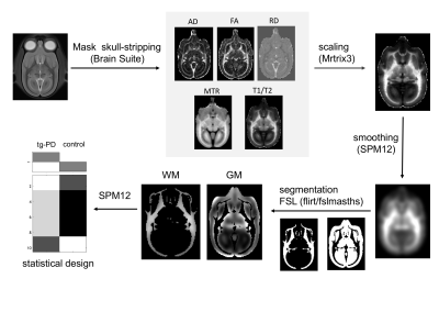
Mai Mizumura, Fumiko Seki, Junichi Hata, Yawara Haga, Marin Nishio, Hideyuki Okano, Akira Furukawa
In this study, we evaluated the characteristics of the brain in a genetically modified marmoset model of Parkinson’s disease. Various contrast images were acquired using magnetic resonance imaging (MRI), and the whole brain underwent explorative investigation with each contrast. For each image, statistical evaluation was performed using SPM. Diffusion tensor MRI showed significance differences in the thalamus, while magnetization transfer ratio images showed a significant difference in the nigral striatum. The findings suggest that the marmoset is useful as a model animal to study human diseases.
|
|
2585.
 |
112 |
Differentiating Parkinson’s disease patients from healthy controls through high iron content deposition in the substantia nigra
Kiarash Ghassaban, Naying He, Sean Sethi, Pei Huang, Shengdi Chen, Fuhua Yan, Ewart Haacke
In this work, 25 Parkinson’s disease patients and 24 healthy controls (HC) were scanned in order to quantify brain iron content in eight major deep gray matter structures. In addition to comparing global iron deposition, a novel threshold-based method was used to assess regional high iron (RII) in these nuclei. Among all the structures, the substantia nigra (SN) was the only one showing significantly higher iron content in PD patients compared to that of the HC cohort with the regional analysis revealing more prominent results. There are two populations of PD patients, those that do not change iron content in SN and those that do. For the abnormally high SN iron content group, there was a significantly higher UPDRS-III than the group showing normal iron content.
|
|
2586
 |
113 |
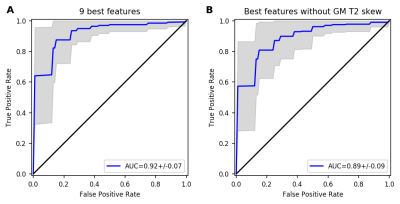
Classification of Parkinson’s disease based on multi-parametric data derived from MR Fingerprinting measurements
Video Permission Withheld
Thomas Amthor, Peter Koken, Mariya Doneva, Vera Keil, Stilyana Bakoeva, Alina Jurcoane, Wolfgang Block, Ullrich Wüllner, Burkhard Mädler, Elke Hattingen
We investigated the potential of multi-parametric MR Fingerprinting measurements for the classification of Parkinson’s disease. For each measured quantity (T1, T2, proton density) and each segmented brain region, several statistical parameters were determined and used to train a Random Forest classification algorithm. An AUC of 0.92 was achieved for distinguishing Parkinson patients from healthy control subjects.
|
|
2587.
 |
114 |
Free-Water Imaging Improves the Evaluation of White and Gray-Matter in Early Parkinson’s Disease
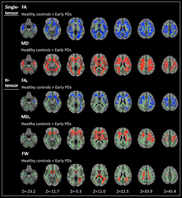
Christina Andica, Koji Kamagata, Taku Hatano, Yuki Takenaka, Asami Saito, Mana Kuramochi, Wataru Uchida, Akifumi Hagiwara, Takashi Ogawa, Haruka Takeshige-Amano, Masaaki Hori, Nobutaka Hattori, Shigeki Aoki
We applied bi-tensor diffusion model to evaluate the microstructural changes of white (WM) and gray matter (GM) in patients with early Parkinson’s disease (PDs). Our results demonstrated that the bi-tensor diffusion model could be used to disentangle neuroinflammation and axonal degeneration in early PDs with more precise estimations of localized microstructural changes compared to the single-tensor model. Our findings also suggest that microstructural changes in early PD may be preceded by neuroinflammation and followed by axonal degeneration, with WM changes preceding GM changes. Finally, the bi-tensor model also enabled to show possible compensatory mechanisms in PD occurring in the cerebellum.
|
|
2588
 |
115 |
Linked alterations in microstructural morphology of white matter in patients with Parkinson’s disease: A multimodal magnetic resonance imaging study
Video Permission Withheld
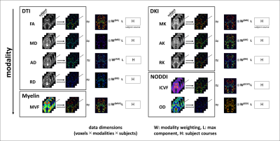
Koji Kamagata, Kouhei Kamiya, Yuya Saito, Taku Hatano, Christina Andica, Tomoko Maekawa, Shohei Fujita, Asami Saito, Takashi Ogawa, Genko Oyama, Haruka Takeshige-Amano, Yasushi Shimo, Akifumi Hagiwara, Masaaki Hori, Nobutaka Hattori, Shigeki Aoki
To identify relationships between Parkinson’s disease (PD) severity and microstructural changes in white matter (WM), we applied a multimodal data-fusion method known as linked independent component analysis (LICA) to a set of diffusion magnetic resonance (MR) and myelin-sensitive imaging data. LICA explained data variance with high sensitivity to PD severity, revealing widespread coordinated decreases in intracellular volume fraction, fractional anisotropy, and myelin volume fraction with increases in radial diffusivity. Our results show coordinated microstructural alterations in WM with disease severity and PD progression.
|
|
2589.
 |
116 |
Atlas based Diffusion Abnormalities in Substantia Nigra in Parkinson's Disease
Apoorva Safai, Shweta Prasad, Jitender Saini, Pramod Pal, Madhura Ingalhalikar
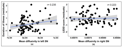
Parkinson’s disease (PD) is characterized by neuronal loss of dopaminergic neurons in the substantia nigra (SN). This study aims to gain deeper insights into the abnormalities in SN by evaluating the diffusion metrics of the SN in a large cohort of patients with PD. To precisely delineate the SN, neuromelanin sensitive MRI images were obtained from a set of healthy controls and were used to create a probabilistic atlas of the SN. Using this atlas, we observed significantly higher radial and mean diffusivity of bilateral SN in patients with PD suggesting microstructural abnormalities that could potentially serve as bio-markers for PD.
|
|
2590.
 |
117 |
Acupuncture Relieve Pain in Parkinson’s Disease Through Modulating Pain Matrix
Sung-han Lin, Shao-Wen Yu, Yu-Chieh Huang, Yih-Ru Wu, Jiun-Jie Wang
Patients with Parkinson’s Disease may suffer from different pain for years, including aching and burning from muscles, skeleton, or even throughout their body. In the current study, we provide that acupuncture could relieve such specific pain in PD patients through modulating regions related to the pain matrix in brain, especially correlated with primary somatosensory cortex and middle temporal pole. This could be an effective and safe analgesic tool that would relieve patients’ suffering.
|
|
2591.
 |
118 |
Analysis of Structural Connectivity using Certain Nuclei as Seeds in Patients with Parkinson’s Disease
Haining Wei, Zhangxuan Hu, Yuhui Xiong, Le He, Yan Tong, Yu Ma, Hua Guo, Xuesong Li
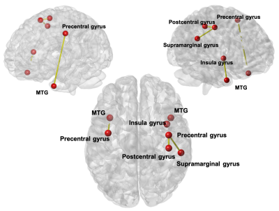
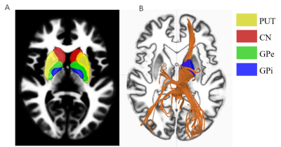
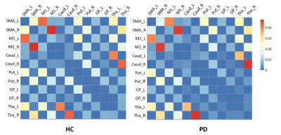
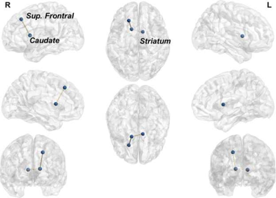
In this study, we aim to evaluate the relationships between fiber connectivity starting from specific nucleus regions to the whole brain and the Unified Parkinson’s Disease Rating Scale (UPDRS)-III scores in patients with Parkinson's disease. The results showed that the structural connectivity to the whole brain starting from bilateral internal global pallidus and caudate nucleus has significant negative correlations with the UPDRS-III scores. In the contrary, no significant correlations was found for the network starting from the putamen and external global pallidus. The strong negative correlation implies that these specific nuclei may play significant roles in the severity of Parkinson's disease. This finding is of great importance for further clinical research.
|
|
2592.
 |
119 |
Temporal Atrophy Predicts the Deterioration of Cognition in Multiple Domains: a Longitudinal Clinical Study in Parkinson’s Disease
Cheng Zhou, Xiaojun Guan, Tao Guo, Minming Zhang
To specify the critical structural alterations of cognitive deterioration in Parkinson’s disease (PD) and explored the underlying mechanism of structural changes. We combined cross-sectional and longitudinal VBM analyses to explore the structural topologies between PD patient who convert to mild cognitive impairment (PD converter). The relationships between dopamine transporter (DAT), CSF proteins and structural alterations were assessed. PD converters showed progressive temporal atrophy associated with multiple cognitive domains. DAT results were significantly associated with temporal atrophy. In conclusion, temporal lobe is a crucial node in modulating cognitive status in multi-domains. Dopamine deficiency may contribute to cognition-related temporal atrophy.
|
|
2593.
 |
120 |
Functional Brain Connectome and Mild Cognitive Impairment in Early Stage Parkinson Disease
Xueling Suo, Du Lei, Nannan Li, Wenbin Li, Lan Cheng, Meiyun Wang, Rong Peng, Qiyong Gong
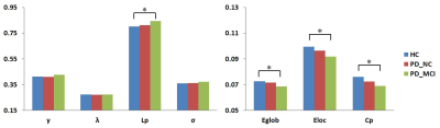
To use graph theory approaches and resting-state functional magnetic resonance imaging (MRI) to explore the brain functional network in patients with early stage Parkinson's disease (PD) and mild cognitive impairment (MCI). The whole-brain functional network was constructed by thresholding the Pearson correlation matrices of 90 brain regions. The results showed a less small-worldization characterized by decreased global integration and decreased local segregation in PD patients relative to healthy controls (HC). On the basis of these between-group difference in global and nodal properties, PD patients with MCI showed the lowest properties values, followed by PD patients with normal cognition and HC.
|
|
2594.
 |
121 |
Altered cerebellar functional connectivity in Parkinson’s disease
Jason Langley, Daniel Huddleston, Xiaoping Hu
Parkinson’s disease is a progressive, neurodegenerative disorder characterized by asymmetrical onset of motor symptoms such as bradykinesia, rigidity, and tremor. Mounting evidence suggests that the cerebellum plays a major role in the pathophysiology of PD. Prior imaging studies have found altered cerebellar activation during motor execution and motor learning, suggesting that altered activation in the cerebellum may reflect Parkinsonian-related impairment. Here, we use resting-state function MRI (fMRI) to ascertain connectivity changes in the cerebellum from Parkinson's disease found reduced connectivity in lobule V of the cerebellum as well as reduced connectivity between dentate nucleus and the cerebellar cortex.
|
|
2595.
 |
122 |
Disruption of cortical-basal ganglia motor network affects cognitive function in Parkinson’s disease: a high angular resolution diffusion imaging study
Chenfei Ye, Zehong Yan, Tao Wu, Ting Ma
Whether clinical phenotype in Parkinson's disease (PD) is affected by cortical-basal ganglia motor circuit (CBG) dysfunction remains to be investigated. In this study, we utilized a high angular resolution diffusion imaging (HARDI) technique to investigate association between white matter structural connectivity within CBG and cognitive function related to PD. We found that the structural connectivity between the M1 cortical area and other regions within the CBG circuit decreased for those PD patients with severe cognitive symptoms, indicating that the less effective information processing between cortical and subcortical regions in CBG network could lead to cognitive deficits in PD patients.
|
|
2596.
 |
123 |
Influence of Analytic Techniques on Comparing Diffusion Derived Measurements in Early Stage Parkinson’s Disease
Virendra Mishra, Karthik Sreenivasan, Xiaowei Zhuang, Zhengshi Yang, Dietmar Cordes, Ryan Walsh
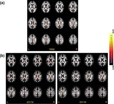
Using a well-characterized multi-site diffusion MRI (dMRI) dataset of early Parkinson’s disease (PD), this study suggests that DTI-TK and TBSS perform at the same accuracy of detection if skeletonwise analysis are conducted. However, DTI-TK is more sensitive than TBSS. Further, results from voxelwise or ROI-based analysis should be reported with caution as this study found a significant smoothing effect to detect differences in scalar dMRI-derived measures. Overall, our findings suggest that the conclusions for the hypothesis being tested are strongly dependent upon the choice of analytic techniques as different analysis techniques can lead to different conclusions.
|
|
2597.
 |
124 |
Towards identification of neuroanatomical correlates of neuropsychological scores in Parkinson’s disease patients, with and without, memory impairment
Virendra Mishra, Karthik Sreenivasan, Ece Bayram, Sarah Banks, Jason Longhurst, Zhengshi Yang, Xiaowei Zhuang, Dietmar Cordes, Aaron Ritter, Jessica Caldwell, Brent Bluett
With a well-characterized dataset of Parkinson’s disease (PD) participants, with and without memory impairment, this study shows that there is a distinct structural network organization between PD with mild cognitive impairment (PD-MCI) and without MCI(PD-nMCI). This study further shows that while there are no discernible differences in scalar diffusion-MRI derived measures, fractional anisotropy in PD-nMCI is negatively associated with trail making test-A. Our study demonstrates the feasibility towards identifying neuroanatomical correlates of neuropsychological scores that will not only aid in our understanding of the underlying neural correlates of cognitive impairment in PD, as well as differentiating PD-MCI and PD-nMCI in an objective and reproducible manner.
|
|
2598.
 |
125 |
Altered Claustral Functional Connectivity in Parkinson’s Disease Patients with Mild Cognitive Impairment.
Karthik Sreenivasan, Ece Bayram, Sarah Banks, Jason Longhurst, Xiaowei Zhuang, Zhengshi Yang, Dietmar Cordes, Aaron Ritter, Jessica Caldwell, Brent Bluett, Virendra Mishra
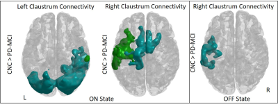
Studies have shown α-synuclein pathology in the claustrum of Parkinson’s disease (PD) patients and its correlation with the onset of cognitive dysfunction in PD. In this study we use resting state fMRI to examine claustral functional connectivity network changes in PD patients with mild cognitive impairment. Our results show increased claustral-cortical connectivity in the PD-MCI group, which may indicate additional effort is required in the PD-MCI group to maintain network integration. The increased load of claustrum is somewhat mitigated by medication in PD patients with cognitive impairment.
|
|
| Top |
Treasure Chest of Neuro Gems
Digital Poster
Neuro
Tuesday, 14 May 2019
| Exhibition Hall |
09:15 - 10:15 |
| |
|
Computer # |
|
2599.
 |
126 |
Investigation of brain plasticity during prolonged Braille learning in sighted subjects: a longitudinal diffusion MRI (dMRI) study
Malwina Molendowska, Bartosz Kossowski, Jacek Matuszewski, Lukasz Bola, Marcin Szwed, Katarzyna Jednoróg, Artur Marchewka
Diffusion MRI can be used to evaluate the brain plasticity processes that occur during new skills acquisition. Commonly, one of the tasks used to investigate neuroplasticity of both blind and sighted subjects is Braille reading. In this work, we analyze DTI metrics based on 5 time points data and investigate the dynamics of the brain reorganization processes. In particular, our preliminary results depict neuronal brain changes within main WM tracts associated with somatosensory area development.
|
|
2600.
 |
127 |
Increased Intracortical R1 in the Motor Cortex of Exercising Older Adults
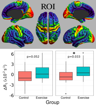
Christopher Rowley, Ralf Deichmann, Tobias Engeroff, Elke Hattingen, Rainer Hellweg, Ulrich Pilatus, Eszter Füzéki, Sina Gerten, Lutz Vogt, Winfried Banzer, Johannes Pantel, Johannes Fleckenstein, Silke Matura, Nicholas Bock
Exercise is known to be beneficial for brain health and performance; however, it is not known if changes in cortical microstructure underlie this effect. To investigate this, R1 maps acquired on cognitively healthy older adults (n=24, 65-90 years old) were analyzed before and after a 12-week exercise intervention. R1 prolongation indicating increased myelin levels were significant in the right (p=0.033) and trending in the left (p=0.052) leg motor regions with respect to a control group (n=22). ΔR1 correlated with aerobic cycling performance improvements (left: p=0.012, right: p=0.011). This study demonstrates that exercise promotes myelination in cortical motor regions.
|
|
2601.
 |
128 |
Investigating premanifest synucleinopathy: structural connectome of brainstem nuclei in REM sleep behavior disorder
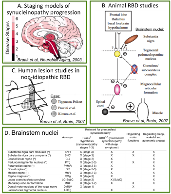
Maria Garcia Gomar, Laura Lewis, Lawrence Wald, Bruce Rosen, Aleksandar Videnovic, Marta Bianciardi
REM-sleep-behavior-disorder (RBD) is characterized by the absence of muscle-atonia during REM-sleep. RBD is strongly associated with presymptomatic-manifestations of neurodegenerative-synucleinopathies. Thus, it allows the investigation of early/premanifest neurodegenerative-stages when treatment can be most effective in delaying the development of full-blown-disease. Changes in brainstem-nuclei-connectivity are expected in RBD/premanifest-synucleinopathy based on animal- and ex-vivo-human-studies. Yet, their investigation in living-humans is understudied. Through high-spatial-resolution 7Tesla-MRI and a recently-developed probabilistic-brainstem-nuclei-atlas, we built a brainstem-based structural-connectome in living RBD-patients and age-matched controls. Interestingly, in RBD-patients we detected structural-connectivity-changes within the brainstem, with the striatum and cerebellum in line with the pathophysiology of RBD in animal-models.
|
|
2602.
 |
129 |
Changes in GABA associated with a sham-controlled transcranial direct current stimulation language intervention for primary progressive aphasia.
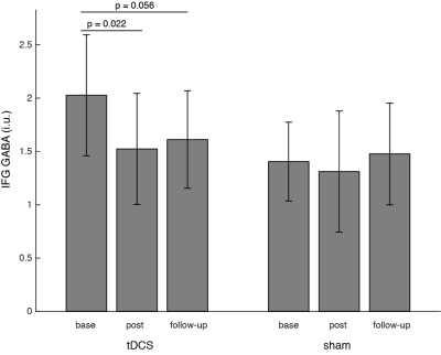
Ashley Harris, Zeyi Wang, Bronte Ficek, Kim Webster, Richard Edden, Kyrana Tsapkini
Primary Progressive Aphasia is a neurodegenerative disorder primarily affecting language. We applied GABA-edited MRS to examine GABA changes with anodal tDCS to augment language-therapy for patients with PPA. With tDCS targeting the left inferior frontal gyus, we see a decrease in IFG GABA following the intervention. No changes were observed in the sham group. While all patients showed improvements with language therapy, those receiving tDCS showed greater improvements that were maintained at 2 months follow-up. This work supports the use of tDCS to augment language therapy in PPA.
|
|
2603.
 |
130 |
Structural MRI abnormalities and the immune system are correlated with neuroinflammation in Neuropsychiatric Systemic Lupus Erythematosus: a retrospective study on a large and well-defined patient cohort.
Francesca Inglese, Ilse Kant, Ece Ercan, Mark van Buchem, Margreet Steup-Beekman, Tom Huizinga, Cesar Magro-Checa, Itamar Ronen, Jeroen de Bresser
Neuropsychiatric Systemic Lupus Erythematosus (NP-SLE) is associated with cerebral abnormalities, but their relation to the inflammatory and ischemic clinical phenotypes is unknown. We performed a retrospective structural brain study within a large, clinically well-defined patient cohort of NP-SLE patients (inflammatory and ischemic) and non-NP-SLE patients. Patients with inflammatory, but not ischemic, NP-SLE showed lower grey matter and white matter volumes, and higher White Matter Hyperintensity volumes compared to non-NP-SLE patients. Brain abnormalities were also associated with the complement system. In conclusion, only inflammatory NP-SLE showed more severe structural brain abnormalities, and these were associated with a specific complement component.
|
|
2604.
 |
131 |
Elucidating the influence of healthy aging on white matter microstructure: A comparison of different diffusion MRI models
Salman Shahid, Qixiang Lin, Antoine Hone-Blanchet, Allan Levey, James Lah, Bruce Crosson, Deqiang Qiu
To understand microstructural changes associated with healthy aging, multi-shell diffusion-weighted images were acquired in a group of 71 cognitively normal volunteers (31-young, 40-old). Signal representation and tissue specific models were used to assess relationship between age and WM microstructural changes. TBSS was performed for group-comparison. Results showed that FA and NODDI-based indices exhibited highest degree of sensitivity with overlap in much wider regions. The results also showed regional differences among FA and ODI. The influence of DKI was more regionalized and complemented by FA. The study demonstrated the sensitivity of higher-order models to the age-related changes in tissue microstructure.
|
|
2605.
 |
132 |
MRI Detection of Amyloid Related Imaging Abnormalities (ARIA) in a Non-Human Primate Model of Sporadic Cerebral Amyloid Angiography at 7-Tesla
Dina Ramadane, Thomas Genovese, Lori Hill, Charles Kingsley, Lawrence Williams, Thomas Wisniewski, Henrieta Scholtzova, Youssef Zaim Wadghiri
Here we describe a non-invasive brain imaging method studying the pathogenesis and long-term effects of ARIA (amyloid-related imaging abnormalities) in an aged squirrel monkey (Saimiri Boliviensis), a non-human primate model of naturally occurring cerebral amyloid angiopathy. We investigated both ARIA-E, characterized by vasogenic edema, and ARIA-H, characterized by MRI evidence of hemosiderin deposits as potential biomarkers to use in a MRI methodology to monitor newly developed AD treatments.
|
|
2606.
 |
133 |
Dynamics of white matter tract covariance across lifespan assessed with diffusion spectrum imaging
Yi-Xi Peng, Chang-Le Chen, Yung-Chin Hsu, Wen-Yih Tseng
In this study, we calculated tract covariance to describe the phenomenon of white matter differentiation and de-differentiation across lifespan, using diffusion spectrum imaging (DSI) and whole brain tract-based automatic analysis (TBAA) techniques. Differentiation was found to be highest in the 2nd decade and de-differentiation started to emerge at 3rd decade and peaked at 6th decade.
|
|
2607.
 |
134 |
Investigation of hypoxia after brain injury using a hypoxia-binding T1 contrast agent GdDO3NI
Babak Moghadas, Vimala Bharadwaj, Sarah Stabenfeldt, Vikram Kodibagkar
In this study we have used the hypoxia-targeted MR contrast agent GdDO3NI, (a nitroimidazole-based T1 MRI contrast agent) to image the development of hypoxia in the rodent brain after traumatic brain injury (TBI). Our results indicate a statistically significant ~ 50% signal enhancement over baseline in the injury region using GdDO3NI compared to baseline values (~ 0%) observed with non-specific Gadoteridol (as control) at 3hours post injection. This study further demonstrates the utility of GdDO3NI in imaging tissue hypoxia and applicability to traumatic brain injury.
|
|
2608.
 |
135 |
Pituitary R2 values at 3T to assess risk of iron-mediated hypogonadal hypogonadism.
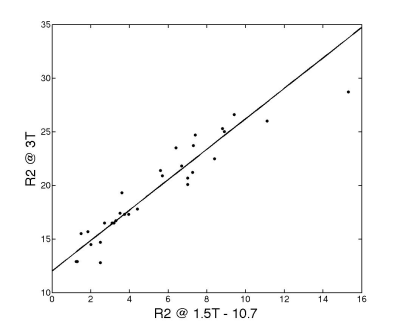
Andrew Cheng, Thomas Coates, John Wood
Pituitary R2 at 1.5 Tesla has been validated as a sensitive marker of pituitary siderosis and risk of clinical hypogonadism. We cross-validated pituitary R2 measurements at 3T and 1.5T in 26 patients with iron overload syndromes. Pituitary R2 scaled linearly across field strength with a relative enhancement of 42%, consistent with previous liver R2 cross-field validations. When 3T pituitary values were transformed into equivalent 1.5T R2 values, the resulting Z-score estimates were unbiased with native 1.5T R2 estimates. Thus it is not necessary to acquire normative R2 data at 3 Tesla in order to interpret 3T pituitary R2 values.
|
|
2609.
 |
136 |
Thalamus in chronic low back pain: vertex-based morphometry and connectivity-based thalamic white-matter studies
Huiling Peng, Jason Craggs, Kelly Boland, Carmen Cirstea
Chronic low back pain (CLBP) is now considered a central nervous system disease. Thalamus is a key relay station for processing and transmission of nociceptive information to the cerebral cortex. We used vertex-based morphometry and connectivity-based diffusion tractography to test the hypothesis that the CLBP is associated with altered thalamic shape and altered white matter integrity of the thalamic projections to cortical regions in frontal and parietal lobes. Compare to controls, CLBP exhibited significant surface depression in left thalamus and lower fractional anisotropy in left thalamic projections to the posterior parietal cortex. This may represent a degenerative pain-related process.
|
|
2610.
 |
137 |
HIV alters brain activation during semantic memory processing demands
Joanna Poweska, Anna Egbert, Marta Sobanska, Agnieszka Pluta, Tomasz Wolak, Lukasz Okruszek, Natalia Gawron, Bogna Szymanska-Kotwica, Ewa Firlag-Burkacka, Andrzej Horban, Przemyslaw Bienkowski, Halina Sienkiewicz-Jarosz, Anna Scinska-Bienkowska, Robert Bornstein, Stephen Rao, Emilia Lojek
Memory and executive dysfunctions burden HIV patients even in the highly active antiretroviral treatment (HAART) era. The neurobiological correlates of these cognitive symptoms remain unclear limiting development of targeted treatment options. Functional magnetic resonance imaging (fMRI) is a promising route to estimate neural signature of HIV-related neurocognitive decline. We examined brain activity in HIV+/HAART+ vs. healthy individuals during execution of semantic memory task. Results show that famous names induce lower activation in left caudate, right thalamus and left middle occipital gyrus in HIV+ vs. healthy group, despite lack of behavioral differences. Such hypoactivation suggests brain functional reorganization in HIV+/HAART+ patients.
|
|
2611.
 |
138 |
A Method for Evaluating Whole Brain Health of the Aging Brain: Assessment of Multiple MRI Detectable Brain Changes using the Brain Atrophy and Lesion Index (BALI)
Lukas Grajauskas, Tory Frizzell, Hui Guo, Caressa Liu, Ryan D'Arcy, Xiaowei Song
As our population ages, there is a need for better methods of assessing neurodegeneration. However, current methods are based on a diagnostic model and assess changes in isolation, failing to account for the interconnected nature of the brain and the heterogeneity of the aging process. To address this, we introduced the Brain Atrophy and Lesion Index (BALI) an MRI based tool for the assessment of structural neurodegeneration across the whole brain. Here, we compare results from eight datasets to which BALI was applied (n=3295), and present a literature review to understand consensus regarding the brain changes assessed by the BALI.
|
|
2612.
 |
139 |
Plastic Changes of the Language-related Brain Regions for Children with Non-syndrome Cleft of Lip with or without Palate (NSCL/P)
Presentation Not Submitted
Bo Rao, Hua Cheng, Yang Fan, Wenjing Zhang, Xuhong Liao, Renji Chen, Yun Peng
Using multimode MRI technique, this study attempt to find structural and functional alterations of brain regions for children with non-syndrome cleft of lip with or without palate (NSCL/P). Compared with control group, both structural and functional changes were detected in distributed cortical regions for NSCL/P group, which mainly located on the dorsal stream of language pathways. Besides, significant correlations were found between ALFF values and Chinese language clear degree scales for NSCL/P children.
|
|
2613.
 |
140 |
A multiparametric study of prion disease
Eleni Demetriou, Mohamed Tachrount, Matthew Ellis, Jackie Linehan, Sebastian Brandner, Karin Shmueli, Mark Farrow, Xavier Golay
In this work, we hypothesize that the metabolic changes occurring in the brain of prion-infected mice due to conformational changes of prion protein can be mapped using CEST MRI following previous in vivo work. Our previous findings include reduced Nuclear Overhauser Effect mediated by exchange-relayed signals in thalamus and cortex of prion-infected mice possibly related to up normal prion protein folding. Here we extend our studies by including a rich multipower acquisition scheme for targeting exchange processes falling at different regimes. For understanding the origin of CEST signal alternations T1, T2, and MT maps are included as well as histological findings.
|
|
2614.
 |
141 |
MR neuroimaging and pons proton spectroscopy in type 1 narcolepsy
Stefania Evangelisti, Claudia Testa, Laura Gramegna, Fabio Pizza, David Manners, Elena Antelmi, Lia Talozzi, Claudio Bianchini, Giuseppe Plazzi, Raffaele Lodi, Caterina Tonon
Narcolepsy type 1 (NT1) is a rare and life-long disease, characterized by central hypersomnia and cataplexy typically triggered by emotions. NT1 is linked to a selective loss of hypothalamic hypocretin neurons. To characterise neurodegeneration, we combined pons 1H-MRS and whole brain structural analysis in a large and homogenous sample of adult NT1 patients. 1H-MRS showed evidence of pontine neuronal dysfunction, consistent with its key role in REM sleep regulation. Grey matter loss was detected in brain regions implicated in the disease pathophysiology, including frontal-prefrontal cortices, putamen nuclei, thalami, hypothalamus, amygdalae, cerebellum, and widespread subtle tissue microstructural alterations were also found.
|
|
2615.
 |
142 |
Ex-vivo MR investigation of microstructures in globus pallidus in QSM: a histological validation study
Jinhee Jang, Yoonho Nam, Tae-Ryong Riew, Sung Won Jung, Sang Hyun Kim, In-Beom Kim
Linear paramagnetic structures were frequently seen in globus pallidus (GP), and interesting calcific densities are overlapping on these paramagnetic structures. This study aimed to explore the microstructural findings of GP using ex-vivo MRI scan and histologic validation. We found that the source of paramagnetism were mineral deposition of perforating vessels in GP. Those mineral depositions were paramagnetic on MR images, and calcific density on CT scan. Histologic study showed simultaneous deposition of iron and calcium along the arterial wall. High resolution MRI might have potential to demonstrate vascular degeneration and mineral deposition, which might be associated with aging and metabolic brain diseases.
|
|
2616
 |
143 |
The change of cerebral cortex in children with Tourette syndrome
Video Permission Withheld
Lei Kong, Yue Liu, Shao Cao, Lv Bin , Yun Peng
Tourette syndrome (TS) is a developmental neuropsychiatric disorder and is characterized by multiple motor and vocal tics. To understand the developmental cause of such changes, we investigated microstructural changes of cortical thickness , cortical sulcus, cortical curvature, and LGI in TS children by using sagittal three-dimensional T1-weighted image (3DT1WI) Magnetization. The TS children had the significant differences in cortical thickness, cortical sulcus, cortical curvature, and LGI compared with controls.
|
|
2617.
 |
144 |
Altered cortical thickness relevance in the early blind, late blind during the critical developmental time
Ankeeta Ankeeta, S Kumaran, N Jagannathan, Rohit Saxena
Visual impairment induces structural and functional modification in the visual cortex. There is significant modification observed between early blind subjects with thicker V1 compared to sighted controls, however late blind subjects with showed no significant differences in the V1 in age group of 6-12 years but there is differences observed between 13-19 years age range. Hence implicating the role of age of blindness onset may induce significant possessions on the expansion of cortical thickness of the V1.
|
|
2618.
 |
145 |
Gradient profiles of myelin and microstructure metrics across the developing brain
Erika Raven, Maxime Chamberland, Sila Genc, Kate Duffy, Chantal Tax, Greg Parker, Maxime Descoteaux, Derek Jones
Myelinogenesis follows a protracted sequence, with distinct pathways being myelinated at various times throughout development. To test this with MRI, we used magnetization transfer and diffusion metrics with tractography to investigate along-tract profiles of myelin and microstructure metrics in children and adolescents. Profiles demonstrated sensitivity to along-tract metrics, with midline regions having increased myelin and restricted diffusion indices indicative of maturation.
|
|
2619.
 |
146 |
Altered brain structure associated with cognitive changes of end-stage renal disease patients without dialysis and with maintenance hemodialysis
Presentation Not Submitted
Xueying Ma, Dun Ding, Peng Li, Pan Zhang, Shaohui Ma, Ming Zhang
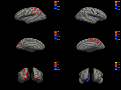
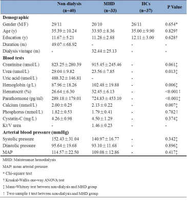
The theory of kidney-brain axis has provided more information for the interpretation of brain damage in ESRD patients. However, the factor of dialysis was ignored in this theoretical system. We analyzed the cortical structural changes and cognitive changes from different dimensions and also analyzed their relationship in ESRD patients with and without hemodialysis. We found that both the patients with dialysis and the patients without dialysis showed decreased cortical thickness when compared with healthy people, while the patients without dialysis presented with a more extensive decreased cortical thickness when compared with patients with maintenance hemodialysis. The brain structural changes were correlated with the cognitive changes. Our results suggested that the hemodialysis might be a protective factor for the brain, but the protective effect of hemodialysis was limited.
|
|
2620.
 |
147 |
Chemotherapy-induced gray matter abnormalities in cancer survivals: a voxel-wise neuroimaging meta-analysis.
Presentation Not Submitted
Running Niu, Mingying Du, Lu Lu, Peng Zhou
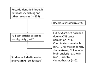
The present meta-analysis investigated the grey matter abnormalities in non-CNS cancer survivals treated with chemotherapy using Anisotropic Effect Size Signed Differential Mapping (AES-SDM) according to the Preferred Reporting Items for Systematic Reviews and Meta-Analyses (PRISMA) guides. Compared with controls, the non-CNS cancer survivals treated with chemotherapy exhibit widespread grey matter abnormalities in brain, especially in prefrontal-temporal pathway, which was significantly affected by the time length since chemotherapy. This pattern of grey matter volume changes might improve our understanding of the pathophysiological nature of chemotherapy related cognitive dysfunctions.
|
|
2621.
 |
148 |
Quantitative R2 mapping reveals information of myelin content in rat brain at 7T
Ping-Huei Tsai, Tsai-Jou Su, Hua-Shan Liu, Fei-Ting Hsu, Yu-Chieh Kao, Chia-Feng Lu, Hsiao-Wen Chung, Cheng-Yu Chen
Information of myelin content can reflect the microstructural difference between brain white matter (WM) and gray matter (GM), and particularly facilitating in detection of WM abnormalities during disease progression. This study aims to optimize a quantitative R2 mapping method of rat brain at 7T MRI and to evaluate the relationship between the measured R2 values and myelin content in discrepant brain tissues. Our findings demonstrated that quantitative R2 measurements could be an alternative to provide information of myelin content in rat brain at 7T, which may have potential to assess microstructural changes of brain WM and GM in demyelinating diseases.
|
|
2622.
 |
149 |
White Matter Tract Abnormalities are Associated with Cognitive Dysfunction in CADASIL
Shiyu Ban, Jingjing Su, Mengxing Wang, Shuai Xu, Xiaoxia Du*
This study was to investigate the white matter microstructural abnormalities and the relationship between white matter alterations and cognitive impairment in patients with cerebral autosomal dominant arteriopathy with subcortical infarcts and leukoencephalopathy (CADASIL), by diffusion tensor imaging (DTI). Patients with CADASIL showed significant extensive reductions in fractional anisotropy (FA), and increases in axial diffusivity (AD), radial diffusivity (RD), mean diffusivity (MD) compared to healthy controls. Furthermore, these white matter microstructural alterations were significantly correlated with Cognitive scores, and Stroke scale scores. It indicated that damage of white matter play an important role in cognition impairment in CADASIL
|
|
2623
 |
150 |
Preoperative brain MRI features and postoperative delirium
Video Permission Withheld
Ilse Kant, Jeroen de Bresser, Simone van Montfort, Myriam Jaarsma-Coes, Theo Witkamp, Henri Mutsaerts, Claudia Spies, Jeroen Hendrikse, Arjen Slooter
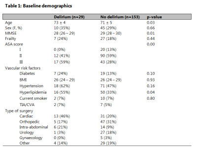
Postoperative delirium is associated with impaired cognitive outcome, longer hospital stay and an increased risk of dementia. To date, the pathophysiology of delirium remains largely unknown. Therefore, we studied the association of preoperative brain MRI features and the occurrence of postoperative delirium in a large group of older patients. We measured preoperative brain volumes, white matter hyperintensity shape, cerebral infarcts and cerebral perfusion. Preoperative cortical brain infarct volume was associated with postoperative delirium. Other preoperative brain MRI features were not significantly associated with postoperative delirium. Patients with a larger burden of cortical infarcts may have a decreased brain reserve, increasing the risk of postoperative delirium.
|
|
| Top |
Neuroanatomy: Seeing Is Believing
Digital Poster
Neuro
Tuesday, 14 May 2019
| Exhibition Hall |
09:15 - 10:15 |
| |
|
Computer # |
|
2624.
 |
151 |
Development of a Standardized Normative Pediatric Spinal Cord structural template: Demonstration of an automatic estimation of Spinal Cord Cross Sectional Area measurements (SCCSA).
Shiva Shahrampour, Benjamin De Leener, Devon Middelton, Kavya Jonnavithula, Mahdi Alizadeh, Hiba Pediyakkal, Laura Krisa, Adam Flanders, Scott Faro, Julien Cohen-Adad, Feroze Mohamed
Template-based analysis of MRI data of the spinal cord lay the foundation for standardization and reproducibility , improves patient diagnosis and helps the discovery of new biomarkers of spinal-related diseases.
|
|
2625.
 |
152 |
Automated MP2RAGE-based Brain Volumetry for Pediatric Patients: A Clinical Usability Study
Maxence Serru, Benedicte Marechal, Tobias Kober, Leo Ribier, Catherine Sembely Taveau, Jean Philippe Cottier, Dominique Sirinelli, Baptiste MOREL
Antenatal and mostly post-natal periods are crucial for brain development, characterized by volume increase, brain maturation, neuronal proliferation, neural migration, and myelination. Head circumference is a reliable clinical assessment of brain volume, correlated with neurodevelopmental outcomes (psychomotor and cognitive development). Particularly in young children, it is a fast and inexpensive tool for brain growth follow-up. Complementarily, brain MRI is becoming more frequently used as a first-line examination for suspected brain development abnormality. In this work, we evaluate the potential of an automated MP2RAGE-based brain volumetry method to objectively support radiologists to better assess brain physiological and pathological cerebral growth.
|
|
2626.
 |
153 |
Sex differences in structural variability of brain regions in development and young adults
Natalie Forde, Grace Jacobs, Erin Dickie, Aristotle Voineskos, Stephanie Ameis
Increased variability of brain metrics is suggested to relate to increased vulnerability for psychiatric disorders. Here we investigate sex differences in variability of brain structure (global and subcortical volume, regional cortical thickness and surface area) in young adults (n=1,032, 22-35 years, Human Connectome Project [HCP]) and through development (n=1,347, 8-21 years, Philadelphia Neurodevelopmental Cohort [PNC]).
Both volume and surface area were observed to be generally more variable in males compared to females in both development and adulthood. This increased variability may relate to the elevated vulnerability for psychiatric disorders seen in males compared to females.
|
|
2627.
 |
154 |
Brain Microstructure Changes in Healthy Aging Revealed by Quantitative Multi-parametric MRI
Qixiang Lin, Salman Shahid, Antoine Hone-Blanchet, Allan Levey, James Lah, Bruce Crosson, Deqiang Qiu
This study aims to reveal the alterations of biologically relevant measurements in healthy aging using multi-parametric quantitative MRI. Multi-parametric quantitative MRI scans of the whole brain were performed in 20 healthy elderly and 21 young adults. Whole-brain voxel-wise analysis showed increased quantitative T1 value in the right hippocampus and right insula, and widespread increases in the subcortical and cortical area of R2*, suggesting microstructural alteration associated with healthy aging in these regions. Quantitative multi-parametric measurements might provide sensitive neuroimaging biomarkers for the microstructure changes during normal aging and related neurodegeneration diseases.
|
|
2628.
 |
155 |
Development and evaluation of a 0.5mm isotropic resolution structural template of the older adult brain
Mohammad Rakeen Niaz, Abdur Raquib Ridwan, Xiaoxiao Qi, David A. Bennett, Konstantinos Arfanakis
Human brain structural MRI templates with low spatial resolution lack important fine details due to partial volume effects. The purpose of this work was twofold: a) to introduce a novel approach for high-resolution template construction based on principles of super-resolution, and b) using this technique, to develop a high-resolution structural template of the older adult brain based on MRI data from 222 non-demented older adults.
|
|
2629.
 |
156 |
Longitudinal tensor-based morphometry in healthy aging
Laura Reyes, Neda Sadeghi, François Lalonde, Carlo Pierpaoli
Previous studies of healthy elderly populations combined a longitudinal design with tensor-based morphometry (TBM) and found significant gray matter (GM) atrophy over short time periods. We examined a separate healthy elderly population using a different method to determine if previous results are biologically driven, and investigated the relationship between GM and cognition. We also detected significant GM atrophy, but did not find a link between GM, age, and cognition. Our longitudinal TBM approach is sensitive to subtle, short-term GM changes, but further investigation is necessary to examine the effect of different methodological approaches on the relationship between GM and cognition.
|
|
2630
 |
157 |
A knowledge-based linear registration for brain MRI morphology
Video Permission Withheld
Xinyuan Zhang, Yanqiu Feng, Qianjin Feng, Susumu Mori
Linear registration is an essential first step for image registration. However, linear registration often fails when the brain shapes, locations, orientations of the target and template images are severely different. To solve this problem, we proposed a knowledge-based approach, in which a large number of MR images were prepared as intermediate images, which were semi-automatically registered to the template a priori to ensure accurate registration. A new target image was first registered to all intermediate images and best intermediate image was selected based on a goodness-of-fit metric. The final transformation was then calculated by combining the pre-determined intermediate-to-target transformation.
|
|
2631.
 |
158 |
Evaluating lifespan tissue structure: Comparing CSD signal fraction and VBM grey matter density
Jamie Blair, Benjamin Newman, Lisa Post, T Druzgal
Constrained spherical deconvolution (CSD), a recently developed diffusion MRI analysis technique, can be used to obtain whole-brain signal fractions from grey-matter-like, white-matter-like, and CSF-like tissue. This study evaluates the CSF compartment present in grey matter (GM-CSF) over the lifespan, and compares it to grey matter density (GMD), obtained through Voxel Based Morphometry. Results of this study reveal a complimentary relationship between GM-CSF and GMD across the lifespan, but not amongst a younger cohort. Results suggest further research is necessary to understand differences between these techniques, and how they may relate to tissue structure.
|
|
2632.
 |
159 |
Characterizing age-related microstructural changes in locus coeruleus and substantia nigra
Jason Langley, Justino Flores, Sana Hussain, Ilana Bennett, Xiaoping Hu
Characterization of age-related alterations in composition and morphology of locus coeruleus and substantia nigra pars compacta will aid in the development of new biomarkers and may provide insight in the development of novel interventions to arrest progression of Alzheimer's disease or Parkinson's disease. Imaging these structures with diffusion-weighted images is difficult due to their small stature (locus coeruleus is 1.5 mm in diameter and 15 mm long) and location in the brain stem. In this abstract, we utilize a high resolution diffusion-weighted protocol to examine age-related microstructural changes in locus coeruleus and substantia nigra pars compacta.
|
|
2633.
 |
160 |
Microstructural Changes in Human Substantia Nigra with Aging as Revealed by Non-Gaussian Diffusion MRI
Zheng Zhong, Muge Karaman, Kaibao Sun, Xiaohong Zhou
Aging is considered a major factor in the development of neurodegenerative disease. The aging process can result in brain tissue microstructural alterations, particularly in regions relevant to neurodegeneration, such as the substantia nigra (SN). In this study, we employed a non-Gaussian diffusion model – the continuous-time random-walk (CTRW) model – together with a high-resolution diffusion acquisition technique to investigate the possible microstructural changes in the SN in normal aging. Two CTRW model parameters have exhibited significant differences in the SN between young and elderly healthy human subjects.
|
|
2634.
 |
161 |
The visualization of the morphology change within depigmented substantia nigra using high field postmortem MRI
Hansol Lee, Sun-Yong Baek, Eun-Joo Kim, Gi Yeong Huh, Jae-Hyeok Lee, HyungJoon Cho
The purpose of this study was to determine the alteration of the morphology in the substantia nigra using MRI with histopathological validation for the patients of atypical Parkinsonism. MR experiments for formalin fixed autopsied brains were operated using a 7T imaging system. Specific visualization of ferric iron and neuromelanin from MR relaxometry was used to identify the neuromelanin distribution within the normal brain and the brain of Perry syndrome. The loss of neuromelanin pigment within the substantia nigra of Perry syndrome was consistently confirmed both from MR relaxometry and from the directly captured picture during the cryo-section.
|
|
2635.
 |
162 |
Simultaneous imaging of neuromelanin and nigrosome 1 in substantia nigra using 3D multi-echo gradient echo acquisition with magnetization transfer preparation
Yoonho Nam, Na-Young Shin, Eung Yeop Kim
Recently, neuromelanin and nigrosome 1 imaging techniques have been developed to assess the substantia nigra in Parkinsons’s disease. Typically, the neuromlanin contrast is maximized by magnetization transfer pulses and the nigrosome 1 contrast is maximized by susceptibility weighting for the surrounding iron-rich regions, respectively. Since the contrast mechanisms of the two images are different, the separated scans are required to obtain the two contrasts. In this study, we investigate the potential utility of a 3D multi-echo gradient echo acquisition for simultaneous imaging of neuromelanin and nigrosome 1 in the substantia nigra.
|
|
2636.
 |
163 |
Visualization of the Substantia Nigra Pars Compacta: comparison between DANTE T1-SPACE and T1-SPACE
Sonoko Oshima, Yasutaka Fushimi, Tomohisa Okada, Akira Yamamoto, Satoshi Nakajima, Gosuke Okubo, Hikaru Fukutomi, Yusuke Yokota, John Grinstead, Sinyeob Ahn, Kaori Togashi
Neuromelanin-sensitive magnetic resonance techniques have been used for depicting neuromelanin-rich structures such as the substantia nigra pars compacta (SNpc). We compared visualization of the SNpc between delay alternating with nutation for tailored excitation-prepared T1-weighted variable flip angle turbo spin echo (DANTE T1-SPACE) and T1-SPACE without DANTE pulse (T1-SPACE) in 8 healthy volunteers. DANTE T1-SPACE provided better delineation of the SNpc and showed higher contrast than T1-SPACE. DANTE T1-SPACE may be a viable tool for evaluating the SNpc.
|
|
2637.
 |
164 |
Assessment of inter-fractional positional accuracy of anterior visual pathway in a frameless stereotactic radiosurgery using an MR-simulator
Jing Yuan, Yihang Zhou, Oilei Wong, Winky Fung, Franky Cheng, Kin Yin Cheung, George Chiu, Siu Ki Yu
In hypofractionized stereotactic radiosurgery (SRS), radiation-induced optic neuropathy (RION) might associate with the local radiation injury to anterior visual pathway (AVP), while the irradiated dose to AVP is much influenced by its positional variation. We for the first time assessed the inter-fractional AVP positional variation in a hypofractionized frameless SRS setting on 13 volunteers using a 1.5T MR-simulator. The results suggested that sub-millimeter AVP positional accuracy could be achieved in the frameless SRS after brain alignment. However, the dose uncertainty in the most anterior optical nerves should be concerned (1.2±2.6 mm positional variability) in a sharp dose gradient of SRS.
|
|
2638.
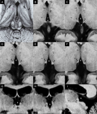 |
165 |
Visualizing and Characterizing the Habenula with Magnetic Resonance Imaging
Naying He, Sean Sethi, Chencheng Zhang, Yan Li, Yongsheng Chen, Bomin Sun, Fuhua Yan, Ewart Haacke
The habenulae are a small pair of nuclei which serve as a hub between the limbic forebrain and midbrain monoameric neurons. It is a target for the treatment of major depressive disorder using deep brain stimulation, which requires precise pre-treatment mapping. We visualized and characterized the habenula using multiple MRI contrasts and maps to quantify its properties and delineate the structure between lateral and medial side. Axially, we observed elevated iron in the posterior aspect, which we believe to be the lateral habenula. Quantitatively, we also noted similarities of the lateral habenula specifically to white matter.
|
|
2639.
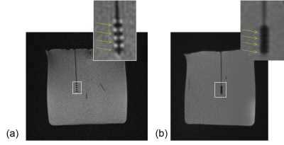 |
166 |
Localization of the habenula and stimulating electrodes in pre/post-DBS surgery using MRI
Yan Li, Naying He, Ewart Haacke, Fuhua Yan
Deep brain stimulation of the lateral habenula is a common approach to treat refractory depression and other psychiatric diseases. It is very important to know the exact position of the habenula before positioning the electrodes. We conducted phantom experiments using a clinical DBS wire to determine the characteristics of the artifacts stemming from the electrodes and also scanned 6 pre/post-DBS patients on a 1.5T scanner. Both T2W TSE and high resolution GRE imaging clearly visualized the electrodes through the geometric distortion artifacts. 3D T1 MPRAGE, T2W TSE and 3D GRE provided a rapid protocol for scanning patients pre/post-DBS treatment.
|
|
2640.
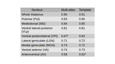 |
167 |
An anatomical atlas for segmentation of thalamic nuclei from conventional 3T MRI
Manojkumar Saranathan, Jennifer Becker, Stephen Rapcsak
Thalamic nuclei are typically invisible on conventional T1 and T2 MRI. We propose here an anatomical atlas based on 7T White matter nulled MP-RAGE data which can be used for a variety of applications including targeting the VIM nucleus for neurosurgical applications and thalamic nuclear volumetry for tracking disease progression using conventional MRI sequences like MP-RAGE or FLAIR.
|
|
2641.
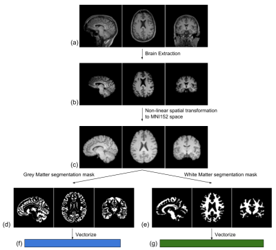 |
168 |
Biological brain age prediction using structural MRI: Insights from dimensionality reduction techniques
Arna Ghosh, Alba Xifra-Porxas, Georgios Mitsis, Marie-Hélène Boudrias
The human brain changes with age and these age-related changes have been used as biomarkers for several brain-related disorders. Therefore, being able to accurately predict the biological age of the brain from T1-weighted MR images yields significant potential for clinical applications. The present study evaluates regression models coupled with dimensionality reduction techniques for biological brain age prediction and concludes that Canonical Correlation Analysis (CCA) enhances prediction performance of Gaussian Process Regression (GPR) models. The proposed analysis also reveals brain areas that are strongly anti-correlated with age, in agreement with previous aging studies.
|
|
2642.
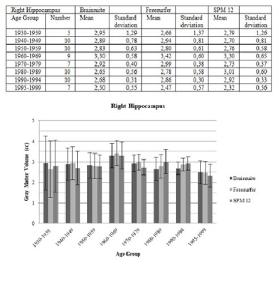 |
169 |
Volumetric analysis of selected brain regions for multi-parametric diagnostic investigation of atrophic brain diseases - Comparison of different volumetric analysis methods
Svea Seehafer, Olav Jansen, Thomas Lindner
In this study three freeware tools for volumetric image post-processing were compared. The study population was subdivided by age decades. Our results show no major deviations between the selected analytical methods.
|
|
2643
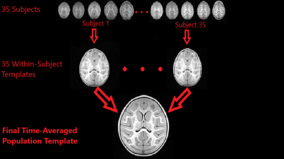 |
170 |
A Time-Averaged MRI Brain Template for the Infant Rhesus Macaque
Video Permission Withheld
Jason Moody, Steve Kecskemeti, Douglas Dean III, Jonathan Oler, Do Tromp, Andrew Alexander, Ned Kalin
Anatomical templates are extremely prevalent in human brain imaging research, but comparatively rare in non-human primate MRI studies, particularly for the early-developing brain. Utilizing a custom T1-weighted imaging sequence (MPnRAGE), we have constructed a finely-sampled (0.469 mm isotropic), T1-weighted, time-averaged, population template of the infant rhesus macaque brain, generated from 35 rhesus monkeys, scanned at five different time points throughout their first year of life (including 4 scans within the first 6 months). This time-averaged template of the early-developing rhesus macaque brain provides an invaluable anatomical framework for characterizing and assessing early brain development in non-human primates.
|
|
2644.
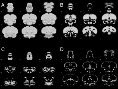 |
171 |
A novel ex vivo MR imaging template of the Japanese quail to study stress-selected lines.
David Barrière, Raïssa Yebga Hot, Marine Siwiaszczyk, Justine Beaujoin, Ivy Uszynski, Scott Love, Baptiste Mulot, Ludovic Calandreau, Élodie Chaillou, Cyril Poupon
In neuroscience, birds are becoming interesting animal models to study learning and memory but also response to stress. Nevertheless, bird’s brain organization and physiology suffers from a lack of neuroimaging tools to perform non-invasive and longitudinal investigations. In this study, we proposed a novel brain template of the Japanese quail (Coturnix Japonica), built from twenty animals dedicated to voxel-based morphometry approach. Using these tools we investigate differences in grey matter concentration (GMC) between two divergent lines of quails selected from their response to fear. Our results report structural differences between the both quail lineages within cognitives, motivational and motor systems.
|
|
2645.
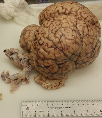 |
172 |
High-Resolution Magnetic Resonance Imaging of Juvenile Minke Whale Brain at 7T
Gaurav Verma, Alan Seifert, Bridget Wicinski, Kuang-Han Huang, John Rutland, Rebecca Feldman, Bradley Delman, Patrick Hof, Priti Balchandani
The brain of a 15-foot juvenile brain that washed ashore in the Bronx, New York City was scanned using a 7T whole-body MRI scanner. After fixation in PBS solution and vacuum removal of air pockets, the specimen was scanned using a battery of high-resolution anatomical MRI sequences, including T1-weighted MP2RAGE, 3D MERGE, Proton Density weighted imaging, T2-weighted FLAIR imaging and Diffusion Tensor Imaging. Post-processing included brain masking to alleviate bright background from the PBS solution in most imaging modalities. Anatomical imaging from three high resolution datasets is presented along with a 3D reconstruction generated by volumetric projection of data segmented using the FreeSurfer 6 algorithm.
|
|
2646.
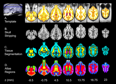 |
173 |
A high-resolution MRI template for adult Beagle dog
Xueru Liu, Rui Tian, Zhentao Zuo, Hui Zhao, Liang Wu, Yan Zhuo, Yongqing Zhang, Lin Chen
High-resolution T1w and T2w templates from 10 male adult purebred beagles were created in this study. According to the tissue probability map, descriptive statistics of brain tissue volumes and brain sizes exhibit our template with smaller variance. Significant correlation between brain size from dorsal to ventral and gray matter volume was found. This high-resolution purebred canine brain template lays the foundation for further studies aimed at in-vivo analysis of the development of canine brain anatomy and function.
|
|
2647.
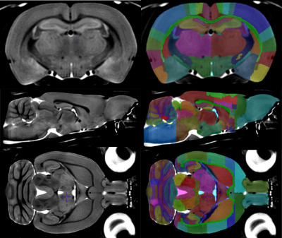 |
174 |
An MRI-Derived Neuroanatomical Atlas of the Fischer 344 Rat Brain
Dana Goerzen, Caitlin Fowler, Gabriel Devenyi, Jurgen Germann, Dan Madularu, Mallar Chakravarty, Jamie Near
Neuroscientific research involving preclinical rodent models often requires the ability to precisely identify anatomical brain regions. This project reports the development of a high-resolution MRI atlas of the Fischer 344 adult rat. The atlas is composed of 98 manually delineated structures through 256 coronal slices. The atlas was developed using 41 adult Fischer 344 rats to generate a co-registered average brain. The template was segmented by intensity contrast in conjunction with the Paxinos and Watson paper atlas. This atlas is intended to be a resource for researchers working with Fischer 344 rats and is provided open-access in MINC2.0 and NIfTI.
|
|
2648.
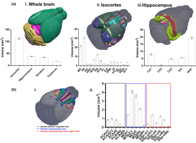 |
175 |
High resolution diffusion magnetic resonance imaging based atlas of the C57BL/6J adult mouse brain: a tool for examining mouse brain structures
Tanzil Mahmud Arefin, Choong Heon Lee, Orlando Aristizábal, Youssef Zaim Wadghiri, Daniel Turnbull, Jiangyang Zhang
Mouse models have been widely used in the neuroscience research to evaluate brain development, micro-structural and functional phenotypes in response to gene mutations and neurological diseases which require a baseline for comparison, such as an atlas. Where existing atlases vary in contrast mechanisms, number of structures and resolution, very few reports detailed neuroanatomical parcellations based on diffusion magnetic resonance imaging. This study was therefore aimed to develop high resolution diffusion MR-based mouse brain atlas database with thorough labels for cortical and subcortical structures compatible with the Allen Mouse Brain Atlas (AMBA) which will be freely available to the research community.
|
|
Back to Program-at-a-Glance |  Back to Top Back to Top |
| The International Society for Magnetic Resonance in Medicine is accredited by the Accreditation Council for Continuing Medical Education to provide continuing medical education for physicians. |
