Digital Poster Session
Neuro Back to Program-at-a-Glance Back to Program-at-a-Glance
|
Tuesday, 14 May 2019
Digital PosterNeuro
2649 -2673 Neurovascular 1
2674 -2698 Psychoradiology: Depression, Bipolar Disorder, Anxiety & More
2699 -2723 Segmentation & Processing
2724 -2748 Stroke
2749 -2772 Psychoradiology: Schizophrenia, Psychosis, OCD & More
2773 -2796 Aging & Neurodegeneration (Other than AD)
2797 -2821 Neuroimaging: Flying High at 7T & Beyond
2822 -2846 Artificial Intelligence Is Taking Over Your Brain 1
2847 -2871 Brain Tumors: Pre-Treatment
2872 -2896 Spinal Cords, Disks & Nerves
2897 -2921 Brain Tumors: Post-Treatment
2922 -2946 Neurovascular 2
2947 -2968 Head & Neck
2969 -2993 Novel Neuroimaging Methods |
| |
Neurovascular 1
Digital Poster
Neuro
Tuesday, 14 May 2019
| Exhibition Hall |
13:30 - 14:30 |
| |
|
Computer # |
|
2649.
 |
1 |
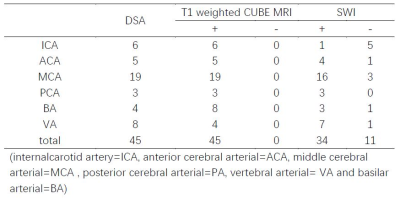
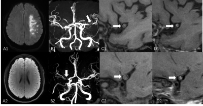
High resolution MRI in Diagnosis of Cerebral Arterial Thrombosis

Chao Zhang, Xinyi Wang, Weiqiang Dou
This study aimed to investigate the feasibility of CUBE MRI for high resolution imaging in the detection of intraluminal thrombi in acute stroke patients. The T1-weighted CUBE images showed dark blood signal in arteries and high signal or iso-signal filling in the lumen. In our study, the sensitivity of T1 weighted CUBE in the detection of intraluminal thrombi reached 100% and the corresponding area under curve(AUC) value was higher than SWI. We therefore demonstrated that the T1-weighted CUBE MRI can effectively help to diagnosis the intraluminal thrombi.
|
|
2650.
 |
2 |
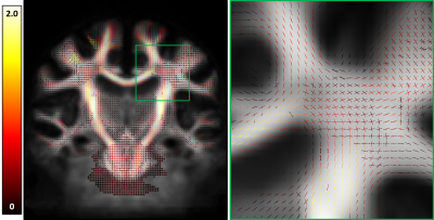
Functional and Microstructural Changes in the Brain After Carotid Endarterectomy
Marc Lindley, Adam Bernstein, Andrew McKinnon, Chidi Ugonna, Denise Bruck, Kevin Johnson, Maria Altbach, Lee Ryan, Gloria Guzman, Nan-kuei Chen, Ying-hui Chou, Theodore Trouard, Craig Weinkauf
Carotid endarterectomy (CEA) for clinically asymptomatic patients has been shown effective in reducing stroke risk. The impact that CEA has on functional connectivity or microstructure in the brain has not been studied. 14 clinically asymptomatic underwent resting state fMRI (rs-fMRI), diffusion MRI (dMRI), and neurocognitive testing pre-operatively and 4-6 months post-operatively. Functional correlation analysis on rs-fMRI was performed by analyzing the average within network correlations. Apparent fiber density calculations were performed to assess the microstructural changes before and after surgery. RS-fMRI and dMRI analysis showed changes before and after CEA.
|
|
2651.
 |
3 |
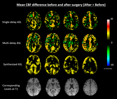
Deep Learning Augmented Cerebral Blood Flow Measurement Using Arterial Spin Labeling Technique in Moyamoya Disease Before and After Direct Bypass Surgery
David Chen, Yosuke Ishii, Jia Guo, Audrey Fan, Greg Zaharchuk
We used single-delayed (SD) pseudo-continuous arterial spin labeling (PCASL), multi-delay (MD) ASL and a new, synthesized (Synth) ASL to longitudinally monitor cerebral blood flow (CBF) before and after direct bypass surgery in Moyamoya disease. The Synth-ASL was generated from a deep convolutional neural network, previously trained on a simultaneous [15O]-water PET/MRI dataset to generate a PET-like CBF map from MRI inputs. The Synth-ASL demonstrated a more homogenous CBF change across the brain and significantly greater CBF increase globally and regionally than SD-ASL and MD-ASL after surgery. Synth-ASL reduces bias in long arterial delay and measurement noise, and may enable robust CBF imaging follow-up in cerebrovascular patients.
|
|
2652.
 |
4 |
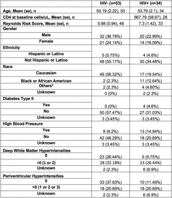
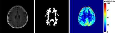
HIV-Associated Cerebral Small Vessel Disease Measured by Quantitative Susceptibility Mapping
Kyle Murray, Arun Venkataraman, Pascal Spincemaille, Lu Wang, Yi Wang, Madalina Tivarus, Xing Qui, Jianhui Zhong, Giovanni Schifitto
HIV-infected older individuals are at increased risk of developing cerebral small vessel disease (CSVD). Quantitative susceptibility mapping (QSM) can be used to asses tissue susceptibility, which can be a measure of CSVD. CSVD tends to occur more frequently in HIV-positive individuals. Limited information in the literature is available on HIV-associated changes in brain tissue susceptibility. In this abstract, we seek to discover relationships between HIV and QSM measures. Brain segmentation and region-based statistics were performed to discover region-based links between HIV and QSM measures and cardiovascular risk factors.
|
|
2653.
 |
5 |
A multi-site round robin assessment of ASL using a perfusion phantom
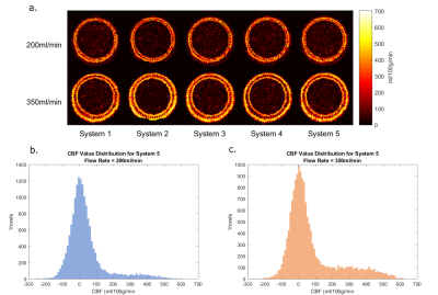
Aaron Oliver-Taylor, Thomas Hampshire, Henk-Jan Mutsaerts, Patricia Clement, Esther Warnert, Joost Kuijer, Koen Baas, Jan Petr, Jeroen Siero, José Marques, Stefan Sunaert, Ronald Borra, Matthias van Osch, Xavier Golay, Eric Achten
Arterial Spin Labelling shows great promise for perfusion measurements; however, despite numerous volunteer reproducibility studies, comparisons have not been made using a phantom to establish differences due to the acquisition hardware and pulse sequences. We present data from a multi-site study using a perfusion phantom, targeting 3T MRI systems from a single vendor running the same software version.
|
|
2654.
 |
6 |
Test-retest Reproducibility and associations with cognitive impairment of 3D PCASL in Elderly Subjects at Risk of Small Vessel Disease
Kay Jann, Xingfeng Shao, Samantha Ma, Giuseppe Barisano, Marlene Casey, Lina D'Orazio, John Ringman, Danny Wang
We assessed the reproducibility 3D pCASL in an elderly cohort with risk for small vessel disease and its associations with clinical assessments and vascular risk factors. We found a high test-retest reproducibility of regional CBF and an association of subcortical MCA perfusion territories of the lenticulostriate arteries with cognition and vascular risks. Hence, 3D pCASL perfusion in MCA perfusion territory might be a potential imaging marker to identify early small vessel changes related to vascular cognitive impairment and dementia.
|
|
2655.
 |
7 |
The value of high-resolution magnetic resonance vascular wall imaging in the diagnosis and treatment of central nervous system vasculitis
Shuai Han, Xinyi Wang, Weiqiang Dou
Three-dimensional (3D) CUBE MRI for high-resolution vascular wall imaging can reveal the morphological characteristics of vessel wall. To investigate its feasibility in the diagnosis of central nervous system vasculitis, we applied the contrast-enhanced 3D T1-weighted CUBE imaging in the patients with vasculitis. We found significant alterations of the vessel wall imaging in signal-to-noise-ratio and contrast-to-noise-ratio before and after clinical treatment. With these, we can demonstrate that 3D CUBE MRI can effectively help to diagnose the central nervous system vasculitis and evaluate the treatment effect.
|
|
2656
 |
8 |
A test-retest multi-site reproducibility study of 4D flow MRI on neurovascular system
Video Permission Withheld
Yang Fan, Xiaocheng Wei, Long Qian, Jing Wang, Bing Wu
4D flow MRI shows great potential in neurovascular disorders such as stenosis, atherosclerotic disease, aneurysms, and vascular malformations. Its widespread application in neurovascular system requires evidence of good test-retest multi-center reproducibility. The purpose of this study is to assess the multi-center reproducibility and test-retest reliability of 4D flow MRI in measurements of cerebral blood flow/velocity in main intracranial vessels. As a result, high multi-center reproducibility and test-retest reliability was shown for 4D flow MRI in the measurements of blood flow and peak velocity of main intracranial vessels for healthy volunteers.
|
|
2657
 |
9 |
Evaluation of image quality of pituitary dynamic contrast-enhanced MRI using TWIST and IT-TWIST.
Video Permission Withheld
Yusuke Yokota, Yasutaka Fushimi, Tomohisa Okada, Hikaru Fukutomi, Akira Yamamoto, Satoshi Nakajima, Gosuke Okubo, Sonoko Oshima, Koji Fujimoto, Kaori Togashi
To compare the image quality of pituitary dynamic contrast-enhanced (DCE) MRI using TWIST and iterative reconstruction TWIST (IT-TWIST). IT-TWIST images were created from the identical rawdata of TWIST. ROI analyses were conducted to evaluate enhancement slope in PS, PL, bilateral cavernous sinus (CS) in enhancement slope map. Four ROIs were applied to temporal SD map as an indicator of temporal noise to evaluate image noise. Enhancement slope of all ROIs but PS was significantly higher in IT-TWIST than that in TWIST. Temporal noise in IT-TWIST was significantly less than that in TWIST in all ROIs.
|
 |
2658.
 |
10 |
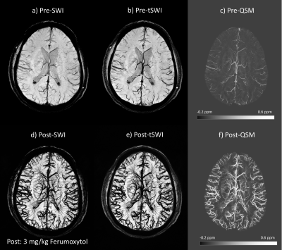
Imaging the Cerebral Vasculature Using Ferumoxytol Enhanced Susceptibility Weighted Imaging and Quantitative Susceptibility Mapping at 3T
Yongsheng Chen, Yulin Ge, Saifeng Liu, Jiani Hu, E. Mark Haacke
Imaging the major arteries in the brain is straightforward using MR angiography either with or without a contrast agent. However, imaging vessels at the 250μm level is challenging and imaging vessels at the 50μm to 100μm level is essentially impossible even with high field systems. One potential approach to bring them to life is using an iron-based contrast agent to enhance SWI. In this work, we extend the use of Ferumoxytol to image the small cerebral arteries and veins to 3T and show that within a reasonable scanning time, one can obtain superb images of the vasculature of the brain.
|
|
2659.
 |
11 |
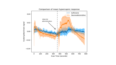
Comparison of the BOLD-evoked response to hypercapnic challenge in mice anesthetized under isoflurane and dexmedetomidine.
Gabriel Desrosiers-Grégoire, Daniel Gallino, Gabriel Devenyi, M. Mallar Chakravarty
Small animal functional magnetic resonance imaging has great potential in a range of basic neuroscientific applications. To maintain stable experimental conditions, animals are usually anesthetized during acquisition. However, anesthesia regimes influence neural activity through their influence on neurovascular coupling. To investigate these mechanisms, we compared the BOLD response following hypercapnia in mice anesthetized under isoflurane or dexmedetomidine. We found that the impact of hypercapnia is much more potent in animals anesthetized under dexmedetomidine, but that FC is much stronger under isoflurane, suggesting that this response does not predict a more pronounced reduction in FC as a consequence of anesthesia.
|
|
2660.
 |
12 |
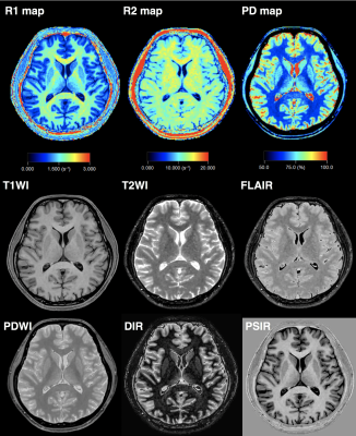
Synthetic MR Angiography: A Feasibility Study of MR Angiography based on 3D Synthetic MRI
Shohei Fujita, Akifumi Hagiwara, Masaaki Hori, Otsuka Yujiro, Fukunaga Issei, Nao Takano, Christina Andica, Tomoko Maekawa, Ryusuke Irie, Koji Kamagata, Akihiko Wada, Michimasa Suzuki, Shigeki Aoki
Quantitative synthetic MRI allows creation of various contrast-weighted image that can be used in clinical settings from a single acquisition. However, clinically widely used MRA was unable to obtain using synthetic MRI. We demonstrate an arithmetic approach to produce MRA-like images from the 5 raw images of 3D-QALAS. Qualitative and quantitative evaluations were performed to compare image qualities of synthetic MRA with TOF-MRA. Proximal segments of intracranial arteries were clearly visualized on synthetic MRA, with comparable quality to that of TOF-MRA. Synthetic MRA may function as a screening tool to detect lesions of major intracranial arteries, without additional scanning time.
|
|
2661.
 |
13 |
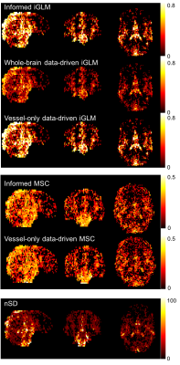
Assessment of cerebral pulsatility using high temporal-resolution MRI
Kevin Ray, Alastair Webb, Peter Jezzard
High frequency resting state BOLD MRI (rs-fMRI, TR=0.43s) detects effects of blood flow pulsatility on the cerebrovasculature, but no systematic comparison of analysis methods has been performed. In ten healthy subjects, we compared three pulsatility quantification methods (iterative GLM, mean-squared coherence (MSC), number of standard deviations (nSD)), with or without external physiological measurements. MSC detected the greatest proportion of voxels with significant pulsatility, but iGLM analysis was the most specific method, identified greater normalised pulsatility magnitude in arteries, and was the only approach that produced similar estimates of pulsatility magnitude and extent independently of external physiological data.
|
|
2662.
 |
14 |
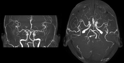
High-resolution Brain 3D-TOF MRA of Critical Fine Branches from Major Trunks Using Deep Learning Reconstruction and High-gradient Magnetic Field
Miho Gomyo, Kazuhiro Tsuchiya, Yoshioka Tatsuya, Sanae Takahashi, Shichirou Katase, Arisa Ohara, Isao Miyazaki, Haruhiko Machida, Kenichi Yokoyama
ProblemUsing a 3-T MRI scanner with a high gradient magnetic field (100mT/m), we evaluated the depiction of the intracranial fine branches on high-resolution 3D-TOF MRA (HR-TOF). We also assessed whether depiction can be improved by deep learning reconstruction (DLR).
Methods
Ten healthy volunteers were imaged by HR-TOF with DLR, and the sharpness of origin and the overall depiction of branches were assessed.
Results
SNR, the sharpness of the origin and the overall depiction of branches were superior in HR-TOF with DLR.
Conclusion
HR-TOF can well depict fine branches from major trunks. By performing DLR processing, depiction can be improved.
|
|
2663.
 |
15 |
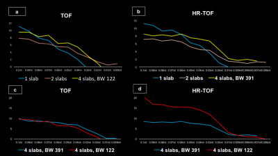
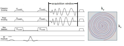
Optimization of a new accelerated time-of-flight Brain MR angiography using spiral data acquisition: Spiral MRA
Yutaka Hamatani, Kayoko Abe, Yasuhiro Goto, Masami Yoneyama, Isao Shiina, Kazuo Kodaira, Yoshihiro Ikeda, Mamoru Takeyama, Isao Tanaka, Shuji Sakai
Spiral MRA is a new accelerated time-of-flight MR angiography (TOF MRA) with spiral data acquisition, which acquires MR data by traveling through k-space with spirals. Acquisition window (AW) is a new parameter, which indicates the degree of under-sampling related to image quality and acquisition time. In this study, suitable flip angle (FA) and AW for Spiral MRA was evaluated by a 5-point scale and signal profile analysis. In conclusion, the suitable FA was 25° to demonstrate each artery and suppress the background signals. AW should be set to 10 or less to avoid vessel blurring.
|
|
2664.
 |
16 |
A new accelerated time-of-flight Brain MR angiography (Spiral MRA) with a combination technique of spiral acquisition and fat suppression: ProSet
Kayoko Abe, Kazufumi Suzuki, Masami Yoneyama, Shuji Sakai
Spiral MRA is a new accelerated time-of-flight MR angiography (TOF-MRA), the k space is filled with data in a spiral trajectory on the frequency and phase encoding directions. In this study, the effect of TONE and ProSet on Spiral MRA was evaluated by comparing image quality between Spiral MRA and conventional TOF-MRA. As the result, TONE was rarely effective on Spiral MRA, and Spiral MRA with ProSet provided high quality images, and reduced the acquisition time by approximately 70%, compared to conventional TOF-MRA with ProSet. In conclusion, Spiral MRA with ProSet is a useful, accelerated technique without image quality deterioration.
|
|
2665.
 |
17 |
Dual Coil Continuous ASL of the human brain at 9.4 T
Markus Schreiyäck, Jonas Bause, Klaus Scheffler, Rolf Pohmann
Arterial Spin Labeling (ASL) is expected to profit highly from ultra high magnetic fields because of the high SNR and the long longitudinal relaxation time. Here we show first images from dual coil continuous ASL measurements in the human brain at 9.4 T. A separate transmit channel was established to feed two small labeling coils placed at the neck. A power limiter was used to ensure subject safety. First images show strong perfusion contrast and high SNR.
|
|
2666.
 |
18 |
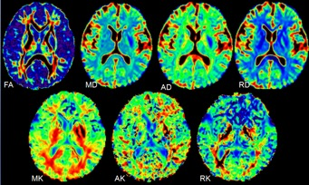
Measure Cerebral Microstructure Alterations in SVD and BVD Using Diffusion Kurtosis Imaging and Investigate the Correlation with Cognitive Impairment
Presentation Not Submitted
Wenjing Lan, Shuang Xu, Yang Liu, Kaiyu Wang, Lizhi Xie
Diffusion tensor imaging (DTI) is one of the most popular diffusion MRI methods in the study of ageing. Diffusion kurtosis imaging, which is a recent novel extension of DTI to provide additional metrics quantifying non-Gaussianity of water diffusion in brain tissues, was applied throughout the study. We investigated diffusional alternations arising from brain small vessel disease, and compared results with age and educational level-matched big vessel disease and healthy controls. We also investigated the correlation between these diseases and cognitive impairment.
|
|
2667.
 |
19 |
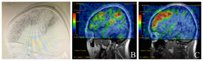
Super selective arterial spin labeling technique in the assessment of blood supply from external carotid artery in Moyamoya Disease: comparison with digital subtraction angiography
Jing Yuan, Jianxun Qu, Yaou Liu
Super selective arterial spin labeling (ssASL) is a MR territory perfusion technique based on arterial spin labeling. The efficacy of this technique to demonstrate the blood supply of external carotid artery (ECA) into the brain has not been studied. This study demonstrated ssASL was in good agreement with DSA, the gold standard for cerebral vessels, in the evaluation of preoperative ECA collaterals, superficial temporal artery to middle cerebral artery bypass and synangiosis-induced vessels in Moyamoya disease.
|
|
2668.
 |
20 |
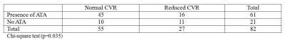
Correlation of cerebrovascular reserve assessed by acetazolamide-stress SPECT with collaterals on arterial spin-labeling MRI in patients with carotid occlusive disease
Hyunkoo Kang, Yoone Kim
We evaluated the correlation between cerebrovascular reserve (CVR) on acetazolamide (ACZ) -stress SPECT brain scans and collaterals on ASL MRI in ICA stenosis. 86 patients with ICA stenosis (>70%) were enrolled in this study. On ASL, late-arriving flow appears as serpiginous high ASL signal within cortical vessels, which has been termed arterial transit artifact (ATA). 82/86 ICA stenosis patients underwent SPECT imagings with Tc-99m-ECD in the resting and after ACZ challenge. Significant positive relationship was observed between normal CVR group and ATA showing group in ICA stenosis patients on ASL brain perfusion (p=0.035, chi-square test).
|
|
2669.
 |
21 |
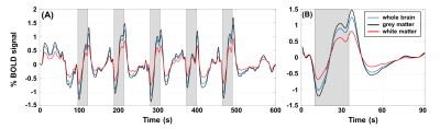
MRI Evaluation of Cerebrovascular Reactivity in Obstructive Sleep Apnea
Pei-Hsin Wu, Ana Rodríguez-Soto, Erin Englund, Michael Langham, John Detre, Richard Schwab, Andrew Wiemken, Felix Wehrli
Obstructive sleep apnea (OSA) is a chronic disorder caused by intermittent obstruction of the upper airways during sleep. OSA patients are prone to cardiovascular disease and stroke. Cerebrovascular reactivity (CVR) is an index to assess the degree of impairment of cerebrovascular regulation. Here, a breath-hold index (BHI) was introduced as a surrogate for CVR to evaluate subjects with OSA and their controls. Preliminary results from an ongoing study found BHI to be significantly elevated in OSA for both BOLD based regional, and global CBF. The results agree with a recent MRI-based CVR study using an exogenously administered hypercapnia stimulus.
|
|
2670.
 |
22 |
Changes to Blood-Brain Barrier Water Permeability After CPAP Treatment in Patients with Obstructive Sleep Apnea
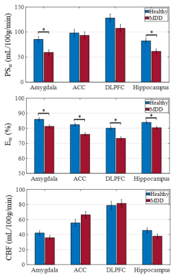
Xiang He, Kenneth Wengler, Karl Spuhler, Muhammed Amin, Chuan Huang
In patients with obstructive sleep apnea (OSA), intermittent ischemia and re-oxygenation leads to disruption of blood-brain barrier (BBB) integrity. In this study changes in BBB water permeability parameters, water extraction fraction (Ew) and water permeability surface area product (PSw), in patients with OSA before and after 8-weeks of continuous positive airway pressure (CPAP) treatment were investigated using the recently developed Intrinsic Diffusivity Encoding of Arterial Labeled Spins (IDEALS) technique. Compared to healthy controls, OSA patients exhibited lower CBF, PSw and Ew before CPAP. After 8-weeks of CPAP, patients showed increased CBF, PSw and Ew demonstrating the improvement of BBB integrity.
|
|
2671.
 |
23 |
4D Flow MRI Analysis of Cerebral Blood Flow Before and After The Superficial Temporal Artery to Middle Cerebral Artery Bypass Surgery for Atherosclerotic Disease
Tetsuro Sekine, Erika Orita, Yasuo Murai, Ryo Takagi, Yasuo Amano, Takahiro Ando, Kotomi Iwata, Masashi Ogawa, Makoto Obara, Shin-ichiro Kumita
The purpose of this study was to clarify the change in the hemodynamics after superficial temporal artery to middle cerebral artery (STA-MCA) bypass surgery using 4D Flow MRI. We enrolled 20 patients who underwent 4D Flow MRI preoperatively and 3 weeks after the surgery. The blood flow volume (BFV) of ipsilateral STA and ipsilateral ICA significantly increased after the surgery (0.53±0.22 vs. 1.78±0.54 ml/sec (p< 0.001); 2.37±5.09 vs. 1.82±3.42 ml/sec (p=0.03)). While, no significant difference was observed in total-BFV (p = 0.24). It may indicate that ipsilateral STA and intracranial native artery supply blood flow complementarily after surgery.
|
|
2672.
 |
24 |
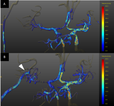
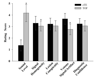
Validation of zTE MRA in the characterization of cerebrovascular diseases: a feasibility study
Presentation Not Submitted
Song'an Shang, Jing Ye, Weiqiang Dou, Jianxun Qu, Xianfu Luo, Jingtao Wu
In this study, we aimed to investigate the feasibility of zero echo time magnetic resonance angiography (zTE-MRA) in the characterization of cerebrovascular diseases. Comparing with the time of flight (TOF) MRA, zTE-MRA showed more robust performance in depicting cerebrovascular diseases with dramatically reduced acoustic noise, higher signal homogeneity, less venous signal/artifact and higher inter-modality agreement and correlation with computed tomography angiography (CTA). We therefore demonstrated that zTE MRA could be a promising technique and further applied routinely in the clinic for patients with cerebrovascular diseases.
|
|
2673.
 |
25 |
Estimating hemodynamic response functions using motor task and resting-state EEG-fMRI data acquired during wakefulness with eyes open
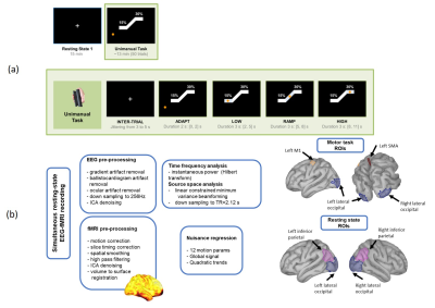
Prokopis Prokopiou, Alba Xifra-Porxas, Michalis Kassinopoulos, Marie-Helene Boudrias, Georgios Mitsis
In this work, we quantify the fMRI hemodynamic response function (HRF) using task-based (motor) and resting-state EEG-fMRI. We developed a methodology that does not require any assumptions regarding the HRF shape or the relative contribution of different EEG spectral bands to obtain region-specific estimates of the HRF. During the motor task, the EEG β-band was found to have a more pronounced contribution to BOLD variations compared to other bands, and the HRF was mainly negative due to β-band desynchronization and post-movement β-rebound. During resting-state, the contribution of different EEG bands and the HRF estimates varied between subjects, possibly due to low SNR and differences in the subjects’ brain state.
|
|
| Top |
Psychoradiology: Depression, Bipolar Disorder, Anxiety & More
Digital Poster
Neuro
Tuesday, 14 May 2019
| Exhibition Hall |
13:30 - 14:30 |
| |
|
Computer # |
|
2674.
 |
26 |
Cortical structure mediates the effect of childhood maltreatment on depression relapse during longitudinal follow-up
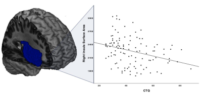
Harald Kugel, Nils Opel, Ronny Redlich, Katharina Dohm, Dario Zaremba, Janik Goltermann, Jonathan Repple Repple, Claas Kaehler, Dominik Grotegerd, Elisabeth J. Leehr, Joscha Böhnlein, Katharina Förster, Susanne Meinert, Verena Enneking, Lisa Sindermann, Fanny Dzvonyar, Daniel Emden, Ramona Leenings, Nils Winter, Tim Hahn, Walter Heindel, Ulrike Buhlmann, Bernhard Baune, Volker Arolt, Udo Dannlowski
Childhood maltreatment is a strong risk factor for the onset of major depressive disorder (MDD) and associated with unfavorable course of the disease. Both, maltreatment and MDD have been independently associated with structural alterations in partly overlapping brain regions suggesting that brain structural changes could mediate the adverse influence of maltreatment on clinical outcome in MDD. In this study the relationship between childhood trauma, brain structural alterations and adverse disease course was investigated in a longitudinal design. Our results suggest that cortical surface area reductions might mediate the prospective association between early life stress and future depression relapse.
|
|
2675.
 |
27 |
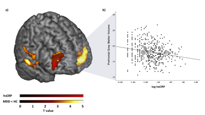
Evidence for an association between low-grade peripheral inflammation and brain structural alterations in major depression
Harald Kugel, Nils Opel, Micah Cearns, Scott Clark, Catherine Toben, Dominik Grotegerd, Walter Heindel, Anja Teuber, Heike Minnerup, Matthias Nauck, Klaus Berger, Udo Dannlowski, Bernhard Baune
Preliminary research suggests that major depressive disorder (MDD) is associated with structural alterations of brain regions relevant for emotion regulation and associated with low-grade peripheral inflammation as indicated by high sensitive C-reactive protein (hsCRP) serum levels. This association between structural brain alterations and low-grade inflammation as potentially interrelated biological correlates of MDD was investigated. In MDD patients, but not healthy controls, prefrontal gray matter volume reductions were significantly associated with higher hsCRP levels.
|
|
2676.
 |
28 |
Abnormal functional connectivity of ACC sub-regions in patients with major depressive disorders
Xiaolong Peng, Xiaoping Wu, Pan Lin, Ruxue Gong, Rui Yang, Wenzhen Zhu
Major depressive disorder (MDD) is a common mental disorder characterized by cognitive and affective deficits. Prior works indicated that anterior cingulate cortex (ACC) is related to high-level cognitive and emotion process, which is also thought to be pivotal to depression. Here, we examined the resting FC of ACC sub-regions in fist-episode MDD patients. The current results revealed reduced ACC sub-regional FC with IPL and SPL while increased FC was found in dmPFC. Additionally, FC with IPL also negatively correlated with symptom severity (HDRS), indicating that depression may disrupt the normal interactions within the DMN. These findings on alteration of ACC sub-regional FC may contribute to the comprehension in pathophysiology of MDD.
|
|
2677
 |
29 |
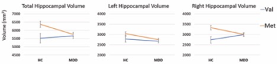
Structural MRI at 7T reveals amygdala nuclei and hippocampal subfield volumetric association with Major Depressive Disorder symptom severity
Video Permission Withheld
Stephanie Brown, John Rutland, Gaurav Verma, Rebecca Feldman, Judy Alper, Molly Schneider, Bradley Delman, James Murrough, Priti Balchandani
Subcortical volumetric changes in MDD have been purported to underlie the symptoms of MDD, however, the evidence to date remains inconsistent. Here, we investigated the relationship between structural limbic brain measurements and MDD symptomology through high-resolution segmentation of the amygdala and hippocampus. We report the novel finding that MDD severity is consistently negatively associated with amygdala nuclei, linking volumetric reductions with worsening depressive symptoms.
|
|
2678.
 |
30 |
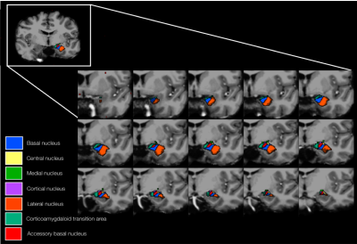
Anomalous functional connectivity in subregional amygdala networks in major depressive disorder
Shi Tang, Hailong Li, Lu Lu, Lianqing Zhang, Xuan Bu, Xiaoxiao Hu, Yingxue Gao, Xinyu Hu, Yanlin Wang, Qiyong Gong, Xiaoqi Huang
The LB, CM, SF and Astr are four main subregions of the amygdala. In this study, we use seed-based functional connectivity method to determine amygdala network dysfunction in MDD. Compared with HC, patients with major depressive disorder showed hypoconncetivity in AStr/LB- OFC circuits, in CM /SF-brainstem/cerebellum circuits and in AStr/CM/SF-thalamus/striatum circuits. These dysfunction in amygdala networks may modulate different emotional and cognitive function in derpession.
|
|
2679.
 |
31 |
Abnormal Blood-Brain Barrier Water Permeability in Major Depressive Disorder
Kenneth Wengler, Kwan Chen, Turhan Canli, Christine DeLorenzo, Mark Schweitzer, Xiang He
Blood-brain barrier (BBB) disruption may be the key mechanism leading to neuronal dysfunction and neuroinflammation in major depressive disorder (MDD). Active pathways account for a large portion of trans-membrane water exchange, providing a link between BBB water permeability and metabolism. In this study alterations in BBB water permeability parameters, water extraction fraction (Ew) and water permeability surface area product (PSw), in patients with MDD were investigated using the recently developed Intrinsic Diffusivity Encoding of Arterial Labeled Spins (IDEALS) technique. Compared to healthy subjects, MDD patients exhibited significantly lower PSw and Ewwith no differences in cerebral blood flow.
|
|
2680.
 |
32 |
Investigation of Cerebral Small Vessel Disease induced Depression using Diffusion Kurtosis Imaging - A preliminary Region-specific Study
Kun Li, Dongtao Liu, Qiao Bu, Xiuqin Jia, Rui Jia, Xiaojiao Pei, Yuchang Yan, Xiang Feng, Qinglei Shi, Zhenyu Pan, Tao Jiang
This abstract presents a preliminary study of cerebral small vessel disease induced depression using diffusion kurtosis imaging (DKI). Different DKI-derived parameters in specific brain structures were compared between depression and non-depression groups, as well as between anxiety and non-anxiety groups. The correlation between DTI- and DKI-derived parameters and clinical scores were also investigated.
|
|
2681.
 |
33 |
7 Tesla Phase Sensitive Imaging of Brain Regions with Metabolic Alterations in Major Depressive Disorder
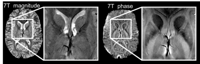
Angela Jakary, Audrey Yin, Scott Mackin, Stuart Eisendrath, Janine Lupo, Yan Li
Ultra high-field phase sensitive imaging can help elucidate subtle changes in brain iron content. Recent research implicates brain iron deposition in the pathophysiology of major depressive disorder (MDD). Our previous work involving MDD patients detected symptom-related metabolic alterations in deep brain structures and anterior cingulate cortex. In our current analysis, we apply 7T phase sensitive imaging in these same brain regions to evaluate the role of iron accumulation in neurocognitive and depressive symptoms in this vulnerable population.
|
|
2682.
 |
34 |
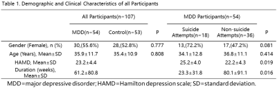
Relationship Between Gray Matter Volume Reductions and TPH1 Polymorphisms in Depressive Disorder Patients with Suicidal Attempts
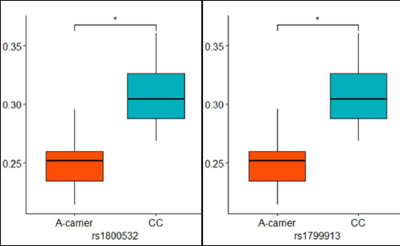
Geon-Ho Jahng, Jin Kyung Park, Seong Jong Yun, Chang-Woo Ryu, Wook Jin, Dal Mo Yang
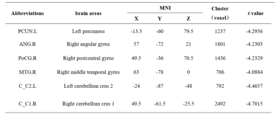
To investigate relationship between gray matter volume (GMV) changes and TPH1 polymorphisms in depressive disorder (DD) patients with suicidal attempts (SA), 13 DD-SA patients and 20 healthy controls were scanned three-dimensional (3D) T1-weighted image to obtain GMV in the brain. In addition, TPH1 rs1800532 and rs1799913 polymorphisms were obtained. The patients showed significant GMV reduction. The right precentral and postcentral gyri GMV values of AA and CA genotypes patients were significantly decreased compared to those of CC genotype subjects, indicating that both GMV reductions and TPH1 A allele may be involved in the pathogenesis of DD-SA patients.
|
|
2683.
 |
35 |
Alterations of White Matter Tracts in Suicidal and Non-suicidal Brain with Major Depressive Disorder
Kaili Liang, Xuan Bu, Lianqing Zhang, Yanli Wang, Xinyu Hu, Lu Lu, Hailong Li, Xiaoxiao Hu, Shi Tang, Yingxue Gao, Xiaoqi Huang
We investigated the white matter alterations at the individual level in MDD patients with and without suicide attempts using Automated Fiber Quantification (AFQ) approach. The three major left hemispheric white matter tracts including arcuate, CST and ATR suggested to play an important role in suicidal brain, which implies deficits of dominant hemisphere specialization with cognitive processes such as reading, writing and speaking. Our study contributes to revealing neurobiological mechanism of suicide attempts.
|
|
2684.
 |
36 |
Study of gender differences in major depressive disorder by using resting state brain functional magnetic resonance imaging
Lihua Qiu, Lan Mei, Jingping Mou, Xinyu Hu, Qiyong Gong
Sex differences are observed in epidemiological and clinical symptoms of major depressive disorder (MDD); yet, little is known about about the gender difference of brain function in MDD. In this work, variance analysis were used to assess the sex differences of amplitude of low frequency fluctuation (ALFF) alterations in male, female MDD patients and matched controls. We found the gender differences of ALFF in bilateral caudate nucleus and posterior cingulate gyrus. Our findings suggest that sex specific functional alterations existed in MDD, and these alterations may associated with the clinical symptoms.
|
|
2685.
 |
37 |
Structural brain abnormalities in MDD patients with suicide: A DARTEL-enhanced voxel-based morphometry study
Huiru Li, Huawei Zhang, Li Yin, Zhiyun Jia, Qiyong Gong
We performed a VBM analysis with DARTEL to analysis the different structure in healthy controls, MDD patients with or without suicidal actors. The result shows suicidal patients had reduced GMV than patient controls in precuneus/cuneus, anterior cingulate cortex and orbital frontal gyrus. Particularly, we found suicidal ideators have reduced GMV in middle frontal gyrus compared to suicidal attempters. Negative correlation was found between clinical characters and volume of some regions. The dysfunction of self-awareness, emotional processing and impulsivity control function caused by the abnormalities of these brain regions may be associated suicidal behavior.
|
|
2686.
 |
38 |
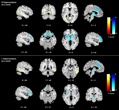
Hippocampus-related regional and network functional deficits in first-episode drug-naïve major depressive disorder: a resting-state functional MRI study
Xiaoxiao Hu, Xinyu Hu, Hailong Li, Lianqing Zhang, Lu Lu, Xuan Bu, Shi Tang, Yingxue Gao, Yanlin Wang, Qiyong Gong, Xiaoqi Huang
Previous neuroimaging studies have suggested that major depressive disorder (MDD) may be correlated with changes in regional- or network-level brain function. The purposes of the present study were to investigate changes of amplitude of low-frequency fluctuation (ALFF) and functional connectivity (FC) in bilateral hippocampus by resting-state functional magnetic resonance imaging (rs-fMRI) in first-episode drug-naive major depressive disorder (MDD) patients. Our findings demonstrate that the hippocampus and dACC contribute to the underlying pathophysiology of MDD at an early-stage.
|
|
2687.
 |
39 |
The Importance of Identifying Functional Val158Met Polymorphism in Catechol-O- Methyltransferase (COMT) when Assessing MRI-based Volumetric Measurements in Major Depressive Disorder
Mario Serrano-Sosa, Kruthika Sampathgiri, Christine DeLorenzo, Ramin Parsey, Chuan Huang
Using voxel-based morphology we investigated the relationship between COMT gene polymorphism and volumetric abnormalities in major depressive disorder patients and healthy controls. A significant difference in the right hippocampus (p=0.015) was found between the interaction of diagnosis and genotype, which suggests that COMT polymorphism must be considered during any volumetric analysis for depression.
|
|
2688.
 |
40 |
Differences in Brain Microstructural Alterations between Bipolar and Major Depression Revealed by Diffusion Kurtosis Imaging
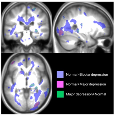
Daisuke Sawamura, Khin Tha, Naoki Hashimoto, Hisashi Narita, Shin Nakagawa, Hiroki Shirato
This prospective study evaluated if bipolar and major depression patients had microstructural brain alterations detectable on DKI. The results showed significant alterations in these patients, of which some clusters correlated with clinical symptoms. Mean kurtosis also differed significantly between the two groups.
|
|
2689.
 |
41 |
Studying disease-related brain alterations in bipolar disorder with combined analysis of DKI and VBM
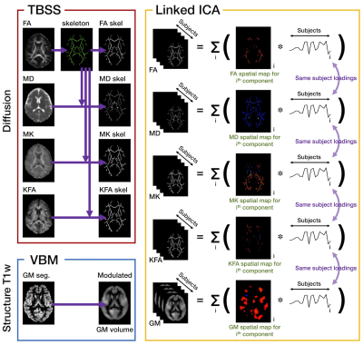
Kouhei Kamiya, Naohiro Okada, Kingo Sawada, Kentaro Morita, Susumu Morita, Shintaro Kawakami, Yuichi Suzuki, Shiori Amemiya, Harushi Mori, Akira Kunimatsu, Koji Kamagata, Masaaki Hori, Shigeki Aoki, Kiyoto Kasai, Osamu Abe
Brain abnormalities in bipolar disorder were investigated with diffusion kurtosis imaging and voxel-based morphometry, using a framework for data-driven feature extraction from multivariate data. The result showed two components capturing effect of diagnosis, and these were driven by diffusion kurtosis measures in the white matter including the prefrontal-striatal-thalamic pathways, cerebellum, and medial temporal lobes. Our results indicate diffusion kurtosis imaging can provide unique information that is sensitive to the abnormalities in bipolar disorder, and that interrelationship among different measures is a promising avenue to study neuronal circuits relevant to the disease.
|
|
2690.
 |
42 |
Altered white matter microstructure correlates with cognitive functions in children and adolescents with bipolar disorder
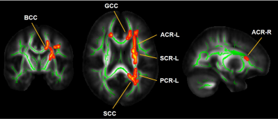
Tianjia Zhu, Chenying Zhao, Minhui Ouyang, Ruchir Arvind, Johanna Saxena, Sherin Kurian, Kirti Saxena, Hao Huang
Cognitive impairments and white matter (WM) microstructural alterations have been found in subjects with bipolar disorder (BD). However, the relationship between WM microstructural alterations and impulsivity, a prominent cognitive trait, in children/adolescents with BD is not known. In this study, diffusion MRI and cognitive assessments were obtained from 19 children/adolescents diagnosed with BD and 23 age-matched healthy controls. We found increased radial diffusivity(RD), reflecting disrupted myelin, in major WM tracts such as corpus callosum. Significant correlation between RD in WM tracts regulating impulsivity and response time to affective words was found, suggesting the association between WM myelin disruption and impulsivity.
|
|
2691.
 |
43 |
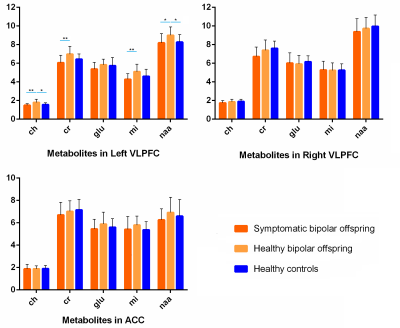
Individual Prediction of Symptomatic Converters in Youth Offspring of Bipolar Parents Using Magnetic Resonance Spectroscopy
Wenjing Zhang, Maxwell Tallman, Li Yao, Su Lui, Qiyong Gong, Melissa DelBello
Whether the neurochemicals are associated with the vulnerability of bipolar disorder has not been studied before, findings of which may extend our understanding of neurobiological factors associated with the pathogenesis. In this study, a cohort of bipolar offsprings were enrolled and later divided into two symptomatic (converters) and healthy bipolar offspring (non-converters). Baseline MRS data was obtained and examined in predicting the disorder conversion. The measures of mI, Cr and Cho in the left VLPFC achieved the highest prediction accuracy, which indicated that some specific neurochemicals are associated with the vulnerability of bipolar disorder.
|
|
2692.
 |
44 |
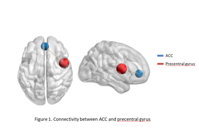
Altered functional connectivity and spectroscopic metabolites related to treatment response in adolescents with bipolar disorder
Siyi Li, Wenjing Zhang, Bo Tao, Su Lui
The reason for the inconsistency of bipolar disorder (BD) patients’ brain functional status and metabolic levels of treatment response is still not clear. This task-based fMRI study was carried out to figure out the relationship between medication treatment and brain status in function and metabolites. By analyzing functional connectivity and correlating metabolic markers in treatment response and no response BD patients, we found medication can affect the brain functional status and metabolic level in BD patients, and precentral gyrus is a key region during BD illness course.
|
|
2693.
 |
45 |
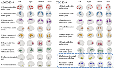
Exploring White Matter Functional Networks at Rest in Boys with Attention Deficit/Hyperactivity Disorder Using Clustering Analysis and Tractography
Xuan Bu, Yingxue Gao, Hailong Li, Yanlin Wang, Lianqing Zhang, Xinyu Hu, Shi Tang, Lu Lu, Xiaoxiao Hu, Lanting Guo, Xiaoqi Huang
In current study, we identified nine white matter functional networks and their relations to structural white matter fibers identified by DTI. Sensorimotor network and dorsal attention network, which show good spatial correspondence with specific anatomical tracts, present higher amplitude in ADHD. Our results uncover the altered intrinsic functional organization of white matter in ADHD, and indicate that changes in neural activity are encoded in BOLD variations within white matter.
|
|
2694.
 |
46 |
Stronger small-worldizition of structural networks in drug-naïve children and adolescents with ADHD:A graph theory analysis
Lu Lu, Shi Tang, Lianqing Zhang , Xinyu Hu, Xuan Bu, Hailong Li, Xiaoxiao Hu, Yingxue Gao, Yanlin Wang, John Sweeney, Qiyong Gong, Xiaoqi Huang
Structural connectomes of patients with ADHD showed a shift toward “stronger small-worldization” which provided a structural basis for higher rates of information transfer in this disorder. These global network alterations, together with increased connectivity within and among DMN and task-positive networks including FPN, DAN and VAN, could lead to disruptions of attention and goal-oriented behavior that are the primary clinical hallmarks of ADHD.
|
|
2695.
 |
47 |
Association of explicit memory dysfunction with regional brain volume alterations in patients with generalized anxiety disorder
Chung Man Moon, Gwang Woo Jeong
Generalized anxiety disorder (GAD) causes emotional dysregulations and/or cognitive deficits, including excessive anger, impairments of explicit and implicit memories and poor attention. A DARTEL-based voxel-based morphometry (VBM) study for assessing the relationship between morphometric abnormalities and explicit memory dysfunction in patients with GAD has not yet been reported. Therefore, the purpose of this study is to investigate the regional gray matter (GM) and white matter (WM) volume alterations over the whole brain in patients with GAD, as well as the correlation between the brain structural abnormality and explicit memory dysfunction. Our findings would be helpful to understand the association between the brain structure abnormality and the functional deficit in the explicit memory in GAD.
|
|
2696.
 |
48 |
Brain regional connectome-wide search identified a resting-state functional connectivity locus within precunes associated with rumination symptom severity in mood and anxiety disorders
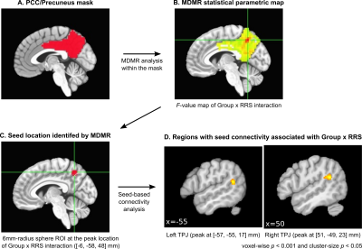
Masaya Misaki, Aki Tsuchiyagaito, Obada A Zoubi, Martin Paulus, Jerzy Bodurka
We identified a precise locus within the precuneus that has resting-state functional connectivity (rsFC) associated with rumination symptom severity for mood and anxiety (MA) disorder patients. We devised brain regional connectome-wide association analysis, which used multivariate distance matrix regression for searching voxels with connectivity correlated with the Ruminative Responses Scale (RRS) within the posterior cingulate cortex and the precuneus. The analysis identified voxels in the precuneus having rsFC significantly associated with RRS. Functional connectivity between the precuneus and bilateral temporoparietal junction (TPJ) had a significant positive correlation with RRS in MA patients but not in the healthy participants.
|
|
2697.
 |
49 |
Age-related alteration in topological efficiency of structural network in children with autism aged 2-7 years
Minhui Ouyang, Hua Cheng, Di Hu, Limei Song, Yun Peng, Hao Huang
Relatively flat white matter (WM) microstructural changes have been found in children with autism spectrum disorder (ASD) aged 2-7 years yet faster WM microstructural maturation in typically developing (TD) children were observed. In this study, we further investigated the WM structural networks in children with ASD and TD children using diffusion MRI tractography and graph-theory-based analysis. Higher global and local topological efficiencies were found in the ASD. Similar to age-related WM microstructural maturation pattern, the global, local and nodal efficiencies established with structural network increase significantly faster in TD children than those in children with ASD.
|
|
2698.
 |
50 |
Functional and Structural Abnormality in Patients with Alcohol Use Disorder Combined VBM and FC Analysis
Presentation Not Submitted
Yaqi Wang, Jun Chen
We combined voxel-based morphometry (VBM) and seed-based functional connectivity (FC) analysis to identify functional and structural characteristics in patients with alcohol use disorder using high resolution T1-weighted structure images and functional MRI. AUD group showed significantly decreased gray matter volume mainly in the default mode network, and decreased FC in the default mode network and executive control network when compared with the HC group. Combining VBM and FC provides a new perspective on the pathophysiological and clinical manifestations in AUD patients.
|
|
| Top |
Segmentation & Processing
Digital Poster
Neuro
Tuesday, 14 May 2019
| Exhibition Hall |
13:30 - 14:30 |
| |
|
Computer # |
|
2699.
 |
51 |
Along-tract statistics of NODDI diffusion metrics to enhance MR tractography quantitative analysis in healthy controls and in patients with glioma
Valentina Pieri, Francesco Sanvito, Sara Cirillo, Marco Riva, Andrea Falini, Antonella Castellano
Along-tract statistical extraction of quantitative diffusion metrics is crucial to unravel the variability of these parameters within white matter fiber bundles. Here for the first time we extracted NODDI-derived microstructural diffusion estimates along the main eloquent fiber tracts in fifteen healthy subjects and in a pilot cohort of glioma patients. We constructed a robust reference database of normative along-tract microstructural values to describe the anatomical variability of NODDI metrics within tracts and to localize and quantify differences in pathological cases. Normal and pathological conditions can be statistically compared between-groups, as well as at the single-subject level.
|
|
2700.
 |
52 |
Evaluation of Compressed SENSE in Quantitative Susceptibility Mapping
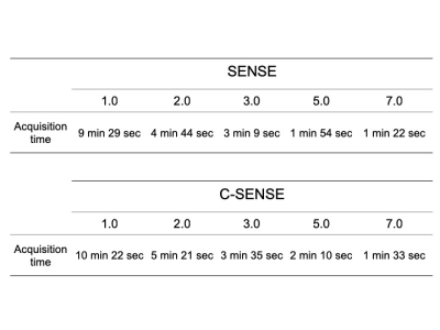
Katsuhiro Inoue, Shiho Isoshima, Maki Umino, Tsunehiro Yamahata, Shinichi Takase, Makoto Obara, Hajime Sakuma, Masayuki Maeda
Quantitative Susceptibility Mapping (QSM) is reportedly useful for the early diagnosis of Parkinson’s disease. However, the imaging time for QSM is very long because of the additional acquisition of 3D FFE; compressed SENSE (C-SENSE) could resolve this problem. The susceptibility values of the putamen, globus pallidus, caudate nucleus, substantia nigra, and nucleus ruber in seven healthy volunteers were measured as well as evaluated using SENSE and C-SENSE QSM. The results suggest that good reproducibility and validity for C-SENSE QSM can be obtained when high factors are used. C-SENSE QSM can reduce acquisition time, and is therefore expected to be widely used in the clinical setting.
|
|
2701.
 |
53 |
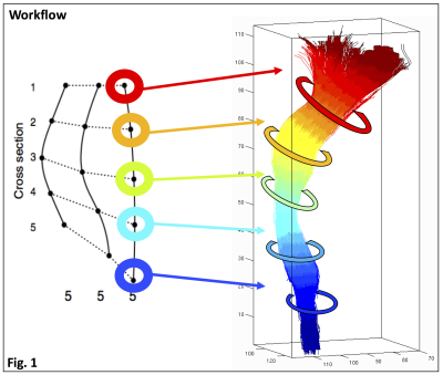
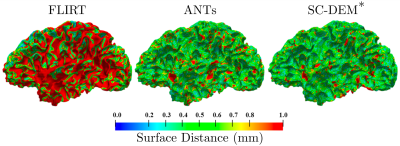
A Surface-Constrained Dynamic Elasticity Model for Deformable Registration of Infant Brain MRI
Sahar Ahmad, Zhengwang Wu, Gang Li, Li Wang, Weili Lin, Pew-Thian Yap, Dinggang Shen
Spatial registration of infant brain images is challenging owing to significant changes in image appearance in association with rapid growth in the first year of life. In this abstract, we introduce a volumetric registration method that is constrained by cortical correspondences for consistent cortical and sub-cortical alignment.
|
|
2702.
 |
54 |
Robust, Atlas-Free, Automatic Segmentation of Brain MRI in Health and Disease
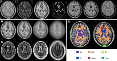
Govind Nair, Kartiga Selvaganesan, Emily Whitehead, Paba DeAlwis, Mathew Schindler, Bryan Smith, Avindra Nath, Steven Jacobson, Daniel Reich, Ziad Saad, Souheil Inati, Sara Inati
An atlas-free, brain-segmentation algorithm that uses derivative-based features and logistic regression classifier was optimized and tested on images of healthy volunteers and individuals clinically diagnosed with a variety of neuroimmunological diseases.The algorithm was trained to classify gray and white matter, CSF, globus pallidus, white matter lesions, and “other” tissue classes from all the images routinely acquired at our center. The algorithm achieved highly accurate brain segmentations and outperformed widely used techniques for brain segmentation and lesion detection. The algorithm has been found to be versatile in brain segmentation using images acquired at other collaborator sites.
|
|
2703.
 |
55 |
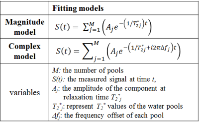
Effect of fitting models and its error analysis in GRE based MWI
Junghyeob Kim, Hongpyo Lee, Dong-Hyun Kim
The MWF fitting through the GRE sequence was performed in various models to determine which model is effective. Models such as magnitude 2-, 3-pool, complex 2-, and 3-pool modeling were used.
|
|
2704.
 |
56 |
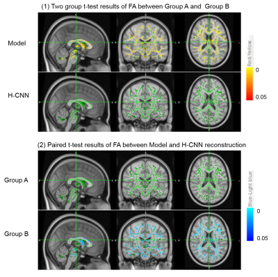
Deep learning-based diffusion method alleviates spurious group differences due to head motion
Ting Gong, Hongjian He, Zhiwei Li, Zhichao Lin, Feng Yu, Jianhui Zhong
Head motion occurring during the acquisition of diffusion-weighted (DW) images will cause deterioration in quality of diffusion model reconstruction, which could lead to spurious group differences of DW measures when there is difference in head motion for different groups. We have previously developed a method for robust diffusion kurtosis mapping of motion-contaminated data. In this study, we applied it in a group level, and the results demonstrated its ability in ameliorating spurious group differences due to head motion. The method can be applied to data with different motion level thus improving the utilization and statistic power of some valuable but motion-corrupted DW data.
|
|
2705.
 |
57 |
ExploreASL: a collaborative effort to process and explore multi-center ASL data
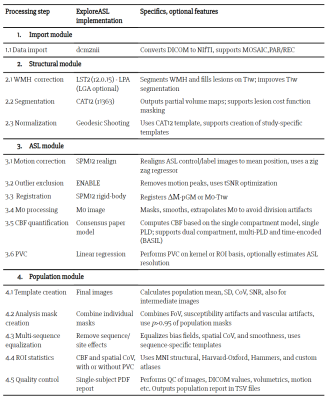
Henk Mutsaerts, Jan Petr, Paul Groot, Silvia Ingala, Andrew Robertson, Lena Vaclavu, Inge Groote, Hugo Kuijf, Owen O'Daly, Fernando Zelaya, Pieter Vandemaele, Alle Meije Wink, Ilse Kant, Matthan Caan, Catherine Morgan, Jeroen de Bresser, Elisabeth Lysvik, Anouk Schrantee, Zahra Shirzadi, Joost Kuijer, Udunna Anazodo, Edo Richard, Reinoud Bokkers, Liesbeth Reneman, Mario Masellis, Eric Achten, Matthias Günther, Bradley MacIntosh, Xavier Golay, Jeroen Hendrikse, Michael Chapell, Matthias van Osch, David Thomas, Enrico De Vita, Atle Bjornerud, Aart Nederveen, Iris Asllani, Frederik Barkhof
Arterial spin labeling (ASL) has undergone significant development since its inception; yet, standardized images processing procedures remain elusive. We present ExploreASL, a robust open source ASL image processing pipeline for clinical studies. Initiated through the European COST action ASL network, this joint effort provides integration and analysis of both single- and multi-center datasets across different operating systems. ExploreASL is optimized for both native- and standard-space analyses, and provides visual and automatic quality control on all intermediate and final images, allowing exploration of ASL datasets from multiple perspectives.
|
|
2706.
 |
58 |
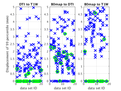
Artificial Observer and Cost Function for Image Registration, MARLINA: Mean Absolute Regional LINear correlation Algorithm
Roman Fleysher, Lazar Fleysher, Asif Suri, Molly Zimmerman, Mark Jenkinson, Craig Branch, Michael Lipton
Upon visual inspection of intra-subject rigid body registrations in large studies, we have observed higher than desired rate of unsatisfactory alignments. To address misregistartions, we designed a battery of 13 candidate transformations, one of which was selected as best during visual inspection. Tediousness of the inspections stimulated development of artificial observer to aid and subsequently to replace the human inspector. Here, we describe artificial observer MARLINA, characterize its ability to identify the best rigid body transformation as compared to human inspectors and propose it as a future cost function.
|
|
2707.
 |
59 |
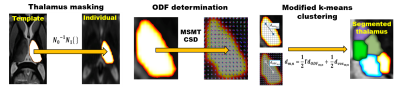
A novel DWI-based thalamus segmentation method using Constrained Spherical Deconvolution
Charles Iglehart, Adam Bernstein, Martin Monti, Joshua Cain, Manojkumar Saranathan
Existing methods to segment the thalamus via diffusion weighted MRI are inhibited by several factors. The largely gray matter composition of the thalamus makes the local diffusion activity indistinct and some of the more successful DWI-based methods require time consuming and computationally expensive cortical parcellation for thalamus masking. This study addresses these limitations by using multi-tissue constrained spherical deconvolution to isolate desired diffusion activity and a novel template based technique for thalamus masking. Segmentation outputs are evaluated and we conclude with a discussion of the method’s advantages over existing techniques.
|
|
2708.
 |
60 |
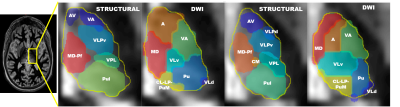
A comparison of structural and diffusion-based MRI thalamus segmentation methods
Charles Iglehart, Martin Monti, Joshua Cain, Manojkumar Saranathan
Automatic thalamus segmentation is a field of study with rapidly evolving applications. Both structural and diffusion weighted MRI can be used to drive parcellations of thalamus nuclei. In this study we present a comparison of leading structural and DWI-based segmentation techniques as implemented on a common set of subject datasets. Results for each are compared, both against an established anatomical atlas and each other. Spatial consistency of nuclei are examined in common template space. Finally, strengths and weaknesses of both techniques are discussed.
|
|
2709.
 |
61 |
Comparison of Phase-Sensitive Inversion Recovery from MPRAGE and MP2RAGE
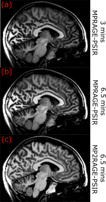
Jing Zhang, Danny Kim, Dan Rettmann, Bruce Bjornson
In this work, we obtained phase sensitive inversion recovery (PSIR) signal from MPRAGE and MP2RAGE sequences. Both PSIR images have better image contrast than magnitude images. The PSIR from MPRAGE requires shorter acquisition time, however, PSIR from MP2RAGE provides better contrast and has no B1B1 field
inhomogeneity effect. Selection of which PSIR technique to use may depend on study aims.
|
|
2710.
 |
62 |
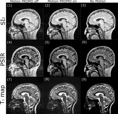
Phase-Sensitive Inversion Recovery and T1 Mapping with Motion Correction
Jing Zhang, Danny Kim, Dan Rettmann, Bruce Bjornson
In this work, we propose a novel motion corrected Phase Sensitive Inversion Recovery (PSIR) method with integrated T1 mapping derived from MP2RAGE acquisition. Motion correction is achieved using PROMO (PROspective MOtion correction), as well as Optimal Weighted Average (OWA) combination of multichannel data. This proposed method will be useful in obtaining high quality T1 images for children and other subjects who are prone to move during scans.
|
|
2711.
 |
63 |
Towards Validating Structural Connectivity in the Human Language System: an Intraoperative Cortico-Cortical Stimulation Experiment
Patryk Filipiak, Fabien Almairac, Théodore Papadopoulo, Denys Fontaine, Lydiane Mondot, Stéphan Chanelet, Maxime Descoteaux, Rachid Deriche, Maureen Clerc, Demian Wassermann
We validate structural connectivity measures based on diffusion MRI with Electrical Stimulation (ES) of the human brain cortex. For this, we combine white matter fiber tractography with propagation of Cortico-Cortical Evoked Potentials (CCEPs) induced by intrasurgical ES in the language system of brain tumor patients. Our results show high correlation (Pearson's coefficient 0.5-0.9) between delays of CCEPs and pathways connecting stimulation sites with recording electrodes. Our approach outperforms earlier study based on Diffusion Tensor Imaging. This potentially indicates that probabilistic tractography is an effective tool to quantify cortico-cortical communication non-invasively.
|
|
2712.
 |
64 |
Assessment of cerebral venous outflow rates with 4D arterial spin labeling vessel-selective angiography
Sidy Fall, Serge Metanbou, Garance Arbeaumont, Caroline Fournez, Olivier Baledent
4D arterial spin labeling (ASL) angiography has gained attention in the diagnosis of cerebrovascular diseases. The aim of this study was to evaluate the feasibility for estimating blood flow rates of the cerebral drainage system using data obtained by a 4D ASL angiography sequence. Data of a 4D ASL angiography acquisition provided comparable flow measurements to those of a standard 2D phase-contrast MR imaging sequence in 12 subjects. We demonstrated that both detailed morphological information and flows rates can be obtained by using a single 4D ASL angiography acquisition.
|
|
2713.
 |
65 |
Toblerone: partial volume estimation on the cortical ribbon
Thomas Kirk, Timothy Coalson, Flora Kennedy McConnell, Michael Chappell
Toblerone is a new method for estimating partial volumes on the cortical ribbon using surfaces as input (eg those produced by FreeSurfer). Evaluation has been performed using both simulations and subjects drawn from the Human Connectome Project. The estimates returned differ from those produced by existing tools such as FSL's FAST, which will have implications for the analysis of functional imaging data (notably ASL). A preliminary analysis of an ASL dataset has been performed using Toblerone's PV estimates.
|
|
2714.
 |
66 |
Automatic segmentation of thalamic nuclei using multiple imaging modalities at ultrahigh field
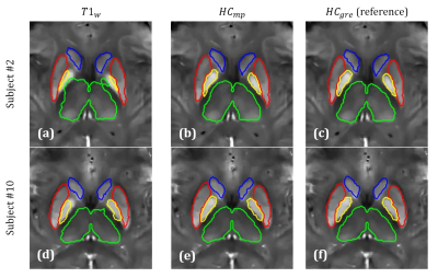
Gaurav Verma, John Rutland, Rebecca Feldman, Bradley Delman, Priti Balchandani
Segmenting gray matter structures within the thalamus is complicated by poor inherent T1/T2 contrast. Most existing approaches focus on clustering diffusion data including fiber orientation and short & long distance diffusion directions. We propose a hybrid approach incorporating diffusion data with a recently-developed high T1 contrast imaging sequence known as FGATIR. The proposed algorithm clusters on spatial position, fiber orientation distribution coefficients and anatomical contrast to provide robust, yet fast and fully-automatic segmentation of the thalamic nuclei showing strong agreement to manual segmentation performed by a neuroradiologist. Reliable thalamic nuclei segmentation could facilitate targeted therapies like deep brain stimulation.
|
|
2715.
 |
67 |
Automatic segmentation of deep grey matter structures for iron quantification
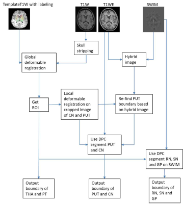
Ying Wang, Yongsheng Chen, David Utriainen, Ewart Haacke
Quantitative susceptibility mapping (QSM) is a promising iron quantification method for assessing subcortical deep gray matter (SGM) in various neurodegenerative diseases. The accuracy of the measurement depends largely on the accuracy of the structural segmentation. Manually drawn regions-of-interest from a well-trained specialist are often the best but are very time-consuming. In this work, we propose an automatic segmentation method for DGM iron quantification by taking advantage of a hybrid image approach combining T1W images and QSM data. Preliminary results on 5 stroke patients presented an overall 77.8±5.8% Dice coefficient compared to the manually drawn ground truth. The measured susceptibility of the DGM showed good agreement between both methods.
|
|
2716.
 |
68 |
A Simple Homogeneity Correction for Neuroimaging at 7T
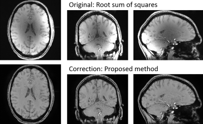
Korbinian Eckstein, Siegfried Trattnig, Simon Robinson
A wide range of MR sequences produce inhomogeneous magnitude images due to the coil sensitivity variation over the head, which is especially severe for ultra-high field strengths. The optimum solution would be a homogeneous reference coil, which however is not possible at 7T due to the shorter wavelength. To date, correction methods require a very long computation time rendering them impractical for on-console imaging. We propose a new magnitude inhomogeneity correction approach, which is based on simplified segmentation and fast interpolation to estimate the bias field. The resulting images show high homogeneity across all three dimensions without any visible artifacts.
|
|
2717.
 |
69 |
Exploiting MPRAGE phase to improve Globus Pallidus segmentation
Nashwan Naji, Alan Wilman
A Quantitative Susceptibility Map can be generated from MPRAGE phase and used to improve Globus Pallidus segmentation. This proposal does not require an additional GRE scan and thus saves time and minimizes possible motion and intermodal registration/interpolation related errors.
|
|
2718.
 |
70 |
Quantitative measurements of three-dimensional vessel tortuosity for cerebrovascular risk assessment: A pilot study
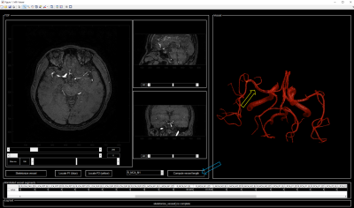
Yoon-Chul Kim, Ha-Na Song, Ji-Eun Lee, In-Young Baek, Woo-Keun Seo
Knowledge of intracranial vessel morphology may be important in predicting the risk of acute ischemic stroke. The three-dimensional nature of the vessels would make it challenging to measure vessels' segmental lengths, unless a software tool dedicated to the purpose is available. The goal of this study is to develop a customized graphical user interface that facilitates users' measurement of intracranial vessel tortuosity in an easy and interactive manner. Using the proposed tool, vessel branch lengths and vessel tortuosity data were collected from 11 proximal vessel segments (e.g., middle cerebral artery, anterior cerebral artery) of 532 subjects.
|
|
2719.
 |
71 |
Quantitative Analysis of Punctate White Matter Lesions Using Quantitative Susceptibility Mapping and R2* Relaxation
Yuting Zhang, Alexander Rauscher, Alexander Weber
Objectives: Our aim was to distinguish PWMLs and focal hemorrhage lesions using quantitative measures. Materials and Methods: In the current study, we acquired multi-echo gradient echo MRI data in neonates with hypoxic ischemic encephalopathy, and post-processed them as R2* relaxation maps and quantitative susceptibility maps (QSM). Manually drawing regions of interest (ROIs) on R2* maps, we measured R2* and susceptibility values of the lesions. Results: We found that R2* and susceptibility values are significantly increased in focal hemorrhage lesions, compared to PWMLs. Conclusions: R2* and QSM can be used to help clinicians distinguish and measure these lesions.
|
|
2720
 |
72 |
Reproducibility of SIENAX volumetric outputs over intra-session, inter-session and inter-scanner acquisitions
Video Permission Withheld
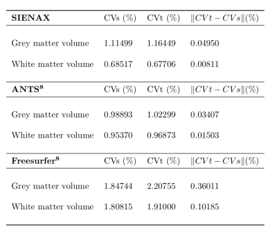
Guillem Garcia, David Moreno-Dominguez, Matt Rowe, Vesna Prckovska, Paulo Rodrigues
Automatic tissue segmentation tools are common in the neuroimaging field. Evaluating their reliability is necessary to validate the findings of studies that use these tools. We conducted a reliability analysis for SIENAX in a test-retest dataset and a multi-site dataset. The results were analysed and compared with other automatic segmentation tools. The volumetric outputs of SIENAX show low coefficients of variance for the test-retest dataset in both grey matter (1.11%) and white matter (0.69%). In the multi-site data the results were to 3.95% and 6.47% respectively, suggesting a possible need for data harmonization in multi-site studies.
|
|
2721
 |
73 |
Comparison of Gradient Echo and Gradient Echo Sampling of Spin Echo Sequence for the Quantification of the Oxygen Extraction Fraction by Combining Quantitative Susceptibility Mapping and Blood Oxygenation Level Dependency
Video Permission Withheld
Simon Hubertus, Sebastian Thomas, Junghun Cho, Shun Zhang, Yi Wang, Lothar R. Schad
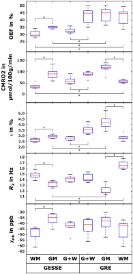
The oxygen extraction fraction (OEF) is a promising biomarker for cerebral tissue vitality. Combining quantitative blood oxygenation level-dependent (qBOLD) modelling and quantitative susceptibility mapping (QSM) from gradient echo (GRE) data revealed promising results but still suffered from biases in white matter and required good parameter initialization. We showed that using an additional gradient echo sampling of spin echo (GESSE) sequence enables OEF reconstruction with higher accuracy, precision and robustness to parameter initialization in simulation. Yet, this increased robustness did still not allow for parameter initialization without prior knowledge of local distributions in vivo, which lead to a non-physiological gray-white matter contrast in the OEF.
|
|
2722.
 |
74 |
Direct reconstruction of arterial blood flow (aBF) from undersampled golden-angle radial non-contrast enhanced dynamic 4D MR angiography
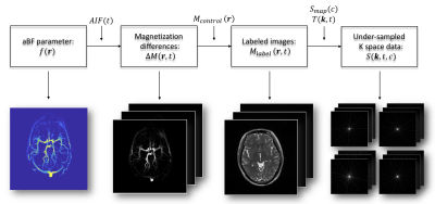
Ziwei Zhao, Kai Wang, Danny JJ Wang, Lirong Yan
Quantification of hemodynamics benefits clinical diagnosis. Non-contrast enhanced MRA with golden-angle radial acquisition has capability of characterization of dynamic flow with high spatiotemporal resolution within a short scan time. Here, we proposed a direct reconstruction framework of arterial blood flow (aBF) from undersampled radial dMRA K-t space data, which mitigated streaking artifacts induced by image-based reconstruction. Both simulation and experimental data suggested that direct optimization method provides reliable aBF under different undersampling rates while preserving detailed delineation of vascular structures, compared to the conventional post-processing singular value decomposition (SVD) method.
|
|
2723.
 |
75 |
Ultra-fast EPI sampling of pulsatile flow waveforms in cerebral arteries via retrospective binning of k-space lines
Joseph Whittaker, Marcello Venzi, Fabrizio Fasano, Daniel Gallichan, Kevin Murphy
Flow related signal enhancement in ultra-fast EPI allows imaging of cardiac pulsatile blood flow profiles in cerebral arteries. We present a novel method that uses retrospective binning of k-space lines to make cardiac phase ‘composite’ k-space planes, from which pulsatile waveforms can be reconstructed with extremely high temporal resolution (~2ms). We demonstrate the proof-of-principle for obtaining pulse wave velocity measures in cerebral arteries, paving the way for mapping quantitative arterial stiffness measures across the brain.
|
|
| Top |
Stroke
Digital Poster
Neuro
Tuesday, 14 May 2019
| Exhibition Hall |
13:30 - 14:30 |
| |
|
Computer # |
|
2724.
 |
76 |
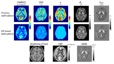
Data-driven regularized inversion (DRI) for improved QSM+qBOLD based CMRO2 Mapping: a feasibility study in healthy subjects and ischemic stroke patients
Junghun Cho, Shun Zhang, Youngwook Kee, Pascal Spincemaille, Thanh Nguyen, Simon Hubertus, Ajay Gupta, Yi Wang
We propose the use of machine-learning to improve the accuracy of a QSM+qBOLD model based Cerebral metabolic rate of oxygen (CMRO2) and oxygen extraction fraction (OEF) mapping. The proposed method, data-driven regularized inversion or DRI, significantly outperformed, in simulation, the current method at all SNR levels. In n=11 healthy subjects, uniform OEF maps were obtained as expected. In n=18 ischemic stroke patients, low OEF regions were clearly located within the lesion region as defined by DWI.
|
|
2725.
 |
77 |
The effect of scan length on the assessment of perfusion using BOLD delay in ischemic stroke
Ayse Ceren Tanritanir, Kersten Villringer, Ivana Galinovic, Ulrike Grittner, Evgeniya Kirilina, Jochen B. Fiebach, Arno Villringer, Ahmed A. Khalil
Hypoperfusion in acute stroke can be detected without exogenous contrast agents using BOLD delay. However the effect of scan duration on assessing perfusion using this method hasn’t been systematically evaluated. This study researched the effect of different scan lengths on diagnostic accuracy and image quality of BOLD delay maps while accounting for head motion. Our results revealed that scan time can be reduced to 3 min and 24 sec without compromising diagnostic power and image quality. However, lesion volumes were robust down to a scan length of 1 min and 8 sec.
|
|
2726.
 |
78 |
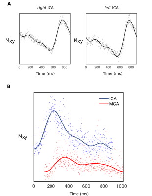
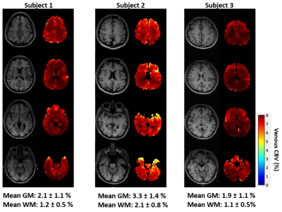
Cerebral Venous Blood Volume Estimation Using Velocity-Selective Spin Labeling Prepared Single-Slab Three-Dimensional Turbo Spin Echo Imaging
Hyunyeol Lee, Felix Wehrli
Venous CBV (CBVv) is of relevance to brain oxygenation level changes during functional activation. To date, MRI techniques for CBVv mapping fall into two categories, based on a 1) quantitative BOLD (qBOLD) model of extravascular signals, and 2) hyperoxic stimulus induced changes in intravascular signal. However, in the former estimation accuracy is impaired due to mutual coupling between CBVv and Yv in the model, while the latter suffers from the complexities in both experiments and estimation involving multiple parameters. Here, we propose velocity-selective spin labeling prepared single-slab 3D TSE imaging for straightforward derivation of CBVv maps in the whole brain. Results from three subjects show plausible values of CBVv estimates in the range of 1.9 - 3.3 % and 1.1 - 2.1 % for gray and white matter, respectively.
|
|
2727.
 |
79 |
Intrinsic vulnerability of low blood flow watershed to white matter hyperintensities in cerebral small vessel disease
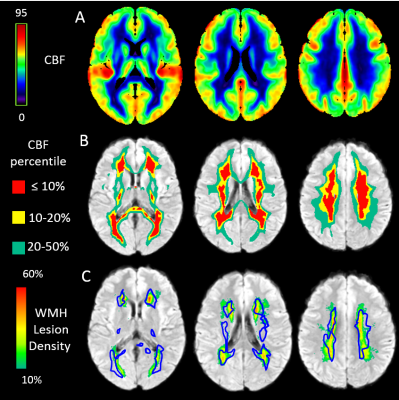
Chunwei Ying, Andria Ford, Peter Kang, Alla Al-Habib, Slim Fellah, Yasheng Chen, Jin-Moo Lee, Hongyu An
White matter hyperintensities (WMH), a major neuroradiological feature of cerebral small vessel disease (CSVD), have a characteristic spatial distribution in the deep white matter and periventricular regions. In this study, we demonstrated a striking spatial overlap between WMH lesion hot spots and the watershed region, defined by a nadir in CBF within the white matter, suggesting that watershed is a region with intrinsic vulnerability to CSVD-related injury.
|
|
2728.
 |
80 |
Focal corticospinal tract volume loss following stroke characterized by diffusion tensor based morphometry (D-TBM)
Amritha Nayak, Matthew Edwardson, Pooja Modi, Neda Sadeghi, Carlo Pierpaoli
Use of a diffusion tensor-based registration method to compare different scans within each subject and to map the results into a population template that can ultimately be used to stratify patients with different motor recovery outcome in stroke.
|
|
2729.
 |
81 |
Evaluation clinical outcome using mismatch between baseline mean diffusion and kurtosis MRI in focal ischemic stroke
Presentation Not Submitted
Min Tang, Wei Di, Xin Zhang, Jie Gao, Xiaoling Zhang, Xiaohong Wu, Zhizheng Zhuo
To observe the cerebral microstructural alterations after focal ischemic stroke by using DKI and assess whether patients are likely to benefit from treated with intravenous tPA at onset of stroke when mean diffusion and kurtosis MRI mismatchs. 58 patients were enrolled. AK, RK and MK values were increased in ischemic lesions, which indicate heterogeneity and complexity of microstructural tissues at onset of stroke. MD-AK mismatch patients? recovered reasonably well with intravenous tPA at onset of stroke, whereas MD-AK mismatch patients without intravenous tPA and coincidence MD-AK of lesions volume showed poor recovery. MD-AK mismatch could be used to identify patients from baseline DKI who are likely to benefit from intravenous thrombolysis at onset of stroke.
|
|
2730.
 |
82 |
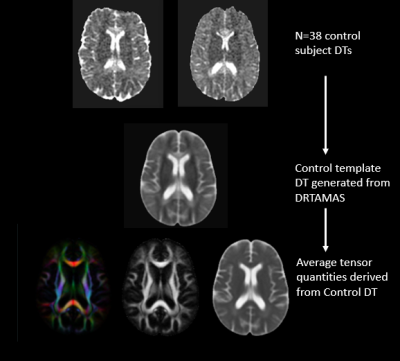
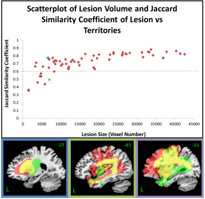
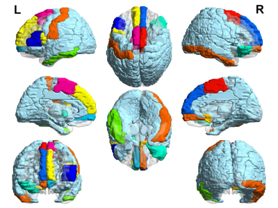
Using Vascular Territories to Predict Disconnection Profiles in Post-Stroke Aphasia
Natalie Busby, Ajay Halai, Ying Zhao, Geoff Parker, Matt Lambon Ralph
Damage sustained to the brain post-stroke appears random but it may be constrained by the underlying neurovasculature; brain regions supplied by the occluded arterial branch will be affected. Combinations of vascular territories were matched to lesions from 62 post-stroke patients. Anatomical connectivity mapping, a measure of whole-brain connectivity, was used to estimate disconnection in each patient through summing disconnection associated with the territories which best matched their lesion. This novel methodology demonstrated that disconnection following a left-hemispheric stroke can be explained by the underlying neurovasculature and may be of particular interest when no diffusion data is available in the patient.
|
|
2731.
 |
83 |
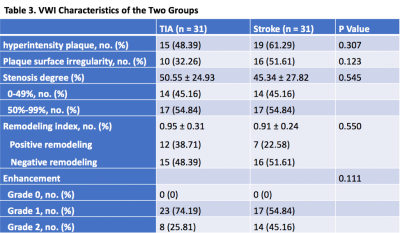
Differential Middle Cerebral Artery Plaque Characteristics in Patients with Transient Ischemic Attack and Ischemic Stroke: A High-Resolution MR Vessel Wall Imaging Study
Jiayu Xiao, Qi Yang, Zhaoyang Fan, Shujuan Li, Fang Wu, Debiao Li, Tao Jiang
This study is to compare the characteristics of intracranial plaques between TIA and stroke patients using VWI. Sixty-two patients (31 TIA and 31 stroke) with MCA stenosis were enrolled in the study. Routine brain MRI, TOF-MRA, pre and post- contrast VWI were performed on each patient. Morphological features of the culprit plaque were compared between the two groups. TIA group had a lower occurrence of hyperintensity plaque, plaque surface irregularity and enhancement grade, those features showed no statistically significant differences and also the degree of stenosis and RI. VWI is useful modality for assessing the intracranial plaques in TIA patients.
|
|
2732.
 |
84 |
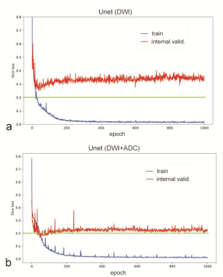
Diffusion lesion segmentation with deep learning in acute ischemic stroke: A combined use of DWI and ADC
Yoon-Chul Kim, Ji-Eun Lee, Inwu Yu, Ha-Na Song, In-Young Baek, Joon-Kyung Seong, Woo-Keun Seo
Conventional deep learning methods for cerebral infarct segmentation rely on diffusion weighted images (DWI) only. Meanwhile, traditional cerebral diffusion lesion segmentation is typically based on a fixed apparent diffusion coefficient (ADC) threshold. It may be worthwhile to combine DWI and ADC images and use them as input for model training. The objective of this study is to develop a deep-learning segmentation model that takes DWI and ADC as input and produces a segmentation map as output and evaluate its performance.
|
|
2733.
 |
85 |
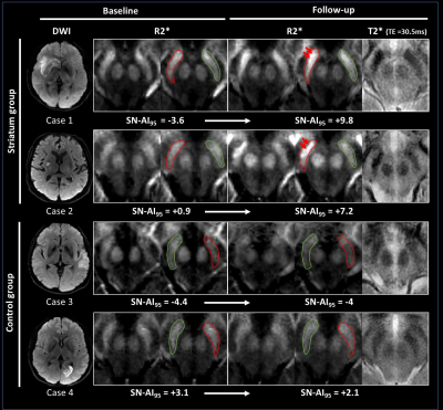
Neurodegeneration of the substantia nigra after ipsilateral infarct: quantification with MRI R2* mapping and relationship to clinical outcome
Tourdias Thomas, Pierre Antoine Linck, Gregory Kuchcinski, Fanny Munsch, Romain Griffier, Renaud Lopes, Gosuke Okubo, Sharmila Sagnier, Pauline Renou, Julien Asselineau, Paul Perez, Vincent Dousset, Igor Sibon
We tested whether long-term neurodegeneration of substantia nigra (SN) secondary to disconnection by supra-tentorial infarcts can be quantified with iron-sensitive imaging and contributes to clinical outcome. 181 stroke patients (75 striatum infarcts, 106 other locations) were prospectively evaluated at 24-to-72h and at one-year clinically and with MRI to quantify iron through R2*. We showed a delayed increase of R2* within SN that was strongly and independently associated with infarct location along known anatomic projections from SN. Such increase of R2* was an independent contributor of poor motor outcome. Iron-sensitive imaging can monitor neurodegeneration non-invasively within SN and potentially other areas.
|
|
2734.
 |
86 |
Remote Effect of Ischemic Stroke: Anatomical Specification of Oxygenation Alteration Investigated by Voxel Based R2' Quantification
Chunxiang Jiang, Xiaojing Long, Siqi Cai, Li Yi, Lijuan Zhang
Ischemic stroke (IS) may induce oxygenation alterations in brain regions remote to the lesion. Remote effect of IS in terms of oxygen metabolism was evaluated based on the voxel wise R2' quantification for subjects with first ever single lesioned IS in corona radiata (CR) (n=10) and brainstem (n=6) using R2' of the superior sagittal sinus as the reference. Both CR and brainstem IS groups showed significant changes of R2' in distributed brain regions with anatomical specifications, suggesting that IS rather represents a spectrum of pathophysiological events of hemodynamic and metabolic impairments at the global level than a focal vascular failure.
|
|
2735.
 |
87 |
Age Specific Differences in Association Between White Matter Cerebral Blood Flow and Ischemic Lesion Severity
Hualu Han, Dongye Li, Huiyu Qiao, Dandan Yang, Zhensen Chen, Runhua Zhang, Gaifen Liu, Xihai Zhao
White matter lesion (WML), one of the sequelae of cerebral hypoperfusion, accumulates with age. This study sought to investigate the relationship between cerebral blood flow (CBF) and WML severity with age in asymptomatic adults. We found that WML scores were strongly associated with WM CBF, suggesting that WM CBF might be an effective indicator for severity of WMLs. We also found that the WM CBF increased with age, consistent with the greater WM cerebrovascular reactivity response in elderly individuals. In addition, our findings of ascending WM CBF cut-off values revealed that the risk of developing WML increases with age.
|
|
2736.
 |
88 |
The value of different plaque indicators in predicting stroke
Yan Wang, Xiaoyue Ma, Yan Bai, Qiang Li, Xianchang Zhang, Yusong Lin, Meiyun Wang
Emerging evidence suggests that the characteristics of intracranial plaques detected by high-resolution vessel wall imaging may serve as an important in-vivo biomarker for predicting ischemic stroke. Different indicators have been introduced to depict plaque features, such as remodeling index, contrast enhancement, and stenosis rate. The purpose of this study was to comprehensively assess the diagnostic performance of these indicators by a quantitative comparison between ischemic stroke patients and transient-ischemic-attack patients. Our results indicated that the remodeling index may have a predictive power similar to contrast enhancement while stenosis rate was a poor predictor.
|
|
2737
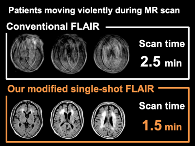 |
89 |
The optimization of single-shot FLAIR for motion artifact reduction and scan time reduction in stroke imaging: A Comparative Analysis with Conventional FLAIR
Video Permission Withheld
Yoshihiro Kubota, Hajime Yokota, Takayuki Sakai, Masami Yoneyama, Hiroki Mukai, Takuro Horikoshi, Takashi Uno
For stroke imaging, motion correction and scan time reduction are essential. We optimized the single-shot FLAIR sequence and implemented it into our stroke MR protocol. 48 patients suspected of stroke were retrospectively involved, and board-certified radiologists evaluated the images of our modified-single-shot FLAIR and conventional FLAIR at the terms of degree of motion artifact, image quality, delineation of hyperintense vessel and contribution for diagnosis. Motion artifact was significantly reduced (P < 0.001) and scan time was decreased by 40% in single-shot FLAIR. Hyperintense vessels were equally detected in both the sequences. Radiologists considered modified-single-shot FLAIR more useful for diagnosis.
|
|
2738.
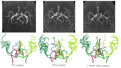 |
90 |
Quantitative assessment of cerebrovascular structure after carotid revascularization using intraCranial Artery Feature Extraction (iCafe) Technique
Manabu Shirakawa, Li Chen, Niranjan Balu, Wenjin Liu, Dakota Ortega, Jinmei Chen, Theodore Trouard, Diane Bock, Wei Zhou, Chun Yuan, Thomas Hatsukami
The aim is to evaluate the change in intracranial arterial vasculature after carotid revascularization using an intracranial feature extraction (iCafe) technique for quantitative analysis of intracranial arteries from 3D time-of-flight magnetic resonance angiography (TOF MRA). Twenty subjects who received carotid revascularization were enrolled and all patients underwent MRA scans three times: before, within 3 days after, and six months after revascularization. The dataset was processed blindly by 4 reviewers using iCafe. Length and volume of intracranial artery and number of intracranial artery branches increased after surgery. This result suggested increased cerebral blood flow after carotid revascularization.
|
|
2739.
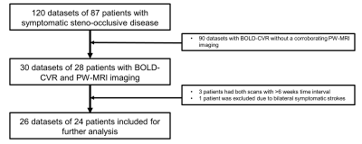 |
91 |
Crossed cerebellar diaschisis characterization with BOLD-fMRI cerebrovascular reactivity and T2*-perfusion MRI
Marco Piccirelli, Martina Sebök, Christiaan Hendrik Bas van Niftrik, Giovanni Muscas, Athina Pangalu, Susanne Wegener, Luca Regli, Christoph Stippich, Jorn Fierstra
Crossed cerebellar diaschisis (CCD) in stroke patients has been associated with worse neurological performance and outcome, but clinical routine CCD imaging is limited. We therefore tested the diagnostic value of blood oxygenation-level dependent cerebrovascular reactivity (BOLD-CVR) and T2* perfusion weighted-MRI in patients with unilateral symptomatic cerebrovascular steno-occlusive disease exhibiting crossed cerebellar diaschisis. Secondly, we assessed the clinical significance of a CCD diagnosis based on BOLD-CVR imaging. BOLD-CVR is a valid clinical diagnostic tool for CCD, whereas perfusion MRI derived parameters were shown to be unsuitable. Furthermore, stroke patients exhibiting CCD showed a worse neurological performance and outcome, unrelated to stroke volume.
|
|
2740.
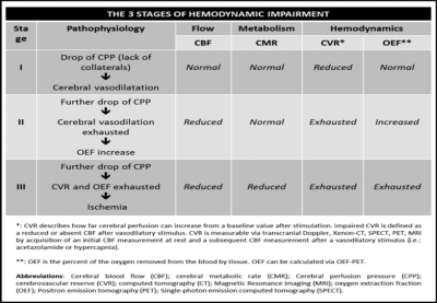 |
92 |
Negative BOLD cerebrovascular reactivity in stroke patients: a sign of misery perfusion of the affected hemisphere
Marco Piccirelli, Christiaan Hendrik Bas van Niftrik, Giovanni Muscas, Martina Sebök, Lita von Bieberstein, Susanne Wegener, Oliver Bozinov, Giuseppe Esposito, Andreas Luft, Luca Regli, Christoph Stippich, Jorn Fierstra
Subjects with hemodynamic failure stage 2 (i.e. misery-perfusion) have heightened risk of acute and chronic brain tissue damage. One of the most important signs of misery-perfusion is a negative cerebrovascular reactivity (CVR). CVR is defined as a blood flow response to a vasoactive stimulus. Recently blood-oxygenation-level-dependent (BOLD) CVR was proposed to detect misery-perfusion. However, BOLD-CVR MRI signal does not reflect CBF changes directly and discrepancies between negative BOLD-CVR and negative CBF changes have been reported. To better assess these discrepancies, we performed a multimodal clinical misery-perfusion assessment with perfusion-weighted-MRI and transcranial-Doppler complimentary to BOLD-CVR in patients with symptomatic steno-occlusive disease.
|
|
2741.
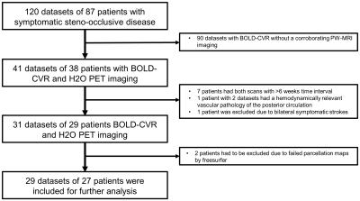 |
93 |
Ipsilateral Thalamic Diaschisis in Stroke Patients
Marco Piccirelli, Christiaan Hendrik Bas van Niftrik, Martina Sebök, Giovanni Muscas, Carlo Serra, Athina Pangalu, Oliver Bozinov, Andreas Luft, Luca Regli, Christoph Stippich, Jorn Fierstra
Presence of ipsilateral thalamic diaschisis in stroke patients, detected using BOLD-CVR, is characterized by thalamic volume reduction, reduced thalamic blood flow, and worse stroke severity scores at admission as well as 3 months follow-up. This finding suggests that ipsilateral thalamic diaschisis may be an important clinical imaging marker in stroke patients.
|
|
2742.
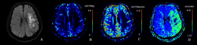 |
94 |
Absolute quantitative dynamic susceptibility contrast cerebral perfusion imaging using the Self-Calibrated EPI sequence in patients with ischemic stroke
Xiaoyue Ma, Yan Wang, Qiang Li, Menghuan Zhang, Xianchang Zhang, Yusong Lin, Meiyun Wang
This study aims to explore the clinical value of the absolute quantitative dynamic susceptibility contrast cerebral perfusion-weighted imaging using Self-Calibrated EPI sequence (SCALE-PWI) in patients with ischemic stroke. SCALE-PWI could provide reliable quantitative measurement of cerebral blood flow (CBF), cerebral blood volume and mean transit time in a quite short scan time of 2:14 mins. Results suggest the CBF values in infarct core are significantly lower than the values in ischemic penumbra. In conclusion, the SCALE-PWI could provide quantitative hemodynamic information in a quite short scan time, thus may serve as a guide for tissue-based decision making and personalized treatment planning in acute stroke.
|
|
2743.
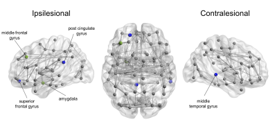 |
95 |
Disrupted structural brain network configuration in patients with post-stroke depression
Presentation Not Submitted
Xiaopei Xu, Rui Tang, Siyu Zhu, Zhijian Cao
To better understand the underlying mechanisms for a wide range of emotional disturbances in post-stroke depression (PSD) patients, we used structural brain connectivity analysis to investigate the differences in global and local network organization of stroke patients with PSD and no PSD. Our results demonstrated that the efficiency of both local and regional network of patients with PSD were higher compared to those without, and that higher depression severity was significantly associated with increased network efficiency. These results indicated that the disrupted network architecture might be the cause of depressive symptoms in PSD patients, and brain network analysis is a useful tool to link psychological disorders with their underlying anatomical substrate.
|
|
2744
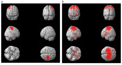 |
96 |
Evaluation of physiotherapy induced changes in post-stroke recovery using MRI
Video Permission Withheld
Rajesh Mishra, Dushyant Kumar, Surbhi Kaura, Senthil Kumaran, Priyanka Bhagat, Padma Srivastava, Rama Jayasundar
This study has evaluated the role of MRI in determining physiotherapy-induced changes in post-stroke recovery in 21 first-ever ischemic patients. Physiotherapy was given as intervention for 45 minutes every day for consecutive 6 months. Pre- and post- (3, 6 months) intervention assessment involved NIHSS, mRS and MRI studies (3T MR scanner). MRI studies included 3D-T1, 3D-FLAIR, DWI, and fMRI (motor task). Preliminary findings showed individual patients’ positive response to physiotherapy reflected in the NIHSS and mRS scores, and in the recovery of fMRI activation in the affected motor cortex post-intervention and other MR markers.
|
|
2745
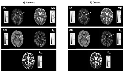 |
97 |
Use of NODDI for Microstructural Characterization of Posterior Limb of the Internal Capsule in Subacute and Chronic Stroke Patients
Video Permission Withheld
Alfonso Mastropietro, Maria Malosio, Lucia Fontana, Laura Straffi, Simona Marcheselli, Bruno Bernardini, Sara Ghirmai, Nunzio Nuzzi, Giovanna Rizzo, Marco Grimaldi
This preliminary work shows that NODDI, supplementing the classical DTI approach, could provide a new insight into the subtle micro-architectural modifications occurring in the posterior limb of the internal capsule of 10 stroke patients in subacute and chronic phase.
|
|
2746
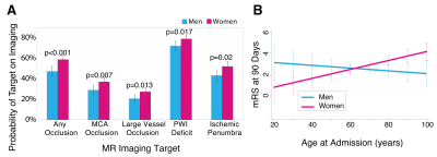 |
98 |
Ischemic Stroke Imaging and Outcomes: Differences between Sexes
Video Permission Withheld
Adrienne Dula, Steven Warach
Stroke is a treatable disease and neuroimaging can identify salvageable tissue, directly impacting treatment decisions. Women experience stroke differently than men with higher severity, worse outcomes, and varying therapeutic response. We examined the contribution of sex and age to stroke outcome with emphasis on therapeutic targets on MRI. MR images were evaluated for predictive imaging factors. A 90-day mRS was obtained to assess functional independence. Women present more often with treatable ischemic stroke than men as defined by MR imaging factors. Sex modulates the age-dependent stroke outcome but upon stratification for treatment, difference in outcome favoring men was not observed.
|
|
2747.
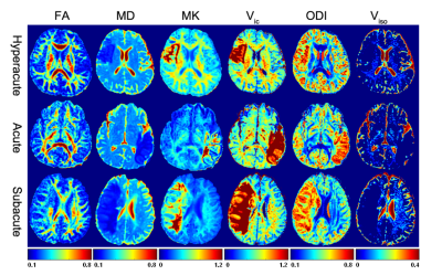 |
99 |
A study of neurite orientation dispersion and density imaging in ischemic stroke
Zhenxiong Wang, Shun Zhang, Yang Fan, Wenzhen Zhu
Neurite orientation dispersion and density imaging (NODDI) can quantitatively evaluate specific microstructural changes in terms of neurite density and orientation distribution of axons and dendrites. In the study, we attempt to demonstrate the feasibility of NODDI in characterizing the microstructural alterations in brain tissues during ischemic stroke and to compare its sensitivity with diffusion tensor imaging and diffusion kurtosis imaging. Results demonstrated that NODDI is a potential technique for quantitatively evaluating ischemic stroke and showed higher sensitivity compared with diffusion tensor imaging (DTI) and diffusion kurtosis imaging (DKI).
|
|
2748.
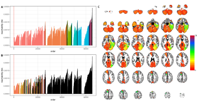 |
100 |
Stroke atlas of the brain: A voxel-wise density-based clustering of infarct lesion topographic distribution
Yanlu Wang, Hadrien Van Loo, Julia Juliano, Sook-Lei Liew, Alexander McKinney IV, Sam Payabvash
In stroke patients, both infarct volume and location affect functional outcome; however, infarct topography is far less commonly incorporated in prognostic models, given the complexity of assessing infarct topographic distribution. In this study, we applied data-driven density clustering analysis, using the OPTICS algorithm, on 793 infarct lesions from 438 stroke patients to devise a “stroke-atlas of the brain” stratifying brain voxels likely to infarct together. This atlas can help with differentiation of infarct lesions in clinical practice, assess topographic distribution of infarct in prognostic models for stroke patients, or be applied for defining regional infarct thresholds in CT/MR perfusion maps.
|
|
| Top |
Psychoradiology: Schizophrenia, Psychosis, OCD & More
Digital Poster
Neuro
Tuesday, 14 May 2019
| Exhibition Hall |
13:30 - 14:30 |
| |
|
Computer # |
|
2749.
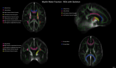 |
101 |
Myelin-associated clinical and physical correlates in a cohort of chronic schizophrenia patients.
Senften Peter, Melissa Woodward, Randall White, Allen Thornton, Kristina Gicas, Cornelia Laule, Darren Warburton, A. Talia Vertinsky, William Honer, Wayne Su, Donna Lang
Aberrant myelination and tandem cardiovascular deficits may contribute to emergence of the schizophrenias. To explore this hypothesis, a pilot study of Myelin Water Fraction (MWF), V02max capacity, and symptom severity was done in 15 chronic schizophrenia/schizoaffective patients. MWF was positively correlated with age in some, but not all, fronto-medial and fronto-temporal regions, 2. V02max was positively correlated with MWF the superior longitudinal fasciculus, the genu, and the forceps minor, and 3. Social functioning was positively correlated to MWF in the forceps major. These data indicate the presence of relationships between MWF measures, social functioning and cardiovascular capacity in schizophrenia.
|
|
2750.
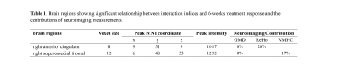 |
102 |
Contribution and Interaction of Brain Structure and Function in Treatment Response Prediction of First-episode Drug-naïve Schizophrenia
Presentation Not Submitted
GUI FU, Ningxuan Chen, Chaogan Yan, Wenjing Zhang, Jiaxin Zeng, Yuan Xiao, Su Lui
Magnetic Resonance Imaging (MRI) including structural and functional neuroimaging has been applied extensively to examine response to antipsychotic treatment, however, questions remain regarding the interaction between these measurements and their unique role. Our study provided a comprehensive examination of interaction and contribution for voxel-wise measurements related with treatment response. We found that brain functional measurements in certain brain regions have advantages in predicting treatment response. Furthermore, the functional activities were different between short- and long-term treatment of antipsychotic drugs. These findings revealed that functional changes were more sensitive to the antipsychotic treatment and could be promising biomarkers in treatment prediction.
|
|
2751.
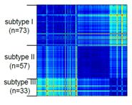 |
103 |
Heterogeneity of brain structure alterations in patients with never-treated first episode schizophrenia
Yuan Xiao, Bo Tao, Jiaxin Zeng, Gui Fu, Biqu Tang, Wenjing Zhang, Siyi Li, Su Lui, Qiyong Gong
Although schizophrenia is a heterogeneous clinical syndrome, one important question that remains largely unanswered is whether the complex and subtle deficits revealed by MRI could be used as objective biomarkers to resolve neurobiological heterogeneity within this disorder. Using clustering analysis and structural MRI, first-episode schizophrenia patients were classified into three subtypes. The three subtypes of patients showed different morphological alterations.
|
|
2752.
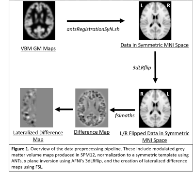 |
104 |
Source-based Laterality of Grey Matter in Schizophrenia
Thomas DeRamus, Jiayu Chen, Armin Iraji, Eswar Damaraju, Rogers Silva, Vince Calhoun
Altered brain laterality is frequently reported in morphological brain studies of individuals with schizophrenia. However, these studies utilize voxel/vertex-wise univariate methods which may not be optimal for examining brain laterality. We introduce a novel multivariate approach to estimate covarying lateralized networks. In our approach, lateralized grey matter maps were computed by subtracting volumetric data one hemisphere from the other, and analyzed via independent component analysis (ICA), followed by testing loading parameters from components identifying covariation within laterality networks. Results display significant relationships with temporal lobe and cerebellar laterality and negative symptoms of schizophrenia that warrant further exploration with multimodal analyses.
|
|
2753
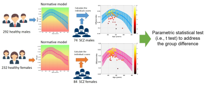 |
105 |
White matter microstructural changes in schizophrenia: A study using normative-model-based statistical analysis
Video Permission Withheld
Li-Ying Yang, Chih-Min Liu, Tzung-Jeng Hwang, Chang-Le Chen, Yung-Chin Hsu, Wen-Yih Tseng
White matter microstructural changes have been found in schizophrenia but the effects of gender and age on these changes remains entangled. This study aimed to quantify the white matter changes in 158 schizophrenia patients using a novel approach which calculated the z scores based on the normative models built from 524 healthy subjects across lifespan. Our results showed that twelve tracts had significant differences between schizophrenia patients and controls.
|
|
2754.
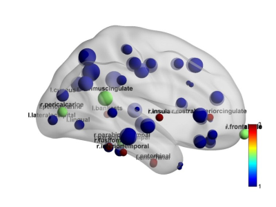 |
106 |
Gray matter network changes with aging and duration in a large group of never-treated patients with schizophrenia.
Presentation Not Submitted
Beisheng Yang, Wenjing Zhang, Bo Tao, Wenbing Li, Su Lui
A large group of never-treated schizophrenia patient was enrolled to investigate the pattern of network changes with aging and illness duration. Cortical thickness preprocessed by FreeSurfer, and correlation matrix was constructed by correlating the cortical thickness of every pair of regions. Compared to healthy controls, all patient subgroups stratified along age and illness duration showed common changes while distinct changes, mainly involved DMN and CN. The alternations within DMN and CN may represent trait-related structural network changes in schizophrenia, while distinct changes may represent illness progression with more-wide spread brain abnormalities.
|
|
2755.
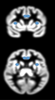 |
107 |
Evaluation of structural brain modifications related to First Episode Psychosis
Francesca Saviola, Marcella Bellani, Letizia Squarcina, Eleonora Maggioni, Domenico Zacà, Cinzia Perlini, Mirella Ruggeri, Paolo Brambilla, Jorge Jovicich
Psychiatric disorders are characterized by a complex range of symptoms. Psychosis, known as one of the most severe mental illness, is still lacking specific neuroimaging markers complementing clinical information for early differential diagnostics, disease progression monitoring and treatment response evaluations. Most previous studies in the field considered psychotic patients undergoing chronic pharmacologic treatment or long duration of illness, which may confound morphometric or functional findings. Here we study a cohort of First Episode Psychosis (FEP) patients to investigated grey matter changes using structural MRI, in FEP relative to healthy controls, with univariate and multivariate analysis.
|
|
2756.
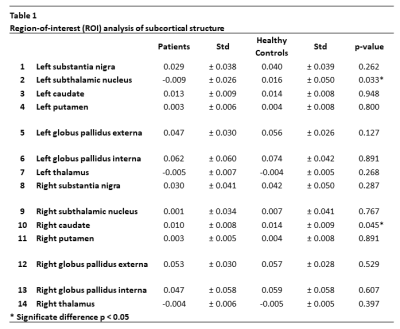 |
108 |
Quantitative Susceptibility Mapping MRI shows changes in dorsal striatum in patients with a first Episode of Psychosis compared to controls.
Marisleydis García, Néstor Muñoz, Manuel Chappa, Carlos Milovic, Cristian Montalba, Julio Acosta-Carbonero, Luz Alliende, Bárbara Iruretagoyena, Juan Undurraga, Alfonzo González, Carmen Castañeda, Marcelo Andia, Sergio Uribe, Nicolas Crossley, Cristián Tejos
Dopamine is a neurotransmitter that plays an important role in psychosis. Neuromelanin is a by-product of the synthesis of dopamine. In First Episode of Psychosis (FEP) is reported the effect that causes dopamine and its relationship with neuromelanin. However, it has not been reported signal change due to accumulation of heavy metal using magnetic resonance imaging techniques. We found susceptibility changes in two areas of brain using QSM, the left subthalamic nucleus and right caudate. This finding might help to discriminate between FEP patients and healthy subjects.
|
|
2757.
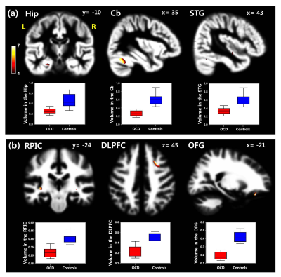 |
109 |
Brain morphometric and cellular metabolic alterations in patients with obsessive–compulsive disorder
Presentation Not Submitted
Chung Man Moon, Gwang Woo Jeong
Obsessive–compulsive disorder (OCD) causes neural dysfunction associated with cognitive deficit and emotional dysregulation. To our knowledge, however, no one has applied the combined neuroimaging study of morphometric and metabolic brain abnormalities in patients with OCD. Therefore, this study assessed the associations of the gray matter (GM) and white matter (WM) volume alterations in conjunction with in vivo cellular metabolic changes in patients with OCD. Our findings will be helpful to aid us in the understanding of neurocognitive impairment in OCD, and thus, enhancing the diagnostic accuracy for OCD by additional information on the associated cerebral volume change and metabolic abnormality.
|
|
2758.
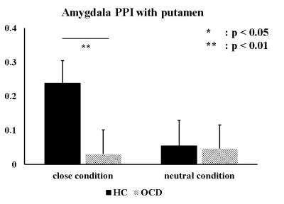 |
110 |
Amygdala dysfunction during negative emotional control in Obsessive-Compulsive Disorder: an fMRI study
Hyunsil Cha, Sang Won Lee, Kyungeun Jang, Eunji Kim, Heajeong Choi, Jiung Yang, Seungho Kim, Jinsu Park, Moon Jung Hwang, Huijin Song, Hui joong Lee, Seung Jae Lee, Yongmin Chang
We investigated brain activation in obsessive-compulsive disorder (OCD) patient using thought-action fusion (TAF) task to assess the influence of OCD symptom on amygdala response to the task. Context-dependent psycho-physiological interaction (PPI) analysis of close condition showed decreased amygdala PPI with putamen in patients with OCD compared to healthy controls.
|
|
2759.
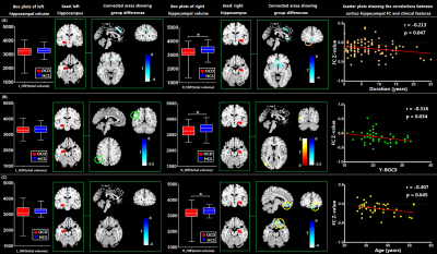 |
111 |
Alterations of structural anatomy and functional connectivity regarding hippocampus in obsessive–compulsive disorder
Xinyu Hu, Lianqing Zhang, Xuan Bu, Hailong Li, Bin Li, Wanjie Tang, Lu Lu, Xiaoxiao Hu, Shi Tang, Yingxue Gao, Yanlin Wang, Yanchun Yang, Qiyong Gong, Xiaoqi Huang
Besides the classical cortico-striato-thalamo-cortical circuits, the hippocampus has received increasing attention in the psychopathology of obsessive-compulsive disorder (OCD). We aimed to investigate the abnormalities of structural anatomy and functional connectivity (FC) regarding hippocampus in a relatively large sample of unmedicated OCD patients and explore the effects of onset age on these neural correlates. Our findings (i) identified significant volumetric reductions of right hippocampus in OCD; (ii) revealed abnormal cortico-hippocampal connectivity in the prefrontal-limbic networks of OCD and (iii) indicated distinct patterns of cerebral-hippocampal connectivity alterations in early-onset and late-onset OCD, which highlighted the potential importance of neurodevelopmental changes in OCD.
|
|
2760.
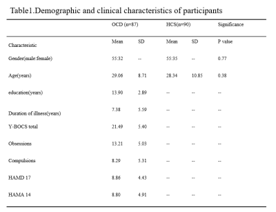 |
112 |
Abnormal Static and Dynamic Functional Network Connectivity Patterns in Patients with Obsessive-compulsive Disorder
Hailong Li, Yingxue Gao, Xuan Bu, Xinyu Hu, Lianqing Zhang, Lu Lu, Shi Tang, Xiaoxiao Hu, Kaili Liang, Yanlin Wang, Qiyong Gong, Xiaoqi Huang
Static and dynamic functional network connectivity (FNC) analyses were applied to determine the abnormal connectivity patterns among the large-scale brain networks in obsessive-compulsive disorder patients. We found that static FNC analysis showed more obvious group differences than dynamic FNC. Decreased functional connectivity between visual network and DMN has been shown in both static and dynamic FNC analysis, it could be considered as the most stable connectivity change of functional brain networks in OCD patients. These findings advocate the using of both static and dynamic FNC to help truly understanding the alterations of brain networks.
|
|
2761.
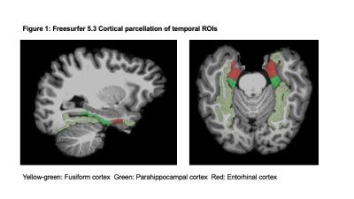 |
113 |
Medial temporal cortical changes in response to yoga and aerobic exercise interventions in early psychosis patients
Melissa Woodward, Jingxia Lin, Wayne Song, William Honer, Eric Chen, Donna Lang
Early psychosis patients exhibit cortical reductions and poor cardiovascular health, which may be worsened by antipsychotic medication. Aerobic exercise and yoga may be able to remediate cortical loss and improve symptom severity. First-episode psychosis patients who completed a twelve-week exercise program showed increased cortical volume and thickness compared to waitlist controls with differential effects of aerobic exercise and yoga. Exercise-mediated changes in brain measures were associated with greater improvement in symptom severity scores. Both aerobic exercise and yoga may have neuroanatomical and clinical benefits for early psychosis patients and may be a safe, cost-effective adjunct treatment.
|
|
2762.
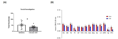 |
114 |
Acute and Chronic intranasal oxytocin differentially affect brain functional connectivity
Alessia De Felice, Marco Pagani, Ludovico Coletta, Alberto Galbusera, Alessandro Gozzi
Intranasal oxytocin (OXT) administration has shown promise as a putative treatment for disorders characterized by social impairments. However, the brain-wide substrates engaged by this neuropeptide remain elusive. By using mouse fMRI, we show that the circuits engaged by intranasal OXT are differentially affected by the duration of OXT dosing. Specifically, acute OXT administration increases brain connectivity in key nodes of the social brain. By contrast, repeated dosing exacerbates inter-regional coupling and results in paradoxical social impairments in control “wild type” mice. These result have implications for clinical testing of OXT in control and pathological conditions.
|
|
2763.
 |
115 |
Brain gray matter correlates of extraversion: A systematic review and meta-analysis of voxel-based morphometry studies
Presentation Not Submitted
Han Lai, Song Wang, Qiyong Gong
Extraversion is a fundamental personality dimension closely related to individuals’ physical and mental health. Although increasing studies have attempted to identify the neurostructural markers of extraversion but have yielded inconsistent and heterogeneous results. The current study aims to reach a comprehensive understanding of brain gray matter (GM) correlates of extraversion by using a systematic review and meta-analysis approach. Our review revealed a preliminary outline of the brain GM differences related to extraversion in distributed brain regions. Our meta-analysis of voxel-based morphometry studies identified six core brain regions correlated with extraversion and revealed the potential effect of gender and age.
|
|
2764.
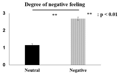 |
116 |
Alteration in negative emotional regulation associated with childhood abuse: fMRI study
Seungho Kim, Sang Won Lee, Kyungeun Jang, Hyunsil Cha, Heajung Choi, Eunji Kim, Jiung Yang, Jinsu Park, Huijin Song, Hui Joong Lee, Moon Jung Hwang, Seung Jae Lee, Yongmin Chang
We assessed negative emotional regulation in young adults who experienced childhood abuse. We investigated a relationship between psychological data and brain activation during emotion regulation task. The ventromedial prefrontal cortex (vmPFC) showed significant correlation with the degree of childhood abuse in negative emotion task.
|
|
2765.
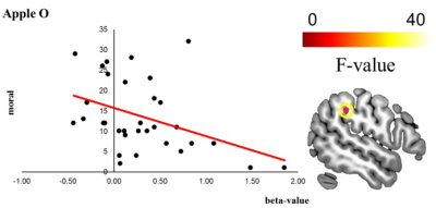 |
117 |
Inferior parietal lobule controls moral thought-action fusion: an fMRI study
Eunji Kim, Sang Won Lee, Hyunsil Cha, Kyungeun Jang, Heajung Choi, Seungho Kim, Jinsu Park, Jiung Yang, Huijin Song, Moon Jung Hwang, Hui Joong Lee, Seung Jae Lee, Yongmin Chang
Thought-action fusion (TAF) could make people feel that action to happen1. We investigated the difference from control thinking and administration of electrical shocks to another person in healthy control with moral related TAF task by using fMRI. The inferior parietal lobule (IPL) activation showed a negative correlation with moral score in TAF among the activated regions from a shock factor. Therefore, when considering the functional role of IPL in making another have some irritation and the appropriate empathic response, such as mentalization, might have an important role in TAF related situation.
|
|
2766.
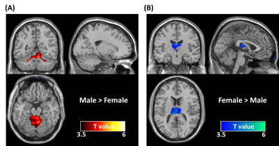 |
118 |
Sexual dimorphism in the young adult brain using magnetic resonance imaging: The effect of the field strengths
Jyun-Ru Chen, Hui-Chieh Yang, Yen-Chih Huang, Chun-Ming Chen, Chih-Feng Chen, Shin-Lei Peng
The existing reports regarding sexual dimorphism in brain structures are not confluent and generally heterogeneous. MRI instrument-related factor such as field strength is one of the greatest contributors to the brain quantification variability, but its effect on sexual dimorphism in brain structures remains unclear. In this study, we found that due to the image contrast differences arising from differences in field strengths, the sex dimorphism in brain morphology appears to exist dependent of field strengths. It suggests field strength should be considered as one important factor that contributes to the inconsistency in the sex dimorphism in brain across literatures.
|
|
2767.
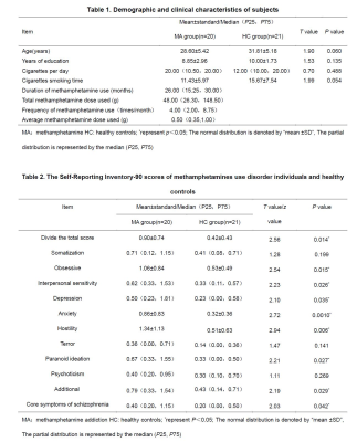 |
119 |
Altered gray matter volume and the relationship with the psychiatric symptoms in methamphetamine use disorder individuals
Presentation Not Submitted
Shan Dang, Wei Li, Jia Zhu, Qiang Li, Wei Wang, Jing Chen
The study used voxel-based morphological to explore the differences in gray matter volume and psychiatric symptoms of Self-reporting Inventory-90 between methamphetamine use disorders and healthy controls. It was found that many of the SCL-90 symptoms and the grey matter volume have changed in methamphetamine use disorder individuals. Meanwhile, the abnormal grey matter volume is associated with psychiatric symptoms.
|
|
2768.
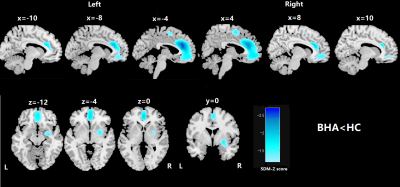 |
120 |
Common gray matter atrophy in individuals with different behavioral addictions: a voxel-wise meta-analysis
Kun Qin, Feifei Zhang, Qiyong Gong
In order to figure out the common structural alterations in behavioral addicts among various publications and to find a biomarker for future improvement of diagnostic category, we searched for voxel based morphometry studies compared between patients and healthy controls and pooled them together in a meta-analytic way by using AES-SDM. 22 studies comprising 5 different addictive behaviors were included. 576 patients showed GM reduction in the left ACC, right striatum and right SMA compared with 635 HCs. In summary, our findings revealed common GM decreases in frontostriatal circuitry, consistent with previous multimodal neuroimaging findings in addiction.
|
|
2769.
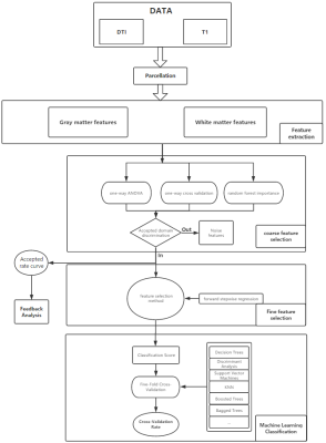 |
121 |
The diagnosis of Internet gaming disorder by MR imaging: A MRI based analysis
Lei Wei, Zhe Wang , Qianfeng Wang, Xu Han, Yawen Sun, Weina Ding, Yan Zhou, He Wang
The internet gaming disorder has become one of the most serious healthy problem among teenagers, the questionnaire and scale are widely used to IGD diagnostic. However, the underlying neural mechanism of IGD was still unclear. Current study present an evidence that cerebral morphometric alteration could be used to identified IGD from normal, and may also help for further study about IGD.
|
|
2770.
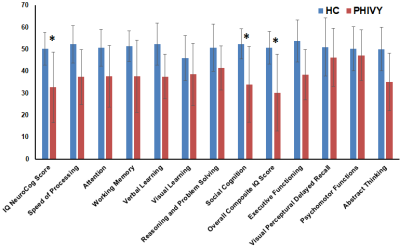 |
122 |
Regional Cortical Thickness Changes and Neurocognitive Performance in Perinatally HIV-infected Youth
Manoj Sarma, Margaret Keller, Paul Macey, Tamara Welikson, David Michalik, Karin Nielsen-Saines , Jaime Deville , Joseph Church, Eva Operskalski , Andrea Kovacs , Irwin Walot, Joseph Ventura, M. Albert Thomas
Despite effective viral suppression, youth with perinatal HIV (PHIVY) often demonstrate long-term cognitive deficits. We measured grey matter cortical thickness as a measure of brain structural integrity in 11 PHIVY receiving long term cART compared to 16 age-matched controls and assessed neurocognitive performance. The PHIVY group performed significantly worse than controls. Regions of significantly thinner and thicker cortex in PHIVY were observed which may contribute to these deficits in neurocognitive function. Cortical thickness in PHIVY was correlated with current CD4 count and neurocognitive performance. Our findings suggest the potential importance of continued monitoring of PHIVY.
|
|
2771.
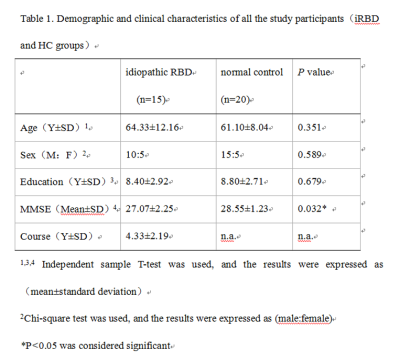 |
123 |
Brain Microstructural Abnormalities in Patients with Idiopathic Rapid Eye Movement Sleep Behavior Disorder: A Voxel Based Morphometry and Diffusion Tensor Imaging Study
Wanqun Yang, Caihong Zhou, Yingjie Mei, Biao Huang
Idiopathic rapid eye movement sleep behavior disorder (iRBD) has close relationship with neurodegenerative disorder. Many researches has validated that almost all of iRBD patients evolved into Parkinson’s disease, Lewy body dementia, or multiple system atrophy over time. However the pathogenesis of iRBD still remains unclear. In this research, VBM and voxel-based DTI analysis were combined to detect the microstructural abnormalities in the iRBD patients. A wide range of changes in brain structure in iRBD group was observed, which may reveal pathophysiologic mechanism on cognitive function disorder,which can be valuable for the early diagnosis and treatment of iRBD.
|
|
2772.
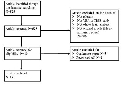 |
124 |
Altered white matter microstructure in anorexia nervosa: A voxel-based meta-analysis of diffusion tensor imaging
Simin Zhang, Weina Wang, Xiaorui Su, Huaiqiang Sun, Qiang Yue, Qiyong Gong
The neurobiological underpinnings of anorexia nervosa (AN) remain unclear. Altered white matter microarchitecture has been described, but findings are inconclusive. To investigate a statistical consensus among published DTI studies of altered white matter microarchitecture in AN, we conducted a quantitative voxel-based meta-analysis of fractional anisotropy using Seed-based d Mapping. The pooled results in AN showed robust reduction fractional anisotropy in the interhemispheric connections, frontal-subcortical circuitry and limbic association fibers. This study provides a thorough profile of WM microarchitecture alterations in patients with AN and these intrinsic alterations may aid in developing effective treatments in AN.
|
|
| Top |
Aging & Neurodegeneration (Other than AD)
Digital Poster
Neuro
Tuesday, 14 May 2019
| Exhibition Hall |
13:30 - 14:30 |
| |
|
Computer # |
|
2773.
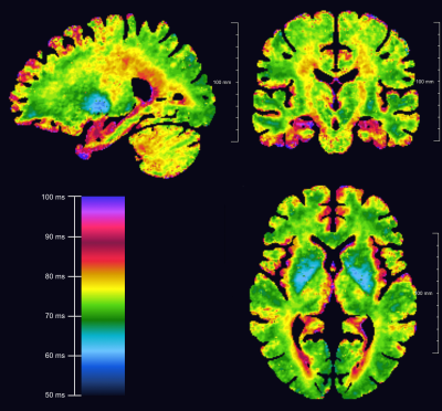 |
126 |
T1-rho in the aging brain: results from large-scale population imaging
Johanna Kramme, Eberhard Pracht, Gerard Sanroma, Tony Stöcker, Monique Breteler
Within the Rhineland Study we investigated and report normative brain T1-rho values and their change over age, for a large cohort of 547 participants. Investigated regions were GM, WM, deep gray matter and selected white matter tracts. All investigated regions, except amygdala and accumbens, show a positive trend with age. Total scan time was under six minutes (whole brain), showing the feasibility to provide normative values for a wide range of brain regions in a reasonable amount of time.
|
|
2774.
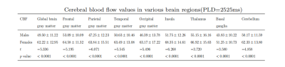 |
127 |
Gender difference on cerebral blood flow in people aged over 80 years: A pseudo-continuous arterial spin labeling study
Heng Zhang, Xian Xu, Ningyu An, Zhentao Zuo
This study focuses on cerebral blood flow(CBF) of the elderly over 80years old, with a large sample size. Different from the conclusions ofprevious studies, females brain perfusion CBF values are higher than males,in the whole brain and various brain lobes.
|
|
2775.
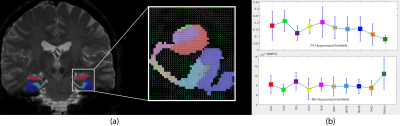 |
128 |
Diffusion magnetic resonance imaging in the human hippocampal subfields using super-resolution HYDI
Nahla Elsaid, Pierrick Coupé, Yu-Chien Wu
The hippocampal atrophy is known to be the most validated biomarker of Alzheimer’s disease. Accordingly, in this study, we develop a method to enable the structural connectivity mapping through tractography of the hippocampal subfields using super-resolution diffusion data.
|
|
2776.
 |
129 |
Differential Relationship of GABA and GLX in Dorsal vs. Ventral Prefrontal Cortex and Their Relationship To Age and Gender
Mark Brown, Harry Smolker, Rebecca Helmuth, Marie Banich
MRS was employed in two regionally distinct prefrontal voxels (dorsal and ventral) in 62 adult females and 119 adolescents (60 males, 59 females) to determine the variations of GABA and GLX concentrations with regional specificity, participant gender, and age. The results indicate that levels of prefrontal neurotransmitter concentrations are influenced by age. Furthermore, the results suggest that the relationship between neurotransmitter levels can vary for adjacent portions of cortex, being more highly correlated for the dorsal than ventral voxel.
|
|
2777.
|
130 |
T2*-weighted imaging and quantitative susceptibility mapping (QSM) in patients with amyotrophic lateral sclerosis and bulbar impairment
Graziella Donatelli, Mauro Costagli, Elena Caldarazzo-Ienco, Gianmichele Migaleddu, Paolo Cecchi, Michela Tosetti, Gabriele Siciliano, Mirco Cosottini
The T2* hypointensity of the primary motor cortex (M1), associated to an increase in iron deposits related to neuroinflammatory reaction and cortical microgliosis, has been suggested as possible MRI marker of upper motor neuron impairment in patients with amyotrophic lateral sclerosis (ALS). This study investigates the orofacial subregion of M1 (fM1) in 36 patients with ALS. The evaluation of T2* signal hypointensity and quantitative susceptibility mapping (QSM) values in fM1 were related to patients’ bulbar functions (such as speech and swallowing) assessed clinically. Results demonstrate that QSM values were significantly higher in patients with bulbar dysfunction than in those without (p≤0.0001).
|
|
2778
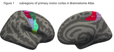 |
131 |
Different Cortical Thinning Pattern in Primary Motor Cortex Correspond to Clinical Characteristics of Amyotrophic Lateral Sclerosis Subtypes
Video Permission Withheld
Haining Li, Qiuli Zhang, Qianqian Duan, Jingmei Xie, Ming Zhang
Heterogeneity of motor phenotypes is a clinically well-recognized fundamental aspect of amyotrophic lateral sclerosis (ALS).The body region of onset is one of independent primary attributes of ALS motor phenotype heterogeneity. In order to investigate the patterns of brain atrophy between ALS patients with bulbar and limb onset and analyse its correlation with clinical characteristics , cortical thickness analyses were performed. ALS Patients with limb onset revealed the majority of significant cortical thinning in the limb segment of the motor cortex, and patients with bulbar onset, in the bulbar segments. The findings suggest that neuroimaging could be a helpful objective measure to estimate of upper motor neuron loss.
|
|
2779.
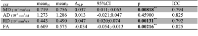 |
132 |
Comparative Corticospinal Tract Relaxation and Diffusion MRI Measures in Amyotrophic Lateral Sclerosis
Hagen Kitzler, Carolin Schwamborn, Paul Kuntke, Hannes Wahl, Rene Guenther, Sean Deoni, Andreas Hermann, Jennifer Linn
Amyotrophic Lateral Sclerosis (ALS) is a fatal disease that primarily affects the human primary motor system. Selective neurodegeneration leads to systemic functional motor decay. We aimed to understand the relationship between cortical degeneration and the desintegration of the related motor corticospinal tract (CST) by applying both Diffusion Tensor Imaging (DTI) and the multi-component driven equilibrium single pulse observation of T1 and T2 (mcDESPOT). We found early changes in diffusion and relaxation measures indicating WM tract degeneration secondary to cortical neurodegeneration. Besides the loss of structural integrity early alterations of the myelin characteristics indicate toward changes of its compositional condition instead of early myelin loss.
|
|
2780.
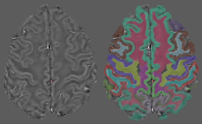 |
133 |
Fully-automatic quantitative susceptibility mapping of the precentral gyrus in motor neuron disease
Valeria Contarino, Giorgio Conte, Claudia Morelli, Sonia Calloni, Luis Sanmiguel Serpa, Elisa Scola, Francesca Trogu, Vincenzo Silani, Fabio Triulzi
The diagnosis of Motor Neuron Disease (MND) is a long process that involves careful clinical and neurological examination during a long period of time. As iron overload is recognized as one of the main pathogenic mechanisms, previous studies focused on hand-drawn ROI-based measures of susceptibility in the precentral gyrus in MND. In contrast to the manually drawn ROIs approach guided by pathology localization and lateralization, this study suggests that the building of a MND biomarker might rely on susceptibility properties of the precentral gyrus measured on clinical images with a fully-automatic pipeline.
|
|
2781
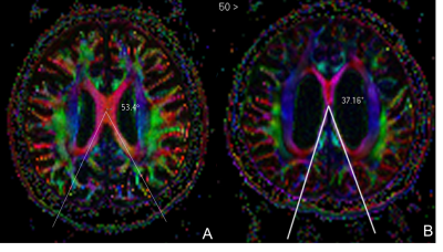 |
134 |
The Splenial Angle: A Novel Index in Idiopathic Normal Pressure Hydrocephalus
Video Permission Withheld
Ling Ling Chan, Robert Chen, Wei Ying Go, Huihua Li, Soo Lee Lim, Sumeet Kumar, Nicole Keong
The callosal angle (CA) is a useful tool in idiopathic normal pressure hydrocephalus (iNPH) for diagnosis and patient selection for shunt surgery. We evaluated for (1) differences in a novel splenial angle (SA) in iNPH compared to healthy controls (HC), and (2) temporal changes in SA, CA and Evan’s index in shunted and non-shunted iNPH patients. Significant differences (p<0.0001) existed in the EI, CA and SA between iNPH and HC. Amongst iNPH patients with or without shunting, significant temporal changes were also found in in all indices on follow-up MRI scans compared to baseline measurements.
|
|
2782.
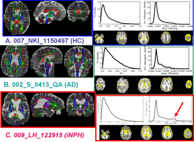 |
135 |
Diffusion Tensor Imaging of the Superior Thalamic Radiation and Cerebrospinal Fluid Distribution in Idiopathic Normal Pressure Hydrocephalus
Khader Hasan, Kyan Younes, Arash Kamali, Zafer Keser, Pejman Rabiei, Christine McGough, Omar Hasan, Tomas Melicher, Larry Kramer, Paul Schulz
Ventricular enlargement in elderly raises a challenging differential diagnosis to physicians. While Alzheimer`s disease is the most common form of dementia, idiopathic Normal Pressure Hydrocephalus (iNPH) constitutes a potentially reversible syndrome. iNPH has a unique pathophysiology pertaining to cerebrospinal fluid dynamics and periventricular white matter. We aimed to determine the effects of iNPH on periventricular white matter bundles and to further characterize its ventricular and sulcal CSF distribution by using diffusion tensor tractography (DTT) and cerebrospinal fluid (CSF) volumetrics on high resolution T1-weighted MRI data.
|
|
2783.
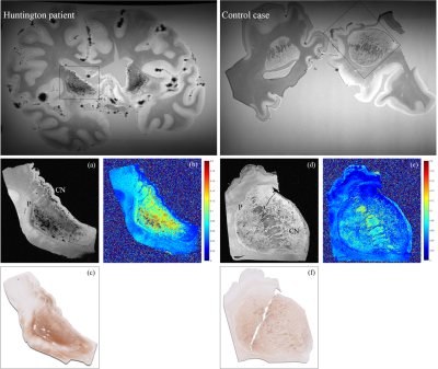 |
136 |
Detection of accumulated iron and microglia in the striatum of Huntington’s Disease patients: evidence from post-mortem MRI and histology
Marjolein Bulk, Ernst Suidgeest, Ingrid Hegeman-Kleinn, Sjoerd van Duinen, Jan Lewerenz, Bernhard Landwehrmeyer, Itamar Ronen, Louise van der Weerd
We detected increased iron deposits in the striatum of post-mortem brain tissue of Huntington’s Disease (HD) patients. High-field T2*-weighted MRI of the striatum showed a different imaging phenotype in HD patients compared to controls, and spatially correlated with the iron distribution obtained from histology. Increased iron was observed in the matrix and in cells morphologically resembling glial cells. These findings bridge the gap between neuropathological and clinical imaging findings and point to iron accumulation as a potential imaging biomarker for disease progression in vivo, possibly reflecting neuroinflammation.
|
|
2784.
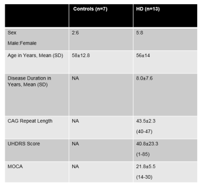 |
137 |
Investigating Microglial Activation and white matter changes in Huntington Disease Patients
wafaa sweidan, navid seraji bozorgzad, edwin george, fen bao, rachel darling
This study will investigate the effects of Huntington disease (HD) progression on white matter microstructure and microglial activation using diffusion magnetic resonance imaging and positron emission tomography in HD patients over the course of 6 months (baseline and 6 month visit). Age-matched healthy controls will be followed up similarly. Baseline differences between HD and healthy controls will likely reflect effect of HD pathology on white matter tracts and assessing longitudinal changes accompanied by disease progression will reflect the temporal and spatial changes.
|
|
2785.
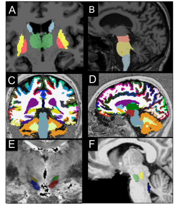 |
138 |
Progressive Supranuclear Palsy versus Parkinson’s disease-related damage using quantitative multimodal MRI
Nadya Pyatigorskaya, Lydia Yahia-cherif, Rahul Gaurav, Claire Ewenczyk, Cecile Gallea, Romain Valabregue, Fatma Gargouri, Eric Bardinet, Cyril Poupon, Marie Vidailhet, Stephane Lehericy
Our primary objective was to generate a precise in vivo model of neurodegeneration of brainstem nuclei, cerebellum, basal ganglia, basal forebrain, and cortex using multimodal MRI in progressive supranuclear palsy (PSP). Secondary objective was to use multimodal imaging biomarkers to efficiently differentiate PSP from Parkinson disease (PD) patients and healthy control subjects (HC). Multiple factorial analyses of the regional damage allowed to efficiently differentiate PSP from HC and PD, in agreement with previous pathological studies. These results suggest the possibility of direct non-invasive assessment of brain damage at multiple level of the central nervous system in PSP and efficient multimodal multiregional based differential diagnosis between PSP and PD patients
|
|
2786.
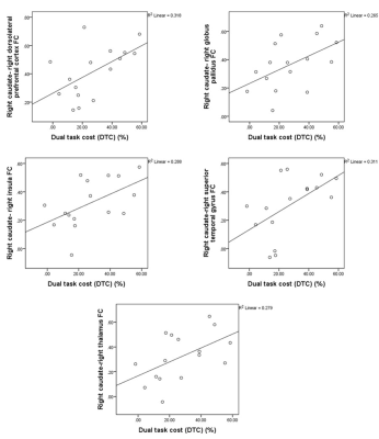 |
139 |
Associations Between Dual Task Cost and Striatal Functional Connectivity in Parkinson’s Disease with Mild Cognitive Impairment
Ece Bayram, Karthik Sreenivasan, Jason Longhurst, Sarah Banks, Zhengshi Yang, Xiaowei Zhuang, Dietmar Cordes, Aaron Ritter, Jessica Caldwell, Brent Bluett, Virendra Mishra
This study investigated striatal functional connectivity correlates of dual-tasking in Parkinson’s disease with and without mild cognitive impairment using resting state fMRI. Increased caudate functional connectivity with frontotemporal, insular and subcortical regions were associated with increased dual task cost in Parkinson’s disease with mild cognitive impairment, whereas correlations were only seen in increased putamen and supplementary motor area functional connectivity and increased dual task cost in PD participants. These results reveal that dual tasking is associated with different striatal functional connectivity patterns in PD participants, with and without, cognitive impairment suggesting compensatory mechanisms in Parkinson’s disease with mild cognitive impairment.
|
|
2787.
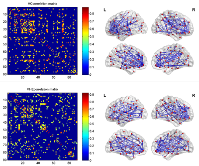 |
140 |
Altered topological properties of gray matter structural covariance networks inminimal hepatic encephalopathy
Presentation Not Submitted
Tian-Xiu Zou, Hua-Jun Chen
Despite the abnormal structural findings in cirrhotic patients with MHE, previous studies have only focused on regional structural changes in cirrhosis and did not consider brain network-level architecture. Recent progress in structural MRI analysis has facilitated the development of a human brain structural network model that is based on statistical correlations of morphological descriptors, including the thickness of the cortex or regional gray matter volume (RGMV). We investigated the topological alterations involving brain structural covariance networks in MHE patients for the first time. Our results suggest that MHE patients exhibit an unoptimizable architecture involving the gray matter structural covariance network and provide structural evidence supporting that MHE is a neurological complication related to disrupted neural networks.
|
|
2788.
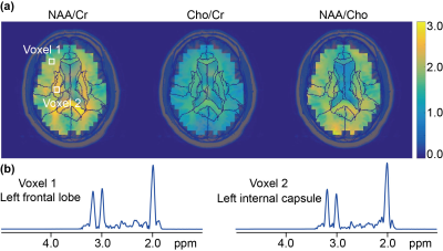 |
141 |
Factor analysis of atlas-segmented brain MRSI data in HIV infection
Liangjie Lin, Kalpana Kallianpur, Cecilia Shikuma, Andreia Faria, Hubert Liu, Anna Wang, Zhong Chen, Peter Barker
HIV-infection may cause HIV-associated neurocognitive disorder. Here, factor analysis is applied to atlas-segmented brain MRSI data from a cohort of HIV-positive subjects to evaluate the relationships of MRSI measures with neuropsychological test performance and immunologic markers. Results indicate that distribution of NAA in right-hemisphere brain regions of basal ganglia, thalamus, etc. may be positively correlated with CD4 counts, and distribution of Cho in both hemispheres of similar regions positively correlated with CD8 counts in HIV-infected subjects. Higher neuropsychological z-scores tends to be associated with higher NAA and/or lower Cho distributions in specific brain regions.
|
|
2789.
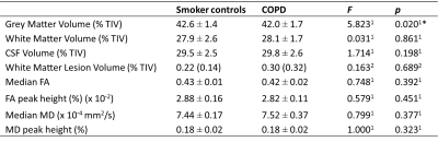 |
142 |
Contributions of cardiovascular risk and smoking to chronic obstructive pulmonary disease (COPD)-related changes in brain structure and function
Catherine Spilling, Mohani-Preet Bajaj, Daniel Burrage, Sachelle Ruickbie, Jade Thai, Emma Baker, Paul Jones, Thomas Barrick, James Dodd
Structural and functional brain abnormalities have been reported in chronic obstructive pulmonary disease (COPD), however, it is unclear whether these occur independently of cardiovascular risk. Neuroimaging and clinical markers of brain structure and function were compared between 27 COPD patients and 23 age-matched non-COPD smoker controls. Clinical relationships and group interactions with brain structure were tested. COPD patients showed a specific pattern of structural (lower grey matter volume) and functional (lower cognitive function and psychological status) brain abnormalities that could not be explained by cardiovascular risk. Lower lung function and psychological ill-health were associated with markers of white matter damage.
|
|
2790.
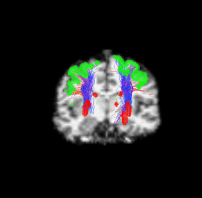 |
143 |
Microstructural changes of the cortico-striatal pathway in Fabry disease: a diffusion MRI connectometry study
Matteo Battocchio, Sirio Cocozza, Simona Schiavi, Giuseppe Pontillo, Camilla Russo, Antonio Pisani, Alessandro Daducci, Arturo Brunetti
Aim of this study was to investigate the presence of microstructural alterations along the cortico-striatal pathway of Fabry Disease (FD) patients. Mean Fractional Anisotropy (FA) values of bundles connecting the motor cortex with the striatum were extracted from 15 FD patients and 14 controls.We found a reduction of mean FA values, alongbundles of the cortico-striatal pathway in FD patients compared to HC (p=0.04 and p=0.01 for the right and left side, respectively).Our preliminary results confirm the presence of an extrapyramidal involvement in FD patients, showing the presence of microstructural changes affecting the cortico-striatal pathway in this condition.
|
|
2791.
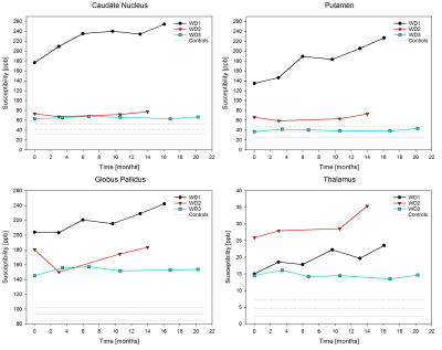 |
144 |
Longitudinal monitoring of the cerebral iron load in de novo neurological Wilson disease
Monika Dezortova, Petr Dusek, Artem Lescinskij, Julio Acosta-Cabronero, Radan Bruha, Milan Hajek
We report a long-term study of three de novo diagnosed Wilson disease patients with neurological form who repeatedly underwent magnetic resonance imaging and neurological examinations for 2 years after treatment initiation. The quantitative measurement of susceptibility revealed higher values in basal ganglia and thalamus compared to controls which correspond to higher iron accumulation. Degree of iron load reflected the clinical severity of neurological impairment. Thus, we can suppose that the increase of the brain iron concentration can be a marker of suboptimal response to anti-copper therapy and unfavorable outcome.
|
|
2792.
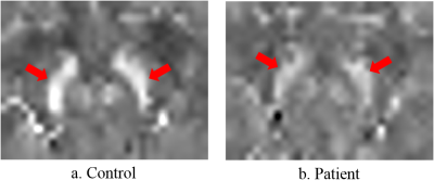 |
145 |
Quantitative Susceptibility Mapping shows differences in substantia nigra of individuals with 22q11.2 deletion syndrome and controls
Nestor Muñoz , Marisleydis García, Carlos Milovic, Manuel Chapa, Cristian Montalba, Julio Acosta-Cabronero, Sergio Uribe, Marcelo Andia, Gabriela Repetto, Analía Cuiza, Cristian Tejos, Nicolás Crossley
Unlike individuals with Parkinson’s disease, patients with 22q11.2 deletion syndrome at risk of Parkinson show an increase in dopamine at striatal regions. Since iron levels are related to dopamine levels, we studied the difference of magnetic susceptibility between 17 patients with the deletion and 19 healthy individuals. Susceptibility measurements were obtained with QSM and then compared using a Mann Whitney U test. Results showed a significant difference in the substantia nigra, which indicates a possible cause for the increased levels of dopamine in 22q11.2 individuals at Parkinson’s risk.
|
|
2793.
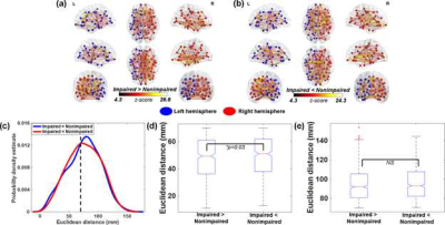 |
146 |
Disrupted topological brain organizations in large-scale cortical networks between impaired and nonimpaired active fighters
Virendra Mishra, Karthik Sreenivasan, Xiaowei Zhuang, Zhengshi Yang, Sarah Banks, Dietmar Cordes, Charles Bernick
Using neuropsychological scores from the Professional Fighters Brain Health Study (PFBHS), this study first identified 70 cognitively impaired active professional fighters, and then matched 70 nonimpaired fighters but matched on demographics, and other fighting criteria. This study shows that repeated head trauma is associated with altered coordination of large-scale structural brain networks, especially in the long-range connections. Furthermore, the cortical thickness of regions identified as hubs has the potential of developing into a predictive biomarker for identifying the fighters that will develop cognitive decline due to repeated head trauma.
|
|
2794.
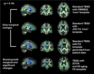 |
147 |
Improved sensitivity to longitudinal changes with advanced DTI analysis in a rare neurodegenerative disease
Young Woo Park, James Joers, Diane Hutter, Khalaf Bushara, Gulin Oz, Christophe Lenglet
We present an optimized processing pipeline for longitudinal DTI data analysis in spinocerebellar ataxia type 1 (SCA1) patients, a rare neurodegenerative disease. Tract-based spatial statistics (TBSS) analysis was used to investigate longitudinal changes in white matter (WM) integrity in patients. While no significant changes in WM integrity were observed using the standard TBSS analysis pipeline, TBSS with advanced spatial normalization with DTITK tool showed significant longitudinal alterations in WM integrity. This result suggests that the use of advanced spatial normalization must be considered for longitudinal group studies of DTI data, especially when small to moderate disease effects are expected.
|
|
2795.
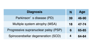 |
148 |
Chemical exchange saturation transfer imaging depending on several neurodegenerative diseases at 3T
Yuki Matsumoto, Masafumi Harada, Yuki Kanazawa, Takashi Abe, Maki Otomo, Mitsuharu Miyoshi
In this study, CEST imaging was performed on the substantial nigra, the basal ganglia, and cerebral cortex level to reveal the mechanism each neurodegenerative disease. For this study, patients with Parkinson’s disease, multiple system atrophy, progressive supranuclear palsy, and spinocerebellar degeneration were examined. Region-of-interest analysis was performed in the substantia nigra, red nuclei, lentiform nucleus, and supplementary motor area. As the results, the CEST parameters were significantly different for each of the neurodegenerative diseases. CEST imaging might have the ability to obtain abnormal proteins each of the neurodegenerative diseases.
|
|
2796.
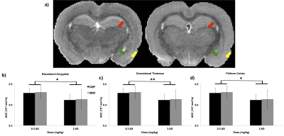 |
149 |
Evaluating Countermeasures Against Chemical Warfare Nerve Agents using Diffusion Tensor Imaging
Kevin Lee, Matthew Bouchard, Sara Bohnert, Steven Klaassen, Roland van den Berg, Marloes Joosen, Jeff Dunn
Seizures induced by chemical warfare nerve agents cause debilitating neurological damage. It is widely accepted that the main contributor to this neuropathology is excitotoxic damage. Current countermeasures to the damage are effective in preventing mortality, but do not always prevent nerve agent-induced seizures and related neuropathology. We have applied diffusion tensor imaging to study the microstructural changes in the brain to evaluate the efficacy of countermeasures against chemical warfare nerve agents.
|
|
| Top |
Neuroimaging: Flying High at 7T & Beyond
Digital Poster
Neuro
Tuesday, 14 May 2019
| Exhibition Hall |
13:30 - 14:30 |
| |
|
Computer # |
 |
2797.
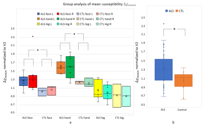 |
151 |
Quantitative susceptibility mapping of post-mortem ALS brains at 7T with quantitative iron histopathology validation
Chaoyue Wang, Benjamin Tendler, Menuka Pallebage-Gamarallage, Olaf Ansorge, Sarah Bangerter-Christensen, Ricarda Menke, Martin Turner, Sean Foxley, Karla Miller
Amyotrophic lateral sclerosis (ALS) is a fatal neurodegenerative disease of the motor system and its wider cortical connections. Progress in therapeutic development in ALS is compromised by a lack of specific biomarkers. In this work, we describe a platform for QSM data acquisition and post-processing protocol for postmortem brains. Preliminary results of 10 brains (validated with quantitative ferritin staining) have shown that ALS brains had significant higher mean susceptibility in motor cortex than control brains, which indicates that QSM has the potential to accurately quantify iron concentration and thus serve as an imaging biomarker for ALS.
|
|
2798.
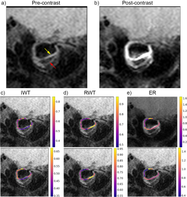 |
152 |
Quantitative analysis of the wall thickness and enhancement ratio of intracranial aneurysms using high-resolution black-blood imaging at 7T
Zihao Zhang, Qingle Kong, Xinke Liu, Chengcheng Zhu, Zhaoyang Fan, Jing An, Youxiang Li, Yan Zhuo
Three-dimensional turbo-spin-echo (3D-TSE) is increasingly being used in black-blood imaging of intracranial aneurysms. In this study, we optimized the protocol of T1-weighted (T1w) 3D-TSE to reach a high isotropic resolution of 0.40 mm. The inner wall thickness and enhancement ratio were analyzed in sections from ten aneurysms. The segments of aneurysmal walls with a higher wall thickness tend to represent a higher enhancement ratio (Pearson correlation, r = 0.32, p < 0.001). The wall thickness and enhancement ratio should be comprehensively considered to predict the prognosis of intracranial aneurysms.
|
|
2799.
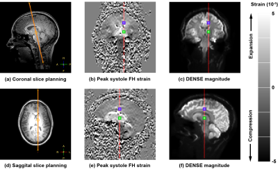 |
153 |
Cardiac and Respiratory induced 3D Brain Tissue Strain as Marker of Physiological Blood Volume Dynamics at 7T MRI.
Jacob Jan Sloots, Peter Luijten, Geert Jan Biessels, Jaco Zwanenburg
Brain tissue deformation induced by the cardiac and respiration cycles could be a valuable source of information on the physiology of the brains tissue properties. In this work, we assess the tissue deformation by computing the tissue strain from DENSE displacement data sets and unravel cardiac and respiratory contributions by using a linear model. We observed consistent trends in the three strain components due to cardiac and respiration cycles, which agree with blood volume changes. In contrast to tissue displacement, the tissue strain may serve as a reliable novel marker of physiological blood volume dynamics in the brain.
|
|
2800.
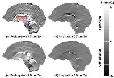 |
154 |
Optimizing the DENSE Sequence for Accurate Brain Tissue Strain Measurements at 7T MRI.
Jacob Jan Sloots, Peter Luijten, Geert Jan Biessels, Jaco Zwanenburg
Brain tissue strain could be a valuable source of information on the brains tissue properties. Therefore, accurate DENSE measurements are crucial, since the computation of tissue strain requires spatial derivatives, which amplifies noise present in the displacement maps. In this work, we optimize the SNR in the displacement maps and substantiate the theory with both computer simulations and measurements. We tested the optimized settings in one volunteer and found a factor of 1.66 SNR increase compared to previously reported experiments. Preliminary results in one volunteer in the basal ganglia showed heartbeat-induced strain of approximately 2.1·10-3 and inspiration-induced strain of -0.54·10-3.
|
|
2801.
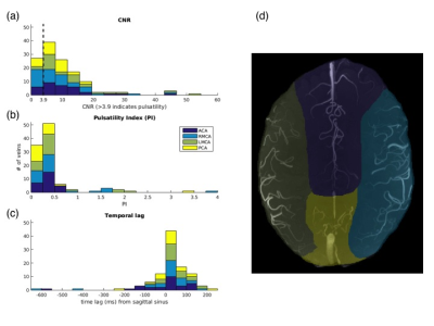 |
155 |
Most small cerebral cortical veins demonstrate significant flow pulsatility: a human phase contrast MRI study at 7T
Ian Driver, Maarika Traat, Richard Wise
We demonstrate new methods to identify and quantify the characteristics of flow pulsatility in small cerebral cortical veins to aid better understanding of the haemodynamics of this little-studied vascular compartment. 7T cardiac-gated motion sensitive phase contrast MRI was combined with an automated method for establishing where venous flow is pulsatile, revealing pulsatile flow in 104 out of 132 veins assessed in parietal and frontal regions. Distributions of pulsatility index and pulse waveform delay were characterized, indicating a small delay in cortical veins compared to the superior sagittal sinus, but no differences between veins draining different arterial supply territories.
|
|
2802.
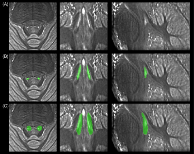 |
156 |
Multi-modal 7T imaging of the Locus Coeruleus in healthy older adults
Catarina Rua, Claire O'Callaghan, Luca Passamonti, P Simon Jones, Kamen Tsvetanov, James Rowe, Rong Ye, Adrian Carpenter, Christopher Rodgers, Guy Williams
The Locus Coeruleus (LC) is the main source of cerebral noradrenaline, which modulates many cognitive domains from attention and motivation to mood and memory. However, the LC is a small structure located in the mid-brain, proven difficult to detect in-vivo. We studied healthy individuals using high-resolution MT-w, R2*, QSM, and fMRI at 7T to characterize the LC’s shape and size, magneto-chemical and functional properties. While no R2* or QSM contrast was found in the LC, it was clearly detected in MT-images and showed widespread functional connectivity towards cortex and cerebellum, These results are the benchmark for future studies in dementia.
|
|
2803.
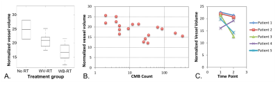 |
157 |
Global and Focal Effects of Radiation Therapy on the Cerebral Vasculature in Pediatric Brain Tumor Survivors using simultaneous MRA-SWI at 7T
Sivakami Avadiappan, Melanie Morrison, Angela Jakary, Erin Felton, Schuyler Stoller, Christopher Hess, Sabine Mueller, Janine Lupo
With the improved survival of children with brain tumors, understanding the late effects of the treatment has become critical. This study explores the effects of RT on vascular structure using a combined MRA-SWI sequence at 7T and a new method for arterial segmentation and quantification. Normalized arterial volume was significantly reduced with increasing RT treatment volume, number of CMBs, and at follow-up. CMBs were located closer to veins than arteries and were larger when further away. Our findings demonstrate the feasibility of our approach for quantifying subtle vascular changes in arterial structure and CMB properties due to RT.
|
|
2804
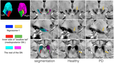 |
158 |
Investigation of microstructural differences in the nigrosome-1 region of the substantia nigra between healthy and Parkinson’s disease subjects at 7T
Video Permission Withheld
Yiming Xiao, Jonathan Lau, Terry Peters, Ali Khan
The loss of hyperintense signals of the nigrosome-1 region within the substantia nigra on T2*w MRI has previously been investigated as a potential biomarker for Parkinson’s disease (PD). Although the radiological observation is expected to be induced by microstructural changes, which can provide more insights into the mechanisms of PD, no relevant MRI studies have been conducted so far. With ultra-high-field 7T MRI, we compared the microstructural features of the nigrosome-1 regions between healthy and PD subjects using quantitative MRI, and revealed alterations in T1, R2*, mode of anisotropy, and fractional anisotropy within this subregion due to the disease.
|
|
2805.
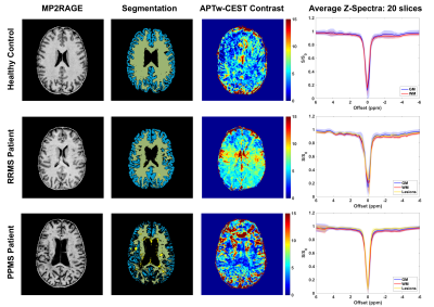 |
159 |
Longitudinal Study of APT-weighted CEST in the Brain at 7T in Patients with Multiple Sclerosis
Kristin O'Grady, Seth Smith, Sanjana Satish, Nirbhay Yadav, Margaret Spini, Seongjin Choi, Daniel Harrison
Amide proton transfer weighted chemical exchange saturation transfer (APTw-CEST) MRI has been applied with whole-brain coverage in a longitudinal study in patients with relapsing-remitting, secondary progressive, and primary progressive multiple sclerosis (MS). APTw-CEST is sensitive to amide protons on proteins and peptides and may provide insight into the pathological processes underlying symptoms and disease progression in MS. Our preliminary results show increased APTw-CEST contrast in relapsing-remitting MS patients and decreased contrast in primary progressive MS patients, as well as differences in CEST contrast in lesions for these disease subtypes.
|
|
2806.
 |
160 |
Volumetric Brain Analysis in Older HIV-positive and -negative Subjects using 7T MRI
Mona Mohamed, Richard Skolasky, Andreia Faria, Peter Barker, Ned Sacktor
Structural brain MRI is often performed to evaluate regional brain structure and atrophy in elderly subjects with cognitive impairment. In this study, 7T MRI was applied to 62 individuals 50 years and older (40 HIV+); an age-appropriate adult brain atlas was then used to segment and compare regional brain volumes as a function of age, HIV serostatus and neurocognitive classification.
|
|
2807.
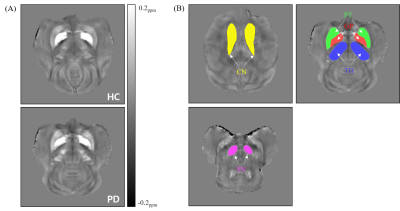 |
161 |
Observation of Iron Deposition in Cynomolgus Monkeys with the Parkinson’s Disease using Quantitative Susceptibility Mapping at 7T MRI
Sangwoo Kim, Sukhoon Oh, Youngjeon Lee, Chang-Yeop Jeon, Philyong Kang, Keunil Kim, Youngjae Jeon, Jincheol Seo, Seung Ho Baek, Yeung Bae Jin, Chulhyun Lee
The anatomic sensitivity of iron deposition in the Parkinson’s disease (PD) study remains controversial with exception of the substantia nigra (SN). A 14-year-old PD-modeled cynomolgus monkey and an age-matched healthy control were scanned at 7T MRI. The average susceptibility ratio of the caudate nucleus, putamen, thalamus (TH) and SN were increased approximately 21~76 %. We found significantly increased susceptibility values in the SN pars compacta (SNc) and TH. In addition to the high-sensitivity of the SNc in PD, we have also seen that the TH could be a diagnostic biomarker of PD in terms of the movement disorder.
|
|
2808.
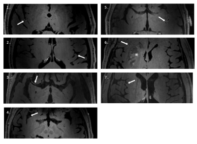 |
162 |
Endovascular thrombectomy using stentretrievers affects the vessel wall: impact demonstrated by wall enhancement at 7T
My Truong, Karin Markenroth Bloch, Mads Andersen, Gunnar Andsberg, Johannes Töger, Johan Wasselius
Stroke is increasingly treated by removing the vessel obstruction via endovascular thrombectomy. 3T imaging has shown wall enhancement after endovascular thrombectomy, and we hypothesize that 7T MR will be beneficial to visualize this enhancement as the higher field strength allows for higher resolution imaging. Seven patients were examined at 7T within two days after endovascular thrombectomy. Vessel wall images pre- and post-contrast administration were reviewed by two neuroadiologists, who rated the image quality as excellent in all cases. In all patients, wall enhancement was found, and correlated to the site of the stent-retriever rather than the site of the embolus.
|
|
2809.
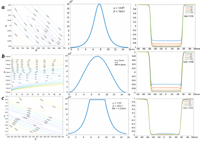 |
163 |
Optimization of adiabatic pulses for Pulsed Arterial Spin Labeling (pASL) at 7T
Kai Wang, Jin Jin, Lirong Yan, Danny Wang
The goal of this work was to optimize and evaluate three types of adiabatic pulses for pulsed ASL (PASL) at 7T including Hyperbolic Secant (HS)[1], WURST[2] and FOCI[3] pulses using theoretical simulation, phantom and in vivo scans. WURST has the lowest loss in simulation and outperforms HS/FOCI in experiments. The optimized WURST pulse with the maximally allowed B1 is preferred to HS and FOCI pulses for adiabatic inversion in 7T PASL.
|
|
2810.
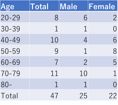 |
164 |
Age-related changes in tissue T1, T2 values of the human brain quantified by 2D PnP-MRF at 7T MRI
Koji Fujimoto, Martijn Cloos, Atsushi Shima, Dinh Ha Duy Thuy, Nobukatsu Sawamoto, Ryosuke Takahashi, Tadashi Isa, Tomohisa Okada
To investigate age-related changes in T1 and T2 of brain tissues at 7T, healthy volunteers (N=45, age range 20-81) were scanned using a 1ch-Tx/32ch-Rx coil and a Plug-and-Play MR Fingeprinting (prototype) sequence. Six brain regions (putamen, globus pallidus, caudate head, thalamus, frontal white matter (WM) and frontal gray matter (GM)) were evaluated. Linear and quadratic curve fitting was performed. Significant linear correlation in T2 was found for the putamen and frontal WM and a quadratic correlation was found for the putamen (T2), globus pallidus (T2), thalamus (T1) and frontal WM (T1/T2).
|
|
2811.
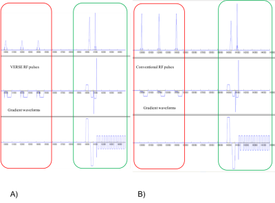 |
165 |
Reduction of power deposition using VERSE RF pulses to decrease acquisition time in pulsed ASL sequence at high field.
Sagar Buch, Omer Oran, Ravi Menon, Hacene Serrai
The use of Arterial spin labelling at high field, allow for a gain in sensitivity in brain perfusion images. However, this necessitates an increase in acquisition time due to SAR limits. To address this problem, the conventional selective RF pulses used for saturation of the blood in the PICORE Q2TIPS tagging technique of the Pulsed ASL sequence were replaced by their corresponding VERSE RF pulses, to achieve the same task with less power. As a result, perfusion images were obtained in a shorter acquisition time allowing for an increase of both the number of acquisitions and SNR.
|
|
2812.
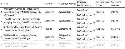 |
166 |
Inter-site, inter-subject and inter-session variability of B1+ and B0 in the human brain at 7 tesla.
William Clarke, Olivier Mougin, Ian Driver, Catarina Rua, Andrew Morgan, Susan Francis, Richard Bowtell, Richard Wise, Adrian Carpenter, Christopher Rodgers, Keith Muir, Stuart Clare
At ultra-high field strengths, e.g. 7 tesla, the homogeneity of the transmit B 1+-field and the B 0-field decreases compared to lower fields. This can strongly affect the contrast in images especially when imaging large volumes, such as the whole brain.
In this work we measure the mean and variance of B1+- and B0-fields in whole brain images acquired on four, whole-body human 7T scanners. We demonstrate that inter-subject differences dominate over inter-site differences in explaining the observed B0- and B1+-field variability. Inter-session differences are not significant.
|
|
2813.
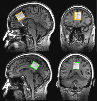 |
167 |
7T MRS of neurometabolites and associations with cognitive deficits in mild cognitive impairment
Georg Oeltzschner, S. Andrea Wijtenburg, Mark Mikkelsen, Richard Edden, Peter Barker, Jin Joo, Jeannie-Marie Leoutsakos, Laura Rowland, Clifford Workman, Gwenn Smith
Mild cognitive impairment (MCI) is associated with increased risk of developing Alzheimer’s Disease (AD). Here, levels of multiple brain metabolites in healthy controls and MCI patients using 7T magnetic resonance spectroscopy in the anterior (ACC) and posterior (PCC) cingulate cortex were studied. MCI patients showed decreased GABA/tCr (ACC, PCC), Glu/tCr (PCC), NAA/tCr (PCC), and increased mI/tCr (ACC). Worse episodic verbal memory performance correlated with lower Glu/tCr (PCC), lower NAA/tCr (PCC), and higher mI/tCr (ACC, PCC). Worse verbal fluency performance correlated with lower GSH/tCr (PCC). In summary, MCI is associated with decreased GABA and glutamate, most consistently in the PCC.
|
|
2814.
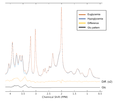 |
168 |
Monitoring the neurotransmitter response to hypoglycemia in the prefrontal cortex in diabetes using an advanced MRS protocol at 7T
Young Woo Park, Dinesh Deelchand, James Joers, Elizebeth Seaquist, Gulin Oz
Studies of neurotransmitter response to changing glycemia levels are needed to address mechanisms of cerebral adaptations to hypoglycemia. Here we present an advanced MRS acquisition protocol that combines commercial 7T hardware, dielectric padding, optical motion tracking and frequency and B0 shim correction to ensure optimal stability of data quality during a challenging infusion protocol to maintain euglycemic and hypoglycemic blood glucose levels. Data obtained from the prefrontal cortex in patients with type 1 diabetes demonstrate excellent quality and reproducibility during euglycemia and hypoglycemia, which will allow robust mechanistic investigations of the primary neurotransmitters, glutamate and GABA, under changing glycemic conditions.
|
|
2815.
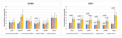 |
169 |
GABA and GSH at 7T: to edit or not to edit?
Anouk Marsman, Anna Lind, Esben Petersen
With the number of 7T sites increasing, more brain-MRS studies will be conducted at 7T. Particularly GABA and GSH are of interest and benefit from the increased resolution. To date, studies have applied both conventional and edited MRS to measure these metabolites, however, there is no consensus on whether to use edited MRS. This study shows that it depends on brain region and potentially voxel size and number of acquisitions, whether it is preferable to use editing for GABA. There is less reason to argue that editing benefits measurement of GSH, but this study indicates a slight preference for editing.
|
|
2816.
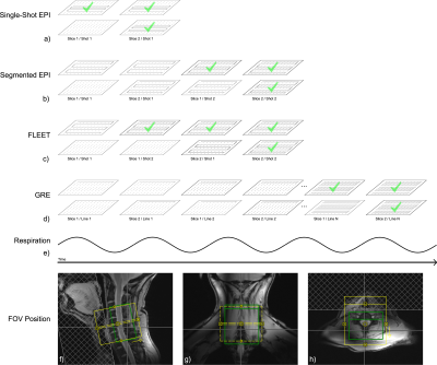 |
170 |
Impact of Autocalibration Method on Accelerated Echo-Planar Imaging of the Cervical Spinal Cord at 7 T
Alan Seifert, Junqian Xu
Respiration-induced B0 fluctuations are significantly greater in the cervical spinal cord than in the brain at 7T, increasing k-space phase inconsistencies and necessitating a separate evaluation of autocalibration scan (ACS) methods for accelerated EPI. We tested four ACS methods (single-shot EPI, segmented EPI, FLEET, and GRE) under three physiological conditions (end-expiration breath-hold, free-breathing, and intentional swallowing). GRE and single-shot EPI ACS methods, which are robust to respiration-induced phase errors between k-space segments, produce images with fewer and less severe artifacts than either FLEET or conventionally segmented EPI ACS methods for accelerated EPI of the cervical spinal cord at 7T.
|
|
2817.
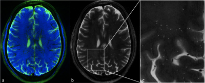 |
171 |
Comparison of Perivenular Perivascular Spaces at 7 Tesla in Relapsing-Remitting Multiple Sclerosis Patients and Healthy Controls
Annie Arrighi-Allisan, Ilena George, John Rutland, Gaurav Verma, Bradley Delman, Priti Balchandani, Sam Horng, Rebecca Feldman
Multiple sclerosis (MS) is a CNS autoimmune disease of unknown etiology. Prior MS studies have characterized an inflammatory process involving the perivascular spaces (PVSs) of a central vein, but have reported conflicting results regarding which aspects of PVS anatomy correlate to clinical aspects of the disease. This preliminary study compared percentages of manually marked perivenular and non-perivenular PVSs in relapsing-remitting MS patients and healthy controls, and detected a higher rate of vein-coincident PVSs in the former group. We posit that perivenular PVSs have potential as a candidate imaging marker for MS diagnosis, disease activity, and treatment efficacy.
|
|
2818.
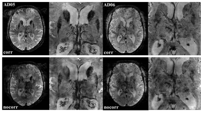 |
172 |
Improvement of R2* mapping in Alzheimer’s Disease at 9.4T through reduction of B0 fluctuation-induced image artifacts using Navigator Echoes
Elisa Tuzzi, Gisela Hagberg, Alexander Loktyushin , Rolf Pohmann, Anja Zeller, Christoph Laske , Klaus Scheffler
Ultra-High-Field (UHF) MRI provides a potential non-invasive means to investigate Alzheimer’s Disease (AD). R2* map is one of the most established MRI techniques to detect iron concentration in the brain. However, image quality can be substantially affected by B0 fluctuation-related-artifacts at UHF. In this study we showed an improvement of the quality of R2* maps in AD at 9.4T using Navigator Echos approach.
|
|
2819.
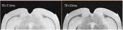 |
173 |
High Resolution MR Imaging of Amyloid Deposition in 5xFAD Mice Brains at 21.1 T
David Hike, Tara Palin, Scott Boebinger, Samuel Grant
This study utilizes high resolution 3D images acquired at 21.1 T to display the presence of amyloid plaques, which temporally correlate with the progression of structural connectivity alterations in this transgenic preclinical model. As a function of phenotype, age and gender, high resolution scans were performed on APP/PS1 mouse brains and age-matched wild-type controls. Data indicate that plaques may be visible in mice as young as 4 months without the use of any magnetic contrast agents.
|
|
2820.
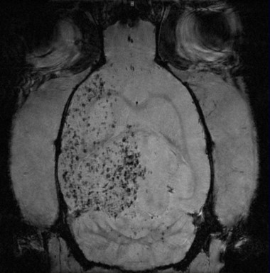 |
174 |
Quantitative Assessment of Treatment Efficacy in Acute Ischemia using Perfusion and Diffusion Weighted Imaging at 21.1-T
F. Bagdasarian, Shannon Helsper, Xuegang Yuan, Jens Rosenberg, Nastaren Abad, Teng Ma, Samuel Grant
This study employs perfusion and diffusion weighted imaging (PDWI) at 21.1 T to identify the pattern of potential recovery of cerebral blood flow (CBF) and apparent diffusion coefficient (ADC) in ischemic stroke with a novel stem cell therapy using dissociated aggregate human mesenchymal stem cells (d-hMSC) in a rodent stroke model. Spanning a week post ischemia, high field MRI was performed with the goal of quantitatively assessing treatment efficacy longitudinally. Results show longer term increase of ADC in the ischemic region and shorter term CBF stabilization in the contralateral region.
|
|
2821.
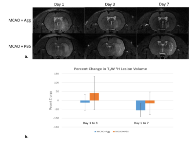 |
175 |
Sodium MRI at 21.1 T to Assess Stroke Recovery Following Adult Human Mesenchymal Cell Transplantation
Shannon Helsper, F. Bagdasarian, Xuegang Yuan, Jens Rosenberg, Jea-Young Lee, Kaya Xu, Teng Ma, Cesar Borlongan, Samuel Grant
This study evaluates recovery following transplantation of human mesenchymal stem cell (hMSC) aggregates labeled with micron-sized particles of iron oxide (MPIO) in a rodent model of ischemic stroke at 21.1 T. Unlike previous studies using single cells, intact aggregates were implanted directly into the contralateral ventricle. Longitudinal MRI post-implantation was used to investigate T2-weighted lesion volume and sodium (23Na) distribution as well as hMSC aggregate localization and cell migration following middle cerebral artery occlusion. MR results correlate with behavioral assessments and histology of tissue recovery. In particular, sodium decreases indicate recovery of tissue homeostasis following hMSC aggregate implantation.
|
|
| Top |
Artificial Intelligence Is Taking Over Your Brain 1
Digital Poster
Neuro
Tuesday, 14 May 2019
| Exhibition Hall |
14:30 - 15:30 |
| |
|
Computer # |
|
2822.
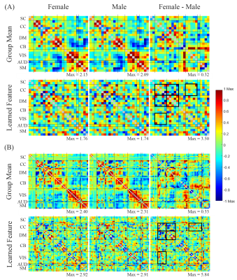 |
1 |
Deep Neural Network based Feature Selection in rs-fMRI Brain Functional Connectivity
Gengyan Zhao, Gyujoon Hwang, Cole Cook, Fang Liu, Mary Meyerand, Rasmus Birn
Deep neural networks (DNN) have been successfully applied to various prediction tasks in rs-fMRI, but the feature selection mechanism of it often appear to be a black box. We developed understanding of DNN’s prediction mechanism and proposed a feature selection method based on each feature’s contribution to the prediction. Experiments were done on the functional connectivity (FC) gender prediction to extract gender related brain FC patterns with 1003 subjects’ rs-fMRI data. The proposed method was validated by the cross-entropy loss of each feature’s prediction, and results showed the selected features are robust and consistent with the findings in previous studies.
|
|
2823.
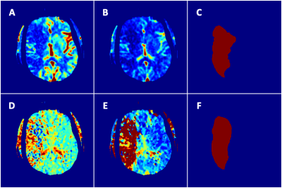 |
2 |
Automated Prediction of Stroke Lesion Outcome using Multiparametric Deep Neural Network
Presentation Not Submitted
Zexin Yan, Lian Ding, Hongkun Yin, Haiyan Lou, JUN YANG
Recent stroke trials raised a demand for triage decision intelligence of ischemic lesion progression. This study aimed to develop a multiparametric deep neural network to segment regions that predicted final infarct formation. The PWI-derived CBF, CBV, MTT and Tmax maps served as multi-channel inputs to algorithm training. We used a 2.5D U-Net to generate lesion segmentation. Our approach showed a good sensitivity and specificity with AUC of 0.868 in predicting the final lesions, and a comparable performance of DICE and IOU. In conclusion, we demonstrated feasibility for predicting tissue outcome in acute ischemic stroke with multiparametric deep learning algorithm
|
|
2824.
 |
3 |
Ultrafast Brain Segmentation using a 3D Fully Convolutional Neural Network for Risk Stratification of Cognitive Impairment and Dementia
Jian Wu, Alex Graff, Jason Deckman, Dmitry Tkach, Hyun-Kyung Chung, Natalie Schenker-Ahmed, David Karow, Christine Swisher
Early detection and intervention has the potential for slowing disease progression of dementia. Brain segmentation of T1W structural MRI is an effective biomarker for assessing cognitive decline. However, automatic brain segmentation has required lengthy processing time and utilizes single structure for risk stratification. In this study, we developed a 3D fully convolutional neural network for ultrafast brain segmentation. Both qualitative and quantitative analysis demonstrated that our segmentation method has a strong generalization capability achieving promising experimental results. Furthermore, we utilized multiple regions in combination and defined a new biomarker to better differentiate early disease progression of normal versus MCI in addition to MCI versus Dementia. Our multi-region approach outperforms conventional single biomarkers.
|
|
2825.
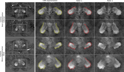 |
4 |
Automatic Dentate Nuclei Segmentation based on Quantitative Susceptibility Maps using a Convolutional Neural Network: Application to Healthy Controls and Cerebellar Ataxia Patients
Nina Jacobsen, Dominik Jäschke, Sophia Luise Goericke, Jürgen Reichenbach, Dagmar Timmann, Daniel Güllmar, Andreas Deistung
Non-invasive visualization and segmentation of the dentate nucleus is helpful for characterizing neurological diseases. Therefore, we set up an automatic segmentation strategy relying on a convolutional neural network (CNN) for the delineation of the dentate nucleus based on quantitative susceptibility maps. We trained the network on 101 healthy controls and 118 patients suffering from various types of cerebellar ataxia. We were able to demonstrate that the CNN accurately segments the dentate nuclei in 26 healthy controls and 21 SCA6 patients with volume estimates being in agreement with literature.
|
|
2826.
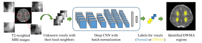 |
5 |
Segmentation of Diffuse White Matter Abnormality in Preterm Infants using Deep Learning
Hailong Li, Nehal Parikh, Jinghua Wang, Stephanie Merhar, Ming Chen, Milan Parikh, Scott Holland, Lili He
Diffuse white matter abnormality (DWMA) is observed in 50-80% of very preterm infants at term-equivalent age. Despite autopsy studies showing correlation with neuropathology, the relationship of DWMA with long term neurodevelopmental impairments remains controversial. The controversy may be due to the qualitative nature of previous studies of DWMA, likely resulting in measurement error and perhaps contributing to the lack of association with neurodevelopmental impairments in some studies. In this study, we developed a deep learning approach to objectively and automatically segment DWMA regions on T2-weighted MRI images. The internal and external validations demonstrated very accurate and reproducible DWMA segmentation performance.
|
|
2827.
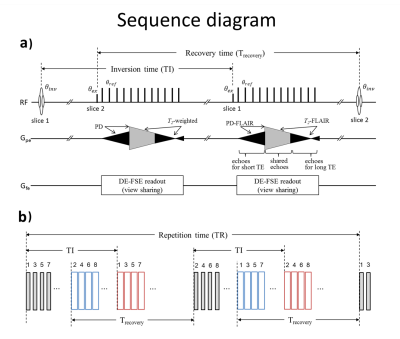 |
6 |
Quad-contrast imaging with deep learning-powered reconstruction: 2-minute neuro-evaluation
Sooyeon Ji, Doohee Lee, Se-Hong Oh, Jongho Lee
A 2D multi-contrast sequence with deep learning-powered reconstruction is developed to generate four contrast images (PDw, T1w, T2w, and FLAIR) and two quantitative maps (T1 and T2) in 2 minutes of scan time. For the reconstruction, a new deep learning method that assures both data consistency and image fidelity is applied with the joint reconstruction of the quad-contrast k-space data.
|
|
2828.
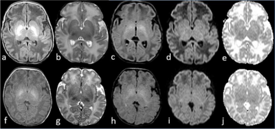 |
7 |
Machine Learning Approach Assisted MRI Characterization for Diagnosis of Neonatal Bilirubin Encephalopathy
Zhou Liu, Bing Ji, Ge Cui, Ling Ding, Xiaofeng Yang, Liya Wang, Hui Mao
Diagnosis of acute bilirubin encephalopathy (ABE) in newborns based on T1-weighted spin echo images in clinical routine is challenging due to subtle signal intensity change in the basal ganglia caused by ABE often overlapping with the presence of signal enhancement from the normal myelination in the infant brain. We used a feature-extraction-based machine learning approach to identify ABE associated morphological features from T1-weighted images of 34 ABE neonates. We found textural features along with intensity-based features added specific information for distinguishing ABE from normal myelination, demonstrating the feasibility of using this approach to assist MRI diagnosis of neonatal ABE.
|
|
2829.
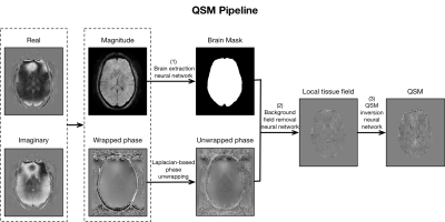 |
8 |
Application of a Clinically Viable Deep-Learning-Based QSM Workflow on Stroke Cases
Juan Liu, Kevin Koch
Quantitative Susceptibility Mapping (QSM) can quantitatively estimate tissue magnetic susceptibility, which enables differentiation of diamagnetic calcifications and paramagnetic hemorrhages. The translation of QSM into clinical practice faces technical implementation challenges, particularly the QSM inversion process. In the clinical practice current QSM post-processing techniques are constrained due to large slick thicknesses, which result in compromised background field removal and streaking artifacts in QSM images. To address these limitations, here we a apply a deep-learning-based QSM pipeline, including: (1) a 2D neural network to construct brain masks, (2) a background field removal deep neural network reveal local tissue fields, and (3) a QSM inversion deep neural network. Nine patients with stroke were scanned using a clinical susceptibility-weighted MR protocol were used to demonstrate that the proposed clinically viable QSM workflow can effectively detect microbleeds and differentiate calcifications from hemorrhages.
|
|
2830.
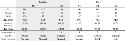 |
9 |
Age prediction from time-of-flight MR angiography using deep learning: Comparison of the predicted age among normal, MCI and AD.
Hunjin Chung, Yoonho Nam, KoungMi Kang, Chul-Ho Sohn, KangHyun Ryu, Jinhee Jang, Dong-Hyun Kim
Time-of-Flight (TOF) MR angiography (MRA) provides meaningful vascular information related to aging. Recently, we have developed a deep learning based chronological age prediction model from 3D TOF data and demonstrated its accuracy in predicting the age of normal volunteers. In this study, to investigate its clinical utilities, we applied the deep learning model to subjects with mild cognitive impairment and Alzheimer’s disease.
|
|
2831.
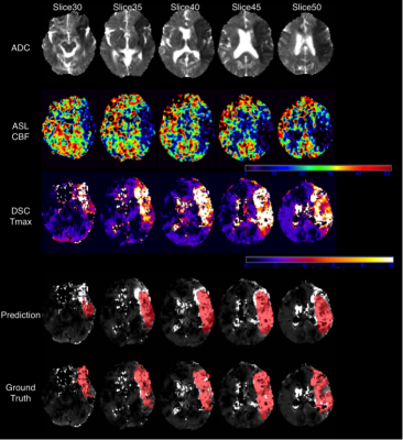 |
10 |
Deep Learning-based Detection of DSC-Defined Penumbral Tissue on pCASL in Acute Ischemic Stroke
Kai Wang, Qinyang Shou, Samantha Ma, David Liebeskind, Xin Qiao, Fabien Scalzo, Jeffrey Saver, Noriko Salamon, Danny Wang
Arterial spin labeled (ASL) techniques can provide cerebral blood flow (CBF) measures without the use of a contrast agent, and it has been shown to provide largely consistent results with DSC perfusion in delineating hypoperfused brain regions in AIS while also providing information on hyperemic lesion. In this study, we develop a deep learning-based model to identify the hypoperfusion lesion on ASL images based on the DSC perfusion-defined penumbra region and diffusion weighted imaging (DWI). Our results show that deep learning can predict the DSC-defined penumbral region in ASL with dice coefficient=0.43.
|
|
2832.
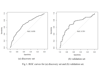 |
11 |
Radiomics and machine learning analysis of brain lesions in MOG-ab-positive and AQP4-ab-positive patients
Presentation Not Submitted
Liqin Yang, Wei Xia, Haiqing Li, Daoying Geng, Yuxin Li
Little is known about the differences between the lesion features of the recently discovered MOG-ab-positive and well-demonstrated AQP4-ab-positive patients till now. We studied the radiomics features of 747 lesions from AQP4 patients, and 295 lesions from MOG patients. Seventy radiomic features were calculated and compared. Features with significant between-group discrimination ability input to the classifier and trained. A radiomics signature was obtained for the discrimination of MOG-ab-positive and AQP4-ab-positive patients. These results provide valuable information for understanding of pathogenesis and imaging-based initial diagnosis in the two subsets of patients.
|
|
2833.
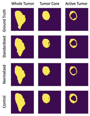 |
12 |
Standardization of MRI as Pre-Processing Method for Machine Learning Based Segmentation
Robert Wujek, Kathleen Schmainda
In this study, standardization methods are used to pre-process brain MRI to generate a machine learning dataset for tumor segmentation. This method was chosen for previously documented repeatability properties as compared to more widely used normalization methods, which could potentially lead to a more generalized segmentation model. When applied to the publicly available BRATS dataset, the standardization methods performed equally as well as the normalization method used in this study, which supports further analysis of the methods beyond the highly controlled BRATS dataset.
|
|
2834.
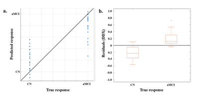 |
13 |
Combined Use of Gray Matter Volume and Quantitative Susceptibility Mapping to Predict Early Alzheimer’s Disease Using a Machine Learning-based Optimized Combination-Feature Set
Hyug-Gi Kim, Soonchan Park, Hak Young Rhee, Kyung Mi Lee, Chang-Woo Ryu, Soo Yeol Lee, Seong Jong Yun, Eui Jong Kim, Yi Wang, Wook Jin, Dal Mo Yang, Geon-Ho Jahng
To investigate the approach of classification and prediction methods using the machine learning (ML)-based optimized combination-feature (OCF) set on gray matter volume (GMV) and QSM in elderly subjects with a cognitive normal (CN) profile, those with amnestic MCI (aMCI), and mild and moderate AD patients, GMV and QSM in the brain were calculated. To differentiate the three subject groups, the support vector machine (SVM) with the three different kernels and with the OCF set was conducted with GMV and QSM values. To predict the aMCI stage, regression-based ML models were developed with the OCF set.
|
|
2835.
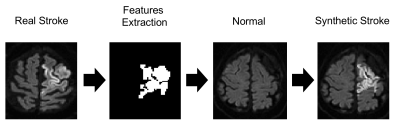 |
14 |
Deep Learning with Synthetic Diffusion-Weighted Images for Acute Ischemic Stroke Detection
CHRISTIAN FEDERAU, Javier Montoya-Zegarra, Soren Christensen, Julian Maclaren, Johanna Ospel, Victor Schulze-Zachau, Maarten Lansberg, Sebastian Kozerke
We studied the feasibility and accuracy of a deep learning algorithm trained on one million realistic synthetic acute stroke lesion images to detect and segment stroke lesions on clinical MR DW images. We compared this method to a more conventional approach, where a deep learning algorithm was trained on 10’000 human labelled images.
|
|
2836.
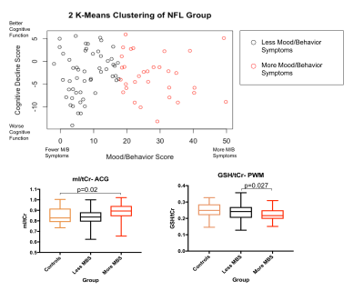 |
15 |
Cohort Stratification by Clinical Symptoms through Unsupervised Learning Reveals Metabolic and Microstructural Brain Alterations in Former American Football Players
Molly Charney, Janna Kochsiek, Tyler Starr, Michael Alosco, Brett Martin, Huijun Liao, David Kaufmann, Lauren O'Donnell, Sylvain Bouix, Fan Zhang, Nikos Makris, Martha Shenton, Robert Stern, Inga Koerte, Alexander Lin
Chronic Traumatic Encephalopathy (CTE) is a neurodegenerative disease associated with exposure to repetitive head impacts. This study aims to characterize the differing clinical presentations of CTE using MR Spectroscopy and Diffusion Tensor Imaging. Unsupervised Learning was first used to divide a cohort of former NFL players into sub-groups based on the degree of mood/behavior symptoms and cognitive impairment relative to controls. The neurometabolite concentrations and measures of diffusivity were then compared between the sub-groups. Athletes with increased mood/behavior symptoms showed alterations reflective of neuroinflammation, whereas the cognitive impairment sub-group showed more neuronal and structural alterations.
|
|
2837.
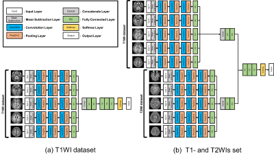 |
16 |
Estimating the Age of Healthy Children Based on Myelination Pattern in Brain MRI using a Deep Learning Neural Network Method
Yuya Saito, Akihiko Wada, Masaaki Hori, Koji Kamagata, Shigeki Aoki
In this study, we used deep learning model to estimate the age of children based on the MR signal changes associated with myelination process on T1 and T2-weighed images. Brain MR images of 119 children age ranging from 0.25 to 24 months were first used as a training and test dataset. The age was then estimated by deep learning model based on the T1-WI and T2-WI dataset and T1-WI only dataset. Our results showed that convolution neural network model using T1WI and T2WI dataset demonstrated higher correlation and lower mean absolute error (MAE) compared to T1-WI only dataset.
|
|
2838.
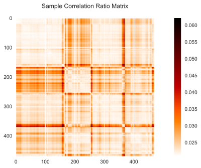 |
17 |
Applying Unsupervised Machine Learning Techniques to Resting-State BOLD Multicenter Neuroimaging of Pre-adolescent Complex Congenital Heart Disease Patients to Enhance Image Harmonization and Predict Motion Artifact Characteristics
Jenna Schabdach, Vincent Schmithorst, Vince Lee, Rafael Ceschin, Ashok Panigrahy
Resting-state BOLD MR images are invaluable for evaluating the neurocognitive state of patients, particularly populations at high risk for neurodevelopmental impairment; however, BOLD images are highly susceptible to motion. The combination of machine learning and image reconstruction techniques during and after BOLD image acquisition holds great promise for harmonizing images and recovering motion-corrupted data. However, there is little information about the relationship between unsupervised ML techniques and characteristics of resting BOLD images. We examined resting state BOLD image harmonization and motion in a set of complex congenital heart disease case and healthy control adolescent subjects acquired through a multi-center study.
|
|
2839.
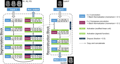 |
18 |
The utility of a convolutional neural network for generating a myelin volume index map from rapid simultaneous relaxometry imaging
Yasuhiko Tachibana, Akifumi Hagiwara, Masaaki Hori, Jeff Kershaw, Misaki Nakazawa, Tokuhiko Omatsu, Riwa Kishimoto, Shigeki Aoki, Tatsuya Higashi, Takayuki Obata
Myelin volume measurement based on rapid-simultaneous-relaxometry-imaging (RSRI) is useful for the clinic. However, the software that translates RSRI into the index map contains a potential weak point: the index is obtained using a pixelwise table-lookup that doesn't incorporate information from surrounding pixels. A novel deep-learning-based method was developed to overcome this problem. A myelin volume index based on magnetization-transfer saturation imaging was used as a reference for training and testing. The proposed method was evaluated by leave-one-out cross-validation using images from 20 healthy volunteers. Correlation with the reference was significantly higher for the proposed method.
|
|
2840
|
19 |
Automatic classification of early-onset neurodegenerative dementia patients using artificial neural networks
Video Permission Withheld
Camilla Cividini, Federica Agosta, Silvia Basaia, Luca Wagner, Maura Cosseddu, Elisa Canu, Stefano Gazzina, Giuseppe Magnani, Elka Stefanova, Vladimir S. Kostic, Roberto Gasparotti, Alessandro Padovani, Barbara Borroni, Massimo Filippi
We built and validated a convolutional neural network (CNN) to predict the individual diagnosis of early-onset Alzheimer’s disease (EOAD) and behavioral variant of frontotemporal dementia (bvFTD) based on a single T1-weighted image. The analysis showed that CNN procedure was able to discriminate EOAD from healthy controls with an accuracy of 83% (sensitivity=85% and specificity=80%). CNNs differentiated bvFTD patients from controls with an accuracy of 73% (sensitivity=63% and specificity=83%). CNNs provide a powerful tool for the automatic classification of early-onset neurodegenerative dementia and perform well without any prior feature engineering and regardless the variability of imaging protocols and scanners.
|
|
2841.
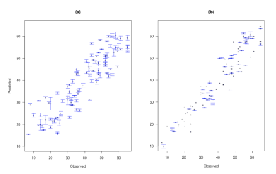 |
20 |
Predicting response to motor therapy in chronic stroke patients based on clinical and connectivity measurements using Machine Learning
Ceren Tozlu, Dylan Edwards, Aaron Boes, K. Zoe Tsagaris, Joshua Silverstein, Heather Pepper Lane, Mert R. Sabuncu, Charles Liu, Amy Kuceyeski
Statistical methods, including machine learning, are a highly promising avenue with which to improve prediction accuracy in clinical practice. The main objective of this study was to use machine learning methods to predict a chronic stroke individual’s motor function after 6 weeks of intervention from demographic, neurophysiological and imaging measurements. Our main finding was that Elastic-net outperformed Support Vector Machine, Artificial Neural Network, Random Forest, and Classification and Regression Trees in predicting post-intervention Fugl-Meyer Assessment. The addition of structural dysconnectivity measurements to the demographic and neurophysiological data did not improve the performance of the methods.
|
|
2842
|
21 |
Automated brain extraction of multi-sequence MRI using artificial neural networks
Video Permission Withheld
Irada Tursunova, Marianne Schell, Fabian Isensee, Ulf Neuberger, Gianluca Brugnara, David Bonekamp, Wolfgang Wick, Martin Bendszus, Klaus H Maier-Hein, Philipp Kickingereder
Brain extraction is a preliminary but critical step in many neuroimaging studies and determines the accuracy of subsequent analyses. Standard brain extraction algorithms are, however, limited to the processing of precontrast T1-weighted (T1-w) MRI and frequently fail in the presence of pathologically altered brain. Here we developed a new algorithm based on artificial neuroal networks (ANN) that enables rapid, automated and robust brain extraction irrespective of pathology, sequence type, hardware or acquisition parameters and lays the groundwork for automated, high-throughput processing of neuroimaging data.
|
|
2843.
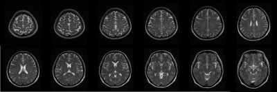 |
22 |
Prediction of Chronological Age from Routine T2-weighted Spin-echo Brain Magnetic Resonance Images by Deep Convolutional Neural Network
Inpyeong Hwang, Hyeonjin Kim, Ji-hoon Kim
Brain-predicted age may be used as a potential biomarker of brain aging. Given that 2D T2-weighted images are more routinely acquired from patients than those 3D images, this study investigated the potential applicability of 2D images in deep learning-based prediction of brain age with an assumption that each individual slice of the T2-weighted brain images possesses brain age-associated features learnable by a convolutional neural network (CNN). The purpose of this study was to investigate whether there are learnable features by a CNN in each slice of routine T2-weighted spin-echo brain MR images that might be associated with normal aging.
|
|
2844.
 |
23 |
Exploiting Heterogeneous Data for Automatic Classification of Multiple Sclerosis Clinical Forms through Neural Networks
Aldo Marzullo, Claudio Stamile, Gabriel Kocevar, Francesco Calimeri, Giorgio Terracina, Françoise Durand-Dubief, Dominique Sappey-Marinier
This work is aimed at producing a fully automatic model for the classification of MS clinical profiles exploiting heterogeneous source of data. The task is addressed by extracting the connectivity graph of the subject as well as the corresponding MRI-derived feature vector, and then by applying a two-branches NN procedure to obtain the corresponding classification.
|
|
2845.
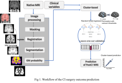 |
24 |
Structural changes in auditory and language-processing cortices and thalamus are predictors of word recognition ability after cochlear implantation
Zhe Sun, Ji Won Seo, Jee Yeon Lee, Min Young Kwak, Yehree Kim, Je Yeon Lee, Hong Ju Park, Hosung Kim
Long-term hearing loss may lead structural changes in brain cortices. These alterations may affect language outcome of the cochlear implant (CI) surgery. We aimed to predict the surgery outcome using imaging features of such cortical structural changes. We individually calculated GM density maps. We applied a random-forest regression to the GM density map and clinical variables, and found the competence of the imaging features in CI outcome prediction. The most accurate prediction was made with GM density changes in the middle temporal cortex, a critical network node of language processing, and in the thalamus, a structure (de-)coupling cortical language operations.
|
|
2846.
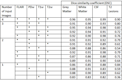 |
25 |
Deep Learning for Characterizing Image Sequence Significance in Brain Tissue Segmentation
IVAN CORONADO, REFAAT GABR, SUSHMITTA DATTA, SHEEBA SUJIT, FRED LUBLIN, JERRY WOLINSKY, PONNADA NARAYANA
Deep learning (DL) is an effective way for performing automatic multi-channel (or contrast) semantic segmentation. Here we investigated the accuracy of tissue segmentation as a function of the number and combinations of contrasts to the input of a fully convolutional neural network. The multi-contrast images included FLAIR, pre-contrast T1-, T2-, and proton density-weighted images, acquired on a large cohort of multiple sclerosis patients. Our results show that the number of input channels affects the segmentation accuracy in a tissue-dependent manner and that FLAIR is the major determinant of segmentation accuracy.
|
|
| Top |
Brain Tumors: Pre-Treatment
Digital Poster
Neuro
Tuesday, 14 May 2019
| Exhibition Hall |
14:30 - 15:30 |
| |
|
Computer # |
|
2847.
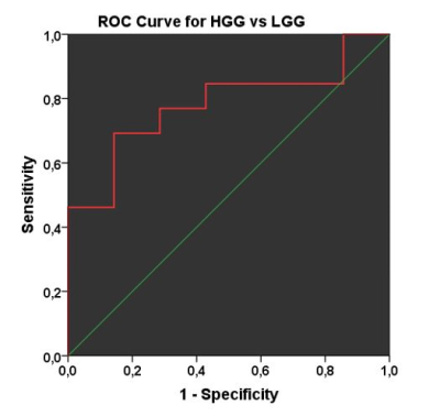 |
26 |
APT-weighted imaging for differentiation between brain tumors of glial origin
Faris Durmo, Jimmy Lätt, Anna Rydhög, Johan Bengzon, Jonas Svensson, Anina Seidemo, Frederik Testud, Benjamin Schmitt, Peter van Zijl, Linda Knutsson, Pia Sundgren
Gliomas of different histopathological grades have diametrically different prognosis and need biopsy-verification for diagnosis. Amide Proton Transfer weighted imaging (APTw), a non-invasive CEST MRI technique, was utilised for tumor differentiation of low grade and high grade brain gliomas. Mean lesional APTw-effect was quantified in regions with highest signal in 20 individuals. The information proved to be useful as the findings demonstrated that APTw increased the diagnostic accuracy in the clinical setting which may reduce treatment delay and improve diagnostics. APTw-imaging therefore shows promise as a clinical tool.
|
|
2848.
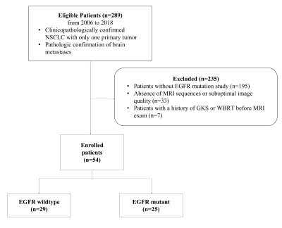 |
27 |
Diffusion Tensor and Conventional Imaging Radiomics Features to Differentiate the EGFR Mutation Status of Brain Metastases from Non-Small Cell Lung Cancer
Yae Won Park, Seng Chan You, Jongmin Oh, Sang Wook Kim, Kyunghwa Han, Sung Soo Ahn, Sung Jun Ahn, Hwiyoung Kim, Jong Hee Chang, Se Hoon Kim, Seung-Koo Lee
We assessed whether radiomics features on diffusion tensor imaging and postcontrast T1-weighted (T1C) images differentiates the epidermal growth factor receptor (EGFR) status in brain metastases from non-small cell lung cancer (NSCLC). Radiomics features (n=5046) were extracted from 54 brain metastases patients with NSCLC (29 EGFR-wildtype, 25 EGFR-mutant). After feature selection, radiomics models were constructed by various machine learning algorithms. Diagnostic performances were compared between multiparametric and single MRI radiomics models. The best performing multiparametric radiomics model (AUC 0.97) showed better performance than any single radiomics model using ADC (AUC 0.79, p=0.007), FA (AUC 0.75, p=0.001), or T1C (AUC 0.96, p=0.678).
|
|
2849.
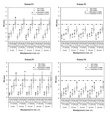 |
28 |
Visualization of Brain Shift Corrected fMRI data for Intraoperative Brain Mapping
Sanam Maknojia, Fred Tam, Sunit Das, Tom Schweizer, Simon Graham
Functional Magnetic Resonance Imaging (fMRI) facilitates the presurgical planning of awake craniotomies, but its use during the procedures remains limited. During intraoperative brain mapping by direct cortical electrical stimulation (DCS), fMRI is typically displayed separately or mentally recalled. The use of fMRI data in this manner is difficult and is further complicated by brain deformation or "shift". An image registration pipeline is presented that addresses this issue by providing covisualization of fMRI and DCS with acceptable accuracy for intraoperative use. This visualization method has the potential to improve the workflow of intraoperative brain mapping.
|
|
2850.
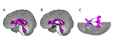 |
29 |
MRI features of the third ventricle subventricular zone suggest differing optic pathway glioma pathology for neurofibromatosis type-1 and sporadic paediatric cases
Natalie Boonzaier, Patrick Hales, Felice D'Arco, Bronwen Walters, Ramneek Kaur, Kshitij Mankad, Jessica Cooper, Alki Liasis, Sian Handley, Patricia O'Hare, Darren Hargrave, Christopher Clark
Paediatric optic pathway glioma (OPG) is a low-grade glioma whose cells likely originate from the subventricular zone of the third ventricle (TVZ). Neurofibromatosis type-1 (NF1) OPG is less clinically aggressive than sporadic OPG, being less likely to cause visual function deficit. Using measures of migrating tumour activity, this study analysed the imaging profiles at the TVZ. Apparent diffusion coefficient (ADC) and cerebral blood flow (CBF) profiles associated with migrative tumour cell activity were detected at the TVZ and correlated with poor visual function in sporadic, but not NF1-associated, cases. These results suggest that MRI can identify imaging characteristics that differentiate sporadic and NF1-associated OPG.
|
|
2851.
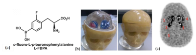 |
30 |
The comparison of L-FBPA PET/MR and PET/CT for BNCT treatment evaluation
Yi-Wen Lo, Ko-Han Lin, Yi-Wei Chen, Rong-Hong Jhou, Chien-Ying Li, Li-Chen Shen, Bang-Hung Yang , Chi-Wei Chang, Wen-Sheng Huang
We hypothesized that o-fluoro-L-p-boronophenylalanine (L-18FPBA) PET/MR could provide more precise treatment planning for Boron neutron capture therapy (BNCT) compared with L-18FBPA PET/CT. We implemented the phantom trial to measure the radioactivity correlation between PET/CT and PET/MR. Results have high comparability. We accessed patients who were potentially eligible for BNCT with same paradigms. The initial results represented acceptable correlation between these two modalities. Furthermore, because of high sensitivity and sophisticated imaging resolution, medics can easy to organize the more accurate treatment planning. In conclusion, PET/MR provides comparable L-18FBPA quantitative measurement and more diagnostic accuracy than PET/CT.
|
|
2852.
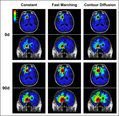 |
31 |
Initial Condition Assessment from Patient MRI Data for Reaction-Diffusion Glioma Growth Models
Corentin Martens, Thierry Metens, Olivier Debeir, Serge Goldman, Gaetan Van Simaeys
Reaction-diffusion tumour growth models have been proposed to assess glioma invasion beyond the MR visible margins as well as for patient follow-up and radiotherapy planning. However, initial tumour cell density assessment from patient imaging data remains an open problem, limiting the clinical application of the model. In this work, three initialisation methods are proposed and simulations from actual glioblastoma patient imaging data are compared. This work demonstrates the strong dependence of the model in its initial condition and highlights the need to integrate additional MR sequences and modalities for the assessment of a patient-specific tumour cell density profile.
|
|
2853.
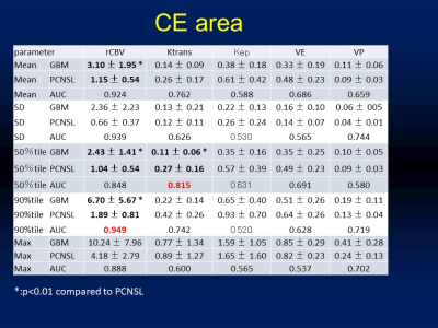 |
32 |
Diagnostic accuracy of T1-weighted Dynamic Contrast-enhanced-MRI and Dynamic Susceptibility MRI for Differentiation of Glioblastoma and Primary CNS Lymphoma
Yoshiyuki Watanabe, Masahiro Fujiwara, Takuya Fujiwara, Hiroto Takahashi, Chisato Matsuo, Hisashi Tanaka, Hideyuki Arita, Manabu Kinoshita, Naoyuki Kagawa, Noriyuki Tomiyama
We retrospectively studied 33 consecutive patients with a diagnosis of GBM or PCNSL performed both DCE and DSC imaging. The rCBV of GBM are significantly larger, and Ktrans of GBM was significantly lower than that of PCNSL in CE ROIs. There was no significant difference between two tumors in CE ROI about other DCE parameters, Ve, Vp Kep and surrounding ROIS. The ROC analysis performed with respect to the GBM and PCNSL groups revealed that 90% tile rCBV and 50% tile of Ktrans showed the largest area under the curve (AUC) of 0.949 and 0.815, respectively. PCNSL can be differentiated from GBM with rCBV value and Ktrans. rCBV was superior to Ktrans in differentiating two tumors and Ktrans had no additional value in differentiating these tumors.
|
|
2854.
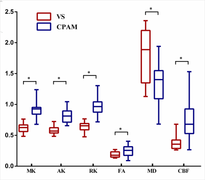 |
33 |
Differentiation between Vestibular Schwannomas and Meningiomas with Atypical Appearance using Diffusion Kurtosis Imaging and Three-dimensional Arterial Spin Labeling Imaging
Xiaodan Chen, Lin Lin, Zhongping Zhang, Yunbin Chen, Ying Chen
An accurate differentiation between vestibular schwannomas (VS) and meningiomas is critical in determining treatment strategies and clinical prognoses, but misdiagnoses may occur when typical imaging appearances are absent. We prospectively assessed the performances of diffusion kurtosis imaging (DKI) and three-dimensional arterial spin labeling imaging (3D-ASL) in the differentiation of VS and meningiomas with atypical appearance. Our study showed that DKI and 3D-ASL are useful for differentiating VS and meningiomas with atypical appearance, with kurtosis values of DKI have the best diagnostic efficiency.
|
|
2855.
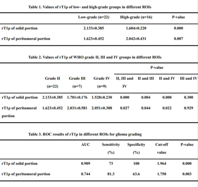 |
34 |
T1 relaxation time in the rotating frame MR imaging of gliomas:efficacy in preoperative grading
Zeyu Zheng, Jing Zhang, Yuefa Tan, Huiyan Li, Yingjie Mei, Queenie Chan, Yikai Xu
Accurate glioma grading is crucial for therapeutic strategy and prognosis. T1ρ MRI could probe the interactions between motion-restricted water and macromolecules in tissues. Our study aimed to determine the diagnostic efficiency of T1ρ MRI in glioma grading. Significant differences were found by rT1ρ between low- and high-grade, WHO grade ? and ?, WHO grade ? and ? gliomas when we placed the ROIs in the solid portion and the peritumoral portion respectively. rT1ρ demonstrated a high diagnostic performance in the solid portion and a moderate diagnostic performance in the peritumoral portion in grading. T1ρ MRI has potential to be a noninvasive quantitative method for preoperatively grading gliomas.
|
|
2856.
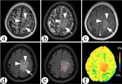 |
35 |
The study of relationship between amide proton transfer(APT) imaging and IDH mutation status in patients with high-grade gliomas
Yonglu Chen, Xinfeng Xie, Yingjie Mei, Yuelong Yang, Xiaoling Wu, Hongjun Liu, Biao Huang
We want to investigate the association of amide proton transfer weighted imaging (APTw) with isocitrate dehydrogenase (IDH) wild-type and IDH mutant of high-grade gliomas.All patients underwent MR scan, including APT san to getting the APT values of the whole tumor. The APT value of the IDH wild type group was significantly higher than that of the IDH mutant group. We conclude that APTw can be used to predict the gene type of IDH in high-grade gliomas before surgery.
|
|
2857.
 |
36 |
Towards the development of a microstructural model for Brain tumours
Samira Bouyagoub, Antonella Castellano, Maira Tariq, Luisa Altabella, Andrew Falini, Eleftheria Panagiotaki, Mara Cercignani
While several multi-compartment models of diffusion have been developed to describe the healthy neural tissue, most of these models fail in the presence of tumours, because they are unable to capture their heterogeneity. By contrast, models of diffusion able to quantify histologic features of colorectal and prostate tumors in vivo have been proposed. Here we combine the sensitivity of these models to tumour histology and the ability of multi-compartment models of dMRI in the brain to describe the complexity of the neural tissue.
|
|
2858.
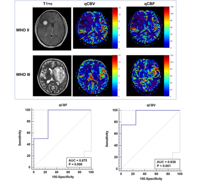 |
37 |
Quantitative cerebral perfusion with Scale-PWI demonstrates WHO II/III grading in glioma patients
Hao Wu, Yulong Zhang, Xiaoyue Zhou, Weiguo Zhang
This study aimed to demonstrate that the Scale-PWI sequence with absolute quantification for cerebral perfusion in the solid region of the tumors was a meaningful diagnostic factor in grade-II and grade-III gliomas. Total 8 glioma patients (4 grade II , 4 grade III) were subjected Scale-PWI scanning. ROC analysis confirmed that has great accuracy in grading World Health Organization (WHO) II and III gliomas.
|
|
2859.
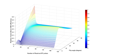 |
38 |
Novel Sequence for Improving Detection and Identification of Brain Tumor Lesions
Jinghua Wang, Mary Gaskill-Shipley, Michael Lamba, Kim Cecil, Lily Wang, Bin Zhang, Hui Wang, Lois Carpenter, Achala Vagal
Early detection and accurate identification of lesions are very important for diagnosis and treatment of brain tumors. Contrast-enhanced (CE) magnetic resonance imaging (MRI) is the gold standard method for the non-invasive identification of primary brain tumors and cerebral metastases. In clinical practice, current CE MRI protocols are acquired with an isotropic resolution around 1.0 millimeter at 3.0 T that is limited by tumor-tissue contrast to noise efficiency. In this investigation, our purpose was to improve the efficiency by incorporating inversion recovery prepared spoiled gradient recalled (IR-GRE) sequence with hybrid k-space and variable flip angles to reach submillimeter resolution in clinical acceptable scan time.
|
|
2860.
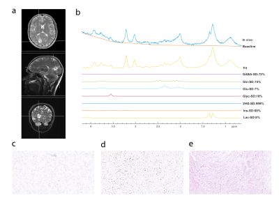 |
39 |
Classification of IDH and TP53 Mutational Subgroups of High-Grade Astrocytic Gliomas Based on Magnetic Resonance Spectroscopy at 3T
Esin Ozturk-Isik, Sevim Cengiz, Alpay Ozcan, Cengiz Yakicier, M. Necmettin Pamir, Koray Ozduman, Alp Dinçer
The goal of this study is to define MR spectroscopic differences of high-grade astrocytic glioma subgroups classified by IDH and TP53 mutations at 3T. TP-53 mutated (TP53-mut) and TP53 wild-type (TP53-wt) gliomas had similar metabolic profiles. IDH mutation (IDH-mut) was the main factor contributing to the metabolic differences between IDH and TP53 mutational subgroups. IDH-mut/TP53-mut gliomas had lower Glyc (P=0.017), GSH (P=0.009), Glu (P=0.038), and Glx (P=0.002) than IDH-wt/TP53-mut gliomas. Gliomas who were IDH wild-type (IDH-wt) and TP53-mut had the highest Glu, Glyc, GSH, and Glx among all high-grade astrocytic gliomas, which might be indicators of poor overall survival.
|
|
2861.
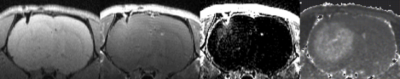 |
40 |
The Initial Tumor Microenvironment in Cerebral Patient-Derived Glioma Xenografts Affects Their Phenotypical Presentation
James Ewing, Ana deCarvalho, Brent Griffith, Stephen Brown, George Divine, Susan Irtenkauf, Robert Knight, Ian Lee, Swayamprava Panda, Glauber Cabral, Taverekere Nagaraja
Patient-derived xenografts (PDXs) of human gliomas in murine models are unmatched in representing the molecular heterogeneity of the disease, but typically do not present with MRI contrast, and thus are not phenotypical. This may pose a limit to the assessment of trial therapies. PDX preparations were tweaked by co-injecting Matrigel with neurospheres. In one of the two cell lines studied, Matrigel promoted the formation of brain tumors whose genetic composition was that of the original human GBMs and whose radiologic appearance on MRI was similar to that seen in humans. In the second cell line, no BBB breakdown occurred.
|
|
2862.
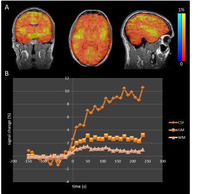 |
41 |
An optimised protocol for dynamic oxygen enhanced imaging of brain tumours
William Lloyd, Erjon Agushi, David Coope, Daniel Lewis, Alan Jackson, Geoff Parker
Hypoxia is an important factor in tumour resistance and regression. The use of oxygen as an MR contrast agent is gaining interest but the technique has been hampered by relatively low contrast, noisy data. Here we present an optimised protocol for dynamic oxygen-enhanced MRI (dOE-MRI) of brain tumours. We present data from healthy normal volunteers that demonstrate the method’s capability of producing reliable oxygen-induced signal change and data from glioblastoma (GBM) patients demonstrating tumour sub-division specific changes, showing the potential of this method for mapping tumour hypoxia.
|
|
2863.
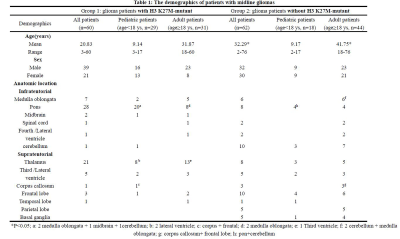 |
42 |
Clinical and Radiological Characteristics of Histone H3 K27M Mutations in Midline Gliomas
Xiaorui Su, Huaiqiang Sun, Weina Wang, Simin Zhang, Qiyong Gong, Qiang Yue
The aim of the present study is to report the clinical and imaging characteristics of midline gliomas with H3 K27M mutations and to compare them with those gliomas without histone H3 K27M mutation in different age groups. There were 60 midline gliomas with H3 K27M mutation and 62 glioma patients without mutation included in this study. Mutant tumors were more likely diagnosed at an early age and located at infratentorial brain, with less contrast enhancement or edema.
|
|
2864.
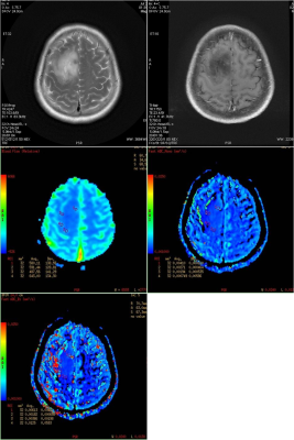 |
43 |
A comparative study of dynamic susceptibility contrast enhanced perfusion weighted imaging with perfusion-related parameters of multiple b values diffusion weighted imaging for neuroepithelial tumors grading
Huafeng Xiao, Xinjing Chen, Yan Wang, Lin Ma
The tumour grading is of great importance in predicting the biological behaviour of the tumours and offers important guidance for prediction of prognosis and the selection of clinical interventions. For these reasons, with dynamic susceptibility contrast enhanced perfusion weighted imaging (DSC) as reference standards, we retrospectively appraised the diagnostic efficacy of perfusion-related parameters of multiple b values diffusion weighted image (DWI) compared to appraise normalized lesion/normal tissue relative cerebral blood flow (nrCBF)in the prediction of neuroepithelial tumors grading. 62 patients with pathologically proved neuroepithelial tumors underwent conventional magnetic resonance sequences, multiple b values DWI, DSC. The group differences were not significant for these perfusion-related parameters of mono-exponential and biexponential model of multiple b values DWI.
|
|
2865
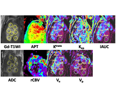 |
44 |
Amide proton transfer in high grade glioma: Correlation with apparent diffusion coefficient, relative cerebral blood volume and pharmacokinetic imaging parameters
Video Permission Withheld
Masanori Nakajo, Kiyohisa Kamimura, Tomohide Yoneyama, Yoshihiko Fukukura, Takashi Iwanaga, Yuta Akamine, Jochen Keupp, Takashi Yoshiura
Our purpose was to investigate potential correlations amide proton transfer signal intensity (APTSI) with apparent diffusion coefficient, relative cerebral blood volume and pharmacokinetic imaging parameters in high grade glioma (HGG). APTSI showed significant positive correlations with ADC (ρ = 0.582, p < 0.001) and with Ve (ρ = 0.300, p = 0.002), and a significant negative correlation with Kep (ρ = -0.337, p = 0.002). These results suggested that high APTSI in HGG may reflect proteins and peptides in large extracellular space, in addition to those in the intracellular space.
|
|
2866
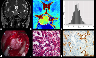 |
45 |
MR Textural Analysis on Contrast Enhanced 3D-SPACE Images in Assessment of Consistency of Pituitary Macroadenoma
Video Permission Withheld
Wenting Rui
This study aimed to explore the value of MR textural analysis in assessing consistency of pituitary macroadenoma (PMA) based on contrast enhanced (CE) 3D-SPACE images. Fifty-three patients with PMAs that underwent CE 3D-SPACE scanning and endoscopic trans-sphenoidal surgery were included. First-order energy and second-order correlation negatively correlated with hard PMAs, while first-order entropy and second-order variance, sum variance, and sum entropy positively correlated with stiffness (P<0.05). Diagnostic accuracy of combined negative features could achieve an AUC of 0.819, and an AUC of 0.836 of positive features (P<0.001). MRTA using CE 3D-SPACE images is helpful for assessing PMA consistency preoperatively and noninvasively.
|
|
2867.
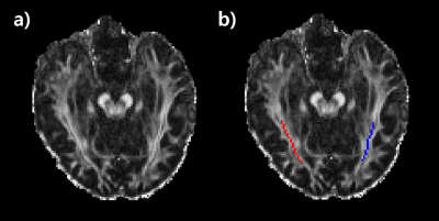 |
46 |
DTI assessment of optic pathway function in patients with anterior visual pathway compression
Eun-Jung Choi, Koung Mi Kang, Woojin Jung, Jongho Lee, Seung Hong Choi, Yong Hwy Kim
We explored the diffusion tensor imaging features of the optic radiations in patients with anterior visual pathway compression. The measurements were correlated with the visual tract impairment score. Significant differences in fractional anisotropy (FA), mean diffusivity (MD) and radial diffusivity (RD) were measured in the bilateral optic radiations depending on the visual disturbance. In addition, FA, MD and RD were correlated with the severity of visual field defects in the visual disturbance group. The results demonstrate the anterior visual pathway compression influences visual field defect affecting optic radiations in the brain.
|
|
2868.
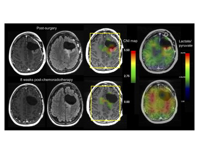 |
47 |
Serial HP [1-13C] pyruvate and 1H metabolic imaging in multiply recurrent high-grade glioma
Javier Villanueva-Meyer, Adam Autry, Jeremy Gordon, Hsin-Yu Chen, Daniele Mammoli, Marisa LaFontaine, Susan Chang, Duan Xu, Peder Larson, Daniel Vigneron, Sarah Nelson, Yan Li
This study demonstrates the feasibility of repeatable serial HP 13C MRI in the neuro-oncologic clinical setting and proposes the utility of pyruvate-to-lactate to monitor treatment response.
|
|
2869.
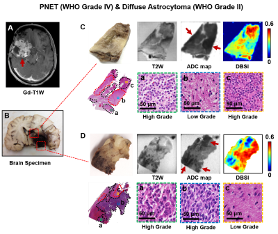 |
48 |
Isotropic Restricted Diffusion Correlates with Tumor Cellularity in Pediatric and Adult Malignant Brain Tumors
Zezhong Ye, Jeffrey Viox, Joshua Lin, Sam Gary, Jie Zhan, Chunyu Song, Ruimeng Yang, Joshua Rubin, Sonika Dahiya, Sheng-Kwei Song
We demonstrated how ADC inconsistently correlates with histologically determined tumor cellularity rendering it an unreliable imaging biomarker of tumor cellularity in pediatric and adult malignant brain tumors. In contrast, modified-diffusion basis spectrum imaging (DBSI)-derived restricted isotropic diffusion fraction correlated with histology-determined cellularity in pediatric and adult malignant brain tumors.
|
|
2870
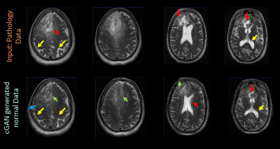 |
49 |
GAN based transformation of pathology data into its normative state for interventional neuro-therapy planning
Video Permission Withheld
Arathi Sreekumari, Sandeep Kaushik, Suresh Joel, Dattesh Shanbhag
In this work, we demonstrate that images with pathologies can be transformed to its non-pathological normal state using a cycle Generative Adversarial Network (cGAN). Potential applications of the work are in surgical planning, radiation therapy planning and longitudinal studies.
|
|
2871.
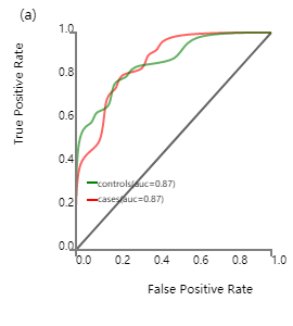 |
50 |
A predictive model for postlaminar optic nerve invasion in retinoblastoma basedon radiomic features from MR images
Presentation Not Submitted
Zhenzhen Li, Jian Guo, Junfang Xian
Retinoblastomas (RB) with postlaminar optic nerve invasion (PLONI) increases the risk for systemic metastasis or local recurrence. MRI is the only method to detect the PLONI in patients with eye-saving treatment strategies. But accuracy of MRI in identifying PLONI was relatively limited. Radiomics is an emerging field with a number of different uses being proposed. We hypothesized that radiomics can have an additional contribution to predicting PLONI in patients with retinoblastoma. This study validated our hypothesis.
|
|
| Top |
Spinal Cords, Disks & Nerves
Digital Poster
Neuro
Tuesday, 14 May 2019
| Exhibition Hall |
14:30 - 15:30 |
| |
|
Computer # |
|
2872.
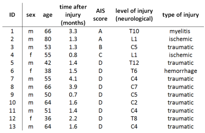 |
51 |
MRI investigation of neurodegeneration in subacute SCI and its relation to functional outcome
Gergely David, Markus Hupp, Kevin Vallotton, Jan Rosner, Tim Emmenegger, Silja Huwyler, Maryam Seif, Armin Curt, Patrick Freund
In this study, for the first time, we performed voxel- and atlas-based DTI analysis along with high-resolution T2*-weighted imaging in subacute spinal cord injury (SCI) to investigate early micro- and macrostructural changes remote from the injury at C2/C3. Thereby, we aimed to test the predictive ability of early tract-specific degeneration for the chronic functional outcome. DTI and tissue area measurements between SCI patients and controls revealed demyelination in the dorsal columns, indicative of early anterograde degeneration of sensory tracts. Importantly, these early microstructural changes predicted 1-year functional recovery, demonstrating the clinical eloquence of early DTI measurements as a prognostic tool.
|
|
2873.
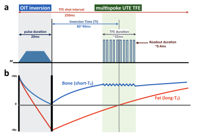 |
52 |
3D broadband IR-prepared UTE bone Imaging for assessment of ossification of the posterior longitudinal ligament (OPLL) in the cervical spine
Masami Yoneyama, Iain Ball, Minako Azuma, Toshinori Hirai, Marc Van Cauteren
We proposed a new technique for the cervical spine MR bone imaging based on broadband inversion recovery prepared segmented multispoke UTE sequence (3D BoneVIEW) for assessment of ossification of the posterior longitudinal ligament (OPLL). 3D BoneVIEW provided robust spinal bone imaging with sufficient background suppression. This sequence might be useful for assessment of the OPLL as an alternative to CT bone imaging.
|
|
2874
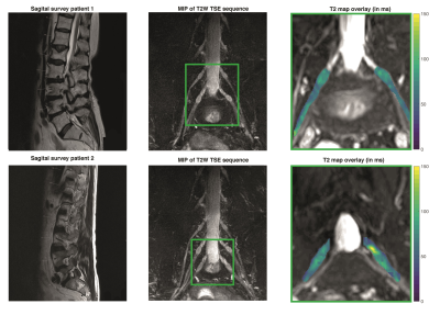 |
53 |
T2 mapping of lumbosacral nerves in patients suffering from unilateral radicular pain due to degenerative disc disease
Video Permission Withheld
Nico Sollmann, Dominik Weidlich, Barbara Cervantes, Elisabeth Klupp, Carl Ganter, Hendrik Kooijman, Claus Zimmer, Ernst Rummeny, Bernhard Meyer, Thomas Baum, Jan Kirschke, Dimitrios Karampinos
It is long known that findings in conventional anatomical imaging do not necessarily correlate with clinical symptoms in patients suffering from unilateral lumbosacral radicular syndrome (LRS), which regularly occurs in the context of disc herniation due to degeneration with unilateral nerve contact. The present study investigates the performance of quantitative imaging by using magnetic resonance neurography (MRN) using T2 mapping for LRS diagnostics at the lumbosacral plexus. As the main finding, it shows that nerves affected by degenerative disc herniation are characterized by elevated T2 values, in contrast to contralateral nerves or a non-affected control level.
|
|
2875.
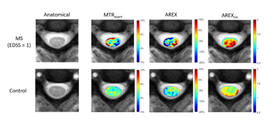 |
54 |
AREX (Apparent Exchange-Dependent Relaxation Rate) CEST MRI in the Spinal Cord at 3T: Application to Multiple Sclerosis
Richard Lawless, Quinn Weinberg, Haley Feiler, Sam By, Francesca Bagnato, Seth Smith
In this study, we evaluated AREX(apparent exchange-dependent relaxation rate) in the human spinal cord in patients with MS. Current clinical MRI is insensitive to biochemical changes which may precede inflammatory lesions in the spinal cord. Development of an MRI biomarker capable of detecting tissue changes prior to lesion formation would improve our diagnostic and prognostic capabilities. APT CEST is an MRI contrast approach sensitive to protein and peptide concentration and AREX removes magnetization transfer, T1-relaxation, and RF-spillover effects. Our results suggest that AREX is an improvement over alternative methods and may increase sensitivity to pathological changes in the spinal cord.
|
|
2876.
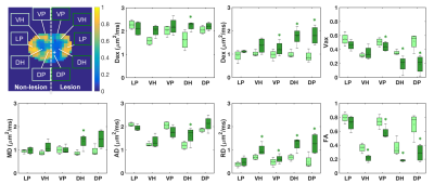 |
55 |
Structural Assessment of Injured Spinal Cord Using the Spherical Mean Technique and Diffusion Tensor Imaging
Feng Wang, Junzhong Xu, Tung-Lin Wu, Pai-Feng Yang, Nellie Byun, Li Min Chen, John Gore
High-resolution spherical mean technique (SMT) MRI may provide specific diffusion measures to detect and characterize structural connectivity before and after spinal cord injury and during repair. This study aims to systematically evaluate the SMT-derived diffusion parameters and compare them to conventional DTI-derived diffusion parameters in assessing injury-associated changes in spinal cords of squirrel monkeys. The main goal is to optimize a rapid and robust high-resolution SMT imaging protocol for spinal cord of monkeys at high field. In addition, the sensitivity of SMT and DTI measures in detecting axonal damage in the dorsal pathway has been evaluated.
|
|
2877
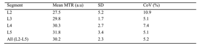 |
56 |
Feasibility of magnetisation transfer ratio measurements in the proximal lumbar plexus using healthy volunteers at 3T
Video Permission Withheld
Marios Yiannakas, Torben Schneider, Masami Yoneyama, Ferran Prados, Olga Ciccarelli, Claudia Gandini Wheeler-Kingshott
Magnetic resonance neurography (MRN) has become a widely used non-invasive tool for assessing the lumbar plexus with application to inflammatory, neoplastic, metabolic and traumatic pathologic conditions. Given that the main pathological process involved in the majority of these neurological conditions results in the alteration of myelin content, this pilot study proposes and demonstrates in healthy controls the feasibility of using MRN to facilitate reliable measurements of magnetisation transfer ratio, a biophysically meaningful MR biomarker, which has been shown to be directly influenced by the amount of myelin in neural tissue.
|
|
2878.
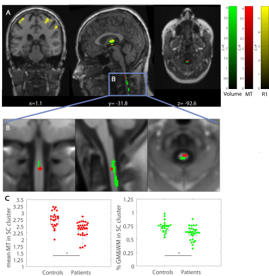 |
57 |
Simultaneous Voxel Based Analysis of Brain and Spinal Cord: Application to Spinal Cord Injury Patients
Michela Azzarito, Claudia Blaiotta2, John Ashburner, Yaël Balbastre, Sreenath Kyathanahally, Martina Callaghan, Maryam Seif, Patrick Freund
Assessing the sequelae of a focal CNS injury across the entire neuroaxis holds great potential to better understand the temporal and spatial distributed pathophysiological changes. Currently, voxel-based analysis is either performed at the level of the spinal cord or brain. As a result, most imaging studies fail to implement and analyse the interactions between remote areas across the CNS. In this study, a new probabilistic-atlas including brain and cervical spinal cord (SC) was used for the simultaneous, fully automated and multi-parametric analysis in SPM framework. This approach was validated by assessing trauma-induced changes in SCI patients and compared with findings from analytical tools assessing the brain and cord separately.
|
|
2879.
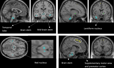 |
58 |
Investigating microstructural correlates of neuropathic pain in spinal cord injury patients using source-based morphometry
Sreenath P Kyathanahally, Michela Azzarito, Patrick Freund
Spinal cord injury (SCI) triggers a cascade of neurodegenerative and compensatory changes across the neuroaxis. The majority of SCI patients develop neuropathic pain (NP) as a secondary complication. In this study, we applied source-based-morphometry to investigate remote microstructural changes sensitive to myelin and iron deposition in brain areas associated with pain processing and explored the associations of these microstructural changes with pain intensity.
|
|
2880.
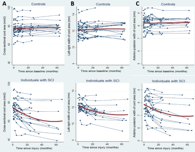 |
59 |
Tracking progressive neurodegenerative processes after spinal cord injury: 5-year follow-up
Sreenath P Kyathanahally, Michela Azzarito, Markus Hupp, Patrick Freund
Spinal cord injury (SCI) triggers a cascade of neurodegenerative and compensatory changes across the neuroaxis. Rehabilitative training has been shown to improve clinical outcome following SCI. However, the spatial and temporal patterns of neurodegenerative processes follow a complicated trajectory over 2-years. Previous studies have already shown trauma-induced neurodegenerative processes and highlight the relationships amongst these in the cord and brain. Here, we investigate the trajectories of neurodegenerative changes that occur over a period of 5 years post-SCI.
|
|
2881.
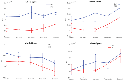 |
60 |
Assessing spinal cord structure changes using diffusion tensor imaging in patients with incomplete traumatic spinal cord injury
Presentation Not Submitted
Bing Yao, Hannah Ovadia, Zhiguo Jiang, Sarah Wood, Gail Forrest, Steven Kirshblum
Physicians rely on self-reports to monitor and evaluate the functional outcome in patients with spinal cord injury during their rehabilitation. These clinical and outcome measurements can be subjective and sometimes impractical if patients have cognitive difficulty. Traditional clinical MRI scans can provide doctors more objective information but they are not sensitive to detect the progression or repair during patient’s recovery. In this study, we investigated the sensitivity of DTI technique in detecting SCI injury and its progression or recovery over the course of rehabilitation in the individuals with SCI.
|
|
2882.
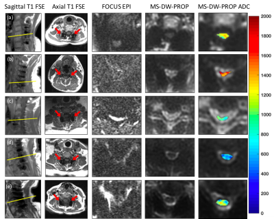 |
61 |
Diffusion Weighted MRI in Cervical Spine with Metal Hardware at 3T
Sampada Bhave, Marjorie Wang, Matthew Budde, Kevin Koch
Surgical repair of the cervical spinal cord to correct instability induced through trauma or degenerative disease often precludes follow-up MRI due to severe artifacts caused by metal stabilization hardware. Postoperative imaging is essential to monitor the hardware positioning, disease progression and new complications that may occur after surgery. In this study, we investigate the imaging capabilities of the recently developed multi-spectral diffusion weighted PROPELLER technique within the spinal cord immediately adjacent to metallic instrumentation. In addition, we assess the quantitative stability of this approach relative to other conventional methods in cohort of normal controls.
|
|
2883.
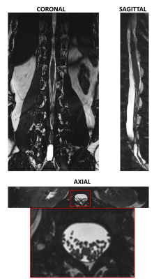 |
62 |
Large field-of-view high resolution 3D imaging of the thoracolumbar nerve roots inside the spinal canal
Stefano Mandija, Martijn Froeling, Anja van der Kolk, Fredy Visser, Andreas Rowald, Loan Nguyen-Mattera, Roberto Martuzzi, Jean-Baptiste Ledoux, Fabio Becce, Jocelyne Bloch, Gregoire Courtine, Cornelis van den Berg
A 3D T2/T1-weighted balanced-SSFP sequence has been developed to visualize the nerve roots inside the thoracolumbar spinal canal and their exit point from the spinal cord for clinical 3T MRI systems and coil setups. This sequence allows acquisition of large field-of-view, high resolution (sub-millimeter), almost isotropic images in less than 5 minutes. Furthermore, it is fully balanced, thus allowing high SNR, and intrinsically flow compensated. Hence, CSF flow artifacts are avoided. The acquired volumes in 15 volunteers showed good contrast between CSF, spinal cord and nerve roots (negative contrast), which could allow adequate visualization of intra-spinal pathologies.
|
|
2884.
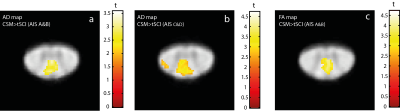 |
63 |
Quantitative MRI investigation above a cervical myelopathy in traumatic and non-traumatic spinal cord injury
Maryam Seif, Gergely David, Eveline Huber , Patrick Grabher, Markus Hupp, Armin Curt, Patrick Freund
Traumatic Cervical Spinal Cord Injury (tSCI) and non-traumatic Cervical Spondylotic Myelopathy (CSM) present a focal damage to the cervical cord with different time profile. Myelin and axonal loss occurs in SCI and CSM; however, little is known how these neurodegenerative changes are comparable in both groups. Therefore, we applied T2*-weighted and DTI MRI at the cervical cord to compare macro/microstructural changes in CSM and tSCI. While the macrostructural changes were similar in both groups, DTI detected more microstructural changes in SCI. Time-dependent compensatory mechanism in CSM might account for less neurodegeneration and better clinical function compared to tSCI.
|
|
2885.
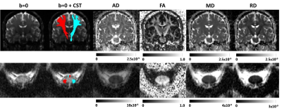 |
64 |
Investigation of DTI parameters in the corticospinal tract: from cervical cord to motor cortex
Rebecca Samson, Jon Stutters, Muhammad Akbar, Julien Cohen-Adad, Armin Curt, Michael Fehlings, Patrick Freund, Maryam Seif, Claudia Gandini Wheeler-Kingshott
We have explored diffusion tensor imaging (DTI) metrics along the corticospinal tract (CST) from the cervical cord to the motor cortex, measured using separate whole brain and cervical cord DTI protocols in healthy subjects at two different sites. We have also explored sensitivity to disease in patients with cervical spondylotic myelopathy (CSM), and spinal cord injury (SCI). Several studies have looked either at brain or cervical cord separately, which makes it difficult to learn about possible interactions between brain and cord. Here, we combine brain and cord measurements and examine how the CST is affected in CSM and SCI.
|
|
2886.
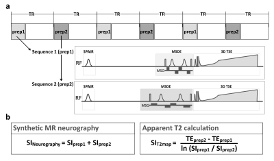 |
65 |
Simultaneous MR Neurography and T2 Mapping (SHINKEI Quant) for Quantitative Evaluation of Lumbar Nerve Roots in Patients with Low Back Pain.
Takayuki Sakai, Masami Yoneyama, Atsuya Watanabe, Daichi Murayama, Shigehiro Ochi, Tosiaki Miyati
This study was to investigate the clinical utility of SHINKEI-Quant ( simultaneously acquire MR neurography and T2 mapping by further optimizing the improved motion-sensitized driven-equilibrium (iMSDE) preparation) for evaluation of lumbar nerve roots in patients with low back pain.
|
|
2887.
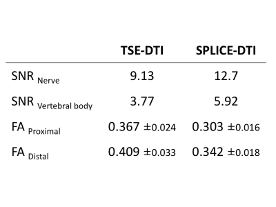 |
66 |
Improvement of distortion-free diffusion tensor imaging for evaluation of lumbar nerve roots: Utility of direct coronal single-shot turbo spin-echo diffusion sequence with a split acquisition.
Takayuki Sakai, Masami Yoneyama, Atsuya Watanabe, Daichi Murayama, Shigehiro Ochi, Tosiaki Miyati
This study was to investigate the availability of SPLICE-DTI (a split acquisition of fast spin-echo signals for diffusion imaging) with high spatial resolution for evaluation of lumbar nerve roots.
|
|
2888.
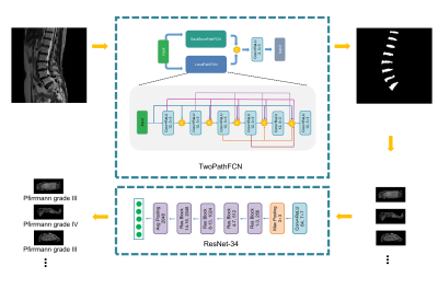 |
67 |
Fully Automated Grading of Lumbar Disc Degeneration Using Two-stage Networks
Presentation Not Submitted
Fei Gao, Shui Liu, Xiaodong Zhang, Jue Zhang, Xiaoying Wang
In this study, we developed a fully automated grading system for lumbar intervertebral disc degeneration with a two-stage network. The quantitative results suggest its efficiency and it can provide valuable tools for clinical diagnosis.
|
|
2889.
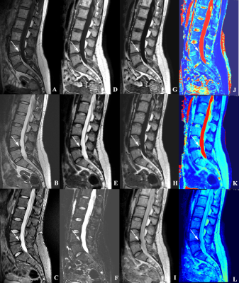 |
68 |
Diagnosis value of Lumbar Spine using synthetic MRI: a comparative study with conventional MRI
Presentation Not Submitted
Weilan Zhang, Guoguang Fan, Fei Bie, Bing Wu
Synthetic MRI is an emerging technique that can produce multiple morphologic contrasts from a single sequence, as well as quantitative T1, T2 and proton density maps. It has been successfully used in the brain, but its role is less well established for MRI of the spine. We aimed to assess the effectiveness and feasibility of synthetic MRI of the lumbar spine by comparing the image quality and diagnostic ability.
|
|
2890.
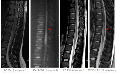 |
69 |
Relevance of spinal white matter suppressed T1 STIR beyond multiple sclerosis
Ullas Acharya, Rupesh Vakkachi Kandi, Narayana Rolla, Jitender Saini, Brian Johnson, Jaladhar Neelavalli
Previous works have shown that white matter suppressed (WMS) T1 STIR imaging can provide better lesion contrast in spine in multiple sclerosis. In this work we have explored the relevance of this technique in non multiple sclerosis related spinal conditions. The results show that, in these conditions as well, T1 STIR provides better lesion conspicuity.
|
|
2891.
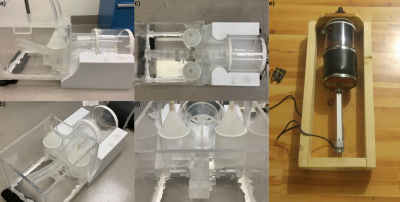 |
70 |
Anthropomorphic Spinal Cord Phantom with Respiratory B0 Field Fluctuations
Alan Seifert, Daniel Villarroel, Amrita Bedi, Anuoluwapo Bolarinwa, Joseph Borrello, Junqian Xu
The spinal cord exists in an unfavorable magnetic field environment; the lungs produce strong B0 field inhomogeneities that vary over time. We have designed and built a phantom that simulates these temporal field distortions to aid in the development of spinal cord imaging methods. The phantom consists of an acrylic tank, two lung simulants, a spinal cord and canal phantom, and a microcontroller-governed air pump. The respiratory waveform is customizable. This phantom accurately reproduced the ~20Hz respiratory-induced field shifts observed in vivo at the C3 vertebral level at 7T, and, being fully synthetic, is stable and replicable.
|
|
2892.
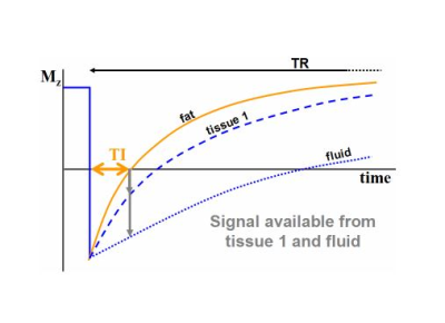 |
71 |
High-resolution Brachial Plexus Imaging using 3-D Short Tau Inversion Recovery (CUBE-STIR) with IV Gadolinium for Vascular Suppression
Darryl Sneag, Jahnavi Curlin, Jaemin Shin, Maggie Fung, Bin Lin, Steven Daniels
Vascular suppression is critical to facilitate confident identification and pathologic changes of small caliber peripheral nerves, many of great clinical importance, that run alongside slow-flowing veins. In our experience, non-contrast flow suppression techniques are suboptimal for complete venous suppression. Previous studies describe the efficacy of intravenous gadolinium for vascular suppression and visualization of the plexus proper using a three-dimensional STIR pulse, but its use for visualizing small plexus branch nerves has not been reported. This study demonstrated that post-STIR imaging significantly improved vascular suppression and visualization of branch nerves compared to non-contrast techniques, with high inter- and intra-rater agreement.
|
|
2893.
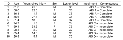 |
72 |
Examining Automatic Segmentation Results of Cervical Spinal Cord MRI in Tetraplegic Spinal Cord Injury
Patrik Wyss, Mihael Abramovic, Alexander Fischer, Markus Berger
Accurate detection and segmentation of spinal levels are of utmost importance in determining tissue changes in follow-up examinations of spinal cord injury patients. This preliminary study shows the results of automatic segmentation algorithms applied to data of tetraplegic patients.
|
|
2894.
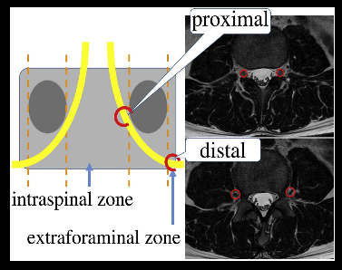 |
73 |
Super multi-directional DTI for lumbar nerve roots using MultiBans SENSE
Daichi Murayama, Takayuki Sakai, Masami Yoneyama, Shigehiro Ochi
An increasing the number of MPG directions causes prolongation of scan time. Recently, MultiBand SENSE (MB-SENSE) has been developed to allow the simultaneous acquisition of multiple slices and it enabled to shorten the scan time. Our purpose in this study was to investigate whether super multi-directional DTI using MB-SENSE is more useful for evaluation of lumbar nerve disorders than conventional method. An increasing the number of MPG directions improved its continuity and accuracy in tractography and tended to decrease the standard deviation of FA values.
|
|
2895.
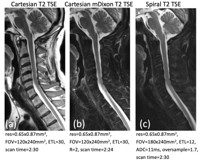 |
74 |
T1, T2, and STIR-weighted spine MRI using spiral SE/TSE techniques
Zhiqiang Li, James Pipe
Spine MRI constitutes a significant portion of neuro exams but is difficult in the clinical practice. The challenges include inhomogeneous field, motion and pulsation induced artifacts, long scan times, high SAR, etc. To overcome these drawbacks, in this work we explore the fast spiral sampling approach with SE and TSE sequences to provide rapid T1 and T2 weighted water-fat imaging, as well as STIR imaging. The feasibility is demonstrated with sagittal c-spine scans on volunteers.
|
|
2896.
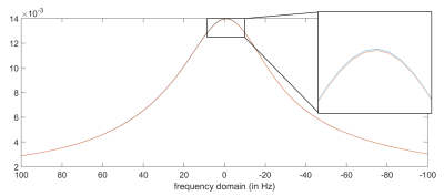 |
75 |
Measurement of peripheral nerve perfusion using FAIR PRESS
Ke Zhang, Johann Jende, Volker Sturm, Lukas Buschle, Artur Hahn, Sabine Heiland, Heinz-Peter Schlemmer, Martin Bendszus, Christian Ziener, Felix Kurz
Peripheral neuropathy in diabetes is a common and poorly understood disease; previous methods to evaluate nerve-associated microvascular angiopathy are contrast-based and exclude patients with severe nephropathies or allergies to contrast agents. We adapt a combined a ?ow-sensitive alternating inversion recovery (FAIR)-sequence with a single-voxel readout such as Point RESolved Spectroscopy (PRESS) to measure peripheral nerve perfusion in healthy subjects and diabetes patients. Our preliminary results suggest that diabetes patients have a lower nerve/muscle perfusion ratio than non-diabetic subjects.
|
|
| Top |
Brain Tumors: Post-Treatment
Digital Poster
Neuro
Tuesday, 14 May 2019
| Exhibition Hall |
14:30 - 15:30 |
| |
|
Computer # |
|
2897.
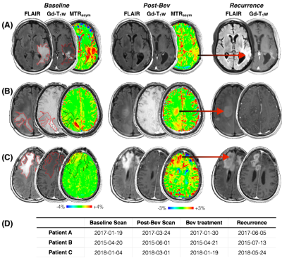 |
76 |
pH-weighted amine CEST-EPI as a fast clinical imaging biomarker for early bevacizumab treatment response in recurrent GBM
Jingwen Yao, Caleb Tan, Ararat Chakhoyan, Catalina Raymond, Noriko Salamon, Linda Liau, Phioanh Nghiemphu, Albert Lai, Whitney Pope, Timothy Cloughesy, Benjamin Ellingson
In the current study we used amine CEST-EPI, a pH-sensitive fast chemical exchange saturation transfer (CEST) technique, as a potential non-invasive imaging biomarker for treatment response in recurrent GBM patients treated with bevacizumab. Results suggest a significantly lower MTRasym at 3.0ppm in recurrent GBM tumors after bevacizumab treatment may be associated with better patient outcome (PFS), indicating that pH-weighted amine CEST MRI could serve as a potential non-invasive imaging biomarker for treatment response evaluation. The colocalization of recurrence tumor site and high MTRasym contrast post-treatment further suggest pH-weighted amine CEST may provide valuable information for early detection of tumor progression.
|
|
2898.
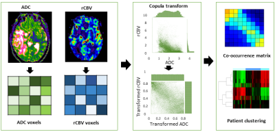 |
77 |
Copula Transform Characterizing the Inter-dependence of Multi-parametric Magnetic Resonance Imaging for Glioblastoma Patients
Chao Li, Shuo Wang, Pan Liu, Bart van Dijken, Carola-Bibiane Schönlieb, Stephen Price
The purpose of this study was to interrogate the inter-dependence of perfusion and diffusion imaging, and further investigate the clinical relevance of the inter-dependence of perfusion and diffusion, using a glioblastoma cohort containing 115 patients. A statistical method, the empirical copula transform, was applied obtain the joint distribution of perfusion and diffusion imaging, which was then discretized to extract second-order features for hierarchical patient clustering. Three patient subgroups were identified which showed significantly different overall survival and progression-free survival. The results showed that the inter-dependence between perfusion and diffusion imaging may be useful in stratifying patients and evaluating tumor invasiveness.
|
|
2899.
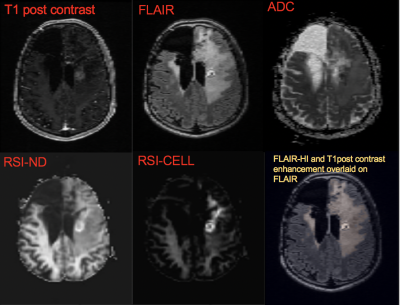 |
78 |
Restriction Spectrum Imaging differentiates True Progression from Pseudoprogression in patients with high grade glioma following treatment with immunotherapy
Presentation Not Submitted
Naeim Bahrami, Shadi Daghighi, William Tom, Nikdokht Farid, Carrie McDonald
Immunotherapy is becoming more frequently used in patients with high grade glioma (HGG) following standard chemoradiation. However, immune-mediated pseudoprogression (PsP) is a growing problem that remains poorly characterized and renders conventional imaging less useful. We evaluate the ability of a novel, advanced diffusion model, restriction spectrum imaging (RSI) to differentiate PsP from TP following immunotherapy. We demonstrate that an RSI-derived measure of axonal integrity (neurite density; ND) can differentiate TP from PsP, correctly classifying 73% of patients. We conclude that lower RSI-ND reflects disruption of axonal microstructure and may provide an imaging marker of tumor infiltration in patients with TP.
|
|
2900
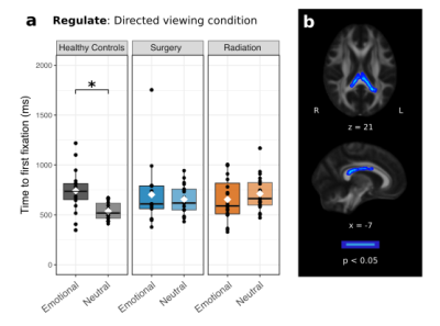 |
79 |
Eye-movements and white matter predict emotional control in children treated for brain tumors
Video Permission Withheld
Iska Moxon-Emre, Michael Taylor, Adeoye Oyefiade , Eric Bouffet, Suzanne Laughlin, Jovanka Skocic , Cynthia de Medeiros , Donald Mabbott
Children who survive a brain tumor diagnosis often suffer from emotional difficulties that decrease their quality of life. We monitored eye-movements during the control of attention to emotional faces to measure emotion regulation. Brain tumor survivors had difficulty regulating their initial attention away from emotional faces, and those who exhibited poor emotion regulation displayed the least emotional control in daily life. White matter of the splenium of the corpus callosum predicted emotion regulation. Our findings may improve the identification of children at risk for poor functional outcomes, and suggest the splenium as a candidate neuroanatomic substrate of emotion regulation.
|
|
2901.
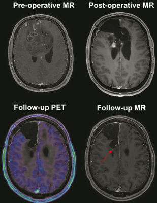 |
80 |
Discriminate glioblastoma recurrence versus radionecrosis: consider cerebrovascular reactivity mapping
Marco Piccirelli, Giovanni Muscas, Christiaan Hendrik Bas van Niftrik, Nicolaus Andratschke, Michelle Leanne Brown, Oliver Bozinov, Luca Regli, Christoph Stippich, Jorn Fierstra
Exploiting the different vascular pathophysiology in brain glioblastomas compared to radionecrosis, we aim to improve the differential diagnosis of post-treatment contrast-enhancing lesions. To recognize recurrent glioblastoma after treatment, we investigate if relevant CVR differences exist between newly diagnosed brain glioblastomas and radionecrosis. For this purpose, we utilized blood oxygenation-level-dependent functional MRI (BOLD-fMRI) to study CVR.
|
|
2902.
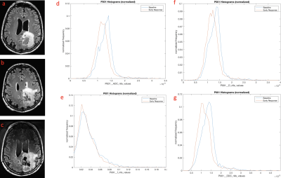 |
81 |
Assessment of early therapeutic response in glioblastoma using advanced DWI-MRI and histogram analysis
Shah Islam, Melanie Morrison, Anastasia Gontsarova, Matthew Orton, Matt Grech-Sollars, Eric Aboagye, Adam Waldman
Glioblastoma is one of the most aggressive brain tumours, with a life expectancy of 12-15 months. Structural MRI lacks sensitivity for detecting early therapeutic response. DWI is sensitive to changes in cellular density and provides a marker of cell kill. There is little published data on which of the diffusion components detectable in diseased brain tissue in vivo is most sensitive to this. Voxel by voxel pre- and post-treatment analysis in ‘functional diffusion map’ studies are confounded by change in tumour size. We assess the utility of a spatially-independent histogram approach to detect early treatment response using advanced DWI models.
|
|
2903.
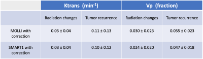 |
82 |
MOLLI versus SMART1 mapping for quantitative brain tumor DCE MRI on a 3T GE 750w: Initial experience
Greg Cron, Michael Gulak, Nicholas Majtenyi, Ian Cameron, Thanh Nguyen
In radiotherapy of high-grade gliomas, radiation changes (RC) are common. RC and tumor recurrence (TR) may both demonstrate progressive Gd contrast enhancement on follow-up, rendering conventional MRI unhelpful. DCE MRI, however, shows promise for distinguishing RC from TR. We set up a new protocol for brain tumor DCE-MRI on a 3T GE 750w. This protocol involved bookend T1 mapping performed with both MOLLI and SMART1. We found that both T1 mapping sequences gave similar results. Although patient numbers are low at this time, the technique appears promising for distinguishing radiation changes from tumor recurrence.
|
|
2904.
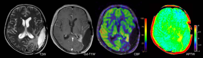 |
83 |
Differentiating the treatment-related effect from tumor recurrence using amide proton transfer-weighted (APTW) and pseudo-continuous arterial spin labeling (pcASL) MRI
Presentation Not Submitted
Qihong Rui, Dexia Kong, Tianyu Zou, Yingjie Mei, Hao Yu, Shanshan Jiang, Xianlong Wang, Jinyuan Zhou, Zhibo Wen
Discriminating treatment-related effect from tumor recurrence with is critical for treatment decision-making. Amide proton transfer (APT) and pseudo-continuous arterial spin labeling (pcASL) are two non-invasive advanced magnetic resonance imaging (MRI) techniques. In this study we combined two techniques hoping to explore their diagnostic performance in differentiating treatment-related effect from tumor recurrence.
|
|
2905.
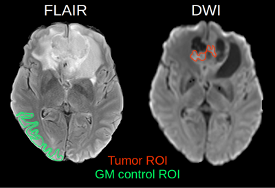 |
84 |
Comparison of quantitative blood oxygenation imaging methods for the assessment of brain tumor oxygen consumption in native MRI.
Jan Sedlacik, Julia Götz, Patrick Borchert, Divya Bolar, Lasse Dührsen, Nils Schmidt, Jan-Hendrik Buhk, Jens Fiehler
The comparison of the blood oxygen extract fraction (OEF) of solid tumor regions between the QUantitative Imaging of eXtraction of Oxygen and TIssue Consumption (QUIXOTIC) and quantitative Blood Oxygenation Level Dependent (qBOLD) methods showed an opposite trend of qBOLD-OEF and QUIXOTIC-OEF between tumor and cortical GM. Further analysis suggests that QUIXOTIC-OEF may be compromised by the strong and long lasting magnetization of the increased interstitial water due to tumor edema and, therefore, no reliable parameter to assess tumor OEF. On the other hand, qBOLD-OEF may have correctly detected a higher OEF of solid tumor regions.
|
|
2906.
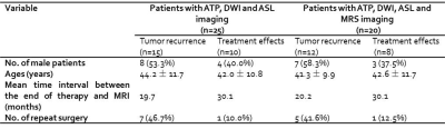 |
85 |
Differentiation between glioma recurrence and treatment effects using DWI, ASL, proton MRS and amide proton transfer (APT) imaging at 3 Tesla
Jie Liu, Yinsheng Chen, Cong Li, Xiaofei Lv, Zhongping Chen, Hairong Zheng, Yin Wu
Conventional MR structural imaging is insufficient for assessment of posttreatment gliomas and advanced imaging is needed to improve diagnostic certainty. The present study compared DWI, ASL, proton MRS and APT for distinction between glioma recurrence and treatment effects. Total 25 patients were enrolled. Results show similar diagnostic performance of APT and ASL, both of which could distinguish recurrent tumor from treatment lesion (P<0.05) with AUC greater than 0.8, superior to that of ADC and proton MRS. Herein, APT and ASL would facilitate posttreatment glioma diagnosis.
|
|
2907.
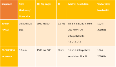 |
86 |
Restricted ketogenic diet and fasting in combination with re-irradiation in glioblastoma impact intracellular pH and intracerebral metabolism
Katharina Wenger, Elke Hattingen, Joachim Steinbach, Johannes Rieger, Martin Voss, Ulrich Pilatus
Inhibition of glycolysis by decreasing blood glucoses levels and increasing ketone bodies (KB) could force tumor cells to shift their metabolism towards potentially impaired mitochondria. The aim of this study was to explore the combination of six days of calorie restricted ketogenic diet (crKD) and three days of fasting (as a possible radiosensitizer) with a re-irradiation therapy in patients with recurrent glioblastoma. Intracerebral concentrations of KB as well as pHi and ATP were non-invasively monitored using MR-Spectroscopy. We were able to show evidence of intratumoral acetone using 1H-MRS in some patients with malignant glioma at day 6 of crKD/fasting. Changes in pHi and ATP during crKD/fasting will remain subject to preclinical studies.
|
|
2908.
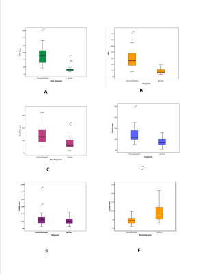 |
87 |
Diagnostic accuracy of serial 99mTc MDM (bis-Methionine-DTPA) SPECT imaging in differentiation of glioma recurrence from radiation Necrosis: A comparative study with Dynamic Susceptibility Contrast-Enhanced (DSCE)-MRI and Magnetic Resonance Spectroscopy (MRS)
Nisha Rani, Prof Baljinder Singh, Dr Naredra Kumar, Dr Paramjeet Singh, Dr Anil Mishra, Dr Puja Hazari
The use of complimentary imaging modalities in the glioma management of individual patients can provide additional information for further treatment strategy. In view of the limitations of anatomical imaging, cumbersome radio labeling procedures of PET tracers with amino-acids, in the present study, we have performed multimodality imaging using 99mTc-methionine SPECT (as cost effective substitute for expensive amino acid PET imaging), perfusion MRI and MRS for the the differentiation of radiation necrosis from recurrent/residual glioma by the means of serial imaging.
|
|
2909.
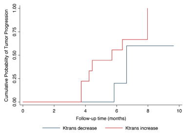 |
88 |
Parameters From Dynamic Contrast-Enhanced Magnetic Resonance Imaging Are Biomarkers Predicting Response after Radiotherapy to Brain Metastases
Presentation Not Submitted
Peng Wang, Zhuo Shi, Lizhi Xie, Yuqing Shang, XinMing Zhao, Han Ou-Yang
Dynamic contrast-enhanced (DCE) MRI provides additional information regarding blood-brain barrier integrity, and Ktrans is directly proportional to the level of permeability of the blood-brain barrier. In our study, we demonstrated that SRS of cerebral metastasis was associated with a decrease of Ktrans values in the early post-treatment period. DCE-MRI derived parameters can be a promising imaging biomarker of tumor aggressiveness.
|
|
2910.
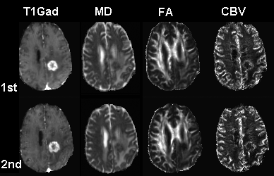 |
89 |
Detection of Tumor Progression in Patients with Glioblastoma using Multiparametric MRI
Sumei Wang, Sanjeev Chawla, Tianyu Yin, Chakri Madla, Ruyun Jin, MacLean Nasrallah, Arati Desai, Steven Brem, Ronald Wolf, Suyash Mohan, Harish Poptani
The study was performed to determine whether progression probabilities (PP) from DTI and DSC parameters can aid in differentiating glioblastomas with true-progression (TP) from pseudo-progression (PsP). MRI data from thirty-nine patients were included. All patients underwent at least two MR scans before pathological confirmation. TP patients tended to have high baseline PP values compared with PsP patients. An increase of PP of more than 25% at follow-up scans was noted in 12/15 TP patients, whereas stable or decreased PP were observed in 21/24 PsP patients. These results indicate that monitoring changes in PP values may aid in identifying TP.
|
|
2911.
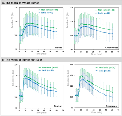 |
90 |
Crossover intraindividual comparison of ionic and non-ionic macrocyclic gadollinium based contrast media in orthotopic glioblastoma model: Pathologic correlation of dynamic contrast enhancement characteristics at 9.4T MRI
Presentation Not Submitted
Chi-Hoon Choi, Kyung Sik Yi, Janggeun Cho, Chulhyun Lee, Sang-Hoon Cha
The DCE parameters including Ktrans and Ve have been used to assess glioblastoma treatment responses reflecting tumor cellularity and tumor vasculature. We hypothesized that the enhancement pattern of the ionic macrocyclic contrast agent used in glioblastoma DCE study, which interacts with the negative charge on the surface of the tumor cell, was different from that of the nonionic one. We demonstrated it with prospective DCE experiments using the orthotopic glioblastoma models as well as Ktrans and Ve from ionic were more correlated with tumor pathology than nonionic. These findings may aid clinical choice of GBCA in DCE-MRI study.
|
|
2912.
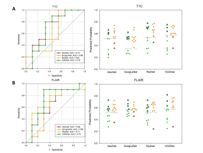 |
91 |
Noninvasive IDH1 genotype prediction in grade ?/? gliomas based on conventional MR images: a transfer learning strategy
Presentation Not Submitted
Jin Zhang, Lin-Feng Yan, Yang Yang, Bo Hu, Ping Chen, Wen Wang, Guang-Bin Cui
Purpose: To evaluate the performance of transfer learning with CNNs in predicting IDH1 genotype. Method and Materials: AlexNet, GoogLeNet, ResNet and VGGNet were pre-trained on the large scale natural image database (ImageNet) and fine-tuned with T1CE and FLAIR images. The outputs of training set were utilized to train LR and SVM models. Besides, fused images combining FLAIR and T1CE were used to fine-tune pre-trained ImageNet models. Results: Performances were improved by fine-tuning the four architectures with fused images. Conclusion: Transfer learning with various CNNs (especially VGGNet) is powerful in predicting IDH1 genotype in grade ?/? gliomas.
|
|
2913.
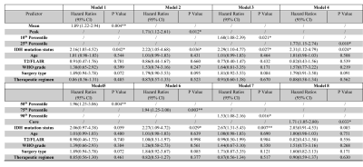 |
92 |
Amide Proton Transfer-Weighted (APTw) MRI as a Marker for Risk Stratification of Overall Survival in Patients with Lower Grade Gliomas
Shanshan Jiang, Qihong Rui, Hao Yu, Tianyu Zou, Zhengyang Zhu, Fangyao Chen, Hye-Young Heo, Peter van Zijl, Zhibo Wen, Jinyuan Zhou
We explored the possibility of using the APTw signal intensity as a noninvasive imaging marker for stratifying the risk of overall survival (OS) in patients with lower grade gliomas. 108 patients with newly diagnosed grade-II/III gliomas were included. APTw histogram data were recorded. Routine MRI based descriptors, clinical parameters, and OS were collected. According to the multiple Cox regression models, higher APTw value and IDH-wildtype together showed significant predictive power for shorter OS in the cohort of patients with lower grade gliomas. APTw MRI has potential for preoperative MRI evaluation of lower grade gliomas in terms of predicting patient outcomes.
|
|
2914.
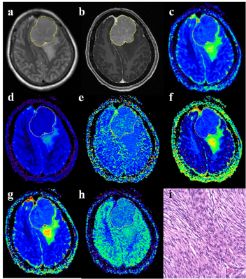 |
93 |
Grading meningiomas using mono-exponential, bi-exponential and stretched exponential model-based diffusion-weighted MR imaging
Lin Lin, Yunjing Xue, Qing Duan, Xiaodan Chen, Zhongshuai Zhang, Daoying Geng, Jun Zhang
Accurate grading is crucial to determine therapeutic strategies and to evaluate the prognosis, but no specific feature of conventional MRI has been found to be reliable in predicting the grade of the tumor. This study prospectively evaluated and compared the potential of various diffusion metrics obtained from mono-exponential model (MEM), bi-exponential model (BEM) and stretched exponential model (SEM)-based diffusion-weighted imaging (DWI) in the grading of meningiomas. It was found that different models of DWI (MEM, BEM, and SEM) are useful in the differentiation between high-grade and low-grade meningiomas. However, D obtained from BEM is the most promising diffusion parameter for predicting the grade of meningiomas.
|
|
2915.
|
94 |
Volume of anisotropic diffusion abnormality as prognostic imaging marker in treated glioblastoma patients
Bart van Dijken, Chao Li, Peter Jan van Laar, Shuo Wang, Stephen Price, Anouk van der Hoorn
The purpose of this study was to investigate the prognostic value of anisotropic diffusion abnormality in 30 treated glioblastoma patients. Diffusion tensor imaging was performed after chemoradiotherapy and volumes of diffusion abnormalities were extracted. Our results showed that a larger volume of high anisotropic abnormality was associated with worsened patient survival. Anisotropic diffusion can therefore potentially be employed as a prognostic imaging marker in longitudinal management of glioblastoma patients.
|
|
2916.
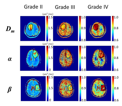 |
95 |
Percentile-based Analysis of Non-Gaussian Diffusion Parameters for Improved Glioma Grading
Christopher Zhou, Jiaxuan Zhang, Wenzhen Zhu, Zheng Zhong, Muge Karaman
Diffusion MRI has been used for tumor grading due to its sensitivity to alterations at the tissue microstructural level. Recognizing the limitations of analysis methods based on region-of-interest (ROI) in which the parameter values are averaged over the tumor ROIs, histogram-based approaches have been proposed for differentiating tumor grades. One challenge in this approach is to determine an optimal percentile over the ROI to be used in the analysis. In this study, we systematically and statistically determined an optimal percentile cut-off for calculating the mean parameters obtained from a non-Gaussian diffusion model based on continuous-time random-walk (CTRW) theory for differentiation among glioma grades.
|
|
2917
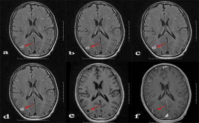 |
96 |
Application of half-dose contrast-enhanced T2-fluid-attenuated inversion recovery (FLAIR) sequence in brain metastasis
Video Permission Withheld
Teng Jin, Zhenwei Yao, Jianxun Qu, Yong Zhang
The treatment for brain metastasis are mainly depended on the number, size and location of lesions, to evaluate the number of brain metastatic lesions sensitively with contrast enhanced series is more important for patient treatment. The aim of this study was to assess the efficiency of half-dose contrast enhanced T2FLAIR used in the detection of intracranial metastases in comparison to full-dose enhanced T1-weighted imaging.
|
|
2918.
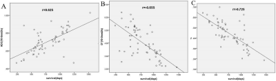 |
97 |
The Value of Intravoxel Incoherent Motion Imaging in Predicting the Survival of Patients with Astrocytoma
Lina Zhu, Jiang Wu, Hui Zhang, Heng Niu, Le Wang, Kaiyu Wang
In this study, two independent sample t-tests was used to evaluate the predictive value of IVIM parameters for the two-year survival rate of 60 patients with astrocytoma, and the correlation between IVIM parameters and survival days was analyzed by Pearson correlation. The results showed that the ADC, D*, f value of IVIM parameters had great potential in predicting the two-year survival rate of astrocytoma patients, which was related to survival days.
|
|
2919.
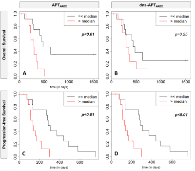 |
98 |
Amide proton transfer (APT) MRI is a predictor of survival and progression in high-grade glioma patients
Daniel Paech, Constantin Dreher, Sebastian Regnery, Jan-Eric Meissner, Steffen Goerke, Johannes Windschuh, Katerina Deike-Hofmann, Sebastian Bickelhaupt, Alexander Radbruch, Moritz Zaiss, Andreas Unterberg, Wolfgang Wick, Martin Bendszus, Peter Bachert, Mark Ladd, Heinz-Peter Schlemmer
In this work we show that relaxation-compensated amide proton transfer (APT) imaging at 7.0 T is associated with overall survival and progression-free survival in newly-diagnosed, previously untreated glioma patients. The current study showed that glioma patients with increased APT values were more likely to progress sooner and live shorter, respectively. This effect may be caused by strong alterations of amino acid concentrations and global upregulation of protein expression in more aggressive brain tumors. Therefore, APT CEST imaging may help to enhance the prognostic value of non-invasive MRI tools at the time of initial diagnosis and during follow-up.
|
|
2920.
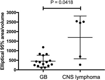 |
99 |
Voxel-based Analyses of SUVs and ADCs with a 18FDG-PET/MR System in Differentiating CNS Lymphomas from Glioblastomas
Daichi Momosaka, Akio Hiwatashi, Koji Sagiyama, Osamu Togao, Kazufumi Kikuchi, Tomohiro Nakayama, Keisuke Ishimatsu, Ryotaro Kamei, Shingo Baba, Hiroshi Honda
Using a hybrid PET/MR system that enables precise image co-registration of 18FDG-PET and DWI, we evaluate the utility of voxel-based analysis of SUVs and ADCs to differentiate CNS lymphomas from GBs. Fifteen GBs and five CNS lymphomas were enrolled. DWI and 18FDG-PET were acquired. Volume, SUVmax, ADCmin, correlation coefficient, elliptical 95% area of bivariate normal distribution of SUVs and ADCs and elliptical 95% area/volume were evaluated. Elliptical 95% area/volume in CNS lymphomas was higher than that of GBs and showed the best diagnostic performance. Voxel-based analysis of 18FDG-PET/MR may be useful to differentiate CNS lymphomas from GBs.
|
|
2921.
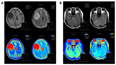 |
100 |
Towards quantitative characterization of brain tumors using synthetic MRI: a preliminary study with pathological confirmation
Hui-ming LIU, Tie-bao MENG, Hao-qiang HE, Liangru Ke, Long Qian, Bing Wu, Guo-ping Yin, Chuan-miao XIE
MRI is widely involved for the diagnosis of brain tumors, and offers qualitative assessment of the tumor with regard to the surrounding brain tissues. However, not only visual inspections may introduce subjective bias, there may be cases that pose challenges for diagnosis even with the use of contrast agent. In this work, a novel relaxation quantification approach named synthetic MRI was applied to 40 patients with glioma or meningioma. Our data showed that both T1 and T2 values of glioma were significant higher than that of meningioma. indicating that synthetic MR may be used for classifying different types of brain tumors.
|
|
| Top |
Neurovascular 2
Digital Poster
Neuro
Tuesday, 14 May 2019
| Exhibition Hall |
14:30 - 15:30 |
| |
|
Computer # |
|
2922.
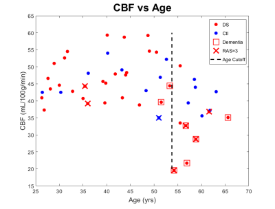 |
101 |
Reduced Cerebral Blood Flow in Aging Adults with Down Syndrome: An Arterial Spin Labeling Study
Scott Thalman, Ai-Ling Lin, Alex Helman, Stacey Brothers, Kathryn O'Connor, Nathan Johnson, Anders Andersen, Katie McCarty, Mary Davis, Gregory Jicha, Allison Caban-Holt, William Robertson, Donita Lightner, David Powell, Elizabeth Head, Frederick Schmitt
Adults with Down syndrome (DS) represent an important model of Alzheimer disease in the absence of cerebrovascular disease (CVD) risk factors like atheroma and hypertension. In this study we used pulsed arterial spin labeling (PASL) to measure cerebral blood flow (CBF) in aging adults with DS and age-matched controls. In adults with DS there is a 31% reduction in CBF after the age of 54 (<54= 46.7mL/100g/min, >54= 32.3mL/100g/min, p=0.011). Despite their CVD protective phenotype, adults with DS do develop vascular dysfunction, however the onset is concurrent with dementia rather than preceding it.
|
|
2923.
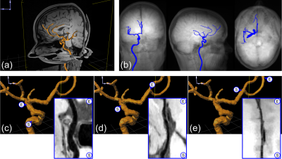 |
102 |
A Dedicated Framework for Intracranial Vessel Wall Imaging: Optimized 3D Dark-blood Imaging and Automated Post-processing
Patrick Vogel, Ute Ludwig, Esther Raithel, Stephan Meckel, Jürgen Hennig, Thorsten Bley, Axel Krafft
MRI has been demonstrated as an indispensable imaging modality in the context of vasculitis – an inflammation of the arterial vessel wall. However, the extent of involvement of intracranial vessels is still part of on-going research. Depiction of intracranial vessels could be achieved with intracranial vessel wall imaging (VWI), which is, however, challenging because of the need for high-resolution within acceptable scan times. Further, the visual assessment of intracranial mural changes is also difficult because of the tortuous vessel course. Our work describes a framework for intracranial VWI with an optimized, compressed-sensing-accelerated sequence prototype and dedicated VWI post-processing.
|
|
2924.
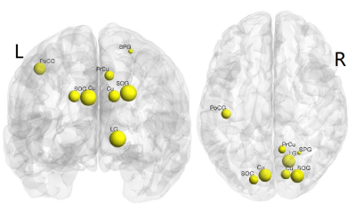 |
103 |
Structural connectivity alterations in CSVD patients with mild cognitive impairment: an atlas-based DTI structural connectome study
Guiling Zhang, Yuanyuan Qin, Wenzhen Zhu
Cerebral small vessel disease(CSVD) is the most common cause of vascular cognitive impairment. Early diagnosis and intervention could prevent patients from progressing to dementia rapidly. DTI deterministic tractography and structural network analysis based on graph theory were utilized to explore the difference of structural connectome between CSVD patients and normal individuals. The results demonstrated the disruption of structural connection at global level and reduction of network efficiency at nodal level. Especially, the decrease of nodal efficiency in the right cuneus gyrus was associated with the mild memory loss in CSVD patients. Our results may provide crucial clues to therapeutic interventions.
|
|
2925
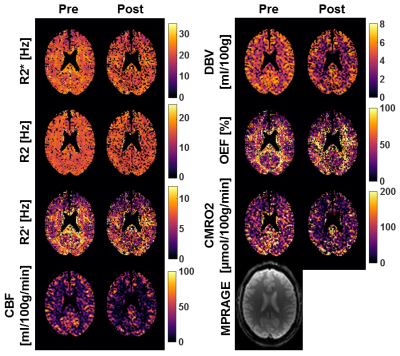 |
104 |
The Acute Effects of Cigarette Smoking on Brain Oxygenation and Perfusion Measured by Multi-Parametric Quantitative BOLD and Arterial Spin Labeling MRI: Preliminary Results
Video Permission Withheld
Sebastian Thomas, Simon Hubertus, Ioanna Skampardoni, Natalie Hartig, Sabine Vollstaedt-Klein*, Lothar R. Schad*
The effects of smoking on human brain hemodynamics have been investigated for many years, but no real consensus was found. In this study, the acute effects of cigarette smoking on the cerebral blood flow, oxygen extraction fraction and cerebral metabolic rate of oxygen are measured with a multi-parametric quantitative BOLD approach. So far, no significant difference between pre- and post-smoking was found. However, the number of subjects will be increased from 5 to 20 in the course of this ongoing study.
|
|
2926.
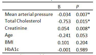 |
105 |
Is cerebral blood flow affected by preventable neurovascular risk factors and physical fitness level?
Lisa van der Kleij, Nina Højland Reislev, Christian Eriksen, Hartwig Siebner, Carl-Johan Boraxbekk, Erik Mortensen, Jeroen Hendrikse, Michael Kjaer, Esben Petersen, Ellen Garde
The health-related effects of aging (biological age) are highly variable across individuals with a similar chronological age. In this study we investigate the relationship between neurovascular health, physical fitness and mean gray matter cerebral blood flow (CBF) in a cohort of 300 home-dwelling volunteers between 62 and 70 years old. In these population-based elderly participants we found that preventable risk factors for neurovascular disease are associated with cerebral blood flow. A high mean arterial pressure, high cholesterol and low creatinine were associated with a decreased gray matter perfusion, whereas physical fitness was not.
|
|
2927.
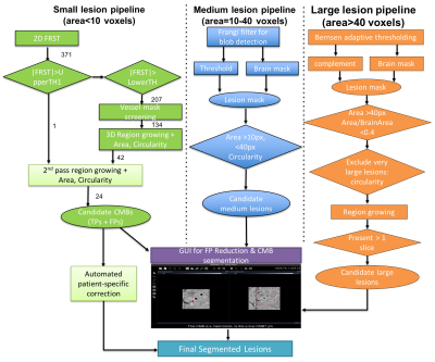 |
106 |
Algorithm for automated lesion segmentation in patients with familial Cerebral Cavernous Malformations(CCM)
Sivakami Avadiappan, Marc Mabray, Blaine Hart, Melanie Morrison, Angela Jakary, Leslie Morrison, Atif Zafar, Helen Kim, Janine Lupo
Cerebral cavernous malformations (CCMs) are collections of small blood vessels in the brain that are enlarged and irregular in structure and have clinical manifestations that include seizures and hemorrhage. In this work, we developed and evaluated an automated algorithm for counting and quantifying different sized CCM lesions on SWI images. The total lesion burden increased with overall symptom score in baseline scans from 50 patients. Large lesion burden increased at follow-up in 16/17 cases. Our automated algorithm is a consistent method for counting microbleeds and accurate volume estimation and can thus facilitate lesion burden calculation and tracking in CCM patients.
|
|
2928.
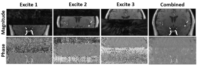 |
107 |
Multiple overlapping slab 4D-flow imaging using distributed-spiral acquisition with magnetization transfer (MT) preparation for inflow signal enhancement and simultaneous 3D time-of-flight (3D-TOF) angiogram
Dahan Kim, Leonardo Rivera-rivera, Patrick Turski, Kevin Johnson
Quantification of 4D-flow MRI relies on signal magnitude and phase of blood but can be affected by high signal intensities from surrounding tissues. By acquiring imaging volume in multiple overlapping slabs, together with MT preparation further to saturate tissue, 3D-TOF inflow enhancement can be used to improve vessel signal in the 4D-flow magnitude image. The scan-time penalty associated with acquiring multiple slabs was avoided by accelerating the scan with undersampled distributed-spiral acquisition. The proposed method greatly increased vessel contrast and improved depiction of blood vessels, which for 4D-flow MRI translate to more reliable flow quantification and vessel segmentations.
|
|
2929.
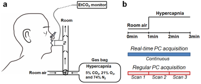 |
108 |
Measuring cerebrovascular reactivity without gas challenge: a real-time phase-contrast MRI approach
Jingran Zhu, Dengrong Jiang, Michael Schär, Dirk Voit, Jens Frahm, Paul Bottomley, Hanzhang Lu, Peiying Liu
Cerebrovascular reactivity (CVR) is typically measured from changes in cerebral perfusion responsive to a hypercapnic gas challenge. Recent attempts to measure CVR using resting-state BOLD fMRI without gas challenge have utilized spontaneous fluctuations in breathing patterns. Since BOLD signal is an indirect measure of cerebral perfusion, a technique that enables cerebral blood flow (CBF)-based CVR under free-breathing conditions is highly desirable. Here, we used a real-time (17 fr/s) PC MRI to measure CBF-based CVR in the resting-state. We evaluated the feasibility of this new approach, and compared it with real-time PC MRI with gas-inhalation, and regular PC MRI with gas-inhalation.
|
|
2930.
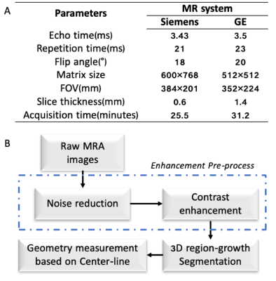 |
109 |
3D Morphological Measurement of Basilar Artery and Its Relationship with CBF Based on TOF-MRA Data in Elders
Zhangyan Yang, Heng Zhang, Zhentao Zuo, Xian Xu, Ningyu An
Morphological features of vascular have a major impact on blood flow and chronic diseases. A 3D semi-automatic vessel geometric feature extraction algorithm was developed in this study, and applied to basilar artery (BA). The relationship among BA morphological parameters can be explained by hemodynamic and flow mechanism. The geometry of BA affected the cerebral blood flow value was demonstrated in this study. This algorithm will save us a lot of efforts for analyzing large dataset, and help clinical doctors to quantify the vessel morphometry rapidly.
|
|
2931.
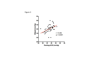 |
110 |
Linearity assessment of the BOLD signal in response to controlled hypercapnea in healthy controls
David Mikulis, Julien Poublanc, Adrian + Laura Crawley
This study addresses concerns over the degree to which the relationship between the cerebral BOLD MRI signal and a CO2 flow stimulus is non-linear. The issue was tested using a quantitative BOLD stimulus in a large population of healthy controls. The results indicate that linear modelling of the response compared to the sigmoid model underestimated the response by ≈ 10%. This would indicate that linear modelling of the BOLD is a reasonable surrogate for flow measurement in subjects undergoing cerebrovascular reactivity testing.
|
|
2932.
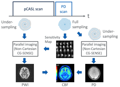 |
111 |
Parallel Imaging Hadamard Encoded pCASL with Efficient Use of Proton Density Weighted Image
Naoyuki Takei, Shota Ishida, Yuki Matta, R Lebel, Hirohiko Kimura, Hiroyuki Kabasawa
A different method of parallel imaging acceleration extracting coil sensitivity maps necessary for using CG SENSE from a proton density weighted image was proposed. Single and multiple post labeling delay (PLD) scan were accelerated while maintaining image quality. The proposed technique is expected to achieve both the robustness of CBF quantification and short imaging time that can be applied clinically.
|
|
2933.
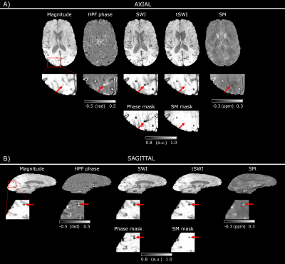 |
112 |
Inaccurate Visualisation of Haemorrhagic Markers in Cerebral Amyloid Angiopathy in Susceptibility Weighted Imaging can be Overcome Using Susceptibility Mapping
Barbara Dymerska, Gargi Banerjee, Emma Dixon, Emma Biondetti, Anna Barnes, Jonathan Schott, Nick Fox, Rolf Jager, Karin Shmueli, David Werring, David Thomas
Cerebral amyloid angiopathy is a small vessel disease characterised by imaging signatures including cerebral microbleeds and cortical superficial siderosis. We show here that non-local phase effects affecting Susceptibility Weighted Imaging (SWI) cause broadening and/or duplication of microbleeds, as well as deformation of superficial siderosis. Furthermore, susceptibility maps and “true SWI”, where local susceptibility values are used, facilitate more accurate microbleed size estimation, reduce the risk of microbleed miscount and provide better delineation of superficial siderosis. Therefore, susceptibility maps and true SWI are likely to be more accurate than SWI in identifying and grading these haemorrhagic markers, with potential clinical relevance.
|
|
2934.
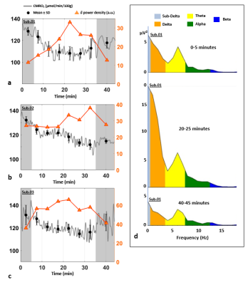 |
113 |
Cerebral Metabolic Rate of Oxygen (CMRO2) during transition from wakefulness to sleep measured by high temporal resolution OxFlow MRI with concurrent EEG recording
Alessandra Caporale, Hyunyeol Lee, Hengyi Rao, Hui Lei, John Detre, Michael Langham, Pei-Hsin Wu, Felix Wehrli
Reduced synaptic transmissions during states of reduced consciousness cause a slowdown in the cerebral metabolic rate of oxygen (CMRO2) and glucose utilization. Prior methods based on PET and tracer kinetics involving repeated blood sampling are not practical in a clinical setting. Here we used whole-brain MR oximetry at 3-second temporal resolution with simultaneous EEG recording to evaluate the feasibility of in-scanner monitoring of brain oxygen metabolism during wakefulness and sleep. The results in three subjects show reduction in CMRO2 up to 15% following onset of sleep paralleling increased delta wave EEG activity and reduction in heart rate.
|
|
2935.
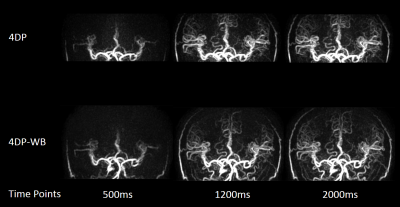 |
114 |
Investigation of Whole Brain Coverage 4D-MR Angiography with Pseudo-Continuous Arterial Spin Labeling Combined with CENTRA-Keyhole and View-sharing (4D-PACK) Accelerated by Compressed-SENSE
Makoto Obara, Osamu Togao, Hiroo Murazaki, Tatsuhiro Wada, Msami Yoneyama, Masanobu Nakamura, Yuta Akamine, Yu Ueda, Marc Van Cauteren
Four dimensional (4D) MR Angiography based on Pseudo-Continuous Arterial Spin Labeling (pCASL) combined with CENTRA-Keyhole and View-sharing (4D-PACK) was accelerated for whole brain 4D-MRA. Compressed-Sensing combined with Sensitivity Encoding (Compressed-SENSE) was used and we demonstrated whole brain 4D-PACK accelerated with Compressed-SENSE. It provides comparable peripheral and posterior cerebral artery visualizations compared with conventional 4D-PACK which has limited anatomical coverage. Validity of whole brain 4D-MRA is suggested using Compressed-SENSE combined 4D-PACK.
|
|
2936.
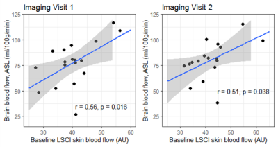 |
115 |
Skin blood flow functions as a potential proxy for cerebral blood flow in adults with sickle cell disease
Brijesh Yadav, Nicolas Currier, Gersham Dent, Paul Swerdlow, Indryas Woldie, Muhammad Shahid, Sean Sethi, Wenting Wang, Kiarash Ghassaban, Willem Birkhoff, Jacobus Burggraaf, Jaspert de Vries, Jaladhar Neelavalli, William Hobbs, E Haacke, Ajay Verma
Even though the prevalence of stroke in adults with sickle cell disease (SCD) is higher compared to children, no accepted screening measures are available for identifying adults with SCD at high risk for strokes. Despite cerebral blood flow (CBF) from transcranial Doppler being an established surrogate measure of stroke risk in children, it is not feasible in adults and MRI is uneconomical and inefficient for screening. We examined skin blood flow using laser speckle contrast imaging as a peripheral surrogate of CBF using MRI. Skin blood flow was highly correlated with CBF in sickle cell patients with excellent test-retest reliability.
|
|
2937.
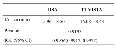 |
116 |
Relationship between morphology and hemodynamics of intracranial aneurysms: A preliminary study
Miaoqi Zhang, Yunduo Li, Fei Peng, Aihua Liu, Rui Li, Chun Yuan
In this study, the purpose was to investigate the relationship between morphology and hemodynamics of IA. Flowavg-APA and Flowmax-IA was positive correlated with IA size and was negatively correlated with mWT. Flowavg-APA was positive correlated with IA size when excluding mWT dependence. And no indices were correlated with mWT when excluding IA size dependence. As blood flows into IA, the volume of aneurysm is enlarged, resulting in the reduction of WT. IA size correlated stronger with blood flow than mWT. This study suggests that mWT and hemodynamics may provide additional information other than size, thus improve current IA risk standard.
|
|
2938.
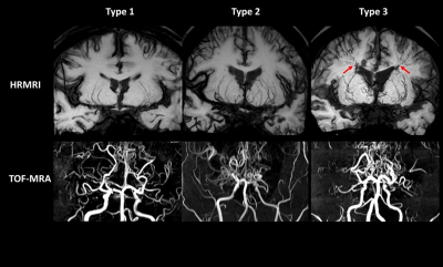 |
117 |
Dilatation, Proliferation, and Abnormal Anastomosis of Lenticulostriate Arteries on Whole-Brain Vessel Wall Imaging as a Predictor of Hemorrhage in Patients with Moyamoya Disease
Fang Wu, Cong Han, Zhiwen Liu, Debiao Li, Zhaoyang Fan, Qi Yang
In this study, we explored distinct branching pattern of lenticulostriate arteries (LSAs) in moyamoya disease (MMD) and identified collateral features of hemorrhagic MMD, using whole-brain vessel wall imaging (WB-VWI). Results showed that patients with MMD more frequently presented with dilated and proliferated LSA branches than patients with atherosclerotic moyamoya syndrome. In patients with MMD, type 3 LSA pattern (dilatation and proliferation of LSA branches with abnormal anastomosis) was associated with hemorrhagic events and vanished middle cerebral artery. These findings suggest that WB-VWI can provide complementary information of abnormal moyamoya vessels and facilitate to predict hemorrhage in MMD.
|
|
2939.
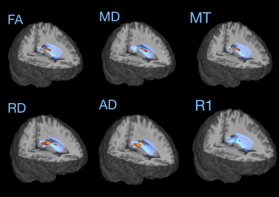 |
118 |
Quantitative Multi-Parametric Mapping reveals specific white-matter tissue changes in Sickle Cell Disease
Hanne Stotesbury, Sara Lorio, Fenella Kirkham , Melanie Koelbel, Sati Sahota, Dawn Saunders, Chris Clark, Jamie Kawadler
Diffusion studies have revealed loss of white-matter integrity in sickle cell disease (SCD), but the underlying pathophysiology is poorly understood. We combined diffusion tensor imaging with multi-parametric mapping in 23 patients and 23 controls to examine more specific imaging markers of myelin, iron, and water content. Voxel-based region of interest analyses revealed a pattern of decreased MT, R1, FA, and AD, and increased RD and MD in patients, consistent with lower myelination. Indices of lower myelination were associated with oxygen desaturation and processing speed. Our results offer insight into pathology of SCD brain changes and potential biomarkers for future trials.
|
|
2940.
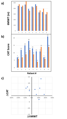 |
119 |
Alteration of cerebrovascular response for COPD patients underwent non-invasive nerve stimulation treatment in nasal cavity
Presentation Not Submitted
Li Tieqiang, Jan-Erik Juto, Karin Sandek, Daniel Alamidi
Chronic obstructive pulmonary disease (COPD) is currently the third leading mortality cause in the world. There is neither a reliable blood biomarkers to confirm its diagnosis nor curative treatment. In this study we tested the hypothesis that the lung function and cerebrovascular responsiveness for COPD patients may be altered by a novel therapy through restoration of ANS homeostasis along with the improvement of clinical symptoms.
|
|
2941.
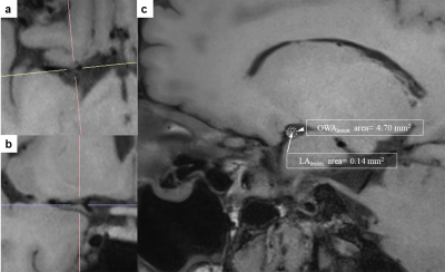 |
120 |
Plaque Characteristics Contribute to Misery Perfusion in Patients with Symptomatic Middle Cerebral Artery Stenosis: A Study of Combined Perfusion MR and High-Resolution Vessel Wall Imaging
Song Liu, Ruowei Tang, Weiwei Xie, Shengting Chai, Wen Shen, Shuang Xia
High-resolution vessel wall imaging(HR-VWI) is an novel technique used to assess intracranial artery stenosis, and it has been useful in clinical practice. In addition, time-to-maximum (Tmax) maps, derived from PWI, are increasingly being used in studies of ischemic stroke, and can provide more prominent cerebral hemodynamic information. Our study aimed to investigate the plaque characteristics in middle cerebral artery (MCA) between patients with and without hypoperfusion area, and to identify the risk factors affecting hypoperfusion area.
|
|
2942.
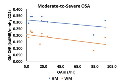 |
121 |
Assessment of obstructive sleep apnea on cerebrovascular health in children with obesity using MRI
Akshdeep Bhatia, Tanvi Naik, Indra Narang, Andrea Kassner
Obstructive sleep apnea (OSA) is a significant contributor to co-morbid disease in obese children. Cerebral endothelial dysfunction and neurocognitive deficits have been linked to obesity-related OSA. However, the effect of the chronic hypoxic environment caused by untreated OSA on cerebrovascular health in the pediatric population remains unclear. Cerebrovascular reactivity (CVR) measured with BOLD MRI in response to a CO2 stimulus is a useful tool in the clinical assessment of cerebrovascular disease. This study seeks to understand the effect of OSA on the cerebrovascular health in obese pediatric patients through MRI assessment.
|
|
2943.
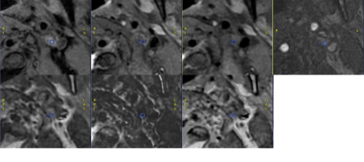 |
122 |
Clinical validation of synthetic MRI in assessing composition of Carotid Atherosclerotic plaques: initial experience
Presentation Not Submitted
Fei Bie, Lihe Cui, Guoguang Fan, Bing Wu, Lizhi Xie
The current study aims to determine the accuracy of synthetic MRI sequences generated from post-acquisition processing of a single sequence compared with sequences imaged from conventional multi-contrast Weighted (MCW) methods in assessing the composition of Carotid artery plaques like calcification and intraplaque hemorrhage (IPH). It was concluded that synthetic MRI sequences are comparable to conventional MCW methods.
|
|
2944.
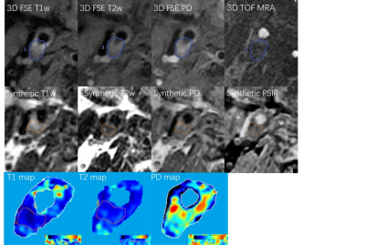 |
123 |
Quantitative characterization of the carotid atherosclerotic plaque composition using synthetic MRI: a preliminary study with histological confirmation
Presentation Not Submitted
Lihe Cui, Fei Bie, Guoguang Fan, Bing Wu
The current study aims to quantify the extent of different composition in carotid artery plaques like intraplaque hemorrhage (IPH) and calcification by Quantitative MR imaging measurements derived from Synthetic MRI technique. We found different components have different distribution range at the value of T1 mapping, T2 mapping and PD mapping. It was concluded that quantitative mapping of synthetic MRI could quantify the extent of different plaque compositions.
|
|
2945.
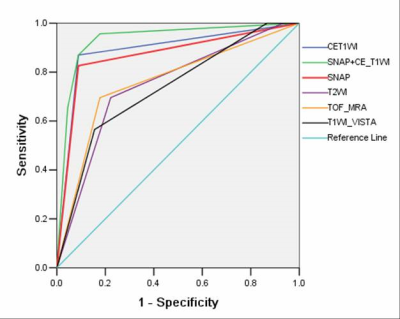 |
124 |
Evaluating of intracranial artery dissection using 3D-SNAP high resolution magnetic resonance:the prospective study
Presentation Not Submitted
Min Tang, Jinglong Gao, Xin Zhang, Jie Gao, Xiaoling Zhang, Xiaohong Wu, Zhizheng Zhuo
The characteristics of intracranial artery dissection were firstly observed using three dimensional simultaneous non-contrast angiography and intra plaque hemorrhage (3D-SNAP) high-resolution magnetic resonance imaging, including intramural hematoma, double lumen and intimal flap. 43 patients were enrolled. The discovery rate of intramural hematoma, false lumen thrombosis were significantly higher in the intracranial artery dissection by 3D-SNAP than other MRI sequences. The discovery rate of double lumen and intimal flap using 3D-SNAP were minor superior to CE-T1WI sequences. The diagnosis efficiency of 3D-SNAP sequences was only lower CE-T1WI, it was the highest diagnostic efficacy in combination with 3D-SNAP and CE-T1WI sequences. Therefore, the application of 3D-SNAP would rise diagnosis rate of intracranial artery dissection, and it may become the first-line screening technology to evaluate the risk factors of stroke caused for intracranial artery dissection.
|
|
2946.
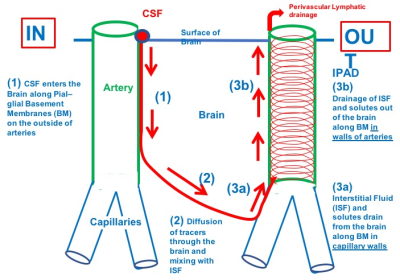 |
125 |
Incidence of enlarged perivascular spaces six-months after aneurysmal subarachnoid hemorrhage: a retrospective MR study
Nivedita Agarwal, Ardalan Zolnourian, Ian Galea, Roxana Carare, Diederik Bulters
One of the major complications of subarachnoid hemorrhage (SAH) is vasospasm which could last days. This could affect the clearance of interstitial fluid (ISF) from the brain parenchyma by restricting its movement along the intramural periarterial drainage (IPAD) pathway. This would result in dilated perivascular spaces (PVS) which have been shown to be an MR biomarker of failure of IPAD. We explore the hypothesis that patients with hemorrhage will present with an increased prevalence of PVS at six months.
|
|
| Top |
Head & Neck
Digital Poster
Neuro
Tuesday, 14 May 2019
| Exhibition Hall |
14:30 - 15:30 |
| |
|
Computer # |
|
2947.
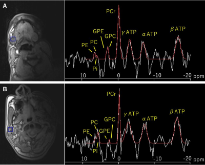 |
126 |
Preliminary Results of 31P Magnetic Resonance Spectroscopy for Human Submandibular and Parotid Glands
Toshiyuki Sato, Hiroyoshi Isoda, Shigeki Arizono, Akihiro Furuta, Hironori Shimizu, Seiya Kawahara, Rinpei Imamine, Tsuyoshi Ohno, Kaori Togashi
We previously performed 31P MRS of the human parotid gland, but no such study of the submandibular gland has yet been reported. The present work was designed to evaluate the potential of 31P MRS, using 3D CSI, to simultaneously measure the phosphorus metabolites of human submandibular and parotid glands. Three healthy volunteers were examined before and after oral intake of vitamin C. Corresponding spectra revealed intense adenosine triphosphate (ATP) and phosphocreatine peaks. Following vitamin C intake, β-ATP decreased in both glands. This preliminary finding indicates that 31P MRS could provide unique information on the bioenergetics of submandibular and parotid glands.
|
|
2948.
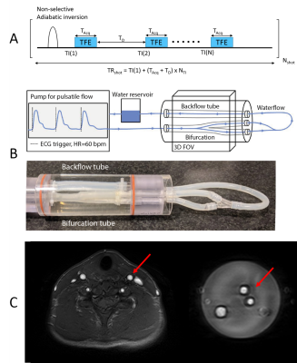 |
127 |
Investigation of arterial blood T1 measurements using cardiac-triggered acquisitions in a pulsatile flow phantom
Koen Baas, Qinwei Zhang, Eva Peper, Gustav Strijkers, Aart Nederveen
We used a pulsatile flow phantom setup to investigate the method by Li et al. (2017)3 to measure arterial blood T1. We found that under different flow conditions the addition of cardiac triggering led to changes in the estimated T1 values. T1 values found in the flowing fluid matched the reference value measured in a no flow condition using a low flip-angle inversion recovery sequence, validating the correctness of our setup.
|
|
2949.
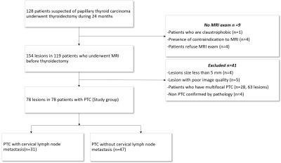 |
128 |
MRI characteristics of papillary thyroid carcinoma for the prediction of cervical lymph node metastasis
Presentation Not Submitted
Hao Wang
Cervical lymph node metastasis is a known prognostic factor in PTC. However, the greatest controversy hampering the routine application of prophylactic central neck dissection is the increased risk of complications in thyroidectomy. In this study, we sought to investigate MRI features for predicting cervical lymph node metastasis in papillary thyroid carcinoma. This study revealed a prediction model built from thyroid contour protrusion sign and poorly tumor margin in contrast enhanced imaging constituted an effective tool for predicting PTC with cervical LNM, which was not reported in previous studies. Meanwhile, age and tumor size could be helpful to distinguish between node-negative and node-positive papillary thyroid cancers.
|
|
2950.
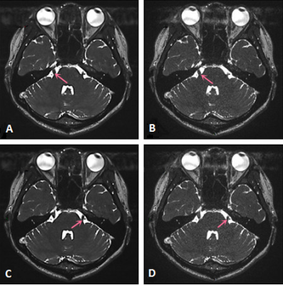 |
129 |
CAIPIRINHA-accelerated 3D constructive interference in the steady state MRI of trigeminal nerve: performance evaluation on image quality at 3.0T
Xiao-qing Sun, Qiong Zhang, Xiao-yue Zhou, Qing-lei Shi, Wei Wang
This study aimed to compare controlled aliasing in parallel imaging results in higher acceleration (CAIPIRINHA) accelerated 3D constructive interference in the steady state (3D-CISS) with conventional CISS, whether image quality can be achieved under the condition of shortening scanning time,we evaluated imaging quality of the trigeminal nerve in 14 healthy subjects,no significant differences of SNR and CNR were found between the two sequences, in other words, CAIPIRINHA- accelerated 3D CISS can improve scanning efficiency without affecting image quality.
|
|
2951.
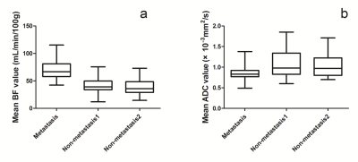 |
130 |
Preliminary study on MRI arterial spin labeling and multi-b value DWI identifying the nature of retropharyngeal lymph nodes in nasopharyngeal carcinoma
Meng Lin, Xiaoduo Yu, Xue Liu, Lizhi Xie, Han Ou-Yang, Denghong Luo
This study using two non-invasive technique as multi-b value DWI and ASL, aimed to compare those derived parameters of metastatic RLNs from NPC patients and non-metastatic retropharyngeal lymph nodes (RLNs) from NPC patients and healthy volunteers, thus to evaluate the diagnostic value of those two series on metastatic RLNs of NPC. It was concluded that the BF, ADC and D could reflect the difference of perfusion and diffusion between metastasis and non-metastasis RLNs, especially the BF which had significant difference. Therefore, ASL and multi-b DWI would be a useful supplement method to conventional diameter criterion to determine the N stage of NPC.
|
|
2952.
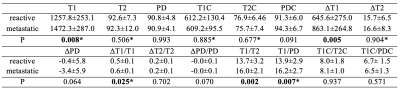 |
131 |
The application of Synthetic MRI in differentiation of metastatic and reactive lymph nodes in patients with nasopharyngeal carcinoma: a preliminary study
Liangru Ke, Tie-bao MENG, Hui-ming LIU, Long Qian, Bing Wu, Hui Li, Yun He, Hao-qiang HE, Chuan-miao XIE
Metastases of nasopharyngeal carcinoma (NPC) in lymph nodes is not uncommon at initial diagnosis, thus the differentiation of the metastatic and reactive lymph nodes is important in accurate staging and precise administration for patients. In current study, a novel MRI quantification method named synthetic MRI was applied to estimate the relaxation values of 71 and 57 metastatic and lymph nodes in 40 patients with NPC, respectively. Our results showed that the relaxation values of lymph nodes were significant different between metastatic and reactive lesions. Moreover, the ration of T1/T2 showed encouraging diagnosis efficiency in differentiation of those two lymph nodes.
|
|
2953.
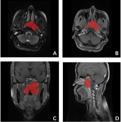 |
132 |
The influence of different MRI scan plane on radiomic features of the Nasopharyngeal Carcinoma
Sulan Wei, Qian Yang, Tianran Li, Miaoru Zhang, Haiyan Pan, Long Qian, Shuangfeng Dai, Dehong Luo
It is known that radiomics has drawn more attentions in radiological research, and has been applied to evaluate the prognosis of nasopharyngeal carcinoma (NPC). However, the influence of different MRI scan plane on the radiomic features of NPC has not been investigated. To address this issue, in current study, contrast-enhanced T1 weighted images (T1WI_C) in axial, coronal and sagittal views with and without axial T2 weighted images were applied to build the radiomics-based models for the prognosis of NPC. Our results showed that radiomics features derived from axial view of T1WI_C had the best performance for the prognosis prediction of NPC.
|
|
2954.
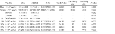 |
133 |
Histogram analysis based on apparent diffusion coefficient maps in differential diagnosis of nasopharyngeal non-Hodgkin lymphoma and nasopharyngeal carcinoma.
Presentation Not Submitted
Keran Ma, Jingliang Cheng, Xiaonan Zhang, Chengru Song
This study aimed to investigate the diagnostic value of the histogram analysis derived from apparent diffusion coefficient (ADC) maps in the differential diagnosis of nasopharyngeal carcinoma (NPC) and nasopharyngeal lymphoma (NPL).Compared with conventional MRI, histogram analysis parameters particularly for 90%ADC and 99%ADC can be used to show the distribution of different intensity signals in the tumor, extract the heterogeneity characteristics of the tumor, understand the tissue density and metabolism inside the lesion, and calculate all the voxels of the whole tumor volume. The results of histogram analysis are more objective, and have a certain advantage compared with the traditional ADC average measurement.
|
|
2955.
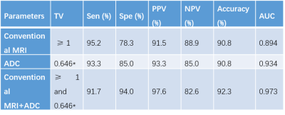 |
134 |
The differentiation between sinonasal natural killer/T-cell lymphomas and diffuse large B-cell lymphomas by high resolution DWI combined with conventional MRI
Mengge He, Zuohua Tang, zebin xiao, Linying Guo, Zhongshuai Zhang
Previous studies already reported that RESOLVE DWI can significantly reduce the susceptibility induced image distortion artifacts [1; 2; 3]. This study used RESOLVE DWI and conventional MRI to differentiate sinonasal natural killer/T-cell lymphomas from diffuse large B-cell lymphomas.
|
|
2956.
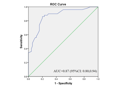 |
135 |
Prediction of Postlaminar Optic Nerve Invasion in Retinoblastoma with MR Imaging Features
Presentation Not Submitted
Zhenzhen Li, Jian Guo, Junfang Xian
Postlaminar optic nerve invasion (PLONI) is a poor prognostic indicator in retinoblastoma. However, it is not easy for both ophthalmologists and radiologists to detect it. Can the combination of conventional MR imaging findings predict PLONI ? This research devoted evidence for this question.
|
|
2957.
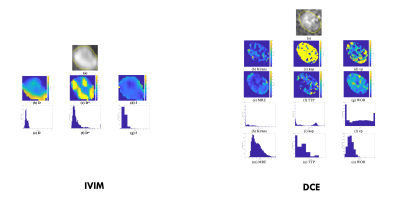 |
136 |
QUANTIFICATION OF HETEROGENEITY TO CLASSIFY BENIGN PAROTID TUMORS
Presentation Not Submitted
Francesca Patella, Mario Sansone, Giuseppe Franceschelli, Roberta Fusco, Mario Petrillo, Filippo Pesapane, Gianpaolo Carrafiello
Warthin tumors (WT) and pleomorphic adenomas (PA) are the most frequent parotid lesions. Despite they are both benign tumors, they are associated with different risk of recurrence and malignant degeneration, which entails diverse management.Benign parotid surgery is challenging since it requires isolation and preservation of the facial nerve, whilst completely removing the tumor.A correct preoperative planning including differential diagnosis, is therefore needed to avoid unnecessary or inadequate surgery. In this study we use functional perfusion and diffusion MR biomarkers and we quantify their heterogeneity by a histogram analysis in order to classify WT and PA.
|
|
2958
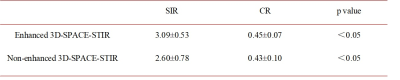 |
137 |
Direct visualization the extracranial segments of the hypoglossal nerve on Enhanced 3D-SPACE-STIR sequence
Video Permission Withheld
Cuifang Chen, Yukun Song, Haiwei Han
In our research, we compared enhanced and non-enhanced 3D-SPACE-STIR sequence in displaying the extracranial segments of the hypoglossal nerve. The SIR and CR of the nerve were higher on enhanced 3D-SPACE-STIR MR imaging than non-enhanced imaging (3.09±0.53 vs. 2.60±0.78 and 0.45±0.07 vs. 0.43±0.10, respectively, p<0.05). Enhanced 3D-SPACE-STIR sequence demostrates better background suppression than non-enhanced. The extracranial segments of the hypoglossal nerve (extracranial-carotid space and extracranial-anterior segments) can be traced continuously on non-enhanced 3D-SPACE-STIR sequence.
|
|
2959.
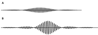 |
138 |
Feasibility of brachial plexus DWI with high acceleration rate using multiband SENSE at 1.5 T
Presentation Not Submitted
Jiazheng Wang, Yajing Zhang, Yishi Wang
Diffusion weighted imaging (DWI) for brachial plexus can provide valuable information for clinical practice, and the accelerated imaging techniques have been extensively evaluated. This study aims to demonstrate the DWI with high acceleration rate on brachial plexus using a multiband SENSE technique at 1.5T system.
|
|
2960.
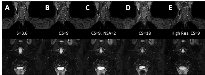 |
139 |
Diagnostic Image Quality and Time reduction Using Compressed SENSE with 3D NerveVIEW for MR Neurography
Brian Johnson, Ivan Dimitrov, Sandeep Ganji, Sergey Cheshkov, Avneesh Chhabra
Magnetic resonance imaging of nerves, also known as MR neurography (MRN) is an emerging technique used to assess peripheral neuropathies. 3D NerveVIEW is a heavily T2-weighted 3D acquisition that has been developed to address common challenges when scanning brachial and lumbosacral plexuses. However, 3D NerveVIEW suffers from long acquisition times. Therefore, in this study we acquired 3D NerveVIEW with compressed SENSE to accelerate scan times and assessed images for motion and pulsation artefacts, nerve signal-to-noise ratio, contrast-to-noise ratio, and nerve signal-to-fat signal ratio. 3D NerveVIEW with compressed SENSE leads to significant time savings without significant changes in image quality.
|
|
2961.
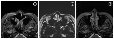 |
140 |
Magnetic resonance Propeller technique in reducing signal loss artifacts in sinus
Presentation Not Submitted
zhang yu, zha yun fei, LIU Chang sheng, yang ren jie
To discuss the clinical application of MR Propeller technique in eliminating the signal loss artifacts of sinus images. 30 cases were performed routine sinus MR examination, underwent Propeller T2 weighted imaging (WI), and the images were compared with IDEAL T2WI sequence. The two image sets were evaluated for overall image quality, signal-to-noise rate(SNR)and contrast-to-noise rate(CNR),respectively. The signal loss artifacts were significantly reduced by Propeller T2WI scanning, and the SNR and CNR, image quality were significantly improved. Propeller T2WI technique can obviously reduce the signal loss artifacts by magnetic-susceptibility, and can significantly improve the image quality for clinical diagnosis
|
|
2962.
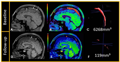 |
141 |
Isointense Clot Sign as a Predictor of Complete Recanalization in Patients with Cerebral Venous Sinus Thrombosis
Presentation Not Submitted
Xiaoxu Yang, Fang Wu, Qi Yang, Yuehong Liu, Jiangang Duan, Ran Meng, Jian Chen, Debiao Li, Zhaoyang Fan, Fisher Marc, Xunming Ji
This study aims to investigate the association of thrombus characteristics with complete recanalization in cerebral venous sinus thrombosis (CVST) patients undergoing endovascular treatment using Magnetic Resonance Black-blood Thrombus Imaging (MRBTI). There was more detection of isointense clot sign (ICS) in complete recanalization group than partial recanalization group. Logistic regression analysis showed that ICS was associated with complete recanalization, which means ICS on MRBTI can be a predictor of complete recanalization in CVST patients having endovascular treatment. So, we think that MRBTI will improve efficacy of endovascular therapy and help eliminate adverse outcomes.
|
|
2963.
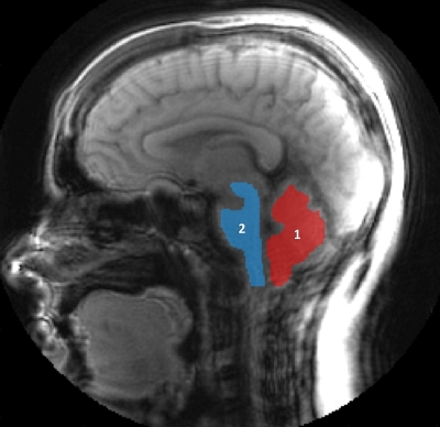 |
142 |
Changes in Cerebellar and Brainstem Tissue Motion in Chiari Malformation Type I Patients Following Posterior Fossa Decompression Surgery
Maggie Eppelheimer, Soroush Heidari Pahlavian, Rouzbeh Amini, Francis Loth, Daniel Barrow, John Oshinski
Chiari type I malformation (CMI) patients have altered brain morphology and tissue motion compared to healthy controls. This motion may be the underlying cause of Chiari symptoms, such as occipital headaches. Highly symptomatic patients may be treated with posterior fossa decompression (PFD) surgery to restore cerebrospinal fluid flow and relieve brainstem compression. To quantitatively assess changes in brain tissue motion before and after surgery, we examined brainstem and cerebellar tissue displacement before and after surgery in 10 patients using spiral cine DENSE MRI. We found that tissue displacement in these structures in CMI patients is significantly reduced by PFD surgery.
|
|
2964.
|
143 |
An investigation of the spatial FOV fold-over oversampling effects in the phase signal of flow-encoding cerebral 2D Cine PC-MRI: an application in phantom and volunteers.
Malek MAKKI, Caroline Fournez, Garance Arbeaumont Trocme, Serge Metanbou
Acquiring high spatial resolution cine phase contrast MRI to quantify the blood flow (arteries or veins) and the cerebrospinal fluid flow (aqueduct or cervical) is challenging. The need for large number of voxels in such small section area is required to accurately quantify the flows measurements. Reduced FOV with fold-over suppression instead of spatial pre-saturation slab is an alternative to such application. The purpose of this study is to investigate the effect of oversampling option in the VNR and both the blood and CSF flow velocities measurements ex-vivo and in-vivo.
|
|
2965.
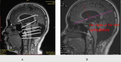 |
144 |
The Use of PC Cine MRI for the Evaluation of Treatment Efficacy of ETV in Patients with Obstructive Hydrocephalus
Presentation Not Submitted
Deying Wen, Xiaoyue Zhou, Jiayu Sun
Phase contrast cine MRI technology can help us understand the pathogenesis of diseases impacting the cerebral ventricular system by quantitatively analyzing intracranial flow of blood and cerebrospinal fluid and can provide a basis for research on new clinical treatments on these diseases. In this study, the non-invasive phase contrast cine MRI technique was used to quantitatively analyze the circulatory dynamics of cerebrospinal fluid and cerebral blood flow in patients with obstructive hydrocephalus. Early postoperative prognosis indicators were determined by comparing patients with healthy controls and by comparing pre- and post-operative results within the patient group.
|
|
2966.
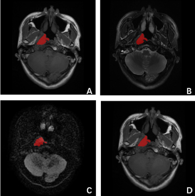 |
145 |
The value of DWI in the pre-treatment prediction of progression in Nasopharyngeal Carcinoma based on MRI radiomics
Miaoru Zhang, Sulan Wei, Tianran Li, Haiyan Pan, Qian Yang, Long Qian, Shuangfeng Dai, Dehong Luo
Radiomics provides a new evaluation method for the prognosis of nasopharyngeal carcinoma (NPC) by extracting high throughput of quantitative descriptors from routinely acquired medical images. However, the research of the prognosis prediction of NPC based on radiomics derived from DWI images hasn’t been reported yet. The present study explores the value of DWI images in the pretreatment predictive of NPC with radiomics methods. By comparing the performance of three classifiers and different MRI sequences, we found that combined DWI and T2W showed optimal pretreatment predictive performance of NPC.
|
|
2967.
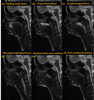 |
146 |
Cine imaging using water (saline) bolus for investigating swallowing problems (dysphagia) in Parkinson’s Disease
Sunita Gudwani, Prabhakar Upadhyay, Kamlesh Sharma, Rajesh Sagar, Rajinder Dhamija, S. Senthil Kumaran
Swallowing a basic oromotor function for survival, investigated with videofluoroscopy (VFS) and flexible endoscopic evaluation (FEES) have limitations either of ionizing radiation, soft-tissue details or of invasiveness. Further clinical examination is challenged with cognitive impairment in degenerative diseases, so non-invasive technique is required for appropriate management. Cine magnetic resonance imaging (dynamic MRI, dMRI) and T2 weighting visualize saline water hyperintensity signal for tissue-bolus contrast without any gadolinium agent. This pilot study reveals dMRI as sensitive non-invasive technique for measuring all the four phases. In motor degenerative diseases like Parkinson’s Disease aids detecting early signs of dysphagia for optimal intervention.
|
|
2968.
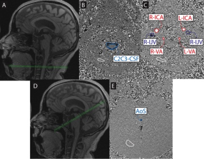 |
147 |
Inefficient cranial venous outflow in patients with Meniere’s disease
Nivedita Agarwal, Christian Contarino, Giuseppenicolò Frau, Eleuterio Toro
Meniere’s disease (MD) patients have a high incidence of abnormal neck venous vessels. This results in hampered cerebral venous outflow, disturbed intracranial dynamics, hampered CFS reabsorption, increased venous pressure and intracranial pressure (ICP), disturbed inner ear circulation and possibly disturbed perilymphatic and endolymphatics fluid spaces. Such disturbed intracranial dynamics may help explain endolymphatic hydrops and underlying mechanisms in MD. In this study we performed magnetic resonance venography (MRV) and phase contrast MRI (PC-MRI) in patients with MD to study the anatomy of the extracranial venous vessels and flow in various cranial compartments.
|
|
| Top |
Novel Neuroimaging Methods
Digital Poster
Neuro
Tuesday, 14 May 2019
| Exhibition Hall |
14:30 - 15:30 |
| |
|
Computer # |
|
2969.
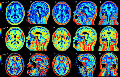 |
151 |
Automatic Brain Tissue Detection using Rapid 3D Quantification of R1 and R2 Rates and Proton Density
Marcel Warntjes, Ida Blystad, Peter Lundberg, Anders Tisell
Absolute quantification of R1 and R2 relaxation rates and proton density PD has been gaining considerable attention in recent years. Quantification provides an absolute scaling of patient properties that is independent of MR scanner settings or imperfections. Simultaneous quantification of R1, R2 and PD has mostly been restricted to 2D acquisitions methods, with relatively thick slices. A novel rapid 3D quantification method provides the maps at 1.2 mm isotropic resolution in 6 minutes scan time. The method was validated against a more established 2D method to measure white matter, grey matter, CSF and myelin volumes in the brain
|
|
2970.
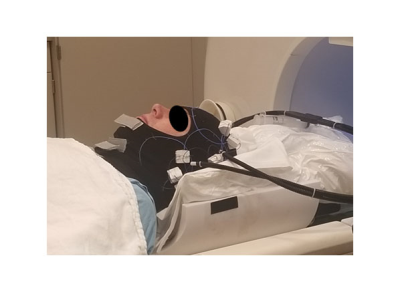 |
152 |
Comparison of AIR Coil Technology to Conventional Head Coils for Neuro Imaging
Petrice Cogswell, Joshua Trzasko, Norbert Campeau, Erin Gray, Phillip Rossman, Daehun Kang, Matt Bernstein, Kiaran McGee, Fraser Robb, Robert Stormont, Scott Lindsay, John Huston III
A 16-channel head coil using novel Adaptive Image Receive (AIR) technology was compared to 8-channel and 32-channel head coils on the Compact 3T system in nine healthy subjects using MPRAGE, FLAIR, GRE, and T2 FSE pulse sequences. Two neuroradiologists graded the AIR coil against the 8-channel and 32-channel coils on signal-to-noise ratio, gray-white matter contrast, lesion depiction, artifact, and overall image quality. On average the AIR coil performed similar to the 8-channel head coil, though not as well as the 32-channel coil. This study demonstrates feasibility of AIR coil technology for imaging of the brain and denotes areas on which future coil designs may improve.
|
|
2971.
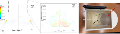 |
153 |
A Single-Compartment Perfusion Phantom for ASL-MRI
Marianna Gabrielyan, Christopher Higgins, Paulo Arratia, John Detre, M. Dylan Tisdall
ASL MRI uses arterial blood water as a diffusible tracer for noninvasive quantification of CBF in physiological units. To produce reliable results across platforms and across subjects, ASL MRI protocols need to be calibrated against a sample with known properties, but it is challenging to simulate tissue perfusion in a mechanical phantom. In this study we present a 3D printed ASL perfusion phantom based on a fluid dynamics model.
|
|
2972.
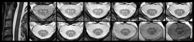 |
154 |
Rapid Volumetric Imaging of the Spinal Cord using AMIRA-3D
Matthias Weigel, Tanja Haas, Oliver Bieri
For fast volumetric spinal cord imaging, a 3D acquisition strategy for the recently published Averaged Magnetization Inversion Recovery Acquisitions (AMIRA) approach was developed: AMIRA-3D. It typically acquires 12 slices with a resolution of 0.67 x 0.67 x 3.0 mm3 in 5:27 min, which represents an acquisition time of just 27 secs per slice. As common to all AMIRA approaches, images with remarkable different tissue contrast are acquired for each slice (or partition) simultaneously. Some of these images can be combined to both enhance and fine-tune the desired tissue contrast.
|
|
2973.
 |
155 |
Clinically viable g-ratio imaging with unified readout at 3T: evaluation and comparison
Francesco Grussu, Marco Battiston, Ferran Prados, Torben Schneider, Enrico Kaden, Rebecca Samson, Daniel Alexander, Claudia Gandini Wheeler-Kingshott
The current way of performing g-ratio imaging is via multi-modal MRI with mixed readouts. This approach can limit the geometrical correspondence of multi-modal maps required for g- ratio calculation. Here we compare g-ratio imaging performed with innovative unified spin echo EPI readout to standard mixed-readout measurements (spoiled gradient echo and spin echo EPI). A unified readout is a feasible alternative to mixed readouts: both provide biologically plausible metrics, with comparable scan-rescan repeatability. Additionally, a unified EPI readout is compatible with multiband acceleration, and enables joint multi-contrast modelling. Our work paves the way for richer multi-contrast acquisitions that could improve g-ratio imaging.
|
|
2974.
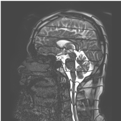 |
156 |
Continuously visualizing slow flow using SSFP
Koichi Oshio, Masao Yui, Seiko Shimizu, Shinya Yamada
A novel technique that can visualize relatively slow flow continously. It is based on SSFP sequence, and gradient pulses are added to introduce spatially linear field gradient. When the steady state is reached, dark bands appear due to the large linear field gradient. When the spins move, these dark band moves following the spin motion, and the flow can be visualized as the motion of the dark bands. Numerical simulation was performed to verify the response to flow, and volunteer images were also taken. It can be a powerful tool to investigate CSF flow.
|
|
2975.
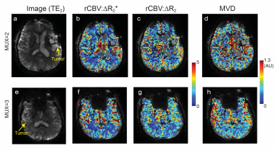 |
157 |
Simultaneous Multi-Slice Spin and Gradient Echo Dynamic Susceptibility-Contrast Perfusion Imaging of Gliomas
Misung Han, Baolian Yang, Brice Fernandez, Marisa Lafontaine, Brian Burns, Suchandrima Banerjee, Janine Lupo
DSC MRI allows for the characterization of first-pass hemodynamics by measuring signal intensity variation resulting from susceptibility changes during the injection of a paramagnetic contrast agent. Simultaneous spin and gradient-echo (SAGE) DSC MRI sequences can provide perfusion parameters that are sensitive to both microvasculature and larger vessels facilitating the measurement of vessel size and correcting for T1 leakage effects. This work evaluates the integration of SAGE EPI with a simultaneous multi-slice excitation to attain high temporal resolution DSC perfusion with full brain coverage. Two protocols with different multi-band acceleration factors were compared in patients with glioma.
|
|
2976.
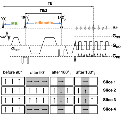 |
158 |
Slice-Accelerated Inner-Volume Cervical Spinal Cord Diffusion MRI
Junqian Xu, Alan Seifert, Joo-won Kim
Cardiac-gated reduced-field of view (FOV) axial cervical spinal cord (C-spine) diffusion MRI (dMRI) has become a mainstay acquisition protocol to achieve tract-specific quantification of tissue microstructure in the C-spine. Building on our previous work in tract-specific C-spine dMRI, we summarize our experiences in combining slice-acceleration (i.e., multiband) with inner-volume-imaging (IVI) for quantitative C-spine dMRI at both 3T and 7T.
|
|
2977.
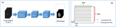 |
159 |
Smart BrainQuant: Ten high quality 3D clinically meaningful contrasts/maps in 6 min on 1.5T using DNN
Mingliang Chen, Aiqi Sun, Wei Xu, Xingxing Zhang, Ruibo Song, Lu Han, Dong Han, Feng Huang
Multi-contrast MR imaging is necessary for clinical diagnosis. Conventional methods require long acquisition time which impede its clinical application. Last year, we proposed a method to acquire 12 high quality contrasts/maps with 0.67×1.33×2.7 mm3 in 8 minutes on 1.5T [1]. In this work, based on deep learning with complete data fidelity, a novel technique called Smart BrainQuant is proposed to further reduce scan time and improve image quality. Feasibility experiments demonstrate that it is achievable to acquire ten 3D contrast/maps with 0.67×0.89×2.7 mm3 in less than 6 minutes on 1.5T with similar image quality to images using the fully acquired data.
|
|
2978.
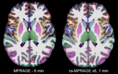 |
160 |
Towards 1 min brain morphometry - evaluating compressed-sensing MPRAGE
Ross Mair, Lindsay Hanford, Emilie Mussard, Tom Hilbert, Tobias Kober, Randy Buckner
A new MR scan acceleration method, employing incoherent undersampling and compressed-sensing reconstructions, can reduce the scan time for a 1.0 mm MPRAGE to 60-90 seconds, depending on acceleration level. We have validated the morphometrics from a prototype compressed-sensing MPRAGE sequence, with different levels of acceleration, with those from conventional MPRAGE scans. Surfaces created from the compressed-sensing MPRAGE images match those from the conventional scan well. Bulk morphometric values such as total gray and white matter volume, and average cortical thickness are similar to those determined with a conventional MPRAGE scan.
|
|
2979.
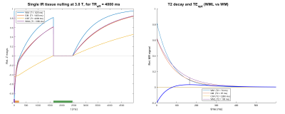 |
161 |
MR Inversion Recovery Simulation and Scanning of Subjects with Focus on White Matter Lesion Contrast Optimization
Presentation Not Submitted
Øystein Gadmar, Anne-Hilde Farstad, Berit Elstad, Piotr Sowa, Wibeke Nordhøy
A MatLab-based inversion recovery sequence simulator/calculator was developed with the purpose of determining and testing optimal parameters for 3D IR acquisitions with the purpose of detecting Multiple Sclerosis lesions in brains. Single inversion FLAIR and dual inversion DIR sequences were studied including a “T1-nulled” DIR sequence removing the undesired T1 weighting inherent in IR sequences to improve lesion-WM contrast. A T2 preparation phase further helps facilitate T1 suppression.
Optimized IR sequences were tested on healthy volunteers and some MS patients, on 1.5 T and 3.0 T MR scanners. Good lesion contrast efficiency with high SNR was found for T1-nulled DIR.
|
|
2980.
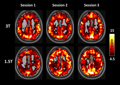 |
162 |
Short- and long-term reliability of BOLD signal change induced by breath holding: Comparison and validation at 1.5T and 3T
Hui-Chieh Yang, Jyun-Ru Chen, Chun-Ming Chen, Yen-Chih Huang, Shin-Lei Peng
cerebrovascular reactivity (CVR) plays important role in the normalization and/or correction of functional magnetic resonance imaging (fMRI) data. But its reliability at the relatively lower field strengths such as 1.5T, to our knowledge, no study has systematically been conducted to date. By performing the systemic evaluations to compare the short- and long-term reliability of BH-induced CVR at 1.5T and 3T, we found reliable measurements of hemodynamic lag of the BOLD response, t-statistics, and activated cortical gray matter at both field strengths, suggesting that CVR measurement can be performed equally well at 1.5T and 3T.
|
|
2981.
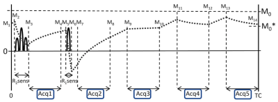 |
163 |
Accelerated Compressed Sensing 3D multi-parametric imaging Toward Isotropic1mm3 Imaging
Naoyuki Takei, Akifumi Hagiwara, Shohei Fujita, Masaaki Hori, Ken-Pin Hwang, Marcel Warntjes, Suchandrima Banerjee, Florian Wiesinger, Shigeki Aoki, Hiroyuki Kabasawa
A multi-parametric technique, 3D QALAS, accelerated using compressed sensing, was implemented to yield images of the brain at high spatial resolution of 1mm isotropic with multiple contrast weightings as well as parametric maps from a single scan. Compressed sensing factor of 1.5 combined with parallel imaging was used to accelerate scan time. The proposed 3D technique is expected to achieve both stable image quality and tissue segmentation accuracy with short imaging time.
|
|
2982.
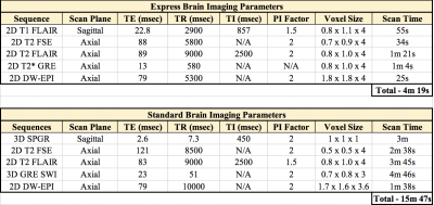 |
164 |
Development of an Express MRI Protocol for Pediatric Brain Imaging in a Clinical Setting
Presentation Not Submitted
Patrick Quarterman, Carly Schwartz, Angela Lignelli-Dipple
The purpose of this study was to develop a routine express MRI protocol for pediatric brain imaging that would provide consistent and robust image quality in one-fourth the scan time of current industry standard protocols, utilizing Fast Spin Echo (FSE) T1, FLAIR, T2w, Gradient Echo (GRE) T2*, and Diffusion Weighted Echo Plane Imaging (DW-EPI) sequences. Evaluation was performed on a group of 20 patients and results indicated that imaging using the express brain protocol was of clinically sufficient diagnostic quality and allowed for a significant reduction in overall scan times compared to industry standard routine brain imaging protocols.
|
|
2983.
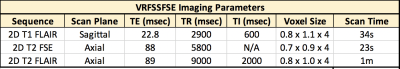 |
165 |
Motion-Robust, Multi-Contrast Rapid Brain Screening Employing Single-Shot FSE with Variable Refocusing Flip Angle
Presentation Not Submitted
Patrick Quarterman, Daniel Litwiller, Jing Zhang, Angela Lignelli-Dipple
This study’s purpose was to compare single-shot with variable refocusing flip angle (vrfSSFSE) and standard multi-shot Fast Spin Echo (FSE) T1, FLAIR and T2w for pediatric brain evaluation. The resulting data shows in 5 subjects that vrfSSFSE provides clinically-acceptable diagnostic quality with a significant reduction in overall examination scan times and reduced motion/ghosting related artifacts typically observed with standard FSE imaging sequences.
|
|
2984.
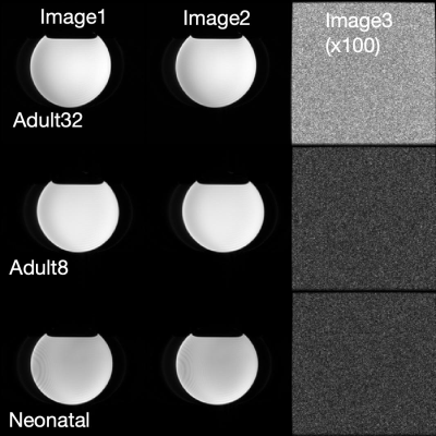 |
166 |
SNR Comparisons of the Coils used for Neonatal Imaging
Li Zhao, Zungho Zun, Subechhya Pradhan, Catherine Limperopoulos
Neonatal brain MRI is widely used in clinical and research scans, in which images are acquired with adult head coils or special neonatal head coils. However, the performances of these coils are not very clear. In this work, we provided detailed comparisons of the signal-to-noise ratio with five noise measurement methods, among adult 8-channel head coil, adult 32-channel head coil, and neonatal 8-channel head coil. The results suggested that adult 8-channel coil has the highest signal-to-noise ratio.
|
|
2985.
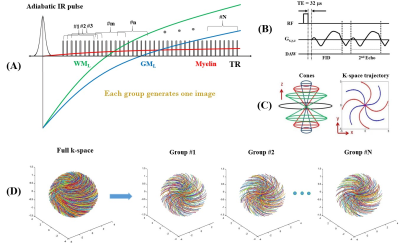 |
167 |
Whole brain myelin imaging using a Double Echo Sliding Inversion Recovery ultrashort echo time (DESIRE-UTE) magnetic resonance imaging
Ya-Jun Ma, Hyungseok Jang, Adam Searleman, Eric Chang, Jody Corey-Bloom, Graeme Bydder, Jiang Du
To image myelin in brain more robustly on clinical scanners, we propose a Double Echo Sliding Inversion Recovery ultrashort echo time (DESIRE-UTE) technique for volumetric myelin imaging in vivo. A series of images with different T1 contrasts can be generated by a single scan. The images with a best signal nulling of long T2 components can be easily found in the second echo image series with a minimum long T2 signals. Then myelin images were generated by dual-echo subtraction for both volunteer and multiple sclerosis (MS) patients. The myelin imaging show clearly signal loss in MS lesions in patient study.
|
|
2986.
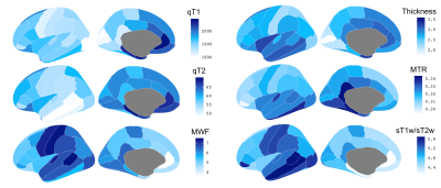 |
168 |
Cellular correlates of multi-parametric magnetic resonance imaging in the human cerebral cortex
Yash Patel, Mark Drakesmith, John Evans, Tomas Paus
To investigate the neurobiological underpinnings of common parametric MRI maps, including T1, T2 relaxation, myelin water fraction (MWF), magnetization transfer ratio (MTR), and synthetic T1w/T2w ratio, using a virtual histology approach. Simply put, we relate inter-regional profiles of these MR measures to inter-regional profiles of cell- specific gene-expression profiles. We observe a relationship between oligodendrocyte- specific gene-expression with profiles of T2, MWF, synthetic T1w/T2w – interpreted as sensitive to intra-cortical myelination. Lastly, MTR was not correlated with oligodendrocyte specific profile, rather with profiles of dendritic arbour enriched gene sets (CA1 pyramidal, and ependymal cells).
|
|
2987.
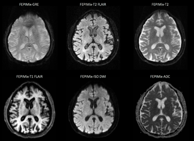 |
169 |
Fast multi-contrast EPI protocol: Preliminary experience with acute intracranial pathologies in the emergency department.
Otto Rapalino, Julian He, Pamela Schaefer, Matthew Christensen, Arnaud Guidon, Stefan Skare, Tim Sprenger, John Kirsch, Ramon Gilberto Gonzalez
Prospective analysis of the diagnostic performance of a fast multi-contrast EPI sequence compared to conventional sequences. A sample of 30 cases (out of 190) was evaluated and head-to-head comparisons were performed for the visualization of clinically relevant intracranial pathologies. Our findings suggest that this multi-contrast EPI sequence could potentially provide an acceptable diagnostic alternative to the core conventional brain MRI sequences in selected populations that cannot tolerate long MRI examinations in the Emergency Department.
|
|
2988.
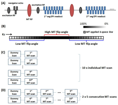 |
170 |
Efficient Quantitative MT (qMT) imaging using 3D Segmented EPI readout Variable Flip angle Magnetization Transfer (EP-vfMT) methods
Se-Hong Oh, Ken Sakaie, Mark Lowe
In this study, newly proposed combined consecutive qMT acquisition scheme was compared with individual MT scan method. Proposed method provides a similar but slightly improved qMT map with slightly higher MT saturation in a clinically feasible scan time. The effect of varying NEX on signal variation of F-map was also investigated. These results suggest that NEX with 2 or 3 is appropriate for the patient scan when considering acquisition time. The EP-vfMT F-map shows high reproducibility (averaged voxel-wise correlation was 0.94 ± 0.03, n=5). These features make the proposed method appealing for clinical neuroimaging applications.
|
|
2989.
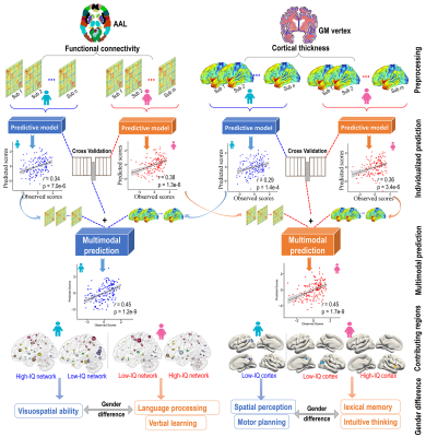 |
171 |
Multimodal data revealed different biological substrates underlying intelligence for young males and females
Rongtao Jiang, Vince Calhoun, Shile Qi, Tianzi Jiang, Jing Sui
Individual differences in intelligence are usually measured using psychometric tests, which cover multifaceted cognitive domains and are strongly predictive of various life outcomes. Here, we employed connectome-based predictive modeling (CPM) to estimate individual’s IQ scores using either cortical thickness in grey matter or resting-state functional connectivity within fully cross-validations for males and females separately. Importantly, integrating multimodal neuroimaging data using CPM achieved improved prediction performance. Interestingly, we found that males and females use distinctively structured brains to achieve similar level of intelligence, consistent with their superiority in cognitive behavior domains (visuospatial processing and logical thinking vs. verbal and memory ability).
|
|
2990.
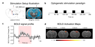 |
172 |
Non-invasive Deep-brain Optogenetic fMRI Mediated by 808 nm Infrared-sensitized Upconversion Nanoparticles
Audrey S. Guo, Alex T.L. Leong, Xunda Wang, Celia M. Dong, Eddie C.S. Wong, Kannie W. Y. Chan, Liming Bian, Ed X. Wu
Typically, optogenetic fMRI is presented at the target region through an implanted optical fiber. Despite the use of small fibers that range from 100µm-400µm in diameter that will ensure minimal brain tissue injury during insertion, it remains an invasive procedure as small brain regions could be easily damaged. In this study, we aim to demonstrate a solution to make non-invasive optogenetic stimulation viable, particularly when used in combination with fMRI to stimulate deep brain regions. We propose the use of our recently developed upconversion nanoparticles (UCNPs), which can be triggered to emit blue light by penetrative near-infrared light (NIR; 808nm) and excite channelrhodopsins (ChR2) expressed in ventral posteromedial (VPM) thalamocortical excitatory neurons.
|
|
2991.
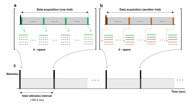 |
173 |
Progress towards direct in-vivo detection and mapping of neuronal activity
Tan-Toi Phan, Hansol Lee, Jeong-Taek Lee, Seung-Kyun Lee, Jang-Yeon Park
Currently, despite much effort to probe the possibility of using MRI for directly detecting and mapping the neuronal activity, the results are inconclusive. Herein, to build upon our previous works, we present new results of using MRI to directly detect and map the neuronal activity with a high temporal resolution up to 4.5 ms. Using a low-flip angle gradient-echo line-scan method, we observed indications of propagating image intensity changes along an electrically stimulated live earthworm at 9.4 T.
|
|
2992.
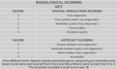 |
174 |
The clinical condition to be assessed by radiological assessment significantly influences the radiological scores of Compressed Sensing accelerated 3D brain MRI
Arnold Julian Vinoj Benjamin, Wajiha Bano, Grant Mair, Gerard Thompson, Michael Davies, Ian Marshall
This study shows that it is important to clearly define the correct clinical question that needs to be answered before the radiological assessment of accelerated 3D gradient echo brain scans for clinical diagnosis.
|
|
2993.
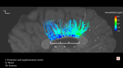 |
175 |
Improved quantitative accuracy using ultrahigh resolution DTI-guided QSM
Lijia Zhang, Chris Petty, Allen Song
Quantitative susceptibility mapping (QSM) has been increasingly used to help access the brain development, especially white matter myelination. However, the quantitative accuracy is limited by its angle dependence to the magnetic field. In this study, ultrahigh resolution diffusion tensor imaging (DTI) was used to delineate the fiber bundles (i.e. corpus callosal fibers), followed by tract-based QSM to minimize the angle dependence and accurately assess magnetic susceptibility changes in different brain regions.
|
|
Back to Program-at-a-Glance |  Back to Top Back to Top |
| The International Society for Magnetic Resonance in Medicine is accredited by the Accreditation Council for Continuing Medical Education to provide continuing medical education for physicians. |
