Digital Poster Session
Neuro Back to Program-at-a-Glance Back to Program-at-a-Glance
|
Tuesday, 14 May 2019
Digital PosterNeuro
2994 -3017 Clinical Traumatic Brain Injury
3018 -3041 Pediatric Neuroradiology: Little Brains
3042 -3066 Alzheimer's Disease: From Mice to People
3067 -3091 Artificial Intelligence Is Taking Over Your Brain 2
3092 -3116 Emerging Technology & Translational Imaging 1
3117 -3139 Epilepsy
3140 -3164 Multiple Sclerosis: Connections & Disruptions
3165 -3189 Alzheimer's & Related Dementia
3190 -3214 Experimental Models of CNS Disease: Functional/Spectroscopy
3215 -3239 Experimental Model of CNS Disease: Structural/Diffusion
3240 -3264 Cerebral Vessel Imaging
3265 -3289 Brains: Functional & Dysfunctional
3290 -3314 Multiple Sclerosis: Quantitative Imaging of Axons, Myelin & More
3315 -3338 Emerging Technology & Translational Imaging 2 |
| |
Clinical Traumatic Brain Injury
Digital Poster
Neuro
Tuesday, 14 May 2019
| Exhibition Hall |
15:45 - 16:45 |
| |
|
Computer # |
|
2994.
 |
1 |
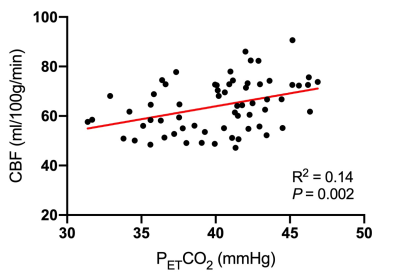
The relationship between baseline PETCO2 measurements and cerebral blood flow: The importance of resting vascular tension in perfusion-based studies

Nicole Coverdale, Allen Champagne, DJ Cook
Measures of cerebral blood flow (CBF) are often used to examine cerebral physiology after sport-related concussion. Carbon dioxide modulates CBF and determines resting vascular tension yet studies rarely account for this. This study examined the effect of the end tidal partial pressure of carbon dioxide (PETCO2) on CBF in athletes. PETCO2accounted for 14% of the variance in CBF and this increased to 37% when age and sex were included. No prior studies examining SRC and CBF have accounted for resting PETCO2. Future studies should move from univariate to multivariate methods to ensure that CBF-based estimates are interpreted correctly.
|
|
2995.
 |
2 |
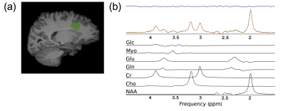
Metabolite Levels Differ in Contact and Non-Contact Sport Female Varsity Athletes
Amy Schranz, Gregory Dekaban, Lisa Fischer, Kevin Blackney, Christy Barreira, Timothy Doherty, Douglas Fraser, Arthur Brown, Jeff Holmes, Ravi Menon, Robert Bartha
Reduced glutamine levels were previously found in the prefrontal white matter of female varsity rugby athletes after a season of play potentially induced by exercise or caused by sub-concussive hits. The current study examined a group of non-contact female varsity athletes and found no changes in glutamine levels, ruling out an exercise effect. Additionally, differences in absolute N-acetyl aspartate, creatine, myo-inositol, glutamate and glutamine were found between rugby players and non-contact athletes. With the future addition of a sedentary group, these data have the potential to elucidate the beneficial and negative effects of exercise and contact play.
|
 |
2996.
 |
3 |
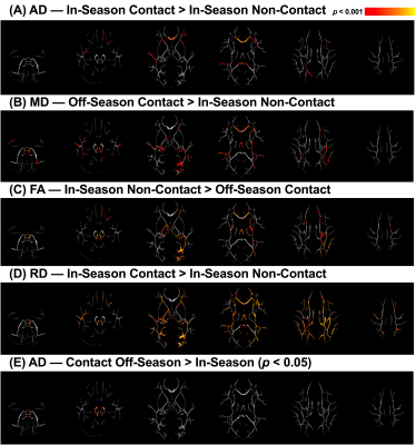
Structural and functional neuroimaging changes in female rugby players with and without a history of concussion
Kathryn Manning, Jeffrey Brooks, Lisa Fischer, Kevin Blackney, Alexandra Harriss, Arthur Brown, Robert Bartha, Tim Doherty, James Dickey, Tatiana Jevremovic, Christy Barreira, Douglas Fraser, Jeff Holmes, Gregory Dekaban, Ravi Menon
In this study we acquired diffusion and resting state fMRI data from female varsity rugby players, rowers and swimmers during the in- and off-season and found (a) significant alterations in the corpus callosum that correlated with altered default mode network connectivity with the posterior cingulate cortex as well as (b) fluctuations in white matter diffusion measures within the brainstem in contact athletes compared to non-contact athletes. Together this suggests that repetitive subclinical impacts incur both acute and long-term changes to brain microstructure and function despite lack of symptoms or even a history of concussion.
|
|
2997.
 |
4 |
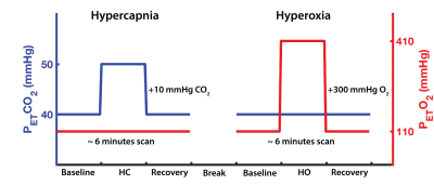
Multi-parametric analysis reveals metabolic and vascular effects driving differences in BOLD cerebrovascular reactivity associated with a history of sport concussion
Allen Champagne, Mike Germuska, Nicole Coverdale, Douglas Cook
In this study, we identified robust differences in BOLD-CVR across the brain which were explained, in part, by hemodynamic parameters relating to CBF modulation, and resting metabolic and vascular physiology. These results emphasize that while BOLD-CVR offers promises as a surrogate biomarker for cerebrovascular health, following sport-concussion, multiple hemodynamic parameters can affect its relative measurements. Thus, multi-parametric approaches like the one proposed here should be considered, in order to better understand how head injuries can relate to changes in the vascular reactivity of the brain, post-injury, and avoid naïve interpretation of neuroimaging findings.
|
|
2998.
 |
5 |
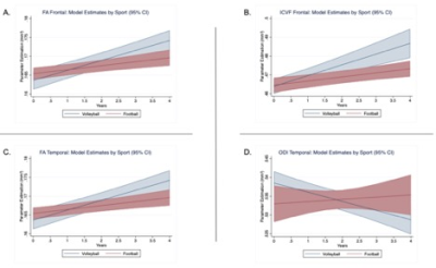
High impact sports and microstructural changes in cortical brain tissue: a 4-year longitudinal study of collegiate athletes
Brian Mills, Maged Goubran, Christian Thaler, Sherveen Parivash, Paymon Rezaii, Wei Bian, Phillip DiGiacomo, Lex Mitchell, Brian Boldt, Jarrett Rosenberg, David Douglas, Jitsupa Wongsripuemtet , Huy Do, Jens Fiehler, Chiara Giordano, Max Wintermark, Gerald Grant, David Camarillo, Michael Zeineh
Exposure to repeated high-velocity impacts may contribute to increased risk of cognitive impairment. However, there has been limited long-term longitudinal investigations into brain tissue change in high-impact sports. In this large 4-year longitudinal DTI study, high (football) and low-contact (volleyball) athletes show a temporal double dissociation in cortical microstructure: in both the frontal and temporal lobes, cortical FA increases over time in volleyball compared to football. While an increase in ICVF underlies this FA increase in frontal cortex, a decrease in ODI underlies this FA increase in temporal cortex. Exposure to high-impact sports may alter cortical microstructural development.
|
|
2999.
 |
6 |
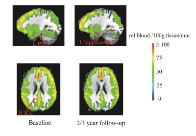
Significant Reductions in Brain Cortical Volumes and Regional Cerebral Blood Flows after Playing College Football 2 to 3 Years
David Zhu, Sally Nogle, Scarlett Doyle, Doozie Russell, David Kaufman
There has been growing concern over sports-related brain injuries and their long-term effects. However, the cumulative effect of sub-concussive hits on the brain is still poorly understood. Twenty-one male Division I collegiate football athletes completed T1 volumetric and arterial spin labeling MRI scans at freshman year with follow-up 2-3 years later. Significant reductions in both brain global and regional cortical volumes were observed. Interestingly, cerebral blood flow was significantly reduced in regions associated with the default-mode network. These changes point to potential long-term effects of sub-concussive hits on the brain.
|
|
3000
 |
7 |
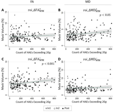
Low-Magnitude Hits Matter: Single-Season Longitudinal DTI Study on Asymptomatic High School Football Players
Video Permission Withheld
Ikbeom Jang, Taylor Lee, Trey Shenk, Victoria Poole, Eric Nauman, Larry Leverenz, Thomas Talavage
The potential consequences of repeated low-magnitude head acceleration events (HAEs) have been less frequently investigated compared to immense HAEs. Retrospective examination of diffusion-weighted-imaging data collected longitudinally from male high school athletes was used to examine the hypotheses that athletes who experience repetitive HAEs will exhibit greater changes in diffusivity than athletes who do not experience repetitive HAEs and that low-magnitude HAEs will affect the integrity of the white matter.
|
|
3001.
 |
8 |
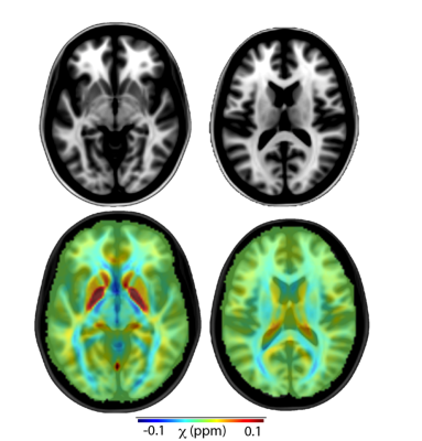
QSM Detects Post-Concussion Changes in Subcortical Gray Matter Susceptibility
Kevin Koch, Brad Swearingen, Robin Karr, Andrew Nencka, L. Tugan Muftuler, Timothy Meier, Michael McCrea
A longitudinal QSM study of sports concussion in 80 injured and control athletes is presented. Regional ROI analysis demonstrated group susceptibility effects that reproduced a previous smaller cohort study finding that QSM diffusely increased in the white matter after sports concussion. In addition, this larger cohort study identified a significant acute trend of decreased susceptibility in sub-cortical gray matter, which is indicative of the calcium influx that is known to occur during the neurometabolic cascade following brain injury. The subcortical gray matter QSM decrease correlated strongly with clinical injury severity metrics.
|
|
3002.
 |
9 |
Neurometabolite changes in College Hockey Players Correlated with Repetitive Head Impacts
Tyler Starr, Katherine Breedlove, Monica Lininger, Molly Charney, Melissa DiFabio, Eduardo Coello, Huijun Liao, Curtis Johnson, Thomas Buckley, Alexander Lin
Repetitive head impacts can lead to long-term cognitive deficits and neurodegenerative diseases such as chronic traumatic encephalopathy. To further understand the effects of repetitive subconcussive head impacts, this study aimed to measure neurochemical concentrations throughout a season of collegiate hockey and examine the relation between subconcussive impacts and neurochemical changes using telemetry and MRS data. As seen in previous studies, players experienced an increase in N-acetyl aspartate and choline. Interestingly, post season NAA was negatively correlated with some telemetry metrics.
|
|
3003.
 |
10 |
Decreased Brain Temperature in Former NFL Athletes
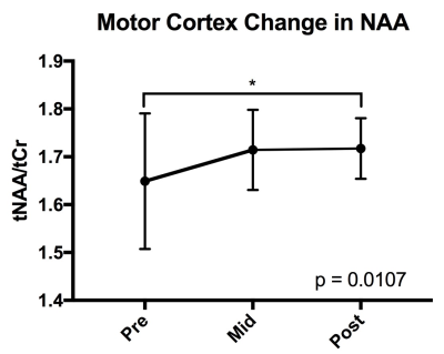
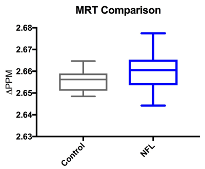
Tyler Starr, Molly Charney, Michael Alosco, Jeffrey Qu, Eduardo Coello, Huijun Liao, Inga Koerte, David Kaufmann, Martha Shenton, Robert Stern, Alexander Lin
Currently, Chronic Traumatic Encephalopathy (CTE) is only diagnosed post-mortem, therefore advanced imaging has an opportunity to identify biomarkers for this disease. This study’s goal is to use Magnetic Resonance Spectroscopy (MRS) and MR Thermography (MRT) to measure cerebral temperature differences between retired former NFL players (n = 50) suspected of CTE and controls (n = 13). The NFL players were found to have lower brain temperature than the controls (p = 0.0340). These finding suggest there is a metabolic difference between those suspected of CTE and healthy controls.
|
|
3004
 |
11 |
Changes in Cerebral Blood Flow after Youth Sport-Related Concussion and with Recovery
Video Permission Withheld
Najratun Nayem Pinky, Carolyn Emery, Chantel Debert, Bradley Goodyear
Mild traumatic brain injury (mTBI), including sport-related concussion, is a major health issue. Changes in cerebral blood flow (CBF) following concussion, as measured by arterial spin labeling (ASL) MRI, may potentially be an indicator of injury or recovery. Compared to healthy controls, we found that CBF was significantly decreased in recently concussed youth ( within 14 days post-injury) within the regions of the occipital and parietal lobes, including the right precuneus. For these regions, CBF of recovered youth was greater than recently concussed and less than controls, though not significantly different from either group.
|
|
3005.
 |
12 |
MR Elastography (MRE)-assessed skull-brain coupling is affected by sports-related repetitive head impacts (RHI)
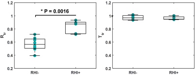
Ziying Yin, Yi Sui, Armando Manduca, Anthony Romano, Richard Ehman, John Huston III
Repetitive head impacts (RHI) in contact sports are known to be associated with altered brain structure and increased concussion susceptibility. Here, MR elastography-based assessment of skull-brain mechanical coupling was used as a new biomarker to assess the RHI-related injury. With novel MRE techniques to directly measure skull-brain displacement, this study aimed to determine the repeatability of MRE-measured mechanical coupling parameters and to assess their changes in RHI subjects. Results demonstrate good repeatability and show preliminary evidence that rotational transmission is significantly higher in RHI group, presumably due to the degradation of the damping capabilities of the protective pia-arachnoid complex following RHI.
|
|
3006.
 |
13 |
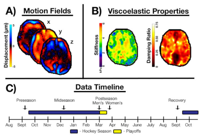
Magnetic resonance elastography of repeated head impacts: Mechanical properties of the brain in collegiate hockey players
Daniel Smith, Melissa DiFabio, Peyton Delgorio, Elizabeth Dickinson, Thomas Buckley, Curtis Johnson
In this study, we use magnetic resonance elastography (MRE) to examine the effects of a season of collegiate hockey on brain biomechanics to better understand the neurological impact of traumatic brain injury. We scanned 13 collegiate-level hockey players at four time points over the course over year using MRE to quantify the possible changes to the viscoelastic mechanical properties caused by repeated head impacts. We discovered that both stiffness and damping ratio changed over the course of the hockey season and then had some recovery after the season, indicating a complex pathology that can be quantified with MRE.
|
|
3007.
 |
14 |
Monitoring subtle brain structural changes following concussion through advanced texture analysis of standard MRI scans
Munib Ali, Shrushrita Sharma, Glen Pridham, Shahnewaz Hossain, Mike Jarrett, Jack Taunton, David Li, Alex Rauscher, Yunyan Zhang
Concussion is a severe health problem and occurs extremely common in contact sports. Clinical MRI is typically used to detect brain abnormalities following injury. However, focal brain pathologies are rarely found. We applied a local spatial frequency-based texture analysis method to evaluate whether invisible MRI changes exist and how they evolve following concussion. Results show that T2 texture spectra decreased uniformly at 2 weeks, continuing at 2 months before recovering thereafter towards baseline in concussed subjects. There were no changes in the non-concussed groups. Advanced texture analysis of clinical MRI may help monitor subtle brain structural changes following concussion.
|
|
3008.
 |
15 |
Magnetic Resonance Imaging to Determine Cerebro-Spinal Fluid Volume to Verify the Role Dehydration Plays in Traumatic Brain Injury
Abi Spicer, Michael Newton, Robert Morris
In contact sports, it is common to see frequent head injuries or indeed traumatic brain injuries (TBI) from minor impacts which anecdotally appear increased as a result of concussion. There is little agreement in the literature regarding the change in CSF volume as a function of dehydration. Here we measure the volume using TrueFISP at 1.5T (Avanto, Siemens, DE) and thresholding images to determine the number of CSF voxels. Imaging reveals a decrease in CSF owing to dehydration. New rehydration regimens should allow for reduction in TBI incidence.
|
|
3009.
 |
16 |
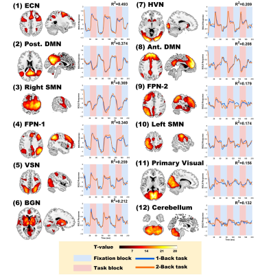
Altered Functional Connectivity during N-Back Task is Associated with Cognitive Deficits in Mild Traumatic Brain Injury
Nai-Chi Chen, Chia-Feng Lu, Li-Chun Hsieh, Sho-Jen Cheng, Yu-Chieh Kao, Cheng-Yu Chen
A subgroup of patients with mild traumatic brain injury (mTBI) suffers from a series of cognitive symptoms, including the memory loss and attention deficit. In our study, we investigated the alterations of functional connectivity during N-back working memory task in 46 mTBI and 43 HC using independent component analysis. Despite both groups revealed comparable performances during task, mTBI showed lower functional connectivity in several task-related neural networks that can be correlated with the cognitive complaints. We concluded that the alterations of neural networks may indicate cognitive symptoms after mTBI.
|
|
3010.
 |
17 |
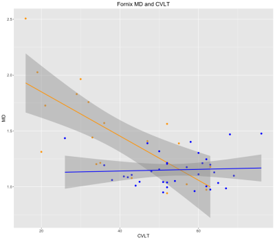
TBI-induced alterations in white matter microstructure relate to impairments in cognition and psychological functioning
Jose Guerrero Gonzalez, Benjamin Yeske, Nagesh Adluru, Gregory Kirk, Peter Ferrazzano, Andrew Alexander
Diffusion tensor imaging (DTI) was performed one-two years after severe traumatic brain injury in a cohort of pre-adolescent and adolescent children. The study investigated the relationships between DTI measures and variations in performance for memory and executive function, and overall clinical dysfunction. Mean diffusivity (MD) in the fornix correlated with learning and verbal tasks. MD in the corpus callosum and global white matter was related to global fuction.
|
|
3011.
 |
18 |
White Matter Microstructural Change Following Traumatic Brain Injury Assessed by Biophysical Modeling using Simultaneous Multi-Slice Multi-Shell Diffusion MRI
Presentation Not Submitted
Ping-Hong Yeh, Chihwa Song, Cheng Guan Koay, Wei Liu, Grant Bonavia, John Ollinger, Gerard Riedy
Mild traumatic brain injury (mTBI) is difficult to diagnose and characterize. In this study, we applied simultaneous multi-slice multi-shell diffusion MRI to assess white matter microstructural changes in chronic military mTBI. Preliminary results showed parameters derived from diffusion MRI biophysical modeling are superior to the parameters derived from diffusion tensor imaging in differentiating tissues with distinct structural and architectural features, and thus has increased ability to identify microstructural changes in mTBI.
|
|
3012.
 |
19 |
Mild TBI Patients Continue to Recover from Perfusion Deficits Months after Initial Injury Evaluated with Dynamic Susceptibility Contrast Imaging
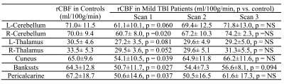
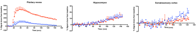
Wei Liu, Ping-Hong Yeh, Chihwa Song, Dominic Nathan, Rael Lange, Louis French, Tracey Brickell, Sara Lippa, Grant Bonavia, John Ollinger, Gerard Riedy
Dynamic susceptibility imaging was performed on 7 mild TBI (mTBI) patients and 16 aged matched controls. Patients were scanned at three intervals: 143 ± 56 days, 277 ± 72 days and 918 ± 353 days after injury. The rCBF of mTBI patients in the cerebellum and cuneus was lower compared to the controls at the fist scan, but continued to increase over time. As a result, mTBI patients demonstrated similar rCBF of the control subjects at the last scan. This finding suggests mTBI patients continue to recover from perfusion deficits months after their initial injury.
|
|
3013.
 |
20 |
Perfusion and brain volume loss after traumatic brain injury
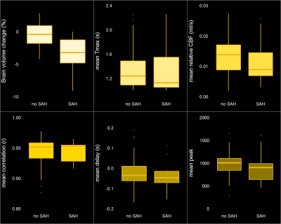
Lisa van der Kleij, Jill De Vis, Matthew Restivo, Lisa Turtzo, Jeroen Hendrikse, Lawrence Latour
How can we identify traumatic brain injury (TBI) patients at risk for long-term brain injury? In this longitudinal study, 57 patients with a relatively good clinical status on admission underwent MRI within 48 hours and at 90 days after injury. Brain volume changes were markedly larger in patients with subarachnoid hemorrhage (-3.2%) compared to patients without subarachnoid hemorrhage (-0.4%; P <0.001). Perfusion was moderately correlated with brain volume change at 90 days (ρ = 0.39; P = 0.003). This demonstrates the utility of imaging markers on acute MRI, especially subarachnoid blood, to identify patients at risk for long-term brain injury.
|
|
3014.
 |
21 |
Analysis of automatically extracted structural features to describe traumatic brain injury severity
Marianna La Rocca, Giuseppe Barisano, Ryan Cabeen, Paul Vespa, Arthur W. Toga, Dominique Duncan
Post-traumatic epilepsy (PTE) prediction is one of the greatest challenges in recent years. The probability of developing PTE is strongly connected with injury severity. Accordingly, having an automated alternative to clinician scoring, to measure injury severity, could be helpful to measure the progression of the disease in view of finding PTE biomarkers. Therefore, we have conducted a study aimed to evaluate if injury severity can be established from automatic analyses of MRI data in a way comparable to manual clinical scoring. We found a statistical association between morphological features and two clinical scores used to quantify injury severity.
|
|
3015.
 |
22 |
Elevated serum inflammation-related cytokines predict longitudinal changes of white matter integrity in mild traumatic brain injury
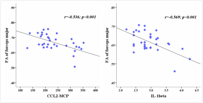
Zhuonan Wang, Lijun Bai, Yingxiang Sun, Bo Yin, Guanghui Bai, Feng Zhu, Kevin Wang, Ming Zhang
Mild traumatic brain injury (mTBI) is the most prevalent neurological insult and approximately 30% of patients have persistently poor clinical outcomes after injury. Evidence indicated both the inflammatory process and white matter (WM) tracts integrity play the crucial roles in clinical outcomes after mTBI. Our study combined inflammation-related cytokines and WM structure changes to examine the dynamic association between white matter integrity and cytokines in mTBI with longitudinal observations. The results had the potential clinical significance and suggested an early intervention on the inflammatory cytokines in order to decrease the structural integrity loss in the WM tracts.
|
|
3016.
 |
23 |
Longitudinal White-Matter Abnormalities in Sports-Related Concussion: A Study of Diffusion Magnetic Resonance Imaging from the NCAA-DoD CARE Consortium
Yu-Chien Wu, Sourajit Mustafi, Jaroslaw Harezlak, Nahla Elsaid, Zikai Lin, Larry Riggen, Kevin Koch, Andrew Nencka, Timothy Meier, Yang Wang, Christopher Giza, John DiFiori, Kevin Guskiewicz, Jason Mihalik, Stephen LaConte, Stefan Duma, Steven Broglio, Michael McCrea, Andrew Saykin, Thomas McAllister
In this study, we investigated the longitudinal recovery trajectories of white-matter microstructures in collegiate athletes who sustained sports-related concussion (SRC). We use diffusion tensor imaging (DTI) to detect white-matter alterations in collegiate athletes longitudinally at four timepoints: 24-48 hours postinjury, the point at which asymptomatic (cleared for return-to-play), seven days following return-to-play, and six months postinjury. We are interested in the extent of white-matter abnormalities over time and whether the white-matter changes persist beyond the point when athletes are considered clinical recovered (i.e., with normal clinical assessments).
|
|
3017.
 |
24 |
Effects of Tract Length in White Matter Alterations After Sports-Related Concussion: A Diffusion MRI Study from the NCAA-DoD CARE Consortium
Yu-Chien Wu, Sourajit Mustafi, Jaroslaw Harezlak, Nahla Elsaid, Larry Riggen, Kevin Koch, Andrew Nencka, Timothy Meier, Yang Wang, Christopher Giza, John DiFiori, Kevin Guskiewicz, Jason Mihalik, Stephen LaConte, Stefan Duma, Steven Broglio, Michael McCrea, Andrew Saykin, Thomas McAllister
In the present study, we performed streamline tractography to characterize effects of tract length on white-matter microstructural alterations after sports-related concussion. Streamline length and counts were studied in affected white-matter fiber tracts that were found to have impaired white-matter integrity at some points along the tracts using voxel-based analyses. The results suggested that long fibers in the brains of collegiate athletes who sustained sports-related concussion are more vulnerable to this mild traumatic brain injury.
|
|
| Top |
Pediatric Neuroradiology: Little Brains
Digital Poster
Neuro
Tuesday, 14 May 2019
| Exhibition Hall |
15:45 - 16:45 |
| |
|
Computer # |
|
3018.
 |
26 |
Prediction of language lateralization in pediatric epilepsy patients: nodal efficiencies of clinical diffusion connectomes
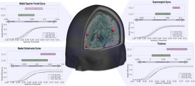
Nolan O'Hara, Min-Hee Lee, Eishi Asano, Jeong-Won Jeong
Language typically utilizes left lateralized brain structures, but its specific localization is heterogeneous, which can complicate surgical approaches to pediatric epilepsy. This study used diffusion weighted connectome to explore the structural network properties of patients clinically characterized as “left language dominant” or “bilateral language dominant.” Nodal efficiency values in canonical language regions were found to be more left lateralized in left language dominant patients, improving prediction of group membership beyond clinical variables and identifying pairwise connections that further distinguished lateralization groups. Our findings support the utility of diffusion connectome in predicting language-dominant hemisphere for presurgical evaluation of pediatric epilepsy surgery.
|
|
3019.
 |
27 |
Detection of abnormal cortical morphology in children and adolescence with intermittent exotropia by anatomic magnetic resonance imaging
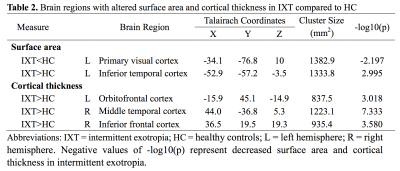
Xi Wang, Lu Lu, Shi Tang, Xiaohang Chen, Meng Liao, Qiyong Gong, Xiaoqi Huang, Longqian Liu
The current study used anatomic magnetic resonance (MR) imaging to evaluate cortical structure alterations, age-related cortical and structural co-variance differences between children with intermittent exotropia (IXT) and healthy controls. The morphologic changes in the visual cortex and associations cortices, different anatomical-age correlation, and abnormal structural co-variance were detected in IXT group. These findings suggest possible disruptions of the cortical visual networks and the cortical maturation in IXT.
|
|
3020.
 |
28 |
Monozygotic twin differences in structural connectivity networks underlying Autism Spectrum Disorder (ASD) symptom severity
Emmanuel Pua, Joseph Yang, Gareth Ball, Jeffrey Craig, Marc Seal
Neurodevelopmental abnormalities in autism spectrum disorders (ASD) have yet to be reliably identified. Recent work suggests that a likely roadblock is the high degree of subject-specific variation in ASD. We previously implemented a validated network analysis method that identified an atypical functional network underlying individual differences in ASD symptom severity. Here we applied the same approach cross-modally to investigate the association between intra-pair differences in structural connectivity networks and within-twin-pair differences in ASD symptom severity in monozygotic twins. A single structural subnetwork was identified with similar hubs implicating the salience and face-perception networks in severity of social deficits in ASD.
|
|
3021.
 |
29 |
Strategically Acquired Gradient Echo (STAGE) Imaging as a Means for Multi-Contrast Quantitative Pediatric Neuroimaging with Minimized Sedation: A Pilot Study in Sturge-Weber Syndrome
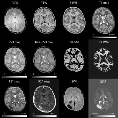
Yongsheng Chen, Yang Xuan, Csaba Juhasz, Jiani Hu, E. Mark Haacke
Non-sedated, non-contrast rapid pediatric magnetic resonance imaging methods are of great interest to pediatric radiology. In this work, we explore the possibility of a multi-contrast, quantitative method referred to as STAGE imaging for minimizing or eliminating sedation in Sturge-Weber Syndrome by using a k-space sharing strategy which increases the resolution of susceptibility weighted imaging and quantitative susceptibility mapping. Preliminary results show the potential of STAGE which generates more than 10 pieces of qualitative and quantitative information in one 5-minute protocol at 3T.
|
|
3022.
 |
30 |
Comparison of 2D BLADE and Spin-Echo Echo-Planar Diffusion-Weighted Brain MRI at 3 Tesla: Preliminary Experience in Children
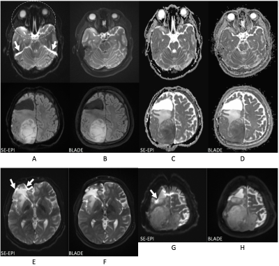
Aaron McAllister, Lacey Lubeley, Bhavani Selvaraj, Ning Jin, Kun Zhou, Mark Smith, Ramkumar Krishnamurthy, Houchun Hu
We describe our preliminary experience using a GRASE (gradient-echo and spin-echo hybrid) based DWI-BLADE pulse sequence in 53 pediatric patients at 3T. On a 4-point scale for rating diagnostic image quality and impact of artifacts, 1 (best) – 4 (worst), a neuroradiologist scored conventional spin-echo EPI DWI 2.4±0.7 whilst BLADE scored 1.1±0.3 (p<0.01). Overall, DWI-BLADE exhibited less geometric distortion at the periphery of the brain, and reduced signal pile-ups at areas of high susceptibility. The pulse sequence is particularly useful in patients with shunts and dental fixtures and is a viable alternative to conventional spin-echo EPI DWI.
|
|
3023.
 |
31 |
MRI quantitative assessment of neonatal hyperbilirubin brain damage
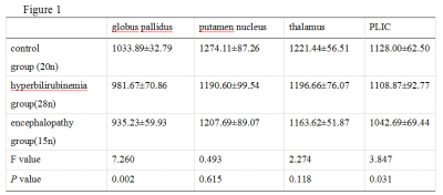
Meng Xiaoli, Wei Xiaocheng, Zhang Lei, Ren Zhuanqin, Yang Ruwu
Neonatal hyperbilirubinemia (NHB) is a common clinical disease and can cause bilirubin encephalopathy in severe cases,which may lead to serious sequelae such as hearing impairment, visual abnormality and mental retardation in children. Quantitatively evaluating the degree of brain damage in neonates with hyperbilirubinemia is of great significance for the prognosis of neonates.In this study, T1 value was measured in different brain regions of newborns with different serum bilirubin levels using T1 mapping, a new magnetic resonance imaging technology of 3.0T.The threshold value of T1 in neonates with bilirubin brain injury was obtained.It provides the quantitative reference index of neonatal hyperbilirubin brain damage for clinic.
|
|
3024.
 |
32 |
INCORPORATING MRS BIOMARKERS INTO MULTICENTER CLINICAL TRIALS: QUALITY ASSURANCE RESULTS FROM THE HIGH-DOSE ERYTHROPOIETIN FOR ASPHYXIA AND ENCEPHALOPATHY (HEAL) TRIAL
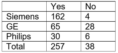
Jessica Wisnowski, Yvonne Wu, Ashok Panigrahy, Amit Mathur, Sandra Juul, Robert McKinstry, Stefan Bluml
MR Spectroscopy (MRS) provides early biomarkers of brain injury and treatment response in neonates with hypoxic-ischemic encephalopathy. We present preliminary data from the High-dose Erythropoietin for Asphyxia and Encephalopathy (HEAL) Trial (NCT02811263), comparing quality assurance parameters across MR vendors. Overall, we have been able to analyze MRS data obtained from 85% of patients who underwent MRI, although this rate is lower at sites operating GE MR systems. 92% of spectra met quality standards, with slight differences in FWHM and SNR by vendor. Overall, these data demonstrate the feasibility of obtaining reliable MRS data in a multicenter neonatal randomized controlled trial.
|
|
3025.
 |
33 |
R2* relaxation rate of white matter in neonates and correlation with clinical predictors of hypoxic ischemic encephalopathy
Yuting Zhang, Alexander Rauscher, Alexander Weber
Objective: To evaluate the potential correlation between the clinical predictors of hypoxic ischemic encephalopathy (HIE) and R2* relaxation rate, and the correlation between R2* and radial diffusivity (RD), axial diffusivity (AD). Methods: We obtained mean R2*, RD and AD within the whole white matter of 19 term infants with clinical diagnosis of HIE and 12 healthy controls. Results: R2*, RD and AD did not differ significantly between the healthy controls and infants with HIE. R2* did not associate with the clinical predictors of HIE. Reduced R2* was correlated with increased RD and AD. Conclusion: R2*, RD and AD do not show a clear relationship with clinically defined HIE.
|
|
3026.
 |
34 |
Alterations of brain white matter in infants with begin enlargement of subarachnoid space by assessing conventional diffusion and WMTI metrics.
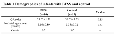
Congcong Liu, Miaomiao Wang, Xianjun Li, Chao Jin, Yannan Cheng, Huifang Zhao, Xingxing Tao, Xiaoyu Wang, Fan Wu, Yuli Zhang, Jian Yang
BESS is characterized by increased cerebrospinal fluid in subarachnoid spaces; some infants with BESS were accompanied with mildly motor and language delay. White matter (WM) development is important to neurodevelopmental outcomes, but relationships between BESS and WM maturation are not very clear. This study aims to quantitative assess WM microstructures of in infant with BESS aged 4-6 months by conventional diffusion and WMTI metrics. Significant decreased FA, α and increased RD, AD, MD, RDe were found in infants with BESS. All results suggested underlying alterations of WM, especially for myelination outside axons. It may provide additional information for neurodevelopment outcomes.
|
|
3027.
 |
35 |
Altered global white matter microstructure and structural brain connectivity in children born with extremely low birth weight
Ulrika Roine, Timo Roine, Viena Tommiska, Terhi Ahola, Aulikko Lano, Taina Autti, Vineta Fellman
Using diffusion MRI, we investigated white matter microstructure and structural brain connectivity in 11-year old children born with extremely low birth weight (ELBW) in comparison with full-term born children. Microstructural white matter properties were investigated within the tract skeleton, and constrained spherical deconvolution based probabilistic tractography and gray matter parcellation were used to reconstruct structural brain connectivity networks. We found decreased integrity and complexity of the white matter microstructure in ELBW, and increased segregation of the structural brain connectivity networks. In addition, the microstructural changes were associated with the administration of antenatal corticosteroids and with retinopathy of prematurity.
|
|
3028.
 |
36 |
Characterization of neurobiochemical profiles in the neonates using GABA-edited Multi-voxel MR spectroscopy
Yan Li, Trevor Flynn, Dawn Gano, Hannah Glass, Donna Ferriero, Anthony James Barkovich, Duan Xu
This study evaluated neurobiochemical profiles in 11 neonates using GABA-edited MR spectroscopy.
|
|
3029.
 |
37 |
What’s shape got to do with it? Exploring subcortical shape and volume alterations in youth with congenital heart disease
Kimberly Fontes, Charles Rohlicek, Christine Saint-Martin, Guillaume Gilbert, Kaitlyn Easson, Annette Majnemer, Mallar Chakravarty, Marie Brossard-Racine
Congenital heart disease (CHD) is a leading cause of long-lasting neurodevelopmental impairment. Evaluating subtle neuroanatomical variation using magnetic resonance imaging data has been shown to be sensitive for capturing morphometric signatures related to neurodevelopmental disorders. In this study, we found morphometric differences in subcortical structures of youth with CHD even in the absence of volumetric differences. While we did not find any significant morphometric differences between groups for the striatum, we did find smaller surface area and inward bilateral inward displacement across the lateral surfaces of the globus pallidus and the thalamus in the CHD group compared to controls.
|
|
3030.
 |
38 |
Characterization of Diffusion Anisotropy Alterations Associated with Dilated Virchow Robin Spaces in Simple Febrile Seizure Children between 12 and 48 Months
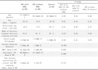
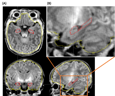
Mustafa Abdelkareem, Xianjun Li, Miaomiao Wang, Congcong Liu, Habib Tafawa, Anuja Pradhan, Martha Singh, Xiaocheng Wei, Guanyu Yang, Jian Yang
Dilated Virchow Robin spaces (dVRs) are common in febrile seizure (FS) patients. However, little is known about how dVRs affect the white matter structure in developing brains. This study aimed to characterize the anisotropy alterations in white matter associated with dVRs in simple FS children by using fractional anisotropy (FA). Through inter-group comparisons, FA was larger in simple FS with dVRs children than that in FS without dVRs and control groups. Significant positive correlations between FA and VRs count, seizure duration were found. These results suggest that dVRs can affect the structure of white matter by increasing FA values.
|
|
3031.
 |
39 |
Age-specific Optimization Strategies of T1-weighted Image Contrasts in Infant Brain
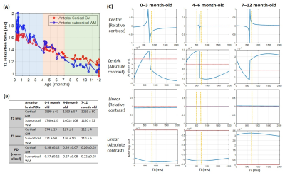
Hongxi Zhang, Ruibin Liu, Tingting Liu, Yi Zhang, Dan Wu
T1-weighted images of the infant brains (≤ 1-year-old) have the inherently low and rapidly-changing contrasts. Previous optimization methods focused on the neonatal brains (≤ 1-month-old), yet the image contrasts in the rest of the infancy are more dynamic and challenging. Here we measured T1, T2 and proton density maps in 58 infant brains at 3T, and performed simulations to maximize the relative white/gray matter contrast using a centrically encoded 3D-MPRAGE sequence. We proposed differential optimization strategies for 0-3 month-old, 4-6 month-old and 7-12 month-old infants. Results demonstrated improved relative contrasts, even in 4-6 month-old infants who had nearly isointense images.
|
|
3032
 |
40 |
The brain-derived neurotrophic factor Val66Met variant is associated with hippocampal volumes in newborn infants
Video Permission Withheld
Yukako Kawasaki, Kenichi Oishi, Antonette Hernandez, Dan Wu, Yoshihisa Otsuka, Can Ceritoglu, Thomas Ernst, Linda Chang
The brain-derived neurotrophic factor (BDNF) Val66Met variant (Met+) is associated with onset of neuropsychiatric disorders. Met+ individuals had smaller hippocampi than those with Met-; whether this phenotype is present at birth is unknown. To minimize postnatal environmental influences, we studied newborn Met+ and Met- infants and compared their hippocampal volumes relative to the intracranial volumes (ICV). Hippocampal volumes and ICVs were automatically parcellated. The Met+ group had significantly smaller % hippocampal volumes than the Met- group (p=0.011). The BDNF Val66Met variant is associated with smaller hippocampal volumes in newborn infants, suggesting the gene’s effects on prenatal hippocampal development.
|
|
3033.
 |
41 |
Atlas-based analysis of brain development from newborn to adolescence using NODDI
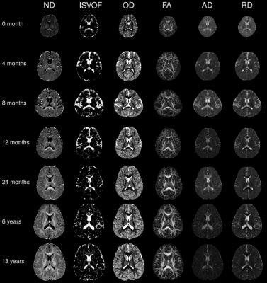
Xueying Zhao, Jingjing Shi, Fei Dai, Wenzhen Zhu, He Wang
Neurite orientation dispersion and density imaging (NODDI) is a specific designed diffusion model for brain, which provides insights into intra-cellular water contents. Here, we investigated the brain development from 0 to 14 years old using NODDI. The whole brain was divided into 159 regions including cortical gray matter, deep gray matter (dGM) and white matter, and was analyzed through exponential regression. Neurite density presented a higher sensitivity to age-related changes than FA, especially in gray matters. Regional specific asymmetry was found between hemispheres in dGM. Sex difference was observed in the developmental rate of GM.
|
|
3034.
 |
42 |
Heterogeneous increase of regional cerebral blood flow and its correlation to functional connectivity during infant brain development
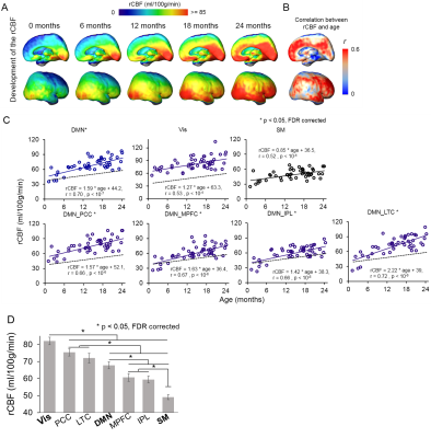
Qinlin Yu, Huiying Kang, Minhui Ouyang, Yun Peng, John Detre, Fang Fang, Hao Huang
The dynamic brain processes during infancy are supported by rapid maturational changes of regional cerebral blood flow (rCBF) to meet the metabolic demands of brain growth. However, the 4D spatiotemporal distribution of regional cerebral blood flow (rCBF) and its relationship to emergence of functional networks are not known. We acquired pseudo-continuous arterial-spin-labeling perfusion MRI and resting-state fMRI from 48 infants to quantify rCBF and functional connectivity (FC), respectively. The rCBF increased significantly faster in default-mode network (DMN) regions than sensorimotor network regions. The rCBF increases at the DMN regions were revealed as the physiological underpinning of emergence of DMN FC.
|
|
3035.
 |
43 |
Longitudinal Multi-contrast Atlas of the Paediatric Brain Acquired with 3 Tesla scanners
Elisa Marchetta, Cristina Baldoli, Antonella Iadanza, Pasquale Della Rosa, Matteo Canini, Sara Cirillo, Andrea Falini, Paola Scifo
This work aims to set up a methodology for the creation of paediatric brain longitudinal atlases by using multimodal 3Tesla MR images. These atlases can be used as a reference of normality and enable to show the developmental trajectories of the brain and its tissues, enhancing the modifications that occur from birth to adulthood.
|
|
3036.
 |
44 |
Validation of Synthetic MRI Brain Volume Segmentation Results in Very Preterm Infants
Maarten Naeyaert, Tim Vanderhasselt, Marcel Warntjes, Hubert Raeymaekers
A novel brain segmentation method, based on quantitative R1, R2 and PD maps measured using a multi-delay multi-echo sequence, was tested on very preterm neonates. The intracranial, brain and CSF volumes and fractions were determined using both the quantitative method and MANTiS, an established atlas-based segmentation method. The results of both methods were compared by using Bland-Altman plots and by quantifying the overlap by co-registering the different segment maps and calculating the Dice score. Despite some systematic differences in the volumetric results, both methods agree well. This study shows that segmentation using quantitative data functions well even for neonates.
|
|
3037.
 |
45 |
Global and regional white matter development in early childhood
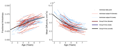
Jess Reynolds, Melody Grohs, Deborah Dewey, Catherine Lebel
White matter development continues into early adulthood, but specific regional trajectories in early childhood remain unclear. We aimed to characterize developmental trajectories and sex differences of white matter in healthy young children. 391 diffusion tensor imaging datasets from 118 children (59 male; 2-7.5 years) were analyzed using tractography. Fractional anisotropy increased and mean diffusivity decreased by 5-15% over the 5.5-year period, likely reflecting increases in myelination and axonal packing. Faster and greater development was observed in males during this period. The preschool period appears to be a critical period for the occipital and limbic connections, which underwent the largest changes.
|
|
3038.
 |
46 |
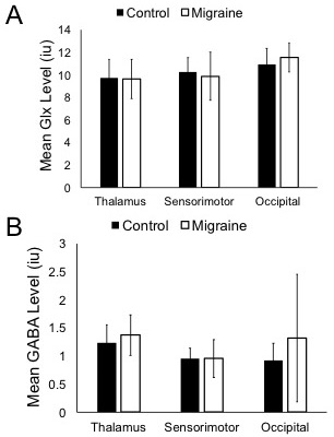
Quantifying changes in excitation and inhibition in childhood migraine
Tiffany Bell, Megan Webb, Melanie Noel, Farnaz Amoozegar, Ashley Harris
Though migraine is one of the top five most common childhood diseases, there has been relatively little investigation into migraine in children. There is evidence of abnormal excitability in the cortex of children with migraine, but levels of excitatory and inhibitory neurotransmitters have not been investigated. We used MRS to compare levels of the neurotransmitters GABA (inhibitory) and Glx (glutamate + glutamine; excitatory) between children with migraine and typically developing controls. We found no significant difference in neurotransmitter levels in the brain of children with migraine; however we found a relationship between neurotransmitter levels and migraine characteristics.
|
|
3039.
|
47 |
Cerebellar anatomical alterations in youth with complex congenital heart disorder
Athena Buckthought, Gabriel Devenyi, Guillaume Gilbert, Christine Saint-Martin, Kimberly Fontes, Kaitlyn Easson, Mallar Chakravarty, Marie Brossard-Racine
Individuals with congenital heart defects (CHD) are vulnerable to long-lasting neurodevelopmental impairments. In this study, we found that youth with CHD had overall smaller total and regional volumes in the cerebellum, when compared to healthy controls of the same age. These differences were statistically significant in 18 of 26 bilateral cerebellar regions, but were not significant in lobules I, II, VI and IX as well as Crus I (bilaterally). These anatomical alterations in many regions could lead to functional impairments since the cerebellum plays a role in many aspects of behavior, including movement, cognition and emotional regulation.
|
|
3040.
 |
48 |
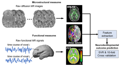
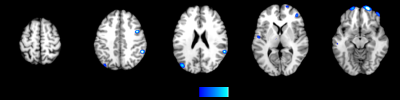
Multimodal brain MRI at birth predicts neurodevelopmental outcome at 2 years of age
Minhui Ouyang, Qinmu Peng, Lina Chalak, Nancy Rollins, Hao Huang
Structural and functional maturation level of brain at birth can be quantified with multimodal MRI, including DTI and resting-state fMRI (rs-fMRI). We hypothesized that features extracted from multimodal brain MRI at birth could better predict the neurodevelopmental outcomes at 2 years of age compared to features extracted from only DTI or rs-fMRI. With combined features of white matter fractional anisotropy and cortical functional connectivity strength from neonatal DTI and rs-fMRI respectively, higher accuracies were achieved using machine learning models to predict Bayley scores at 2 years of age. Heterogeneous feature-contribution patterns were observed across cortical and white matter regions.
|
|
3041.
 |
49 |
MRI study of cortical thickness and regional brain volume in pediatric cancer survivors
Patricia Stefancin, Christine Cahaney, Robert Parker, Thomas Preston, Jessica Goldstein, Rina Meyer, Cara Giannillo, Debra Giugliano, Tim Duong, Laura Hogan
The concept of pediatric chemobrain and the neural mechanisms that underlie its development have not been adequately studied. In this study, MRI was used to examine the neuroanatomy of childhood cancer survivors. We found reduced brain volumes and cortical thicknesses in childhood cancer survivors compared to age-matched controls. These changes were in regions known to be involved in working-memory function and executive function, which could account for the development of executive function difficulties observed in childhood cancer survivors. These findings may prove useful to inform treatment strategies and modify behavioral programs to help survivors combat these issues.
|
|
| Top |
Alzheimer's Disease: From Mice to People
Digital Poster
Neuro
Tuesday, 14 May 2019
| Exhibition Hall |
15:45 - 16:45 |
| |
|
Computer # |
|
3042.
 |
51 |
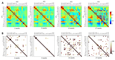
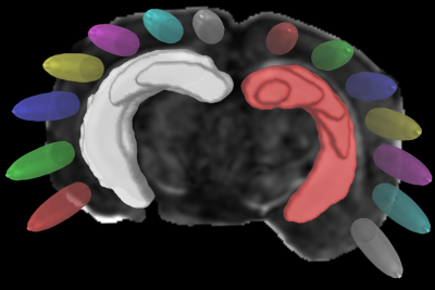
Spatio-temporal alterations in functional connectivity in a rat model of brain glucose metabolism disruption and sporadic Alzheimer’s disease
Yujian Diao, Catarina Tristão Pereira, Ting Yin, Analina da Silva, Rolf Gruetter, Ileana Jelescu
Impaired brain glucose consumption is a possible trigger of Alzheimer’s disease (AD). Animal models can help characterize each contributor to the cascade independently. Here we perform a first-time longitudinal study of brain connectivity in the intracerebroventricular-streptozotocin rat model of AD. We report altered brain circuitry as early as two weeks in regions notoriously affected by AD (cingulate cortices, posterior parietal cortex and hippocampus), and widespread gradual breakdown of connectivity with time. The changes in brain connectivity induced by glucose metabolism disruption can bring further insight into the role of this mechanism in AD.
|
|
3043.
 |
52 |
Sex and Hemispheric Dependent DTI-based Network Analysis of an Alzheimer’s Disease Mouse Model
David Hike, Casey Weiner, Scott Boebinger, Tara Palin, Samuel Grant
This study utilizes DTI and graph theory as a novel way for early detection of pathology and connectivity changes related to Alzheimer’s Disease. As a function of phenotype, age and sex, DTI studies were performed on APP/PS1 mouse brains and age-matched wild type controls at 11.75 T. Current hemisphere-dependentdata shows differences between hemispheres within age and phenotype for the parameters observed.
|
|
3044.
 |
53 |
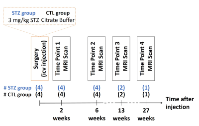
Longitudinal characterization of white matter degeneration in a rat model of brain glucose hypometabolism and sporadic Alzheimer's disease
Catarina Pereira, Ting Yin, Yujian Diao, Analina da Silva, Ileana Jelescu
Impaired brain glucose consumption is a possible trigger of Alzheimer’s disease (AD). Animal models can help characterize each contributor to the cascade independently. Here we use the intracerebroventricular-streptozotocin rat model of AD in a first-time longitudinal study of white matter degeneration using diffusion MRI. Diffusion and kurtosis tensor metrics reveal alterations in the cingulum, fimbria and fornix. The two-compartment WMTI-Watson biophysical model further characterizes the cingulum damage as axonal injury and loss - consistent with previous histopathological studies. White matter degeneration induced by brain glucose metabolism disruption can bring further insight into the role of this mechanism in AD.
|
|
3045.
 |
54 |
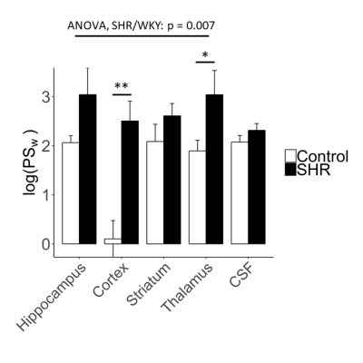
Increased water-exchange across the blood-brain barrier in spontaneously hypertensive rats
Ben Dickie, Hervè Boutin, Geoff Parker, Stuart Allan, Laura Parkes
Blood-brain barrier (BBB) and blood-cerebrospinal fluid barrier (BCSFB) dysfunction are increasingly recognised as pathological hallmarks of vascular dementia and Alzheimer’s disease. Chronic hypertension increases the risk of developing both types of dementia, and may contribute by disrupting the function of blood-brain interfaces. Here we study the permeability of blood-brain and blood-CSF barriers to water using our recently developed multi-flip angle multi-echo (MFAME)-MRI protocol in spontaneously hypertensive rats (SHR). SHRs display increased BBB permeability surface area product to water, relative to age matched controls. Such changes may alter brain water and ion balance and/or contribute to glymphatic dysfunction. Blood-CSF barriers were unaffected.
|
|
3046.
 |
55 |
Hyperactivity of hippocampus-amygdala network during cue-reward association learning of APP/PS1 Alzheimer’s disease model mice detected by 14T-fMRI
Keisuke Sakurai, Teppei Shintani, Tatsuhiro Hisatsune
Dys-regulation of neural network is a biomarker for neurodegenerative disease, such as Alzheimer’s disease. We prepared numbers of 12-month-old APP/PS1 Alzheimer’s disease (AD) model mice and obtained series of BOLD fMRI (SE-EPI) data during cue-reward association learning. We pointed out hyperactivity of amygdala as well as hippocampus in the AD mice, which may involve in a cause for alteration of behavior phenotype in the AD.
|
|
3047.
 |
56 |
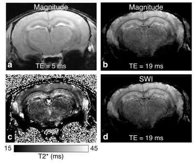
In vivo manganese-enhanced MRI of amyloid pathology in the 5xFAD mouse model of Alzheimer’s disease
Eugene Kim, Diana Cash, Camilla Simmons, Michel Mesquita, Steve Williams, Clive Ballard, Richard Killick
Amyloid plaques are a hallmark of Alzheimer’s disease (AD) but are difficult to detect in vivo due to their small size. We investigated the utility of manganese-enhanced MRI (MEMRI) for visualizing plaques in the 5xFAD mouse model of AD. Plaque-like hypointensities were present in 3D gradient-echo images in all transgenic mice (n=4) but not wild type littermates (n=4). MP2RAGE T1-mapping (n=2/2) revealed reduced manganese uptake in 5xFAD brains, suggesting neurodegeneration. These results demonstrate the potential for MEMRI to provide biomarkers of AD-related neuropathologies that can be useful for monitoring disease progression and therapeutic response in animal models of AD.
|
|
3048.
 |
57 |
Concomitant reduction of glymphatic flow and CBF in mouse model of Alzheimer’s disease
Yunpeng Wang, Zengmin Li, Kai-Hsiang Chuang, Elizabeth Coulson
An impaired glymphatic system has been implicated in the accumulation of toxins such as amyloid beta (Ab) in Alzheimer’s disease. As both glymphatic flow and cerebral blood flow are driven by arterial pressure, we hypothesized that vascular dysfunction represented by reduced cerebral blood flow might contribute to reduced glymphatic flow. Our preliminary results indicate that aged AD mice have both reduced tissue glymphatic flow and reduced cerebral blood flow. Our results suggests that impairment of the glymphatic system in AD may be partly due to impaired cerebrovascular function.
|
|
3049
 |
58 |
Novel diagnosis index for Alzheimer's disease based on a hybrid sequence of QSM and VBM
Video Permission Withheld
Ryota Sato, Kohsuke Kudo, Yasuo Kawata, Niki Udo, Masaaki Matsushima, Ichiro Yabe, Akinori Yamaguchi, Hisaaki Ochi, Yoshitaka Bito, Toru Shirai
Voxel-based morphometry (VBM) is widely used to diagnose Alzheimer’s disease (AD). Recently, several studies have showed that quantitative susceptibility mapping (QSM) is also useful for detecting iron deposition in the early stages of AD. In this study, we propose a novel diagnosis index for AD based on both QSM and VBM, which are simultaneously executed by a single scan. The diagnostic performance of the proposed index in regard to AD and MCI patients is compared with that of the conventional VBM-based index. The comparison results show that the proposed index improved diagnostic performance for discriminating MCI patients and control groups.
|
|
3050.
 |
59 |
An extended-2D CNN approach for diagnosis of Alzheimer’s disease through structural MRI
Mariana Pereira, Roberto Lotufo, Leticia Rittner
Alzheimer's disease (AD) is a devastating type of dementia that affects millions of people around the world. To date, there is no cure for Alzheimer's and its early-diagnosis has been a challenging task. The current techniques for AD diagnosis have explored the structural information of MRI. The aim of this work is to investigate the use of 2D-CNN approaches to distinguish AD patients from MCI and NC using T1-weighted MRI, since most of the works either explored the classic machine-learning or 3D-CNN approaches. The main novelty of our methodology is the use of an extended-2D approach, which explores the volumetric information of the MRI data while maintaining the low costs associated with a 2D approach.
|
|
3051.
 |
60 |
Gaussian Map Descriptors for Alzheimer Detection Using T1-weighted Magnetic Resonance Imaging
Inas Yassine, Nourhan Zayed, Shereen Morsy
Recently, Alzheimer’s Disease (AD) is one of the most emerging elderly diseases. In this study, we propose employing Gaussian map descriptors to discriminate between AD, Mild Cognitive Impairment (MCI) and Normal subjects using T1-weighted Magnetic resonance images (MRI) downloaded from Alzheimer's disease Neuroimaging Initiative (ADNI) website. Extracted Gaussian map descriptors, calculated for the hippocampus, such as Gaussian curvature and mean curvature, were then fed to the support vector machine (SVM) for classification purposes. The Gaussian curvature outperformed mean curvature in case of normal to abnormal, and AD to MCI discrimination with accuracies of 69.5%, and 98.3% respectively.
|
|
3052.
 |
61 |
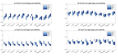
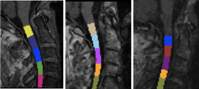
Diagnosis of Alzheimer’s diseases using hippocampal metabolite ratios at the subfield level
Dadi Zhao, Zhao Qing, Bing Zhang, Yu Sun
Hippocampal metabolite ratios can be used as clinical biomarkers to diagnose Alzheimer’s disease (AD), yet the metabolite ratios at a subfield level have been rarely reported, neither for its clinical diagnostic power. We aim to investigate the diagnostic power of metabolite ratios at a subfield level in AD with comparison to the whole level. A quantitative method of metabolite ratios was used, where 2D 1H-MRSI and 3D T1W volumetric MRI were co-registered. Statistical results show subfields have better diagnostic power than the whole hippocampus through metabolite ratios, and also prove the accuracy of the method for AD diagnosis.
|
|
3053.
 |
62 |
Cervical spinal cord atrophy contributes to classification of Alzheimer’s disease and vascular dementia patients
Roberta Lorenzi, Fulvia Palesi, Paolo Vitali, Alfredo Costa, Gloria Castellazzi, Elena Sinforiani, Giuseppe Micieli, Egidio D'Angelo, Claudia Gandini Wheeler-Kingshott
Brain atrophy is an established biomarker for dementia. Here we tested the hypothesis that spinal cord atrophy is also an important in vivo imaging biomarker for neurodegeneration associated with dementia. 3DT1 images of Alzheimer Disease, Vascular Dementia and healthy subjects were processed to calculate spinal cord morphological parameters, such as vertebral spinal cord cross sectional areas and volumes. We confirmed the presence of significant spinal cord atrophy in dementia compared to healthy subjects. In particular, the C2-C3 vertebrae area resulted to have a considerable weight both for discriminating and classifying Alzheimer Disease from Vascular Dementia and Healthy control subjects.
|
|
3054.
 |
63 |
Assessment of dilated perivascular spaces in Alzheimer’s patient and normal aging using 3.0T MR images
Anuja Pradhan, Martha Singh, Tafawa Habib, Mustafa Salimeen, Xianjun Li, Miaomiao Wang, Congcong Liu, Quqiu Min, Guanyu Yang, Jian Yang
Assessment of frequently enlarged perivascular space (EPVS) is essential for assessing Alzheimer’s disease (AD) patients. Recently, EPVS density has been found to be related to the early diagnosis of mild cognitive impairment. However, characteristics of the EPVS density in AD patients are not well understood. We evaluated 44 AD patients and 40 controls by assessing the frequency and density of EPVS in quantitative and semi-quantitative methods. The density and frequency of EPVS is higher in AD patients than that in controls. These results suggest that EPVS density could be used as an indicator in the assessment of EPVS in AD.
|
|
3055.
 |
64 |
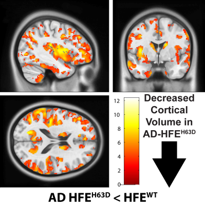
Reduced Brain Volume and Integrity in Alzheimer’s HFEH63D Carriers
Carson Purnell, Jian-Li Wang, Qing Yang, James Connor, Mark Meadowcroft
The data demonstrate that the HFEH63D polymorphism reduces apparent brain integrity in AD carriers. AD-HFEH63D carriers have reduced white matter integrity, increased cortical loss, increased amyloid-beta (Aβ) deposition, and an accelerated disease course trajectory compared to HFEWT carriers in regions susceptible to AD pathology. This work helps decipher how HFE mutations affect AD trajectory, regional susceptibility to AD pathology, brain aging integrity, and cognitive decline.
|
|
3056.
 |
65 |
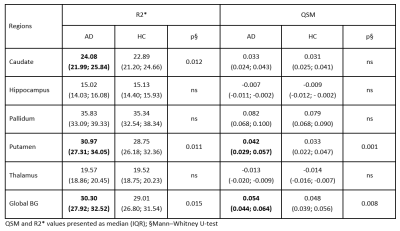
Quantitative Susceptibility Mapping in Alzheimer's Disease
Christian Langkammer, Anna Damulina, Lukas Pirpamer, Maximilian Sackl, Martin Soellradl, Edith Hofer, Marisa Koini, Franz Fazekas, Stefan Ropele, Reinhold Schmidt
Using QSM and R2* mapping we found higher iron levels in specific basal ganglia structures in a cohort of 100 patients with AD when compared to 100 age-matched controls. Iron load in the basal ganglia was negatively correlated with brain volume measures.
|
|
3057.
 |
66 |
Association between T1rho relaxation time and iron deposition among AD patients and normal controls
Chun Ki Au, Jill Abrigo, Chung Tong MOK, Wing Chi AU, Weitian Chen
In this study, we investigated the relationship of iron deposition and T1rho measurement in thalamus among AD patients and healthy controls. Despite the theory indicates elevated iron concentration can decrease T1rho value, we did not observe obvious correlation between iron content and T1rho in this study. This can be due to the fact that the change of iron level is not significant enough to alter T1rho and/or other confounding factors.
|
|
3058.
 |
67 |
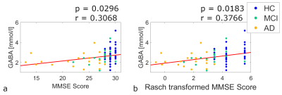
Towards a better understanding of Alzheimer’s Disease: Rasch transformation of cognitive assessment data yields better linear description of cognition using neurometabolite concentrations as explanatory variables
Ariane Fillmer, Jeanette Melin, Leslie Pendrill, Laura Göschel, Stefan Cano, Semiha Aydin, Theresa Köbe, Agnes Flöel, Bernd Ittermann
Due to its non-invasive nature, magnetic resonance spectroscopy is a promising tool for investigating neurochemical disease processes, monitoring potential therapy responses, and diagnosis of Alzheimer’s disease (AD). Changes of γ-amino-butyric acid (GABA) and glutamate (Glu) concentrations have been associated with AD, however, their relationship to other disease parameters is still unknown. This work aims to investigate the relationship of GABA and Glu with cognitive measures and demonstrates that the application of Rasch transformation to cognitive assessment data yields more reliable descriptions of cognitive outcome using metabolite concentrations as explanatory variables.
|
|
3059.
 |
68 |
Cortical Surface-based Index Change in Alzheimer's Continuum: a Structural MRI Study
Qingze Zeng, Xiao Luo, Kaicheng Li, Peiyu Huang, Yong Zhang, Minming Zhang
Alzheimer’s disease remains the most common cause of dementia. To identify morphological difference in an early stage, we used surface-based method to detect the cerebral alternation in the Alzheimer’s continuum (subdivided into Alzheimer’s pathologic change and AD) based on the 2018 NIA-AA research framework. We found that the reductions in surface measures were greater in individuals labeled as AD than in participants with Alzheimer’s pathologic change, while these metrics were more significantly decreased in AD dementia patients. Our findings suggest that AD biological definition would be beneficial for earlier detection which could lead to early diagnosis and intervention.
|
|
3060.
 |
69 |
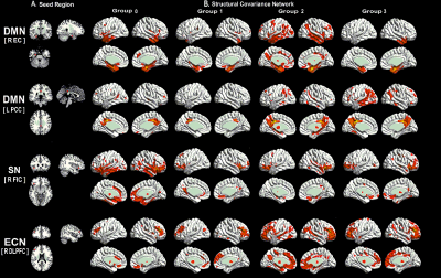
Gray Matter Structural Covariance Networks Changes along the Alzheimer's Disease Continuum
Kaicheng li, Xiao Luo, Qingze Zeng, Peiyu Huang, Yong Zhang, Min-Ming Zhang
Alzheimer’s disease (AD) is a clinical-pathologic entity with a long pathological phase before the dementia onset. The latest ATN classification system is a effective tool in AD research and can provide a more accurate AD stages. Here, we aim to explore the evolution patterns of gray matter structural covariance networks (SCNs) along AD continuum by using the ATN classification system.
|
|
3061.
 |
70 |
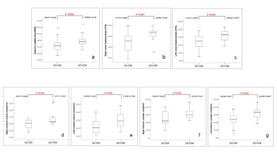
Cerebral venous oxygen saturation variations in Alzheimer's disease complicated with type 2 diabetes patients using susceptibility weighted imaging mapping
Liang Han, Yanwei Miao, Junyi Dong, Xiaoxin Li, Lizhi Xie , Qingwei Song, Ailian Liu
Alzheimer's disease (AD) is a progressive neurodegenerative disease. Epidemiological studies suggest that type 2 diabetes (T2DM) patients are 2 times more likely to develop AD than healthy people. But it is unclear yet why more decreased saturation of blood oxygen and secondary more severe cognitive impairment are present in AD patients with diabetes. Susceptibility weighted imaging (SWI) is widely used in the diagnosis of central nervous system diseases and venous oxygen content is the basis of SWI angiography. As such, this study used SWI mapping to measure the changes of magnetic susceptibility as well as the change of blood oxygen.
|
|
3062.
 |
71 |
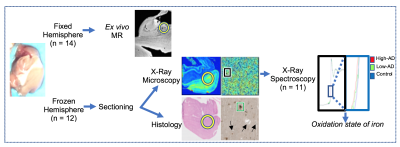
Multimodal microscopic imaging of iron accumulation and oxidation state in the Alzheimer’s disease hippocampus
Phillip DiGiacomo, Samuel Webb, Ed Plowey, Maged Goubran, Sherveen Parivash, Don Born, Brian Rutt, Michael Zeineh
Recent evidence suggests that iron, specifically ferrous Fe2+, may produce oxidative stress in Alzheimer’s disease (AD). However, there remains a gap in our understanding of the progression of iron deposition and its oxidation state. Here, we use X-ray fluorescence imaging (XFI), absorption spectroscopy (XAS), and ultra-high resolution ex vivo MRI in human AD specimens to show that elevated levels of iron correlate with disease severity and to demonstrate that elevated levels of ferrous Fe2+ are present in AD, supporting a neuroinflammatory mechanism. This supports the further development of iron-sensitive MRI as an AD biomarker.
|
|
3063.
 |
72 |
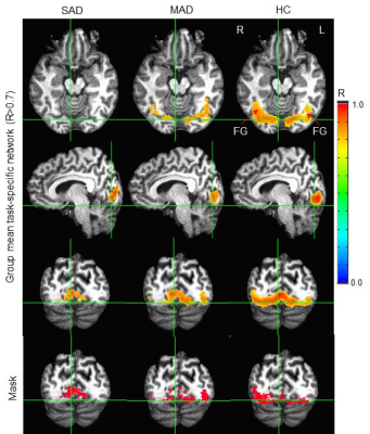
Alzheimer’s disease progressively weakens the face-processing network
Jie Huang, Paul Beach, Andrea Bozoki, David Zhu
A functional area of unitary pooled activity (FAUPA) is defined as an area in which the temporal variation of the activity is the same across the entire area. Using the signal time course of a task-associated FAUPA may identify the functional network specific for the task, and comparing these task-specific networks between healthy controls and those with neurologic diseases may reveal the relationship between task-specific networks and the disease. A cardinal manifestation of later-stage Alzheimer’s disease (AD) is the progressive disintegration of biographical memory and semantic knowledge. This study found an association of task-specific network disruption with AD severity.
|
|
3064.
 |
73 |
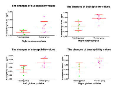
Alters of brain iron deposition in Alzheimer's disease patients after cognitive training: A prospective study
Lei Du, Zifang Zhao, Lizhi Xie, Guolin Ma
The increased iron deposition on brain quantitative susceptibility mapping (QSM) has been proved to be correlated with the decreased cognitive function of Alzheimer’s disease (AD) patients, while cognitive training seems an effective intervention for AD patients in clinic. This study quantifies the change of iron deposition of brain tissues before and after cognitive training and further explores the correlation between the change of iron deposition and the change of mini-mental state examination (MMSE) and Montreal cognitive assessment (MoCA) scores of mild AD patients. The results indicate that cognitive training can relieve iron deposition in right caudate nucleus (p<0.05), right hippocampus (p<0.01) and bilateral globus pallidus (p<0.05). However, there is no correlation between the change of iron deposition of brain tissues and the change of MMSE and MoCA scores, suggesting that cognitive training might be helpful to diminish disease progression of mild AD patients.
|
|
3065.
 |
74 |
Brain connectivity and gray matter volume changes following donepezil treatment in Alzheimer’s disease
Gwang-Won Kim, Gwang-Woo Jeong
Donepezil treatment is associated with improved cognitive performance in patients with Alzheimer’s disease (AD), and its clinical effectiveness has been demonstrated. However, it has been unknown how donepezil treatment influences white matter (WM) connectivity and gray matter (GM) morphology in AD. The purpose of this study was to evaluate the thalamo-cortical white matter connectivity and GM volume after donepezil treatment in patients with AD using probabilistic tractography and voxel-based morphometry (VBM).
|
|
3066.
 |
75 |
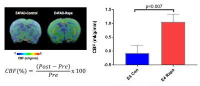
Identifying Rapamycin as a potential preventative therapeutic for Alzheimer’s disease through Multimodal MRl
Mengfan Xia, Ai-Ling Lin
The ε4 allele of apolipoprotein E gene (APOE4) is the strongest genetic risk factor for Alzheimer’s disease (AD). Studies have indicated that APOE4 carriers develop vascular and metabolic dysfunctions several decades prior to the clinical symptom of dementia occurs. In this study, we used multi-modal MRI markers to investigate the effect of Rapamycin, a FDA approved drug, on genetically modified pre-symptomatic E4FAD mice, as a preventative therapeutic for AD. Cerebral blood flow and crucial brain metabolites detected by MR spectroscopy were restored in Rapamycin fed mice, consistent with lower BOLD responses, lower cerebrovascular-reactivity (CVR) and decreased Amyloid-beta deposition.
|
|
| Top |
Artificial Intelligence Is Taking Over Your Brain 2
Digital Poster
Neuro
Tuesday, 14 May 2019
| Exhibition Hall |
15:45 - 16:45 |
| |
|
Computer # |
|
3067.
 |
76 |
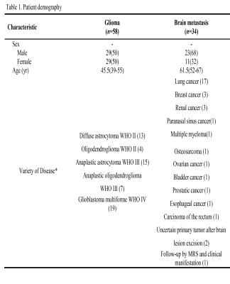
Conventional MR-based machine learning for distinguishing brain glioma and solitary metastasis
Presentation Not Submitted
Zhe Liu, Chao Jin, Xiaotong Liu, Changchang Yin, Ting Liang, Yitong Bian, Yonghao Du, Qinli Sun, Zhongqiang Shi, Buyue Qian, Jian Yang
Differentiation of brain glioma and solitary metastasis is clinically crucial for prescribing the patients’ management and assessing the prognosis. However, indistinguishable signs between two tumors on conventional MRI always embarrass the radiologists and thus lead to high misdiagnosis rate. To address such issue, series of MR features like grey level co-occurrence matrix, histograms of oriented gradient, shape and etc. were first extracted to detail the tumors’ histologic and morphologic characteristics. Then, a gradient-boosting machine learning approach was employed to distinguish the two tumors by the MR features. A good performance with area under receiver operating characteristic curve 0.80, sensitivity 85% and specificity 78% was obtained, suggesting the potential role of our approach in identifying brain glioma and solitary metastasis.
|
|
3068.
 |
77 |
Enhanced isocitrate dehydrogenase mutation classification in lower grade gliomas with deep learning and CEST and MTC MRI
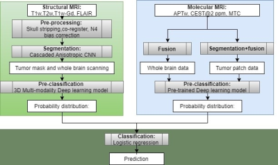
Zuo Wang, Meiyappan Solaiyappan, Qihong Rui, Hye-Young Heo, Zhibo Wen, Gregory Hager, Jinyuan Zhou, Shanshan Jiang
we assess the feasibility of using molecular MRI with deep learning to differentiate IDH mutation status in patients with lower grade gliomas. Two separate deep learning models were used to analyze routine MRI and molecular MRI, and then, a combined model was also devised. 18% and 11% higher AUCs were obtained by the combined system, with respect to the routine MRI subsystem and the molecular MRI subsystem, respectively. Molecular MRI with deep learning algorithm demonstrated a great potential to diagnose IDH mutation status, which could be implemented as a robust approach to enhance routine MRI classification performance.
|
|
3069.
 |
78 |
Grading of glioma using a machine learning framework based on optimized features obtained from T1 perfusion MRI and volumes of tumor components
Anirban Sengupta, Sumeet Agarwal, Rakesh Gupta, Dinil Sasi, Ayan Debnath, Anup Singh
Grading of glioma based on T1 perfusion MRI parameters is well reported but it has certain challenges specially in differentiating intermediate glioma grades (Grade II vs. III and Grade III vs. IV). In this study, we have differentiated intermediate as well as multiclass glioma grades (Grade II vs. III vs. IV) using an optimized machine learning framework which uses quantitative T1 perfusion MRI parameters in combination with volume of different components of tumor as a feature set. The results show that it is feasible to obtain low error in glioma grading using the proposed methodology. The results also emphasizes the utility of using volume of tumor subparts in conjunction with T1 perfusion MRI parameters for glioma grading.
|
|
3070.
 |
79 |
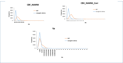
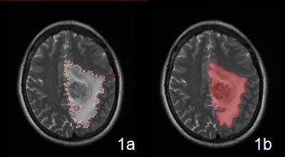
Differentiation of Non-enhancing tumor region from Vasogenic edema in high-grade glioma using a machine learning framework based upon conventional MRI feature
Anirban Sengupta, Neha Vats, Sumeet Agarwal, Rakesh Gupta, Dinil Sasi, Ayan Debnath, Anup Singh
Differentiation between non-enhancing tumor (NET) from vasogenic edema (VE) in glioma patients is difficult using conventional MRI parameters (CMP) such as FLAIR, T2-W, T1-W and PD-W as they appear similar in intensity in both the regions. T1 perfusion MRI parameters (T1-PMP) have been found useful in differentiating between NET and VE previously. The work in this study shows that combining different CMP using a machine learning algorithm improves differentiation between NET and VE substantially over using any individual CMP. However, combination of T1-PMP still performs slightly better than combination of CMP in differentiating NET from VE.
|
|
3071.
 |
80 |
Grading of glioma using a machine learning framework based on optimized features obtained from quantitative DCE-MRI and SWI
Banasmita Kar, Anirban Sengupta, Rupsa Bhattacharjee, Neha Vats, Virendra Yadav, Dinil Sasi, Rakesh Kumar Gupta, Anup Singh
Potential of quantitative dynamic-contrast-enhanced(DCE) MRI parameters in glioma is well reported. However, in some glioma cases, biological behavior of tumors overlap between grades, therefore, correct grading becomes necessary for true classification and further treatment planning. In such cases histopathological glioma grading doesn’t necessarily correlate with DCE-MRI parameters based grading. Objective of this study is to improve the accuracy of grading of glioma using multi-parametric analysis i.e. combining Intra-Tumoral-Susceptibility-Signal(ITSS) volume generated from SWI with DCE-MRI parameters. Using a supervised-machine-learning based approach glioma grading can be improved particularly for the cases where DCE-MRI parameters underperform or vice-versa.
|
|
3072.
 |
81 |
Comparing supervised and unsupervised machine learning frameworks based upon quantitative-MRI features in differentiation between non-enhancing tumor and vasogenic edema of glioma patients and validation using histopathological ground-truth
Neha Vats, Anirban Sengupta, Dinil Sasi, Rakesh Gupta, R.P. Chauhan, Virendra Yadav, Sumeet Agarwal, Anup Singh
The aim of this study was to compare the efficacy of unsupervised machine learning technique in differentiating non-enhancing tumor(NET) from surrounding vasogenic edema (VE) in high-grade glioma patients using T1-perfusion MRI parameters. Two unsupervised machine learning techniques, k-means clustering and Gaussian mixture model (GMM) were optimized with respect to their hyper-parameters for differentiating NET from VE and the results were compared with previously published results obtained using a supervised classifier Support Vector Machine (SVM). The results showed that SVM classifier was slightly superior to GMM and K-means clustering in differentiating NET from VE.
|
|
3073.
 |
82 |
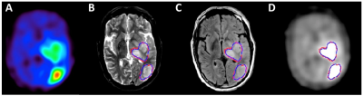
MRI-based deep learning prediction of high amino acid uptake region to improve survival prediction in patents with glioblastoma: A 3D U-net study with deeply learned inter-scanner multi-modal MRI and alpha-[11C]-methyl-L-tryptophan (AMT) PET
Presentation Not Submitted
Jeong-Won Jeong, Min-Hee Lee, Flora John, Sandeep Mittal, Csaba Juhasz
Previous studies found that high amino acid uptake measured by alpha-[11C]-methyl-L-tryptophan (AMT)-PET can accurately detect glioblastoma cell infiltration both in enhancing and non-enhancing tumor portions. However, AMT-PET is not widely available for clinical use. This study explores a novel U-Net which can accurately detect high tryptophan uptake glioblastoma regions using clinical multi-modal MRI data. The resulting U-Net led to 0.85±0.08 sensitivity and 0.99±0.00 specificity to predict AMT-PET tumor regions showing significant negative correlation with survival period, suggesting that an end-to-end deep learning of multi-modal MRI data may be effective for survival prediction of glioblastoma patient without the need of AMT-PET.
|
|
3074.
 |
83 |
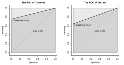
Radiomics approach in differentiating the true progression from pseudoprogression in malignant gliomas treated with concurrent radiotherapy and temozolomide chemotherapy
Yan Bai, Jing Zhou, Wei Wei, Yusong Lin, Meiyun Wang
The conventional magnetic resonance imaging could not confirm the enhancing lesion in malignant gliomas after the standard postsurgical treatment is due to the ture progression or pseudoprogression. The radiomics model based on the selected magnetic resonance imaging features was established to predict the ture progression and pseudoprogression. The radiomics model yielded the AUC value of 0.875 and 0.821 for the train set and test set, respectively. The radiomics model based on the selected contrast-enhanced T1WI features is useful in differentiating the true progression from pseudoprogression in malignant gliomas treated with concurrent radiotherapy and temozolomide chemotherapy after the surgical resection.
|
|
3075.
 |
84 |
Development and Validation of a Radiomics model Based on Conventional MRI for Preoperative Prediction of Gliomas with IDH1 Mutations
Liang Han, Yanwei Miao, Junyi Dong, Xiaoxin Li, Lizhi Xie, KaiYu Wang, Qingwei Song, Ailian Liu
As the most common malignant tumor in the central nervous system, glioma is characterized by low progression-free survival. Studies show that abnormal expression of isocitrate dehydrogenase 1(IDH1) is closely related with the occurrence of brain tumors, especially gliomas. Evidence suggests that gliomas with mutated IDH1 have improved prognosis compared to those with wild-type IDH1.Radiomics means the high-throughput extraction of large amounts of quantitative image features from radiographic images, including segmenting tumors, building models, and then predicting and analyzing those massive feature data to assistphysicians. In this study, the IDH1 mutation was predicted by such radiomics modeling.
|
|
3076.
 |
85 |
A cluster-based diffusion spectrum analysis of diffusion-attenuated signal in glioma
Xueying Zhao, Yan Ren, Xiaoyuan Feng, He Wang
We used a cluster-based method to investigate the diffusion-attenuated signal of glioma patients with different grades. The clustering results were analyzed by diffusion spectrum, which returns a continuous distribution of diffusion coefficient for a given attenuated signal. CSF, gray matter and white matter were clearly separated by Fuzzy C-means clustering. And some clusters showed sensitivity to interface between glioma-related tissues and normal tissues, which can be used for tumor delineation. High grade glioma tended to have clusters with smaller diffusivity and contained more types of clusters than low grade glioma.
|
|
3077.
|
86 |
Testing Machine Learning Algorithms using Anisotropy Indices of Normal Appearing White Matter as Predictors of Molecular Grouping of Gliomas
Hande Halilibrahimoglu, Korhan Polat, Seda Keskin, Oguzhan Aslan, Ozan Genc, Koray Ozduman, Cengiz Yakicier, Esin Ozturk Isik, M. Necmettin Pamir, Alp Dincer, Alpay Ozcan
Grouping gliomas using the telomerase reverse transcriptase (TERT) gene and IDH mutations, and 1p/19q co-deletion status was demonstrated to be useful previously for clinical decisions. MR based radiogenomics might potentially be advantageous. The aim of this study was to determine for the first time whether full distributions of the fractional anisotropy, relative anisotropy and ADC in normal appearing white matter were adequate predictors for machine learning algorithms to classify molecular subgroups based on TERT, IDH and 1p/19q co-deletion information.
|
|
3078.
 |
87 |
Integrating Machine Learning and Image Inpainting to Predict Tumour Invasion in Glioblastoma using multi-parametric MRI
Chao Li, Pan Liu, Shuo Wang, Carola-Bibiane Schönlieb, Stephen Price
The multi-parametric MRI has the potential to compensate for the non-specific contrast-enhancing imaging in delineating tumor margin. The purpose of this study was to propose a method by integrating machine learning with image inpainting to predict the glioblastoma invasion using advanced multi-parametric MRI. The predictive tumor regions using this approach showed significance for patient prognosis, in a cohort containing 115 glioblastoma patients. This approach could advance the scenario of mathematical image analysis by considering both imaging features and brain structure. The predictive region may have significant clinical impact on personalized and targeted surgical treatment of patients.
|
|
3079.
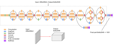 |
88 |
A Transfer Learning-based Radiomics Model for Prediction MGMT Promotor Methylation Status in Glioblastoma Multiforme
Xin Chen, Tianjing Zhang, Zhongping Zhang, Zaiyi Liu
Glioblastoma multiforme (GBM) is the most common malignant brain tumor. MGMT promoter methylation is associated with beneficial chemotherapy. We extract deep features from a pre-trained deep neural network model via transfer learning and generate an effective feature vector model together with radiomics features for an optimal pretreatment prediction of MGMT promoter methylation status. The deep feature set achieved the higher predictive accuracy of 0.86 and 0.70 for validation and test group comparing to handcrafted radiomics feature and combined feature sets. The deep feature model may serve as a potential imaging biomarker for pretreatment prediction of MGMT methylation in GBM.
|
|
3080.
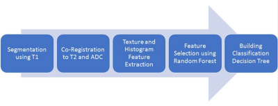 |
89 |
Radiomics Approach for Prediction of Tumor Recurrence and Progression of Skull Base Meningioma
Ching-Chung Ko, Yang Zhang, Jeon Hor Chen, Peter Chang, Daniel Chow, Tiffany Kwong, Min-Ying Su
A subset of low grade skull base meningiomas (SBM) shows early progression/recurrence (P/R). In clinical practice, one of the main challenges in the treatment of SBM is to determine factors that correlate with P/R. This study investigated the role of radiomics for the prediction of P/R. Sixty patients diagnosed with benign SBM were studied. Totally 99 descriptors were extracted from the various MR sequences. The prediction accuracy of P/R was 90% and the AUC of the prediction model was 0.94. Our study also noted that subsequent P/R of SBM after surgery was not associated with the completeness of tumor resection.
|
|
3081.
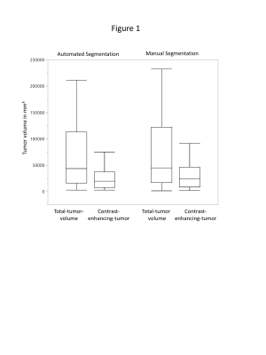 |
90 |
Fully automated segmentation of meningiomas using a specially trained deep-learning-model on multiparametric MRI
Kai Laukamp, Frank Thiele, Lenhard Pennig, Robert Reimer, Georgy Shakirin, David Zopfs, Simon Lennartz, Marco Timmer, David Maintz, Michael Perkuhn, Jan Borggrefe
Volumetric assessment of meningiomas plays an instrumental role in primary assessment and detection of tumor growth. We used a specially trained deep-learning-model on multiparametric MR-data of 116 patients to evaluate performance in automated-segmentation. The deep-learning-model was trained on 249 gliomas, then further adapted by a subgroup of our meningioma patients (n=60). A second group of meningiomas (n=56) was used for testing performance of the deep-learning-model compared to manual-segmentations. The automated-segmentations showed strong correlation to the manual-segmentations: dice-coefficients were 0.87±0.15 for contrast-enhancing-tumor in T1CE and 0.82±0.12 for total-tumor-volume (union of contrast-enhancing-tumor and edema). Automated-segmentation yielded accurate results comparable to manual interreader-variabilities.
|
|
3082.
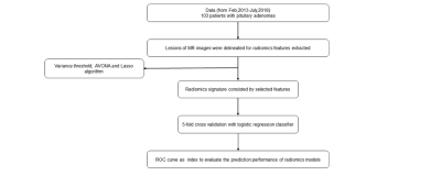 |
91 |
MRI-based radiomics in pituitary adenomas: pre-treatment prediction of prolactin expression
Presentation Not Submitted
Mo Zhanhao, Li Xuejia, Sui He, Liu Lin
To investigate the potential of radiomics features based on MRI in predicting the prolactin expression of pituitary adenomas before treatment. We build the logistic model and validation, which can offer a noninvasive approach to predict the PRL expression of pituitary adenomas by the way of machine learning. It may be a reference in the diagnosis, treatment and prognosis evaluation for some pituitary adenoma subtypes.
|
|
3083.
 |
92 |
Improved classification of paediatric brain tumours through whole spectra from in vivo magnetic resonance spectroscopy
Dadi Zhao, James Grist, Yu Sun, Andrew Peet
Single voxel magnetic resonance spectroscopy (SVS) is a non-invasive technique that can be used to probe metabolic activity in tumours. Previous studies have used metabolite concentrations to classify paediatric brain tumours from good quality data. However, the use of in vivo MRS whole spectra and wavelet de-noising for paediatric brain tumours have been rarely reported. In this study, we investigated the performance of spectra in classifying paediatric brain tumours by employing wavelet-based de-noising, and found significantly reduced error rate of classification based on the whole spectra, compared to that from metabolite concentrations and fits.
|
|
3084
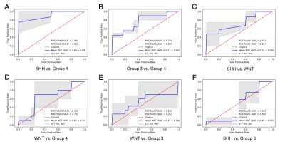 |
93 |
Predicting molecular subgroups of medulloblastoma using a quantitative radiomics approach
Video Permission Withheld
Jing Yan, Haiyang Geng, Binke Yuan, Zhenyu Zhang, Jingliang Cheng
Machine learning-based radiomics have been introduced in providing information on molecular biology and genomics of tumors. Here, we used features of MRI to predict molecular subgroups of medulloblastoma. MRI-based radiomics features were extracted from 37 patients with medulloblastoma (WNT = 11, SHH = 9, Group 3 = 8 , and Group 4 = 9). The molecular subgroups of medulloblastoma were classified with accepted accuracies by using support vector machine (SVM). In conclusion, MRI-based radiomics can effectively predict molecular subgroups of medulloblastoma using the machine-learning approach to benefit the treatment and prognosis of medulloblastoma.
|
|
3085.
|
94 |
Presurgical Differentiation between Malignant Haemangiopericytoma and Angiomatous Meningioma by a Radiomics Approach based on Texture Analysis
Presentation Not Submitted
Xuanxuan Li, Yiping Lu, Jianxun Qu, Bo Yin, Daoying Geng
We attempted to assess whether a machine-learning model based on texture analysis (TA) could yield a more accurate diagnosis in differentiating malignant haemangiopericytoma (HPC) from angiomatous meningioma (AM). Our sample population consisted of 23 malignant HPCs and 43 AM. We compared the diagnostic ability of three classifiers based on texture features extracted from each modality (T2FLAIR, T1-CE, and DWI) to the classifier based on clinical features from three neuro-radiologists. The T1W-CE classifier performed the best.
|
|
3086.
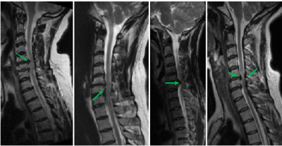 |
95 |
Deep learning for prognosis in Degenerative cervical myelopathy
Lucas Rouhier, Matthieu Parizet, Muhammad Akbar, Michael Weber, Michael Fehlings, Julien Cohen-Adad
Degenerative cervical myelopathy is an important cause of spinal cord dysfunction in adults worldwide 1,2. This study’s goal is to use boosting algorithm and deep learning on MRI and clinical data to predict the condition of a patient 6 months after baseline. Results show an improvement of prediction accuracy when combining MRI with clinical data (82.3%) versus with clinical data only (78.5%). The heterogeneity of the data makes it difficult for the learning algorithm to generalize, however future work exploiting boosting algorithm for structural data, and dimensionality reduction (e.g., via MRI feature extraction) could further improve prognosis accuracy.
|
|
3087.
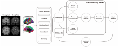 |
96 |
Automated machine learning classification of first-episode schizophrenia and controls using cerebral morphometric features
Huaiqiang Sun, Haoyang Xing, Su Lui, Xiaoqi Huang, Xiaoyue Zhou, John Sweeney, Qiyong Gong
Discriminate the first-episode schizophrenia patient with optimal feature set and classification model identified by automated machine learning algorithm
|
|
3088.
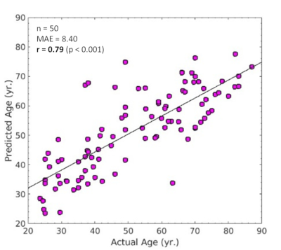 |
97 |
Prediction of Chronological Age from Intracranial Time-of-Flight Magnetic Resonance Angiography Images by Deep Convolutional Neural Network
Inpyeong Hwang, Hyeonjin Kim, Ji-hoon Kim
Brain-predicted age may be used as a potential biomarker of brain aging, and there also may be features related to cerebrovascular aging, such as decline of visualization of the arteries, or tortuosity. Therefore, the purpose of this study was to investigate whether there are learnable features by a deep convolutional neural network in the maximum intensity projection images of time-of-flight magnetic resonance angiography that might be associated with cerebrovascular aging.
|
|
3089.
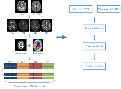 |
98 |
Classification of Parkinson‘s disease with diffusion magnetic resonance imaging by machine learning methods
Wenliang Fan
The diagnosis of PD is mainly based on clinical features and does not rely on imaging biomarkers. Neuroimaging studies help us better understand the pathophysiology and symptoms of PD. The aim of this study is to investigate the feasibility and performance of the PD classification using diffusion MRI of different machine learning methods.
|
|
3090.
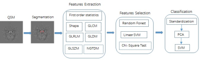 |
99 |
Radiomic Features of the Nigrosome-1 Region of the Substantia Nigra: Using Quantitative Susceptibility Mapping to Assist in the Diagnosis of Idiopathic Parkinson’s Disease
Zenghui Cheng, Jiping Zhang, Naying He, Fuhua Yan, E. Mark Haacke, Dahong Qian
There has been a major effort to study iron deposition in the substantia nigra (SN) because of its relationship to depigmentation of iron in the nigrosome-1 area. Recently, the swallow tail sign (STS) has been introduced as a new biomarker for idiopathic Parkinson’s disease (IPD). In this work, we analyzed the STS region of the SN based on quantitative susceptibility mapping (QSM) via a support vector machine (SVM) classifier and found that this radiomic approach could help to differentiate IPD patients from healthy controls.
|
|
3091.
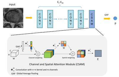 |
100 |
Evidence pinpointing of Intervertebral disc herniation with weak supervision
Presentation Not Submitted
Fei Gao, Shui Liu, Xiaodong Zhang, Jue Zhang, Xiaoying Wang
Deep learning has shown encouraging performance for lesion detection, but it is limited due to the high requirement of data labeling. In the task of lumbar intervertebral disc herniation recognition, we proposed to develop a recognition method based on axial images, which include more anatomical information about the disc, using a convolutional network. And we attempt to provide possible pathological evidence from the weakly labeled training data (normal/herniated label on image level).
|
|
| Top |
Emerging Technology & Translational Imaging 1
Digital Poster
Neuro
Tuesday, 14 May 2019
| Exhibition Hall |
15:45 - 16:45 |
| |
|
Computer # |
|
3092.
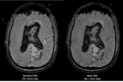 |
101 |
Prospective Evaluation of Wave-CAIPI Susceptibility-Weighted Imaging (SWI) Compared to Conventional 3D SWI in a Clinical Setting
John Conklin, Maria Gabriela Longo, Stephen Cauley, Kawin Setsompop, John Kirsch , Wei Liu, Sinyeob Ahn, Thomas Beck, Ramon Gonzalez, Pamela Schaefer, Otto Rapalino, Susie Huang
We present the first large-scale evaluation of Wave-CAIPI susceptibility-weighted imaging (Wave-SWI) for clinical brain imaging. Wave-SWI was compared to conventional SWI in 107 patients undergoing 3T MRI for a range of indications in both inpatient and outpatient settings. Two neuroradiologists assessed the images in individual and head-to-head comparisons, and found no significant difference between the two sequences for detection of microhemorrhages, visualization of pathology and normal anatomy, and overall diagnostic quality, despite a nearly 5-fold decrease in acquisition time using Wave-SWI. Broader application of highly-accelerated 3D imaging may improve utilization of MRI resources while reducing motion artifacts and patient anxiety.
|
|
3093.
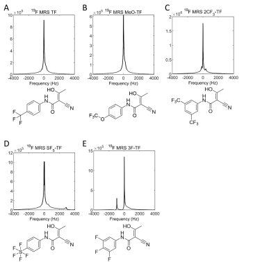 |
102 |
Enhancing Fluorine-19 Magnetic Resonance Drug Imaging: Chemical Variations in the Fluorine Side-Groups of the Immunomodulatory Drug Teriflunomide
Christian Prinz, Vera Martos Riaño, Tizian-Frank Ramspoth, Ludger Starke, Martin Neuenschwander, Jens-Peter von Kries, Andreas Pohlmann, Marc Nazaré, Thoralf Niendorf, Sonia Waiczies
Fluorine-19 (19F)-MR is of high relevance for the study of fluorinated drugs in vivo. Due to low drug concentrations and low numbers of fluorine atoms per molecule, the signal to be detected is very low. To address this drawback this work enhances 19F MRI of the antiinflammatory drug teriflunomide. For this purpose, derivatives of the trifluorinated drug were synthesized including modifications of the number and position of fluorine atoms in the 19F side-chains. We studied the 19F NMR characteristics and compared the SNR efficiencies of these compounds. The inhibitory activity was studied and correlated with the detectability of the compounds. By this, we can select drugs which provide a better signal than the original teriflunomide and which show an equal or even better biological activity.
|
|
3094.
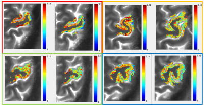 |
103 |
Using Glutamate-Weighted Imaging (GluCEST) to Detect Effects of Transcranial Magnetic Stimulation to the Motor Cortex
Abigail Cember, Brian Erickson, Olufunsho Faseyitan, Apoorva Kelkar, Neil Wilson, Ravi Prakash Reddy Nanga, Hari Hariharan, Ravinder Reddy, John Medaglia
We used glutamate weighted Chemical Exchange Saturation Transfer (GluCEST) imaging to investigate changes in glutamate contrast in the brains of young, healthy adults undergoing transcranial magnetic stimulation (TMS) to the motor cortex. Subjects were scanned to acquire a 2D GluCEST map of a slice which includes the motor cortex, then removed from the scanner and given continuous theta burst stimulation (cTBS). Subjects were scanned again post-stimulation. The resulting images show a trend of decreasing GluCEST contrast in the gray matter of the motor cortex where stimulation was administered. Interestingly, initial GluCEST values appear to predict response to TMS.
|
|
3095.
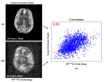 |
104 |
Investigating gas-exchange and tissue perfusion in the human brain using a combination of proton and hyperpolarized xenon-129 MRI
Madhwesha Rao, Graham Norquay, Jim Wild
This study establishes a correlation between cerebral perfusion and gas uptake using 1H arterial spin labeling and T2 weighted imaging for cerebral blood perfusion, and inhaled hyperpolarized 129Xe brain MR imaging for cerebral uptake of a free-diffusible noble gas. Using arterial transit time and cerebral blood volume maps, along with xenon images, correlation coefficients between 0.34 and 0.63 was observed for healthy subjects between the ages 26 and 36 years. The distinct properties of water and noble gas opens up the opportunity to use them in conjunction to understand aspects of brain physiology.
|
|
3096.
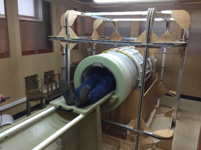 |
105 |
Fast Field-Cycling MRI identifies ischaemic stroke at ultra-low magnetic field strength
P. Ross, Lionel Broche, Mary Joan MacLeod, German Guzman-Guttierez, Alison Murray, David Lurie
In this work we present the first patient images from our home built Fast Field-Cycling MRI (FFC-MRI) scanner. By varying the external magnetic field during the imaging process, FFC-MRI allows us to probe the variation of T1 with magnetic field, known as “T1 dispersion”. This T1 dispersion has potential value as a diagnostic biomarker in a range of conditions. Here we present images demonstrating that endogenous T1 contrast at 20 mT and below can be used to identify ischaemic stroke.
|
|
3097.
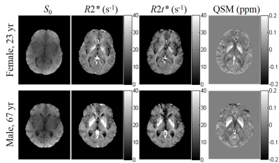 |
106 |
Theoretical model and experimental evaluation of cellular, BOLD and nonheme Iron contributions to the quantitative Gradient Recalled Echo (qGRE) and quantitative susceptibility mapping (QSM) signals in basal ganglia.
Dmitriy Yablonskiy, Jie Wen, Satya Kothapalli, Alexander Sukstanskii
Nonheme iron is an important element supporting the structure and functioning of biological tissues. Misbalance in nonheme iron can lead to different neurological disorders. Several MRI approaches have been developed for iron quantification relying either on the relaxation or susceptibility features of MRI signal. Specific quantification of the nonheme iron can, however, be tempered by the heme iron in the deoxygenated blood. The goal of this presentation is to introduce theoretical background and experimental method allowing disentangling contributions of heme and nonheme irons simultaneously with evaluation of tissue neuronal density in the basal ganglia.
|
|
3098.
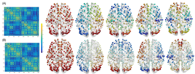 |
107 |
How does functional brain activity depend on the underlying structural connectome? A graph signal processing perspective
Maria Giulia Preti, Dimitri Van De Ville
How to integrate the information of structural connectivity and brain activity measured with magnetic resonance imaging (MRI) represents still an open question. Here, we addressed this problem by applying graph signal processing (GSP) to human brain data, aiming at exploring significant excursions of functional activity and its degree of alignment to the underlying structural connectivity. Two contrasting functional networks were highlighted: a primary sensory one, more aligned to the structure and characterized by less excursions, and a high-level cognitive one, more liberal and showing more fluctuations. This advanced framework opens new perspectives in the interpretation of the brain structure/function interplay.
|
|
3099.
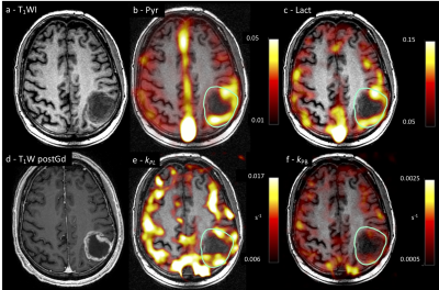 |
108 |
Magnetic resonance spectroscopic imaging of hyperpolarized [1-13C]pyruvate in glioblastoma: a promising tool for investigating tumor metabolism and heterogeneity.
Fulvio Zaccagna, James Grist, Mary McLean, Charlie Daniels, Frank Riemer, Joshua Kaggie, Surrin Deen, Ramona Woitek, Rolf Schulte , Kieren Allinson, Anita Chhabra , Marie-Christine Laurent, Amy Frary, Tomasz Matys, Ilse Patterson, Bruno Do Carmo, Stephan Urpsrung , Ian Wilkinson, Bristi Basu, Colin Watts, Stephen Price, Sarah Jefferies, Jonathan Gillard, Martin Graves, Kevin Brindle, Ferdia Gallagher
Glioblastomas (GBM) are characterized by diffuse infiltration, a high level of intratumoral and intertumoral heterogeneity and a very poor prognosis. Characterising tumor heterogeneity in vivo may improve diagnosis, therapy planning and treatment assessment. Dissolution dynamic nuclear polarization (DNP) is a novel technique that allows dynamic and non-invasive assessment of the metabolism of hyperpolarized (HP) 13C-labelled molecules in vivo, such as the preferential exchange of [1-13C]pyruvate to [1-13C]lactate within tumors (Warburg effect). In this study we explore metabolic reprogramming within glioblastoma (GBM) and its microenvironment using HP [1-13C]pyruvate to demonstrate the heterogeneity of pyruvate’s metabolic fate.
|
|
3100.
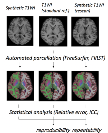 |
109 |
3D-QALAS Sequence for Brain Volumetry and Cortical Thickness: Comparison with 3D MPRAGE and Scan-Rescan Repeatability
Shohei Fujita, Akifumi Hagiwara, Masaaki Hori, Fukunaga Issei, Christina Andica, Tomoko Maekawa, Ryusuke Irie, Koji Kamagata, Kanako Kumamaru, Akihiko Wada, Shigeki Aoki
Previous quantitative synthetic MRI of the brain has been solely performed in 2D. Here, we evaluated the feasibility of the recently developed 3D-QALASsequence for brain cortical thickness and volumetric analysis in healthy volunteers. 3D-QALAS and MPRAGE were performed twice and once, respectively, in the same session at 1.5 T. FreeSurfer and FIRST were used to measure cortical thickness and volume of subcortical structures, respectively. High agreement with MPRAGE and scan-rescan repeatability in both cortical thickness and subcortical structures were shown. 3D-QALAS could be reliably used for measuring cortical thickness and subcortical volumes in most brain regions.
|
|
3101.
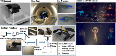 |
110 |
A Non-Intrusive Eye Tracking Based MRI Compatible VR System
Kun Qian, Tomoki Arichi, Jonathan Eden, Sofia Dall'Orso, Rui Pedro A G Teixeira, Kawal Rhode, Mark Neil, Etienne Burdet, A Edwards, Jo Hajnal
Achieving compatibility of VR systems with MRI scanners is challenging and for applications such as fMRI, it is highly desirable to avoid local distortions of the static magnetic field. We have developed a non-intrusive MR compatible VR system which avoids disturbing the magnetic environment and uses eye tracking as the main interface. Our system demonstrates a capability to bring the VR world into MRI systems, including dynamic interaction with VR content based on gaze, with performance that it is competitive to the current leading commercial gaming eye tracker.
|
|
3102.
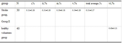 |
111 |
Preliminary Research of ischemic penumbra in human subacute stroke patients combinating use of amide proton transfer (APT) Chemical Exchange Saturation Transfer (CEST) and arterial spin-labeling (ASL) MRI
Presentation Not Submitted
Yuefa Tan, Bin Chen, Yingjie Mei, Yikai Xu
The purpose of this word was to explore the feasibility of amide proton transfer(APT) assisting arterial spin-labeling(ASL) and diffusion-weighted imaging(DWI) in identiflication and definition of ischemic penumbra in subacute stroke .Our results showed that APTWI deficits were always larger than or equal to DWI deficits and smaller than or equal to ASL-CBF deficits in subacute stroke. ATPWI deficits coincided with the resulting infarct area at follow-up endpoint. Final infarcts were smaller than CBF deficits and larger than or equal to subacute DWI deficits.APT can provide information complementary on cell metabolism to ASL and DWI in the definition of ischemic tissue.
|
|
3103.
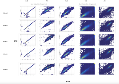 |
112 |
Accelerated quantification of ultrashort-T2 brain components with ultrashort-TE relaxometry
Nikhil Deveshwar, Peng Cao, Shuyu Tang, Duan Xu, Peder Larson
This study presents accelerated quantification of the ultrashort-T2 components in the brain with approximately 4-fold reduction in scan time. Retrospective undersampling across 5 healthy volunteers showed strong correlations between the ultrashort-T2 component amplitude, relaxation times, and frequency shifts between 8 TE and 32 TE datasets, showing that it is possible to rapidly obtain high quality images of brain ultrashort T2 components that are associated with myelin membrane protons.
|
|
3104.
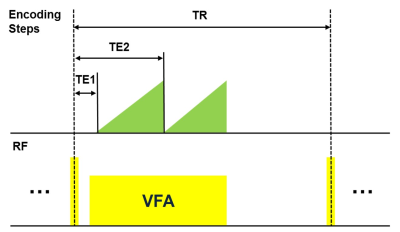 |
113 |
Cerebral venous thrombus staging in a single magnetic resonance (MR) scan: A dual-contrast approach
Yunduo Li, Shuo Chen, Zechen Zhou, Miaoqi Zhang, Rui Li, Chun Yuan
This study proposed a dual-contrast Volumetric Isotropic Turbo spin echo Acquisition (dVISTA) sequence that allows both T1 and T2 cerebral venous thrombus imaging. In-vivo experiments indicated that dVISTA provide adequate image contrast as conventional T1/T2 imaging, and the clinical feasibility of this technique was further validated by CVT patients’ scan. By assembling flow-suppression, T1/T2 contrast in one 6-min whole brain scan, dVISTA has the potential to detect and differentiate thrombus in clinical routine.
|
|
3105.
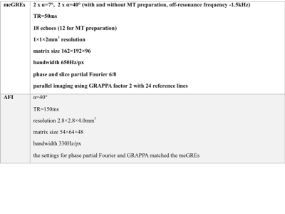 |
114 |
A high-resolution multi-parametric quantitative method to investigate tissue (micro)structure
Ana-Maria Oros, Melissa Schall, N. Jon Shah
We report on a multi-parametric quantitative method based on four 1x1x2mm3 3D multiple-echo gradient-echo acquisitions (GRE), complemented by AFI for B1+ mapping (whole-brain TA=21min). The most notable parameters derived are water content, T1 and T2*, magnetization transfer ratio (MTR), bound proton fraction (fbound) and magnetization exchange rate (kex). Results are reported from seven volunteers, one post-mortem brains and one tumour patient. The use of multiple contrasts for tissue segmentation is illustrated. Correlations between parameters are investigated with the aim of better understanding sources of T1 relaxation. f_bound and k_ex are found to be lower in tumour than in healthy tissue.
|
|
3106.
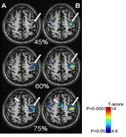 |
115 |
Online brain fMRI using a novel magnetic resonance compatible hand induced robotic device provides accurate monitoring and can be used in rehabilitation
Zeba Qadri, Loukas Astrakas, Lawrence Wald, Michael Moskowitz , Bruce Rosen, Aria Tzika
Using a hand motor task, we investigated brain activation after chronic stroke by combining fMRI at 3T with a novel MR-compatible hand-induced, robotic device (MR_CHIROD). Patients trained at home using a gel ball; serial neuroimaging was performed before, during, upon completion of training, and after a non-training period, to assess permanence of effects. Training significantly increased the number of activated voxels in the cortex as a function of effort level, suggesting functional cortical plasticity in chronic stroke. The result’s persistence indicates permanence of rehabilitation, which is remarkable given that training is generally effective during a narrow window after stroke.
|
|
3107.
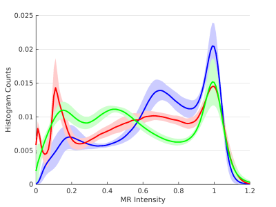 |
116 |
Synthetic harmonization of multi-site multi-vendor MRI data to improve MS lesion segmentation
Snehashis Roy, Blake Dewey, Peter Calabresi, John Butman, Dzung Pham
Segmentation of lesions from magnetic resonance images of patients with multiple sclerosis is a challenging task, especially when involving multi-center or multi-scanner data. State-of-the-art lesion segmentation algorithms require training data to use identical acquisition protocols as the input data, but this is often difficult to control. In this work, we employ image synthesis to allow data from one scanner to resemble the data acquired in a different scanner. Overall lesion segmentation accuracy improves and the amount of false positives are reduced using synthesized images, indicating image synthesis can improve segmentation consistency in a heterogeneous dataset.
|
|
3108.
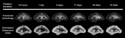 |
117 |
Ex-vivo diffusion MRI of human hemispheres: the effect of tissue fixation and the relation to in-vivo diffusion MRI
Arnold Evia, Yingjuan Wu, David Bennett, Konstantinos Arfanakis
Until the relationship between in-vivo and ex-vivo diffusion measures has been established, the clinical relevance of findings using ex-vivo diffusion MRI is unclear and precludes potential studies. Therefore, this study investigated the effects of tissue fixation on basic diffusion measures, and established the relationship of diffusion measures recorded in-vivo and ex-vivo. Basic diffusion measures of postmortem hemispheres were observed over 5 weeks. The relationship between in-vivo and ex-vivo diffusion measures was studied using linear mixed regression. Appreciable changes to diffusion measures were seen early on in fixation, and ex-vivo measurements of FA and RD were linked to their in-vivo measurements.
|
|
3109.
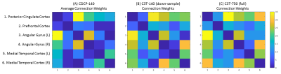 |
118 |
Improved Structural Imaging of the Default Mode Network on the Compact 3T
Thomas Welton, Matthew Lyon, Jerome Maller, Myung-Ho In, Ek-Tsoon Tan, Matt Bernstein, Erin Gray, Yunhong Shu, John Huston, Stuart Grieve
We compared diffusion MRI tractographic representations of the default mode network using high-angular resolution scans from the Compact 3T with high-performance gradients to equivalent data acquired on a standard clinical scanner. Overall performance in terms of strength, accuracy and visualisation of the DMN was superior for the Compact 3T data, with improved global tracking performance and improved measurement of weaker connections.
|
|
3110.
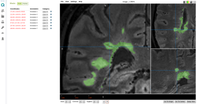 |
119 |
Semi-automatic cloud-based workflow for evaluating the central vein sign for MS diagnosis in a multicenter clinical setting
David Moreno-Dominguez, Marc Ramos, Daniel Reich, Daniel Ontaneda, Paulo Rodrigues, Pascal Sati
The central vein sign (CVS) is novel MRI marker for improving the accuracy and reducing the time to diagnose patients with multiple sclerosis (MS). Recent advancements have introduced the MRI FLAIR* contrast, allowing for CVS to be easily identified. In this work, we developed a semi-automatic cloud-based workflow for evaluating the clinical value of the CVS for MS diagnosis using FLAIR* in a multicenter setting. This novel workflow is a powerful tool that has the potential to significantly accelerate the clinical research imaging studies in MS.
|
|
3111.
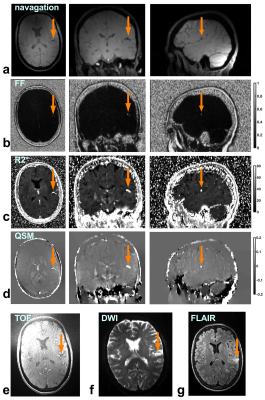 |
120 |
Initial experience of imaging stroke patients with a 5-minute whole head multi-echo GRE acquisition
Junmin Liu, Luciano Sposato, Spencer Christiansen, Maria Drangova
We report on our initial experience of imaging stroke patients using a technique that achieves quantification of fat fraction (FF), QSM, and R2* simultaneously from a single multi-echo GRE (mGRE) acquisition. We collected 3T data from three stroke patients with a ~ 30-minute whole head multi-sequence protocol. By performing the joint analysis of TOF MRA, DWI, FLAIR and mGRE, we evaluated the capability of using the mGRE-based maps and images to characterize thrombus, differentiate intracranial calcifications from hemorrhages and detect white-matter lesions. Our initial results have shown the feasibility of using the mGRE technique to image stroke patients.
|
|
3112.
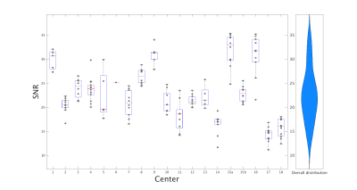 |
121 |
Towards a harmonized protocol for structural MRI imaging of the brain for multisite studies in the Italian IRCCS advanced neuroimaging network
Paolo Bosco, Laura Biagi, Alessandra Retico, Anna Nigri, Domenico Aquino, Fulvia Palesi, Maria Grazia Bruzzone, Claudia Gandini Wheeler-Kingshott, Michela Tosetti, The Italian IRCCS advanced neuroimaging network
MRI derived brain structural measurements from multicenter datasets can strongly be affected by factors such as the acquisition protocol, the static magnetic field strength and the scanner manufacturer. A preliminary study was performed to assess the homogeneity of population metrics from 3DT1 scans acquired with already established routine protocols in a dataset of 174 healthy subjects from 18 Italian Research Hospital Centers (IRCCS). The impact of each center acquisition parameters on outcomes was assessed with quality control measurements and FreeSurfer volumetric metrics of cortical and subcortical structures. Future multicenter studies will benefit from harmonizing the acquisition protocols.
|
|
3113.
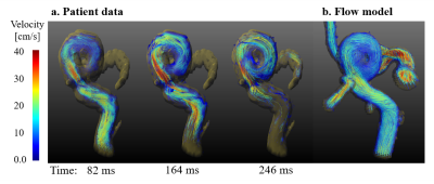 |
122 |
3D printed, patient-specific vascular models for 4D flow MRI: production workflow and imaging aneurysm treatment
Mariya Pravdivtseva, Eva Peschke, Thomas Lindner, Olav Jansen, Jan Hövener
Patient-specific models of the human vessels could be used for the different purposes varying from the visualization of a vasculature to examination of a strategy of the endovascular treatment. Current work represents a step by step workflow of the production of vascular models: optimisation of the geometry of the digital model, design of the connectors, wall thickness of the model etc. The flow in the models was compared with in vivo flow data of the patient.
The influence of different flow diverter stents on the flow in the model with an aneurysm was evaluated with MRI (TOF and 4D Flow).
|
|
3114.
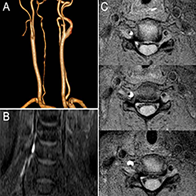 |
123 |
Improved imaging method for comprehensive diagnosis of cervical artery disssection
Hong-Wei Zhou, Dezheng Kong, Linjing Wang, Tianjing Zhang, Zhongping Zhang
Cervical artery dissection (CAD) is a significant cause of ischemic stroke in young adults. As a result, the early and accurate diagnosis of CAD is helpful for appropriate treatment decision-making to prevent stroke. Traditional imaging methods, such as CTA, MRA or DSA are difficult to differentiate CAD from other mimics because of similar luminal findings1,2. 3D T1-weighted black blood sequence would demonstrate the abnormality of vessel wall and could potentially provide diagnosis information for vasculopathy patients. This study aims at introducing a 3D black-blood sequence’s application in the diagnosis of CAD, as well as atypical artery dissection.
|
|
3115.
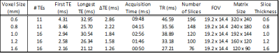 |
124 |
Optimisation of QSM measurements of venous oxygen saturation
John McFadden, Julian Matthews, Maélène Lohézic, Geoff Parker, Laura Parkes
Quantitative Susceptibility Mapping has been shown to be capable of making estimates of venous oxygen saturation (YvYv)
which are comparable to those obtained using MR methods such as calibrated BOLD. While there have been a few studies which have considered optimal acquisition parameters for QSM1,2 none have focussed on the specific case of deoxyhaemoglobin. In this work, we propose a protocol for which voxel dimensions, final echo time, and readout polarity have been optimised. Demonstrations of reasonably precise estimates of which are in line with the broader literature recommend suitability of the protocol for future studies.
|
|
3116.
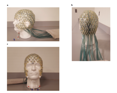 |
125 |
High-quality FLAIR and diffusion imaging with presence of EEG nets
Nina Fultz, Catherine Poulsen , Michael Lev, Avery Berman , Jingyuan Chen , Giorgio Bonmassar, Laura Lewis
EEG nets are typically removed before MR imaging due to their negative effects on image quality, which is time-consuming and interrupts monitoring of brain activity. We tested whether the InkNet, a high-resistance polymer thick-film based EEG net, could improve images by reducing RF-shielding caused by copper leads. We imaged subjects with FLAIR and diffusion at 3 Tesla, wearing a conventional, copper-lead net, the InkNet, or no EEG net (NoNet). The InkNet induced less artifact than conventional nets, and produced similar image quality to the NoNet control. Results suggest that high-quality imaging can be achieved while wearing an EEG net.
|
|
| Top |
Epilepsy
Digital Poster
Neuro
Tuesday, 14 May 2019
| Exhibition Hall |
15:45 - 16:45 |
| |
|
Computer # |
|
3117.
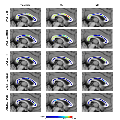 |
126 |
Corpus callosum involvement in mesial temporal lobe epilepsy and non lesional frontal lobe epilepsy: a multimodal MRI study
Maria Eugenia Caligiuri, Angelo Labate, Aldo Quattrone, Antonio Gambardella
Mesial Temporal Lobe Epilepsy (MTLE) and Frontal Lobe Epilepsy (FLE) are the two most common forms of partial epilepsy. While MTLE has been widely studied, FLE has been less investigated. Patients with FLE in which there is no clearly identifiable abnormality on MRI (non lesional FLE, nlFLE) represent an ideal sample to study the epileptic syndrome itself, regardless of the nature and location of the epileptogenic focus in the frontal lobe. Here, we studied the involvement of the corpus callosum in temporal and frontal lobe epilepsy, considering non-lesional FLE, refractory MTLE and mild MTLE, a particularly drug-responsive phenotype. Neuroimaging characteristics of CC seem to be indeed altered with patterns that are specific to the different epileptic syndromes.
|
|
3118.
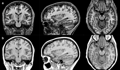 |
127 |
Voxel Based Morphometry in Temporal Lobe Epilepsy: a pilot study using MT maps instead of conventional T1-weighted data
Fulvia Palesi, Paul Summers, Claudia Gandini Wheeler-Kingshott, Giancarlo Germani, Valeria Mariani, Laura Tassi, Paolo Vitali
Temporal Lobe Epilepsy (TLE) is the most common form of focal epilepsy. Neuroimaging and neuropathological studies indicates that the structural network affected in TLE extends to both temporal and extra-temporal structures. In this work, quantitative 3DMT maps were used in a Voxel Based Morphometry(VBM) framework to assess atrophy in left and right TLE compared to controls. Our findings revealed that 3DMT maps, thankfully to their excellent grey-white matter contrast, can be successfully employed for VBM in epilepsy identifying temporal and extra-temporal grey and white matter alterations in patients. This study is a proof-of-principle to adopt 3DMT for voxel based analysis in TLE.
|
|
3119.
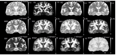 |
128 |
Comparative analysis of Mean Apparent Propagator (MAP)-MRI and traditional DTI for the diagnosis of hippocampal sclerosis in temporal lobe epilepsy
Presentation Not Submitted
Keran Ma, Jingliang Cheng, Xiaonan Zhang, Ankang Gao, Chengru Song, Shaoyu Wang, Xu Yan, Huiting Zhang
This study aimed to preliminarily investigate the utility of MAP-MRI parameters in hippocampal sclerosis and compare them with traditional DTI parameters to explore the changes in hippocampal microstructure and understand its pathophysiological mechanisms. The swelling of the hippocampal nerve fiber axon presynaptic terminal can be reflected by MAP-MRI, which greatly assists in the clinical diagnosis of early hippocampal sclerosis. Compared with DTI parameters, DSI parameters showed higher diagnostic efficacy particularly for RTPP and QIV. This study extracted more objective and reproducible parameters of fixed hippocampal sclerosis. More samples are needed for future research.
|
|
3120.
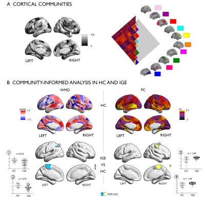 |
129 |
Community-informed connectomics of the thalamo-cortical system in idiopathic generalized epilepsy
Presentation Not Submitted
Zhengge Wang, Sara Lariviere, Qiang Xu, Reinder Vos de Wael, Seok-Jun Hong, Zhongyuan Wang, Xu Yun, Bin Zhu, Neda Bernasconi, Andrea Bernasconi, Bing Zhang, Zhiqiang Zhang, Boris Bernhardt
Idiopathic generalized epilepsy with tonic-clonic seizures (IGE-GTCS) has been associated to the thalamo-cortical circuitry. By quantifying the interplay between macroscale functional communities via resting-state fMRI (rs-fMRI) connectome analysis, we assessed the intrinsic organization of this network and its relation to drug-response. Compared to controls, IGE-GTCS showed a more constrained network embedding of the thalamus, while frontocentral neocortical regions expressed increased functional diversity. Findings remained significant after regressing out thalamic volume and cortical thickness, suggesting independence from structural alterations. We observed more marked network imbalances in drug-resistant compared to seizure-free patients. Our findings suggest a pathoconnectomic mechanism of IGE, centered on diverging changes in cortical and thalamic connectivity. More restricted thalamic connectivity could reflect the tendency to engage in recursive thalamo-cortical loops, which may contribute to hyper-excitability.
|
|
3121.
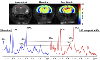 |
130 |
Mapping astroglial glutamine synthetase activity in vivo in a preclinical model of epilepsy using glutamate-weighted CEST (GluCEST) MRI
Puneet Bagga, Stephen Pickup, Julien Flament, John Detre, Hari Hariharan, Ravinder Reddy
Epilepsy is broadly characterized by aberrant neuronal excitability causing seizures. Glutamate is the major excitatory neurotransmitter in the brain and can be detected using MRS and glutamate weighted chemical exchange saturation transfer (GluCEST) MRI. In this study, we performed high-resolution GluCEST MRI in a preclinical model of epilepsy to evaluate glutamatergic alterations in the hippocampus. GluCEST contrast was increased in the hippocampus of mice treated with methionine sulfoximine acutely, while mice treated chronically with MSO showed reduced hippocampal GluCEST contrast. The GluCEST results were also corroborated with in vivo 1H MRS.
|
|
3122
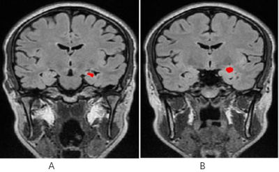 |
131 |
Diagnostic value of signal intensity histogram analysis based on magnetic resonance cube sequence on hippocampal sclerosis in temporal lobe epilepsy
Video Permission Withheld
liu fan, SONG FAN, WANG YING, SUN BO, miao wei, dong yi, li xin, qiu jia, song wei
Hippocampal sclerosis(HS)is the most common pathological form of medial temporal lobe epilepsy, characterized by loss of hippocampus and related structural selective neurons and reactive gliosis. With magnetic resonance(MR) imaging, the detection rate for HS has been found to vary largely and such substantial variations in the detection rate have been primarily attributed to the subjective nature of the assessment of scans. Texture analysis is a technique used to quantify image textures
|
|
3123.
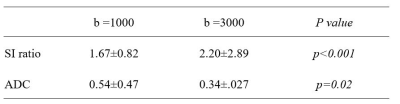 |
132 |
A comparison of high b-value and standard b-value diffusion weighted imaging for status epilepticus in pediatric patients
Presentation Not Submitted
Haiwei Han, Hua Wu
In this study series with status epilepticus (SE) pediatric patients, we investigated the utility diffusion weighted magnetic resonance imaging with a high b-value (b = 3000 s/mm2) compared with standard b-value (b = 1000 s/mm2) for acute ictal MRI changes in pediatric patients. High b-value diffusion weighted imaging (DWI) could be beneficial for detecting additional lesions and improving the contrast between lesions and normal tissue. Therefore, high b-value DWI may be a better noninvasive imaging method for exploration of the acute ictal MRI changes in pediatric patients with SE.
|
|
3124.
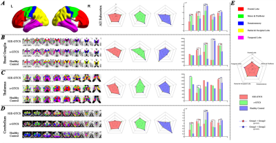 |
133 |
Altered Cortex-Subcortex structural connectivity pattern in different types of generalized epilepsy
Presentation Not Submitted
Qiang Xu, Xinyu Xie, Zhiqiang Zhang, Guangming Lu
Different types of GTCS could be explained by the different brain network. Cortex-subcortex structural covariate connectivity might help us to understand the mechanism in the new insight. Thalamus was the important regions in classification the two types of GTCS.
|
|
3125.
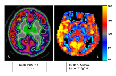 |
134 |
Mapping brain oxygen metabolism with dual calibrated fMRI in pre-surgical evaluation of epilepsy: a case report comparison with FDG-PET.
Hannah Chandler, Michael Germuska, Rhodri Smith, Patrick Fielding , Christopher Marshall, Khalid Hamandi, Richard Wise
Positron emission tomography (PET) with fluorodeoxyglucose (FDG) is a widely used approach to help identify putative epileptogenic areas in patients with epilepsy, as part of the epilepsy surgery evaluation. Epileptogenic areas typically show a regional reduction in glucose metabolism. Here we present a dual-calibrated fMRI method (acquiring BOLD and ASL CBF data simultaneously), which permits mapping of the cerebral metabolic rate of oxygen consumption noninvasively across grey matter. In this case report, we demonstrate close agreement between the two methods (dc-fMRI and PET-FDG) in localising a region of cerebral hypometabolism in epilepsy.
|
|
3126.
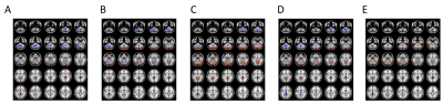 |
135 |
Cortical and subcortical networks in frontal lobe epilepsy with generalized tonic clonic seizures
Presentation Not Submitted
xinyu xie, qiang xu, zhiqiang zhang, guangming lu
Cortico-subcortical networks are considered core pathologic substrates for frontal lobe epilepsy with generalized tonic clonic seizures; however, the mechanism is still unknown. This study aims to identify the changes of cortico-subcortical networks by resting-state functional connectivity. 60 patients with frontal lobe epilepsy and healthy controls were enrolled. Bilateral hemispheres were divided into 5 nonoverlapping cortical lobes. Functional connectivity between each cortical lobe and the subcortical regions were calculated, and functional connectivity strength was used to evaluate the interconnectivity. Our results indicate that the decrease connection between prefrontal cortex and subcortical structures suggests it maybe the epicenters of frontal lobe epilepsy.
|
|
3127.
 |
136 |
Based on MR-T2WI Haar-like and MB-LBP Features findings of temporal lobe glioma-related epilepsy
Presentation Not Submitted
ankang Gao, Jie Bai, Jingliang Cheng, Yuan Hong
There are many other strong risk factors for glioma-related epilepsy that have been highlighted in previous research, including high levels of glutamate and adenosine kinase and low levels of gamma-aminobutryic acid. How the value machine vision application of study on this?
|
|
3128.
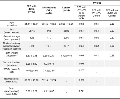 |
137 |
Morphology Analysis of Dilated Virchow Robin Spaces in Simple Febrile Seizure Children Between 12 and 48 Months Based on the Automated Segmentation
Mustafa Salimeen, Xianjun Li, Miaomiao Wang, Congcong Liu, Habib Tafawa, Anuja Pradhan, Martha singh, Xiaocheng Wei, Guanyu Yang, Jian Yang
Febrile seizure (FS) has become a common problem in childhood and imposes acute effects on the brain. Currently dilated Virchow -Robin spaces (dVRs) has become hot point in research for the explanation the mechanism how its involving brain in inflammatory lesions. we conducted a quantitative method to assessment dVRs that visible onT2WI in simple FS. Our study aim is to describe an effective uses of automatic software method to recognize VRs and to get intergroup differences. Our result suggests that dVRs count, volume and head circumference are greater simple FS than in the control group.
|
|
3129.
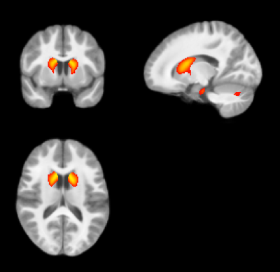 |
138 |
The gray matter volume and shape abnormal in angelman syndromes
Lei Wei, He Wang, Xiaonan Du, Shasha Long, Baofeng Yang, Yonghui Jiang, Jianfeng Feng, Yi Wang
The angelman syndromes is a neurogenetic disease and clinically characterized by the developmental delay, movement or balance disorder, seizures, and frequent smiling. Our study compare the alteration of gray matter volume and shape between angelman syndromes and healthy controls
|
|
3130
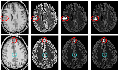 |
139 |
Detection of Focal Cortical Dysplasia via quantitative T1-mapping
Video Permission Withheld
Ulrike Nöth, René-Maxime Gracien, Michelle Maiworm, Philipp Reif, Elke Hattingen, Susanne Knake, Marlies Wagner, Ralf Deichmann
Focal cortical dysplasias (FCD) are characterized by an increased cortical thickness and blurred junctions between white (WM) and gray matter (GM). A method for improved FCD detection is proposed, which is only based on quantitative maps of T1 relaxation time. Masks of WM, GM and CSF are derived from the measured T1 values. The local cortical extent (CE) is calculated from the GM mask and the local smoothness (SM) of GM-WM junctions is derived from the T1 gradients. Synthetic double inversion recovery data sets are calculated from the T1 map and further enhanced in areas of increased CE and SM.
|
|
3131.
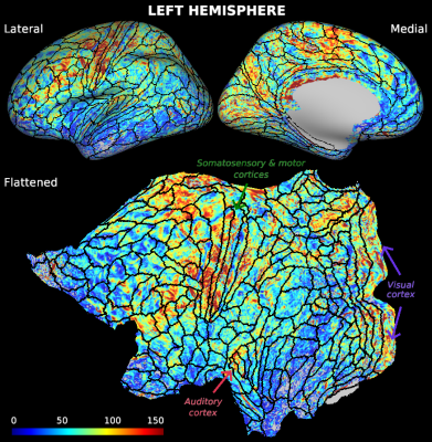 |
140 |
Sub-millimeter blood flow mapping of cortical and hippocampal gray matter
Roy Haast, Dimo Ivanov, Jordan DeKraker, Sriranga Kashyap, Shanice Janssens, Benedikt Poser, Ali Khan, Kamil Uludag
Acquisition of sub-millimeter whole-brain blood flow (CBF) maps was recently demonstrated to be feasible using 7T MRI. Here, we show that such high resolution CBF maps can be used to differentiate cortical regions, in general, and subregions within the hippocampal formation, in particular. We found that higher baseline perfusion was especially present in regions known to be highly myelinated and/or characterized by low quantitative T1 values. Moreover, these initial results warrant the use of CBF data to improve the interpretability of fMRI activation maps at a finer scale.
|
|
3132.
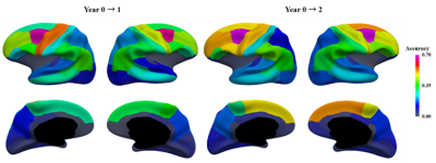 |
141 |
Cortical FoldingPrint for Individual Identification During Dynamic Postnatal Development
Dingna Duan, Shunren xia, Zhengwang Wu, Fan Wang, Li Wang, Weili Lin, John H Gilmore , Dinggang Shen, Gang Li
Human cortical folding is highly convoluted and characterizes the inter-subject variability. Recent studies found that the adult brain cortex is unique for individual identification. However, little is known about whether the infant brain cortex, which develops dynamically in the first postnatal years, is reliable for individual identification. To this end, we proposed a novel morphological folding descriptor, called FoldingPrint, to perform the infant identification in a large longitudinal dataset with 472 infants. Successful identification results indicate the effectiveness of the proposed FoldingPrint. In addition, we found that the regions with high identification accuracy are mainly distributed in high-order association cortices.
|
|
3133.
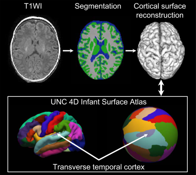 |
142 |
Surfaces Area and Cortical Volume Development of Infant Transverse Temporal Cortex Influenced by Preterm Birth
Xianjun Li, Jing Xia, Jian Chen, Li Wang, Miaomiao Wang, Congcong Liu, Mengxuan Li, Xiaocheng Wei, Gang Li, Dinggang Shen, Jian Yang
Development of transverse temporal cortex is essential for speech perception in infants. However, the early morphological development of this cortex has not been fully understood. Additionally, influences of preterm birth on cortical development remain to be investigated. This study assessed cortical development of transverse temporal cortex in preterm and term infants based on surface reconstruction. We found that surface area and cortical volume of transverse temporal cortex underwent rapid changes. Term infants held higher surface area, cortical volume, and asymmetry than preterm infants. These results suggest that preterm birth influences the asymmetry and developmental trajectory of transverse temporal cortex.
|
|
3134.
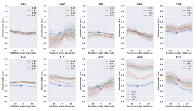 |
143 |
Hippocampal subfield segmentation and partial volume effects - reliability assessment
Arturo Cardenas-Blanco, Yi Chen, Jose-Pedro Valdes-Herrera, Renat Yakupov, Hendrik Mattern, Alessandro Sciarra, David Berron, Anne Maass, Oliver Speck, Emrah Duezel
The hippocampus is involved in a variety of cognitive and functional tasks. Research groups rely on volumetric segmentations to assess: the integrity of the HC and its subfields as well as their involvement in cognitive tasks. Unfortunately, due to its size and location, most studies use non-isotropic T2-weighted images to segment the HC. The aim of this project is to determine whether partial volume effects due to T2-weighted slice angulation and non-isotropic resolution have an impact in the segmentation process. The results indicate that both, angulation and non-isotropic acquisition have a significant impact in specific subfields.
|
|
3135.
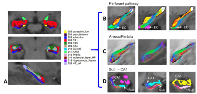 |
144 |
Three-dimensional reconstruction and dissection of hippocampal fiber pathways with human connectome data
Jing Li, Zifei Liang, Weihong Zhang, Thomas Wisniewski, Jiangyang Zhang, Yulin Ge
The hippocampus plays a vital role in learning and memory and consists of multiple subfields with distinct functional pathways. Despite many volumetric investigations, in vivo human studies on hippocampal pathways remain scarce. In this study, we show that the Perforant, Alveus/Fimbria, and CA1-Subiculum pathways can consistently reconstructed from the Human Connectome Project (HCP) diffusion MRI dataset aided by automated brain and hippocampal subfield segmentation methods. This demonstrates the feasibility of in vivo mapping of the major hippocampal pathways at 3T, which may lead to new research avenue of the functional pathways of hippocampus in normal and disease states.
|
|
3136.
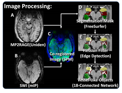 |
145 |
Asymmetry of hippocampus vasculature in temporal lobe epilepsy
Rebecca Feldman, Gaurav Verma, Stephanie Brown, John Rutland, Madeline Fields, Lara Marcuse, Bradley Delman, Priti Balchandani
Epilepsy is a chronic and prevalent disease. When epilepsy is drug resistant, resection of epileptogenic abnormalities may control seizures. However, there exist focal epileptogenic abnormalities which are not well visualized by current imaging techniques. Hippocampal vascularity has yet to be explored as an in vivo marker for the epileptic brain. Using susceptibility weighted imaging at 7T MRI we are able to visualize vessels in the hippocampus, and have automatically segmented the vessels in the hippocampus of 19 patients with temporal lobe epilepsy and 19 healthy controls. We found significant asymmetry in the vessel density in patients with epilepsy when compared against healthy controls.
|
|
3137.
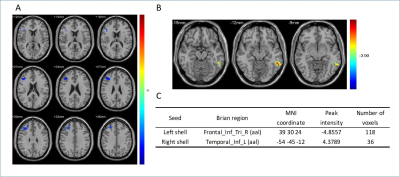 |
146 |
Functional connectivity changes of nucleus accumbens subdivisions in left mTLE patients
Presentation Not Submitted
Ru Yang, Wenhan Yang, Jun Liu, Xixi Zhao
Within the nucleus accumbens(NAC) there is a clear distinction between the shell and core portions. Growing evidences have supported that the NAC, especially its shell portion has been involved in epileptogenesis. However relevant studies on vivo human brains are quite limited. in this study, we investigated Left mTLE related function connectivity changes of NAC subregions . Our result indicated an decrease in FC between left shell and right frontal area and an increase FC between right shell and left temporal area. But no significant FC changes appear on core, which suggest that shell portion play important roles on mTLE.
|
|
3138.
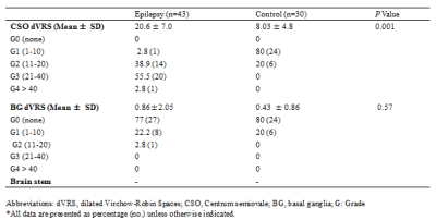 |
147 |
Dilatation of Virchow-Robin Spaces is An Acute Phase Response in Children with Epileptic Seizure
Tafawa Habib, Mustafa Salimeen, Xianjun Li, Guanyu Yang, Anuja Pradhan, Martha Singh, Lu Gao, Miaomiao Wang, Congcong Liu, Jian Yang
Dysfunction of Virchow -Robin spaces (VRS) has become an exciting avenue of research for the exploration of the pathophysiology involving CNS neurodegenerative and neuroinflammatory disorders. To investigate the pathophysiological link of the dilated VRS (dVRS) in children with epilepsy, we conducted semiquantitative and quantitative assessment of dVRS visible on standard 3.0T magnetic resonance imaging (MRI) in children with clinical history of symptomatic generalized tonic clonic, clonic or myoclonic seizures. Our results revealed the tendency of increased number of dVRS in the early phase following epileptic seizure, suggesting that dilated VRS might be related to the cascade of immune response during epileptic seizures.
|
|
3139.
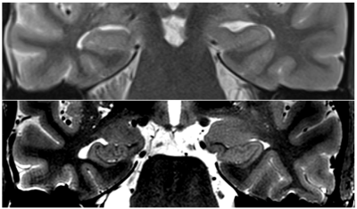 |
148 |
Multimodal 7T MRI of epilepsy surgery candidates: Prospective evaluation of impact on presurgical decisions
Giske Opheim, Melanie Ganz-Benjaminsen, Patrick Fisher, Ulrich Lindberg, Mark Vestergaard, Helle Simonsen, Henrik Larsson, Anne-Mette Leffers, Camilla Madsen, Olaf Paulson, Lars Pinborg
Identifying lesions at 3T MRI remains the most important correlate to epilepsy surgery outcome. Since 45% of candidates present with negative 3T MRI, investigation of diagnostic yield of clinical 7T MR protocols along with post-processing markers is urged. This ongoing study will evaluate how radiological descriptions and computational morphometrics affect presurgical decisions. So far, 19 patients and 31 controls are scanned at the Philips Achieva 7T system at Hvidovre Hospital, Denmark. Preliminary analyses of automatic segmentations show promising potential. We are in radiological training, but expect first case with evaluation of impact on presurgical decision to start around new-year 2018/2019.
|
|
| Top |
Multiple Sclerosis: Connections & Disruptions
Digital Poster
Neuro
Tuesday, 14 May 2019
| Exhibition Hall |
15:45 - 16:45 |
| |
|
Computer # |
 |
3140.
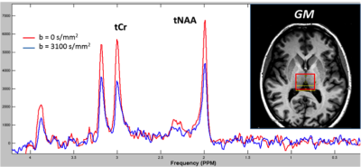 |
151 |
Thalamic energy dysregulation drives microstructural changes of thalamo-cortical projections in multiple sclerosis
Vito RICIGLIANO, Matteo Tonietto, Raffaele Palladino, Emilie Poirion, Francesca Branzoli, Geraldine Bera, Elisabeth Maillart, Bruno Stankoff, Benedetta Bodini
Our objective was to investigate whether thalamic energy alterations in multiple sclerosis (MS) are associated with microstructural degeneration of thalamo-cortical tracts. In 17 patients and 13 healthy controls (HCs), the apparent diffusion coefficient of creatine-phosphocreatine ADC(tCr) in the thalami, reflecting energy dysregulation, was evaluated with diffusion-weighted magnetic resonance spectroscopy. Integrity of thalamo-cortical and non-thalamic tracts was evaluated by measuring mean diffusivity (MD) with standard diffusion weighted imaging. In patients but not in HCs, lower thalamic ADC(tCr) was associated with higher MD of thalamo-cortical tracts only, suggesting that thalamic energy dysfunction may induce the selective anterograde degeneration of thalamo-cortical networks.
|
|
3141
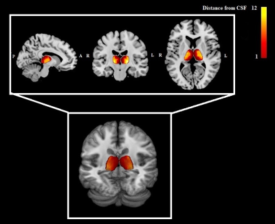 |
152 |
In-vivo mapping of thalamic pathological mechanisms in pediatric patients with MS
Video Permission Withheld
Loredana Storelli, Ermelinda De Meo, Lucia Moiola, Maria Pia Amato, Angelo Ghezzi, Pierangelo Veggiotti, Ruggero Capra, Maria A. Rocca, Massimo Filippi
Despite widely recognized in pediatric population with multiple sclerosis (MS), the pathogenesis of thalamic damage remains largely unknown. This study was performed to explore the microstructural abnormalities within this structure through the use of quantitative MRI metrics (diffusion tensor, T1/T2-weighted ratio) considering the two different thalamic interfaces (cerebrospinal fluid/thalamus and thalamus/white matter) as sites of two different pathogenic processes: the first one accounting for cerebrospinal fluid-mediated factor damage and the second one for diffuse neurodegenerative damage. The study demonstrated an heterogeneous pathogenesis for thalamic damage since the beginning of the disease.
|
|
3142.
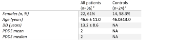 |
153 |
Gray matter pathology changes in multiple sclerosis: a comparison of diffusion kurtosis and volumetrics.
Lily Cao, Benjamin Ades-Aron, Katarina Yaros, Nicolas Gillingham, Dmitry Novikov, Yvonne Lui, Ilya Kister, Timothy Shepherd, Els Fieremans
Along with well-characterized abnormalities in normal appearing and lesional white matter, gray matter (GM) pathology has been observed in multiple sclerosis (MS) both with diffusion tensor imaging (DTI) and structural MRI. We used diffusion kurtosis imaging (DKI), a clinically feasible extension of DTI, to characterize pathology in cortical and subcortical GM in MS and uncovered correlations with disease severity, quantified in terms of the Patient Determined Disease Score (PDDS). Our results suggest that DKI metrics are sensitive to changes in GM and could be helpful to use alongside standard markers of disease progression, such as GM volume atrophy.
|
|
3143
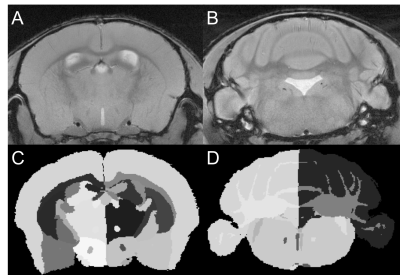 |
154 |
Grey matter atrophy measured with MRI correlates with reduced neuronal density in the experimental autoimmune encephalomyelitis model of multiple sclerosis
Video Permission Withheld
A. Max Hamilton, Nils D. Forkert, Ying Wu, James A. Rogers, V. Wee Yong, Jeff F. Dunn
Atrophy is a clinical marker of neurodegeneration and progressive disability in multiple sclerosis (MS). To test neuroprotective treatments aimed at reducing atrophy, mouse models featuring atrophy are needed. We have shown the experimental autoimmune encephalomyelitis (EAE) mouse model features atrophy, though we do not know if EAE atrophy is caused by neurodegeneration, as it is in MS. We used MRI and atlas-based regional volumetrics to measure atrophy in EAE, while using immunohistochemistry to measure neurodegeneration. Atrophy measured in the cerebellum and cerebral cortex correlated with neuronal loss, suggesting we can use EAE along with MRI to test neuroprotective therapies.
|
|
3144.
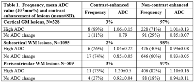 |
155 |
Characterization of gray-matter multiple sclerosis lesions using double inversion recovery, diffusion, contrast-enhanced, and volumetric MRI
M. Andrea Parra, Sindhuja T Govindarajan, Lev Bangiyev, Patricia K Coyle, Tim Q Duong
This study characterized gray-matter (GM) multiple sclerosis (MS) lesions using double-inversion recovery (DIR), contrast-enhanced and diffusion at 3T MRI. Lesion segmentation was based on DIR. We determined GM lesion prevalence, characterize their contrast-enhancement and diffusion characteristics, and compared them with white-matter (WM) lesions. Correlated GM lesion count and volume with total brain, WM, GM and deep GM volumes, as well as clinical disability. Comparisons were also made with healthy controls. We tested the hypothesis that GM MS lesions are highly prevalent, contrast-enhanced GM lesions have higher ADC values, and GM MS lesion counts and volumes are correlated with brain atrophy.
|
|
3145.
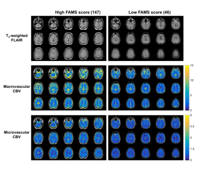 |
156 |
Characterization of Multi-Scale Perfusion in Patients with Relapsing-Remitting Multiple Sclerosis (RR-MS)
Ashley Nespodzany, Charrid Simpson, Aimee Borazanci, Ashley Stokes
The purpose of this study is to characterize multi-scale cerebral perfusion in patients with multiple sclerosis (MS) using an advanced spin- and gradient-echo (SAGE) dynamic susceptibility contrast (DSC) MRI method. SAGE DSC-MRI provides macro- and micro-vascular perfusion, permeability, and other vascular parameters. We have acquired SAGE DSC-MRI data in 24 patients, along with a quality of life metric. Preliminary analysis of the first five patients demonstrated reduced perfusion in patients with lower quality of life scores than patients with higher quality of life scores. Work is ongoing to characterize the full set of SAGE-based hemodynamic metrics in these patients.
|
|
3146.
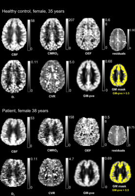 |
157 |
Quantification of cerebral grey matter vascular and metabolic function in multiple sclerosis using dual-calibrated fMRI.
Hannah Chandler, Rachael Stickland, Mike Germuska, Eleonora Patitucci , Catherine Foster, Sharmila Khot, Neeraj Saxena, Valentina Tomassini, Richard Wise
Dual-calibrated fMRI (dc-fMRI) relies on the simultaneous acquisition of BOLD and ASL during a respiratory challenge to quantitatively map cerebral blood flow (CBF), cerebral metabolic rate of oxygen (CMRO2), oxygen extraction fraction (OEF), cerebrovascular reactivity (CVR) and effective oxygen diffusivity (D). Here, we use this method to investigate alterations in brain physiology in patients with multiple sclerosis (and matched healthy participants), demonstrating significant reductions in CBF and CMRO2per unit of remaining grey matter in patients. We suggest that this method not only provides novel markers of tissue dysfunction, it also extends the methodological armamentarium for non-invasive investigation of brain pathophysiology in disease.
|
|
3147.
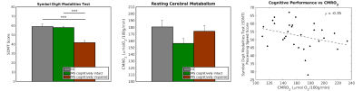 |
158 |
Evaluating brain oxygen metabolism and cognition in multiple sclerosis
Kathryn West, Dinesh Sivakolundu, Mark Zuppichini, Gayathri Maruthy, Darin Okuda, Hanzhang Lu, Bart Rypma
We used phase contrast (PC) and T2-Relaxation-Under-Spin-Tagging (TRUST) to evaluate whole brain cerebral metabolic rate of oxygen (CMRO2) in MS patients and healthy controls. We compared CMRO2 with cognitive performance and diffusion kurtosis imaging. CMRO2 correlates negatively with cognitive function in MS patients, suggesting a marker of ongoing disease activity leading to cognitive decline.
|
|
3148.
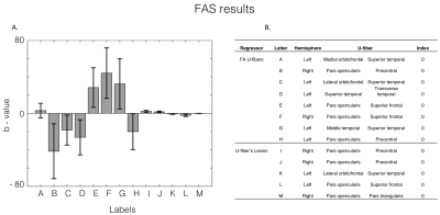 |
159 |
Relevance of cortical and subcortical integrity in cognitive performance in Relapsing-Remitting Multiple Sclerosis patients.
Cristian Montalba, Mariana Zurita, Tomás Labbé, Marcelo Andia, Miguel Guevara, Jean-François Mangin, Juan Pablo Cruz, Ethel Ciampi, Cristian Tejos, Claudia Cárcamo, Pamela Guevara, Sergio Uribe
In this work, we evaluated if structural characteristics of cortical regions and the U-fibers connected to these regions allows predicting multiple sclerosis patients’ score in FAS, SDMT and PASAT tests. For this purpose, we represented structural characteristics as the proportion of the regions and fibers covered by lesion and the U-fiber’s mean fractional anisotropy. In our sample, FAS test score results significantly correlated with structural damage in U-fibers from Frontal and Temporal lobes. PASAT test scoresignificantly correlated with structural damage in U-fibers in the bilateral Frontal and Parietal lobes. No significant correlations were found for the SDMT.
|
|
3149
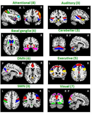 |
160 |
Action observation training increases dynamic functional connectivity in patients with multiple sclerosis
Video Permission Withheld
Maria A. Rocca, Loredana Storelli, Claudio Cordani, Paola Valsasina, Luca Gavazzeni, Alessandro Meani, Paolo Preziosa, Federica Esposito, Massimo Filippi
Action Observation Training (AOT) seems to be a promising tool to improve upper limb function. We applied a novel method of analysis, which allows a time-varying (dynamic) assessment of resting state functional connectivity on two randomized experimental groups of healthy controls and multiple sclerosis (MS) patients and two control groups. Between-group differences and dynamic functional network connectivity (dFNC) changes over-time in each group were evaluated. After a training of 2 weeks, MS groups improved in right upper limb functions and AOT showed a modulation of dFNC of several functional networks in MS patients.
|
|
3150.
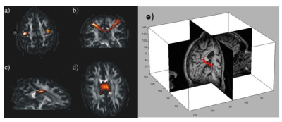 |
161 |
Combining Structural and Functional Connectivity as Biomarkers for Disease Progression in Neurologic Disease: A Longitudinal Multiple Sclerosis Study
Mark Lowe, Katherine Koenig, Jian Lin, Bhaskar Thoomukuntla, Ken Sakaie, Daniel Ontaneda, Lael Stone, Kunio Nakamura, Stephen Rao, Stephen Jones
Sensitive outcome measures are required to test novel therapies designed to target neurodegeneration in Multiple Sclerosis(MS). We have previously shown that functional connectivity using resting-state fMRI (rs-fMRI) and anatomic connectivity using DTI are related in the transcallosal motor pathway and along the memory pathway connecting hippocampus to posterior cingulate. We propose a combined metric incorporating anatomic and functional connectivity along these pathways as a potential biomarker of disease progression in MS. In this study, we present results from a 2 year study that assessed this biomarker in 19 MS patients at six timepoints. We show that our metric is sensitive to changes in MS disease over this time interval. We also show that our metric is more sensitive to change than typically used imaging biomarkers.
|
|
3151.
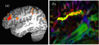 |
162 |
Evolution of functional and structural connectivity of cognitive frontoparietal network during 2 years of fingolimod therapy of multiple sclerosis
Jian Lin, Pallab Bhattacharyya, Hong Li, Ken Sakaie, Robert Fox, Mark Lowe
It has been reported that the cognitive impairment in MS is related to the dysfunction of frontoparietal network (FPN). In a longitudinal study of MS patients undergoing fingolimod treatment, we investigated the structural and function connectivity of FPN over 2 years. The trend of changes in functional connectivity MRI, and transverse diffusivity measured by DTI probabilistic tractography indicate that fingolimod treatment stabilized damage of structural and functional connectivity of FPN sometime around/after the 1st year of treatment similar to that reported for motor network.
|
|
3152
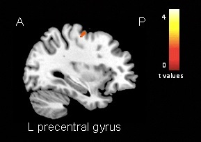 |
163 |
Structural and functional damage of the sensorimotor network contribute to predict disability progression and phenotype evolution in patients with multiple sclerosis: a 6.5-year follow-up study
Video Permission Withheld
Massimo Filippi, Loredana Storelli, Alessandro Meani, Chiara Cervellin, Paola Valsasina, Claudio Cordani, Elisabetta Pagani, Paolo Preziosa, Maria A. Rocca
Multiple sclerosis (MS) is a complex disease, characterized by a highly heterogeneous disease evolution. The prognostic value of magnetic resonance imaging (MRI) in clinically definite MS is still debated. The aim of this study was to find possible structural and functional MR imaging prognostic biomarkers able to guide treatment decisions in MS disease course. The analysis of structural and functional MRI networks was able to improve our understanding of the extreme variability in MS and allowed prognosis prediction at an individual level.
|
|
3153
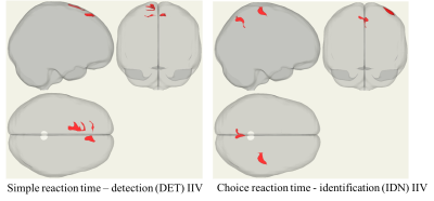 |
164 |
Functional connectivity correlates of altered intraindividual variability of processing speed in multiple sclerosis patients with young adult onset
Video Permission Withheld
Sindhuja T Govindarajan, Gregory Bodik, Yilin Liu, Leigh Charvet, Lauren Krupp, Timothy Duong
Intra-individual variability of processing speed (IIV-PS) is a more sensitive discriminator between controls and MS patients in early disease stage than the normative mean scores of PS. We used rsfMRI to interrogate the neural correlates of IIV-PS and means of PS in MS patients with young-adult onset. Significant correlation of the amplitude of low-frequency fluctuations (ALFF) was found with IIV-PS but not with mean PS. Seed-to-voxel functional connectivity analysis showed significant connections associated with altered IIV of identification task, but not the IIV of detection task. This approach identified the neural networks associated with altered IIV-PS in MS.
|
|
3154.
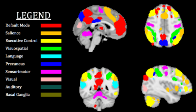 |
165 |
Connectomics of Brain Demyelination in Multiple Sclerosis
Shangming Liu, Bratislav Misic, Joseph Gati, Ravi Menon, Sridar Narayanan, Douglas Arnold, David Rudko
Resting-state connectivity alterations associated with demyelination and neurodegeneration occur in multiple sclerosis, but the specific network connections that are affected are not well understood. Moreover, temporal alterations in these networks across MS patient lifespan and MS phenotype have not been robustly characterized. In this study, we sought to isolate the influence of MS phenotype on resting-state functional connectivity in a cohort of early and later stage MS patients imaged using 7 T MRI. Through single-subject ICA denoising, followed by group-level connectivity matrix analysis with partial-least squares methods, we evaluated group level differences in 7 T resting-state connectivity between MS phenotypes.
|
|
3155.
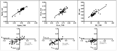 |
166 |
Individual simulation of brain functional reorganization in early multiple sclerosis - The Virtual Brain approach
Rebecca Marchetti, Bertrand Audoin, Marmaduke Woodmann, Arnaud Le Troter, Paul Bartelemy, Manon Philibert, Pierre Besson, Jean Pelletier, Maxime Guye, Viktor Jirsa, Jean-Philippe RANJEVA
We demonstrate here that 'The Virtual Brain' (TVB), a computational neural mass model fed with structural connectome derived from individual diffusion MRI can successfully generate reorganized individual resting-state functional connectomes in early multiple sclerosis patients with similar topologies that those obtained with real rs-fMRI data. This opens new perspectives to predict and better understand the brain functional reorganization occuring in individual MS patients during disease progression or treatment.
|
|
3156.
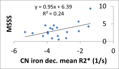 |
167 |
Five Year Changes in Iron and Myelin in Relapsing-Remitting Multiple Sclerosis Deep Gray Matter Compared to Healthy Controls
Ahmed Elkady, Dana Cobzas, Hongfu Sun, Gregg Blevins, Peter Seres, Alan Wilman
This study evaluated RRMS and age-matched control longitudinal changes in iron/myelin-sensitive quantitative MRI of the deep gray matter (DGM) over 5 years. In addition to standard bulk analysis of the DGM, we have used a recently introduced analysis framework that utilizes combined R2* and QS for Discriminative Analysis of Regional Evolution (DARE) to identify regions of iron and myelin longitudinal change. The main findings of the study are the significantly different volume shrinkage rates of the thalamus and iron decrease rates of the caudate, thalamus, and globus pallidus over 5 years compared to age-matched healthy controls. Additionally, iron decrease in the CN and GP was shown to be correlated to clinical disease severity and duration, respectively.
|
|
3157.
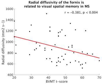 |
168 |
A preliminary assessment of the relationship of radial diffusivity of the fornix to future episodic memory performance in Multiple Sclerosis
Katherine Koenig, Jian Lin, Daniel Ontaneda, Kedar Mahajan, Jenny Feng, Stephen Rao, Sanghoon Kim, Stephen Jones, Mark Lowe
Here we present a preliminary report assessing the relationship of DTI measures of the fornix to future episodic memory in patients with multiple sclerosis (MS). We find that radial diffusivity of the fornix is longitudinally related to visual spatial episodic memory. This finding suggests this measure is appropriate to investigate as a potential predictive marker of cognitive decline.
|
|
3158.
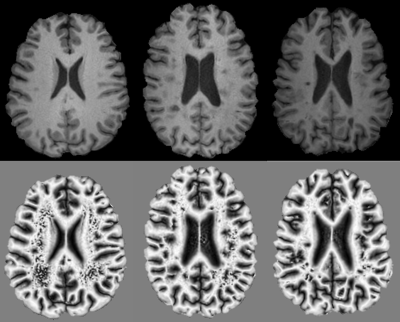 |
169 |
Statistical analysis of phase congruency detects normal appearing white matter changes associated with disease severity between relapsing remitting and secondary progressive multiple sclerosis
Olayinka Oladosu, Yunyan Zhang
Mechanisms of disease progression from relapsing remitting to secondary progressive multiple sclerosis (MS) are still unclear. Here we applied new texture analysis approaches including phase congruency to understand the differences between normal appearing white matter structure in the brain corpus callosum of patients with relapsing or progressive MS, and matched controls. We found that the contrast, energy, and homogeneity of weighted mean phase in the corpus callosum differentiate MS patients from controls, and energy and homogeneity further distinguish relapsing from progressive MS. Advanced analysis of phase congruency outcomes may help detect disease progression in MS.
|
|
3159.
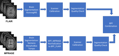 |
170 |
Brain volume measurements in multiple sclerosis patients: a novel combination approach for routine clinical assessment in MS PATHS
Adrian Tsang, Yu-Chen Su, Rodrigo Perea, Ricardo Corredor-Jerez, Mário João Fartaria, Till Huelnhagen, Veronica Ravano, Bénédicte Maréchal, Tobias Kober, Robert Bermel, Stephen Jones, Izlem Izbudak, Yvonne Lui, Lauren Krupp, Ellen Mowry, James Williams, Rick Rudick, Elizabeth Fisher
Highly precise measurements from fully automated techniques are required to quantify brain atrophy in individual multiple sclerosis (MS) patients. We developed a novel approach to reliably estimate brain atrophy in MS that combines two techniques applied to different image contrasts and incorporates inter-scanner calibrations. We validated this approach using data acquired in a scan-rescan study. Mean coefficient of variation (CV) for the new brain parenchymal fraction measurement was 0.18%, which was lower than the CV attained for the individual techniques. This new metric will next be integrated into the radiology workflow in MS PATHS institutions for further testing.
|
|
3160.
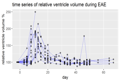 |
171 |
Transient enlargement of brain ventricles during Multiple Sclerosis and Experimental Autoimmune Encephalomyelitis
Jason Millward, Laura Böhmert , Paula Ramos Delgado, Henning Reimann, Joao Periquito, Antje Els, Alina Smorodchenko, Michael Scheel, Judith Belmann-Strobl, Carmen Infante-Duarte, Friedemann Paul, Thoralf Niendorf, Andreas Pohlmann, Sonia Waiczies
Brain ventricle volumes (VV) increased sharply during initial disease in the experimental autoimmune encephalomyelitis model of multiple sclerosis (MS), normalizing upon clinical remission. A cohort of MS patients with 13 monthly MRI scans over one year showed significantly greater VV volatility than healthy controls. Most patients showed VV contractions greater than the ±6% range of variation in healthy controls, and these patients had significantly lower disease severity compared to non-contracting patients. For some patients, the time series of changes in VV showed significant cross-correlations with other MRI and clinical parameters, suggesting that VV variations reflected disease processes related to inflammation.
|
|
3161.
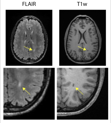 |
172 |
White Matter Tract-Defined Lesion Loads in Relapsing-Remitting Multiple Sclerosis
M. Ethan MacDonald, Wei-Qiao Liu, Sarah Scott, Conrad Rockel, Deepthi Rajashekar, Jacinta Specht, Hongfu Sun, G. Bruce Pike
Lesions in multiple sclerosis (MS) present at various locations throughout the brain. 207 relapsing-remitting MS (RRMS) patients were scanned at 3T. T2w and T1w images were used to segment white matter (WM) hyper- and hypo-intensities, respectively. Using an atlas of WM tracts, the lesion burden was computed for each tract. The dominant lesion load was found in the periventricular regions. The tract percent load is highest in the anterior thalamic radiation, inferior fronto-occipital fasciculus, the forceps major, and forceps minor tracts. The uncinate fasciculus, superior longitudinal fasciculus, and the four cingulum tracts have the lowest lesion loads.
|
|
3162.
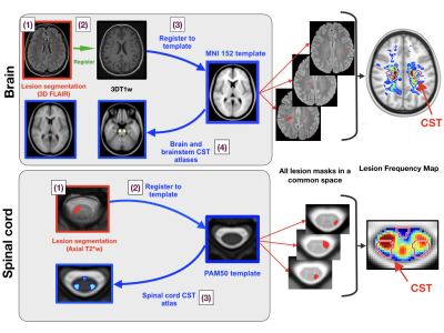 |
173 |
Spatial distribution of multiple sclerosis lesions along the brain and spinal motor tracts and correlation with functional deficits
Anne Kerbrat, Charley Gros, Atef Badji, Elise Bannier, Francesca Galassi, Benoit Combès, Pierre Labauge, Xavier Ayrignac, Clarisse Carra Dallière, Frederic Pinna, Benjamin De Leener, Dominique Eden, Josefina Maranzano, Ren Zhuoquiong, Tobias Granberg, Leszek Stawiarz, Jan Hillert, Russell Ouellette, Jason Talbott, Yasuhiko Tachibana, Masaaki Hori, Kouhei Kamiya, Lydia Chougar, Jennifer Lefeuvre, Daniel Reich, Govind Nair , Paola Valsasina, Maria Rocca, Massimo Filippi, Renxin Chu, Rohit Bakshi, Gilles Edan, Julien Cohen-Adad
We describe the spatial distribution of multiple sclerosis (MS) lesions in the corticospinal tracts (CST), from the cortex to the lower cervical cord, using an open-source automated pipeline. We confirm the high frequency of CST focal damage in MS patients from the first year of relapsing-remitting MS and identify areas of predilection for MS lesions along the CST in the cervical spinal cord. At baseline, no significant correlation was observed between brain or spinal cord CST lesion volume fraction and physical disability scores. However, the baseline spinal cord CST lesion volume fraction correlated with the EDSS at 2 years.
|
|
3163.
 |
174 |
Investigating the contribution of interhemispheric disconnection to disability and fatigue in Progressive Multiple Sclerosis.
Maria Petracca, Matteo Battocchio , Simona Schiavi, Mohamed El Mendili, Lazar Fleysher, Alessandro Daducci, Matilde Inglese
We explored the presence and clinical impact of interhemispheric disconnection in progressive multiple sclerosis (PMS) through a tractography-based approach, quantifying the number of streamlines passing through callosal subregions. In PMS, we identified a reduced number of streamlines in the splenium and the anterior portion of the corpus callosum (CC) body. Patients with primary and secondary progressive phenotype presented different patterns of CC involvement. The reduced number of streamlines in central and anterior CC was related to motor disability and fatigue, while loss of the integrity in the posterior portion of CC was the main feature of cognitively impaired patients.
|
|
3164.
 |
175 |
Vascular disease risk factors in multiple sclerosis is associated with reduced cerebral metabolic activity
Manoj Sammi, Ian Tagge, Dennis Bourdette, Allison Fryman, William Rooney, Vijayshree Yadav
Reduced cerebral metabolic activity is observed with increased vascular disease risk factors like hyperlipidemia, hypertension, obesity, diabetes, and heart disease. High resolution MR techniques are used to measure and characterize capillary water flux and water permeability surface product as potential surrogates of brain metabolic activity.
|
|
| Top |
Alzheimer's & Related Dementia
Digital Poster
Neuro
Tuesday, 14 May 2019
| Exhibition Hall |
16:45 - 17:45 |
| |
|
Computer # |
|
3165.
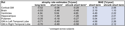 |
1 |
Towards clinically useful individual regional brain atrophy rates: bridging long- and short-term longitudinal volume change estimates
Jonas Richiardi, Bénédicte Maréchal, Ricardo Corredor, Mazen Fouad A-wali Mahdi, Reto Meuli, Tobias Kober
Several brain diseases show prominent, regionally-specific atrophy over time, which is often predictive of patient outcomes. However, reliable estimation of atrophy rates typically relies on repeated observations over several years. This is incompatible with clinical practice in terms of cost and prognosis utility. Here, we propose a statistical regularization approach to dramatically improve the quality of regional atrophy estimates, using only two longitudinal measurements. We evaluate the approach on open data from 43 MR scanners and 599 subjects, showing that we halve the error in most regions, while maintaining discriminability between controls and Alzheimer patients.
|
|
3166.
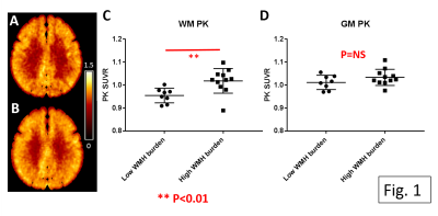 |
2 |
White matter hyperintensity burden is strongly associated with neuro-inflammation but not with amyloid deposition: a multimodal PET MR study
Hongyu An, Chunwei Ying, Yasheng Chen, Peter Kang, Jon Christensen, Qing Wang, Lisa Cash, Jin-Moo Lee, Andria Ford, Tammie Benzinger
Selectively elevated PET 11C-PK11195 uptake within white matter demonstrates that neuro-inflammation is an important pathogenic factor in patients with white matter hyperintensities.
|
|
3167.
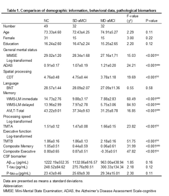 |
3 |
Decreased FDG-PET Uptake and Inter-Hemispheric Functional and Structural Connectivity in Patients with Multi-domain Amnestic Mild Cognitive Impairment
Presentation Not Submitted
Xiao Luo, Kaicheng Li, Qingze Zeng, Peiyu Huang, Yong Zhang, Minming Zhang
Amnestic mild cognitive impairment (aMCI) is not a uniform disease entity. Based on clinical symptoms, aMCI can be further classified as single-domain aMCI (SD-aMCI) with isolated memory deficit, or multi-domain aMCI (MD-aMCI) if memory deficit is combined with other cognitive domains impairment. By examining cerebral metabolism, inter-hemispheric connectivity, behavioral features, and neuropathologies, we observed disrupted bilateral cerebral metabolism, structural and functional disconnection between bilateral hemispheres in MD-aMCI, but not in SD-aMCI. Thus, along with the continuum between normal aging and Alzheimer’s disease, SD-aMCI and MD-aMCI may represent two clinical phases.
|
|
3168.
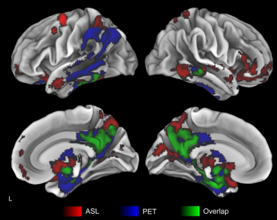 |
4 |
Multimodal Magnetic Resonance Imaging versus 18F-FDG-PET to Identify Mild Cognitive Impairment
Sudipto Dolui, Zhengjun Li, Ilya Nasrallah, David Wolk, John Detre
18F-FDG-PET provides a functional neurodegenerative biomarker in the Alzheimer’s continuum, but it is costly and involves exposure to ionizing radiation. Arterial Spin Labeled (ASL) perfusion MRI can be acquired during routine MRI session to measure cerebral blood flow (CBF), which is tightly coupled with cerebral metabolism. We demonstrated that the ASL hypoperfusion pattern was similar to that of FDG-PET-hypometabolism in patients with mild cognitive impairment. Further, ASL-CBF provided complementary information to hippocampal atrophy measured with structural MRI. Multimodal MRI may provide a cost-effective and totally noninvasive substitute for 18F-FDG-PET in clinical and research setting for detecting Alzheimer’s neurodegeneration.
|
|
3169.
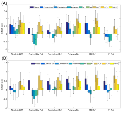 |
5 |
Reference Regions for Computing Relative Cerebral Blood Flow in Mild Cognitive Impairment
Sudipto Dolui, Zhengjun Li, Ilya Nasrallah, David Wolk, John Detre
Alzheimer’s disease (AD) is characterized by reduced cerebral blood flow (CBF) both globally and in AD specific regions, however there is considerable CBF variability even in healthy population. Relative CBF using mean CBF in AD-spared regions as reference removes this variability and can provide higher sensitivity and specificity for regional changes. We compared the effects of using different reference regions in discriminating patients with amnestic mild cognitive impairment (aMCI) and elderly controls using two different arterial spin labeling acquisitions. Putamen and primary motor cortex were most spared in the aMCI cohort and provided best patient-diagnosis when used as reference regions.
|
|
3170.
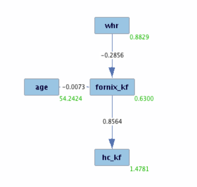 |
6 |
Does obesity cause limbic neuroglia changes that may precede Alzheimer's disease? An MRI causal analysis investigation using quantitative magnetisation transfer
Fabrizio Fasano, John Evans, Cyril Charron, Derek Jones, Claudia Metzler-Baddeley
By a causal analysis approach, we investigated the effect that an “intervention” on healthy population weight would produce on hippocampus macromolecular exchange rates. The quantitative magnetisation transfer index forward exchange rate (kf) is recognised to be lowered by myelin/axonal membrane loss and neuroinflammation. We found that, especially in over 56 years aged male subjects, forcing the weight to stay below the overweight threshold (waist-to-hip ratio 0.9) may dramatically reduce low macromolecular exchange rate occurrences in the population.
|
|
3171.
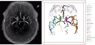 |
7 |
Quantitative Intracranial Vasculature Assessment to detect dementia using the intraCranial Artery Feature Extraction (iCafe) Technique
Li Chen, Thomas Grabowski, Eric Larson, Paul Crane, Thomas Hatsukami, Jenq-Neng Hwang, Chun Yuan, Niranjan Balu
Intracranial artery features measured from 3D magnetic resonance angiography (MRA) may provide new biomarkers for detecting dementia. Quantitative morphometry and intensity features using iCafe analysis of MRA was compared between cognitively normal and abnormal subjects. We found significantly lower total artery length (p=0.0046), distal artery length (p=0.0043), number of branches (p=0.0038) and average order (p=0.0250) in the cognitively abnormal group. These results suggest reduced vascularity for dementia subjects. iCafe is a promising tool to quantitatively characterize intracranial vascular structures for dementia research.
|
|
3172.
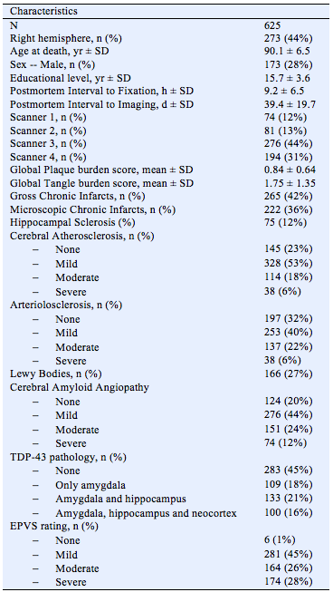 |
8 |
Neuropathologic Correlates of Enlarged Perivascular Spaces in a Community Cohort of Older Adults
Carles Javierre Petit, Ashish Tamhane, Arnold Evia, Nazanin Makkinejad, David Bennett, Julie Schneider, Konstantinos Arfanakis
Perivascular spaces are fluid-filled spaces surrounding blood vessels as they penetrate the brain, forming a brain fluid drainage system that facilitates interstitial fluid exchange and clearance of waste products. Enlargement of perivascular spaces is common in aging, and literature has linked enlarged perivascular spaces (EPVS) to increased risk of stroke, lower cognitive function, and vascular dementia. The links of EPVS burden to age-related neuropathologies have not yet been thoroughly investigated. Therefore, the purpose of this work was to assess the neuropathologic correlates of EPVS by combining ex-vivo MRI and pathology data in a large community cohort of 625 older adults.
|
|
3173.
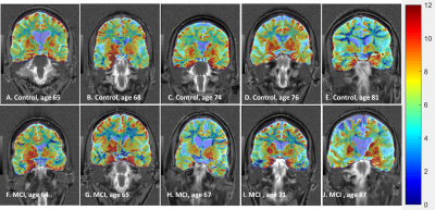 |
9 |
Glutamate Weighted Imaging (GluCEST) as a Biomarker of Cognitive Function: Preliminary Findings from GluCEST MRI in Older Adults
Abigail Cember, Ravi Prakash Reddy Nanga, Sandhitsu Das, Neil Wilson, Deepa Thakuri, David Wolk, John Detre, Ravinder Reddy
We used glutamate weighted Chemical Exchange Saturation Transfer (GluCEST) imaging to investigate changes in glutamate concentrations in the brains of older adults. These are preliminary findings representing the data from subjects presenting with Mild Cognitive Impairment (MCI) (n=5) and similarly-aged healthy controls (n=5). In this cohort, we observed a trend of decreasing GluCEST contrast in multiple regions of the brain of MCI subjects when compared to control subjects. Especially interesting is the apparent global decrease in GluCEST contrast throughout the white matter of the MCI subjects.
|
|
3174.
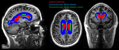 |
10 |
FLAIR-DTI joint analysis of periventricular white matter lesions for normal control, mild cognitive impairment, and Alzheimer’s disease
Jingyun Chen, Yi Li, Artem Mikheev, Zifei Liang, Matthew Gruen, Jiangyang Zhang, Henry Rusinek, Yulin Ge
In patients with mild cognitive impairment from ADNI MRI database, joint analyses of FLAIR and diffusion-tensor MRI (DTI) find significant positive correlation between periventricular white matter lesion (PVWML) and ventricle volumes; and negative correlation between PVWML and grey matter thickness of four cortical regions. Furthermore, significant correlations were found between PVWML and DTI measures (FA, MD, AD and RD) within the WM in close proximity to four cortical regions, indicating the degenerative changes in remote subcortical WM regions that can be quantitatively evaluated with DTI. However, such correlations were not found in either normal controls or patients with AD.
|
|
3175.
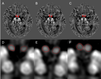 |
11 |
Susceptibility and Volume Changes of the Mammillary Bodies as a Function of Age for Healthy Individuals and Early Stage Dementia
Zhijia Jin, Sean Sethi, Binyin Li, Naying He, Weibo Chen, E. Haacke, Fuhua Yan
The mammillary bodies play an important role in episodic memory and spatial memory. However, few studies have focused on their properties in humans due to the limitations of the imaging techniques, their location and small size. In this study, we evaluate brain iron content and volume of the mammillary bodies in subjects with mild cognitive impairment (MCI), Alzheimer’s Disease and healthy individuals using quantitative susceptibility mapping and 3D T1-weighted imaging. There was a slight reduction in volume and increase in susceptibility with age and there were no differences of either susceptibility or volume among the three groups.
|
|
3176.
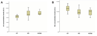 |
12 |
Cerebral myo-inositol and glutamate+glutamine levels assessed by magnetic resonance spectroscopy in healthy aging
Antoine Hone-Blanchet, Lisa Krishnamurthy, Salman Shahid, Qixiang Lin, Candace Fleischer, James Lah, Allan Levey, Deqiang Qiu, Bruce Crosson
This study aims at measuring the effect of aging and expression of protein amyloid-beta (Aβ) on markers of neuroinflammation and glutamate metabolism using magnetic resonance spectroscopy. Measures of medial frontal cortical myo-inositol (mI) and glutamate+glutamine (Glx) were obtained in 21 young adults, 21 healthy older adults and 10 healthy older adults expressing Aβ. Results show an increase in mI in all older adults compared to young adults, and a decrease in Glx in all older adults compared to young adults. These results suggest that MRS is a viable tool in the investigation of biomarkers of inflammation in aging.
|
|
3177.
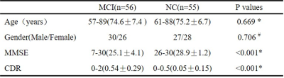 |
13 |
Prediction of Mild Cognitive Impairment Patients based on VBM, DBM and SBM Analysis
Presentation Not Submitted
Zhe Ma, Xiangyu Ma, Zhizheng Zhuo, Lijiang Wei, Yingjie Mei, Haiyun Li
Exploring reliable biomarkers is important for the clinical early detection of mild cognitive impairment (MCI) patients . This study investigated cerebral morphological abnormalities in MCI by combining three widely-used morphometry analysis methods (Voxel-based morphometry (VBM), deformation-based morphometry (DBM) and surface-based morphometry (SBM)) and constructed a set of classifiers to identify MCI patients from normal controls. The highest classification accuracy (91%) was reached when using combined morphological features (including gray matter volume, deformation, cortex thickness, gyrification index, sulcus depth and fractal dimension). Our results indicate that using combined morphological features could improve the performance of MCI prediction compared to using a single morphometry method
|
|
3178.
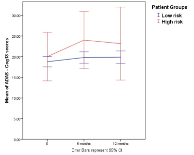 |
14 |
HIGH AND LOW RISK DEMENTIA CLASSIFICATION IN EARLY MCI FROM ADNI1
Trisha Kibaya, Nivedita Agarwal , Jorge Jovicich
Mild cognitive impairment (MCI) has been defined as the stage between the expected cognitive decline of normal aging and the more serious decline of dementia. Its clinical characteristics represent the earliest features of many forms of dementia. Specifically, many studies have reported amnestic MCI (aMCI) as a risk factor for Alzheimer’s disease, but to date there’s still a need for establishing specific, reliable, pre-symptomatic and non-invasive biomarkers associated to the progression of aMCI to AD. In this study we investigated CSF and structural MRI biomarkers for the classification of high and low risk of dementia conversion in aMCI.
|
|
3179.
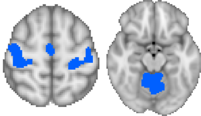 |
15 |
Functional Impact of Theta Burst Stimulation on Motor Cortex
Marc Lindley, Mark Sundman, Koeun Lim, Jack-Morgan Mizell, Viet Ton That, William Mennie, Chidi Ugonna, Nan-kuei Chen, Ying-hui Chou
The effect of transcranial magnetic stimulation using intermittent theta burst stimulation (iTBS) on functional connectivity and cortical excitability was tested in a healthy older adult and individuals with mild cognitive impairment. Individuals underwent an iTBS protocol with single pulse cortical excitability tests and-resting state fMRI(rs-fMRI) being performed before and after. Cortical excitability was tested through a pseudorandomized order of intensities of single pulses. Within functional network RS-fMRI analysis was done of the sensorimotor network. Differences in the response between the two populations was observed in both the functional connectivity changes in the sensorimotor network as well as the cortical excitability.
|
|
3180.
 |
16 |
Quantification of neuronal loss related to cognitive impairment in mild Alzheimer disease in hippocampal subfields using quantitative gradient recalled echo (qGRE) MRI
Satya VVN Kothapalli, Tammie L Benzinger, Jason Hassenstab, Manu S Goyal, John C Morris, Dmitriy A Yablonskiy
Damage of hippocampus leading to cognitive decline is one of the major hallmarks of Alzheimer disease (AD). Differentiating neuronal loss in hippocampal subfields is important as they control different biological functions. In this study, we used MRI-based qGRE technique to evaluate neuronal content (in the remaining after atrophy tissue) of hippocampal subfields in a well-characterized cohort of human subjects recruited from Knight ADRC. Our results showed pronounced left/right asymmetry in the pattern of neuronal damage in mild AD and a significantly stronger (compared to atrophy) correlation between neuronal loss in the remaining tissue of hippocampal subfields and cognitive tests.
|
|
3181.
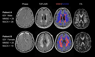 |
17 |
The effect of deep medullary veins disruption on cognitive function is mediated by microstructure integrity in patients with cerebral small vessel disease
Ruiting Zhang, Qingqing Li, Min Lou, Minming Zhang
We aimed to evaluate the effect of deep medullary veins (DMVs) disruption on cognition in cerebral small vessel disease (cSVD) patients and to examine whether white matter microstructure integrity mediate the relationship between DMVs disruption and cognitive impairment. Susceptibility-weighted images were used to observe characteristics of DMVs. White matter was classified into white matter hyperintensities (WMH) and normal appearing white matter (NAWM), and the average Fractional anisotropy (FA) and cerebral blood flow values of each region were extracted and compared. Mediation analyses revealed that the effect of DMVs score on cognition was mediated by microstructure integrity (FA value) in NAWM.
|
|
3182.
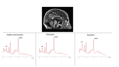 |
18 |
Brain volume asymmetries and 1H-MRS of Posterior Cingulate Cortex in the differential diagnosis of Primary Progressive Aphasia
Micaela Mitolo, Michelangelo Stanzani-Maserati, Stefania Evangelisti, Lia Talozzi, Laura Ludovica Gramegna, Lorenzo Cirignotta, Claudio Bianchini, Federico Oppi, Roberto Poda, Roberto Gallassi, Giovanni Rizzo, Luisa Sambati, Piero Parchi, Sabina Capellari, Rocco Liguori, David Manners, Claudia Testa, Raffaele Lodi, Caterina Tonon
Differential diagnosis of neurodegenerative diseases is a great challenge for both clinic practice and research. We investigated the ability of magnetic resonance spectroscopy (1H-MRS) of Posterior Cingulate Cortex (PCC) and brain volume asymmetries to differentiate patients with Primary Progressive Aphasia (PPA) from patients with Alzheimer’s disease (AD). The N-acetyl-aspartate to myo-inositol ratio (NAA/mI ratio) in the PCC discriminates APP from AD (p = 0.009) with an accuracy of 75.5%. Furthermore, ROC curve analyses of all statistically significant asymmetry indexes were performed and the PCC showed the highest level of accuracy (81.4%) in discriminating between the two neurodegenerative groups.
|
|
3183.
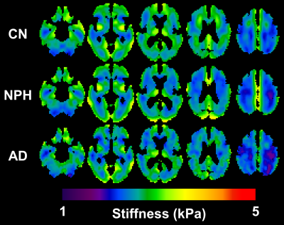 |
19 |
Identification of normal pressure hydrocephalus by disease-specific patterns of brain stiffness and damping ratio
Matthew Murphy, Arvin Arani, Fredric Meyer, Armando Manduca, Kevin Glaser, Richard Ehman, John Huston
Normal pressure hydrocephalus (NPH) is a form of dementia characterized by cognitive impairment, urinary incontinence and abnormal gait. NPH can be difficult to differentiate from other dementias, but it can be treated in many cases if accurately diagnosed. Here we tested whether MR elastography-based measures of brain stiffness and damping ratio could discriminate subjects with NPH from both cognitively normal subjects and those with probable Alzheimer’s disease. Both mechanical parameters exhibited significant group-wise differences in a specific spatial pattern. Further, summary measures of these spatial patterns in individuals discriminated subjects with NPH from the other two groups (area under ROC≥0.94).
|
|
3184.
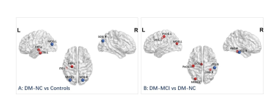 |
20 |
Altered Functional Topological Properties of Brain Networks in Type-2 Diabetes with and without Mild Cognitive Impairment
Ying Xiong, Qiang Zhang, Shuchang Zhou
This study aims to investigate the functional topological properties in T2DM with and without impairment, and characterize its relationships with clinical measurements. Forty-four T2DM patients were divided into two sub-groups (impaired and normal cognition), together with healthy controls, were imaged at a 3T scanner. We found no significant intergroup difference in global measurements among the three group. However, increased or decreased nodal efficiency was detected in some important brain regions. Altered nodal efficiency in FFG and ITG correlated with glycosylated hemoglobinA1c and neuropsychological assessments. The resting-state functional topological properties research shows potential feasibility in characterizing intrinsic alterations of diabetic encephalopathy.
|
|
3185.
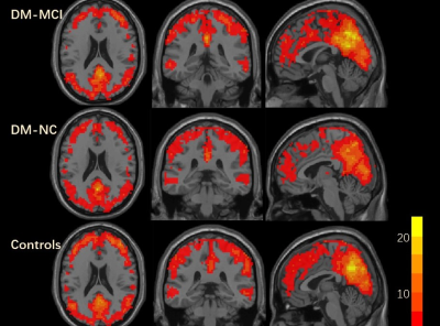 |
21 |
Progressive Alterations in Resting-State Regional Homogeneity in Type 2 Diabetes with and without Mild Cognitive Impairment
Ying Xiong, Qiang Zhang, Wenzhen Zhu
This study aims to detailly investigate the alterations in spontaneous brain activity in T2DM patients with and without MCI and characterize its relationships with clinical measurements. Forty-four T2DM patients were divided into two sub-groups(impaired and normal cognition), together with 25 healthy controls, were scanned at 3Tscanner. T2DM patients with normal cognition had increased regional-homogeneity in many important brain regions than controls. Those with MCI exhibited regional-homogeneity in right inferior frontal gyrus in a further step. Increased regional-homogeneity correlated with neuropsychological assessment, glycosylated-hemoglobinA1c and disease duration. Rs-fMRI can be an appropriate approach for studying the alteration in spontaneous brain activity in diabetes.
|
|
3186.
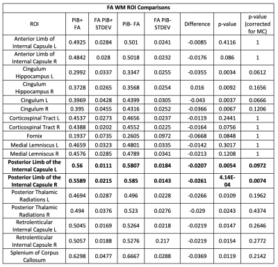 |
22 |
Amyloid- ß Associations with White Matter Integrity in Down Syndrome Assessed Using Diffusion Tensor Imaging and 11C-PiB Positron Emission Tomography
Austin Patrick, Minjie Wu, Patrick Lao, Douglas Dean, Matthew Zammitt, Sterling Johnson, Dana Tudorascu, Annie Cohen, Karly Cody, Charles Laymon, William Klunk, Shahid Zaman, Benjamin Handen, Andrew Alexander, Bradley Christian
Nearly all people with Down Syndrome will develop Alzheimer’s Dementia neuropathology by age 50, often asymptomatically. In this work, diffusion tensor imaging (DTI) was used to characterize white matter (WM) tract microstructure in thirty-three non-demented participants with Down Syndrome. Amyloid plaque burden was assessed using PET imaging with C-11 PiB. DTI measures were used to compare WM microstructure in DS individuals with high amyloid burden (PiB+) versus individuals with low amyloid burden (PIB-). PIB+ adults demonstrated significantly increased mean diffusivity and decreased fractional anisotropy in several bilateral WM regions. These findings are consistent of signs of WM degeneration.
|
|
3187.
|
23 |
Hippocampal cerebrovascular reactivity is associated with obesity in women. An arterial spin labeling study.
Lidia Glodzik, Henry Rusinek, Wai Tsui, Yi Li, Pippa Storey, Ricardo Osorio, Tracy Butler, Mony de Leon
Cerebrovascular reactivity to carbon dioxide (CVR-CO2) is impaired in conditions affecting cerebral vasculature. Obesity increases the risk of Alzheimer’s disease (AD). The hippocampus plays a prominent role in cognition and it is one of the earliest brain structures affected during the progression of AD. It remains uncertain how obesity affects cerebral vasculature in AD vulnerable regions. We examined the relationship between body mass index and neocortical and hippocampal vasoreactivity. Our pulsed ASL sequence combined a flow-sensitive alternating inversion-recovery labeling scheme with balanced steady-state free precession to optimize spatial resolution and lower sensitivity to susceptibility artifacts. Cerebral blood flow (CBF) measurements were done during rest and rebreathing challenge designed to increased CO2 level. In obese women (BMI≥30, n=36) hippocampal vasoreactivity was 80% lower than in their non-obese peers. No relationship was observed in men or with respect to cortical vasoreactivity.
|
|
3188.
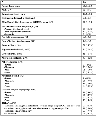 |
24 |
A novel MRI classifier of arteriolar sclerosis in aging: Prediction of pathology and cognitive decline
Nazanin Makkinejad, Arnold Evia, Ashish Tamhane, David Bennett, Julie Schneider, Konstantinos Arfanakis
Arteriolar sclerosis is one of the main pathologies of small vessel disease, is common in the aging brain, and has been associated with lower cognitive performance and higher risk of dementia. Definitive diagnosis of arteriolar sclerosis is only possible at autopsy. In this work, an MRI-based classifier of arteriolar sclerosis was developed, by first training a classifier on ex-vivo MRI and pathology data and then translating it in-vivo, and was evaluated in a large community cohort of older adults.
|
|
3189.
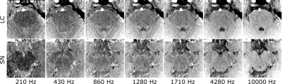 |
25 |
Evaluation of the magnetization exchange in catecholaminergic nuclei at 3 T
Nikos Priovoulos, Stan van Boxel, Heidi Jacobs, Benedikt Poser, Dimo Ivanov
Magnetization Transfer (MT) weighted gradient echo techniques can be used favourably instead of typical spin-echo based approaches for high-resolution imaging of the Locus Coeruleus (LC), which is of interest in various diseases. Examining the MT properties of the LC is needed for the development of quantitative biomarkers. Employing a fast, high-resolution acquisition sequence we show that it is possible to obtain data to fit a two-pool MT exchange model in the LC. The LC shows differential MT behavior compared to other catecholaminergic and adjacent GM regions consistent with reduced macromolecular content.
|
|
| Top |
Experimental Models of CNS Disease: Functional/Spectroscopy
Digital Poster
Neuro
Tuesday, 14 May 2019
| Exhibition Hall |
16:45 - 17:45 |
| |
|
Computer # |
|
3190
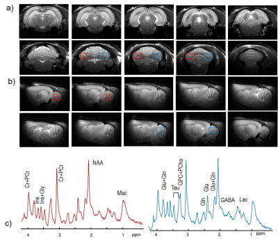 |
26 |
Mild neonatal hypoxia-ischemia in rats induces long-term behavior and cerebellar anormalities
Video Permission Withheld
Yohan Van de Looij, Edurado Sanches, Stephane Sizonenko, Hongxia Lei
How mild neonatal hypoxia-ischemia (HI) in very immature rats affects cerebellum remains not well understood. We showed cerebellar abnormalities, including behavior, motor function and metabolites, in adult rats after HI at postnatal day 3.
|
|
3191.
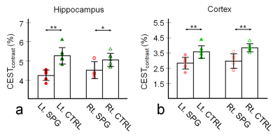 |
27 |
Cerebral mapping of glutamate with GluCEST MRI in a rat model of stress-induced sleep disturbance
Do-Wan Lee, Dong-Hoon Lee, Chul-Woong Woo, Jae-Im Kwon, Yeon-Ji Chae, Su Jung Ham, Ji-Yeon Suh, Sang-Tae Kim, Jeong Kon Kim, Kyung Won Kim, Jin Seong Lee, Choong Gon Choi, Dong-Cheol Woo
GluCEST is a powerful neuroimaging tool, can detect in vivo glutamate signals involving neurotransmitter metabolism in the central nervous system. We measured glutamate signal changes in the hippocampus and cortex of a rat model of stress-induced perturbed sleep, using in vivo GluCEST and high-resolution 1H-MRS. The CEST signal in control and sleep-perturbed rats revealed significant findings on GluCEST contrast values and metabolic concentrations in both regions. Our in vivo GluCEST and 1H-MRS results may yield valuable insights in the alterations of cerebral glutamate signals in sleep disorders.
|
|
3192
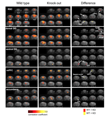 |
28 |
Diffusion tensor imaging and resting-state functional MRI reveal altered brain network hubs on a depression knockout mouse model
Video Permission Withheld
Sheng-Min Huang, Kuan-Hung Cho, Tsung-Ying Yang, Yi-Shan Wu, Hsuan-Kai Huang, Chia-Wen Chiang, Pei-Hsin Huang, Li-Wei Kuo
In this study, diffusion tensor imaging (DTI) and resting-state functional MRI were employed on a depression knock-out mouse model, which shows behaviors of anxiety and depression. The brain network hubs were investigated by region-of-interest (ROI) and connectivity analyses. Our results showed altered resting-state connectivity in prefrontal and hippocampal areas. Also, altered DTI indices were also found in thalamus and hippocampus. These findings are consistent with previous human studies and suggest the brain neuroimaging could be potentially useful to reveal the brain network hubs affected by depression on the proposed mouse model.
|
|
3193.
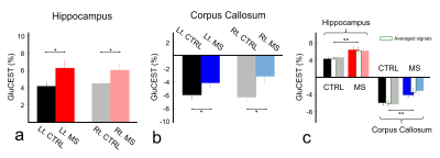 |
29 |
In Vivo Evidence of Brain Glutamate Level Changes in a Multiple Sclerosis Rat Model Using Glutamate Chemical Exchange Saturation Transfer (GluCEST) Imaging
Dong-Hoon Lee, Chul-Woong Woo, Jae-Im kwon, Yeon-Ji Chae, Su Jung Ham, Ji-Yeon Suh, Sang-Tae Kim, Jeong Kon Kim, Kyung Won Kim, Jin Seong Lee, Choong Gon Choi, Dong-Cheol Woo, Do-Wan Lee
GluCEST imaging is a novel molecular MRI imaging technique that provides in vivo image contrast by glutamate concentration changes. In this abstract, we attempted to evaluate signal changes in hippocampus and corpus callosum at a multiple sclerosis rat model based on the quantified GluCEST signals. We also evaluated and compared the signals with those in the control group to demonstrate the glutamate signal differences. Our results clearly showed that GluCEST imaging could be a useful tool to evaluate the brain metabolism in the brain multiple sclerosis, and it provides quantitative results highly related with the in vivo glutamate level changes.
|
|
3194.
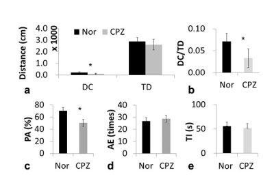 |
30 |
Glutamate imaging in schizophrenia prodrome
Zhuozhi Dai, Handi Zhang, Alan Wilman, Haiyun Xu, Gang Xiao, Hongfu Sun, Zerui Zhuang, Yanlong Jia, Zhiwei Shen, Gen Yan, Renhua Wu
The diagnosis of schizophrenia prodrome is significant since early intervention may prevent the development of full-blown schizophrenia. With increasing evidence indicating that glutamate is involved in the incidence of schizophrenia, we hypothesize that glutamate could be an imaging biomarker in the diagnosis of prodromal schizophrenia. We applied the glutamate CEST imaging technique in schizophrenic models and demonstrates that glutamate changes in different regions of the brain at an early stage, which may provide a powerful indicator of the diagnosis of schizophrenia prodrome. In addition, the glutamate CEST image had excellent correlation with standard MR spectroscopy.
|
|
3195.
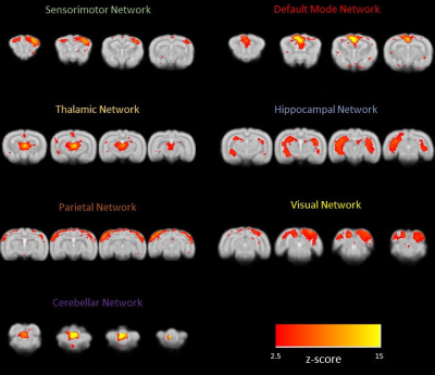 |
31 |
Advanced Experimental setup for awake resting-state fMRI in rabbits
Nicola Bertolino, Daniele Procissi, Craig Weiss, Quinn Smith, John Disterhoft
Awake imaging in pre-clinical research is challenging due to the MRI sensitivity to motion and animal’s natural reactivity to unfamiliar and loud environment. In this work we present an experimental setup for resting-state fMRI of rabbits and preliminary data collected with it. Our setup relies on the natural tolerance of rabbit to restraint and a home-designed animal fixing cradle equipped with a 3-channel receiver coil.
|
|
3196.
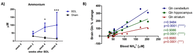 |
32 |
In vivo longitudinal 1H MRS study of hippocampal, cereberal and striatal metabolic changes in the developing brain using an animal model of Chronic Hepatic Encephalopathy
Dunja Simicic, Katarzyna Pierzchala, Veronika Rackayova, Olivier Braissant , Stefanita-Octavian Mitrea, Dario Sessa, Valerie McLin, Cristina Cudalbu Ramona
Chronic hepatic encephalopathy (CHE) is a severe neuropsychiatric disorder associated with chronic liver disease (CLD). For children, the impairment of neurocognitive functions as a consequence of CLD seems to be irreversible. We aimed to investigate longitudinally, using in-vivo 1H-MRS, differences in metabolic changes between hippocampus, cerebellum and striatum of a developing brain in a rat model of CHE. We showed the most pronounced changes in metabolites in cerebellum, suggesting its increased vulnerability. Further delineation of regional changes in the brain in response to CLD may help elucidate the molecular and regional origins of neuromotor and neurocognitive changes associated with CLD.
|
|
3197.
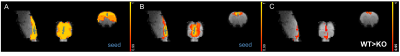 |
33 |
Impaired Functional Connectivity in a Rat Model and Humans with Fragile X Syndrome.
Joanna Smith, Andrew McKechanie, Milou Straathof, Rick Dijkhuizen, Sumantra Chattarji, Andrew Stanfield, Sally Till, Peter Kind
A key requirement for the effective development of novel therapies for Intellectual Disabilities is the ability to directly compare findings from basic neuroscience in rodent models with human studies. Functional magnetic resonance imaging offers a platform to overcome this translational barrier. Here, we use a parallel resting state fMRI approach in individuals with Fragile X Syndrome (FXS) and in a rat model of FXS using a 3T and 7T scanner respectively, and show that the loss of Fragile X mental retardation protein leads to a shared decrease in DMN connectivity in humans with FXS and rats that model this disorder.
|
|
3198.
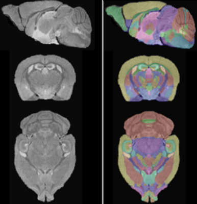 |
34 |
Imaging the evolution acute fear to anxiety: Longitudinal whole brain imaging in living mice of neural activity with MEMRI
Elaine Bearer, Daniel Barto, Rusell Jacobs
Life-threatening events cause extreme fear, which evolves in vulnerable people into a debilitating mental illness--post-traumatic stress disorder (PTSD). Here we directly address how acute fear evolves to anxiety using high field MR in mouse models of PTSD, applying systems-wide longitudinal manganese-enhanced magnetic resonance imaging (MEMRI) to image whole brain responses to unconditioned fear, predator stress (PS), and progression or resolution over time. We report that serotonin transporter knock-out results in sustained anxiety-like behavior and altered neural activity after predator stress. Automated segmentation of SPM maps identifies m regions correlated with progression to PTSD for the first time.
|
|
3199.
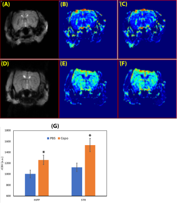 |
35 |
In Vivo MRI Reveals Increased Brain Prefusion in Carbamoylated Erythropoietin Treated Mice
Yutong Liu, Monica Sathyanesan, Samuel Newton
In vivo MRI was used to detect and measure the brain hemodynamic action of carbamoylated erythropoietin (Cepo) in mice. Brain perfusion was measured using dynamic susceptibility contrast MRI, and BBB permeability was detected by pre- and post-contrast T1 mapping. It was found that Cepo caused increased cerebral blood flow and volume. Consistent pre- and post-contrast T1 values indicated no gadolinium leakage from vascular system to brain tissue. In summary, chronic Cepo treatment induced increased brain perfusion and this can be detected using in vivo MRI.
|
|
3200.
|
36 |
Longitudinal 9.4 Tesla 1H MRS in the thalamus of the Theiler’s encephalomyelitis virus (TMEV) mouse model of multiple sclerosis
Poonam Choudhary, Suyog Pol, Marilena Preda, Michele Sveinsson, Robert Zivadinov, Ferdinand Schweser
This study investigated thalamic metabolic alterations related to acute and chronic inflammation in mice infected with TMEV, a model of MS. TMEV-inoculation causes a biphasic neurological disease starting with an early acute inflammation of the subcortical gray matter (<1 month) and progressing into a late chronic demyelinating phase associated with oligodendroglial damage that develops into a neurodegenerative phase (>4 months). Our hypothesis was that the influx of immune cells will result in increased glutamate and myoinositol in the acute phase, and that glutamate will further increase in the chronic phase whereas gamma-aminobutyric acid, N-acetylaspartate, and choline will decrease.
|
|
3201.
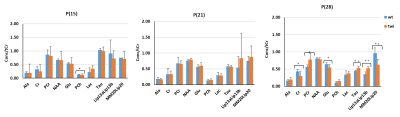 |
37 |
Pre- and post-symptomatic longitudinal metabolic assessment of the Twitcher mouse model of Krabbe Disease at 9.4 Tesla
Poonam Choudhary, Nadav Weinstock, Marilena Preda, Lawrence Wrabetz, Robert Zivadinov, Ferdinand Schweser
Krabbe Disease (KD) is a rare progressive globoid cell leukodystrophy caused by a deficiency of galactocerebrosidase (GALC), necessary for the metabolism of galactosylceramide and psychosine. Accumulation of these neurotoxic sphingolipids results in demyelination, neuroinflammation and ultimately death in infancy. This study aimed to investigate if localized proton magnetic resonance spectroscopy (1H-MRS) may serve as useful markers to detect pre-symptomatic metabolic alterations related to KD.
|
|
3202
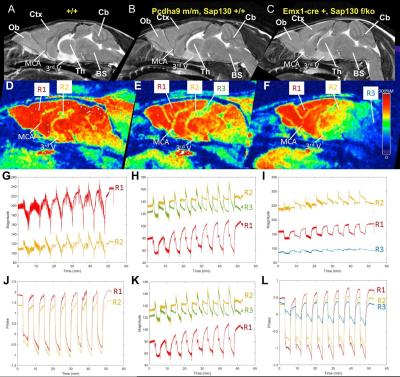 |
38 |
4D Real-time BOLD MRI in Genetically Engineered Mouse Brains with Acute Hypoxia Challenge
Video Permission Withheld
Anthony Christodoulou, George Gabriel, Cecilia Lo, Yijen Wu
The objective of this study is to develop 4D time-resolved high-resolution BOLD MRI by combining low-rank sparse imaging and compressed sensing. By expressing a dynamic BOLD image as the product of a set of basis images and temporal functions, we are able to capture differential dynamic BOLD responses to oscillating hypoxia challenge in high-resolution 3D space in genetically engineered mouse brains.
|
|
3203.
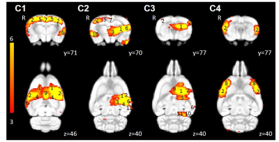 |
39 |
Effects of daily high and low frequency low-intensity repetitive transcranial magnetic stimulation in rats: A longitudinal rs-fMRI study
Bhedita Seewoo, Kirk Feindel, Sarah Etherington, Jennifer Rodger
Repetitive transcranial magnetic stimulation (rTMS), a novel non-invasive brain stimulation technique, has been shown to modulate dysfunctional brain networks in humans. However, despite anecdotal evidence that rTMS effects tend to wear off, there are no reports of fMRI studies, even in humans, mapping the therapeutic duration of rTMS effects. Here, we investigated the cumulative effects of daily low-intensity rTMS on rodent resting-state networks using rs-fMRI and mapped for persistence for up to three weeks. Our study confirms the frequency-specific effects of rTMS and shows that 1 Hz stimulation has milder, but longer-lasting effects on functional connectivity than 10 Hz stimulation.
|
|
3204.
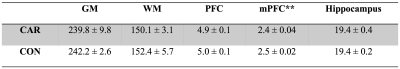 |
40 |
Upregulation of Rac1 activity related to structural and functional neuroimaging changes in mouse brain at 11.7T
Wenjing Xu, Ying Liu , Garth Thompson, Xiaoyong Zhang
Rac1 is critical for synapse remodeling. In this work, we investigated whether Rac1 could induce neuroimaging changes in a transgenic mouse model using structural MRI, resting state-fMRI and MRS at 11.7T. Our data showed that the volume of the medial prefrontal cortex (mPFC) is significantly decreased in Rac1 activation group. Moreover, we found that the upregulation of Rac1 activity is related to increased functional connectivity in mouse brain. However, Glx or GABA levels do not show significant changes in mPFC. We concluded that the upregulation of Rac1 activity is related to structural and functional neuroimaging changes.
|
|
3205.
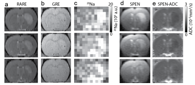 |
41 |
Performance of sodium, ultrafast diffusion, and MPIO stem cell tracking MRI classification of sub-acute ischemic stroke recovery at 21.1T
Avigdor Leftin, Jens Rosenberg, Xuegang Yuan, Teng Ma, Samuel Grant, Lucio Frydman
MRI leverages multiple modes of contrast to characterize stroke. Acute phase stroke detection has focused on multiparametric MRI contrasts such as T2-weighting (T2w), apparent diffusion coefficient (ADC), and sodium level. Evaluation of these contrasts with theranostic cell tracking during the subacute recovery phase at ultrahigh field has not been investigated to a similar extent. Here multiparametric MRI evaluation of ADC, 23Na, and MPIO stem cell tracking in a rodent MCAO model at 21.1T was used to determine parametric correlations and receiver operator performance. Differential parametric time-dependence and sensitivities are observed that inform future high-and low field studies of stroke recovery.
|
|
3206
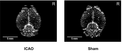 |
42 |
Contrast-enhanced MR microangiography of cortical vascular remodeling after unilateral internal carotid artery occlusion in the mouse
Video Permission Withheld
Philipp Boehm-Sturm, Till de Bortoli, Stefan Koch, Melina Nieminen, Susanne Mueller, Giovanna Ielacqua, Jan Klohs, Ulrich Dirnagl, Peter Vajkoczy, Nils Hecht
Collateral flow is an important, yet poorly understood compensatory mechanism in response to brain hypoperfusion. Unilateral internal carotid artery occlusion in the mouse is a model to study collateral growth of penetrating arterioles. We hypothesized that this process could be assessed in vivo by high resolution MR microangiography with iron oxide nanoparticles. Since our MR vessel density measurements contradicted previous histological findings we established atlas tools to validate angiograms with microscopy on vessel-stained tissue slices or with whole brain serial two photon microscopy.
|
|
3207.
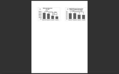 |
43 |
Brain GABA Levels of P5 Wild Type and Fragile X Mouse Model (FMR1 KO): Comparison of Mass Spectroscopy and In vivo 1H MRS
Sanaz Mohajeri, Samantha Reyes, Scarlett Guo, Karolina Krasinska, Laura Pisani, Daniel Spielman, Fredrick Chin
An imbalance between excitatory glutaminergic and inhibitory GABAergic processes is one of the leading hypotheses for fragile X syndrome. We investigated GABAergic differences by measuring brain GABA levels of male five-day-old mouse pups (P5) in Wild-Type (WT) and Fragile X knockout (FMR1 KO) models. .. Ex vivo mass spectrometry detected thalamus and frontal differences in both WT and FMR1 KO models and lower GABA levels in the FMR1 KO animals. In vivo 1H spectra at 7T found similar regional GABA differences between thalamus- and frontal-rich regions, but was unable to detect Wild-Type versus FMR1 KO differences.
|
|
3208.
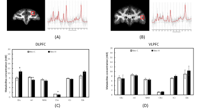 |
44 |
Investigation of cerebral metabolite changes in the dorsolateral and ventrolateral prefrontal cortex of adult macaques with neonatal hippocampal lesions
Chun-Xia Li, Christa Payne, Xiaoping Hu, Jocelyne Bachevalier, Xiaodong Zhang
In the present study, in vivo MR spectroscopy was employed to investigate the neuro-metabolites changes of dorsolateral prefrontal cortex (DLPFC) and ventrolateral prefrontal cortex (VLPFC) in adult macaques with neonatal hippocampus lesion (Neo-H). Significant metabolite changes were seen in the right DLPFC of the Neo-H monkeys but not in the VLPFC, and significantly correlated with the working memory scores. Also, lateralization of the cerebral metabolite changes was observed. The results suggest that the neuro-metabolites changes in DLPFC and VLPFC of adult monkeys resulted from early insult to the hippocampus and the effect is mainly seen in the right hemisphere.
|
|
3209.
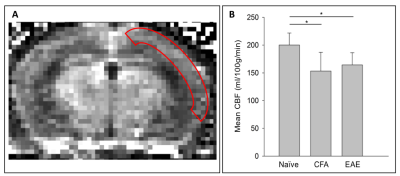 |
45 |
Reduced cerebral blood flow measured in the EAE mouse model of multiple sclerosis using perfusion MRI
Taelor Evans, A. Max Hamilton, Erin Stephenson, V. Wee Yong, Jeff Dunn
Multiple sclerosis (MS) is a chronic neurological disease involving inflammation. Brain hypoxia, or low brain oxygenation, in MS is an emerging field of study. It has been shown that some MS patients have reduced cerebral blood flow (CBF), but the mechanism is unclear. Experimental autoimmune encephalomyelitis (EAE) is a mouse model used to study inflammation-associated neurodegeneration. Using perfusion MRI and immunohistochemistry, we demonstrated that CBF reduction in EAE may be due to cerebral blood vessel occlusion in response to systemic inflammation. CBF reduction, coupled with brain inflammation, is a likely cause of hypoxia in MS.
|
|
3210.
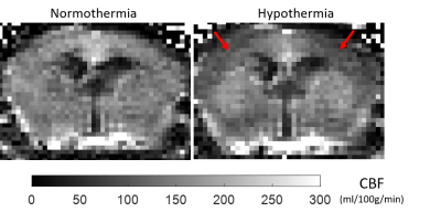 |
46 |
Using a multimodal near-infrared spectroscopy and MRI system to quantify gray matter metabolic rate for oxygen: A hypothermia validation study
Mada Hashem, Ying Wu, Jeff Dunn
Non-invasive quantitative imaging of cerebral oxygen metabolism (CMRO2) in mice is crucial to understand the role of oxidative metabolism in neurological diseases. We are developing a multimodal method combining near-infrared spectroscopy and high-field MRI to non-invasively study oxygen delivery and consumption in the cortex of mouse models of neurological disease. In this study, the feasibility of the NIRS-MRI technique to detect changes in CMRO2 in the mouse brain was assessed using a mild hypothermia, known to reduce metabolic rate. A decrease of 23% in CBF and 46% in CMRO2 was observed, which is consistent with previously published values.
|
|
3211.
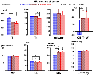 |
47 |
MRI Discriminates between the Aged and Aged Diabetic Brain of Rats
Guangliang Ding, Michael Chopp, Lian Li, Li Zhang, Esmaeil Davood-Bojd, Qingjiang Li, Min Wei, Zhenggang Zhang, Quan Jiang
Aging and diabetes both affect brain structure and physiology. To distinguish changes in brain induced by normal aging from diabetes in the aging brain, MRI measurements were performed on three groups of young, aged non-diabetic and correspondingly aged diabetic rats. MRI measurements, i.e., T1, T2, CBF, CE-T1WI, MD, FA, MK and Entropy were performed. Our data indicate that select MRI metrics FA of white and grey matter, and combination of T1 and T2 of grey matter are able to discriminate cerebral changes caused by aging and age-equivalent diabetes.
|
|
3212.
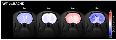 |
48 |
Longitudinal monitoring of brain glutamate levels using gluCEST in a rat model of Huntington’s disease
Julien Flament, Jérémy Pépin, Julien Valette, Emmanuel Brouillet
Huntington’s disease (HD) is an inherited neurodegenerative disease characterized by motor, cognitive and psychiatric symptoms. As glutamate has been shown to be a potential biomarker of neurodegenerative diseases, we used Chemical Exchange Saturation Transfer imaging of glutamate (gluCEST) to map cerebral glutamate distribution in a rat model of HD. The longitudinal follow-up of brain glutamate levels reveals different variations between HD and control animals, suggesting that gluCEST may serve as a potential biomarker of HD, especially at asymptomatic stage.
|
|
3213.
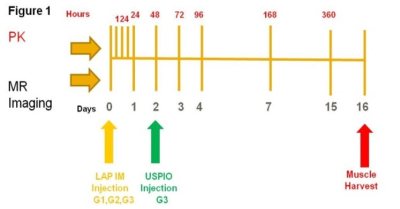 |
49 |
Serial MRI to Assess Effects of Drug Particle Size on Inflammation and Pharmacokinetics to Support Development of Long Acting Parenteral Formulations
Stephen Lenhard, Mary Rambo, Tracy Gales, Taki Kambara, Cindy Fishman, Valeriu Damian, Nima Akhavein, Matt Burke, Beat Jucker
We evaluated the effect of Long Acting Parenteral particle size on drug depot kinetics and inflammation using ultra small paramagnetic iron oxide (USPIO) T2W MRI. Our results showed an immediate post injection difference in inflammatory response and histological confirmation of greater muscle injury in the smaller micronized (1um) particle group compared to larger 20um particle formulation. The imaging of the drug depot in vivo with MRI combined with drug PK, tissue biodistribution, and histology allows for the development of individual Physiological Based PK models of drug biodistribution which would add significant scientific value to drug development.
|
|
3214.
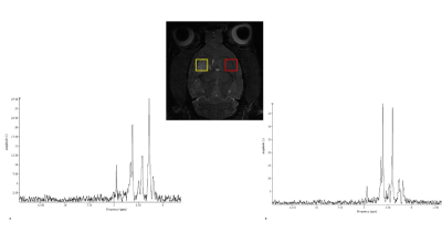 |
50 |
Spectroscopy and sodium analysis of dissociated cellular therapy in acute ischemia at 21.1 T
Shannon Helsper, F. Bagdasarian, Xuegang Yuan, Nastaren Abad, Jens Rosenberg, Teng Ma, Samuel Grant
This study evaluates biochemical markers in a rat model of acute ischemia at 21.1 T following administration of human mesenchymal stem cells dissociated (d-hMSC) from 3D aggregates utilizing sodium (23Na) chemical shift imaging (CSI) and relaxation-enhanced MRS. High field 1H MRS provides sensitive longitudinal metabolic mapping of biological markers including lactate, NAA, creatine and choline in response to cellular treatment. Evaluation of 23Na CSI provides insight into cerebral ionic homeostasis and tissue recovery following acute neurodegeneration by tracking ischemic lesion volumetrics.
|
|
| Top |
Experimental Model of CNS Disease: Structural/Diffusion
Digital Poster
Neuro
Tuesday, 14 May 2019
| Exhibition Hall |
16:45 - 17:45 |
| |
|
Computer # |
|
3215.
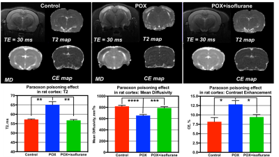 |
51 |
In Vivo MRI characterization of the effect of neuroprotection after nerve agent poisoning in rats
Alexandru Korotcov, Asamoah Bosomtwi, Ranjini Vengilote, Narayanan Puthillathu, John Moffett, Arun Peethambaran, Andrew Knutsen, Shalini Jaiswal, Nathanael Allison, Aryan Namboodiri, Bernard Dardzinski
Organophosphate poisoning is a major public health problem in developing countries. Organophosphates cause prolonged seizures and leads to neurodegeneration and functional defects. Current therapies are largely ineffective. Several advanced neuroimaging techniques demonstrate promise to detect subtle changes in brain activity and morphology related to organophosphate nerve agent poisoning, which would allow for the in vivo assessment of new therapeutics. In this study we use in vivo MRI and immunohistochemistry to demonstrate that fluorinated volatile anesthetics are an effective post-exposure neuroprotectant and can be used for organophosphates poisoning treatment.
|
|
3216.
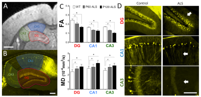 |
52 |
Ultra-High-Resolution Diffusion Tensor MRI Detects Early Axonal Connectivity Anomalies in Hippocampal Regions of ALS Mice
Presentation Not Submitted
Rodolfo Gatto, Manish Amin, Ariel Finkielsztein, Ronen Sumagin, Thomas Mareci, Richard Magin
Ultra-High field (UHF) MRI has been in continuous development as a new tool to investigate ultrastructural microscopic details in neuropathology. In this study we use UHF-MRI (17.6T) to investigate presymptomatic changes in the hippocampus of animal models of ALS (G93A-SOD1 mice). Using an ALS fluorescent transgenic mouse reporter and optical confocal microscopy, we demonstrated that microstructural changes detected by MRI diffusion can be related to very early alterations in axonal connectivity. This study constitutes a stepping stone for the application of more complex diffusion models in inhomogeneous brain tissue as a non-invasive exploration of neuropathology in ALS.
|
|
3217.
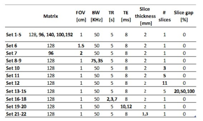 |
53 |
The generalized effect of diffusion on quantitative T2 mapping in preclinical scanners
Natalie Bnaiahu, Ella Wilczynski, Noam Omer, Tamar Blumenfeld-Katzir, Shir Levy, Noam Ben-Eliezer
Diffusion has a clear effect on qT2 measurements especially at high resolutions, caused by the imaging gradients. SSE and MSME protocols were used to scan a phantom and an in-vivo brain. An equation representing the effective b value of the applied pulse sequence (MSME) was developed to estimate the attenuation of the signal caused by diffusion. T2 values calculated without correcting the diffusion effect showed high variability between scans with different parameter sets. After correction T2 values increased and showed excellent agreement. The method demonstrated here is generalized and can apply to different pulse sequences, it improves accuracy and stability.
|
|
3218.
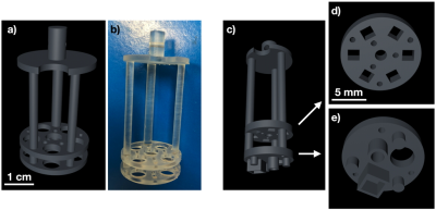 |
54 |
3D printed-phantoms dedicated to pre-clinical imaging systems for quality control
Sylvain Miraux, Aurélien Trotier, Colleen Cardiet, Stéphane Loubrie, Laurence Dallet, Thibaut Faller, William Lefrançois, Emeline Ribot
3D printing is a fantastic tool for creating prototypes. In parallel, pre-clinical imaging systems have been democratized for longitudinal studies of small animal models. However, for these systems, standardized phantoms for quality control do not exist yet. The aim of the work presented here is to show that phantoms of different shapes and sizes can be 3D printed to characterize preclinical imaging systems or sequences developed by research groups. As an example, images were acquired with a 70μm in plane resolution to study the influence of Cartesian and Radial encodings on the spatial resolution using a mouse dedicated phased array coil at 7T.
|
|
3219.
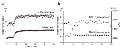 |
55 |
The mechanical signature of the dying brain
Gergely Bertalan, Jürgen Braun, Stefanie Schreyer, Barbara Steiner, Charlotte Klein, Angela Ariza, Eric Barnhill, Ingolf Sack, Jing Guo
In this study the mechanical properties of the mouse brain were continuously sampled by fast magnetic resonance elastography during ketamine/xylazine induced dying. Mechanical properties were correlated with metabolic and physiological imaging markers. Immediately after respiration arrest, stiffness of the whole brain and the hippocampus increased significantly while cardiac functioning was intact and reached a plateau ca. 5 min after ECG stop. Stiffness increase was inversely correlated with diffusion decrease. Results suggest that during the process of dying cytotoxic edema and brain swelling occurs leading to significant tissue stiffening.
|
|
3220.
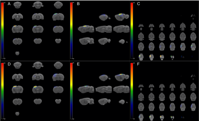 |
56 |
Structural changes in a rat model of traumatic brain injury and challenges of preclinical registration in the presence of lesions
Maria Yanez Lopez, Nicoleta Baxan, Cornelius Donat, Pete Hellyer, Mazdak Ghajari, Magdalena Sastre, David Sharp
The aim of this work was to develop an automated pipeline to study structural changes in a rat model of TBI using T1/T2-weighted images. Our results show that the CCI model causes focal alterations in grey matter in the proximity of the injury, with a visible edema 14 days after injury in T2-weighted images, plus damage in the white matter (corpus callosum). These hyper-intense lesions complicate registration and require optimisation of the affine step, together with skull stripping of the brain of injured rats.
|
|
3221.
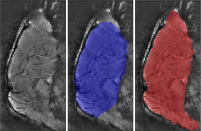 |
57 |
Automatic brain segmentation framework for bias field rich cranial MRI scans of rats and mice via similarity invariant shape priors
Jacob Hansen, François Lauze, Sune Darkner, Julia Huntenburg, Kristian Mortensen, Simon Sanggaard, Hedok Lee, Helene Benveniste, Maiken Nedergaard
This abstract presents an extension to our previous work for the extraction of rat brain tissue and internal cerebrospinal fluid networks in MR imaging of rat crania that display severe bias fields. This work contributes automation and robustness in the skull stripping module by introducing an automatic similarity invariant shape prior segmentation method. We demonstrate the capabilities of our framework on both rat brain as well as mouse brain data, using the same minimal number of rat brain priors.
|
|
3222.
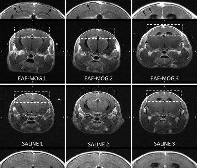 |
58 |
Preclinical detection of leptomeningeal inflammation in the myelin oligodendrocyte glycoprotein (MOG) induced experimental autoimmune encephalomyelitis (EAE) model of multiple sclerosis (MS)
Zachary Smith, Nicola Bertolino, Suyog Pol, Marilena Preda, Michelle Sveinsson, Robert Zivadinov, Ferdinand Schweser
Clusters of inflammatory cells in the leptomeningeal compartment are suspected to contribute directly to subpial cortical demyelination and neurodegeneration in patients with multiple sclerosis (MS). Clinical post-contrast 3D T 2-FLAIR detects these clusters through the leakage of a T 1 contrast agent into inflammatory foci and the subarachnoid space, referred to as leptomeningeal contrast enhancement (LMCE). While leptomeningeal inflammation has been reported in rodent models of MS, LMCE has not been used to study disease pathology in the preclinical setting. In this work, we present an imaging protocol for LMCE imaging in the experimental autoimmune encephalomyelitis myelin oligodendrocyte glycoprotein (EAE-MOG) murine model of MS at 9.4 Tesla.
|
|
3223
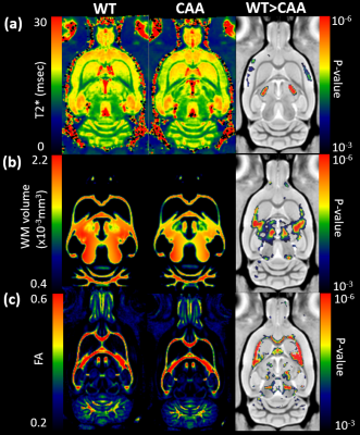 |
59 |
Diffuse white matter disease revealed in novel rat model of cerebral amyloid angiopathy type 1 by multi-modality MRI at 9.4T.
Video Permission Withheld
Hedok Lee, Sunil Koundal, Xiaodan Liu, Feng Xu, Simon Sanggaard, William Van Nostrand, Helene Benveniste
A novel cerebral amyloid angiopathy type 1 rat model which robustly develops microvascular amyloid beta deposits is studied. We report that this animal model develops white matter atrophy, cerebral micro-bleed, and loss of white matter integrity. These findings are corroborated by immunohistochemistry showing axonal disruption and vacuolization.
|
|
3224.
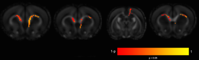 |
60 |
Neuroimaging Microglia: Development of a Quantitative Multi-Compartment Diffusion MRI Biomarkers of Microglial Density
JOHN-PAUL Yu
Neuroinflammation plays a critical role in neurologic and psychiatric disorders of the central nervous system (CNS) from ischemic stroke and traumatic brain injury to Alzheimer’s disease, schizophrenia, and major depression. The application of multi-compartment diffusion MRI techniques for the robust, non-invasive, and quantitative evaluation of microglial morphology and density in the setting of acute and chronic neuroinflammation would represent an important advancement in understanding, identification, and therapeutic monitoring of microglia across a broad spectrum of acute and chronic disorders of the CNS. We present the first evidence and application of multi-compartment diffusion MR techniques for the sensitive detection of changes in microglial density throughout the brain.
|
|
3225.
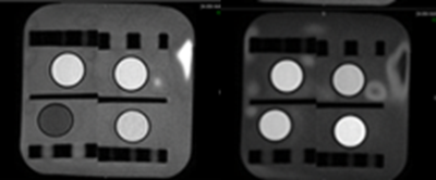 |
61 |
Optimized quantification of T2 relaxation times using gel phantoms in animal models for high-consequence pathogens in a Biosafety Level 4 environment
Marcelo Castro, Ji Hyun Lee, Joseph Laux, Harry Friel, Jeffrey Solomon, David Thomasson, Dima Hammoud
This experiment improved the accuracy of in vivo T2 measures used to evaluate blood brain barrier (BBB) disruption and subtle cerebral edema in non-human primate models of high-consequence viral infections in a Bio-Safety Level 4 (BSL-4) environment. In a healthy non-human primate, we used four gel phantoms to optimize T2relaxation times and investigated dependence of T2 on echo times. This methodology improved the accuracy of T2 estimation using fast spin echo sequences and phantoms. We hypothesized that reliable T2 values can be obtained by adding a phantom-calibration step to the T2 map calculation.
|
|
3226.
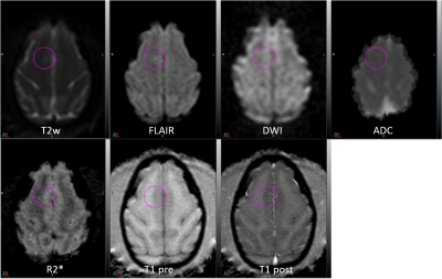 |
62 |
Neuroimaging of Nipah Virus infection in an African Green Monkey Model
Ji Hyun Lee, Yu Cong, Joseph Laux, Marcelo Castro, Matthew Lackemeyer, Jordan Bohannon, Oscar Rojas, Jeffery Solomon, Irwin Feuerstein, Vincent Munster, Dima Hammoud, Michael Holbrook
The purpose of this study was to utilize MRI to assess alterations in the brain that occur in African Green Monkeys infected with Nipah virus (NiV) via aerosol inoculation. Within 15 days of exposure to NiV, signal alterations were observed in the brain in T2-weighted, fluid-attenuated inversion recovery, and diffusion-weighted images, suggestive of infarction, inflammation and edema induced by NiV. The identification of non-invasive imaging biomarkers of acute NiV neurologic disease progression in this animal model could aid in the examination of potential vaccines and therapeutics.
|
|
3227.
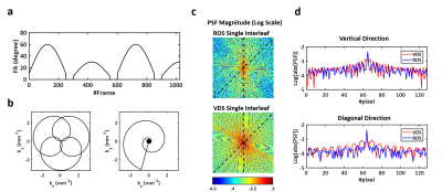 |
63 |
Comparison of Undersampling Capability of Magnetic Resonance Fingerprinting Using Rosette and Variable Density Spiral Trajectories
Yuning Gu, Charlie Wang, Yuchi Liu, Charlie Androjna, Xin Yu
The undersampling capability of magnetic resonance fingerprinting (MRF) using Rosette (ROS) and variable density spiral (VDS) trajectories was compared in this study. With a high undersampling factor, ROS-based MRF showed more uniform T1 and T2 mapping in phantoms with reduced errors. However, ROS reconstructed images of mouse brain showed slightly reduced SNR and off-resonance related artifacts, leading to similar undersampling capability as VDS-based MRF in vivo.
|
|
3228.
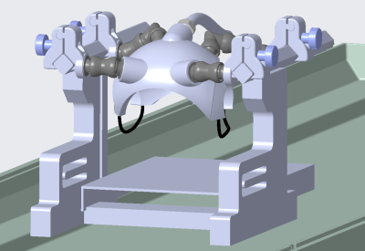 |
64 |
Evaluation of Diffusion-Weighted Imaging of the Macaque Brains Using Diffusion-Prepared TSE
Jialu Zhang, Dingxin Wang, Xiufeng Li, Kamil Ugurbil, Anna Roe, Xiaotong Zhang
A major challenge with EPI-based diffusion-weighted imaging (dMRI) is magnetic field inhomogeneity-associated distortion and signal loss. We implemented a mono-polar diffusion-preparation module for TSE sequence (DP-TSE) as an alternative to achieve distortion-free, high-resolution diffusion imaging with improved signal-to-noise ratio (SNR). Such an approach has been demonstrated in human subjects with a promising potential. We want to further evaluate the robustness of the implemented DP-TSE sequence and the feasibility of applying this approach for anesthetized macaques, and investigate whether DP-TSE is superior to alternative dMRI method in terms of imaging quality and SNR efficiency.
|
|
3229.
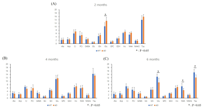 |
65 |
Neurochemical and structural early changes and onset time in 5xFAD model: MRS and VBM studies
Jang Woo Park, In OK Ko, Kyung Jung Kang, Ji Ae Park
We investigated early neurochemical and structural changes in 5xFAD model using magnetic resonance spectroscopy (MRS) and Voxel-based morphometry (VBM). At 6 months, it was confirmed that Glu, NAA, Tau and volume, which is characteristic of Alzheimer’s disease (AD) model, decreased in hippocampus of 5xFAD model.
|
|
3230.
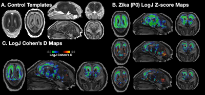 |
66 |
Voxelwise and morphometric analysis using diffusion tensor MRI microscopy reveals distinct microstructural and morphometric abnormalities in ferret brain development after gestational infection with the Zika virus
Elizabeth Hutchinson, Laura Reyes, Okan Irfanoglu, Sharon Juliano, Carlo Pierpaoli
An efficient pipeline for the processing and analysis of diffusion MRI microscopy data has been applied to study neurodevelopmental abnormaities in a ferret model of Zika infection. Individual and group differences in DTI values were found using Z-score and Cohen’s D maps to compare Zika treated and untreated P0 ferret brain specimens. Morphometric abnormalities were also identified using DTI-driven tensor based morphometry (DTBM) to show reduced local volume in the developing cortex. These results highlight the utility of this pipeline and advance the basic understanding of neurodevelopmental abnormalities that can result from exposure to the Zika virus during gestation.
|
|
3231.
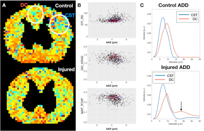 |
67 |
Improvements in specificity by non-Gaussian diffusion modeling and double diffusion encoding (DDE) to characterize axonal injury
Elizabeth Hutchinson, Dan Benjamini, Peter Basser, Carlo Pierpaoli, Michal Komlosh
Diffusion MRI techniques that extend beyond diffusion tensor imaging (DTI) – including acquisition strategies and advanced modeling – could provide more specific tools to probe abnormalities in disease or injury states. To evaluate similarities and distinctions across several prominent diffusion MRI strategies in the context of injury, we acquired multi-shell diffusion weighted images (DWIs) and double diffusion encoded (DDE) DWIs in healthy and injured ferret spinal cords. Scalar metrics from DTI, diffusion kurtosis imaging (DKI), mean apparent propagator MRI (MAP-MRI) and DDE-based axonal modeling were directly compared to reveal the ways in which each approach can specify key features of cellular alterations.
|
|
3232.
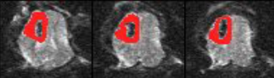 |
68 |
Evaluation of Neuroprotective Effects of Cyclosporine in a Porcine Model of Focal Traumatic Brain Injury using Diffusion Tensor Imaging
Nile Delso, Michael Karlsson, Bryan Pukenas, Johannes Ehinger, Sumei Wang, Matilda Hugerth, Eskil Elmér, Suyash Mohan, Magnus Hansson, Todd Kilbaugh, Sanjeev Chawla
A moderate focal contusion injury model in swine was used to evaluate treatment response to cyclosporine, a neuroprotective agent, using diffusion tensor imaging. This work builds upon the recent work in which we demonstrated improved neurological outcomes after administration of cyclosporine in the acute time period following traumatic brain injury (TBI). Regions of interests were drawn on the peri-contusion regions. We observed significant elevations in FA and CL and significant decline in CS from cyclosporine groups compared to those of placebo. These findings suggest that DTI may be useful in assessing treatment response to cyclosporine in a porcine model of TBI.
|
|
3233.
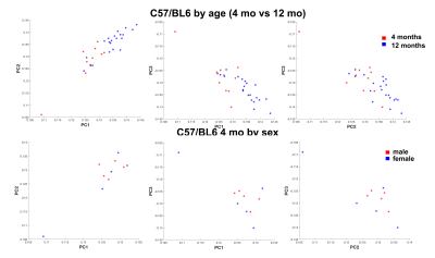 |
69 |
Vulnerable Networks in the Aging Mouse Brain
Wenlin Wu, Robert Anderson, Serge Koudoro, Eleftherios Garyfallidis , David Dunson, Carol Colton, Alexandra Badea
Despite recent advances in aging research, the underlying mechanisms of selective brain vulnerability to aging remain to be elucidated. Mouse models may provide useful tools to dissect the mechanisms behind age and sex associated vulnerability of brain circuits. We used high resolution accelerated protocols and tensor network analyses to reveal structural network differences in aging C57BL/6 mice.
|
|
3234.
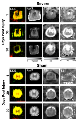 |
70 |
MRI Correlates of Neurological Outcomes in a Rat Spinal Cord Injury Model
Matthew Budde, Natasha Wilkins, Brian Schmit, Shekar Kurpad
This work evaluated the prognostic potential for quantitative MRI measures of the spinal cord compared to neurological assessments in a rat model of spinal cord injury. The results demonstrate in the acute setting, that a double diffusion encoded spectroscopy acquisition has greater accuracy than either DWI-EPI or T2 mapping while in the chronic setting, measures of spinal cord atrophy perform better than DWI measures. These results set the basis for future patient studies to improve MRI biomarkers in spinal cord injury.
|
|
3235.
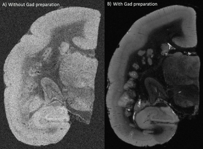 |
71 |
Ultra-high-resolution postmortem MRI of cortical lesions in a nonhuman primate model of multiple sclerosis
Maxime Donadieu, Diego Szczupak, Seung Kwon Ha, Daniel Abraham, Emily Leibovitch, Joseph Guy, Cecil Yen, Erin Beck, Afonso Silva, Steve Jacobson, Pascal Sati, Daniel Reich
Experimental autoimmune encephalomyelitis (EAE) in the common marmoset (Callithrix jacchus) shares important pathological and radiological similarities with MS. However, cortical pathology in this model has not been investigated by MRI. The purpose of this study is to examine, for the first time, whether cortical lesions can be visualized by MRI in this model. Similar to MS patients, we report the MRI detection of MS-like cortical lesions in postmortem EAE marmoset. These findings further reinforce the proximity between this animal model and the human disease.
|
|
3236.
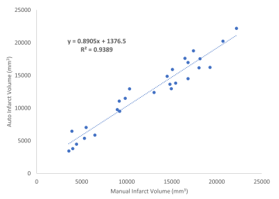 |
72 |
Automatic Measurement of Infarct Volume and Prediction by Pial Collaterals in Experimental Acute Ischemic Stroke
Yong Jeong, Gregory Christoforidis, Niloufar Saadat, Keigo Kawaji, Charles Cantrell, Steven Roth, Marek Niekrasz, Timothy Carroll
Tissue infarct due to major vessel occlusion depends on the compromised blood flow and the time since onset. Compromised blood flow may be sustained by recruitment of pial collateral vessels. In this study, the extent of pial collateral recruitment was used to predict infarct volume in an experimental middle cerebral artery occlusion animal model with and without norepinephrine and hydralazine. An automatic method of infarct volume measurement was developed to minimize user variability and time. The automatically calculated infarct volumes were highly correlated to manually measured volumes, and pial collaterals were predictive of infarct volume.
|
|
3237.
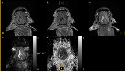 |
73 |
Development of a Swine Model to Evaluate Radiation-Induced Brain Injury: A Preliminary Report
Whitney Perez, Ilektra Athanasiadi, David Edmondson, Jeannie Plantenga, Carlos Perez-Torres
Radiation-induced brain injury (RIBI) is an irreversible and progressive long-term effect of radiation therapy. Current pre-clinical experiments use mouse models, which do not accurately replicate the pathology as seen in humans. We chose to simulate RIBI within a swine model because its brain structures are more comparable to human brain structures. Our preliminary findings show similar resemblances to the pathologies seen in human patients affected by RIBI.
|
|
3238.
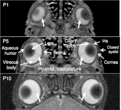 |
74 |
Postnatal Ocular Development in Normal and Visually Impaired Rats using in vivo Multi-Modal MRI
Malack Hamade, Hassan Hamade, Swarupa Kancherla, Kevin Chan
Ocular development is a complex process yet limited tools are currently available for non-invasive and longitudinal studies of the whole eye while minimizing biovariability. Using 7-Tesla MRI, we showed the growth characteristics of the anterior chamber and posterior chamber, the lens and the vitreous humor of rats from postnatal day 1 to 60 and compared the development of these three compartments between normal and impaired states. We conclude that MRI is an effective tool in characterizing normal and abnormal postnatal ocular growth over time before and after eyelid opening.
|
|
3239.
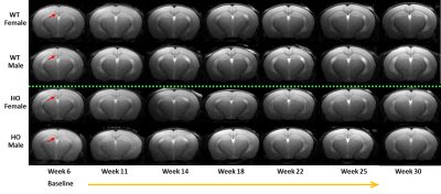 |
75 |
In vivo MRI characterization of a mouse model of vanishing white matter disease
Xiaomeng Zhang, Bradley Hooker , Arthur Nikkel, Holly Robb, Ana Basso, Ann Tovcimak, Carmela Sidrauski, Gerard Fox, Kathleen Martin, Michael Dart, Yanping Luo
Vanishing White Matter Disease (VWMD) is a rare genetic disorder characterized by white matter degeneration. Here we employed MRI to longitudinally characterize pathological changes in white matter with contrasts sensitive to pathology (T2W) and myelin integrity (MTR, DTI). Statistically significant difference were detected in T2W and MTR comparing wild-type and homozygous mice starting at 14 and 22 weeks of age, respectively. These differences between the wild-type and homozygous mice were observed before symptomatic behavioral changes, and became more prominent over the time. Correlations with immunohistochemistry markers provided underlying pathology corresponding to the observed imaging changes.
|
|
| Top |
Cerebral Vessel Imaging
Digital Poster
Neuro
Tuesday, 14 May 2019
| Exhibition Hall |
16:45 - 17:45 |
| |
|
Computer # |
|
3240.
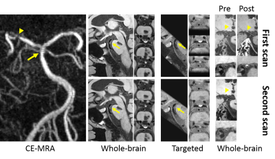 |
76 |
Clinical reliability of 3D whole-brain vessel wall imaging in patients with intracranial atherosclerotic disease: a comparison with conventional targeted imaging
Na Zhang, Xinfeng Liu, Qi Yang, Shlee Song, Zhenliang Xiong, Lei Zhang, Hairong Zheng, Xin Liu, Zhaoyang Fan
Conventional intracranial MR vessel wall imaging (VWI) techniques based on 3D turbo spin-echo (TSE), with a thin, oblique slab to specifically target a limited imaging volume, have been shown to be reliable in quantifying vessel morphology of intracranial atherosclerotic disease (ICAD). Recently, 3D whole-brain VWI was proposed and optimized offering large spatial coverage, improved cerebrospinal fluid suppression, and enhanced T1 weighting and exhibits excellent reproducibility in quantification of vessel dimensions in healthy volunteers. This study is to further evaluate the clinical reliability of 3D whole-brain VWI in patients with ICAD via a comparison with 3D targeted VWI and 2D TSE.
|
|
3241.
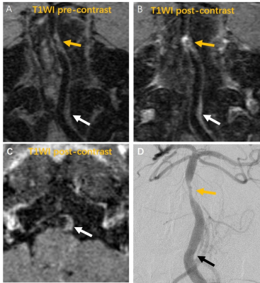 |
77 |
Assessment of intracranial atherosclerotic plaques using 3D non-contrast black blood MRI: comparison with DSA
Xia Tian, Chengcheng Zhu, Bing Tian, Zhang Shi, Luguang Chen, Qi Liu, Jianping Lu
Digital subtraction angiography (DSA) remains the gold standard for assessment of intracranial artery stenosis. However, it is invasive and may miss lesions without luminal stenosis due to vessel wall outward remodeling. We compared 3D black-blood MRI (SPACE) with DSA in 74 intracranial plaques, and found SPACE was in good agreement with DSA for stenosis quantification (ICC=0.82), but the plaque was longer in SPACE than DSA. Moreover, SPACE detected 28 more plaques than DSA, and 14 of 28 plaques showed enhancement. SPACE is promising for evaluating the severity of intracranial atherosclerosis and may improve patient management.
|
|
3242.
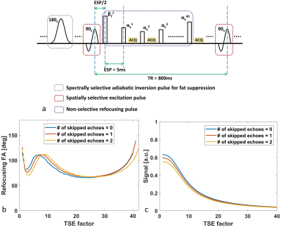 |
78 |
Highly Accelerated Whole Brain Isotropic 0.5mm Intracranial Vessel Wall Imaging with Nonlocal Denoising and Super Resolution Enhancement
Zechen Zhou, Baocheng Chu, Jie Sun, Niranjan Balu, Mahmud Mossa-basha, Thomas Hatsukami, Peter Börnert, Chun Yuan
The three-dimensional (3D) turbo spin echo (TSE) sequence with variable flip angle (VFA) has proven to be clinically useful for high resolution intracranial vessel wall imaging (VWI) at 3T. In this study, a T1 weighted 3D TSE sequence was optimized for whole brain isotropic 0.5mm intracranial VWI in 8mins28secs. The optimized VFA design improves the flow suppression in small vessels and the nonlocal denoising effectively reduces the noise amplification after super resolution enhancement while improving the vessel wall boundary definition. This combined imaging and post-processing technique provides a promising tool for intracranial atherosclerosis and stroke investigation.
|
|
3243.
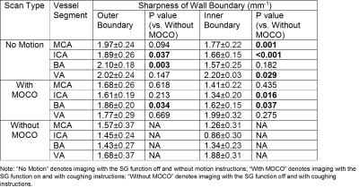 |
79 |
Intracranial Vessel Wall Imaging: Artifactual Effects of Localized Movement and In-line Mitigation with Self-gating
Zhehao Hu, Fei Han, Qi Yang, Shlee Song, Marcel Maya, Anthony Christodoulou, Xiaoming Bi, Debiao Li, Zhaoyang Fan
Intracranial vessel wall imaging can directly visualize the vessel wall and characterize wall pathologies, and has drawn great clinical interest. 3D variable-flip-angle turbo spin-echo is currently the method of choice for intracranial VWI. However, because of its 3D acquisition fashion, this method is inherently susceptible to motion as previously shown in extracranial VWI studies. For intracranial VWI, motion may also result from bulk or localized movement, although the former can be effectively reduced by using foam pads. Reducing motion degradation becomes more critical because of long scan time and demanding spatial resolution. This work aims to investigate the effect of localized movement on intracranial VWI quality and to present an in-line self-gating approach to mitigate quality deterioration.
|
|
3244.
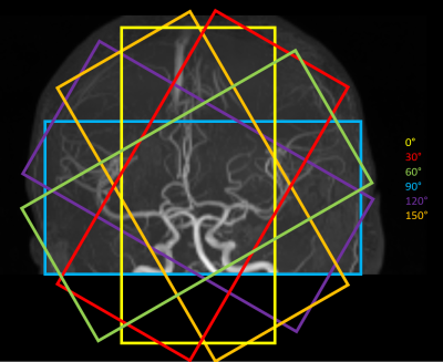 |
80 |
4D high-resolution Angiography maps combining time-of-flight angiography and ASL projection data
Thomas Lindner, Olav Jansen, Michael Helle
This study presents a novel method of combining Arterial Spin Labeling and TOF angiography based on image post-processing after radial projection image readout. Tehreby high-resolution time-resolved angiograms can be obtained.
|
|
3245.
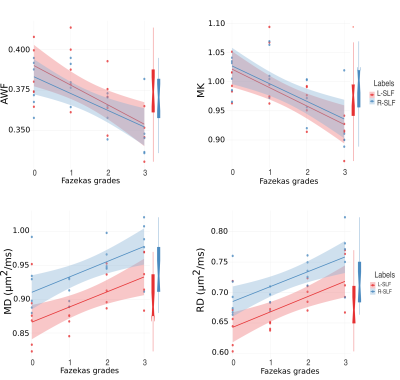 |
81 |
Diffusion Metrics of White Matter in Cerebral Small Vessel Disease: Unravelling the Variance in Microstructural Integrity
Elene Iordanishvili, Ezequiel Farrher, Melissa Schall, Ana-Maria Oros-Peusquens, Ketevan Kotetishvili, Farida Grinberg, Jon Shah
Cerebral small vessel disease (SVD) has a long period of silent progression until it clinically manifests as a stroke or cognitive decline. Early detection of microstructural alterations in the white matter will help to develop targeted therapy and avoid clinical consequences. The need for advanced imaging to reflect the plethora of the changes is increasing. Here, we used the methods for the analysis of the diffusion MRI signal to investigate microstructural alterations in SVD. Our study identified the most frequently changed parameters and the affected regions. We also show increased changes in the diffusion MRI metrics, corresponding with disease severity.
|
|
3246.
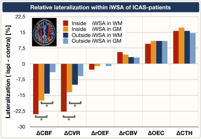 |
82 |
Hemodynamic impairments in asymptomatic unilateral carotid artery stenosis are increased within individual watershed areas
Stephan Kaczmarz, Jens Goettler, Jan Petr, Mikkel Hansen, Jan Kufer, Andreas Hock, Christian Sorg, Claus Zimmer, Kim Mouridsen, Fahmeed Hyder, Christine Preibisch
Internal carotid-artery stenosis (ICAS) causes complex and not yet well understood physiological impairments, which currently limits treatment decisions. We present multimodal perfusion and oxygenation-related MRI-data from unilateral asymptomatic ICAS-patients and age-matched healthy controls. The major aim was to investigate hemodynamic impairments in ICAS within individually defined watershed areas (iWSA’s) to account for individual vascular configurations. We found statistically significant lateralization of hemodynamic parameters within iWSA’s - strongest in WM of iWSA’s. Therefore, our iWSA-based approach facilitates detection of even subtle hemodynamic changes in ICAS. Furthermore, we detected spatially widespread capillary flow heterogeneity increases which are promising future treatment indicators.
|
|
3247.
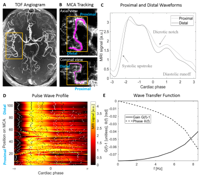 |
83 |
MRI pulse wave profiles of cerebral arteries
Henning Voss, Jonathan Dyke, Douglas Ballon, Ajay Gupta
The pulse pressure wave is an indicator for vascular health. Given the importance of vascular health to the central nervous system, we developed a method to extract intracranial pulse waveforms along the main cerebral arteries from dynamic MRI data. The resulting “pulse wave profiles” track the pulse shape along the main arteries of the brain. The simultaneous acquisition of pulse waveforms within the whole brain at once allows for a system analysis of the vasculature. A Matlab toolbox to compute pulse wave profiles is provided.
|
|
3248
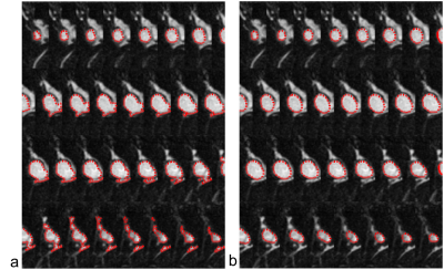 |
84 |
Non-contrast 3D Black Blood MRI for Intracranial Aneurysm Surveillance: A Quantitative Study
Video Permission Withheld
Yan Wang, Jing Liu, David Saloner
Patients with unruptured intracranial aneurysms routinely undergo surveillance imaging to monitor their growth. Contrast enhanced magnetic resonance angiography (CE-MRA) provides good accuracy in measuring size, but requires the use of contrast agents, which is undesirable for repeated imaging. In this study, we propose a semi-automatic method to compare the volumetric difference between 3D non-contrast black blood MRI and CE-MRA on 17 aneurysms. Our results support the use of non-contrast black blood MRI for surveillance of intracranial aneurysm in the clinical setting.
|
|
3249.
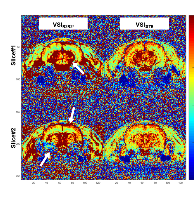 |
85 |
Robust MRI assessment of cerebral microvasculature using stimulated-echo diffusion-imaging method
DongKyu Lee, HyungJoon Cho
Stimulated-echo diffusion-imaging method was employed to reduce the effect of macroscopic field inhomogeneity and large vessel overestimation on the quantification of blood volume fraction and mean vessel radius. Stimulated-echo diffusion-imaging method were compared with conventional spin-echo and gradient-echo methods by Monte Carlo (MC) proton diffusion simulations and in vivo rat experiments on a 7 T system. The results of this study showed that stimulated-echo based MR relaxation-rates, ?RSTE,long TD (ΔR2* like) and ?RSTE,short TD (ΔR2 like), provide the robust means of assessing cerebral microvasculature where the macroscopic field inhomogeneity is severe and signal contamination from adjacent large vessel is significant without necessity of co-registering gradient- and spin-echo images.
|
|
3250.
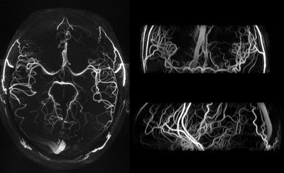 |
86 |
Pulsed ASL prepared 4-Dimensional Dynamic Intracranial MR Angiography at 7T with Improved B1 and B0 robustness
Jin Jin, Kai Wang, Xiaoming Bi, Danny Wang, Lirong Yan
This work enhances the quality of non-contrast enhanced time-resolved 4-dimensional MR angiography (4D-MRA) at 7T, by combining segmented multi-phase acquisition of the FAIR scheme with labeling pulses of improved robustness to B1/B0 inhomogeneity and chemical shift. The gradient-modulated offset independent inversion pulses were optimized for 7T application maximizing MR system performance, while providing uniform inversion, sharp transition and tolerance to chemical shift. 4D-MRA images with higher spatial-temporal resolution were reliably acquired within SAR limit of normal operation mode within 5 minutes, presenting a valuable tool for assessing intracranial vasculature and hemodynamics.
|
|
3251.
 |
87 |
Comparison of cerebrovascular reactivity among patients with sporadic cerebral small vessel disease
Ming-Kang Li, Ting-Yu Chang, Kuo-Lun Huang, Mei-Yu Yeh, Tsong-Hai Lee, Changwei Wu
Sporadic cerebral small vessel disease (SVD) affects small vessels in the brain and causes cerebrovascular-event-related disability. We separated patients into two groups (acute ischemic stroke and spontaneous intracranial hemorrhage) and then compared their cerebrovascular reactivity (CVR) with normal control. We found there was significant BOLD amplitude decline between normal and ischemic group, but no difference in the temporal estimates between groups.
|
|
3252.
 |
88 |
Interactive Correlation between Iron Deposition and Cerebral Blood Flow in the Deep Cerebral Gray Matter Structures of Hemodialysis Patients
Chao Chai, Huiying Wang, Jinping Li, Jinxia Zhu, Xianchang Zhang, E. Mark Haacke, Shuang Xia, Wen Shen
The aim of this study was to explore the correlation between iron deposition (ID) and cerebral blood flow (CBF) in the deep gray matter structures of hemodialysis patients using susceptibility-weighted image mapping and arterial spin labeling. Both ID and CBF of the patients were increased compared with the healthy controls. ID in patients was positively correlated with CBF in the right putamen, while negatively correlated with CBF in the right thalamus. Abnormal calcium-phosphorus metabolism and triglyceride were shared independent factors for increased ID and CBF. Increased ID, rather than CBF, was a risk factor for neurocognitive impairment.
|
|
3253.
 |
89 |
Accurate staging of Cerebral Venous Sinus Thrombosis using T1 SPACE:clinical experience
Presentation Not Submitted
Yanan Ren, Ying Wei, Yanan Lin, Guoxi Xie, Jingliang Cheng
This study aims to explore magnetic resonance High-resolution Variable Flip Angle Turbo-Spin-Echo(T1 SPACE) technique for accurate staging of Cerebral Venous Sinus Thrombus(CVT). CVT patients confirmed by Computed Tomographic (CT) were randomly divided into three groups according to the time from the onset of symptoms to T1 SPACE. Signal to Noise Ratio(SNR) and Contrast to Noise Ratio(CNR) between thrombus and gray tissues/white tissues were calculated on every thrombus segments and difference between different groups were analyzed. Results indicate that T1 SPACE has the potential to be a promising tool for accurate diagnosis and staging of CVT.
|
|
3254.
 |
90 |
Inner volume 3D TSE for isotropic 0.30 mm black-blood images of intracranial perforating arteries at 7T
Qingle Kong, Dehe Weng, Jing An, Yan Zhuo, Zihao Zhang
The impairment of microvessels can lead to neurologic diseases such as stroke and vascular dementia. The imaging of lumen and vessel wall of perforating arteries requires an extremely high resolution due to their small caliber size (50 – 400 um). In this study, we developed a 3D inner-volume (IV) TSE (SPACE) sequence with 2D spatially selective excitation (SSE) RF pulses. High resolution of isotropic 0.30mm was achieved for the black-blood images of lenticulostriate artery (LSA) within 10 minutes. The IV-SPACE images showed clearer delineation of vessel wall and lumen of LSA than conventional SPACE images. IV-SPACE might be a promising method for detecting microvasculopathies of cerebral vascular diseases.
|
|
3255.
 |
91 |
Consistency of FLAIR vascular hyperintensity (FVH) and arterial transit artifact (ATA) in 3D ASL imaging in patients with unilateral MCA stenosis
Peipei Chang, Yanwei Miao, Yiwei Che, Yuhan Jiang, Li Yang, Lizhi Xie, Kaiyu Wang
Arterial spin labeling (ASL), a noncontrast method of measuring CBF has become feasible in the clinical setting. Serpiginous high intensity structures (ASL) are seen in the ischemic tissue on ASL, prior studies suggested that ATA may represent collateral flow. On the other hand, FVHs are related to hemodynamic impairment and represent slow retrograde flow in leptomeningeal collaterals. Our aim was to compare the consistency of the occurrence of ATA relative to that of FVH in patients with proximal MCA occlusion.
|
|
3256.
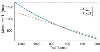 |
92 |
A Single Reference Variable Flip Angle(SR-VFL) Method using a 3D Pseudo Golden Angle Stack of Stars(PGA- SOS) Sequence for Accurate Dynamic T1 Quantification of Contrast Uptake within Vulnerable Plaque
Seong-Eun Kim, Bryant Svedin, Adam de Havenon, Matthew Alexander, Dennis Parker, Gerald S Treiman, J Scott McNally
The carotid artery atherosclerotic disease is one of the most common causes of ischemic stroke. Post-contrast plaque enhancement (PPE), which may result from endothelial dysfunction or be secondary to intraplaque inflammation, is a vulnerable plaque feature that correlates with increased stroke risk independent of stenosis. Although PPE can be detected with vessel wall MRI, better quantitative methods to measure PPE are needed. This work presents a new 3D high resolution dynamic T1mapping technique for accurate T1 quantification of contrast uptake within vulnerable carotid atherosclerotic plaque. The proposed method may provide important mechanistic implications for the pathophysiology of PPE.
|
|
3257
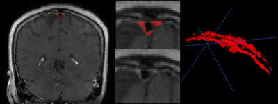 |
93 |
in vivo quantitative assessment of the meningeal lymphatics using 3D black blood T1 imaging: a preliminary study
Video Permission Withheld
Mina Park, Sung Jun Ahn, Sang Hyun Suh
We evaluated the meningeal lymphatics using 3D black blood T1 imaging and its association with clinical parameters as well as enlarged perivascular spaces. This retrospective study included 24 patients who underwent contrast-enhanced 3D black blood T1 imaging on 3T brain MRI and assessed their meningeal lymphatics located parallel to the superior sagittal sinuses. The group with higher meningeal lymphatics volume more frequently have diabetes than the lower group, which is one of the major vascular as well as cognitive risk factors. Furthermore, the higher group had a significantly higher score of enlarged perivascular space in centrum semovale, which is not surprising when considering its close anatomical and functional relationships within the glymphatic system. Therefore, we suggest that the expanded meningeal lymphatics may be an imaging marker of poor function of glymphatics but further studies with disease conditions such as Alzheimer’s disease should be followed to clarify the exact meaning of the volume of the meningeal lymphatics.
|
|
3258
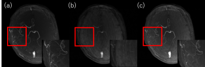 |
94 |
Reducing scan time of selective 3D TOF using the dedicated algorithm based on compressed sensing
Video Permission Withheld
Kosuke Ito, Masahiro Takizawa
Non-contrast enhanced ICA selective 3D TOF using cylinder excitation pre-sat pulse has a possibility which visualizes blood flow from left or right ICA like DSA. It requires one set of 3D TOF scan; non-labeled 3D TOF and ICA labeled 3D TOF, then scan time becomes two times longer than conventional 3D TOF scan. In this study, compressed sensing scheme was applied to reducing the scan time of selective 3D TOF. Proposed method uses similarity between 3D TOF images with and without pre-sat pulse. This realizes only 1 minutes’ additional ICA selective 3D TOF scan to conventional 3D TOF scan.
|
|
3259.
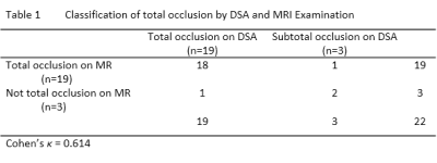 |
95 |
Is It Necessary for Patients with Internal Carotid Artery Occlusion to Undergo 3D Head-neck Combined Vessel Wall Cardiovascular Magnetic Resonance Imaging?
Presentation Not Submitted
Jin Zhang, Huilin Zhao, Beibei Sun, Xiaosheng Liu, Xiaoyue Zhou, Xihai Zhao, Chun Yuan, Jianrong Xu
Definite diagnosis of internal carotid artery occlusion no longer satisfies neurosurgeons’ treatment needs for vascular recanalization. This study sought to investigate what information provided by 3D head-neck combined vessel wall cardiovascular magnetic resonance imaging could be useful for neurosurgeon to make the therapy plan. Our study turned out that 3D MR vessel wall imaging is capable of diagnosing internal carotid artery occlusion, and patients with short extent of occlusion are benefit from the vascular recanalization therapy. Our results suggest it is useful for those patients to undergo 3D MR vessel wall imaging for making their therapy plan.
|
|
3260.
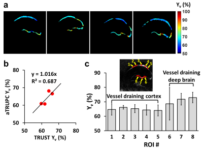 |
96 |
Vessel-specific Quantification of Neonatal Cerebral Venous Oxygenation
Dengrong Jiang, Hanzhang Lu, Charlamaine Parkinson, Pan Su, Zhiliang Wei, Li Pan, Aylin Tekes, Thierry Huisman, W. Golden, Peiying Liu
Cerebral metabolic rate of oxygen (CMRO2) is an important biomarker for normal and pathological neonatal brain development. However, regional measurement of neonatal CMRO2 has been limited due to an inability to evaluate regional venous blood oxygenation (Yv). This study presented a rapid MRI technique, accelerated T2-relaxation-under-phase-contrast (aTRUPC), to measure vessel-specific Yv in neonates. We have improved the reliability and accuracy of aTRUPC by optimizing imaging parameters and calibrating T2-bias. A pilot study on healthy, non-sedated neonates demonstrated the feasibility of aTRUPC to measure vessel-specific Yv. Accuracy of aTRUPC-based Yv measurements was further validated with established whole-brain Yv measurement using T2-relaxation-under-spin-tagging.
|
|
3261.
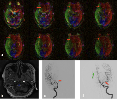 |
97 |
Evaluation of collateral circulation in intracranial atherosclerosis using random vessel-encoded arterial spin labeling
Lirong Yan, Xiaoyue Ma, Hong Zheng, Yan Wang, Meiyun Wang
Collateral circulation plays an important role in predicting the clinical outcomes and risk of recurrent stroke in patients with stroke and transient ischemic attack (TIA). In this study, we evaluated the clinical utility of random vessel-encoded ASL (rVE-ASL) technique for the characterization of vascular territory and collateral circulation in a cohort of patients with stroke or TIA. The assessment of collaterals using rVE-ASL matched well with that of digital subtraction angiography (DSA).
|
|
3262.
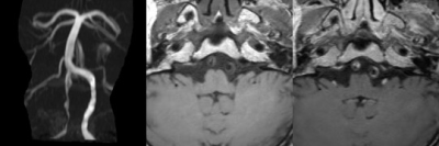 |
98 |
Evaluate the Characteristics of Spontaneous Intracranial Artery Dissection using High Resolution MRI Vessel Wall Imaging
Bing Tian, Xia Tian, Zhang Shi, Qi Liu, Jianping Lu
Compared to luminal angiographic techniques, high resolution magnetic resonance imaging is more helpful to the diagnosis and differential diagnosis the dissection from other vascular pathologies such as atherosclerosis or aneurysm. In this study, we present results obtained with vessel wall imaging to evaluate the characteristics of spontaneous intracranial artery dissection. We divided patients into two groups according to CE-MRA, and compared the characteristics of high resolution magnetic resonance imaging between two groups. The results of our study may be helpful to understand the lumen and wall change of different type of spontaneous intracranial artery dissection shown on luminal angiographic techniques.
|
|
3263.
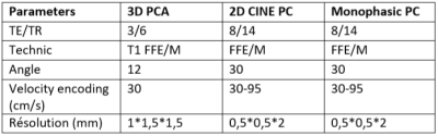 |
99 |
To investigate blood flow in head and neck arteries by phase contrast MRI
Agnès Paasche, Jérémie Bettoni, Stéphanie Dakpé, Bernard Devauchelle, Sylvie Testelin, Olivier Balédent
The 2D-cine PC MRI allows to know precisely the arterial flow in small vessels but it is unsuited for clinical use. The principal aim of this study was to determine the precision loss when rapid monophasic PC sequences (whiches are not synchronised with cardiac cycle) are used compared to the traditional 2D-cine sequences in ten healthy volunteers. Pearson’s coefficient between the two technics was determined and Bland Altman tests were used. The precision loss was between 0,55 % et 27 %, depending on the studied vessel, so the monophasic PC sequences can be used for clinical vascular evaluation in pretherapeutic conditions.
|
|
3264.
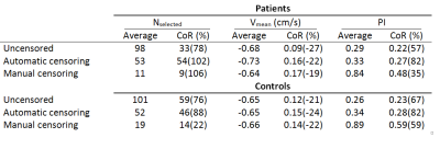 |
100 |
Method for vessel selection effects the outcome and reproducibility of velocity and pulsatility measures in cerebral penetrating arteries
Tine Arts, Jeroen Siero, Geert Jan Biessels, Jaco Zwanenburg
Measuring the function of the cerebral small vessels can greatly benefit our understanding of cerebral small vessel disease. Recent research enabled the assessment of blood flow velocity and pulsatility in the perforating arteries of the cerebral white matter in healthy subjects. However, in patients this method requires manual elimination of artifacts. This paper explores various methods for excluding false positives. The reproducibility of the velocity, pulsatility and number of selected vessels was investigated in a test-retest study. Results show that the reproducibility of these outcome measures highly depends on the chosen method for vessel selection.
|
|
| Top |
Brains: Functional & Dysfunctional
Digital Poster
Neuro
Tuesday, 14 May 2019
| Exhibition Hall |
16:45 - 17:45 |
| |
|
Computer # |
|
3265.
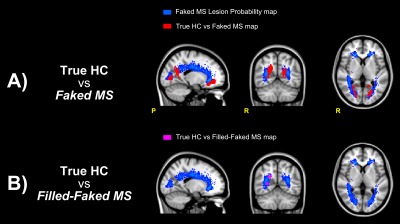 |
101 |
White matter MS lesions effect on resting state fMRI analysis: should we lesion fill functional data?
Gloria Castellazzi, Anisha Doshi, Adnan Alahmadi, Floriana De Angelis, Jeremy Chataway, Ferran Prados, Claudia Gandini Wheeler-Kingshott
Multiple sclerosis (MS) lesions are well known to alter tissue segmentation, shifting tissue boundaries between grey and white matter regions (GM and WM). Despite evidence of these errors occurring when working with anatomical images, little is known about the possible effects of MS lesions on the functional MRI results. Here, we addressed this question by simulating the presence of MS lesions on resting state functional MRI data from healthy controls. Subsequently, we tested whether lesion filling functional MRI data is useful to prevent artefactual results of functional connectivity alterations that are actually due to MS lesions.
|
|
3266.
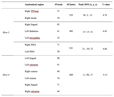 |
102 |
Frequency specific functional connectivity density in Parkinson’s disease: A resting-state fMRI study
Presentation Not Submitted
Xue-hua Peng, Jian-bo Shao, Hui-jing Ma, Zhiyao Tian, Long Qian, Bai-qi Zhu
The functional connectivity of the brain in Parkinson’s disease (PD) has been largely investigated focusing on a low frequency oscillation from 0.01 to 0.1 Hz. Nevertheless, the frequency specificities relating to the functional connectivity have not yet been fully understood. In this study, we utilized analysis of the functional connectivity density (FCD) to determine changes in patients with PD and in healthy controls in wo different frequency bands were analyzed. Our results demonstrated differential FCD maps from both spatial and frequency domain, thus providing a novel insight for investigation of the neuroimaging biomarkers in PD.
|
|
3267.
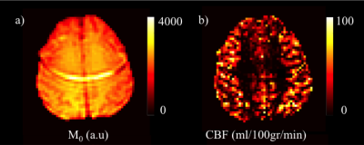 |
103 |
The Cerebral Blood Flow Changes of Executive Dysfunction in Parkinson’s Disease Measured by Arterial Spin Labeling MRI at Cerebral Cortex Parcellations Obtained from Resting State fMRI
Dilek Arslan, Sevim Cengiz, Ani Kicik, Emel Erdogdu, Zerrin Yildirim, Zeynep Tufekcioglu, Basar Bilgic, Hasmet Hanagasi, Aziz Ulug, Tamer Demiralp, Ibrahim Gurvit, Esin Ozturk-Isik
The aim of this study is to find cerebral blood flow (CBF) based biomarkers of executive dysfunction in Parkinson’s disease (PD) at cerebral cortex parcellations obtained from resting state fMRI. CBF maps of PD with mild cognitive impairment, cognitively normal PD and healthy controls were compared at brain regions based on parcellations obtained from resting state fMRI. Additionally, CBF values of the PD subjects were classified according to Stroop scores and COMT genotype. Hypoperfusion in several brain networks related with executive functioning was observed in PD patients with executive dysfunction, and also in patients with COMT Met/Met genotype.
|
|
3268.
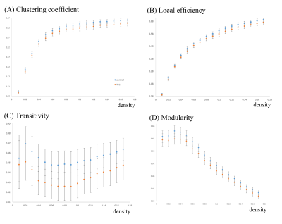 |
104 |
Evaluation of brain functional connectome in tuberous sclerosis complex with neuropsychological disorders using resting-state fMRI
Jun-Cheng Weng, Tzu-Yi Chuang, Chao-Yu Shen, Jeng-Dau Tasi
We aimed to use resting-state fMRI to provide the first findings on disrupted functional brain networks in tuberous sclerosis complex (TSC) patients with graph theoretical analysis (GTA) and network-based statistic (NBS) analysis. We found several topological parameters including clustering coefficient, local efficiency, transitivity, and modularity in the healthy control were better than those in the TSC patients. One subnetwork showed more edges in the healthy control compared with the TSC, including the connections from the frontal lobe to the parietal lobe. Our findings may help better understand the variable clinical phenotypes of TSC and the underlying physiological mechanisms.
|
|
3269.
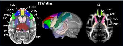 |
105 |
A Framework for Integrating Diffusion and rs-fMRI with Measurements of Cognitive Function in the Rhesus Macaque Frontal Lobe and Striatum
Zheng Liu, Alison Weiss, Xiaojie Wang, Jackie Domire, William Liguore, Christopher Kroenke, Jodi McBride
Brain atlases commonly used for characterizing neurodegenerative processes are frequently referenced to a specific imaging modality. Here we describe a combined gray matter and white matter atlas to be used in the study of a rhesus macaque model of Huntington’s disease. We illustrate how this atlas will be used to integrate diffusion tensor imaging and resting-state functional MRI connectivity with measures of cognitive behavior. Prefrontal WM tracts, cortical and basal ganglia regions are labeled in the same space for characterization of WM microstructure changes and cognitive and motor loop connectivity. The results of preliminary study show that these MRI measurements can identify correlations with cognitive behavior measurements.
|
|
3270.
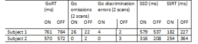 |
106 |
Change in fMRI activation between deep brain stimulation on and off states in patients with Parkinson’s disease during a stop-signal task
Pallab Bhattacharyya, Adam Aron, Jian Lin, Mark Lowe, Anna Crawford, Andre Machado, Mahsa Malekmohammadi, Nader Pouratian, Stephen Jones
Motor regulation pertaining to stopping when necessary is impaired in Parkinson disease (PD) and is modulated by deep brain stimulation (DBS) therapy. We performed fMRI study at 3T using a stop-signal task with PD patients having implanted DBS, and investigated differences in activation in networks responsible for stopping between on and off states of DBS. Overall, larger and stronger activation was observed in this preliminary study when the DBS was turned on for go (when the subject is supposed to press button) minus baseline and successful stop (when the subject successfully stopped) minus go contrasts.
|
|
3271.
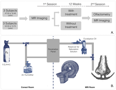 |
107 |
Neuroplasticity in patients with post surgical olfactory dysfunction after olfactory training, assessed with fMRI.
Francisco García-Huidobro, Cristian Montalba, Andrés Rosesbaum, Mariana Zurita, David Jofré, Sergio Uribe, Marcelo Andia, Pablo Villanueva, Claudio Callejas, Claudia González, Cristian Montalba
We evaluated the effect of olfactory training in olfactory pathway brain areas in subjects with olfactory dysfunction after transsphenoidal surgery for pituitary tumors. For this purpose, we compare 2 groups of subjects, one with olfactory training, and a second without training. All of them underwent with an fMRI block designed with eucalyptus odor. An olfactory test were measured in all subjects. The subjects with olfactory training showed a clinical improvement and more activations of compensatory olfactory central brain areas, in comparison with subjects without olfactory training, that they did not show a clinical improvement and activated less compensatory olfactory areas.
|
|
3272.
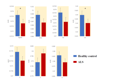 |
108 |
Regularization Small-Worldization in Amyotrophic lateral sclerosis: A Resting-State fMRI Study
Yuan Ai, Du Lei, Qiyong Gong
This study aims to use the graph-based theoretical analysis to investigate the topological properties of the functional brain connectome in amyotrophic lateral sclerosis(ALS).151 healthy controls and 101 patients underwent a rs-fMRI scan. Compared with controls, brain networks of ALS patients were characterized by decreased global efficiency and the characteristic path length increased. Based on these perspectives, the ALS group exhibited “regularization small-worldization”. A network with 10 nodes and 16 edges was identified that was significantly altered in default mode network (DMN) regions. This study may provide novel insights into the pathophysiological mechanisms underlying psychiatric disorders from a connectomic perspective.
|
|
3273.
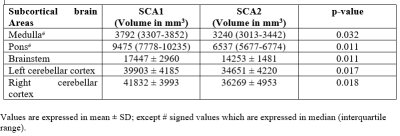 |
109 |
Altered Connectivity Using Resting State fMRI in SCA1 and SCA2 Patients
Presentation Not Submitted
Dibashree Tamuli, Shefali Chaudhary, S. Senthil Kumaran, Ashok Jaryal, Achal Srivastava, Kishore Deepak
Neurodegeneration in cortical and subcortical brain areas are found in SCA type 1 and 2 patients. The structural brain volumetric analysis has been done to know the differential loss of volume of brain areas in SCA type 1 and 2. Especially, to find the functional connectivity of the whole brain in the same SCA patients, we have assessed default mode network using resting state fMRI.
|
|
3274.
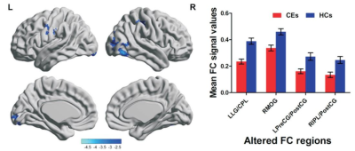 |
110 |
Altered resting-state functional connectivity of primary visual cortex in young patients with comitant exotropia
Presentation Not Submitted
Zhi Wen, Xuefang Lu, Xin Huang, Yang Fan, Yunfei Zha, Baojun Xie
The comitant exotropia (CE) is a common eye disease characterized by outward deviation of the eyes and impairment of stereovision. In this study, we aim to investigate the altered functional connectivity (FC) in patients with CE using resting-state fMRI. The CE patients showed significantly less FC between the left V1 and left lingual gyrus/cerebellum posterior lobe, right MOG, left precentral gyrus/postcentral gyrus and right IPL/postcentral gyrus; as well as less FC between right V1 and right MOG (BA19, 37). Our findings show that CE involves defects in the possessing of stereovision, such as fusion, spatial processing, visual recognition, and oculormotor.
|
|
3275.
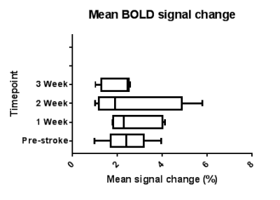 |
111 |
Longitudinal functional MRI mapping changes in neurovascular responses following middle cerebral artery occlusion
Andrew Crofts, Melissa Trotman-Lucas, Justyna Janus, Claire Gibson, Michael Kelly
Due to limitations in anaesthetic protocol, fMRI studies of recovery following ischaemic stroke in rodents show high variability and must use large numbers of animals. This study uses a novel anaesthetic protocol which gives a highly reproducible BOLD signal in healthy animals to study changes in response to forepaw stimulation in 10 rats following middle cerebral artery occlusion. Even with the improved protocol, high variability between animals following MCAO is a confounding factor, and while a trend towards hyperactivation followed by return to baseline is seen, a larger number of animals is still required for longitudinal studies.
|
|
3276.
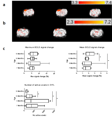 |
112 |
Longitudinal functional MRI for animal studies of neurovascular coupling in healthy ageing
Andrew Crofts, Melissa Trotman-Lucas, Justyna Janus, Claire Gibson, Michael Kelly
Due to effects of anaesthetic on the BOLD signal, or use of toxic anaesthetics, longitudinal preclinical fMRI studies are uncommon, and studies of age-related disease progression show high variability and use large numbers of animals. This study uses a novel anaesthetic protocol in 11 rats to study changes in the BOLD signal with age. A strong, reproducible BOLD response to forepaw stimulation is found between 7 and 12 months old, and at 15 months old the number of active voxels is reduced by half. This shows that the protocol is suitable for longitudinal studies of ageing.
|
|
3277.
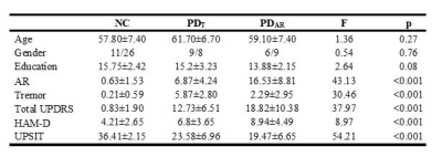 |
113 |
Resting State Olfactory Network Functional Connectivity in Tremor- and Rigidity-predominant Patients with Parkinson’s disease
Lu Jiaming, Rachel Stanford, Prasanna Karunanayaka, Qing Yang, Paul Eslinger, Xuemei Huang
Parkinson's disease (PD) is a neurodegenerative disease, consisting of a broad spectrum of motor and non-motor symptoms. We used resting-state fMRI to investigate differences in functional connectivity (FC) of the olfactory network (ON) in two major PD subtypes: akinetic-rigid (PDAR) and tremor predominant (PDT). Significant differences in ON FC were found between normal controls, PDAR, and PDT groups in six brain regions. Lower FC was observed in the orbitofrontal cortex, insula, and posterior cingulate cortex in the PDAR group compared to the PDT group. Our findings suggest that the ON FC is deferentially affected in the two PD subtypes.
|
|
3278.
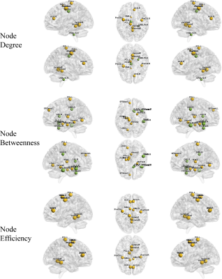 |
114 |
Altered topological organization of whole-brain functional network in patients with postherpetic neuralgia after repetitive transcranial magnetic stimulation treatment
Presentation Not Submitted
Zhizheng Zhuo, Qian Pei, Haiyun Li
This work is to investigate the topological organization alteration of whole-brain functional network in patients with postherpetic neuralgia (PHN) after repetitive transcranial magnetic stimulation (rTMS) treatment and assess whether the functional alteration could be used as a neural biomarker for the post-treatment evaluation.
|
|
3279.
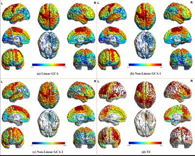 |
115 |
The information flow pattern of the human brain based on high temporal resolution functional MRI
Presentation Not Submitted
Zhizheng Zhuo, Xiangyu Ma, Zhe Ma, Lijiang Wei, Haiyun Li
To investigate the information flow pattern of human brain by using high temporal resolution functional MRI based on linear, non-linear Granger causality analysis and transfer entropy
|
|
3280.
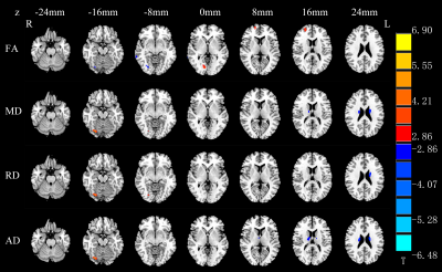 |
116 |
Alterations of the brain microstructure and corresponding functional connectivity in early blind adolescents with and without residual light perception
Zhifeng Zhou, Jinping Xu, Leilei Shi, Xia Liu, Hengguo Li
Previous neuroimaging studies of adult blind have revealed structural and functional neuroplasticity. It is still unclear if the brains of young blind will have the same alterations, and the effects of residual light perception (RLP) on the structure and function of the blind brain cannot be ignored. This study explored the structural and functional brain changes in early blind adolescents (EBAs) with and without RLP using voxel-based analysis method of diffusion tensor imaging data and resting-state functional connectivity analysis. The results provide new insights into the mechanisms underlying the reorganization of brain in EBAs with or without RLP.
|
|
3281.
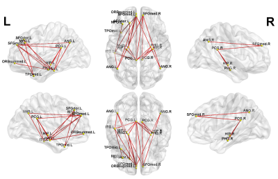 |
117 |
Functional network-based statistics reveal abnormal resting-state functional connectivity in minimal hepatic encephalopathy
Presentation Not Submitted
Tian-Xiu Zou, Hua-Jun Chen, Zhongshuai Zhang
Whole-brain functional network analysis is an emerging methodology for the investigation of the pathophysiology of MHE. A nonparametric statistical approach, called “network-based statistics” (NBS), has also been used in the field of connectome analysis. However, few whole-brain NBS studies have been conducted on MHE patients, which limits the further clarification of the network pathophysiology of MHE. We performed NBS analysis to identify FC changes related to MHE at the whole-brain functional connectome level and indentified two subnetworks with significant differences in FC matrices between patients and controls. Correlation analyses revealed that the PHES score was significantly positively correlated with the strength of two FCs within the first subnetwork. In summary, our findings indicate that DMN dysfunction may be one of the core issues in the pathophysiology of MHE.
|
|
3282.
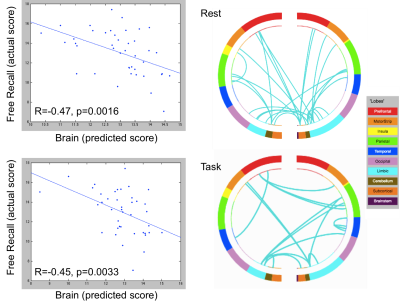 |
118 |
Connectome predictive modeling in subjects with mild cognitive impairment: Resting-state and object location task results
Scott Peltier, Sean Ma, Allison Moll, Julia Laing, Benjamin Hampstead
Connectome predictive analysis was applied to MCI subjects using both resting-state and task data. While a measure of free recall was predicted equally well in rest or task, a measure of total cognition was only predicted succesfully using the task data. This argues for the utility of cognitive "stress tests" to better capture relevant brain biomarkers.
|
|
3283.
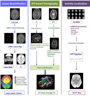 |
119 |
Neural mechanism of processing speed decline induced by anatomically connected white matter tract damage and cortical function abnormality
Shuyue Wang, Jiaerken Yeerfan, Peiyu Huang, Xinfeng Yu, Yong Zhang, Minming Zhang
White matter hyper-intensities (WMH) is considered as an important source of morbidity associated with dementia, stroke and increased mortality risk. Previous studies have suggested that WMH always leads to the decline of information processing speed, which have an impact on activities of daily living. Current hypothesis is that the dysfunctions caused by WMH is the result of “disconnection”, while the connection between the structural and functional alteration has not been fully investigated. We aimed to explore the underlying pathway of information processing speed decline via combining the spatial distribution of WMH, microstructural changes based tractography and cortical activity alterations. The results from different modalities converged in the occipital lobe with precise spatial overlappings. Results show regional WMH may indicate disrupted tract integrity and cause altered brain activities, leading to impaired function. This WMH-tract-function-behavior link is critical for WMH induced dysfunctions and treatment strategies.
|
|
3284.
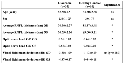 |
120 |
Widespread Structural and Functional Brain Connectivity Changes and Behavioral Relevance in Glaucoma
Vivek Trivedi, Ji Won Bang, Carlos Parra, Max Colbert, Caitlin O'Connell, Muneeb Faiq, Ian Conner, Mark Redfern, Gadi Wollstein, Joel Schuman, Rakie Cham, Kevin Chan
Glaucoma is the second leading cause of blindness worldwide, yet its effects on the brain structure and function and the related behavioral relevance remain unclear. This study shows that glaucoma patients present reduced structural integrity in white matter around the supramarginal gyrus, as well as reduced functional connectivity between supramarginal gyrus and visual occipital and superior sensorimotor areas when compared to healthy controls. Furthermore, decreased functional connectivity between supramarginal gyrus and visual network was associated with changes in postural control. Taken together, our results suggest an involvement of altered brain coordination in the increased risk of falls in glaucoma patients.
|
|
3285.
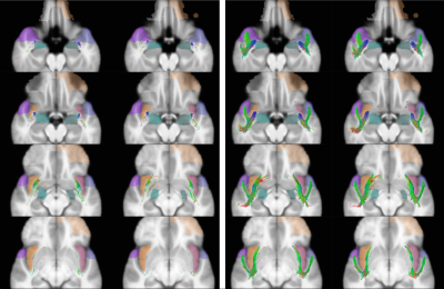 |
121 |
The temporo-insular projection system: a multisubject fiber tractography study using connectome diffusion data
Ahmed Radwan, Pieter Nachtergaele, Stijn Swinnen, Thomas Decramer, Mats Uytterhoeven, Johannes Van Loon, Tom Theys, Stefan Sunaert
The precise structural connections between the amygdala, anterior temporal pole and insula remain poorly understood. These connections have been described with ex-vivo dissection, however the diffusion-based counterparts to these bundles haven’t been described in detail. In a recent study (currently under review) we investigated these connections with ex-vivo dissection on 11 brain specimens. We also explored the possibility of reconstructing the bundles found on dissection with Diffusion MRI. Here we demonstrate the results obtained for reconstructing these white matter connections using 20 randomly selected healthy participants of the HCP young adults data release.
|
|
3286.
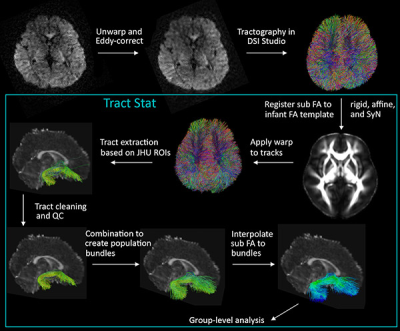 |
122 |
Associations Between Maternal Depression and Infant Fronto-Limbic Connectivity
Emily Dennis, Ananya Singh, Conor Corbin, Neda Jahanshad, Tiffany Ho, Lucy King, Lauren Borchers, Kathryn Humpreys, Paul Thompson, Iang Gotlib
Maternal depression is a well-documented risk factor for psychopathology in children; the origins of this association, however, are not well understood. We present preliminary analyses of 24 infants using a multi-shell diffusion MRI sequence optimized for imaging infant white matter, along with a novel tract clustering and identification workflow, TractStat. We examine the association between maternal depressive symptoms and infant white matter organization in the uncinate fasciculus (UF). Infants whose mothers report experiencing more severe depressive symptoms have lower fractional anisotropy of the right UF, highlighting a possible neurobiological marker of the intergenerational transmission of risk for depression.
|
|
3287.
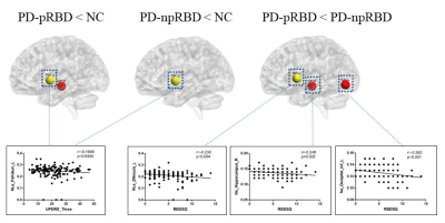 |
123 |
Altered topological organization in white matter connectome in Parkinson’s disease with and without rapid eye movement sleep behavior disorder
Tao Guo, Cheng Zhou, Xiaojun Guan, Xiaojun Xu, Minming Zhang
We detected the alteration of white matter connectome in Parkinson’s disease (PD) patients with and without Rapid eye movement sleep behavior disorder (RBD). 155 PD patients including 66 possible RBD (pRBD) and 89 non-possible RBD (npRBD) and 71 normal controls were included. Diffusion-tensor magnetic resonance imaging and graph theory were used to explore the topologic organization of the brain structural connectome. Significant decreased nodal efficiency were found in specific regions including hippocampus and inferior occipital gyrus in PD-pRBD patients. Both these nodal properties were negatively correlated with RBD severity. This study may contribute to understand the pathophysiology of PD-RBD.
|
|
3288.
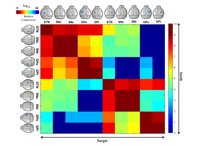 |
124 |
The Structural Connectivity Network of Basal Ganglia in Mouse Brain: MR Diffusion-Tractography at 9.4 T
A-Yoon Kim, Hyeon-Man Baek
Tractography is becoming increasingly common in clinical settings for understanding pathological development and disease, and for assessing pre- and post-operative diagnosis. However, the study on neuronal connectivity network for basal ganglia, a DBS treatment target for Parkinson’s patients, remains unestablished. Therefore, in the present study we have visualized probabilistic diffusion tractography and investigated detailed 3D reconstruction of the projection of basal ganglia structures in mouse model using high-resolution 9.4T MRI. Multi-fiber tractography methods combined with diffusion MRI data have the poential to help identify brain DBS targets in function neurosurgery intervention.
|
|
3289.
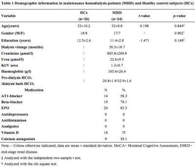 |
125 |
Altered whole-brain functional networks associated with cognitive dysfunction in end-stage renal disease patients with maintenance hemodialysis
Presentation Not Submitted
Peng Li, Xueying Ma, Dun Ding, Huawen Zhang, Shaohui Ma, Ming Zhang
We focused on exploring the whole-brain resting-state functional networks abnormality by using the rs-fMRI with independent component analysis (ICA) algorithm and the relationships with cognitive dysfunction in ESRD patients with hemodialysis. 24 maintenance hemodialysis patients (MHD group) and 26 healthy control subjects (HCs) were evaluated a battery of neuropsychological tests and rs-fMRI scans. Compared with HCs, the MHD group showed worse neuropsychological performances and related decreased functional connectivity in the default mode network, the right frontoparietal network, and the sensorimotor network. Our study might contribute to better understanding underlying neuropathological substrate of cognitive dysfunction in patients with ESRD.
|
|
| Top |
Multiple Sclerosis: Quantitative Imaging of Axons, Myelin & More
Digital Poster
Neuro
Tuesday, 14 May 2019
| Exhibition Hall |
16:45 - 17:45 |
| |
|
Computer # |
|
3290.
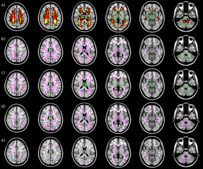 |
126 |
Sensitivity of NODDI and two-compartment SMT parameter maps in multiple sclerosis
Daniel Johnson, Antonio Ricciardi, Wallace Brownlee, Baris Kanber, Ferran Prados, Sara Collorone, Enrico Kaden, Ahmed Toosy, Daniel Alexander, Claudia Gandini Wheeler-Kingshott, Olga Ciccarelli, Francesco Grussu
We make a novel comparison of two diffusion MRI techniques modelling white matter microstructure: neurite orientation dispersion and density imaging (NODDI) and spherical mean technique (SMT) in 63 Multiple Sclerosis (MS) patients and 28 healthy controls using tract based spatial statistics. Both techniques show that there is a reduction in the intracellular volume fraction and an increase in neurite orientation dispersion in lesions and normal appearing white matter in MS patients when compared to controls. Additionally, SMT appears more sensitive to these differences, identifying a larger number of voxels showing significant differences between patients and controls in the studied parameters.
|
|
3291.
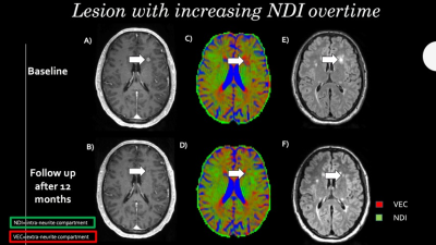 |
127 |
Application of multi-shell NODDI to characterize acute and chronic MS lesions.
Simone Sacco, Eduardo Caverzasi, Tristan Gundel, Shuiting Cheng , Carlo Asteggiano, Christian Cordano, Antjie Bischof, Gina Kirkish, Gillian Rush, Nico Papinutto, Stefano Bastianello, Bruce Cree, Stephen Hauser, Roland Henry
In order to overcome the limitations of conventional MRI in MS, we explored the ability of NODDI to characterize features of acute and chronic lesions. In our study, in line with a recent work that histologically investigated animal models, Orientation Dispersion Index (ODI) was significantly higher in enhancing lesions, thus representing a reliable tool for detecting acute inflammation. After enhancement, lesions could be divided based on their change in Neurite Density Index (NDI): lesions showing increasing NDI values were likely to be characterized by partial remyelination, whereas lesions showing decreasing NDI values might be expression of chronic focal damage.
|
|
3292.
 |
128 |
Neurite orientation dispersion and density imaging predicts disability at 8 years follow-up in relapsing remitting MS patient
Elda Fischi-Gomez, Guillaume Bonnier, Po-Jui Li, Pietro Maggi, Geraldine Le Goff, Ludwig Kappos, Cristina Granziera
Neurite orientation dispersion and density imaging (NODDI) is a diffusion imaging technique that uses diffusion gradients of different strengths to provide novel metrics of axonal and dendrites integrity. In this study, we explored the value of NODDI metrics - in lesions and normal appearing tissue - to predict the long term disability in relapsing-remitting MS (RRMS) patients. NODDI metrics in NAWM and lesions showed significant correlations with patients disability at 8 years follow-up. Future studies should explore the predictive value of NODDI metrics in MS lesions and in larger cohorts of MS patients.
|
|
3293.
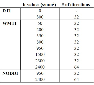 |
129 |
Axonal Damage in the Optic Radiation Detected by Advanced Diffusion MR Metrics is Associated with Retinal Thinning in Multiple Sclerosis
Chanon Ngamsombat, Qiyuan Tian, Qiuyun Fan, Andrew Russo, Thomas Witzel, Lawrence Wald, Eric Klawiter, Susie Huang
Axonal damage diffusely involves the retina, optic nerve, and optic pathway in MS but lacks a specific imaging biomarker. We investigated the presence of alterations in advanced diffusion MRI metrics derived from WMTI and NODDI in the optic radiation (OR) in MS. We found a significant association between thinning of the retinal nerve fiber layer measured by OCT and reduction in axonal water fraction and intracellular water fraction in the OR as measured by WMTI and NODDI. Our results support the idea that axonal damage is widespread throughout the visual pathway in MS and may be mediated through trans-synaptic degeneration.
|
|
3294.
|
130 |
Feasibility of NODDI for the characterization of multiple sclerosis clinical features
Agnese Tamanti, Anna Pisani, Alberto De Luca, Francesca Pizzini, Marco Castellaro, Carmela Zuco, Damiano Marastoni, Francesco Crescenzo, Alessandra Bertoldo, Marco Pitteri, Roberta Magliozzi, Massimiliano Calabrese
Neurite Orientation Dispersion and Density Imaging (NODDI) is a diffusion weighted MRI technique introduced to asses neuronal microstructure. NODDI is used to retrieve indexes such as neurite density and fibers orientation dispersion that might be useful to assess neuronal damage and demyelination in multiple sclerosis (MS) MRI images. For this reason, we investigate its ability to differentiate to different MS phenotypes and clinical features in white matter and in multiple areas of the grey matter.
|
|
3295.
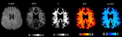 |
131 |
Evaluation of white matter integrity in multiple sclerosis using ultra-high gradient diffusion imaging
Fang Yu, Eric Klawiter, Susie Huang
Conventional MRI of multiple sclerosis lacks specificity for the underlying disease-related processes. The use of ultra-high gradient diffusion imaging increases sensitivity for small caliber axons. When used in combination with myelin-sensitive imaging, estimates of axonal volume fraction (AVF), myelin volume fraction (MVF), and the g-ratio can be obtained. Here, we derived estimates of axonal and myelin metrics on a per-voxel basis using ultra-high gradient diffusion MR and macromolecular tissue volume (MTV) in MS patients and healthy controls. Compared to healthy subjects, focal MS lesions and to a lesser extent the normal appearing white matter demonstrated evidence of demyelination and axonal loss.
|
|
3296.
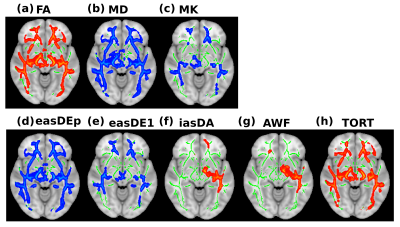 |
132 |
Measuring white matter injury in children with demyelinating syndromes as revealed by non-Gaussian diffusion imaging
Sonya Bells, Ann Yeh, Donald Mabbott
Characterizing whole brain white matter structure in children with demyelinating syndromes (e.g. multiple sclerosis (MS) and neuromyelitis optica spectrum disorders (NMOSD), and myelin oligodendrocyte glycoprotein antibody related disorders (MOG)) may shed light on patterns of injury which are not apparent using conventional imaging techniques. We evaluated group differences in non-Gaussian diffusion data between 26 healthy control, 17 MS and 17 MOG/NMOSD children. We show white-matter microstructure differences between healthy controls and MS patients in areas associated with oculomotor function. Specifically, we show lower axonal density and myelin volume along optic radiations in MS patients than controls or NMOSD/MOG patients.
|
|
3297.
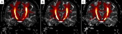 |
133 |
Minimum Requirements for Diffusion Tensor Imaging (DTI) for a Longitudinal Trial
Ken Sakaie, Mark Lowe, Robert Fox
This study examines the optimal number of diffusion-weighting gradients for using DTI as an outcome in a multicenter clinical trial. The results suggest that 6 directions may be sufficient for large, simple fiber tracts. This finding may help simplify the implementation of DTI into clinical trials.
|
|
3298.
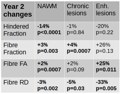 |
134 |
Longitudinal changes in diffusion basis spectrum imaging metrics of normal-appearing white matter and lesions in ocrelizumab-treated relapsing multiple sclerosis
Anika Wurl, Irene Vavasour, Adam Dvorak, Nathalie Ackermans, Cornelia Laule, David Li, Robert Carruthers, Alex Mackay, Roger Tam, Anne Cross, Peng Sun, Sheng-Kwei Song, Hideki Garren, Anthony Traboulsee, Shannon Kolind
Diffusion Basis Spectrum Imaging (DBSI) probes axonal and myelin damage by modeling the diffusion-weighted MRI signal as discrete anisotropic diffusion tensors while simultaneously differentiating and quantifying inflammation and edema through a modeled isotropic diffusion tensor spectrum. We studied 15 RMS patients beginning ocrelizumab treatment over two years as well as 10 healthy controls. DBSI detected microstructural differences between RMS normal-appearing white matter, chronic and enhancing lesions, and healthy control white matter. Further, the metrics were sensitive to changes within two years of follow-up and showed improvement towards healthy control values in patients treated with ocrelizumab.
|
|
3299.
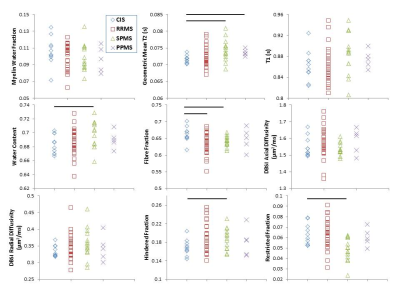 |
135 |
Advanced MRI in subtypes of multiple sclerosis: T1, T2, water content and diffusion basis spectrum imaging
Irene Vavasour, Carina Graf, Shannon Kolind, Peng Sun, Robert Carruthers, Anthony Traboulsee, GR Wayne Moore, Sheng-Kwei Song, David Li, Cornelia Laule
T1, T2, water content (WC) and diffusion basis spectrum metrics were compared from the normal appearing white matter (NAWM) of 10 clinically isolated syndrome (CIS), 27 relapsing-remitting multiple sclerosis (RRMS), 14 secondary progressive multiple sclerosis (SPMS) and 5 primary progressive multiple sclerosis (PPMS) subjects. SPMS showed higher WC, longer geometric mean T2 and a larger hindered fraction than CIS indicating increased oedema. Fibre fraction (apparent axonal density) was lower in RRMS and SPMS than CIS thought to reflect loss of axons or increased oedema. Advanced imaging can show differences between MS subtypes related to the underlying tissue damage.
|
|
3300.
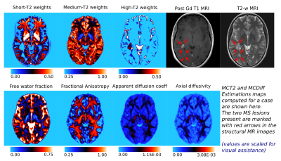 |
136 |
Tissue microstructure information from T2 relaxometry and diffusion MRI can identify multiple sclerosis (MS) lesions undergoing blood-brain barrier breakdown (BBB)
Sudhanya Chatterjee, Olivier Commowick, Onur Afacan, Simon Warfield, Christian Barillot
Gadolinium based contrast agents (GBCA) play a critical role in identifying MS lesions undergoing BBB which is of high clinical importance. However, repeated use of GBCAs over a long period of time and the risks associated with administering it to patients with renal complications has mandated for greater caution in its usage. In this work we explored the plausibility of identifying MS lesions undergoing BBB from tissue microstructure information obtained from T2 relaxometry and dMRI data. We also proposed a framework to predict MS lesions undergoing BBB using the tissue microstructure information and demonstrated its potential on a test case.
|
|
3301.
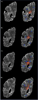 |
137 |
Myelin measurements using GRASE and mcDESPOT are strongly correlated with those from Multi-Echo Spin-Echo myelin water imaging in postmortem Multiple Sclerosis tissue
Heather Yong, Irene Vavasour , Cornelia Laule, Wayne Moore, Roger Tam, Anthony Traboulsee, Bruce Trapp, Shannon Kolind
We compared myelin water fraction (MWF) results from the gold standard multi-spin-echo (MSE) sequence to two accelerated techniques: gradient-and-spin-echo (GRASE), and multicomponent-driven equilibrium single pulse observation of T1/T2 (mcDESPOT) in a formalin-fixed multiple sclerosis brain. All three techniques were sensitive to differences in myelin throughout the sample, with MSE and GRASE producing equivalent MWF values. mcDESPOT estimated significantly higher MWF in both normal appearing white matter and lesion compared to MSE and GRASE. However, the MWF was strongly correlated (p<0.0001) between all three methods (r=0.88 for MSE vs. GRASE; r=0.89 for MSE vs. mcDESPOT; r=0.89 for GRASE vs. mcDESPOT).
|
|
3302.
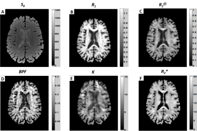 |
138 |
Quantitative Evaluation of Brain Tissue Damage in Multiple Sclerosis with SMART (Simultaneous Multi-Angular Relaxometry of Tissue) MRI
Biao Xiang, Jie Wen, Amber Salter, Anne Cross, Dmitriy Yablonskiy
Quantitative MRI would be beneficial in evaluating tissue damage in Multiple Sclerosis (MS), but existing quantitative techniques are not yet widely used. Recently developed SMART MRI technique generates quantitative magnetization transfer and MR relaxometry data from a single protocol. Herein we present data suggesting that the SMART metrics can serve as biomarkers for MS tissue damage. Our results showed that SMART metrics readily distinguish RRMS from progressive MS subtypes, and correlate with clinical assessments of MS patients. These data suggest that SMART MRI is a highly promising technique for MS monitoring, and for use as an endpoint in clinical trials.
|
|
3303.
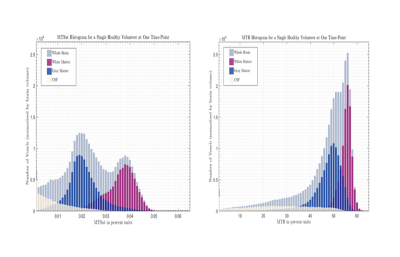 |
139 |
Quantitative imaging biomarkers of demyelination and remyelination: reproducibility of MTsat vs. MTR.
Beth York, Michael Thrippleton, Rozanna Meijboom, Adam Waldman
MTsat provides magnetization transfer information less confounded by B1 and T1 inhomogeneities than MTR (largely derived from bound protons within axonal myelin in the brain). The present study measured the reproducibility of MTsat versus MTR, and compared tissue-type contrast for MTsat and MTR in healthy volunteers and patients with relapsing-remitting multiple sclerosis. Quantitative maps were created for histogram analysis, descriptive statistics and sensitivity comparisons. MTsat and MTR showed similar reproducibility but MTsat showed higher tissue contrast. Our data suggest MTsat is a superior biomarker for myelin integrity, with utility for the study of demyelination and remyelinating therapies in multiple sclerosis.
|
|
3304.
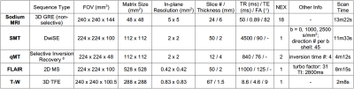 |
140 |
Sodium MRI, SMT, and qMT in Black Holes in Multiple Sclerosis
Ping Wang, Junzhong Xu, Richard Dortch, John Gore, Francesca Bagnato
Black holes in multiple sclerosis (MS) are considered to be more indicative of axonal loss than T2 lesions. We employed three novel MRI techniques, sodium MRI, diffusion MRI via the spherical mean technique, and quantitative magnetization transfer to measure tissue sodium concentration (TSC, for axonal dysfunction), apparent axonal volume fraction (Vax, for axon loss), and macromolecular-to-free pool-size-ratio (PSR, for demyelination degree), respectively. The results showed significant differences of the measures between black holes and the contralateral normal appearing white matters, indicating the potential of using these techniques to provide more specific information on underlying pathology in MS.
|
|
3305
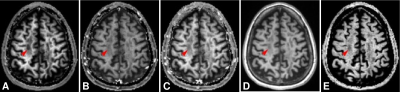 |
141 |
Quantitative magnetization transfer MRI with improved specificity to demyelinating lesions
Video Permission Withheld
Jung-Jiin Hsu, Roland Henry
MRI techniques that utilize the magnetization transfer (MT) effect are highly desirable as they potentially can map the myelin content. However, MT measurements produced by conventional MT-MRI techniques, such as MT ratio imaging, vary with the pulse sequence and the scan parameters used. The MT effect and the intrinsic spin–lattice relaxation, both a component of the MR longitudinal relaxation, must be separated to improve MT-MRI accuracy and precision. To solve the problem, a method for MT-MRI is developed in this work. In patients of multiple sclerosis, the present MT-MRI shows improved specificity to demyelinating lesions in the brain.
|
|
3306.
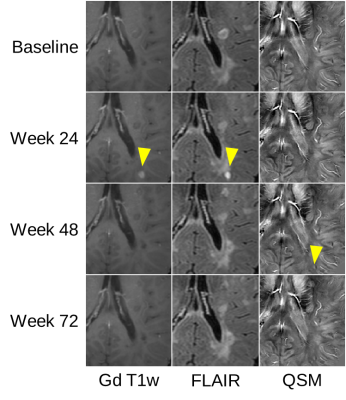 |
142 |
Don’t track edema if you are interested in demyelination: Quantitative susceptibility mapping of MS lesions depends on the image contrast used for lesion definition
Christoph Birkl, Christian Kames, David Li, Anthony Traboulsee, Alexander Rauscher
The purpose of this study was to evaluate how definition of multiple sclerosis lesions affects the measurement of quantitative magnetic susceptibility measurements in these lesions. Masks were drawn on FLAIR and QSM images at baseline and different follow-up time points. QSM was analyzed longitudinally across these lesion masks of the same lesion and compared to NAWM. A strong variability in longitudinal QSM was observed when masks were drawn on FLAIR images. The most consistnt behavior was seen on QSM based masks. The time point of lesion definition had a stronger influence when using FLAIR maks than with QSM based masks.
|
|
3307.
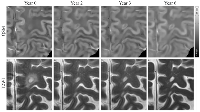 |
143 |
Utilizing QSM to assess long-term longitudinal susceptibility changes in enhancing multiple sclerosis lesions
Shun Zhang, Thanh Nguyen, Sandra Hurtado Rua, Yi Wang, Susan Gauthier
We measured the longitudinal susceptibility change of 32 new Gd-enhancing lesions from 19 multiple sclerosis (MS) patients over six years. Lesion susceptibility increased to peak within 1-2 years and was followed by a steady decline. Lesions with QSM rim had an overall higher susceptibility and demonstrated a decay rate that was significantly slower compared to that of lesions without rim.
|
|
3308.
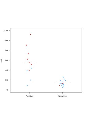 |
144 |
Phase imaging and serum neurofilaments: a combined laboratory-imaging marker of multiple sclerosis chronic inflammation
Pietro Maggi, Martina Absinta, Amandine Mathias, Pascal Sati, Caroline Pot, Reto Meuli, Jens Kuhle, Ludwig Kappos, Du Pasquier Renaud, Daniel S. Reich, Cristina Granziera
In multiple sclerosis (MS), persistent chronic inflammation at the edges of old non-gadolinium-enhancing white matter lesions, is identified with a paramagnetic rim on susceptibility-based-MRI sequences. Serum neurofilaments (sNfL) levels are associated with disease activity and neurodegeneration in acute and chronic phases of MS. Whether the presence of chronic inflammation is accompanied by increased in neuroaxonal destruction is currently unknown. We showed that MS patients featuring chronic inflammation at the lesions edges have higher neuroaxonal destruction than patients without. The combination of paramagnetic rim and sNfL may help in the selection of “chronically” active MS patients who may benefit from disease-modifying-treatments.
|
|
3309.
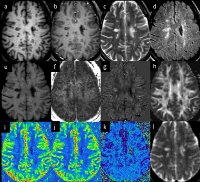 |
145 |
Multi-Parametric White Matter Imaging in Multiple Sclerosis Lesions
Ewart Haacke, Evanthia Bernitsas, Karthikeyan Subramanian, David Utriainen, Vinay Kumar, Sean Sethi, Yongsheng Chen, Zahid Latif, Pavan Kumar, Xiomeng Zheng, Robert Comley, Yanping Luo
A multiparametric quantitative white matter study using MRI was designed to include 15 relapsing remitting MS patients and 10 age and sex matched controls. The imaging protocol acquired 3D conventional imaging, SWI, QSM, DTI, MTC, and STrategically Acquired Gradient Echo (STAGE) imaging. ROI were manually drawn around MS lesions in T2-FLAIR and QSM. Contralateral normal appearing white matter (NAWM) and NAWM in controls were drawn in all modalities to measure mean intensity and volume. We found a good correlation between QSM, MTR, FA, MWF and conventional imaging. QSM’s sensitivity to demyelination best complimented T2-FLAIR’s sensitivity to inflammation and scarring.
|
|
3310.
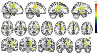 |
146 |
Phase opposed cerebral vasoreactivity in multiple sclerosis: evidences of a link between white matter tracts and vascular alterations
Jeremy Deverdun, Arthur Coget, Victor Vagné, Amel Benali, Pierre Labauge, Nicolas Menjot de Champfleur, Emmanuelle Le Bars
Patients with multiple sclerosis (MS) have a higher risk for ischaemic stroke. The current hypothesis states that white matter (WM) fibers alterations causes, through astrocytes, a cerebral vasoreactivity (CVR) disruption resulting in a hypoperfusion. Due to the location of the astrocytes, we expect an altered vasoreactivity mainly around WM tracts. Using a MR vasoreactivity experiment, we could identify altered WM pathways. In MS patients a path from left anterior insula to both precentral gyrus and right middle and superior frontal gyrus highlighted an altered CVR compared to controls. A negative association was found with fNART in the cingulum limbic pathway.
|
|
3311.
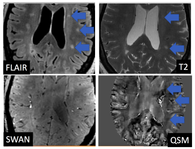 |
147 |
Contrast to Noise Measurements of Multiple Sclerosis Lesions using QSM at 7T: Comparison with Conventional Contrasts
Kevin Koch, Robin Karr, Staley Brod, L. Muftuler
Multiple Sclerosis (MS) has been a condition of particular interest to the QSM community, due to the conspicuity that white matter abnormalities show in QSM contrast images. While several studies have examined small cohort case findings of QSM variations within MS subjects at ultra-high field, an analysis of QSM contrast-to-noise ratio across a wide range of lesions has yet to be performed. Here, we present quantitative CNR analysis of 65 MS lesions spread across 10 subjects scanned with a high resolution 3D protocol at 7T.
|
|
3312
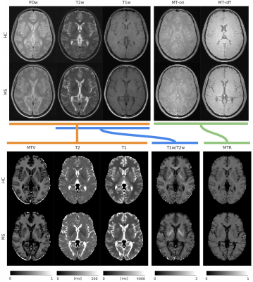 |
148 |
Myelin-sensitive indices in multiple sclerosis: the unseen qualities of qualitative clinical MRI
Video Permission Withheld
Antonio Ricciardi, Francesco Grussu, Marco Battiston, Ferran Prados, Baris Kanber, Rebecca Samson, Daniel Alexander, Declan Chard, Claudia Gandini Wheeler-Kingshott
Specialised quantitative MRI techniques, while considered state-of-the-art for quantitative studies, increase acquisition times and costs. On the other hand, clinical techniques routinely added to every MR-protocol are dismissed from quantitative analyses because labelled as qualitative. In this study, macromolecular tissue volume (MTV) and T1-/T2-weighted ratio (T1w/T2w) maps extracted from clinical images were compared with magnetisation transfer ratio (MTR, gold standard for myelin mapping) to assess whether clinical scans can also be used for myelin mapping in multiple sclerosis. Good correlation and similar sensitivity to disease were observed for both comparisons, with MTV appearing overall more reliable than T1w/T2w when compared with MTR.
|
|
3313.
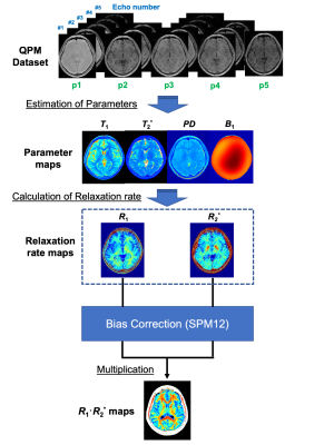 |
149 |
Myelin imaging derived from quantitative parameter mapping
Yuki Kanazawa, Masafumi Harada, Yo Taniguchi, Hiroaki Hayashi, Takashi Abe, Maki Otomo, Yuki Matsumoto, Masaharu Ono, Yoshitaka Bito, Akihiro Haga
We developed a novel method which is applicable to visualize myelin component in the human brain using relaxation time derived from QPM-MRI. Our method demonstrated acknowledgement that the myelin content increased proportionally by times R1 and R2 in healthy volunteers. Linear regression analysis showed a strong and highly significant correlation between conventional T1w/T2w ratios and R1·R2* times derived from QPM (R = 0.61, P < 0.0001). In conclusion, our myelin mapping technique using QPM may replace conventional T1w/T2w ratio mapping and be expected to become independent of measurement conditions due to having quantitative characteristic of QPM itself.
|
|
3314.
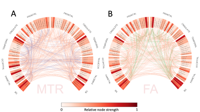 |
150 |
The myelin-weighted connectome: a new look at multiple sclerosis
Tommy Boshkovski, Atef Badji, Predrag Janic, Ljupco Kocarev, Julien Cohen-Adad, Antonio Giorgio, Nicola De Stefano, Bratislav Misic, Nikola Stikov
Myelin imaging has yet to make its way into standard connectomics protocols. Myelin-specific MRI metrics are useful for the assessment of neurological conditions that affect white matter. In this study we compared the structural connectomes of multiple sclerosis (MS) patients and healthy controls. Magnetization transfer ratio was used as a myelin-specific metric computed along each tract (MTR-weighted connectome) and compared against the connectome weighted by fractional anisotropy (FA-weighted connectome). We then identified the connections that were reliably different between MS patients and controls, and found 100 connections that were unique to the MTR-weighted connectome and 62 connections that were unique to the FA-weighted connectome.
|
|
| Top |
Emerging Technology & Translational Imaging 2
Digital Poster
Neuro
Tuesday, 14 May 2019
| Exhibition Hall |
16:45 - 17:45 |
| |
|
Computer # |
|
3315.
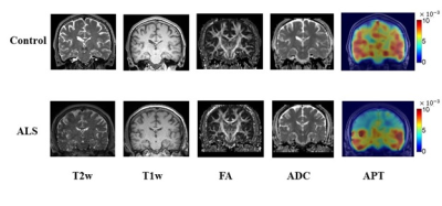 |
151 |
Amide proton transfer in amyotrophic lateral sclerosis
Zhuozhi Dai, Sanjay Kalra, Dennell Mah, Peter Seres, Gen Yan, Hongfu Sun, Zhiwei Shen, Renhua Wu, Alan Wilman
There is lack of objective imaging indicators for ALS diagnosis and assessment. Our hypothesis is that amide is altered due to neurodegeneration in ALS, and this alteration will be visible on APT images. This study aims to explore the value of APT in ALS patients as a possible image biomarker of disease. Thirty-two participants were recruited as part of the Canadian ALS Neuroimaging Consortium. This study first demonstrated changes of APT in the motor cortex and corticospinal tract of ALS patients. The combination of APT and DTI can simultaneously detect changes of metabolism and microstructure in ALS patients.
|
|
3316.
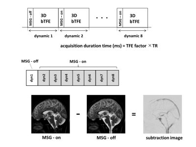 |
152 |
Visualization of CSF flow of whole brain using 3D dynamic iMSDE SSFP
Tomohiko Horie, Nao Kajihara, Haruo Saito, Shuhei Shibukawa, Susumu Takano, Natsuo Konta, Makoto Obara, Tetsuo Ogino, Tetsu Niwa, Kagayaki Kuroda, Mitsunori Matsumae
We reported a technique to visualize the irregular flow of cerebrospinal fluid (CSF) in whole brain by using dynamic improved motion-sensitized driven-equilibrium steady-state free precession (dynamic iMSDE SSFP). The purpose of this study was to propose a new technique using 3D dynamic iMSDE SSPF for visualizing the slow and irregular CSF flow of whole brain. 3D dynamic iMSDE SSPF can visualize the connection between the CSF space and a small lesion. This technique is suggested to contribute to the diagnosis of various diseases in the CSF space.
|
|
3317.
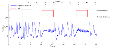 |
153 |
Measurement of cerebral perfusion changes during breath holding using pCASL with an accelerated 3D readout: an approach to quantify cerebrovascular reactivity.
Sergio Solís-Barquero, Rebeca Echeverria, Elena Cacho-Asenjo, Antonio Martínez-Simón, Marta Vidorreta, Pablo Domínguez, Miguel Fernández, María Fernández-Seara
Cerebrovascular reactivity (CVR) can be defined as the change in flow in response to a vasoactive agent. Hypercapnia is known to cause global increases in cerebral blood flow (CBF). ASL can provide a quantitative measure of CBF changes. The objective of this research was to determine whether pseudo-continuous ASL (pCASL) with an accelerated 3D readout, combined with breath-hold induced hypercapnia, is a practicable method for evaluating CVR. Results showed that the faster readout provided whole-brain coverage at isotropic resolution and allowed a post-labeling delay long enough to avoid macrovascular signal artifacts, while keeping TR short to sample several points per breath-hold.
|
|
3318.
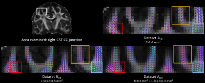 |
154 |
Tractography of complex white matter bundles: limitations of diffusion MRI data upsampling
Dmitri Shastin, Umesh Rudrapatna, Greg Parker, Khalid Hamandi, William Gray, Derek Jones, Maxime Chamberland
Diffusion MRI images for fiber tractography are often acquired at low spatial resolution which may lead to underestimation of smaller tracts with complex morphology. Although upsampling may improve results, this has had mixed observations in the literature. We compared three datasets (2×2×2 mm3, 1.5×1.5×1.5 mm3, and 2×2×2 mm3upsampled to 1.5×1.5×1.5 mm3) obtained and processed using state-of-the-art hardware and methodology. By evaluating the appearances and streamline metrics of the corticospinal tract, anterior commissure and small subcortical U-shaped fibers as test bundles, we demonstrated that the original high-resolution dataset outperformed both the low-resolution and upsampled data in resolving complex regional anatomy.
|
|
3319.
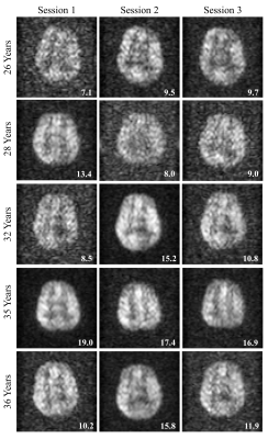 |
155 |
Assessment of repeatability of imaging inhaled hyperpolarized xenon-129 in the human brain
Madhwesha Rao, Graham Norquay, Jim Wild
This study assesses the repeatability of image quality of inhaled hyperpolarized 129Xe brain MRI by assessing the signal-to-noise ratio of 129Xe brain images for five healthy subjects. A maximum signal-to-noise ratio of 18.8 ±6.1 and mean signal-to-noise ratio of 12.1 ±3.8 was observed over the five volunteers. An intra-subject variability between ±6 % and ±30 %, and inter-subject variability of ±30 % was observed. By using an optimized polarizer, RF coil and pulse sequence as in this study, we believe the signal-to-noise ratio is sufficiently reproducible for further clinical evaluation.
|
|
3320.
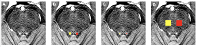 |
156 |
Quantitative Magnetization Transfer of the Human Locus Coeruleus
Paula Trujillo, Kalen Petersen, Seth Smith, Daniel Claassen
The locus coeruleus (LC) is the major source of norepinephrine in the brain and it is affected in several neurodegenerative disorders. We used quantitative magnetization transfer (MT) imaging to create parametric maps of the macromolecular content of the LC and neighboring tissues. We found that the macromolecular content was lower in the LC compared to the surrounding pontine tegmentum, suggesting that LC contrast is related to MT effects.
|
|
3321.
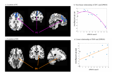 |
157 |
Progesterone, not estrogen modulates cerebral blood flow across the menstrual cycle
Samantha Cote, Russell Butler, Marie Anne Richard, Adrianna Mendrek, Jean-Francois Lepage, Kevin Whittingstall
The sex hormones estrogen (EST) and progesterone (PROG) are known vasodilators, yet little is known about how large changes in concentrations across the menstrual cycle (MC) influences CBF. This study aimed to determine how fluctuations in EST and PROG across the MC influence CBF and cerebral arteries. CBF and arteries were evaluated twice in female participants when PROG was low and high. There were region-specific and dose-dependent effects of PROG (but not EST) on CBF, but not arterial diameters. This indicates that cerebral vascular function in women is dynamic across the MC and is not due to vasodilatory effects of EST or PROG.
|
|
3322
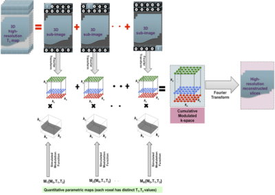 |
158 |
Quantitative Data Driven Voxel-Wise Simulator (QVS): Application to 3D MP-RAGE Optimization for Harmonization of Multi-Centre Brain MRI Studies
Video Permission Withheld
Irtiza Gilani, John Thornton, Martina Callaghan, Marzena Wylezinska-Arridge, Stephen Wastling, Tarek Yousry, David Thomas
Typical MRI simulators are either based on the signal equations or on the solution to the Bloch equations for each voxel, with appropriate image encoding incorporated. In this work, a novel modular approach to simulate MR sequences, termed quantitative voxel-wise simulator (QVS), is proposed. This simulator employs quantitative parametric maps as inputs, models the evolved MR signal as a multi-dimensional filter output and reconstructs the image by using a voxel-by-voxel modulated k-space approach. As a proof-of-concept, 3D MP-RAGE sequence is simulated and the simulated images are compared with in-vivo data. QVS is designed for multi-centre brain MRI harmonization.
|
|
3323.
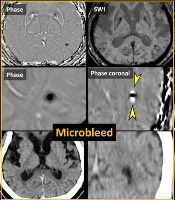 |
159 |
Differentiation between Hemorrhage and Calcifications by Assessing the Dipole Patterns on Unwrapped Phase Images of Gradient-recalled Echo Sequence
Eung Yeop Kim, Sung Hyun Yu, Jongho Lee
Both cerebral hemorrhages and calcifications may show similar internal heterogeneity on phase images, limiting its clinical utility. We assessed a dipole artifact (both the upper/lower poles and the equatorial region) on phase images in 123 patients with hemorrhages (n=119) and calcifications (n=75). All hemorrhagic lesions were perfectly determined, while all but one calcification showed a diamagnetic dipole pattern. The equatorial phase values (degree) were significantly different between hemorrhages and calcifications (-10.41±10.66 versus 10.86±9.53; P<0.0001). The signal intensities of both the lobes and the equatorial rim on GRE phase images accurately differentiate hemorrhages from calcifications irrespective of internal heterogeneity.
|
|
3324.
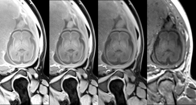 |
160 |
Application of 3T Magnetic Resonance 3D StarVIBE Sequence in Fetal Brain
Presentation Not Submitted
Xuesheng Li, Haibo Qu, Fenglin Jia, Gang Ning, Juncheng Zhu, Zhijun Ye, Shaoyu Wang, Huapeng Zhang, Zhitao Zhang
Fast T2-weighted imaging is a commonly used diagnostic sequence for fetal MR. Because of the limitations of fetal development, the imaging effect of T1 nervous system imaging is difficult to achieve like the post-natal image effect. The 3D StarVIBE sequence is used as a new sequence under 3T MR, using a radial K-space filling, not sensitive to motion, can be applied to the fetal nervous system examination. Recent reports suggest that the TurboFLASH(TFL) sequence with an optimized TI time of 2500 ms is used as an advantage of excellent contrast and overcoming motion artifacts in 2D gradient echo sequences. Therefore, this study will use the 3D StarVIBE sequence and the TFL sequence as a reference to judge its imaging effect.
|
|
3325.
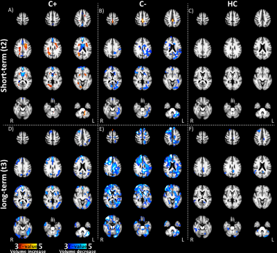 |
161 |
Longitudinal brain volume changes in pre-menopausal breast cancer patients treated with chemotherapy
Gwen Schroyen, Jeroen Blommaert, Mathieu Vandenbulcke, Ahmed Radwan, Ann Smeets, Ron Peeters, Charlotte Sleurs, Patrick Neven, Hans Wildiers, Frederic Amant, Stefan Sunaert, Sabine Deprez
This longitudinal study investigates possible recovery of volumetric brain changes in pre-menopausal patients three years after being treated for early-stage breast cancer. While initial widespread white matter volume increase was previously observed, recovery is seen three years after treatment in the same group of young women treated with chemotherapy. Patients with breast cancer show widespread gray matter volume decrease, observed both in patients treated and not treated with cytotoxic agents. Further studies are necessary to unravel possible acute volumetric changes, possibly neuro-inflammatory mechanisms, in this population as a cause for these findings.
|
|
3326.
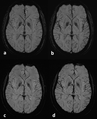 |
162 |
3D Flow Compensated Interleaved EPI for a Fast High-Resolution Susceptibility-Weighted Imaging at 1.5T
Wei Liu, Kun Zhou, Shi Cheng, Kawin Setsompop
We implemented a first order gradient nulling (GMN) based partial flow compensation in 3D interleaved EPI and assessed its feasibility for a fast high-resolution SWI application at 1.5T. Specifically, we used GMN to zero the velocity-induced phase error at the center echo for each shot in both phase and frequency encoding directions. The slice direction implementation was identical to that for 3D GRE, with flow compensation for both slice selective gradient and partition encoding gradient. In addition, each shot was sequentially acquired twice with the swapped readout polarity in the second to further reduce the phase oscillates between the even and odd echoes in each shot. Flow phantom and in-vivo experiments were performed to validate that flow effect is effectively reduced even not all echoes have been fully flow compensated.
|
|
3327.
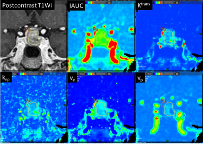 |
163 |
Quantitative pharmacokinetic comparison of adenoma and normal pituitary gland using high-temporal and spatial resolution dynamic contrast enhanced MRI
Kiyohisa Kamimura, Masanori Nakajo, Tomohide Yoneyama, Yoshihiko Fukukura, Shingo Fujio, Takashi Iwanaga, Hiroshi Imai, Takashi Yoshiura
Preoperative localization of the normal pituitary gland is important in patients with pituitary adenoma. Our aim was to evaluate the possible role of high-temporal and spatial resolution dynamic contrast enhanced MR imaging (DCE-MRI) and quantitative pharmacokinetic analysis in differentiation of the normal pituitary gland from pituitary adenoma. The normal pituitary gland showed significantly higher IAUC, Ktrans, kep and ve than pituitary adenoma. The ROC curve analysis showed significance for IAUC, Ktrans, kep and ve (AUC = 0.958, 0.882, 0.781 and 0.851, respectively). These quantitative parameters may be useful for differentiation of the normal pituitary gland from pituitary adenoma.
|
|
3328.
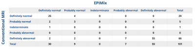 |
164 |
Diagnostic performance of a new multicontrast one minute full brain exam (EPIMix) in neuroradiology
Anna Delgado, Annika Kits, Jessica Bystam, Magnus Kaijser, Mikael Skorpil, Tim Sprenger, Stefan Skare
This study assess if a new one minute multi contrast MRI method has comparable diagnostic performance as conventional MRI in brain imaging. Prospectively consecutively included patients (n=101) underwent a conventional clinical brain MRI in addition to a new 80 seconds multi contrast MRI sequence named EPIMix. The diagnostic performance to categorize a clinical brain MRI scan with EPIMix or conventional MRI as abnormal (symptom-causing lesion) was comparable between EPIMix (AUC 0.97 (95%CI 0.94-1.00) and 0.98 (95% CI 0.96-1.00)) and conventional MRI (AUC 1.00 (95% CI 1.00-1.00)), (n=96-101).
|
|
3329
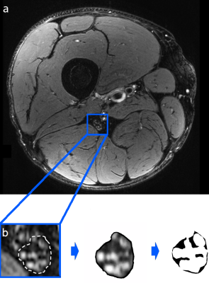 |
165 |
Magnetic resonance neurography reveals association between low serum cholesterol and peripheral nerve damage in type 2 diabetes
Video Permission Withheld
Johann Jende, Jan Groener, Zoltan Kender, Sabine Heiland, Stefan Kopf, Mikro Pham, Peter Nawroth, Martin Bendszus, Felix Kurz
Diabetic polyneuropathy (DPN) is a major contributor for morbidity in diabetes; however, up to this day, there is a lack of sufficient strategies to prevent this disorder especially in patients suffering from type 2 diabetes (T2D). Specifically, it remains controversially discussed if a lowering of serum cholesterol has beneficial effects on the course of T2D DPN. Using in vivo high resolution magnetic resonance neurography in 100 type 2 diabetes patients with and without DPN, we found that lowering of serum cholesterol, especially lowering of LDL, is strongly associated with an increase in visible nerve damage. We further found a significant correlation between the amount of nerve damage and the patients’ statin dose. In T2D patients, this effect is potentially relevant for therapies that promote an aggressive lowering of serum cholesterol.
|
|
3330.
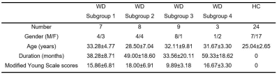 |
166 |
Regional morphometric abnormalities and clinical relevance in Wilson's disease
Presentation Not Submitted
Yukun Song, Jianping Chu, Lin Zou, Xiaoying Tang
In this study, coarse-to-fine evaluations were creatively made in terms of both global volume and local shape to identify Wilson’s disease (WD)-related morphometric abnormalities of eight structures of interest (caudate, putamen, globus pallidus, thalamus, amygdala, hippocampus, red nucleus and substantia nigra). Our results revealed that significant volume reductions and region-specific surface atrophy were detected in all structures of interest except the bilateral hippocampus in patients with WD relative to HC subjects, and the putamen had the strongest global and local atrophy and the amygdala was least affected. These morphometric abnormalities may serve as useful imaging biomarkers for WD.
|
|
3331.
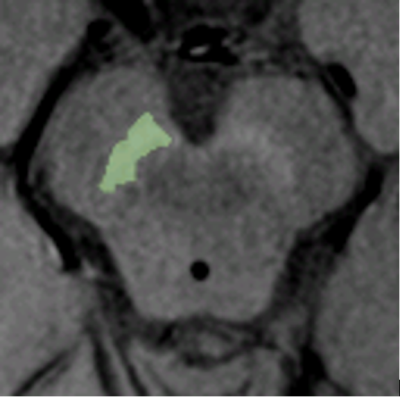 |
167 |
Neuromelanin-sensitive Magnetic Resonance Imaging Study of the Substantia Nigra in Huntington’s Disease
Ricardo Leitão, Sofia Reimão, Leonor Correia-Guedes, Joaquim Ferreira, Rita Nunes
Neuromelanin(NM)-sensitive MRI (NM-MRI) is a promising technique to study pathological changes in NM-containing structures, such as the substantia nigra pars compacta (SNc). This midbrain structure modulates the corticostriatal pathway through the striatum, which is known to degenerate in Huntington’s Disease (HD). Our study used NM-MRI for the first time to study HD, with a semi-automatic segmentation method to evaluate the SNc, compatible with dopaminergic neuronal loss in the SNc of HD patients. SNc NM correlated with the volumes of the caudate, putamen and globus pallidus, suggesting that SNc neuronal loss and basal ganglia atrophy may not be independent processes.
|
|
3332.
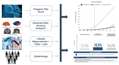 |
168 |
Multimodal Models Provide Earlier Prediction (10 Years Prior to Diagnosis) of Dementia and Cognitive Decline and Personalized Actionability for Risk Mitigation for At-Risk Individuals
Natalie Schenker-Ahmed, Ilan Shomorony, Jian Wu, Alex Graff, Naisha Shah, Peter Garst, Nafisa Bulsara, Krisztina Marosi, Dmitry Tkach, Lei Huang, Axel Bernal, Jason Deckman, Hyun-Kyung Chung, Wayne Delport, David Karow, Christine Swisher
Current approaches for predicting an individual’s risk of developing dementia rely primarily on single modality data and/or single biomarkers. Here we evaluate the utilization of non-invasive MR imaging and genetics for early detection and prediction of cognitive decline and dementia. We demonstrate superior performance of our multi-measurement, multimodal approach. Moreover, our approach performs as well or better than invasive amyloid PET. We further show a method that identifies modifiable factors upon which an individual can act to mitigate their risk with the long-term goal of empowering high-risk individuals with personalized action plans earlier when the disease progression can be slowed.
|
|
3333.
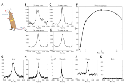 |
169 |
Investigating the biodistribution of the antiinflammatory drug teriflunomide in vivo using 19F MRI
Christian Prinz, Jason Millward, Paula Ramos Delgado, Ludger Starke, Andreas Pohlmann, Thoralf Niendorf, Sonia Waiczies
Teriflunomide is a trifluorinated drug indicated in the treatment of multiple sclerosis (MS). Using fluorine (19F) MR methods, the biodistribution of this anti-inflammatory drug could be examined in vivo to guide pharmacological studies and dosage adjustments en route to individualized therapy. In this study, we administered teriflunomide to healthy rats and an animal model of MS. We could detect teriflunomide non-invasively in various tissues in vivo, during the disease course and ex vivo.
|
|
3334.
 |
170 |
Diagnosing MS using Central Veins at Clinically Relevant Image Resolutions
Edward Peake, Margareta Clarke, Christopher Tench, Nikos Evangelou, Paul Morgan
The central vein sign is well documented as a biomarker for multiple sclerosis at high-resolution MRI. To investigate its potential at lower resolution and inform clinical MR protocol parameters, interpolation was used to demonstrate the effect of decreasing the resolution in 30 MS patients who underwent 7T-MRI. Finding were compared to 5 Non-MS groups (n = 82) at different resolutions using logistic regression. At half the original resolution the proportion of perivenular lesions changed considerably (73% to 47%), however, classification results remained unaffected provided the threshold (proportion of perivenular lesions) used for the differentiation decreased at lower resolution.
|
|
3335.
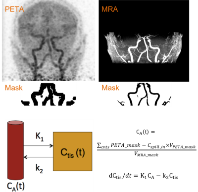 |
171 |
Validation of an Image Derived Input Function Method for 15O-water PET/MR Brain Scans
Mohammad Mehdi Khalighi, Audrey Fan, Mathias Engström, Lieuwe Appel, Gunnar Antoni, Frederick Chin, Eva Kumlien, Greg Zaharchuk, Mark Lubberink
A recently introduced image-derived input function (IDIF) method on a PET/MR scanner addresses the spill-in and spill-over artifacts on the PET images by measuring the true carotid artery volume by an MR-angiogram. This study validates the IDIF method for quantification of cerebral blood flow (CBF) from 15O-water PET, using arterial blood sampling as the gold standard in 20 subjects. CBF measured by IDIF and BSIF were correlated (R2= 0.5) in the gray-matter and whole-brain, with average difference of only 3%.
|
|
3336.
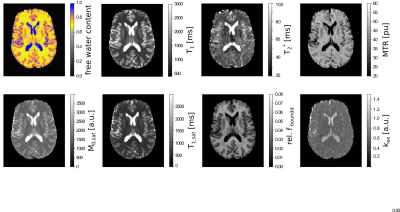 |
172 |
Characterisation of ageing effects using multiparametric quantitative imaging
Ana-Maria Oros-Peusquens, Melissa Schall, Elene Iordanishvili, N. Jon Shah
A multiparametric, quantitative method was implemented in a population-based study of normal ageing - the 1000Brains study. Several parameters are derived with whole-brain, high-resolution imaging in TA=21min: these included water content, T1 and T2* relaxation times, their changes in the presence of saturation and calculated qMT parameters - MTR, bound proton fraction fboundand forward magnetization exchange rate (kex). Methodological precision is high allowing for detecting of ageing effects as illustrated by several parameters (T1, T2*, H2O, fbound) from data sets of 25 healthy subjects (mean age 53±16 years, from 27 to 80, 18 male).
|
|
3337.
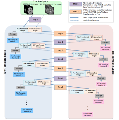 |
173 |
Development of High Quality T1w and DTI Templates of the Older Adult Brain in a Common Space
Yingjuan Wu, Abdur Raquib Ridwan, Xiaoxiao Qi, Shengwei Zhang, David A. Bennett, Konstantinos Arfanakis
The demand for a multimodal MRI atlas of the older adult brain is increasing as large amounts of data are generated in studies of aging. The purpose of this work was to develop high quality T1-weighted (T1w) and diffusion tensor imaging (DTI) templates of the older adult brain in the same space, to allow future multimodal analyses. This was successfully accomplished through a proposed iterative multimodal template construction strategy. The new templates allowed higher spatial normalization accuracy of T1w and DTI data from older adults compared to other available templates.
|
|
3338.
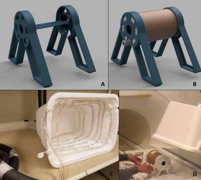 |
174 |
Diffusion imaging of rat brain slices on a human clinical MRI scanner
Natalie Aw, Alex Cerjanic, Jennifer Mitchell, Martha Gillette, Brad Sutton
We demonstrate the feasibility of performing diffusion tensor imaging on a rat brain slice using a 3 T human clinical scanner and a rat coil. Brain slices provide an important platform for performing mechanistic studies in neuroscience. We show that sufficient SNR is available for performing experiments to examine the diffusion properties of white matter through examining age-related differences in FA on 8-week-old and 1-year-old rats.
|
|
Back to Program-at-a-Glance |  Back to Top Back to Top |
| The International Society for Magnetic Resonance in Medicine is accredited by the Accreditation Council for Continuing Medical Education to provide continuing medical education for physicians. |
