Digital Poster Session
Diffusion Back to Program-at-a-Glance Back to Program-at-a-Glance
|
Wednesday, 15 May 2019
Digital PosterDiffusion
3339 -3363 Diffusion MRI: Image Reconstruction
3364 -3388 Diffusion MRI: Fiber Orientations & Tracking
3389 -3413 Diffusion MRI: Artefact Correction
3414 -3437 Diffusion: Neuro Applications
3438 -3461 Diffusion: Body Applications
3462 -3486 Diffusion MRI: Signal Representation & Modelling
3487 -3508 Diffusion MRI: Diffusion Gradient Waveform Design & Optimization
3509 -3533 Diffusion MRI: Data Acquisition
3534 -3558 Microstructural Modelling & Mapping
3559 -3582 Microstructure Modeling: 1
3583 -3606 Diffusion in Disease
3607 -3631 Microstructure Modeling: 2
3632 -3656 Diffusion: Validation |
| |
Diffusion MRI: Image Reconstruction
Digital Poster
Diffusion
Wednesday, 15 May 2019
| Exhibition Hall |
08:15 - 09:15 |
| |
|
Computer # |
|
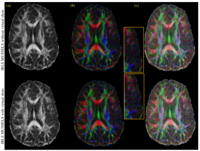
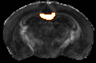
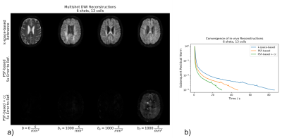
3339.
 |
1 |
Improved MUSSELS Reconstruction for High Resolution Diffusion Weighted Imaging Using Fast Iterative Re-weighted Least Squares Algorithm

Merry Mani, Hemant Aggarwal, Vincent Magnotta, Mathews Jacob
Multi-shot diffusion-weighted imaging reconstructions are challenged by the inter-shot phase inconsistency that exists between the data from different shots. The MUSSELS algorithm enabled the direct reconstruction of the multi-shot k-space data by posing it as a low-rank based matrix recovery problem. The iterative algorithm has been shown to successfully recover the missing k-space samples in accelerated and non-accelerated acquisitions. However, the reconstruction time increases as the number of shots\acceleration increases. We propose a new formulation based on iterative re-weighted least squares that increase the computational efficiency of the matrix completion by several folds to speed up the recovery of multi-shot data.
|
|
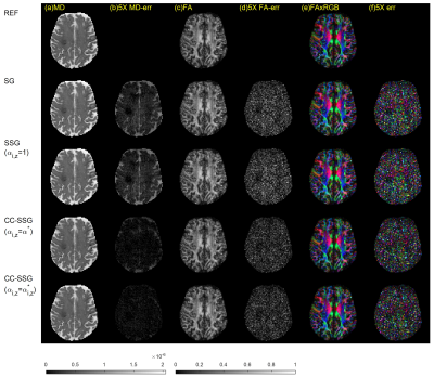
3340.
 |
2 |
Simultaneous Multi-Slice Diffusion MR Reconstruction using Coil-Combined Split Slice-GRAPPA
Kazem Hashemizadeh, Rong-Rong Chen, Edward V. DiBella, Leslie Ying, Ganesh Adluru
Simultaneous multi-slice (SMS) acquisition accelerates diffusion MR imaging by acquiring multiple slices simultaneously. In this work, we propose a new method, termed coil-combined split slice-GRAPPA (CC-SSG), to improve the quality of SMS diffusion imaging reconstruction. By optimizing split-slice-GRAPPA (SSG) kernels specifically for coil combining, our approach allows for a better trade-off for suppressing inter-slice and intra-slice leakages and minimizes the mean-square-error (MSE) of coil-combined images. The proposed CC-SSG method improves the estimation of diffusion tensor imaging (DTI) parameters over existing methods.
|
|
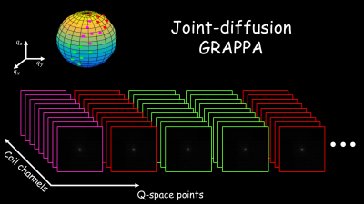
3341.
 |
3 |
Joint-diffusion GRAPPA: enabling higher acceleration rates in dMRI by exploiting joint information from the k- and q-space
Gabriel Ramos-Llordén, Santiago Aja-Fernández, Congyu Liao, Kawin Setsompop, Yogesh Rathi
In this work, we generalize conventional GRAPPA-based dMRI reconstruction by exploiting joint information from the k-and q-space simultaneously. Higher acceleration in-plane rates than those commonly reported may be achieved when the missing k-space lines are learned using all information available in the whole k-space data set, that is, considering multi-coil channel information as well as the k-space data probed at different q-space points. Our novel method, joint-diffusion GRAPPA, is validated with in-vivo multi-slice dMRI data, where we show it always outperforms conventional GRAPPA in terms of image quality, and works reasonably well for regimes where conventional GRAPPA results in significant noise penalty (Rin−planeRin−plane >
3 ).
|
|
3342.
 |
4 |
Use of self-navigation to enable efficient 3D DWI SE-EPI multislab multiband imaging
Steen Moeller, Sudhir Ramanna, Edward Auerbach, Pramod Pisharady, Christophe Lenglet, Mehmet Akcakaya, Kamil Ugurbil
A method is proposed for self-navigation of DWI 3D multislab multiband SE-EPI, to enable whole brain high-resolution imaging, with optimal imaging TR for higher SNR efficiency. Data for high b-value (b=3k s/mm2) and 1mm3 resolution is presented.
|
|
3343.
 |
5 |
Acceleration of 3D diffusion MRI using a kernel low rank compressed method
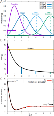
Chaoyi Zhang, Tanzil Mahmud Arefin, Ukash Nakarmi, Hongyu Li, Dong Liang, Jiangyang Zhang, Leslie Ying
Diffusion MRI has showed great potential in probing tissue microstructure and brain structural connectivity. However, high-resolution diffusion MRI with multiple direction is limited by the lengthy scan time. In this abstract, we apply a kernel low rank model to accelerate diffusion imaging by undersampling the k-space. This method is validated using high-resolution mouse brain datasets. Compared with the conventional compressed sensing method, the proposed method demonstrate more accurate mean diffusivity, fractional anisotropy and fiber orientation distribution estimates with acceleration factors up to 8.
|
|
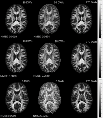
3344.
 |
6 |
Deep Learned Diffusion Tensor Imaging
Hongyu Li, Chaoyi Zhang, Zifei Liang, Dong Liang, Bowen Shen, Yulin Ge, Jiangyang Zhang, Ruiying Liu, Peizhou Huang, Sunil Kumar Gaire, Xiaoliang Zhang, Leslie Ying
Diffusion tensor imaging typically requires acquisition of a large number of diffusion weighted images (DWI) for accurate fitting of the tensor model due to the issue of low SNR. This abstract presents a deep learning method to generate FA color map showing the primary diffusion directions from very few DWIs. The method uses deep convolutional neural networks to learn the nonlinear relationship between the DWIs and the FA color maps, bypassing the conventional DTI models. Experimental results show that the proposed method is able to generate FA color maps from only 6 DWIs with quality comparable to results from 270 DWIs using conventional tensor fitting.
|
|
3345.
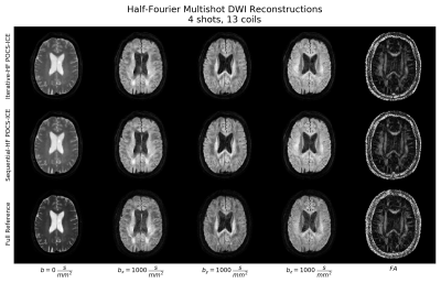 |
7 |
Self-navigated Half-Fourier Multi-shot Echo-planar DWI Reconstructions for Brain Imaging
Malte Steinhoff, Kay Nehrke, Peter Börnert, Alfred Mertins
EPI trajectories using Half-Fourier achieve shorter echo times and therefore higher SNR, which is especially desirable in low-SNR applications like diffusion-weighted MRI. For the same reason, methods enabling phase-corrected image recovery for multi-shot diffusion acquisitions have been intensively studied for both spiral and EPI trajectories. In this work, two algorithms are presented comprising both half-Fourier and the multi-shot same-magnitude constraints to exploit the advantages of both techniques. The algorithms are shown to robustly recover interleaved half-Fourier datasets from in-vivo brain acquisitions.
|
|
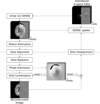
3346.
 |
8 |
Multi-shot Diffusion EPI Reconstruction with Iterative Rigid Motion-correction and Motion-induced Phase-correction for Brain Imaging
Malte Steinhoff, Kay Nehrke, Alfred Mertins, Peter Börnert
Multi-shot diffusion-weighted imaging offers increased SNR and higher resolution, but makes the acquisition vulnerable to shot-specific phase variations and macroscopic inter-shot motion. A wide range of iterative phase-corrected reconstruction schemes have been proposed to overcome the inter-shot phase inconsistencies, but robust motion estimation is still challenging due to the inherently low SNR of DWI. This work moves forward from initial one-time rigid motion estimation to an alternating optimization balancing the joint image, phase and motion estimation. A novel multi-shot echo-planar diffusion algorithm with iterative motion and phase correction is presented in simulations and in-vivo.
|
|
3347.
 |
9 |
Accelerating Iterative SENSE-based Algorithms for Cartesian Trajectories using the Point Spread Function and Coil Compression
Malte Steinhoff, Kay Nehrke, Alfred Mertins, Peter Börnert
For multi-shot diffusion-weighted imaging, iterative SENSE-based algorithms like POCSMUSE are boosting SNR allowing for higher image resolution. These advantages are achieved at the cost of higher computational load, thereby narrowing the clinical use case. The Cartesian implementation of such SENSE algorithms iteratively involves time-consuming 1D-Fast Fourier Transforms. In this abstract, the well-known point spread function for regular Cartesian undersampling is exploited to accelerate gradient- and projection-based SENSE updates. Accelerations of approximately 45% were achieved. Furthermore, coil compression is evaluated for these algorithms.
|
|
3348.
 |
10 |
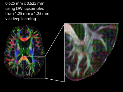
Super-Resolution Diffusion Imaging using Deep Learning: A Feasibility Study
Nahla Elsaid, Yu-Chien Wu
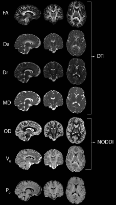
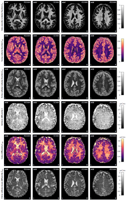
In this study, we present and validate the efficacy of using a state-of-the-art deep-learning method to achieve submillimeter high-resolution diffusion-weighted (DW) images. The 2D-based deep-learning method was validated by comparing diffusion tensor imaging (DTI) and neurite orientation dispersion and density imaging (NODDI) of the deep-learning high-resolution images and the ground-truth.
|
|
3349.
 |
11 |
Super-Resolution Hybrid Diffusion Imaging (SR-HYDI)
Nahla Elsaid, Pierrick Coupé, Yu-Chien Wu
In this study, we present and validate an efficient pipeline for submillimeter super-resolution hybrid diffusion imaging (SR-HYDI). The pipeline employs a collaborative patch-based super-resolution interpolation approach, which uses self-similarity to drive the reconstruction of diffusion-weighted images. The FA and MD generated from the proposed pipeline are compared against the ground-truth for validation.
|
|
3350.
 |
12 |
Complex-valued diffusion MRI data processing: Application to neural soma imaging
Enrico Kaden, Umesh Rudrapatna, Noemi Gyori, Uran Ferizi, Chris Clark, Derek Jones, Daniel Alexander
Microscopic diffusion anisotropy imaging requires averaging the diffusion signal over the gradient directions to regress out the unwanted effects of the fibre orientation distribution. However, Rician noise biases the mean signal calculations especially in the high b-value regime and subsequently the estimation of microstructural tissue features. In this work we develop new data processing methods using complex-valued MRI data that remove the background phase and hence retain the Gaussian characteristics of the signal noise, which is demonstrated in neural soma imaging, a novel application of the Spherical Mean Technique (SMT).
|
|
3351.
 |
13 |
Giving up the ghost: a systematic comparison of 2D phase correction algorithms in multi-shell high angular diffusion weighted imaging
Elizabeth Powell, Torben Schneider, Marco Battiston, Matthew Clemence, Ahmed Toosy, Jonathan Clayden, Claudia Wheeler-Kingshott
The echo planar imaging (EPI) Nyquist ghost often requires complex 2D phase error corrections in order to be robustly removed. Several methods exist but have not yet been systematically evaluated in high b-value diffusion-weighted (DW) EPI, where lower signal-to-noise ratios may affect the phase error estimation. We explore here the influence of different 2D phase-error corrected reconstruction methods on quantitative parameters derived from DW-EPI, and demonstrate that errors in parameter estimations relating to the Nyquist ghost can persist even after 2D phase-error correction.
|
|
3352.
 |
14 |
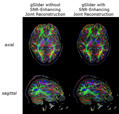
Whole-brain DTI at 860 µm isotropic resolution in 10 minutes on a commercial 3T Scanner
Yunsong Liu, Congyu Liao, Kawin Setsompop, Justin Haldar
We describe an acquisition and reconstruction methodology that enables in vivo human diffusion tensor imaging with whole-brain coverage and 860μμm
isotropic spatial resolution, all within a 10 minute acquisition window on a commercial 3T scanner. Our approach is enabled by combining the gSlider-SMS acquisition approach (which uses simultaneous multi-slab acquisition for increased spatial coverage, combined with highly-efficient RF slab-encoding to achieve high spatial resolution) with an SNR-enhancing joint reconstruction approach that mitigates the noise associated with high-resolution acquisition.
|
|
3353.
 |
15 |
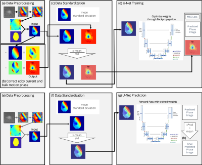
Estimation and Correction of Image Phase in diffusion weighted MRI using Deep Learning
Carolin Hecking-Veltman, Carolin Pirkl, Jonathan Dannenberg, Rolf Schulte, Tim Sprenger, Bjoern Menze, Marion Menzel
The phase of diffusion weighted MR images (DWI) is regularly discarded in clinical application although it might contain valuable information, as it is composed of phase contributions due to rigid motion, eddy currents and brain pulsation among others. In this work we take advantage of a neural network to separate the different phase components in individual DWI phase images. This enables estimating the amount of brain pulsation from DWI and modelling brain pulsation. The gained information may be used for phase correction, which eventually will allow using real-valued DWI (instead of magnitude DWI) to eliminate the Rician bias.
|
|
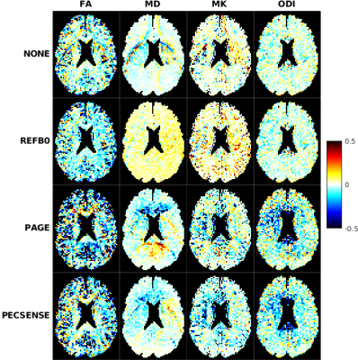
3354
 |
16 |
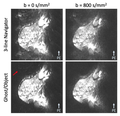
Nyquist Ghost Correction of High-Resolution SMS Breast DWI with Ghost/Object Minimization
Video Permission Withheld
Jessica McKay, Steen Moeller, Sudhir Ramanna, An Church, Michael Nelson, Edward Auerbach, Kamil Ugurbil, Patrick Bolan
A recent novel approach to acquire high-resolution breast DWI uses a simultaneous multi-slice (SMS) SE-EPI sagittal acquisition. In EPI, Nyquist ghosts are typically corrected using a 3-line navigator, which often fails in SMS SE-EPI breast DWI due to low SNR, insufficient fat suppression, and larger B0 inhomogeneity. In this work we compare a referenceless ghost correction method, called Ghost/Object minimization, with the standard 3-line navigator in high-resolution breast DWI. Ghost/Object provides more reliable 1st-order ghost correction in a dynamic and slice-specific way, which improves image quality and reduces bias in ADC values compared to the standard correction.
|
|
3355.
 |
17 |
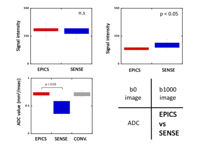
Pseudo-3D Diffusion-Weighted Imaging of the Brain using Echo Planar Imaging with Compressed SENSE (EPICS)
Kosuke Morita, Masami Yoneyama, Takeshi Nakaura, Seitaro Oda, Masahiro Hatemura, Yasuyuki Yamashita
We attempted to obtain brain high-resolution pseudo-3D (2D multi-slice acquisition with very thin slice thickness) diffusion-weighted echo planar imaging (DW-EPI) using a hybrid compressed sensing and sensitivity encoding (Compressed SENSE) framework (EPICS). pseudo-3D-DWI with EPICS achieved high-resolution (1.15 mm3) isotropic DWI within clinically feasible scan time. Furthermore, EPICS clearly improved the accuracy and robustness of ADC values in high b-value brain DWI with pseudo-3D acquisition without any penalty for scan parameters.
|
|
3356.
 |
18 |
Fast reconstruction of fractional anisotropy with two-dimensional principal component analysis based recognition
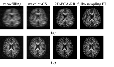
Fangrong Zong, Zihao Zhang, Qingle Kong, Jing An, Yan Zhuo, Xiaoliang Zhang
Reducing the acquisition time for obtaining fractional anisotropy (FA) is of paramount importance to investigate cerebral microstructures and morphologies non-invasively. This is the first time to introduce the two-dimensional principal component analysis recognition reconstruction (i.e. 2D-PCA-RR) in recovering highly under-sampled FA maps with 5-fold acceleration of data acquisition. An in-house data processing procedure is implemented to optimize signal-to-noise ratio and construct a distortion-free database. Our results from two different under-sampling patterns show a superior performance gain from the 2D-PCA-RR algorithm as compared to conventional reconstruction methods.
|
|
3357.
 |
19 |
Acquisition and processing strategy for obtaining high quality, distortion free diffusion MRI of the brainstem and cervical spine
Neda Sadeghi, Joelle Sarlls, Jessica Jordan, Flavia Facio, Elizabeth Hutchinson, M. Okan Irfanoglu, Amritha Nayak, Laura Reyes, Shruti Japee, Irini Manoli, Carlo Pierpaoli, Moebius Syndrome Research Consortium
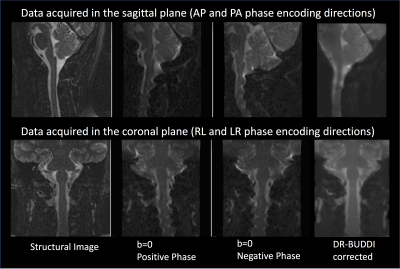
Despite its clear clinical and research usefulness, high quality in-vivo diffusion MRI imaging of the brainstem and the cervical spine has been challenging due to susceptibility-induced distortions and ghosting in echo planar images (EPI). In this study, we propose an acquisition and data processing strategy that can be carried out on clinical scanners with commonly available EPI sequences with good resolution and in a reasonable scan time (less than 30 minutes). We apply this acquisition strategy to the study of pyramidal decussation in subjects with Moebius syndrome and mirror movements.
|
|
3358.
 |
20 |
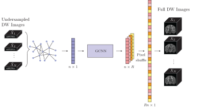
Accelerating Diffusion MRI via Slice Undersampling and Deep-Learning Reconstruction
Yoonmi Hong, Geng Chen, Pew-Thian Yap, Dinggang Shen
In this abstract, we present a proof-of-concept method for effective diffusion MRI reconstruction from slice-undersampled data. Instead of acquiring full diffusion-weighted (DW) image volumes, only a subsample of equally-spaced slices are acquired. We show that the complementary information from DW volumes corresponding to different diffusion wavevectors can be harnessed using graph convolutional neural networks for reconstruction of the full DW volumes.
|
|
3359.
 |
21 |
Deep learning for DSI parameter map generation without image pre-processing
Eric Gibbons, Kyler Hodgson, Ganesh Adluru, Edward DiBella
Recent advances in diffusion spectrum imaging (DSI) have reduced scan time considerably. Through the use of deep learning, DSI parameter maps (NODDI, GFA, etc.) can be generated with only a fraction of the number of q-space samples compared to conventional acquisition strategies. However, image pre-processing prior to the deep learning parameter map generation step is a computational bottleneck. This abstract explores if this bottleneck can be bypassed entirely and use images straight from the scanner as CNN inputs. We show that the image pre-processing is not necessary to generate NODDI and GFA parameter maps--thereby avoiding the image processing computation time.
|
|
3360.
 |
22 |
Phase-Error-Free Multi-shot 3D DWI using Filtered Back Projection Reconstruction
Hai Luo, Gaojie Zhu, Xiang Zhou, Chao Wang, Bei Lv, Xia Liu, Ziyue Wu
Conventional multi-shot DWI is known to suffer from inter-shot phase inconsistencies due to motion and hardware imperfections. In this work, we present a new approach for phase-error-free diffusion imaging by using multi-shot Paddlewheel-shaped EPI acquisition and filtered back projection (FBP) Reconstruction. The necessity of inter-shot phase correction is removed due to the magnitude-only nature of FBP. Reduced FOV excitation scheme is incorporated to reduce scan time and artifacts. Exemplary results of head and prostate DWI are demonstrated to show the efficacy of the proposed methods.
|
|
3361.
 |
23 |
Imaging Cortical Columns in Gray Matter with Sub-Millimeter isotropic DTI
Iain Bruce, Christopher Petty, Allen Song
The microarchitecture in gray matter of the human brain is comprised of short (<3 mm) cortical columns that traverse six cortical layers. To most accurately delineate these columns in-vivo, it is essential to achieve isotropic spatial resolutions on the order of 0.8 mm or less. In this study, we present a means of acquiring diffusion tensor imaging data with ultrahigh spatial resolution to effectively characterize the complex architecture of gray matter.
|
|
3362.
 |
24 |
High-resolution DWI for breast by using multi-band sense technique
Lingyan Kong, Ning Ding, Zhengyu Jin, Dong Liu, Zhizheng Zhuo, Huadan Xue
Two challenges in clinical MRI are the slow image acquisition time and low image resolution compared to other clinical imaging technique such as CT. The purpose of this study is to improve the image quality of diffusion weighted imaging (DWI) for clinical breast examination within a limited scanning time by using multi-band sense technique.
|
|
3363.
 |
25 |
Rapid and Accurate NODDI Parameter Estimation with the Spherical Mean Technique
Ryan Cabeen, Farshid Sepehrband, Arthur Toga
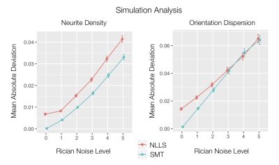
Neurite orientation dispersion and density imaging (NODDI) is a widely used tool for modeling microstructure using diffusion MRI, but its computational cost can be prohibitively expensive. This work investigates the efficacy of integrating the spherical mean technique (SMT) into a non-linear optimization framework to improve NODDI parameter estimation. Through quantitative simulation, comparative, and reliability analyses, we found that integrating SMT into more traditional non-linear optimization enables rapid, accurate, and reliable estimation of neurite density and dispersion compared to other approaches.
|
|
| Top |
Diffusion MRI: Fiber Orientations & Tracking
Digital Poster
Diffusion
Wednesday, 15 May 2019
| Exhibition Hall |
08:15 - 09:15 |
| |
|
Computer # |
|
3364.
 |
26 |
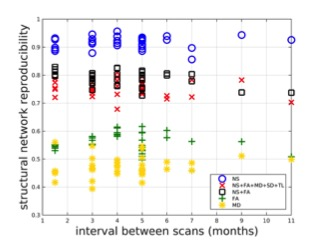
Assessment of the reproducibility of structural brain networks derived using different edge-weighting strategies
Eirini Messaritaki, Stavros Dimitriadis, Derek Jones
Structural brain networks derived from diffusion Magnetic Resonance Imaging data can use various tract metrics to weigh the network edges. In this work we use the Human Connectome Project test-retest diffusion MRI data to assess the reproducibility of structural brain networks, their edges and their graph-theoretical measures derived using different edge-weighting strategies.
|
|
3365.
 |
27 |
White matter parcellation test-retest reproducibility of diffusion MRI tractography fiber clustering
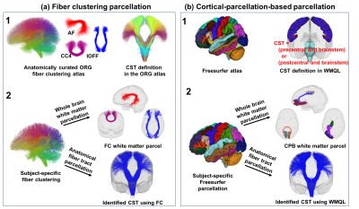
Fan Zhang, Ye Wu, Isaiah Norton, Yogesh Rathi, Alexandra Golby, Lauren O'Donnell
Fiber clustering is a popular strategy for automated white matter parcellation using diffusion MRI tractography. However, there has been no investigation to assess fiber clustering parcellation test-retest reproducibility, i.e. whether white matter parcellations could be reliably reproduced in repeated scans. This work presents the first study of fiber clustering white matter parcellation test-retest reproducibility. We perform evaluation on a large test-retest dataset, including a total of 255 subjects from multiple independently acquired datasets. Our results in general indicate that the fiber clustering method produced more reproducible white matter parcellations than a popular cortical-parcellation-based method.
|
|
3366.
 |
28 |
Connectome analysis of world class gymnasts using probabilistic multi-shell multi-tissue constrained spherical deconvolution tracking
Wataru Uchida, Koji Kamagata, Christina Andica, Hiroyuki Tomita, Hidefumi Waki, Mana Kuramochi, Yuki Takenaka, Akifumi Hagiwara, Makoto Fukuo, Kouhei Tsuruta, Issei Fukuaga, Syo Murata, Mutsumi Harada, Shigeki Aoki, Hisashi Naito
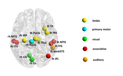
We analyzed the brain anatomical networks between world-class gymnasts and controls using probabilistic Multi-shell, Multi-tissue Constrained Spherical Deconvolution tracking method. Our results showed higher neural connectivities in gymnasts in the brain areas related to motor activity and visual perception. In addition, a positive correlation between difficulty-score (D-score) and brain connectivity was also in the brain areas including auditory, limbic, associative and visual area. In conclusion, our findings can be useful for a better understanding of neural changes related to gymnastic skills.
|
|
3367.
 |
29 |
A novel connectomics metric for investigating the structural-functional relationship in the brain
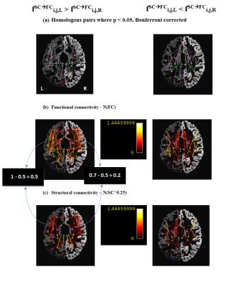
Oren Civier, Marion Sourty, Fernando Calamante
We present a novel connectomics metric that quantifies the relationship between structural connectivity (SC) and functional connectivity (FC) in each connection in the brain. The metric is based on a biologically meaningful and quantitative measure of SC, followed by normalization of both modalities to a common scale. We demonstrated the utility of the metric in examining structural-functional relationships in pairs of homologous connections, detecting homologues that are dissimilar. The metric is more informative than using FC in isolation, and might provide insights into factors that contribute to FC beyond the strength of SC (e.g., indirect connections, organization of fibres).
|
|
3368.
 |
30 |
Bundle Analytics: a computational and statistical analyses framework for tractometric studies
Bramsh Chandio, Jaroslaw Harezlak, Serge Koudoro , David Reagan , Eleftherios Garyfallidis
Bundle Analytics promises fast, robust, and flexible computational and statistical analyses for tractometric studies on clinical data. It uses information from both tractometry, and anatomy to analyze the extracted fiber bundles from challenging clinical datasets. It uses streamline-based efficient algorithms to register and extract fiber bundles from a tractogram, and applies linear mixed models in the extracted bundles to find significant differences at specific locations of the bundles across groups. Finally, the method does not require training, an important advantage over deep learning methods.
|
|
3369.
 |
31 |
Is it feasible to directly access the bundle’s specific myelin content, instead of averaging? A study with Microstructure Informed Tractography
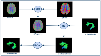
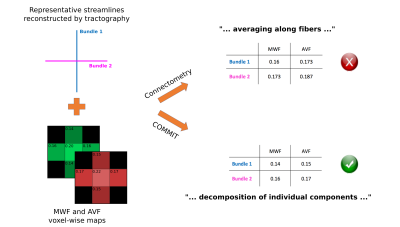
Simona Schiavi, Marco Pizzolato, Mario Ocampo-Pineda, Erick Canales-Rodriguez, Jean-Philippe Thiran, Alessandro Daducci
Diffusion MRI connectometry is a widely used tool to investigate features of structural connectomes that reflect differences in white matter tracks integrity. It consists in averaging microstructural tissues properties (obtained from any voxel-wise map) along streamlines recovered with diffusion tractography. Nevertheless, the average of a microstructural measure is a weak information about an entire bundle. Using microstructure-informed tractography (COMMIT), we were able to simultaneously estimate fiber’s specific myelin water fraction, intra-axonal volume fraction, and g-ratio. We also computed new connectomes with bundles’ specific measures instead of the commonly used averages.
|
|
3370.
 |
32 |
Using HCP data to improve diffusion tractography in routine-quality data: Application to the virtual dissection of the SLF system
Chiara Maffei, Anastasia Yendiki
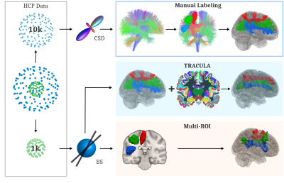
The development of scanners with ultra-high gradients, spearheaded by the Human Connectome Project, has led to dramatic improvements in the spatial, angular, and diffusion resolution that is feasible for in vivo diffusion MRI acquisitions. Here we show that global probabilistic tractography with anatomical priors can be trained on such data, which can only be acquired on a handful of Connectome scanners worldwide, and improve the accuracy of tractography in more widely available, routine-quality diffusion data. We apply this method to reconstruct the three subcomponents of the SLF and show its superior accuracy compared to a conventional multi-ROI approach.
|
|
3371.
 |
33 |
Reducing false-positive connections using hierarchical microstructure-informed tractography
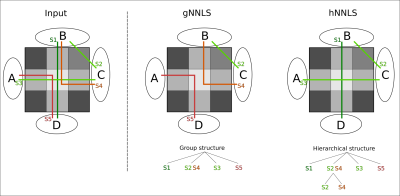
Mario Ocampo-Pineda, Simona Schiavi, Matteo Frigo, Muhamed Barakovic, Gabriel Girard, Maxime Descoteaux, Jean-Philippe Thiran, Alessandro Daducci
Tractography has proven particularly effective for studying non-invasively the neuronal architecture of the brain, but recent studies have showed that the high incidence of false-positives can significantly bias any connectivity analysis. Last year we presented a method that extended COMMIT framework to consider the prior knowledge that white matter fibers are organized in bundles. Inspired by this, here we propose another extension to further improve the quality of the tractography reconstructions. We introduce a novel regularization term based on the multilevel hierarchy organization of the human brain and we test the results on both synthetic phantom and in vivo data.
|
|
3372.
 |
34 |
TractEM: A fast protocol for Whole Brain Tractography
Roza Bayrak, Kurt Schilling, Jasmine Greer, Colin Hansen, Justin Blaber, Christa Greer, Susan Resnick, Owen Williams, Lori Beason-Held, Baxter Rogers, Bennett Landman
We introduce TractEM, a tractography-based whole-brain labeling protocol informed by the Eve Labeling [1] procedures from the single-subject Johns Hopkins white matter atlas [1, 2]. This project proposes to create a resource of manually labelled white matter atlases that is driven by state-of-the-art diffusion tractography, and can be manually created in less than 6 hours. We defined and tested the TractEM protocol on 61 tracts for 20 subjects, with multiple raters per subject, and show moderate to high reproducibility for most labels. TractEM should be a useful resource for generating target templates for automated labeling methods.
|
|
3373.
 |
35 |
Structural anatomy of the executive control network – a high angular resolution diffusion MRI study
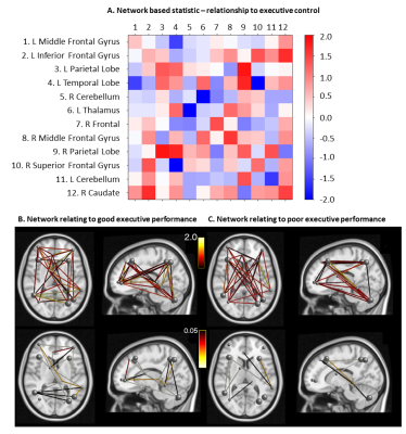
Kaikai Shen, Thomas Welton, Matt Lyon, Jurgen Fripp, Ralph Martins, Stuart Grieve
In this abstract, our aim is to investigate the relationship between executive function and the underlying structures of the executive control network (ECN) in the normal population. To this end, we acquired multi-shell diffusion MRI data with 391 gradient directions to estimate the structural connectivity within this functionally-defined network, and evaluated the executive function of the subjects. We used network-based statistic (NBS) to assess the relationships between executive function and the ECN connectivity, and found that the structural connectivity between hemispheres displayed positive correlation with higher executive function performance, while the connectivity within a sub-network in the right hemisphere showed a negative correlation with executive function.
|
|
3374.
 |
36 |
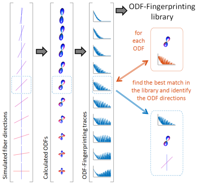
In vivo Diffusion MRI ODF-Fingerprinting performance relative to an HCP reference
Steven Baete, Martijn Cloos, Ying-Chia Lin, Dimitris Placantokanis, Timothy Shepherd, Fernando Boada
High quality diffusion acquisitions are routinely used to study white matter architecture and brain connectivity in vivo. A key step for successful tractography of neuronal tracts is correct identification of the tract directions in each voxel. The recently proposed ODF-Fingerprinting method has been demonstrated in computer simulations and qualitative in vivo results to improve detection of fiber pairs with small crossing angles whilst maintaining fiber direction precision. Here we evaluate the performance of ODF-Fingerprinting and several other fiber direction identification algorithms quantitatively in vivo in a downsampled DWI dataset where the high resolution dataset provides a reference standard.
|
|
3375.
 |
37 |
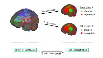
Towards a deep learning model for diffusion-aware tractogram filtering
Daniel Jörgens, Philippe Poulin, Rodrigo Moreno, Pierre-Marc Jodoin, Maxime Descoteaux
We propose a deep learning model that is able to separate a tractogram into sets of anatomically plausible and implausible streamlines. In contrast to existing methods, our model relies solely on the measured diffusion signal as an input ensuring independence of potential misalignments between subjects. The model is shown to generalize to different tractography methods and has the potential to simultaneously learn from multiple supervisor methods.
|
|
3376.
 |
38 |
Tractostorm: Evaluation of intra and inter rater reproducibility in tractography dissection
Francois Rheault, Laurent Petit, Maxime Descoteaux
Investigative studies of white matter structures using tractography often require manual virtual bundle dissection to be performed. Human errors and personal decisions make these manual segmentations hard to reproduce. Reproducibility assessment of raters is common practice in other neuroimaging field where segmentation protocols were refined to maximize reproducibility. However, this has not been done in the field of diffusion tractography. The contribution of this study is to provide the first large-scale, multi-center variability assessment of virtual dissection of tractography dataset.
|
|
3377.
 |
39 |
Brain tumors: a challenge for tracking algorithms
Guillaume Theaud, David Fortin, Felix Morency, Maxime Descoteaux
In tumor cases, several fiber bundles are displaced, destroyed, or infiltrate the tumor zone. For surgical planning, it is important to have the best estimation of the bundles near the tumor and in the edema. In neurosurgical tractography, DTI is the clinical standard and most used tractography method in publications. DTI does not correctly estimate local crossing fibers and is limited by edema contamination. In this work, we compare 4 tracking algorithms (DTI, HARDI deterministic, probabilistic, a new probabilistic edema-informed) applied to tumor cases, show differences and advise on the choice of tractography algorithm to be used in neurosurgical cases.
|
|
3378.
 |
40 |
Optimized DTI acquisition and tractography pipeline for a reliable reconstruction of the facial nerve in patients with vestibular schwannoma
Manuela Moretto, Valentina Baro, Sabrina Brigadoi, Marco Castellaro, Mariagiulia Anglani, Antonio Mazzoni, Elisabetta Zanoletti, Andrea Landi, Luca Denaro, Francesco Causin, Domenico D'Avella, Alessandra Bertoldo
Vestibular schwannomas (VS) are intracranial tumors that can cause the dislocation of the facial nerve (FN). The location of the FN is therefore a priori unknown to the surgeon and this is the main reason why patients with VS may experience FN damage during the surgery, leading to facial paralysis. In this work we used a multi-shell DTI acquisition to perform probabilistic fiber tracking for the preoperative determination of FN course in patients with VS. High-resolution anatomical scans were used to help the fiber tracking algorithm to obtain a reliable reconstruction also when the FN course had a complex configuration.
|
|
3379.
 |
41 |
Comparison of diffusion MRI data with different b-values in trigeminal nerve tracking with unscented Kalman filter tractography
Guoqiang Xie, Fan Zhang, Lorenz Epprecht, Isaiah Norton, Yogesh Rathi, Alexandra Golby, Lauren O'Donnell
Diffusion MRI enables improved identification of the trigeminal nerve by tracking of its 3-D trajectory. To explore optimal methods for trigeminal nerve tracking using diffusion MRI, we compared tracking results from acquisitions with different b-values using single- and multi-fiber tractography methods. We found that the trigeminal nerve can be best tracked using b=3000 data with a two-tensor fiber model. We suggest that these settings can potentially be suitable for clinical applications, e.g., diagnosis and evaluation of trigeminal neuralgia.
|
|
3380.
 |
42 |
Toward identifying individual branches of the trigeminal nerve with dMRI-based tractography at 7 Tesla: methodological considerations
Kellen Mulford, Christophe Lenglet, Pramod Pisharady, Sean Moen, Donald Nixdorf, Bharathi Jagadeesan, Andrew Grande, Pierre-Francois Van de Moortele
Trigeminal Neuralgia is a debilitating neuropathic condition affecting the trigeminal nerve. Tractography allows for the possibility of identifying the three branches of the nerve to assist in refining the etiopathology of trigeminal neuralgia. In this study we identify methodological factors that contribute to reliability in identifying the branches of the nerve through an analysis of public HCP data and methodological experiments at 7 Tesla. We conclude that the choice of phase encoding direction can dramatically impact the fidelity of cranial nerve tractography results, and that spatial resolution plays an important role in CN-V branch identification.
|
|
3381.
 |
43 |
Fibre Tracking of the Arcuate Fasciculus at High Spatial and Angular Resolution
Matthew Lyon, Thomas Welton, Jerome Maller, MyungHo In, Ek Tsoon Tan, Matt Bernstein, Erin Gray, Yunhong Shu, John III Huston, Stuart Grieve
We compared fibre tracking performance in the arcuate fasciculus across a range of angular resolutions, as well as a low distortion dataset using diffusion MRI data from a Compact 3T scanner with high-performance gradients. Tracking performance increased approximately linearly with greater angular resolution. Performance was also improved using a low-distortion diffusion sequence at a single relatively low angular resolution acquisition (33 directions).
|
|
3382.
 |
44 |
Altered structural connectivity in the auditory-related pathway in patients with idiopathic sudden sensorineural hearing loss by diffusion spectrum imaging
Zihao Zhang, Tao Jiang, Xiuqin Jia, Xiaojiao Guan, Qinglei Shi, Jing Yang, Yi Zhang
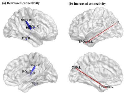
Sensorineural hearing loss is increasingly recognized as the result of alterations in the auditory-related network. The present study aimed to further explore the whole brain abnormalities of neural network connections in idiopathic sudden sensorineural hearing loss (ISSNHL) through diffusion spectrum imaging. It was found left-sided ISSNHL exhibited decreased connectivity between the contralateral inferior temporal gyrus and rolandic operculum, and the contralateral heschl gyrus and superior marginal gyrus; while increased connectivity was detected between the ipsilateral temporal pole and cuneus, which suggests that DSI could help investigate the structural correlates of these imaging abnormalities in this disease.
|
|
3383.
 |
45 |
A surface-based shape analysis of the human corticospinal tract
Gabrielle Grenier, Étienne St-Onge, Maxime Descoteaux
Traditionally, when examining bundles, comparative methods between measures such as volume, streamline density and mean fractional anisotropy are often used. However, these measures can be biased and do not inform about shape of the white matter bundle. In this work, a new method is proposed to compare bundles based on its surface. Indeed, three measures (area, elongation, irregularity) applied along the cross-sections of the surface enable to highlight shape differences. Here, we illustrate the potential of these surface-based shape measures on the right and the left pyramidal tract in a healthy group of 30 datasets.
|
|
3384.
 |
46 |
DIPY Horizon: fast, modular, unified and adaptive visualization
Eleftherios Garyfallidis, Marc-Alexandre Côté, Bramsh Chandio, Shreyas Fadnavis, Javier Guaje, Ranveer Aggarwal, Etienne St-Onge, Karandeep Juneja, Serge Koudoro, David Reagan
DIPY Horizon is fast, modular, unified and adaptive visualization system that resembles a high-end game engine, works on the web and across desktop operating systems. Horizon is suitable both for user, programmers and clinical applications.
|
|
3385.
 |
47 |
On the regression of intracranial volume in Fixel-Based Analysis
Robert Smith, Thijs Dhollander, Alan Connelly
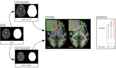
Fixel-Based Analysis (FBA) enables robust whole-brain statistical analysis of both microscopic and macroscopic white matter properties that is both sensitive and specific to crossing fibre geometry. Given the influence of macroscopic brain differences in such experiments, interest has been expressed in how best to account for variations in brain volume across participants. Here we demonstrate the effect of brain volume on FBA by synthetically modulating brain sizes within a healthy cohort and statistically testing FBA metrics with various regressions of estimated intracranial volume. We conclude with recommendations for regression of the influence of global brain size differences in FBA when desired.
|
|
3386.
 |
48 |
Automated Segmentation of the Cortical Boundaries in Native Diffusion Tensor Imaging Space to Measure Anisotropy of the Cerebral Cortex
Graham Little, Christian Beaulieu
Diffusion tensor imaging (DTI) can quantify anisotropic diffusion in the cerebral cortex reflecting its microstructural architecture. However, the analysis is usually performed by defining the inner and outer cortical boundaries on 3D T1-weighted images which are then applied to co-registered DTI, but this is prone to registration errors. Here we present an automatic cortical boundary segmentation method applied directly to 1.5 mm isotropic DTI acquired in 6 minutes at 3T. The cortical surfaces derived from DTI alone demonstrate the radial orientation of the primary eigenvector and appropriate FA/MD showing promise for DTI studies of the cortex in neurological disorders.
|
|
3387.
 |
49 |
Spatial Normalization of Fiber Orientation Distribution (FOD) Maps using Mrtrix and an Image-based Non-uniform Gradient Method
Zifei Liang, Jiangyang Zhang
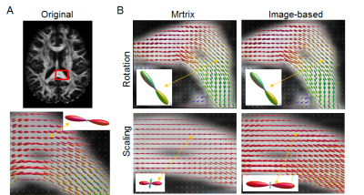
Diffusion MRI based fiber orientation distribution (FOD) estimates are widely used to examine structural connectivity in the brain. For group comparison using nonlinear spatial normalization, FOD needs to be adjusted based on the estimated degree of rotation and scaling at each voxel. We compared the current method implemented in Mrtrix for spatial normalization of FODs with an image-based method. The results suggest that the method in Mrtrix is accurate for rotation but generates potential bias in FOD peak amplitude and orientation when large anisotropic scaling is present. This knowledge is important for studies to use spatially normalized FOD maps.
|
|
3388.
 |
50 |
Characterization of the cerebro-cerebellar loop exploiting advanced tractography and dealing with thalamic synapsis
Fulvia Palesi, Nicolò Rolandi, Fernando Calamante, Egidio D'Angelo, Claudia Gandini Wheeler-Kingshott
Recently, advanced tractography has been used for assessing the feasibility of characterizing cerebro-cerebellar loop, composed of cerebello-thalamo-cortical and cortico-ponto-cerebellar pathways, acknowledging the issue of how tractography deals with polysynaptic connectivity, i.e. at thalamic level. In this work, polysynaptic cerebello-thalamo-cortical and cortico-ponto-cerebellar pathways were reconstructed using a multiplicative hypothesis for thalamic connectivity. Our findings revealed the importance of using such a multiplication factor for streamlines reaching the thalamic synapses to properly reconstruct cerebro-cerebellar connection. Furthermore, findings using polysynaptic tracts support the cerebellar role in cognition showing that cognitive/associative areas are the mainly involved in both the cerebello-thalamo-cortical and cortico-ponto-cerebellar pathways.
|
|
| Top |
Diffusion MRI: Artefact Correction
Digital Poster
Diffusion
Wednesday, 15 May 2019
| Exhibition Hall |
08:15 - 09:15 |
| |
|
Computer # |
|
3389.
 |
51 |
Distortion dominates fibre tracking of the optic chiasm – an evaluation of ultra-high angular resolution compared to low-distortion diffusion MRI on a Compact 3T
Thomas Welton, Matthew Lyon, Jerome Maller, Myung-Ho In, Ek-Tsoon Tan, Matt Bernstein, Erin Gray, Yunhong Shu, John Huston, Stuart Grieve
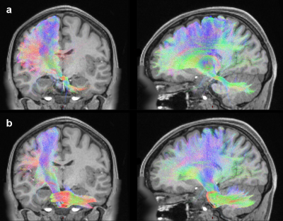
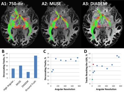
We evaluated the impact of angular resolution and spatial distortion on crossing-fibre tracking accuracy at the optic chiasm using diffusion MRI data from a Compact 3T scanner with high-performance gradients. Contralateral tracking via the chiasm was quantified in acquisitions optimised for q-space resolution or low distortion and compared to the known true rate of decussation. We found that, for chiasmal tracking, minimising the effects of geometric distortion may provide better value than maximising spatial or angular resolution beyond 140 directions. An ideal future diffusion MRI protocol will combine these features for more optimal tracking performance.
|
|
3390.
 |
52 |
Compensation of signal loss induced by scanner table vibrations in high b-value DW-TSE for measuring lipids ADC
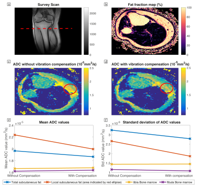
Dominik Weidlich, Stefan Ruschke, MingMing Wu, Andreas Hock, Rainer Burgkart, Dimitrios Karampinos
Fat plays a central role in the incidence of metabolic syndrome but current MRI biomarkers cannot answer questions about fat cell microstructure. Diffusion-weighted measurements are capable of revealing information about fat tissue microstructure but the required strong diffusion weighting induces scanner table vibrations that eventually lead to measurement errors and artifacts. The purpose of this work was to mitigate vibration artifacts by placing a vibration compensation gradient before the diffusion preparation. The approach was tested in a water-fat phantom and in-vivo in the lower leg of a healthy volunteer.
|
|
3391.
 |
53 |
Diffusion Gradient Nonlinearity Correction in a Diffusion Phantom and in Breast Cancer Bone Metastases
Thomas Buus, Anders Jensen, Erik Pedersen
The purpose was to investigate if diffusion gradient nonlinearity (DGNL) ADC-errors could be corrected in vitro and in vivo.
Methods
DWIBS was performed in a diffusion phantom and in breast cancer patients. ADC-maps with and without DGNL correction were created and compared at different positions relative to the isocenter.
Results
In the diffusion phantom uncorrected ADC-values 17.5cm from the isocenter dropped by 29-32% while the corrected ADC-values increased by 2-4%.
In 85 bone metastases uncorrected ADC-values 14cm from the isocenter dropped by 18.7% while corrected ADC-values dropped by 2.8%.
Conclusion
DGNL ADC-errors can be corrected in vitro and in vivo.
|
|
3392.
 |
54 |
Noise2Noise MRI for High-resolution Diffusion-weighted Imaging of the Brain: Deep Learning-based denoising without need for Highly Averaged Ground-truth Images
Motohide Kawamura, Daiki Tamada, Satoshi Funayama, Hiroshi Onishi, Utaroh Motosugi
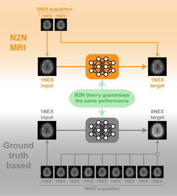
Deep learning (DL)-based denoising is promising to achieve high resolution diffusion-weighted imaging (HR-DWI) by improving SNR without signal averaging. Training supervised DL-based algorithm, however, requires thousands of teaching data, which need long acquisition time. In this study, we propose to use noise2noise (N2N) theory to develop DL-based denoising algorithm, which does not need teaching data with high SNR. In the results, the proposed method (N2N-MRI-based algorithm) outperformed conventional ground-truth-based algorithm in terms of maximum peak SNRs on validation sets during training. The image quality of HR-DWI denoised by N2N-MRI-based algorithm was equivalent to that denoised by conventional algorithm.
|
|
3393.
 |
55 |
Iteration-based motion compensation method for multi-shot diffusion imaging
Zhongbiao Xu, Rongli Zhang, Yingjie Mei, Zhifeng Chen, Yaohui Wang, Ed X. Wu, Feng Huang, Yanqiu Feng
Multi-shot EPI technique is vulnerable to patient motion. Though CIRIS proposed by our group tackles the infrequent macroscopic motion in multi-shot EPI by clustering and registration, it cannot deal with the frequent motion (e.g. shot-wise motion). In this work, an iterative motion compensation frame was introduced to correct for the frequent motion during the multi-shot acquisition. The simulation experiments demonstrated that the proposed method can obtain improved image quality in the presence of infrequent motion, and even correct for the shot-wise motion, compared to CIRIS.
|
|
3394.
 |
56 |
Impact of processing options on histogram metrics extraction from DWI in cerebral small vessel disease
Ana Fouto, Rita G. Nunes, Joana Pinto, Luísa Alves, Sofia Calado, Carina Gonçalves, Margarida Rebolo, Miguel Viana Baptista, Pedro Vilela, Patrícia Figueiredo
Biomarkers based on diffusion-weighted imaging (DWI) have been proposed as potential disease biomarkers in several brain conditions including cerebral small vessel disease (SVD). Often histogram-based metrics are extracted, but findings across studies are somehow inconsistent. Here, we investigated the impact of several processing options for extracting histogram metrics of fractional anisotropy (FA) and mean diffusivity (MD) from DWI. We considered two white matter regions-of-interest with different interpolation and thresholding options, as well as different numbers of bins. We found that processing options significantly impacted histogram metrics, which in some cases significantly affects the ability to discriminate between patient and controls.
|
|
3395.
 |
57 |
Corrupted Data Rejection Strategy in k-space Based Multi-shot Diffusion Reconstruction
Zhe Zhang, Wanlin Zhu, Jing Jing, Hua Guo, Jiazheng Wang, Chun Yuan, Yongjun Wang
In diffusion imaging, bulk and physiological motion together with strong diffusion encoding gradient introduces extra image phase or data corruption in the diffusion-weighted images. Corrupted data identification/rejection procedure has not been integrated in the recently proposed k-space based multi-shot diffusion reconstruction pipelines. In this work, two corrupted data rejection strategies were proposed, compared and evaluated. Results show that using corrupted data identification and rejection after the CK-GRAPPA reconstruction is potentially a robust choice for multi-shot diffusion imaging reconstruction.
|
|
3396.
 |
58 |
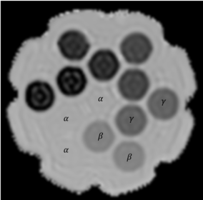
Correction of systematic errors in DTI imaging caused by gradient nonlinearity using gradient field maps measured by diffusion imaging of an isotropic diffusion phantom.
Alan Barnett, Mustafa Irfanoglu, Baxter Rogers, Bennett Landman, Carlo Pierpaoli
Gradient nonlinearity causes systematic errors in the measurement of DTI parameters. These errors can be greatly reduced if the actual fields generated by the gradient coils is known. Although the gradient field maps are known to the manufacturers, many users do not have access to them. We describe a method for measuring the gradient field maps using a set of diffusion weighted images of an isotropic diffusion PVP phantom. We use the field maps to analyze a DTI study of the phantom and compare the results to analysis performed without the field maps. The results show that the method works well.
|
|
3397.
 |
59 |
Motion Compensation for Free-Breathing Diffusion-Weighted Imaging (MoCo DWI)
Christian Dávid, Thomas Vahle, Robert Grimm, Peter Bachert, Marc Kachelrieß
Diffusion-weighted imaging (DWI) of the abdomen has acquisition times of several minutes. For this reason respiratory motion can cause misalignment between acquired slices at the same position but different b-values. To overcome this, we estimate the patients’ respiratory motion using a T1-weighted, stack-of-stars GRE pulse sequence and an advanced 4D reconstruction. This motion estimation is used to compensate for respiratory motion in a common, free-breathing DWI acquisition. In three volunteers an improved alignment of structures in the liver are shown. This allows for a better comparison and potential benefits for further processing (e.g. for ADC-maps).
|
|
3398.
 |
60 |
Effects of Gradient Nonlinearities on Reproducibility and Accuracy of Diffusion MRI Metrics in the Brain.
M. Okan Irfanoglu, Alan Barnett, Neda Sadeghi, Carlo Pierpaoli
Gradient nonlinearities in MRI cause spatially-varying b-values and diffusion gradient directions. In this work, we analyze whether these nonlinearities have a significant impact on data reproducibility and accuracy for brain studies. Our results indicate that not only FA and TR values have an increasing bias away from the isocenter of the magnet, but also differences in subject positioning and head orientation combined with nonlinearities have a significant effect on reproducibility. The effects were also observed in principal eigenvector directions computed with the tensor model.
|
|
3399.
 |
61 |
Progress toward on-line implementation of vendor-provided prospective correction for non-uniform diffusion weighting due to gradient nonlinearity
Dariya Malyarenko, Ajit Devaraj, Ek Tan, Johan Tondeur, Johannes Peeters, Yuxi Pang, Lisa Wilmes, Michael Jacobs, David Newitt, Thomas Chenevert
Feasibility of the prospective correction for non-uniform diffusion weighting due to gradient nonlinearity using scanner-specific gradient design information was previously demonstrated by our academic-industrial partnership (AIP). Here we report on the progress toward implementation of the prospective correction by leading MRI vendor participants of the AIP on their respective scanner platforms. The vendor-provided on-line correction is benchmarked by comparison to previously validated retrospective off-line processing for uniform gel and flood phantoms, and a human volunteer. Vendor efforts enable comprehensive bias correction for standardization of quantitative DWI applications in multi-center clinical trial environments.
|
|
3400.
 |
62 |
Gradient nonlinearity-induced bias calibration and correction in diffusion imaging using DIADEM and a simple, uniform gel phantom
Myung-Ho In, Uten Yarach, Daehun Kang, Ek Tsoon Tan, Erin Gray, Nolan Meyer, Joshua Trzasko, Yunhong Shu, John Huston, Matt Bernstein
This study reports a novel gradient nonlinearity (GNL) calibration approach using DIADEM (Distortion-free Imaging Approach with a Double Encoding Method) diffusion imaging. Unlike standard diffusion-weighted echo-planar-imaging (DW-EPI), DIADEM is free from DW-EPI distortions. This allows GNL calibration with a uniform phantom, since confounding effects between DW-EPI and GNL-induced distortions in the calibration are separated. Direct bias correction could be applied to the corresponding in-vivo data from the DIADEM scans, which results in reliable quantitative diffusion imaging. The feasibility was successfully demonstrated in phantom and in-vivo on a compact 3T system.
|
|
3401.
 |
63 |
Evaluation of an adapted DWI MRI for improved image quality and tissue differentiation in abdominal MRI – a prospective study in oncologic follow-up examinations
Constantin Dreher, Tristan Kuder, Stefan Windhaber, Franziska König, Daniel Paech, Anoshirwan Tavakoli, Frederik Laun, Florian Flothow, Regula Gnirs, Thomas Benkert, Heinz-Peter Schlemmer, Sebastian Bickelhaupt
Diffusion-Weighted imaging is a mainstay for oncologic examinations. However, abdominal examinations with standard DWI can be challenged by signal exploration and motion artifacts. This prospective study therefore compares a standard EPI-DWI with an oncologically optimized-DWI, including complex averaging, motion correction between averages, rescaling of motion corrupted averages, and background suppression, in oncological follow-up MRI investigations. A diffusion resolution phantom demonstrated an improved image quality by the optimized-DWI. Image quality analysis of prospectively acquired abdominal DWI MRI examinations in 52 patients showed a significant improvement in image quality and apparent signal-to-background-noise-ratio by optimized-DWI as compared to standard EPI-DWI in oncologic imaging.
|
|
3402.
 |
64 |
Learned Gibbs Removal in Partial Fourier Acquisitions for Diffusion MRI
Matthew Muckley, Antonios Papaioannou, Benjamin Ades-Aron, Daniel Sodickson, Yvonne Lui, Els Fieremans, Dmitry Novikov, Florian Knoll
Despite significant advances in both denoising and Gibbs artifact removal, in acquisitions such as partial Fourier encoding, noise and Gibbs ringing continue to be an issue. Here we demonstrate that a machine learning approach can extend Gibbs ringing and noise removal to partial Fourier image acquisitions and show results on estimates of diffusion parameters on phantom and brain imaging data.
|
|
3403.
 |
65 |
An independent phase-based measure for slice outlier rejection in diffusion MRI
Daan Christiaens, Lucilio Cordero-Grande, Joseph Hajnal, J-Donald Tournier
Detecting and downweighting damaged slices is vital in analysing motion-corrupted dMRI data. Conventional magnitude-based outlier rejection methods rely on intensity model predictions, with the state of the art using slice-to-volume reconstruction. However, in cases with very high outlier prevalence such model prediction is no longer reliable. Here, we introduce a model-independent phase-based measure for detecting motion-induced slice dropouts. We demonstrate its use in neonatal data, and show that it outperforms model-based magnitude techniques in highly damaged data.
|
|
3404.
 |
66 |
Noise Estimation and Bias Correction of Diffusion Signal Decays: Application to Prostate Diffusion Imaging
Mohammad Alipoor, Stephan Maier
A novel approach to estimate noise and Rician signal bias in diffusion MRI magnitude data is proposed. Rather than relying on repeat measurements for estimation of noise and expected signal, the methods uses multi-b measurements and non-monoexponential signal fits. In addition to noise and bias estimation this approach also provides signal averaging over all b-factors and permits determination of non-monoexponential tissue water diffusion signal decay. Testing was performed with Monte-Carlo simulations and on diffusion-weighted high-b prostate image data obtained with an external coil array.
|
|
3405.
 |
67 |
Correction of motion artifacts in head and neck diffusion-weighted MRI
Nienke Sijtsema, Dirk Poot, Gerda Verduijn, Mischa Hoogeman, Aad van der Lugt, Steven Petit, Juan Hernandez-Tamames
Diffusion-weighted imaging (DWI) is a promising technique for early stratification of responders and non-responders in head and neck chemoradiotherapy. However, data corruption due to swallowing and misalignment reduce the precision of estimated DWI parameters. We investigated different post-processing approaches to improve the reproducibility of the apparent diffusion coefficient (ADC) and demonstrated an improvement in reproducibility from 13.2% to 6.7%. Inter-volume registration showed the largest improvement compared to intra-volume registration and swallowing artifact rejection. The 6.7% reproducibility is sufficient for stratification of responders and non-responders in H&N chemoradiotherapy.
|
|
3406
 |
68 |
Deep Learning based displacement transform estimation for EPI distortion correction
Video Permission Withheld
Dattesh Shanbhag, Chitresh Bhushan
In this work, we propose a DL methodology to estimate the displacement field transform (DL_DispMap) from distorted DWI-EPI images. Compared to direct estimation of distortion-free images, we establish that DL_DispMap provides better training accuracy and mitigates the smoothening effect noticed in DL_Direct. The DL_DispMap corrected images are well matched (SSIM = 0.97) to gold-standard INVERSION method based distortion-free images in performance but might be needed to be tuned for an anatomy and system configuration. The method can potentially be also applied for fMRI studies
|
|
3407
 |
69 |
Validation of an image-based measurement of gradient non-linearity method and voxel-wise correction of b-values in a HARDI dataset
Video Permission Withheld
Yoojin Lee, Adam Kettinger, Bertram Wilm, Ralf Deichmann, Nikolaus Weiskopf, Christian Lambert, Klaas Pruessmann, Zoltan Nagy
Diffusion MRI requires numerous corrective steps in the data processing pipeline. One such step regards the need for voxel-wise correction of erroneous b-values that ensue from the inherent non-linearity of the magnetic field gradients. To estimate the non-linearity we used a simple image-based method that involves a spherical water phantom to measure voxel-wise the apparent diffusion constant. Deviations from the expected value allow estimation of the local b-value. The b-value maps were validated against the spherical harmonic predictions and subsequently used to correct phantom and in-vivo data.
|
|
3408.
 |
70 |
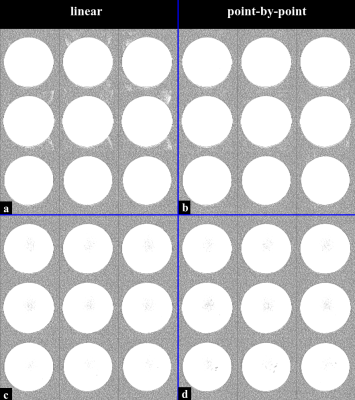
Minimization of Nyquist ghost artifacts for diffusion-weighted single-refocused spin-echo EPI
Manoj Shrestha, Ulrike Nöth, Ralf Deichmann
In diffusion-weighted (DW) imaging with EPI readout, Nyquist ghost (NG) artifacts might be aggravated due to higher order eddy currents, especially when using monopolar DW gradients in single-refocused spin-echo EPI (srSE-EPI). Both linear and point-by-point phase corrections were tested on DW-srSE-EPI and, for comparison, also on DW twice-refocused spin-echo EPI (trSE-EPI) with intrinsic eddy-current compensation. Both phase correction methods performed equally well for DW-trSE-EPI. However, for DW-srSE-EPI with high b-values, the linear phase correction failed to fully correct NG artifacts. In contrast, point-by-point phase correction yielded considerably better results. This was confirmed in vitro and in vivo.
|
|
3409.
 |
71 |
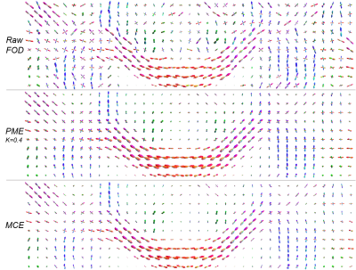
HARDI denoising with mean-curvature enhancement PDE on SE(3)
Etienne St-Onge, Stephan Meesters, Erick J Bekkers, Maxime Descoteaux, Remco Duits
We present a new High Angular Resolution Diffusion-weighted Imaging (HARDI) mean-curvature enhancement (MCE) on the homogeneous space of positions and orientations, embedded in the rigid body motion group SE(3). Its potential for crossing-preserving enhancement of fiber orientation distribution (FOD) fields is demonstrated. Compared to previous partial differential equation (PDE) enhancements on SE(3), denoising FOD fields with MCE better preserve boundaries, resulting in a lower overall angular error.
|
|
3410.
 |
72 |
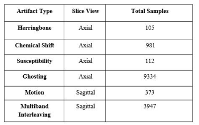
QC-Aautomator: A deep learning based automated artifact detection in dMRI data
Zahra Riahi Samani, Jacob Alappatt, Parker Drew, Ragini Verma
We have developed a deep learning based automated Quality Control (QC) tool, QC-Automator, for diffusion weighted MRI data, that will detect different artifacts. This will ensure that appropriate steps can be taken at the pre-processing stage to improve data quality and ensure that these artifacts do not affect the results of subsequent image analysis. Our tool based on convolutional neural nets has 94 – 98% accuracy in detecting the various artifacts including motion, multiband interleaving artifact, ghosting, susceptibility, herringbone and chemical shift. It is robust and fast and paves the way for efficient and effective artifact detection in large datasets.
|
|
3411.
 |
73 |
D-stripe: correction for stripe artefacts in diffusion MRI using a combined deep neural network and SVR approach
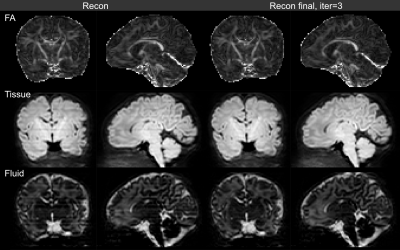
Maximilian Pietsch, Daan Christiaens, J-Donald? Tournier, Joseph? Hajnal
We present a data-driven method for the correction of stripe artefacts in multi-shell diffusion data that might arise for example from spin-history or stimulated echo effects. It relies on a filter, based on a deep neural network trained with simulated data to detect and remove stripe artefacts from single volumes. This is used to destripe signal predictions obtained from a slice to volume reconstruction, which are then projected onto the input data to determine the appropriate modulation field. The corrected input data are then reconstructed again with reduced stripe artefacts. This approach is applied to super-resolution reconstructions of neonatal multi-shell high angular resolution data.
|
|
3412.
 |
74 |
Distortion Correction of Multi-Shot Diffusion-Weighted Echo-Planar Imaging using Reversed Gradient Acquisition and Joint Reconstruction
Xiaoxi Liu, Di Cui, Edward S. Hui, Queenie Chan, Hing-Chiu Chang
Multi-shot diffusion-weighted echo-planar imaging (DW-EPI) with multiplexed sensitivity encoding (MUSE) is a self-navigated technique that can achieve high resolution diffusion-tensor imaging (DTI) without the need of navigator echo. However, even with multi-shot acquisition, the effective echo spacing is still relatively long for acquisition of high resolution DTI, leading to significant geometric distortion. In this study, we aim to reduce the geometric distortion of multi-shot DW-EPI by 1) integrating the reversed gradient acquisition in multi-shot DW-EPI, and 2) developing a joint reconstruction method that can reconstruct non-uniform k-space data by taking the off-resonance effect into account.
|
|
3413.
 |
75 |
Dual-echo blip reversed EPI acquisition enables distortion correction in the presence of motion in diffusion-weighted MRI
Onur Afacan, W. Scott Hoge, Tess Wallace, Ali Gholipour, Sila Kurugol, Simon Warfield
Slice-to-volume registration methods have been shown to provide motion robust reconstruction for large and frequent motions. One challenge with motion correction is the changing magnetic field inhomogeneities with different head positions. In this work we implemented a dual-echo blip reversed EPI acquisition and show that this sequence can be used to reduce distortions in large and frequent motions and can improve slice-to-volume registration results.
|
|
| Top |
Diffusion: Neuro Applications
Digital Poster
Diffusion
Wednesday, 15 May 2019
| Exhibition Hall |
08:15 - 09:15 |
| |
|
Computer # |
|
3414.
 |
76 |
Mapping anatomical connectivity: a Structural Network Analysis in Early and Profoundly Deaf people.
Francesca Saviola, Lisa Novello, Chiara Maffei, Stefania Benetti, Ceren Battal, Stefania Mattioni, Olivier Collignon, Jorge Jovicich
In case of early acquired deafness, auditory deprived temporal regions massively enhance their response to stimuli from remaining senses. This so called cross-modal plasticity also alters functional connectivity between reorganized temporal regions and those from preserved senses. The extent and distribution of white matter structural alterations supporting these functional effects are still poorly understood. In this diffusion MRI study, we investigate white matter reorganization of early deaf relative to hearing controls. Further, since early deaf typically become fluent at sign language, which may itself also induce brain structural reorganization unrelated to deafness, we also include a group of hearing signers.
|
|
3415.
 |
77 |
Structural connectivity between cerebellum and cerebral cortex in idiopathic generalized epilepsy: a diffusion tensor imaging study
Presentation Not Submitted
Sisi Jiang, Xiangkui Li, Yan Chen, Dezhong Yao, Cheng Luo
Probabilistic tracking method was applied to study the cerebellar efferent and afferent fibers in the patients with idiopathic generalized epilepsy (IGE), trying to address the structural connectivity (SC) alteration in the cerebello-cerebral circuit compared with healthy controls. Present findings suggested cerebellar effects on ganglia-thalamo-cortical circuit, which might be responsible for motion and cognition impairments in IGE. Furthermore, unbalanced SC alterations in the efferent and afferent fibers between frontal cortex and cerebellum might help to understand specific physiopathologic mechanism in IGE.
|
|
3416.
 |
78 |
Difference in Tensor Metrics between the Survived and Infarcted Penumbra by Reperfusion in a Rat Model of Cerebral Ischemia
Yu-Chieh Jill Kao, Jun Tazoe, Chia-Feng Lu, Bao-Yu Hsieh, Cheng-Yu Chen
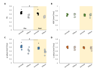
We demonstrated significant difference in FA and q-value between destined-survived and destined-infarcted penumbral tissue by reperfusion in transient MCA occlusion animals, which may imply the possible application of DTI-MRI in the evaluation of salvageable tissue before mechanical thrombectomy for acute stroke.
|
|
3417.
 |
79 |
Graph-based Structural Connectivity is correlated with Children Intelligence Quotient Scores
Ilaria Suprano, Gabriel Kocevar, Claudio Stamile, Salem Hannoun, Pierre Fourneret, Olivier Revol, Fanny Nusbaum, Dominique Sappey-Marinier
The neural substrate of high intelligence performances remains not well understood. We propose to investigate the structural brain connectivity measurements and their relationship with the intelligence performances, as measured by the WISC-IV scores of 57 children. We found strong correlations between children brain network density and intelligence scores. Moreover, several correlations were found between integration and redundancy graph metrics suggesting that intelligence performances are probably related to homogeneous network organization.
|
|
3418.
 |
80 |
High-Resolution FLAIR DTI Tractography of the Fornix in Multiple Sclerosis
Diana Valdés Cabrera, Christian Beaulieu
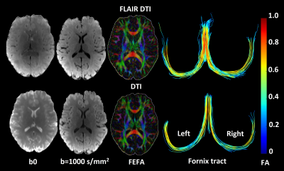
Diffusion tensor imaging (DTI) tractography of the human fornix is biased by partial volume effects from cerebrospinal fluid (CSF) due to its small bundle size and intra-ventricular location, even when using high spatial resolution. These errors in diffusion parameter estimation and tracking of the fornix will worsen with axonal loss in multiple sclerosis (MS). Here we demonstrate the superiority of FLAIR-DTI, even when compared to high-resolution 1.5 mm isotropic DTI, to mitigate CSF contamination of fornix tractography in MS and healthy volunteers. FLAIR-DTI yields more accurate diffusion metrics in both cohorts, and still shows abnormal fornix in MS.
|
|
3419.
 |
81 |
Brain White Matter Abnormality Induced by Chronic Spinal Cord Injury in the Pediatric Population: A Tract Based Spatial Statistic Study
Joshua Fisher, Mahdi Alizadeh, Devon Middleton, Caio Matias, MJ Mulcahey, Feroze Mohamed, Laura Krisa
Few studies have quantified the change in cortical white matter tracts following chronic spinal cord injury in a pediatric population. Additionally, no work has been done to compare chronic SCI subjects with different American Spinal Injury Association Impairment (AIS) scale classifications. We hypothesized that these cortical changes can be detected using tract-based spatial statistics (TBSS). Our efforts revealed that significant changes in fractional anisotropy occur in several motor and sensory related regions. We conclude that TBSS can be effectively used to identify alterations in brain microstructure in a chronic pediatric spinal cord injury population.
|
|
3420.
 |
82 |
Measuring white matter damage in different types of MS
Presentation Not Submitted
Chunyu Song, Peng Sun, Anne Cross, Zezhong Ye, Sheng-Kwei (Victor) Song
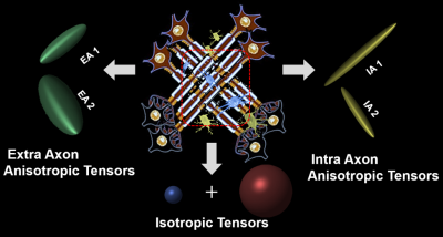
A new diffusion histology imaging (DHI) is proposed to model intra and extra axonal diffusion along with isotropic diffusion within an image voxel of diffusion-weighted MR images. It resolves crossing fibers while more accurately detecting and quantifying axonal injury, axon loss, demyelination, edema and inflammation. Through the multiple-tensor modelling of diffusion-weighted MRI signals, DHI has shown the potential to detect underlying pathologies of normal appearing corpus callosum in all clinical subtypes of multiple sclerosis.
|
|
3421.
 |
83 |
Evaluating Neurite Density and Orientation in the White Matter of Youth Born with Congenital Heart Disease
Kaitlyn Easson, Jean-Christophe Houde, Guillaume Gilbert, Kimberly Fontes, Charles Rohlicek, Christine Saint-Martin, Annette Majnemer, Maxime Descoteaux, Marie Brossard-Racine
In this study, neurite orientation dispersion and density imaging (NODDI) was used to quantify neurite density and orientation in white matter tracts in youth born with congenital heart disease (CHD). Neurite density index was significantly lower in youth born with CHD as compared to control youth in numerous, widespread association tracts. There were no regional differences in orientation dispersion index that survived correction for multiple comparisons. Our findings suggest a predominant role for lower neurite density, rather than lower neurite coherence and organization, in the white matter abnormalities observed in youth born with CHD.
|
|
3422.
 |
84 |
Structural brain connectivity network alterations following mild traumatic brain injury
Timo Roine, Mehrbod Mohammadian, Timo Kurki, Jussi Hirvonen, Olli Tenovuo
We used graph theoretical analysis to investigate structural brain connectivity networks in mild traumatic brain injury (mTBI). Global and local measures of structural connectivity were investigated in acute/sub-acute and chronic phases after TBI. There were no statistically significant differences in the global network measures between patients and controls at either of the stages after TBI. Node-level differences were found between patients and controls in local efficiency, strength, and betweenness centrality in several brain regions. However, only betweenness centrality in the right pars opercularis endured the Bonferroni correction for multiple comparisons.
|
|
3423.
 |
85 |
White matter Fractional Anisotropy measures in Autism Spectrum Disorder. Implications of differences in structural correlation with performance IQ measures and age. Results from EU- Autism Interventions data.
Robert Dallyn, Pedro Luque Laguna, Cate Davidson, EU-AIMS2 TRIALS Consortium, Declan Murphy, Flavio Dell'Acqua
We present voxel-wise statistics on Fractional Anisotropy from EU-AIMS diffusion imaging data on Autism Spectrum Disorder. We validate previous findings of structural white matter abnormalities in a younger cohort. Correlation analysis of white matter development with behavioural tasks points to altered functioning in ASD individuals in visuospatial reasoning tasks, consistent with the central coherence theory of autism. Cohort differences between ASD and control of white matter integrity correlation with age hint toward altered developmental trajectory in Autism Spectrum individuals.
|
|
3424.
 |
86 |
White Matter Microstructural Alternations in Neuropsychiatric Systemic Lupus Erythematosus With Normal Appearing Brain Using Diffusion Tensor Imaging
Jyh-Wen Chai, Ni-Jung Chang, Tsung-Yung Li, Yi-Ying Wu, Li-Ying Fan , Clayton Chi-Chang Chen
Systemic lupus erythematosus (SLE) patient has neuropsychiatric signs and symptoms, called neuropsychiatric SLE (NPSLE), usually with increased mortality and morbidity rates. There was little known about pathogenic mechanisms leading to neuropsychiatric symptoms in SLE. The aims of this study attempt to investigate diffusion tensor imaging (DTI) in detection of white matter micro-structural alternations for NPSLE patients, who had normal appearing brain in conventional MRI. By using the TRACULA analysis, we found significant differences of mean diffusivity (MD) and fraction anisotropy (FA) in several important nerve tracts between NPSLE patients and normal subjects, which would be helpful in understanding the mechanisms of NPSLE.
|
|
3425.
 |
87 |
Reproducibility of the diffusion of the perivascular space in older adults with dementia.
Christopher Steward, Vijay Venkatraman, Elaine Lui, Charles Malpas, Kathyrn Ellis, Terence O'Brien, Nicola Lautenschlager, Patricia Desmond
Recently, there has been interest in the glymphatic system and its role in flushing amyloid ß along with other waste products in the brain. New imaging techniques are being developed to try and measure such activity. Recently, diffusion tensor MRI was used to construct an index "DTI-ALPS" to help link dementia burden with diffusion in the perivascular space. We aimed to replicate this study in 36 patients (16 AD, 16 MCI, and 4 SMC). Significant correlations were found between DTI-ALPS and stratified Mini-Mental State Examination score. Further work is needed to evaluate the feasibility of MRI to measure glymphatic activity.
|
|
3426.
 |
88 |
Multivariate characterization of brain white matter maturation related to intellectual ability in children
Yannan Cheng, Chao Jin, Xianjun Li, Congcong Liu, Miaomiao Wang, Xiaocheng Wei, Yuli Zhang, Fan Wu, Mengxuan Li, Jian Yang
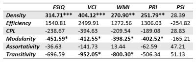
Brain maturations are thought to relate to behavioral acquisitions and cognitive development. Nevertheless, in vivo investigations of such relationships remain scarce in childhood. To bridge this gap, a multivariate index (DM), which delineates the “maturational distance” between children and adults and leverages DTI-metrics complementarity, was utilized to characterize WM variation. We found that DM showed significantly negative correlations with FSIQ in children aged 4-12 yr, especially in cingulum and superior longitudinal fasciculus. Besides, left hemispheric lateralization (higher correlations with FSIQ) was also observed. Our findings suggest DM as a useful biomarker in detailing the brain WM maturation related to intelligence.
|
|
3427.
 |
89 |
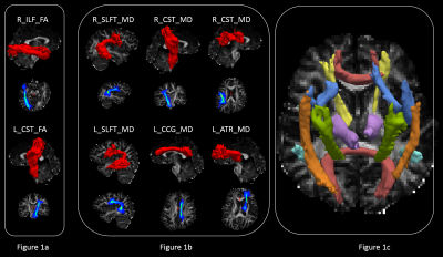
Alterations of brain white matter tracts in children with abnormal intelligence quotient
Yannan Cheng, Chao Jin, Xianjun Li, Congcong Liu, Miaomiao Wang, Xiaocheng Wei, Yuli Zhang, Fan Wu, Jian Yang
Studies have shown that brain white matter microstructure is of crucial importance for intelligence. Here, we detailed the alterations of brain white matter tracts in children with abnormal intelligence quotient by comparing the difference of brain white matter tract between the two groups and exploring correlations with intelligence scores based on DTI parameters. Significant differences of ILF, IFO and SLF were found in children with abnormal intelligence quotient. And all these tracts were strongly correlated with intelligence quotient, suggesting the underlying structural origins of lower cognitive function.
|
|
3428.
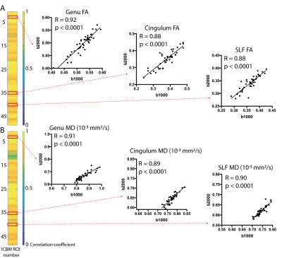 |
90 |
High Spatial Resolution Multi-shell DTI in Children and Young Adults Prenatally Exposed to Alcohol
Pascal Tétreault, Sarah Treit, Graham Little, Emily Stolz, Christian Beaulieu
Diffusion tensor imaging (DTI) has identified white matter differences in individuals with prenatal alcohol exposure (PAE). The current study uses higher spatial resolution of 1.5 mm isotropic (3.4 mm3 voxels) and larger b-values of 2000 s/mm2 (in addition to typical 1000 s/mm2) to assess the brain in children to young adults with PAE. Using the ICBM DTI template regions-of-interest for white matter, the b2000 tensor metrics (but not b1000) showed group differences in the left medial lemniscus and left stria terminalis, the latter also showing correlations with age in the control group but not the PAE group.
|
|
3429.
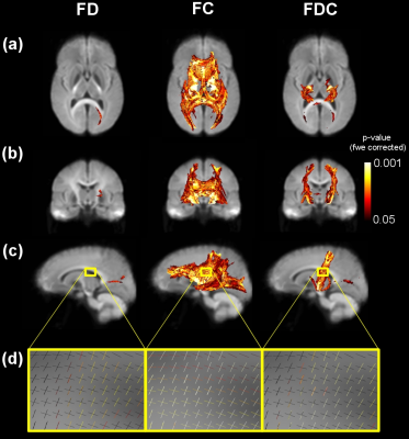 |
91 |
A fixel-based analysis of diffusion properties of white matter fiber in Williams Syndrome
Michael Green, Kelsie Boulton, Richard Webster, Melanie Porter, Caroline Rae
Williams Syndrome (WS) is a genetic neurodevelopmental disorder produced by a hemideletion of around 26 genes on chromosome 7 (7q11.23) that leads to unique changes in physical and cognitive profiles. Behavioural characteristics including hyper-sociability, excessive friendliness and empathy, sensitivity to loud noises and visual-spatial construction deficits infer unique brain structure and connections. WS is also characterised by reduced muscle tone and a decrease in motor coordination and balance1. We show significant differences in the corticospinal tract in WS participants and that white matter changes previously reported are more likely to be due to morphological changes to the fiber bundles rather than microstructural ones.
|
|
3430.
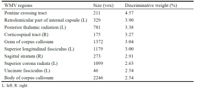 |
92 |
Abnormal brain white matter volume underlying methamphetamine abusers: A machine learning approach
Wentao Lai, Mei Yang, Zhifeng Zhou, Wentao Jiang, Xia Liu, Gangqiang Hou, Long Qian, Zhi Kong, Haiyan Run
The disruption of specific microstructural features of white-matter (WM) has been observed in methamphetamine (MA) abusers. However, it remains unknown whether WM volume is abnormal in MA abusers. To address this issue, a machine learning approach was applied in this study to differentiate between 21 MA abusers and 13 age- and gender- healthy controls. Our results showed that a linear support vector machine classifier achieved an accuracy of 73.53% using the white matter volume as input features. Particularly, the most discriminative WM regions included pontine crossing tract, motor system and the reading related network.
|
|
3431.
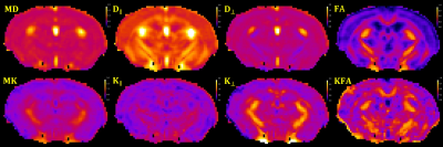 |
93 |
Diffusion MRI detects changes in the Hippocampus and Fimbria-Fornix Circuit in 2 months old 3xTg-AD mice
Maria Fatima Falangola, Xingju Nie, Emilie McKinnon, Joseph Helpern, Jens Jensen
The triple transgenic mouse model (3xTg) of Alzheimer’s disease (AD) exhibits both Aβ and tau pathology. Interestingly, the first detectable pathological features in this model are alterations in overall myelination patterns leading to white matte disruption as early as 2 months of age. Here we investigated the sensitivity of diffusion MRI (dMRI) to detect brain changes in young 3xTg mice. Our results indicate that dMRI is able to capture brain microstructural alterations associated with the hippocampus-fimbria-fornix circuit in 2 months-old 3xTg-AD mice, thus demonstrating dMRI as a viable tool for studying abnormal pathology in this AD mouse model.
|
|
3432.
 |
94 |
3D Diffusion MRI of Perivascular Fluid Movement: Towards Non-Invasive Mapping of Glymphatic Function.
Phoebe Evans, Bernard Siow, Ian Harrison, Ozama Ismail, Yolanda Ohene, Payam Nahavandi , David Thomas, Mark Lythgoe, Jack Wells
Within the glymphatic system, CSF is transported in a network of perivascular channels where it exchanges with ISF to drive drainage of unwanted solutes, like amyloid- β, out of the brain. Perivascular channel impairment may be an early biomarker of neurodegenerative processes. Here, we present a pilot study for 3D non-invasive assessment of glymphatic function in the rat brain using ultra-long echo time diffusion MRI. We show that this technique is sensitive to the fluid movement in downstream perivascular channels that drives glymphatic inflow.
|
|
3433.
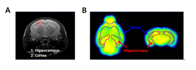 |
95 |
DW-MRI and FDG-PET as an Imaging Biomarker for Biological Response to Fast Neutron Exposure
Kyung Jun Kang, Ki Hye Jung, In Ok Ko, Yong Jin Lee, Kyo Chul Lee, Ji Ae Park
In this study, we monitored biological response in neutronexposed normal mouse brains using DW-MRI and 18F-FDG-PET. DW-MRI and 18F-FDG-PET were sensitive enough to detect physiological changes that were not identified in the H&E results. These results suggest that DW-MRI and 18F-FDG-PET can be used as imaging biomarkers to provide a quantitative indicator of complex in vivo pathological changes. Assessment of radiation exposure using imaging biomarkers is non-invasive and can be used repeatedly in clinical practice. In addition, it should be applied not only for evaluating the therapeutic effects of tumors, but also for evaluating the degree of tissue damage by radiation exposure.
|
|
3434.
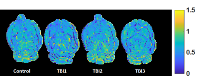 |
96 |
Functional and microstructural alterations in the rat hippocampus after repetitive mild traumatic brain injury
Ahmad Khan, Brian Hansen, Nina Iversen, Jonas Olesen, Mariana Perez, Donald .Kuhn, Sune Jespersen
Repetitive mild traumatic brain injury (mTBI) causes subtle tissue injury, which often reports normal under CT and MRI, leading to suboptimal diagnosis and increase the risk of developing neurological disorders. We probe microstructural alterations using advanced diffusion MRI, and perfusion MRI based cerebral blood flow (CBF) in rats before and after repetitive mTBI. Mean diffusivity and radial kurtosis tensor (WT) indicate the microstructural alteration in the ipsilateral hippocampus (IHP) and ipsilateral corpus callosum (ICC) respectively, and CBF maps show a significant reduction in the IHP. Our study suggests that multimodal imaging can provide useful information for early diagnosis of repetitive mTBI.
|
|
3435.
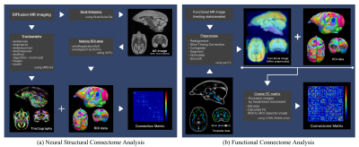 |
97 |
Structural and Functional Characteristics of the Common Marmoset Brain Assessed Using Connectome Analysis
Yawara Haga, Junichi Hata, Fumiko Seki, Akiko Uematsu, Mai Mizumura, Takaaki Kaneko, Hideyuki Okano, Akira Furukawa
Connectome analysis with magnetic resonance imaging (MRI) is a non-invasive method and is an important approach for assessing brain networks and neurological diseases from a multilateral perspective. We examined the structural and functional characteristics of the common marmoset brain using structural and functional connectome analyses. For these connectome analyses, diffusion-weighted imaging and true-awake resting-state functional MRI (rsfMRI) were performed using a 9.4-T MRI scanner. The structural connectome analysis showed that the visual region had the most prominent structural characteristics in the marmoset brain. In addition, functional connectome analysis indicated that the functional connectome results were affected by the rsfMRI environment.
|
|
3436.
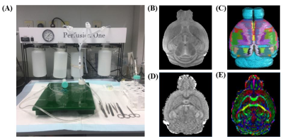 |
98 |
Ex vivo Structural Connectomics of the Mouse Brain Subcortical Regions
Eunbee Kim, Hyeon-Man Baek
Parkinson's disease (PD) is one of the degenerative brain disease and the hallmark of PD is the death of dopaminergic cells. The subcortical area involved in neuronal circuits which play a central role in the motor control. Structural connectivity can identify abnormal connectivity of neural circuits. Diffusion tensor imaging (DTI) is a non-invasive technique that has been used to delineate the internal anatomy by tracing white matter tracts. In this study, we investigated ex vivo diffusion MR images using perfusion methods and identify the structural connectivity in subcortical regions. In conclusion, we provide structural connectional fingerprints in the mouse model.
|
|
3437.
 |
99 |
Readout segmented EPI based Relative apparent diffusion coefficient in early monitoring the treatment effect of low-intensity transcranial ultrasound: evaluation in a rat permanent occlusion model
Lanxiang Liu, Juan Du, Tao Zheng, Xuemei Wang, Huiling Yi, Shuang Wu, Qinglei Shi, Shuo Wu
Acute ischemic stroke is a common and frequently occurring disease that severely harms human health and has high morbidity and mortality. Low-intensity transcranial ultrasound (LIPUS), due to its advantages of higher spatial resolution and greater penetration depth, has emerged as a new modality for noninvasive neuromodulation. This study indicated that the rADC based on readout segmented EPI sequence is a good indicator to evaluate the curative effect of LIPUS on acute cerebral infarction treatment.
|
|
| Top |
Diffusion: Body Applications
Digital Poster
Diffusion
Wednesday, 15 May 2019
| Exhibition Hall |
08:15 - 09:15 |
| |
|
Computer # |
|
3438.
 |
101 |
Comparison of IVIM Model, Kurtosis Model and IVIM-Kurtosis Model for the Grading of Hepatocellular Carcinoma
Yue Lin, Chunmei Li, Min Chen
IVIM model and DKI model cover different aspects of diffusion, thus a combined IVIM-DKI model might describe diffusion in perfused biological tissues more accurately. The aim of the study was to quantitatively compare the potential of various parameters obtained from IVIM model, Kurtosis model and IVIM-Kurtosis model in the grading of hepatocellular carcinoma (HCC). We enrolled 24 HCC patients who underwent multiple b values Diffusion-Weighted Imaging (DWI) with subsequent pathological confirmation. Our results showed that all three models could differentiate high-grade HCC from low-grade HCC and IVIM-Kurtosis model may be superior to IVIM model.
|
|
3439.
 |
102 |
Correlation between IVIM parameters and cT1 in HCV patients
Michele Nicastro, Nicola Martini, Matteo Milanesi, Chloe Hutton, Antonio Salvati, Daniele Della Latta, Maurizia Brunetto, Matthew Robson, Luigi Landini, Dante Chiappino
IVIM imaging has been investigated by several researchers to understand its clinical potential. However, the dependency from b-value distribution and the effect of confounding factors such as iron and steatosis make hard to draw conclusion. In this work, a multiparametric protocol was proposed to address those confounds and the correlation with iron-corrected T1 (cT1) was studied in a group of patients affected by hepatitis C. The vascular volume fraction F shows a statistically relevant correlation with cT1 (p<0.001).
|
|
3440.
 |
103 |
DTI of Spinal Cord Lesions of Varying Severity Based on MRI in the Entire Pediatric Spinal Cord
SONA SAKSENA, Feroze Mohamed, Devon Middleton, Laura Krisa, Mahdi Alizadeh, Shiva Shahrampour, Chris Conklin, Adam Flanders, Jürgen Finsterbusch, MJ Mulcahey, Scott Faro
To determine whether DTI at sites cephalad and caudal to the MRI lesion provides measures of cord abnormalities in different SCI groups (n=23) compared to TD (n=36) and any correlation between the varying severity of MRI abnormality and DTI abnormalities cephalad and caudal to the MRI lesion. to the MRI lesion. The five SCI groups included; 1= SCIWORA (n=6); 2=Syringohydromyelia (n=4); 3=Atrophy+Syringomyelia (n=7); 4=Atrophy+Myelomalacia (n=3); 5=Atrophy+Hemorrhage (n=3). Groups 1 to 5 represent a progressive measure in cord abnormality. Among these SCI groups, we looked at concordant DTI correlation which is defined as the expected change in DTI metrics with increasing cord severity. SCIWORA showed DTI abnormality at segments of the cephalad and caudal cord with the caudal cord more affected consistent with the anterograde degeneration. FA showed the greatest total number of segments in the cephalad and caudal cord that showed abnormal DTI values in comparison to the other DTI metrics.
|
|
3441.
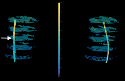 |
104 |
Assessment of Traumatic Peripheral Nerve Injury and Carpal Tunnel Syndrome with Diffusion Tensor Imaging
Michael Pridmore, Wesley Thayer, Mark Does, Richard Dortch
Clinical management following traumatic peripheral nerve injury and carpal tunnel release surgeries requires physicians to rely on qualitative measures from patient history and physical exams. The resulting delays in clinical decision-making can lead to a negative impact on patient outcomes because the regeneration of nerves must occur in a timely fashion to avoid permanent muscle atrophy and loss of sensorimotor function. The current study aims to test the feasibility of performing diffusion tensor imaging in the nerves of the wrist to provide a noninvasive and reliable tool that can monitor patient outcomes and improve clinical decision-making after surgical interventions.
|
|
3442
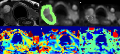 |
105 |
Inter- and intraobserver reproducibility of two different delineation of intravoxel incoherent motion in differentiating benign and malignant thyroid nodule
Video Permission Withheld
Yunlong Yue, Minghui Song, Yanfang Jin, Jinsong Guo, Lili Zuo, Queenie Chan, Zhenchang Wang
To compare the reproducibility and diagnostic performance of IVIM parameters derived from 3D whole-lesion (W-L ROI) and single-section (S-S ROI) delineation respectively . Forty-three patients with 46 pathologically confirmed thyroid nodules were involved. Reduced FOV DWI with 2D RF was employed to decrease the distortion. The ICC values of all IVIM parameters were higher with W-L ROI delineation than with S-S ROI delineation. The 95% limits of agreement (Bland-Altman plots) determined by W-L ROI revealed smaller absolute intra- and interobserver variability and the AUC of W-L ROI delineation was higher than S-S ROI according to ROC analysis.
|
|
3443.
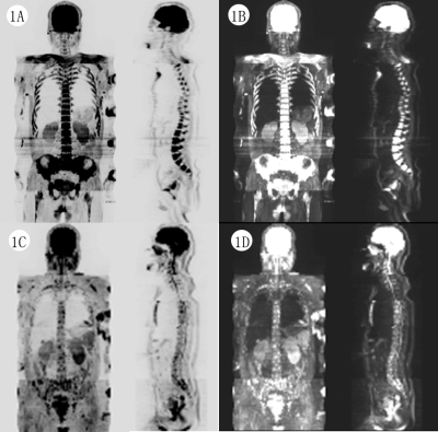 |
106 |
Application of Whole-Body Diffusion-Weighted MRI Imaging with Apparent Diffusion Coefficient Mapping for the Evaluation of Depth of Response to Therapy in Multiple Myeloma
Presentation Not Submitted
Menglong Zheng, Xinxing Ma, Daohai Xie
Whole-body diffusion-weighted magnetic resonance imaging (WB-DWI MRI) is an important imaging technique of great supplementary diagnostic value to the evaluation of treatment response in patients with multiple myeloma (MM). Conventional radiography which has been widely avaiable for the identification of myeloma-related bone lesions both at diagnosis and during disease course is of limited value to the assessment of response to therapy or disease relapse in MM, as lytic bone lesions rarely show signs of healing despite response to therapy. WB-DWI MRI with the measurement of apparent diffusion coefficient (ADC) value is an effective way for quantitatively comparing the differences in water diffusion among normal marrow, active pathologic marrow and that in remission. In this work, we report the preliminary results of the application of WB-DWI MRI in discriminating deep response to induction therapy for newly diagnosed MM patients.
|
|
3444.
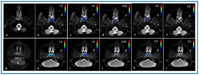 |
107 |
Non-Gaussian IVIM-DWI for prediction of locoregional failure in nasopharyngeal carcinoma
Ramesh Paudyal, Jung Oh, Jonathan Leeman, Xinmao Song, Amresha Shridhar, Eve LoCastro, Vaios Hatzoglou, Nadeem Riaz, Nancy Lee, Amita Shukla-Dave
The study aims to assess quantitative metrics derived from pretreatment NG-IVIM using multiple b-value diffusion weighted imaging data for predicting tumor with and without locoregional failure (LRF) in NPC patients. Kaplan-Meier method and the log-rank test were used to analyze differences between NPC patients with and without locoregional curves. Kaplan Meier results showed that the pre-treatment mean ADC and D value could predict LRF in NPC patients. The pretreatment NG-IVIM diffusion-weighted imaging will provide useful information for the selection of patients appropriate for definitive radiotherapy.
|
|
3445.
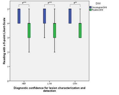 |
108 |
Increasing reader confidence in cancer imaging using an advanced DWI processing – a prospective reader study
Constantin Dreher, Tristan Kuder, Stefan Windhaber, Franziska König, Daniel Paech, Anoshirwan Tavakoli, Regula Gnirs, Thomas Benkert, Heinz-Peter Schlemmer, Sebastian Bickelhaupt
Diffusion-Weighted imaging (DWI) in abdominal MRI is highly valuable for oncological follow-up-investigations. However, intrinsic limitations of the method, including susceptibility to motion artifacts and poor signal in the retroperitoneum, are of special importance regarding its usage in abdominal examinations. This prospective study compared a standard EPI-DWI with an oncologically optimized prototype DWI in 59 patients with regard to the detectability/characterization of suspicious lesions. The study demonstrated that the oncologically optimized prototype DWI sequence with complex averaging, motion correction between averages, rescaling of motion corrupted averages, and background suppression, significantly increased the reader confidence for lesion characterization/detection in oncological abdominal MRI.
|
|
3446.
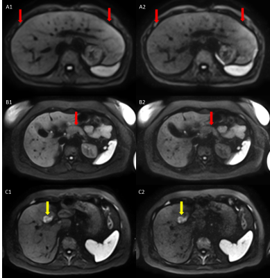 |
109 |
Prospective evaluation of motion correction and complex averaging in ultra-high-b-value DWI for image quality and lesion conspicuity in oncologic follow-up examinations
Constantin Dreher, Tristan Kuder, Stefan Windhaber, Franziska König, Daniel Paech, Anoshirwan Tavakoli, Regula Gnirs, Thomas Benkert, Heinz-Peter Schlemmer, Sebastian Bickelhaupt
In oncological, abdominal MRI, the value of motion corrected Diffusion-Weighted Imaging (DWI) including complex averaging with high and ultra-high b-values remains to be defined. This is of special importance regarding the balance between normal tissue suppression and suspicious lesion demarcation. This prospective study investigated 41 patients with an oncologically optimized prototype-DWI (with complex averaging, motion correction between averages, rescaling of motion corrupted averages, background suppression). Image quality, tissue differentiation and lesion detection/characterization were significantly increased in high (b900) as compared to ultra-high (b1500) DWI. At the same time, apparent signal-intensity ratio of lesion/normal tissue was not significantly different.
|
|
3447.
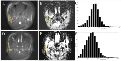 |
110 |
Whole-volume ADC Histogram and Texture Analyses of Parotid Glands as an Image Biomarker in Evaluating Disease Activity of Primary Sjögren's Syndrome
zhengyang zhou, Jian He, Weibo Chen
Fifty-five patients with pSS underwent MRI including DWI to explore whether ADC histogram and texture analyses to evaluate the disease activity of pSS. According to the ESSDAI score, 35 patients were categorized into the low-activity group and 20 into the moderate-high-activity group. ROC analysis showed that the anti-SSB, lip biopsy, MRI morphology, ADC, ADCmean, and entropy values were able to categorize the disease into two groups, particularly the entropy values. The multivariate model, which included anti-SSB, MRI morphology and entropy, had an area under the ROC curve of 0.923. The parotid entropy value distinguished disease activity in patients with pSS.
|
|
3448.
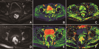 |
111 |
Non-Gaussian Diffusion Imaging with a Fractional Order Calculus Model to predict response to neoadjuvant chemoradiotherapy in local advanced rectal cancer
Presentation Not Submitted
Yanfen Cui, Zhizheng Zhuo, Xiaotang Yang
A novel non-Gaussian diffusion model based on fractional order calculus (FROC) were successfully applied to diffusion MRI of rectal cancer. Statistically significant differences in ?D and ?β values are observed between the responder group and non- responder group (p < 0.01), indicating that FROC-derived parameters from the FROC diffusion model may be useful as imaging biomarkers in predicting the biological properties of rectal cancer in clinical practice.
|
|
3449.
 |
112 |
Reduced field-of-view Diffusion-weighted imaging at 3T for Evaluating Allograft Fibrosis After Kidney Transplantation: A Preliminary Study
Presentation Not Submitted
Yuan Meng Yu, Long Jiang Zhang, Yong Zhang
The extent of renal allograft fibrosis is a critical predictor of adverse allograft outcome. This institutional review board approved study included 109 renal transplantation recipients receiving 3 T multi-b reduced field-of-view intravoxel incoherent motion diffusion-weighted imaging (rFOV IVIM-DWI) examinations. The purpose of the present study is exploring the diagnostic value of evaluating allograft fibrosis with IVIM-DWI. In the present study, we have found that there is downward trend of ADCT and ADCD with the development of kidney fibrosis and both of them had good diagnostic efficiency in the distinguish fibrosis extent.
|
|
3450.
 |
113 |
Diffusion time-dependence of diffusivity and kurtosis in locally advanced head and neck squamous cell carcinoma before and after chemo-radiation therapy
Gregory Lemberskiy, Steven Baete, Dmitry Novikov, Els Fieremans, Elcin Zan, Kenneth Hu, Sungheon Kim
Synopsis: The effect of chemo-radiation therapy on advanced head & neck squamous cell carcinoma was evaluated via diffusion and kurtosis time-dependence. We found opposing diffusion limiting regimes pre and post therapy, where prior to therapy the tissue was well described by the long-time limit (Karger Model applies), and where after therapy the tissue was well described by the short-time S/V limit. This reversal of imaging regimes can serve as a signature of the minimal effective dose required for treatment.
|
|
3451.
 |
114 |
Predicting Renal Glomerular Filtration Rate of Children with Account of Kidney Compensation Using Multiple b Values Diffusion-Weighted Imaging
Presentation Not Submitted
Jianbo Shao, Zhiyao Tian, Xiaowen Wang, Zujun Hou, Xuehua Peng
GFR would fail to tell the functional status of each kidney for CKD cases,so we try to use machine learning methods to predict GFR of pediatric kidneys based on the IVIM diffusion parameters. The results is that,With account of kidney compensation, averaged correlation between predicted and measured GFR up to 0.9 (p < 0.05) was obtained for the combination of perfusion-fraction f and pseudo-flow fD*. For comparison, if not taking into account kidney compensation, the best predictor attained the correlation of 0.3. We conclude that a noninvasive method can predict well the GFR of children with kidney diseases using multiple b values DWI. The best predictions involved the use of perfusion-fraction f and pseudo-flow fD* which are closely related to renal blood perfusion.
|
|
3452.
 |
115 |
The performance of TGSE BLADE DWI in the diagnosis of cholesteatoma compared with RESOLVE DWI technique
Yaru Sheng, Yan Sha, Yang Li, Xinpei Ye, Zhongshuai Zhang, Kun Zhou, Caixia Fu, Mengxiao Liu
Synopsis:This study aimed to compare the performance of two diffusion weighted imaging techniques used to diagnose cholesteatoma: readout-segmented echo-planar imaging (RESOLVE) and turbo gradient spin echo (TGSE) BLADE DWI. The results showed that the TGSE BLADE DWI had better image quality with respect to reducing susceptibility artifacts, distortion, and blurring compared with RESOLVE. However, the SNR of TGSE BLADE DWI needs to be improved in the future.
|
|
3453.
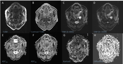 |
116 |
Head and neck squamous cell carcinoma: diagnostic performance of diffusion kurtosis imaging for the prediction of histopathologic prognostic factors
Tong Su, Yu Chen, Zhenyu Jin, Xiaohua Shi, Xingming Chen, Tao Zhang, Jinxia Zhu, Robert Grimm, Huadan Xue, Zhuhua Zhang
This preliminary study evaluated the diagnostic efficacy of diffusion parameters, derived from different models, for differentiating between tumor grades and identifying metastatic lymph nodes in head and neck squamous cell carcinomas (HNSCCs). Superior diagnostic efficiency was achieved when combining ADC values derived from a mono-exponential model with kurtosis and diffusivity values derived from a diffusion kurtosis imaging (DKI) model compared to using each parameter by itself.
|
|
3454.
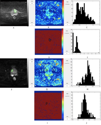 |
117 |
Histogram analysis of diffusion kurtosis imaging in prostate cancer: comparison with pathology using in-bore transrectal MR-guided biopsy
Yuwei Jiang, Lu Yu, Yadong Cui, Chunmei Li, Kaining Shi, Min Chen
Our purpose was to verify the use of DKI histogram analysis for differential diagnosis of prostate cancer and noncancerous foci (benign prostatic hyperplasia and prostatitis). Twenty-three patients underwent MRI subsequently using in-bore transrectal MR-guided biopsy as the pathological reference. Our results showed DKI histogram analysis has favorable effective function and good repeatability for the differential diagnosis. DKI histogram analysis may contribute to improve the diagnostic accuracy.
|
|
3455.
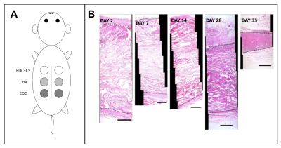 |
118 |
Quantifying diffusion-weighted MRI parameters with cellular infiltration, tissue ingrowth, and angiogenesis to characterize collagen-sponge remodeling.
Mohammed Salman, Karl Helmer, Isa Ahmed, Sivakumar Kandasamy, Staurt Howes, Christopher Sotak, George Pins
In vivo evaluation of biomaterial implant remodeling involves surgical removal of the implant for subsequent histological assessment, which is often destructive and limits the effective evaluation of these materials. Diffusion-weighted MRI has the potential to non-invasively monitor the remodeling of collagen scaffolds. This study investigated the role of apparent diffusion coefficient (ADC) from different diffusion regimes to evaluate the remodeling of implanted collagen scaffolds and correlated the findings to conventional histological techniques. Correlations between ADC and histological parameters demonstrated that MRI is sensitive to specific remodeling parameters which can aid in the design of robust biomaterial scaffolds for tissue regeneration.
|
|
3456.
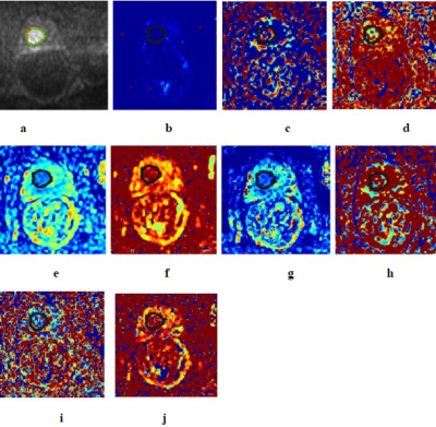 |
119 |
A Comparative Study of IVIM Model, Kurtosis Model and IVIM-Kurtosis Model for the Diagnosis and Aggressiveness Assessment of Prostate Cancer
Presentation Not Submitted
Ying Liu, Chunmei Li, Kaining Shi, Min Chen
The purpose of our study was to quantitatively compare the utility of the parameters obtained from IVIM, kurtosis, and IVIM-kurtosis models in the differential diagnosis and aggressiveness assessment of prostate cancer, by using in-bore transrectal MR-guided biopsy as a reference. Our results displayed that IVIM, kurtosis and IVIM-kurtosis models has effective function for the differential diagnosis of prostate cancer and the diagnostic efficacy among the three models were similar. In aggressiveness assessment, IVIM-kurtosis model may be superior to the others.
|
|
3457.
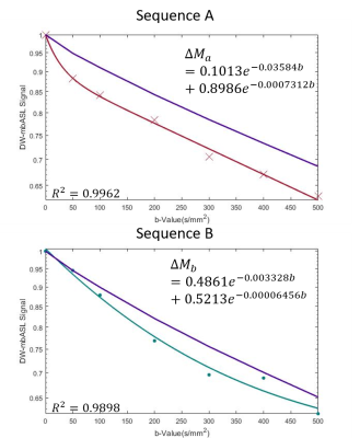 |
120 |
Adapting Diffusion Weighted Arterial Spin Labelling (DWASL) to use a Single Sided Bipolar Diffusion Gradient to Probe Microvascular Signal
Samantha Paterson, Antoine Vallatos, William Holmes
Diffusion Weighted Arterial Spin Labelling uses a pair of bipolar diffusion gradients surrounding a spin echo pulse to image signal in the intravascular and extravascular compartments. We propose moving these gradients to a single sided gradient, reducing Δ and changing where the change to the extravascualar signal starts. Our results show that the ratio has changed from b = 50 to b = 300 s/mm2 for our new single sided sequence. This can give us more information on the intravascular signal, helping to improve the probing of blood brain barrier permeability.
|
|
3458.
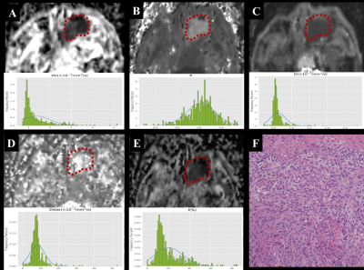 |
121 |
Differentiation of Sinonasal Mucosal Malignant Melanomas from Sinonasal Carcinomas Using Whole-tumour Histogram Analysis of Diffusion Kurtosis Imaging and Intravoxel Incoherent Motion
Zebin Xiao, Zuohua Tang, Jing Zhang, Guang Yang, Yang Song, Linying Guo, Zhongshuai Zhang
This is the first study that used histogram metrics derived from diffusion kurtosis (DKI) and intravoxel incoherent motion (IVIM) to differentiate sinonasal mucosal malignant melanomas (SNMMMs) and squamous cell carcinomas (SCCs) which are sometimes indistinguishable with conventional MRI. Overall, histogram metrics obtained from K, D, D* and f were found to be significantly higher in SNMMMs than in SCCs. Furthermore, the combined use of the two independent indicators, the 75th percentile of K and skewness of D, can effectively differentiate between SNMMMs and SCCs.
|
|
3459.
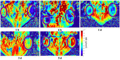 |
122 |
Quantifying diffusivity of ISFIs in vivo using DWI: A novel method to evaluate drug release
Nicole Vike, Xin Li, Kelsey Hopkins, Luis Solorio, Joseph Rispoli
Medication effectiveness relies on patient adherence to a given treatment regimen. Often, patients do not adhere to the temporal guidelines set by their physicians and treatments therefore remain less effective. In situ forming implants (ISFIs) eliminate the need for patient adherence and release an effective dose of drug overtime. However, no methods exist to noninvasively and temporally validate drug release in vivo. We conducted in vivo experiments that validated the use of DWI to monitor ISFI diffusivity overtime. This has enormous implications in pharmaceutical research as this method can robustly quantify diffusivity in ISFIs post-implantation to ensure effective drug release.
|
|
3460.
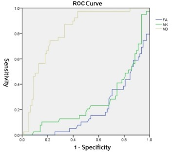 |
123 |
Evaluation the staging of liver fibrosis using diffusion kurtosis imaging: a pilot study
Jiawei Hu, Dongmei Guo, Yang Dong, Yan Xu, Bing Wu, Lizhi Xie
To investigate the diagnostic value of the quantitative diffusion kurtosis imaging(DKI)parameters in the staging diagnosis of liver fibrosis in rabbits’ model. Twenty-nine successful rabbit models were scanned by a 3T MR scanner (Discovery MR750W, GE,Healthcare, USA). GE AW4.6 Workstation and statistical software helps to evaluate the correlation between quantitative analysis of DKI and METAVIR staging. It is concluded that DKI related parameters might have important diagnostic value for the staging of liver fibrosis.
|
|
3461.
 |
124 |
Assessment of microstructural changes during intervertebral disc degeneration by diffusion kurtosis imaging
Li LI, Jicheng Fang, Alessandro Scotti,, Kejia Cai, WenZhen Zhu
To assess the microstructural changes of intervertebral disc degeneration, 18 rats were punctured percutaneously at the intervertebral disc, and the rats’ tail was imaged with T2WI, DWI and DKI sequences. The diffusion parameters changes were consistent with the histological changes at the different time points and showed significant differences between the different groups. And, DKI parameters were better markers to detect the change of microstructure of the degenerated discs than conventional DWI. Therefore, DKI is a noninvasive technique to test the changes of the microstructure of the intervertebral discs.
|
|
| Top |
Diffusion MRI: Signal Representation & Modelling
Digital Poster
Diffusion
Wednesday, 15 May 2019
| Exhibition Hall |
08:15 - 09:15 |
| |
|
Computer # |
|
3462.
 |
126 |
Brain Development in Non-human Primates Assessed with Neurite Orientation Dispersion and Density Imaging
Mayu Iida, Junichi Hata, Marin Nishio, Fumiko Seki, Yawara Haga, Erika Sasaki, Takako Shirakawa
Neurite orientation dispersion and density imaging (NODDI) can specifically estimate the microstructure of neurites. Brain development characteristics were evaluated using NODDI in common marmosets. The study included six common marmosets (age, 5–30 months). In NODDI, the value of both dispersion and density increased. Additionally, the trend of increase varied from region to region. This pattern has previously been observed in human studies. Thus, NODDI can be a good approach to evaluate brain development in the common marmoset, and eventually a suitable parameter to study brain developmental disorders for diagnosis and treatment.
|
|
3463.
 |
127 |
Diffusion Dispersion Imaging: Mapping OGSE Frequency Dependence in the In Vivo Human Brain
Aidin Arbabi, Corey Baron
Oscillating gradient spin-echo (OGSE) diffusion MRI allows measurement of the frequency dependence of the apparent diffusion coefficient (ADC), which gives insight into tissue microstructure. OGSE has been utilized in numerous animal models, but its application in the in vivo human brain is challenging. Further, a parameterization that allows visualization of maps of the frequency dependence of ADC throughout the human brain has thus-far not been demonstrated. In this work, we report on an efficient method to generate maps of diffusion dispersion (DD), which characterizes the frequency dependence of the ADC, and demonstrate full-brain DD mapping in vivo at 7T.
|
|
3464.
 |
128 |
Correlation Tensor Imaging (CTI): resolving diffusion sources of non-Gaussian diffusion
Rafael Neto Henriques, Sune Jespersen, Noam Shemesh
The cumulant expansion of Double Diffusion Encoding (DDE) involving correlation tensors has been previously theoretically presented but never exploited beyond microscopic anisotropy detection. Here, we propose the correlation tensor imaging (CTI) as novel approach capable of mapping correlation tensor features using DDE acquisitions. The correlation tensor can provide unique information: as a first step, we theoretically and experimentally demonstrate that CTI can be used to resolve the different underlying sources of diffusion kurtosis vis-à-vis isotropic and anisotropic variance of tensors and restricted diffusion (µK). The ensuing estimates bode well for many future applications.
|
 |
3465.
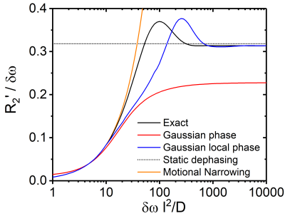 |
129 |
Restricted diffusion in the Gaussian local phase approximation
Lukas Buschle, Christian Ziener, Thomas Kampf, Sabine Heiland, Martin Bendszus, Heinz-Peter Schlemmer, Felix Kurz
Diffusion MRI is highly influenced by diffusion boundaries created by microscopic cells and blood-filled vessels. The Gaussian phase approximation is a common method to analyze the signal evolution in the presence of diffusion. However, the Gaussian phase approximation exhibits several drawbacks as it is only valid for strong diffusion effects, it does not provide phase information and it yields only the total magnetization. In this work, we generalize the traditional Gaussian phase approximation and solve the mentioned issues. The new Gaussian local phase approximation is then applied to diffusion restricted between two slabs, within a cylinder and within a sphere.
|
|
3466.
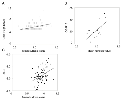 |
130 |
Diffusion kurtosis imaging as an effective predictor of liver function
Daisuke Yoshimaru, Yasuo Takatsu, Yuichi Suzuki, Toshiaki Miyati, Yuhki Hamada, Ayumu Funaki, Ayumi Tabata, Chifumi Maruyama
We evaluated whether DKI analysis as an indicator of liver function can replace laboratory tests. The mean kurtosis value was found to correlate with the Child–Pugh score and the ICG score. However, the correlation between the mean kurtosis valueand the ALBI score was the poorest among these. ADC values correlated with only the ICG score. The mean kurtosis value can be one of predictors for liver function owing to its correlation with typical indicators of liver function by serum data although this method, which shows liver function using DKI alone, cannot completely substitute the laboratory tests.
|
|
3467.
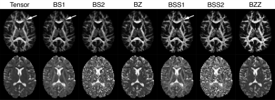 |
131 |
Single-shell diffusion MRI data with b=1000: which non-tensor models are feasible?
Andrew Davis, Geoffrey Hall, Benicio Frey, Stephen Strother, Glenda MacQueen, Stefanie Hassel, Jacqueline Harris, Mojdeh Zamyadi, Stephen Arnott, Jonathan Downar, Sidney Kennedy
Single-shell diffusion data with b=1000 is commonly acquired in clinically focused studies, and fit with a tensor to calculate microstructure parameters, despite that model's limitations. This study fit several multi-compartment models to such data. The most promising models were: (i) two-fiber ball-stick with a single common diffusivity parameter, which provided the most stable results, and (ii) two-fiber ball-zeppelin, which provided low-noise and stable parameter maps and dispersion values with burn-in settings above 20,000 jumps. The default model in bedpostx yielded lower quality results in this data, even with extremely long processing times.
|
|
3468.
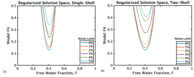 |
132 |
Regularization stabilizes the fit of the two-compartment free water diffusion MRI model
Jordan Chad, J. Chen, Ofer Pasternak
Free water mapping using single-shell diffusion MRI has been proposed by regularizing the ill-posed problem, but the effect of this regularization has yet to be systematically investigated. Here we explore the solution space for the two-compartment free water model. Without regularization, the solution space is flat for one-shell data, and while it has a minimum for two-shell data, this minimum is not particularly steep. Regularizing the fit by exploiting smoothness of the tensor creates a steep minimum in both one- and two-shell data, demonstrating the advantage of regularization.
|
|
3469.
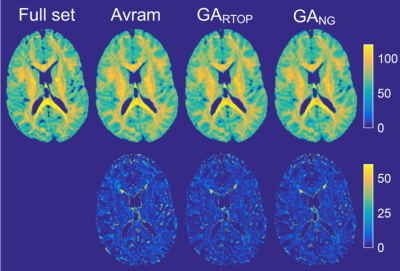 |
133 |
Optimal subsampling of q-space for Mean Apparent Propagator MRI using a genetic algorithm
L. Tugan Muftuler, Daniel Olson, Volkan Arpinar
Mean Apparent Propagator (MAP) MRI provides a robust analytical framework to estimate the diffusion probability density function (PDF). Several scalar metrics are calculated from the PDF, which might better characterize tissue microstructure compared to conventional diffusion methods. The downside of MAP MRI is the long acquisition times of over an hour. In this study we developed a genetic algorithm (GA) to determine optimal q-space subsampling scheme for MAP MRI that will keep total scan time under 10 minutes, while preserving accuracy. Results show that the metrics derived from the optimized schemes match those from the full set closely.
|
|
3470.
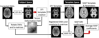 |
134 |
Quantifying voxel-wise differences between diffusion propagators across multiple MAP-MRI datasets
Alexandru Avram, Adam Bernstein, M. Okan Irfanoglu, Craig Weinkauf, Amber Simmons, Martin Cota, Neville Gai, Neekita Jikaria, Anita Moses, L. Christine Turtzo, Lawrence Latour, Dzung Pham, John Butman, Peter Basser
We describe a technique for voxel-wise analysis across multiple mean apparent propagator (MAP) MRI datasets warped using a diffeomorphic tensor-based registration algorithm. By measuring propagators from co-registered MAP datasets with the same MAP basis functions we can directly quantify voxel-wise differences using angular dissimilarity metrics. We show examples from a cohort of healthy volunteers, and from a longitudinal clinical dataset of a patient undergoing carotid endarterectomy. This approach could provide improved sensitivity in the detection and characterization of subtle microstructural tissue changes in cross-sectional group and longitudinal single-subject clinical studies.
|
|
3471.
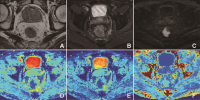 |
135 |
Diffusion kurtosis imaging–derived histogram metrics for prediction of KRAS/NRAS/BRAF mutations in rectal adenocarcinoma: Preliminary findings
Presentation Not Submitted
Yanfen Cui, Zhizheng Zhuo, Xiaotang Yang
It was revealed that DKI metrics with whole-tumor volume histogram analysis, especially the K75th parameter, yielded more preferable AUC and specificity values for predicting KRAS/NRAS/BRAF mutations than ADC and D values, and thus may potentially serve as an optimal imaging biomarker for the prediction of KRAS/NRAS/BRAF mutations for guiding targeted therapy.
|
|
3472.
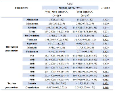 |
136 |
Differential diagnosis of pathologic grade of hepatocellular carcinoma (HCC) using whole-tumor histogram and texture analysis based on apparent diffusion coefficient (ADC) map
Ying Zhao, Ailian Liu
This work aimed for ADC histogram and texture features based strategy to identify poorly differentiated hepatocellular carcinoma (HCC) which may own a high risk of recurrence or metastasis. The results showed that ADC histogram and texture features can discriminate HCC grade. The 95th percentile achieved the best result (AUC: 0.772; sensitivity: 90%; specificity: 72%) on ADC signal intensity map, forming a valuable strategy for clinical practice.
|
|
3473.
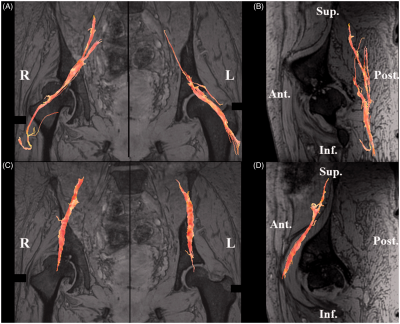 |
137 |
Diffusion tensor imaging of the sciatic and femoral nerves in osteoarthritis of the hip and osteonecrosis of the femoral head
Shigeo Hagiwara, Wako Yasushi, Junichi Nakamura, Miura Michiaki, Yawara Eguchi, Yuya Kawarai, Masahiko Sugano, Kenoto Nawata, Kensike Yoshino, Kenta Konno, Satoshi Yo
We visualizedsciatic and femoral nerves andcompared the fractional anisotropy (FA) values of the sciatic and femoral nerves in patients with osteoarthritis of the hip (OA) and osteonecrosis of the femoral head (ONFH) using diffusion tensor imaging (DTI). This study indicates thatthe FA values for the sciatic and femoral nerves in patients with OA and ONFH showed no significant differences with normal hips indicating no degeneration or demyelination occurred in either nerve. DTI may be able to differentiate hip diseases from lumbar nerve radiculopathy.
|
|
3474.
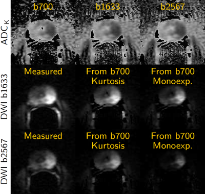 |
138 |
A Simplified Approach for Measuring Tissue ADC: Application to Prostate Diffusion Scans
Stephan Maier
A simplified approach is presented to obtain consistent tissue ADC data, irrespective of diffusion protocol, location within field of view and MR system. Examples of application in prostate diffusion scans are presented. This could greatly facilitate the establishment of suitable cut-off values to differentiate aggressive from indolent disease in prostate cancer, but as detailed in the abstract would entail several other substantial benefits that are universally applicable to diffusion imaging in tissues.
|
|
3475.
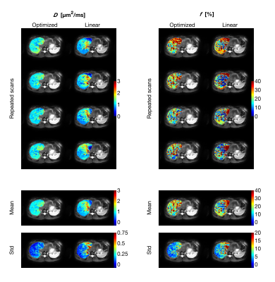 |
139 |
Optimization of b-values for estimation of IVIM D and f
Oscar Jalnefjord, Mikael Montelius, Göran Starck, Maria Ljungberg
IVIM parameter estimation restricted to D and f (avoiding D*) has gained increased popularity. In this study we propose a framework for optimization of b-value schemes for this application. We show that optimized b-values schemes contain exactly three unique b-value, regardless of the total number of acquisitions, and that parameter estimation uncertainty can be substantially reduced by the use of the optimized b-value schemes.
|
|
3476.
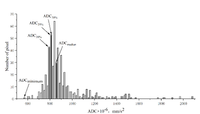 |
140 |
Whole-lesion histogram analysis of the apparent diffusion coefficient - a correlation study with histological grade of hepatocellular
Presentation Not Submitted
Yongsheng Xu, Haifeng Liu, Jinkui Li, Junqiang Lei, Shaoyu Wang
Lately, an increasing number of studies made an effort to investigate the correlation between quantitative analysis of diffusion weighted imaging (DWI) and the histological grade of HCC. However, the optimal ADC parameter for characterization of grade of HCC has yet to be determined. Various investigators have evaluated that ADC value such as the mean ADC and minimum ADC correlate with histological grade of HCC, which were derived from single or several parts slice–based ROIs within tumors. We drew lesions on multiple slices to get more reliable estimate of lesion signal.
|
|
3477.
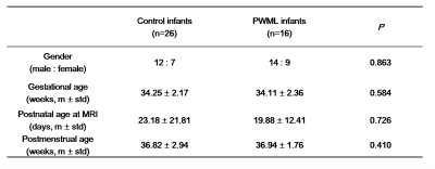 |
141 |
Microstructural Variations in Intra-axonal and Extra-axonal Spaces Associated with Punctate White Matter Lesions in Preterm Infants Reveal by White Matter Tract Integrity Metrics
Presentation Not Submitted
Mengxuan Li, Xianjun Li, Qinli Sun, Miaomiao Wang, Congcong Liu, Yannan Cheng, Xiaocheng Wei, Jian Yang
Punctate white matter lesions (PWML) are common in preterm infants. Extensive microstructural changes were observed previously. However, specific changes related to axons and extra-axonal structures remain to be investigated. White matter tract integrity (WMTI) metrics derived from diffusion kurtosis imaging provide information of intra-axonal or extra-axonal spaces. This study aimed to use these metrics to quantify specific variations associated with PWML. Besides reduced fractional anisotropy and increased radial diffusivity, increased diffusivities in extra-axonal and intra-axonal spaces were found. Furthermore, tortuosity and fiber dispersion decreased obviously. These results suggested that PWML would influence axonal reorganization as well as extra-axonal structural integrity.
|
|
3478.
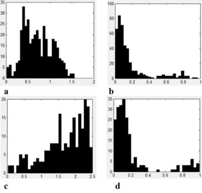 |
142 |
The Histogram Analysis of Intravoxel Incoherent Motion-Kurtosis (IVIM-Kurtosis) model for Detection of Prostate Cancer
Lu Yu, Yuwei Jiang, Yadong Cui, Chunmei Li, Kaining Shi, Min Chen
Diffusion-weighted (DW) MR imaging has a great potential in the study of prostate cancer (PCa). We aimed to assess the feasibility and efficacy of intravoxel incoherent motion-kurtosis (IVIM-Kurtosis) model for the differential diagnosis of PCa, benign prostatic hyperplasia and prostatitis (BPH/prostatitis). Thirty-six patients were enrolled and underwent MRI subsequently using in-bore transrectal MR-guided biopsy as the pathological reference. Significant differences were found for D and f values for the differential diagnosis of PCa and BPH/prostatitis, and D median and f median had the highest area under curve. The f, K and D* were correlated with Gleason score (GS) of PCa.
|
|
3479.
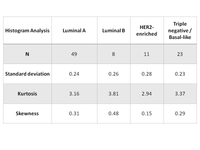 |
143 |
Diffusion Weighted Imaging with Apparent Diffusion Coefficient on the Assessment of Tumor Heterogeneity for the Differentiation of Molecular Subtypes in Breast Cancer
Joao Horvat, Olivia Sutton, Blanca Bernard-Davila, Elizabeth Morris, Katja Pinker, Sunitha Thakur
The study evaluated the utility of diffusion weighted imaging with apparent diffusion coefficient on the assessment of tumor heterogeneity for the differentiation of molecular subtypes in a population of 91 patients with invasive breast cancer. The authors investigated the use of histogram analysis and visual assessment of tumor heterogeneity for the differentiation of breast cancer subtypes derived via immunohistochemistry surrogates. The results obtained demonstrated that HER-2 enriched tumors had higher ADC values than other tumor subtypes. It was also demonstrated that histogram analysis and visual assessment of tumor heterogeneity could not reliably be used to differentiate tumor molecular subtypes.
|
|
3480.
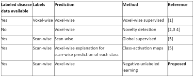 |
144 |
Negative-Unlabeled Learning for Diffusion MRI
Phillip Swazinna, Vladimir Golkov, Ilona Lipp, Eleonora Sgarlata, Valentina Tomassini, Derek Jones, Daniel Cremers
Machine learning strongly enhances diffusion MRI in terms of acquisition speed and quality of results. Different machine learning tasks are applicable in different situations: labels for training might be available only for healthy data or only for common but not rare diseases; training labels might be available voxel-wise, or only scan-wise. This leads to various tasks beyond supervised learning. Here we examine whether it is possible to perform accurate voxel-wise MS lesion detection if only scan-wise training labels are used. We use negative-unlabeled learning (an equivalent of positive-unlabeled learning) and achieve an AUC of 0.77.
|
|
3481.
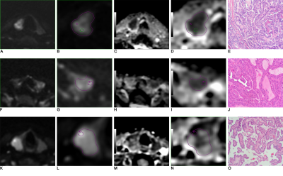 |
145 |
Diagnostic Value of Apparent Diffusion Coefficient for predicting aggressive histological features of papillary thyroid carcinomas
Bin Song
Objective: To investigate the value of preoperative diffusion-weighted magnetic resonance imaging for predicting aggressive histological features in papillary thyroid cancer (PTC). Methods: The mean, minimum apparent diffusion coefficient (ADCmean and ADCmin) and ADC value of the solid component (ADCsolid) were compared among different aggressive histological groups. Analyses of receiver-operating characteristic curve were also performed. Results: The ADC value in the low aggressive PTC were significantly higher than in high aggressive PTC. ADCmin values had the best performance. Conclusion: ADCmin values derived from DWI could be used as quantitative predictors of aggressive histological features in PTC.
|
|
3482.
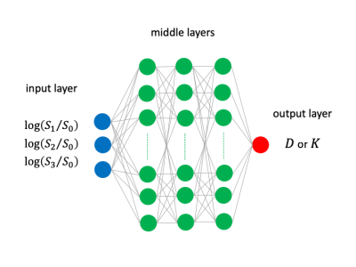 |
146 |
DKI parameter inference by deep neural networks trained by synthetic data
Ko Sasaki, Yoshitaka Masutani
In general, DKI parameters (D and K) are obtained by fitting models to DWI signal values, such as by least-square fitting (LSF) methods. However, when DWI signal values are contaminated by noise of high level, fitting error is often observed especially for diffusional kurtosis K. In this study, we propose a robust method to infer DKI parameters based on deep neural networks trained by only synthetic data to overcome the limitations of real data training. Our experimental results including comparison with LSF showed the potential of our method for robust inference of DKI parameters.
|
|
3483.
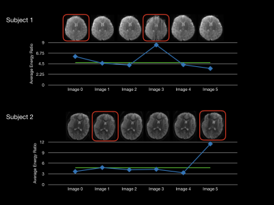 |
147 |
Framework to Identify Motion-Related Artifacts in Non-Diffusion Weighted Reference Images
Douglas Dean III, Andrew Alexander
Motion related artifacts can significantly degrade the quality of quantitative measures estimated from diffusion weighted images. Here, we present a method to identify images with significant motion artifact by inspecting the high and low frequency domain from a 1-D Fourier transform along the phase-encode direction of the data. Our results demonstrate the feasibility of such an approach for identifying images with significant motion artifacts, which can ultimately be used to improve the quality of diffusion parameter estimates. This framework may provide a more objective approach to identify images affected by motion-artifact compared to traditional visual inspection.
|
|
3484.
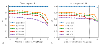 |
148 |
Machine learning can boost the acquisition and estimation of anomalous diffusion model
Boyan Xu, Yang Fan, Jia-Hong Gao
The departure from mono-exponential decay of the diffusion-induced signal loss has promoted the research of anomalous diffusion in MRI. It has been found that anomalous diffusion models offer substantial advantages over the conventional method in clinical applications. However, these models require more diffusion weightings for complicated estimation procedure, which prevents its further application. In this study, we demonstrated that machine learning can be applied to accelerate the estimation of anomalous diffusion parameters. Furthermore, feature selection was used to identify the most relevant signals, thus helping to reduce the extensive sets of diffusion weightings.
|
|
3485.
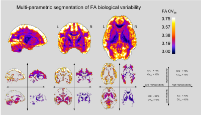 |
149 |
Multi-parametric segmentation of whole-brain variability maps of diffusion MRI metrics
Pedro Luque Laguna, Steve Williams, Flavio Dell'Acqua
The analysis of the whole-brain statistical variability maps corresponding to different statistical coefficients provides quantitative and anatomical information regarding the reproducibility, reliability or biological variability of diffusion MRI data. However, the separate analysis of each statistical map cannot reveal the emerging relationships that exist between these three properties of the data and their interactions across the brain anatomy. In this abstract, we present a new integrated multi-parametric segmentation approach for the combined visualisation and analysis of the reproducibility, reliability and biological variability maps using diffusion MRI data acquired from an older population.
|
|
3486.
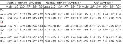 |
150 |
Alteration of IVIM Parameters by Signal Variation of Multiple b-value DWI Measurement
Chun-Jung Juan, Teng-Chieh Cheng, Tzu-Cheng Chao, Teng-Yi Huang, Chia-Wei Lin, Wu-Chung Shen, Yi-Jui Liu
The purpose of this study is to explore the alterations of IVIM measurement among multiple b-value sets with different low b-value and high b-value numbers using EPDWI. EPDWI with 41 different b-values was performed. For simulating the situation in clinic, five sets of 16 b-value numbers were chose from the 41 b values. DWI data of 1000 pixels for each WM, GM and CSF were randomly chose to evaluate the error of IVIM parameters. Our study demonstrated the variation of DWI signal with multi-b-value, affect the evaluation of Df and f but does not affect Ds in pixel-wise IVIM analysis.
|
|
| Top |
Diffusion MRI: Diffusion Gradient Waveform Design & Optimization
Digital Poster
Diffusion
Wednesday, 15 May 2019
| Exhibition Hall |
08:15 - 09:15 |
| |
|
Computer # |
|
3487.
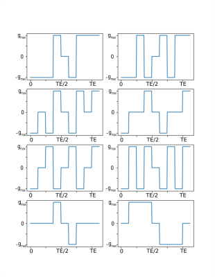 |
151 |
Optimal selection of diffusion-weighting gradient waveforms using compressed sensing and dictionary learning
Raphaël Truffet, Christian Barillot, Emmanuel Caruyer
Acquisition sequences in diffusion MRI rely on the use time-dependent magnetic field gradients. Each gradient waveform encodes a diffusion-weighted measure; a large number of such measurements are necessary for the in vivo reconstruction of microstructure parameters. We propose here a method to select only a subset of the measurements while being able to predict the unseen data using compressed sensing. We learn a dictionary using a training dataset generated with Monte-Carlo simulations; we then compare two different heuristics to select the measures to use for the prediction. We found that an undersampling strategy limiting the redundancy of the measures allows for a more accurate reconstruction when compared with random undersampling with similar sampling rate.
|
|
3488.
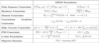 |
152 |
Reduced Eddy Current induced image distortions and Peripheral Nerve Stimulation based on the Optimal Diffusion-weighting Gradient Waveform Design (ODGD) formulation
Óscar Peña-Nogales, Yuxin Zhang, Rodrigo de Luis-Garcia, Santiago Aja-Fernández, James Holmes, Diego Hernando
Diffusion-Weighted MRI (DW-MRI) often suffers from signal attenuation due to long TE, motion-related artefacts, dephasing due to concomitant gradients (CGs), and image distortions due to eddy currents (ECs). Further, the application of rapidly switching gradients may cause peripheral nerve stimulation (PNS). These challenges hinder the progress, application and interpretability of DW-MRI. Therefore, based on the Optimized Diffusion-weighting Gradient waveforms Design (ODGD) formulation, in this work we design optimal (minimum TE) nth-order moment-nulling diffusion-weighting gradient waveforms with or without CG-nulling able to reduce EC induced distortions and PNS-effects. We assessed the feasibility of these waveforms in simulations and phantom experiments.
|
|
3489.
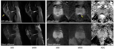 |
153 |
Silent Distortionless DWI
Jianmin Yuan, Yuxin Hu, Anne Menini, Christopher Sandino, Jesse Sandberg, Marcus Alley, Michael Lustig, Brian Hargreaves, Shreyas Vasanawala
Standard clinical EPI based diffusion sequence has high acoustic noise, which is uncomfortable for patients. The images acquired by single-shot EPI also suffer from distortion in areas of large susceptibility variations. To overcome these two limitations, we have developed a silent and distortion-free DWI sequence using a driven equilibrium diffusion prepared multi-segmented Rotating Ultra-Fast Imaging Sequence (RUFIS) readout. The sequence was validated in phantom, volunteers, and patients. The results showed the proposed sequence provides similar diffusion contrast and ADC measurement compared to DW-EPI, with improved image quality and a sound level that approaches that of ambient noise.
|
|
3490.
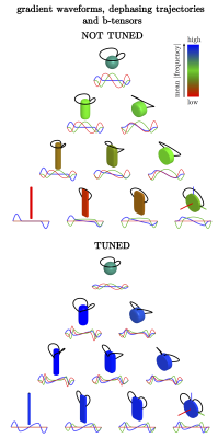 |
154 |
The tuned trinity of b-tensors
Samo Lasic, Henrik Lundell, Daniel Topgaard
Unprecedented microstructural details of heterogeneous materials such as tissue are non-invasively accessible by multidimensional diffusion encoding (MDE) MRI using varying shapes of b-tensors. MDE can probe multidimensional distributions of diffusion tensors in terms of their size, shape and orientation. For robust conclusions, time-dependent diffusion effects need to be considered in MDE. A convenient recipe for generating b-tensors of varying shape was implemented including a single adjustable tuning parameter. The resulting b-tensors can thus be tuned for sensitivity to time-dependent diffusion.
|
|
3491.
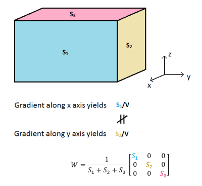 |
155 |
An accurate surface-to-volume ratio estimation by general diffusion gradient waveform
Nicolas Moutal, Ivan Maximov, Denis Grebenkov
We generalize Mitra’s formula for estimating the surface-to-volume ratio of an anisotropic medium with any pore shapes by using arbitrary diffusion gradient waveforms. We show that the surface-to-volume ratio can be significantly misestimated using the original formula without taking into account the applied diffusion gradient profile. For an accurate estimation, we derive new “isotropy” criteria which ensure that the diffusion weighted sequence is insensitive to the mesoscopic anisotropy of the sample. As a result, we introduce a novel class of porous media characteristics which are useful for biomedical applications.
|
|
3492.
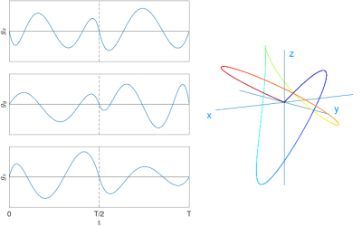 |
156 |
Optimized diffusion gradient waveforms for estimating surface-to-volume ratio of an anisotropic medium
Nicolas Moutal, Ivan Maximov, Denis Grebenkov
We present here a simple and efficient algorithm for designing of diffusion gradient profiles allowing one to implement prescribed theoretical properties. This algorithm generalizes the well-known sine and cosine decomposition and can handle various experimental constraints. Relying on recent diffusion MRI theoretical advances, we apply it to the challenging problem of estimating the surface-to-volume ratios in anisotropic media.
|
|
3493.
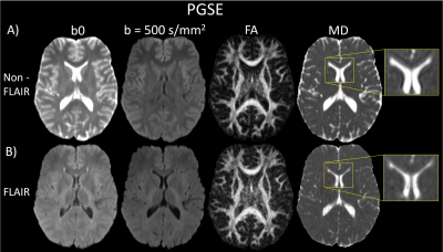 |
157 |
Oscillating Gradient Spin-Echo Diffusion Tensor Imaging of the Human Brain using FLAIR
Fernanda Padron, Thorsten Feiweier, Christian Beaulieu
Oscillating-gradient spin-echo (OGSE) diffusion tensor MRI permits very short diffusion times (e.g. 5 ms) relative to the typical pulsed-gradient spin-echo (PGSE), but it is challenging to acquire in the human brain. Given low b-values and spatial resolution, it is more prone to problematic artifacts such as Gibbs ringing and partial-volume effects that can compromise diffusion parameter measurements. It is shown here that the use of inversion-recovery FLAIR to suppress cerebrospinal fluid can significantly reduce such artifacts, improve overall image/map quality, and provide better quantitation of OGSE-PGSE diffusion parameter differences in the white matter of the human brain.
|
|
3494.
 |
158 |
Impact of different b-value combinations on Radiomics Features of Apparent Diffusion Coefficient in Cervical Cancer
Presentation Not Submitted
Zilong Yuan, Yaoyao He, Yi Rong, Hao Chen, Zhaoxi Zhang, Jianfeng Qiu, Lili Zheng, Stanley Benedict, Yulin Liu
There is potential influence of different b-value combinations on evaluation of ADC-based radiomics features. The aim of this study is to investigate the difference between ADC-based radiomics feature extracted from ADC maps with different b-value combinations in cervical cancer. It was found that variable b-value combinations can play substantially impact on radiomics features extracted from corresponding ADC maps in cervical cancer. Therefore, we should pay more attention to the choice of b-value combinations in retrospective and multi-center studies.
|
|
3495.
 |
159 |
A Real-time Solver for Convex Optimization of Diffusion Encoding Gradient Waveforms
Michael Loecher, Matthew Middione, Kévin Moulin, Daniel Ennis
Recent work has demonstrated the usefulness of designing time optimal gradient waveforms for diffusion encoding, especially when incorporating gradient moment nulling to mitigate bulk motion artifacts. Prolonged optimization times, however, can limit the usability of these methods. This work presents a solver fast enough to run in real-time, and investigates the performance over a range of parameters and tests for convergence. The solver is able to generate diffusion encoding gradient waveforms with gradient moment nulling in 5-50 ms – fast enough to permit real-time optimization on the scanner.
|
|
3496.
 |
160 |
Impact of Residual Gradient Moments on Diffusion Weighted Imaging
Matthew Middione, Michael Loecher, Kévin Moulin, Daniel Ennis
Bulk motion corrupts diffusion measurements in the heart but can be mitigated by nulling first and second gradient moments. When using optimization methods to design diffusion gradient waveforms with moment nulling, imperfect gradient waveforms arise from discrete convergence criteria, which impart residual (non-zero) gradient moments. This leads to intravoxel dephasing, signal loss, and inaccurate ADC measurements. Herein, simulations show that residual M0≤10-2mT/m•s, M1≤10-4mT/m•s2, and M2≤10-5mT/m•s3 leads to ≤5% increase in ADC. This work defines convergence criteria requirements for residual gradient moments that enable faster optimizations and more accurate measurements of ADC when using optimization methods for cardiac DWI sequence design.
|
|
3497.
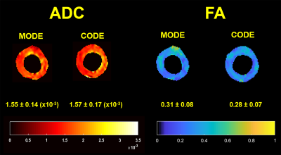 |
161 |
In-vivo cardiac DTI: Comparison between Motion Compensated Optimized Diffusion Encoding (MODE) and Convex Optimized Diffusion Encoding (CODE) Techniques
Prateek Kalra, Waqas Majeed, Mohammad Maddah, Xiaokui Mo, Richard White, Arunark Kolipaka
Diffusion-weighted imaging(DWI) is used to determine defective areas such as fibrosis in the myocardium without the need of contrast agent by calculating apparent diffusion coefficient(ADC) and fractional anisotropy(FA). However, DWI of heart is very challenging due to bulk motion to which diffusion encoding gradients are sensitive. Previous studies proposed CODE (convex optimized diffusion encoding) and MODE (Motion-compensated Optimized Diffusion Encoding). Aim of this study is to compare ADC and FA using MODE and CODE technique in healthy subjects. No significant difference was found between two techniques. However, for a given TE, MODE generated slightly higher b-value compared to CODE.
|
|
3498.
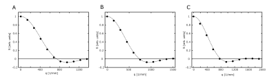 |
162 |
Using symmetrized temporal gradient profiles for water diffusion pore imaging on a 14.1 T spectrometer
Dominik Ludwig, Frederik Laun, Karel Klika, Peter Bachert, Tristan Kuder
Diffusion pore imaging can be used to retrieve the pore-space function of an arbitrary closed pore. While a few publications involving pore imaging using the long–narrow gradient approach exist [1–3], all of them involve the use of sophisticated acquisition schemes like CPMG or involve an additional readout gradient. In this study, we were able to show that it is indeed possible to acquire the pore space function of capillaries with a diameter of 15 µm using a simple modification of the long–narrow gradient scheme on a 14.1 T NMR spectrometer.
|
|
3499.
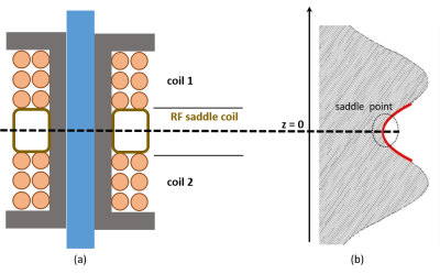 |
163 |
A novel approach to diffusion phase imaging using non-linear gradients in anisotropic media
Pamela Wochner, Jason Stockmann, Torben Schneider, Jack Lee, Ralph Sinkus
Classical methods for Diffusion-Weighted MRI use linear gradients to obtain information about tissue microstructure encoded in the loss of MR magnitude. These linear gradients cause diffusing spins to dephase which prevents the use of phase information. When, however, gradient fields are applied that vary quadratically in space, diffusion in anisotropic media results in a shift of net phase with only a minimal loss in signal magnitude enabling the exploration of MR phase. Both the theory of this new concept and Monte-Carlo simulations predict net phase for diffusion experiments using quadratic gradients in anisotropic media in excellent agreement.
|
|
3500.
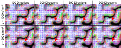 |
164 |
Optimizing Diffusion MRI for Mapping Cortical Fiber Patterns
Grant Yang, John Cocjin, Jennifer McNab
A custom DW pulse sequence is optimized to minimize echo time and evaluate optimal trade-offs between SNR, b-value and spatial resolution for mapping cortical fiber patterns. Using a lower b-value (500 vs. 1000 s/mm2) failed to improve estimates of the principal fiber orientation in the cortex despite the significant boost in signal. Accurate estimates of the principal fiber orientation in the cortex requires a spatial resolution of 1.44 mm isotropic or less.
|
|
3501.
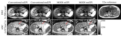 |
165 |
Motion-Robust and Distortion-Corrected Diffusion MR Imaging of the Liver with Optimized Motion-Compensated Waveforms and Multi-shot EPI acquisition
Yuxin Zhang, Ruiqi Geng, Arnaud Guidon, James Holmes, Diego Hernando
Conventional liver diffusion MRI acquisitions suffer from several challenges including low spatial resolution, B0-induced distortions, and elastic motion-induced signal voids. In this work, motion-robust and distortion-corrected liver diffusion-weighted imaging (DWI) was enabled by combining optimized motion compensated diffusion waveforms with multi-shot EPI acquisitions. Diffusion-weighted images of healthy volunteers were acquired to evaluate the effect of motion compensation and distortion correction. Preliminary results demonstrate the feasibility of the proposed approach, including reduced ADC bias in the left lobe (due to the motion-robust waveforms) and reduced distortion (due to the multi-shot acquisition) compared to conventional liver DWI.
|
|
3502.
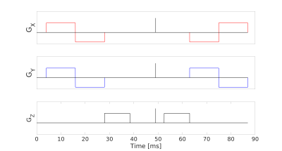 |
166 |
Towards clinically viable and robust measurement of microscopic diffusion anisotropy
Nico Arezza, Aidin Arbabi, Corey Baron
In contrast to fractional anisotropy, microscopic diffusion anisotropy allows measurement of pore structural anisotropy with no influence from orientational coherence. However, acquisitions that allow in vivo measurement of microscopic anisotropy are generally time-consuming and/or low SNR. Here, we introduce an efficient approach to perform isotropic diffusion encoding and measure high quality full-brain microscopic anisotropy in a scan time of only 3 minutes at 7T. Results are demonstrated in a healthy subject and MS patient, where clear delineation of lesions is observed in the latter case.
|
|
3503.
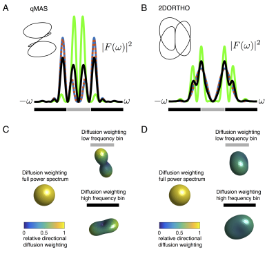 |
167 |
Reducing spectral anisotropy in isotropic b-tensor multidimensional diffusion encoding
Henrik Lundell, Markus Nilsson, Filip Szczepankiewicz, Carl-Fredrik Westin, Daniel Topgaard, Samo Lasic
Multidimensional diffusion encoding isotropic b-tensors eliminate rotational variance from anisotropic Gaussian diffusion, an essential feature for estimating microscopic anisotropy. In anisotropic substrates which exhibit non-Gaussian diffusion on the time scale of the encoding pulse, the variance in temporal characteristics of diffusion encoding across different directions – spectral anisotropy (SA) - may introduce a directional variance in apparent diffusivities. We propose an alternative isotropic encoding with drastically lower SA, which in turn allows accessing intrinsic signatures of non-Gaussian diffusion.
|
|
3504
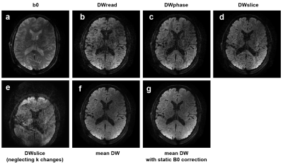 |
168 |
Diffusion MRI of the Brain with a Gradient Strength of 100 mT/m and a Slew Rate of 1200 T/m/s
Video Permission Withheld
Bertram Wilm, Manuela Roesler, Franciszek Hennel , Markus Weiger, Klaas Pruessmann
The performance of diffusion-weighted single-shot EPI is limited by off-resonance artifacts and low signal-to-noise. To address both problems, we employ a recently developed gradient insert (strength: 100 mT/m, slew rate: 1200 T/m/s). Thereby the time for the diffusion encoding as well as the EPI readout train duration can be significantly shortened, resulting in higher signal yield and robustness against off-resonance artifacts. First in-vivo results are presented.
|
|
3505.
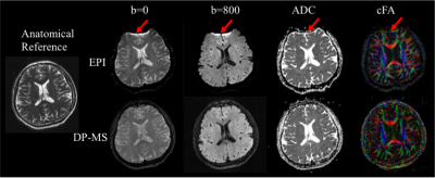 |
169 |
3D distortion-free multi-slab diffusion-prepared magnitude-stabilized bSSFP imaging
Yu Gao, Ziwu Zhou, Xinyu Ye, Hua Guo, Yingli Yang, Peng Hu
A 3D diffusion-prepared magnitude-stabilized bSSFP (DP-MS-bSSFP) sequence was proposed and validated to achieve high resolution distortion-free brain diffusion imaging. Shot-to-shot phase variation was corrected using the plane-by-plane MUSE technique. High-quality 3D diffusion images, ADC maps and cFA maps were obtained from four volunteers. Good agreement of white matter ADC between DW-ssEPI and DP-MS-bSSFP was observed based on the Bland-Altman plot. Geometric accuracy of DP-MS-bSSFP was confirmed by a four-fold reduction of target registration error compared with DW-ssEPI. In conclusion, the proposed DP-MS-bSSFP approach provided high-quality 3D diffusion images and diffusion tensor images with high ADC accuracy and minimal distortion.
|
|
3506.
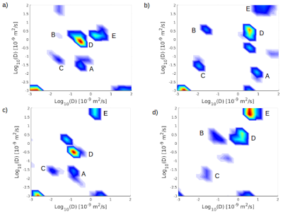 |
170 |
In-vivo Diffusion Exchange Spectroscopy (DEXSY), to measure water exchange in tumours.
James Breen-Norris, Bernard Siow, Claire Walsh, Mark Lythgoe, Andrada Ianus, Daniel Alexander, Simon Walker-Samuel
We describe the first use of Diffusion Exchange Spectroscopy (DEXSY) in-vivo, to measure water exchange in tumours. We present preliminary data suggesting that we can capture information about the micro-structure of tumors, with what appear to be intracellular, extracellular and perfusion peaks present in diffusion-diffusion exchange plots, acquired from subcutaneous tumor xenograft models, in-vivo using DEXSY.
|
|
3507.
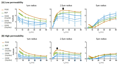 |
171 |
Effects of Diffusion Gradient Waveforms on Time-dependent Kurtosis and Microstructural Properties
Dan Wu, Jiangyang Zhang
Diffusion-time (td) dependent diffusion MRI is a promising tool to probe tissue microstructure. Although different diffusion gradient waveforms (DGW) have been introduced to achieve short and long td’s, whether they have comparable sensitivity to microstructural changes remains to be investigated. Here, we examined the td-dependency of kurtosis measured using pulsed, bipolar, and oscillating gradients with different microstructural substrates. Simulations and in vivo experiments showed that the choice of DGW affects the characteristics of time-dependent kurtosis, and this effect varied with microstructural size and permeability. This knowledge is important for design of td-dependent diffusion MRI experiments.
|
|
3508.
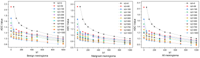 |
172 |
The optimal b value study of endogenous water molecule diffusion imaging in the diagnosis of brain tumors
Presentation Not Submitted
Fen Xing, Guangyao Wu
In this study, thirty-six cases of meningioma were divided into benign and malignant meningioma. ADC values fitted by 11 b values and any pairs of two b values were used to distinguish between benign and malignant meningioma. We found that, the ADC values of benign, malignant, and all meningiomas showed a decreasing trend with the increase of b value, and the smaller the b value, the greater the decrease. when the b value was ≥ 200 s/mm2, any combinations of b values could discriminate between benign and malignant meningioma. The ADC values obtained by fitting b = 700 and b = 1000 s/mm2 had the highest AUC value.
|
|
| Top |
Diffusion MRI: Data Acquisition
Digital Poster
Diffusion
Wednesday, 15 May 2019
| Exhibition Hall |
09:15 - 10:15 |
| |
|
Computer # |
|
3509.
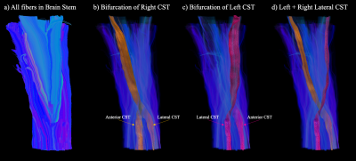 |
1 |
Sub-Millimeter Isotropic DTI and fiber tractography of the human Spinal Cord in-vivo
Iain Bruce, Christopher Petty, Allen Song
The ability to characterize complex microstructures in the small cross-sectional area of the spinal cord through diffusion tensor imaging has traditionally been limited by the achievable spatial resolution in-vivo. Through ultra-high spatial resolution diffusion imaging, this study presents a technique for accurately delineating complex fiber pathways such as the corticospinal tracts. When imaged with sub-millimeter isotropic spatial resolutions, it is possible to characterize intricate details in the spinal cord such as the bifurcations and decussations of the corticospinal tracts. The improved delineation of neural pathways in the spinal cord could facilitate the placement of stimulation electrodes for movement disorder treatments.
|
|
3510.
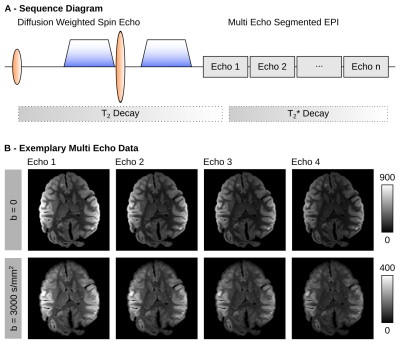 |
2 |
Multi-Echo Segmented Diffusion-Weighted MRI for Ex-Vivo Whole-Brain Measurements with 300mT/m Gradients
Cornelius Eichner, Toralf Mildner, Michael Paquette, Torsten Schlumm, Catherine Crockford, Roman Wittig, Evgeniya Kirilina, Carsten Jäger, Harald Möller, Nikolaus Weiskopf, Angela Friederici, Alfred Anwander
We provide a novel method to increase SNR of segmented diffusion-weighted EPI acquisitions. Multiple gradient echoes were acquired after each diffusion-preparation and combined in an SNR-optimized way using weightings from quantitative T2* maps. The combination of diffusion-weighted echoes yielded an SNR-gain of 58% compared to single-echo dMRI data with an increase in the segmented readout duration by only 23.1 ms. The multi-echo diffusion MRI acquisition and combination were employed to acquire high-quality ex-vivo diffusion-weighted MRI data from a wild chimpanzee brain.
|
|
3511.
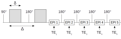 |
3 |
A Multi-Echo Stejskal-Tanner EPI Sequence for Rapid Measurements of T2 and Diffusion Tensor Correlations
Richard Buschbeck, Ezequiel Farrher, Kuan-Hung Cho, Ming-Jye Chen, Seong Dae Yun, Chang-Hoon Choi, Li-Wei Kuo, N. Jon Shah
Initial phantom and in vivo results of a multi-echo Stejskal-Tanner EPI sequence are presented demonstrating the feasibility of rapid measurements for diffusion tensor and T 2 correlations. The diffusion and T 2 maps are artefact-free and are found to be very similar to the ones derived from the conventional individual-echo methods. However, the data tend to display slightly overestimated diffusivities and T 2, which need to be investigated further. Compared to the standard method, the proposed multi-echo sequence reduces the scan time by a factor of five, which is necessary to make multidimensional diffusion techniques feasible in clinical settings.
|
|
3512.
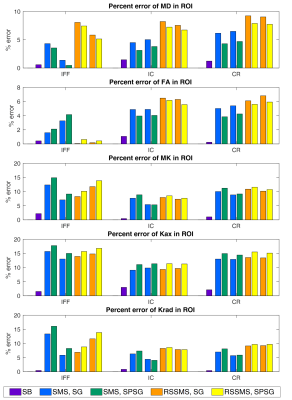 |
4 |
Effects of imperfect slice separation in simultaneous multi-slice for diffusion kurtosis imaging
L. Tugan Muftuler, Daniel Olson, Volkan Arpinar, Andrew Nencka
Simultaneous multislice (SMS) technique accelerates MRI data acquisition. However, slice separation in reconstruction is imperfect. This results in residual leakage between the simultaneously excited slices. For diffusion MRI, repeated pairing of the same slices in the image series leads to repeated leakage of a consistent diffusion signal from the other slice. Studies of this effect on diffusion MRI is sparse. Here, we tested two different SMS acquisition and image reconstruction techniques and quantified resulting errors on all diffusion kurtosis (DKI) metrics in different ROIs. Large errors were observed with SMS compared to singleband acquisitions for all DTI and DKI metrics
|
|
3513.
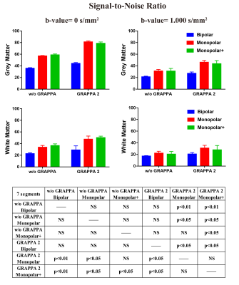 |
5 |
Evaluation of Diffusion-Weighted Readout-Segmented EPI in Macaque Brain Imaging at 7T
Pinyi Wang, Jialu Zhang, Meizhen Qian, Yi Sun, Robin M. Heidemann, Ruiliang Bai, Dingxin Wang, Xiaotong Zhang
Diffusion-weighted imaging (DWI) has been widely used in brain structure and connectivity investigations and other neuroscience applications. To implement an optimized protocol for high spatial resolution DW imaging at ultra-high field, we have conducted macaque brain imaging by using readout segmented echo planar imaging at 7T. Our results indicate that, the rsEPI has potential to achieve DWI with improved signal-to-noise ratio and with the intent of minimizing geometric distortion.
|
|
3514.
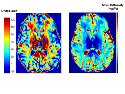 |
6 |
A model free, sparse acquisition approach (« Sindex ») to investigate brain tissue microstructure from diffusion MRI data in the human brain
Denis Le Bihan, Tetsuya Yamamoto, Masaki Fukunaga, Tomohisa Okada, Norihiro Sadato
Diffusion MRI provides noninvasively information of tissue microstructure. Current models allow to empirically analyze data or to provide more insightful information on the tissue features. However, those models require strong assumptions on the underlying tissues and the acquisition of large image data sets with different acquisition parameters. We have investigated a new, model free approach which enables classification of tissue types from the “proximity” or resemblance of their diffusion MRI signal profile at a sparse set of key b values (maximizing sensitivity to tissue microstructure) to a library of “signature” signal profiles (e.g. typical brain grey and white matter).
|
|
3515.
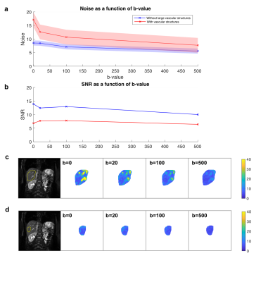 |
7 |
Maximising Repeatability: It’s Time to Reconsider the Sampling Scheme for IVIM
Callie Deng, Pa°l Erik Goa, Matthew Orton, Torill Sjøbakk, Tone Bathen, Neil Jerome
Diffusion-weighted imaging (DWI) has the potential to improve characterisation and detection of breast cancer. A variety of models are available to fit the relationship between MRI signal and b-value, but the repeatability of such models is often overlooked. In this study, we simulate repeated DWI scans and quantify the precision of IVIM parameters from segmented fitting for D and f; our findings suggest that repeatability of f is highly dependent on disproportionate noise at b=0, and repeatability for f and D can be maximised by redistribution of signal averages. Optimal acquisition strategies for varying values of inter-scan noise are reported.
|
|
3516.
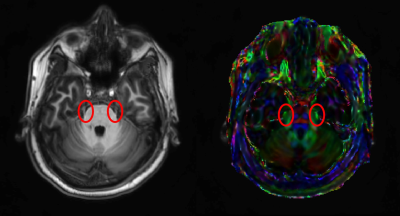 |
8 |
TRACTOGRAPHY OF THE TRIGEMINAL NERVE OUTSIDE THE BRAIN
Sulagna Sahu, Munish Chauhan, Stephen Tillery, Vikram Kodibagkar, Rosalind Sadleir
Diffusion Tensor Imaging (DTI) tractography used for the visualization of cranial nerves has mostly focused on tracts inside the brain. This work focuses on tracking trigeminal nerve branches using DTI outside the brain. Trigeminal nerve efferents in the face can be stimulated using transdermal electrical neuromodulation. Locations of subcutaneous tracts of the ophthalmic and maxillary branches of the trigeminal nerve may assist with electrode placement in these studies. Here, we describe use of HARDI-derived DTI to determine locations of trigeminal nerve projections in the face.
|
|
3517.
 |
9 |
Time-efficient high resolution diffusion weighted SSFP measurements at 14.1T
Dario Bosch, Gisela Hagberg, Rolf Pohmann, Thomas Shiozawa-Bayer, Bernhard Hirt, Klaus Scheffler
This study investigates the use of diffusion weighted SSFP imaging for high-resolution diffusivity measurements of post-mortem tissue, using two different models (Wu-Buxton and Freed) and draws a comparison to conventional DW SE-Measurements. A toolbox was developed to optimize SSFP imaging parameters for selected tissue regions which was then used successfully. We showed that the SSFP approach is highly valuable for time-efficient high-resolution post-mortem diffusion weighted imaging.
|
|
3518.
 |
10 |
Feasibility of 129Xe MRI Morphometry Measurements with Acceleration Factors of 7 and 10
Alexei Ouriadov, Andrew Westcott, David McCormack, Grace Parraga
Hyperpolarized gas pulmonary MRI provides physiologically relevant biomarkers of obstructive lung disease including emphysema, bronchopulmonary dysplasia, congenital lobar emphysema and alpha-1 antitrypsin deficiency. Recently, a stretched-exponential-model combined with under-sampling in the imaging and diffusion direction was used for the generation of 3He static-ventilation, T2* and multiple b-value diffusion-weighted MRI-based ADC/morphometry maps. We hypothesize that the 3He method can be modified to provide whole lung 129Xe MRI-based emphysema biomarkers including static-ventilation, T2*/ADC/morphometry maps with high-spatial-image-resolution in a single breath-hold. Therefore, in this proof-of-concept evaluation, our objective was to demonstrate a feasibility of this approach in a small group of patients.
|
|
3519.
 |
11 |
3D distortion-free diffusion imaging using an accelerated multi-shot diffusion-prepared sequence
Elisa Roccia, Radhouene Neji, Vicky Goh, Isabel Dregely
Current methods for cancer diffusion imaging employ 2D single-shot diffusion weighted echo planar imaging sequences, which are fast but have limited resolution and are prone to distortion artefacts. The purpose of this work is to achieve 3D high-resolution and distortion-free diffusion imaging suitable for cancer precision diagnostics and therapy planning. A multi-shot diffusion-prepared sequence with gradient echo readout, centric encoding, and accelerated variable density acquisition was developed and tested in healthy volunteer brains as an exemplar. A full set of 2.2-fold accelerated diffusion weighted images in three diffusion directions was acquired in 4:30 min producing quantitative 3D distortion-free ADC maps.
|
|
3520.
 |
12 |
Diffusion-Weighted ZOOM-EPI with Simultaneous Multi-Slice Imaging
Jürgen Finsterbusch, Caspar Florin
Zonal oblique multi-slice (ZOOM) EPI uses cross-sectional RF excitations to focus the measurement volume to a small, inner volume which allows to reduce the FOV without aliasing in the phase-encoding direction. Thus, geometric distortions are reduced and the spatial resolution of diffusion-weighted acquisitions can be increased as has been demonstrated for the optic nerve and spinal cord. In this study, ZOOM-EPI is combined with simultaneous multi-slice (SMS), the boundary condition to avoid unwanted signal contributions is determined, and the feasibility to shorten acquisitions times is demonstrated for diffusion-tensor imaging (DTI) of the human spinal cord.
|
|
3521.
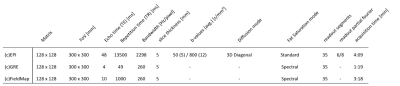 |
13 |
Improvement of EPI-based DWI of the head/neck region using additional local shim coils at 3 Tesla
Sven Walter, Regine Perl, Mike Notohamiprodjo, Konstantin Nikolaou, Sergios Gatidis
Implementing additional local shim coils into the head and neck surface coil may enhance the performance of EPI-based diffusion weighted imaging of the head/neck region at 3 Tesla. Thus, we evaluated single shot echo planar DWI and standard T1-weighted gradient echo acquisition without and with the use of additional local shim coils. In addition, apparent diffusion coefficients were quantified in specific anatomical regions. Local shim coils improved the homogeneity of the static magnetic field, resulting in marked improvement of image quality, signal loss, distortion and fat saturation. ADC values did not significantly differ between the measured anatomical compartments.
|
|
3522.
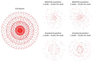 |
14 |
A novel Multishell Acquisition with Increased b-Shells and Sparse ORientations (MAISSOR)
Manish Amin, Thomas Mareci
With improvements in MRI technology, more informative diffusion acquisitions can be obtained to improve tissue microstructure analysis. In this study, a Multi-shell Acquisition with Increased b-Shells and Sparse ORientations (MAISSOR) is proposed to optimize diffusion acquisition. The scheme improves diffusion signal decay fitting while simultaneously improving fiber orientation distribution1 (FOD) calculations as well as diffusivity metrics, such as those derived from diffusion tensor imaging2 (DTI).
|
|
3523.
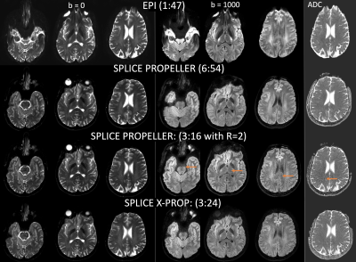 |
15 |
A rapid PROPELLER technique for high resolution diffusion weighted imaging
Zhiqiang Li, Melvyn Ooi, John Karis
Diffusion weighted imaging is routinely used in the clinical settings. EPI is commonly used but suffers from severe geometric distortion. TSE/PROPELLER based DWI techniques are free of geometric distortion, but lead to long scan time and high SAR. In this project we propose a new SPLICE X-PROP technique to provide rapid high resolution DWI, by incorporating a SPLICE acquisition approach and a cross-blade GRASE readout into the PROPELLER technique. The improvement over conventional SPLICE PROPELLER technique is demonstrated with volunteer scans.
|
|
3524.
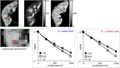 |
16 |
Investigating microscopic diffusion anisotropy in the human kidney using multidimensional diffusion encoding
Fabio Nery, Filip Szczepankiewicz, João de Almeida Martins, Matt Hall, Isky Gordon, David Thomas, Chris Clark
Diffusion tensor imaging (DTI) has been widely used to provide tissue microstructure measures such as fractional anisotropy (FA). Advances in diffusion acquisition methods (e.g. efficient spherical tensor encoding) have enabled more specific microstructural parameters to be derived, including microscopic fractional anisotropy (µFA). This work focused on extending our initial observation of µFA in the kidney through a detailed analysis of the linear and spherical diffusion encoding in healthy subjects, by comparing conventional FA to the µFA in human kidneys, and investigating the technical limitations of the current approach.
|
|
3525.
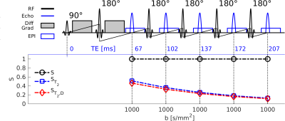 |
17 |
Diffusion weighted multi-spin echo sequence fuses T2-relaxometry and diffusometry
Jelle Veraart, Ying-Chia Lin, Tiejun Zhao, Steven Baete
Combination of diffusion weighted MRI with orthogonal measures such as T22-weighting
has been proposed to overcome the fit degeneracy found in microstructure modeling of diffusion signals. However, the repetition of diffusion measurements at different TE leads to unacceptably long acquisition times, hindering clinical applicability of this approach. Here, we propose an accelerated acquisition using a multi spin echo diffusion and T22-weighted
sequence which samples each diffusion weighting at several TEs with a CPMG read-out train after the standard monopolar diffusion encoding spin echo. In the current configuration this speeds the acquisition up by a factor of 2.5x.
|
|
3526.
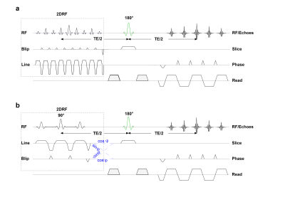 |
18 |
Sensitivity of 2D-Selective RF Excitations for Inner-Field-of-View Diffusion-Weighted Imaging to Field Inhomogeneities
Caspar Florin, Jürgen Finsterbusch
Inner-field-of-view techniques based on 2D-selective RF (2DRF) excitations reduce geometric distortions of echo-planar imaging and improve diffusion-weighted imaging of the human spinal cord. A comparison of two common approaches revealed that a setup with the 2DRF excitation collinear to the image plane suffers from a reduced signal-to-noise ratio (SNR) for tall slice stacks. In this study, it is shown that the SNR loss is related to the larger sensitivity of the corresponding 2DRF excitation to frequency offsets caused by field inhomogeneities and is particularly pronounced for the large fields-of-excitation and 2DRF pulse durations needed for tall slice stacks.
|
|
3527.
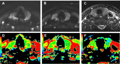 |
19 |
A comparative study of single shot EPI, reduced field of view EPI, PROPELLER FSE based diffusion weighted imaging in the evaluation of normal thyroid gland
Jingjing Bai, Xiaocheng Wei, Xijun Jiao, Yinxia Guo, Yanan Li, Miaomiao Wang, Jianxin Guo, Jian Yang
Thyroid gland diffusion images often suffer from high level of image distortion due to poor magnetic filed homogeneity. We evaluated the image quality and compared apparent diffusion coefficient (ADC) of healthy thyroid gland using EPI DWI, rFOV DWI and PROPELLER DWI images respectively. Our results indicated that the image quality of rFOV is better than that of EPI and PROPELLER DWI, while the image quality has no significant difference between EPI and PROPELLER DWI. Moreover, there was no significant difference in ADC value between EPI and rFOV DWI images, while the ADC value of PROPELLER EPI was significantly higher.
|
|
3528.
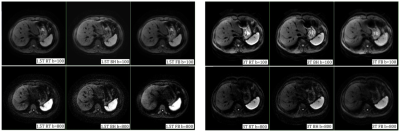 |
20 |
Diffusion-Weighted Imaging Of Upper Abdomen: A Comparative Study In Healthy Volunteers
Presentation Not Submitted
Zhuo Shi, Lizhi Xie, XinMing Zhao, Han Ou-Yang
Nowadays,no standard scanning protocol or parameter for upper abdomen DWI is available. Due to tension, fear or poor tolerance, patients’ respiration may vary during the scan, leading to incorrect ADC measurement and missed diagnosis. In our study, we performed quantitative and qualitative analyses of images acquired via respiratory-triggered, breath-hold and free-breathing DWI at both 1.5T and 3T, to compare their image quality and the apparent diffusion coefficient (ADC) values of upper abdomen, then decide which is the best technique for upper abdomen DWI.
|
|
3529.
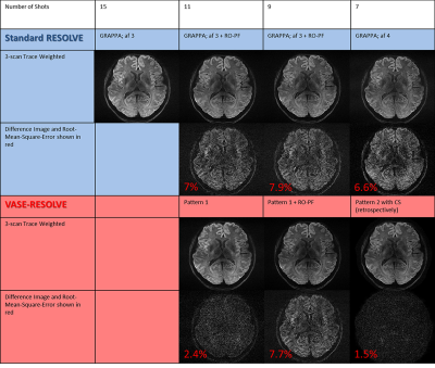 |
21 |
VASE-RESOLVE: Accelerated Readout Segmented Echo Planar Imaging with Compressed Sensing and Variable-Width Readout Segments
Patrick Liebig, Robin Heidemann, Bernhard Hensel, David Porter
Readout-segmented echo-planar imaging (EPI) with 2D navigator correction, also known as RESOLVE, is an established method for performing high-resolution diffusion weighted imaging in clinical studies. However, the method requires long acquisition times compared to single-shot EPI. A reduction in acquisition time could be achieved with Compressed Sensing (CS), but sub-sampling of EPI-based sequences is problematic because of the phase evolution during the echo train. This work introduces a new CS sampling scheme for readout-segmented EPI that varies the readout-segment width as a function of sampling density and preserves the phase relationship between data points.
|
|
3530.
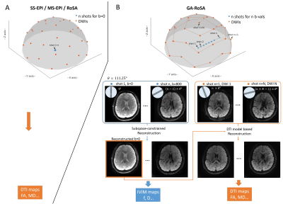 |
22 |
Golden-Angle Rotating Single-Shot Acquisition (GA-RoSA) for Simultaneous High-Resolution DTI and IVIM
Qiuting Wen, Feng Li, Kun Zhou, Christianne Leidecker, Yu-Chien Wu
High-resolution DWI often relies on multi-shot acquisitions, which suffer from long acquisition time and motion-related phase variations. Although effort has been made to accelerate DWI acquisitions, acquiring b=0 images still requires multiple shots and multiple averages. In this work, in addition to accelerating DWI acquisitions, the proposed method (GA-RoSA) further rearranged the b=0 acquisitions to cover a range of lower b-values without increasing scan times, so that IVIM parameters can be obtained for free. With a golden-angle rotation scheme, GA-RoSA introduced sampling incoherence facilitating its combination with sparsity-based reconstruction approaches. We demonstrated its feasibility for simultaneous high-resolution DTI and IVIM.
|
|
3531.
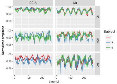 |
23 |
Can unprecedented echo times in human diffusion weighted fMRI help reveal its biological underpinnings?
Suryanarayana Umesh Rudrapatna, Lars Mueller, Marcello Venzi, Richard Wise, Derek Jones
The brain versus vein dilemma in BOLD fMRI has spurred research towards more direct correlates of neuronal activation. Diffusion-weighted fMRI (dfMRI) emerged as a potential alternative 17 years ago. However, its signal origins and utility have been greatly debated. In this work, we combine ultra-high-gradients and spiral readout to characterize dfMRI contrast in humans in parameter spaces (TE, b-value, SNR and resolution) that have never been accessible before. Varying TE over a wide range while keeping the b-value fixed allowed us to detect significant hemodynamic contributions to dfMRI contrast at a b-value of 1200 s/mm2.
|
|
3532.
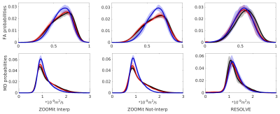 |
24 |
Novel HARDI-ZOOMit protocol detects changes in spinal cord microstructure in patients with asymptomatic non-myelopathic degenerative cervical spinal cord compression
René Labounek, Jan Valošek, Tomáš Horák, Alena Svátková, Petr Bednarík, Pavel Hok, Petr Kudlicka, Magda Horáková, Jan Kocica, Christophe Lenglet, Julien Cohen-Adad, Igor Nestrašil, Josef Bednarík, Petr Hluštík
Detection of degenerative cervical spinal cord compression (DCSCC) at early stage that causes spinal cord (SC) microstructure disruption is limited when current imaging techniques are used. Early detection of damage to SC can predict symptomatic degenerative cervical myelopathy (DCM). We are presenting novel HARDI-ZOOMit protocol (High Angular Resolution Diffusion Imaging + syngo ZOOMit sequence), which is able to detect changes in microstructural diffusion MRI (dMRI) parameters (e.g. fractional anisotropy – FA, mean diffusivity – MD) in asymptomatic DCSCC. Results obtained with HARDI-ZOOMit protocol reveal higher sensitivity when compared to clinical RESOLVE (REadout Segmentation Of Long Variable Echo trains) protocol.
|
|
3533.
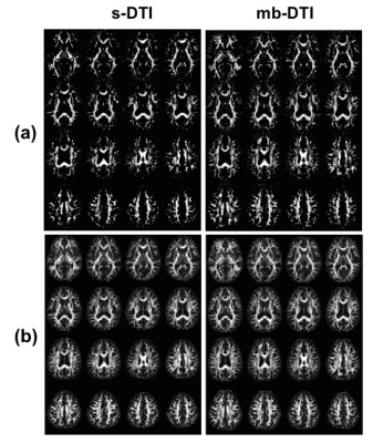 |
25 |
Clinical feasibility of multi-band diffusion tensor imaging (DTI) using simultaneous multi-slice acquisition for presurgical planning: Comparisons with standard DTI in patients with brain tumor
Mehrnaz Jenabi, Madeleine Gene, Nicholas Cho, Ricardo Otazo, Robert Young, Andrei Holodny, Kyung Peck
Standard diffusion tensor imaging (s-DTI) is currently utilized during presurgical planning to visualize white matter tracts despite its long scanning time. Multi-band DTI (mb-DTI) utilizes simultaneous multi-slice excitation, which greatly reduces scanning time and increases coverage. In this study, 42 patients with brain tumor were analyzed using mb-DTI and s-DTI in the arcuate fasciculus and corticospinal tract, two critical white matter tracts for language and motor function, respectively. When using mb-DTI, scanning time was reduced by 40% and the image quality and quantitative parameters were preserved. This study demonstrated the clinical feasibility and potential advantages of mb-DTI during presurgical planning
|
|
| Top |
Microstructural Modelling & Mapping
Digital Poster
Diffusion
Wednesday, 15 May 2019
| Exhibition Hall |
09:15 - 10:15 |
| |
|
Computer # |
|
3534.
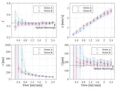 |
26 |
Comparison and Assessment of IVIM-MRI Methods Using a Capillary Flow Phantom
Moritz Schneider, Thomas Gaass, Julien Dinkel, Jens Ricke, Olaf Dietrich
In this study different intravoxel incoherent motion (IVIM) MRI methods were assessed and compared using phantom measurement. The phantom was constructed to mimic a capillary bed and allowed for the controlled application of fluid flow at varying rates. Advanced IVIM MRI methods beyond the biexponential pseudo-diffusion model were shown to be capable of accurately characterizing fluid flow inside a capillary network yielding intuitive parameters in a reproducible manner.
|
|
3535.
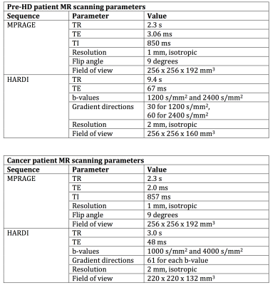 |
27 |
What are the consequences of ignoring non-Gaussian diffusion in models of convection-enhanced drug delivery to the human brain?
Eirini Messaritaki, S Umesh Rudrapatna, Najmus S Iqbal, Hannah Furby, Emma Yhnell, Claudia Metzler-Baddeley, William Gray, Derek Jones
Convection-enhanced drug delivery to the human brain is a promising method for treating neurodegenerative diseases and brain tumors. Accurate predictions of the drug concentration via computational fluid dynamics models are essential, and models not accounting for diffusion non-Gaussianity give predictions that are not in good enough agreement with experimental results. We use a fluid dynamics model recently presented in the literature, which accounts for diffusion non-Gaussianity, to calculate the differences with models that do not account for it, in data from pre-symptomatic Huntington’s disease patients and metastatic brain tumor patients. We make recommendations on the use of fluid dynamics models in the clinical setting.
|
|
3536.
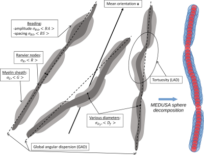 |
28 |
MEDUSA: a GPU-based tool to create realistic phantoms of the brain white matter microstructure using tiny spheres.
Kévin GINSBURGER, Felix MATUSCHKE, Fabrice POUPON, Jean-François MANGIN, Markus AXER, Cyril POUPON
This work proposes a novel Microstructure Environment Designer with Unified Sphere Atoms (MEDUSA) to construct realistic white matter cellular phantoms ensuring the absence of overlapping cells. It relies on a CUDA-based collision solver that enables to reach high values of packing density and angular dispersion, even in the case of multiple fiber populations. MEDUSA thus achieves the fast construction of biomimicking white matter phantoms with fully controlled geometrical properties, including axons, astrocytes and oligodendrocytes.
|
|
3537.
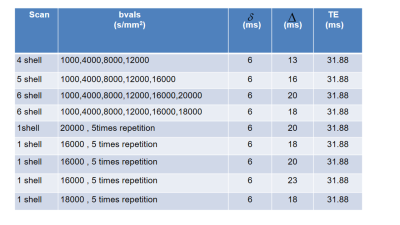 |
29 |
Axon diameter estimation in fixed human optic chiasm using diffusion weighted MR microscopy and microstructure-informed tractography
Mohammad Ashtarayeh, Tobias Streubel, Joao Periquito, Andreas Pohlmann, Thoralf Niendorf, Evgeniya Kirilina, Markus Morawski, Carsten Jäger, Stefan Geyer, Muhamed Barakovic, Alessandro Daducci, Siawoosh Mohammadi
We estimated mean axon diameter of ipsilateral bundles in a fixed human optic chiasm sample using diffusion weighted MR microscopy and COMMIT framework. We computed that the resolution limit for axon diameters of our dMRI data is about 1.3 µm, indicating that our approach is sensitive to a larger range of axon diameter distribution (ADD) than in-vivo dMRI studies. We found a similar pattern in the estimated ADD using MRI as was reported by a histological analysis of macaque monkeys’ optic chiasm, indicating that the estimated ADD provides similar sensitivity to regional differences in microstructure properties as in ex-vivo histology.
|
|
3538
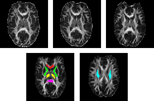 |
30 |
An observational study on the diffusion time and field strength dependency of the diffusion coefficient in human white matter
Video Permission Withheld
Sebastian Endt, Jan Martin, Lipeng Ning, Lars Müller, Andreas Wetscherek, Michael Uder, Bernhard Hensel, Yogesh Rathi, Frederik Laun
Signal dependence on diffusion time has been reported in various tissues and has been connected to cellular restrictions. It is conceivable, however, that it partly originates from relaxation weightings of different compartments. In this study, we investigate the diffusion time dependence of the diffusion tensor in brain tissue at 1.5 T, 3 T and 7 T using a stimulated echo sequence. The results show clear time dependencies of axial and radial diffusivity. 7 T data show a reduction of both diffusivities, indicating a non-negligible role of compartmental relaxation weighting effects.
|
|
3539.
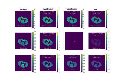 |
31 |
A microstructure fingerprinting approach with heterogeneous axon diameters: validation on feline spinal cord
Gaëtan Rensonnet, David Romascano, Benoit Scherrer, Simon Warfield, Benoit Macq, Jean-Philippe Thiran, Maxime Taquet
A microstructure fingerprinting approach is proposed in which an optimal single-fascicle configuration is selected from a pre-computed collection of Monte Carlo diffusion signals, or fingerprints, uniquely relating the diffusion MRI signals to the underlying axon properties. The simulated 3D geometries feature randomly-placed cylinders with diameter heterogeneity. The approach is validated on a public dataset of ex vivo cat spinal cord and exhibits indices of axon density and of the mean and standard deviation of the axon diameter distribution in agreement with histological measurements. The method is shown to outperform AMICO, which relies on approximate analytical expressions for the signal, and a simpler model using Monte Carlo simulations in a homogeneous packing of identical cylinders.
|
|
3540.
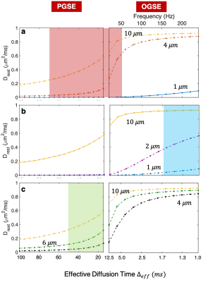 |
32 |
A Diffusion MRI Approach to Tumour Microstructure Modeling with Two Cell Populations of Different Sizes
Shu Xing, Ives Levesque
Advanced diffusion-weighted MRI allows the characterization of cancer tumours noninvasively by estimating cell radius R and volume fraction vin. Existing methods map the apparent R and vin , under the assumption that a given voxel contains one cell population. This work investigates the feasibility of estimating the radii and volume fractions of 2 cell populations co-existing in the same voxel. This method could be useful in studying biphasic tumours like round cell/myxoid liposarcoma, which consist of high-grade and low-grade tumour cells, where the percentage of high-grade cells is strongly related to the risk of metastasis and changes the course of treatment.
|
|
3541.
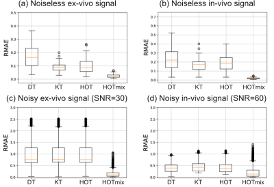 |
33 |
HOTmix: characterizing hindered diffusion using a mixture of generalized higher order tensors
David Romascano, Erick Canales-Rodriguez, Jonathan Rafael-Patino, Marco Pizzolato, Gaëtan Rensonnet, Muhamed Barakovic, Gabriel Girard, Alessandro Daducci, Tim Dyrby, Jean-Philippe Thiran
We present HOTmix, a new model to describe the diffusion MRI signal for molecules undergoing hindered diffusion. HOTmix is based on a mixture of generalized higher order tensors, explicitly incorporating the diffusion sequence’s time-dependent parameters. The method was evaluated on simulated diffusion MRI signals obtained through Monte Carlo simulations, using intermediate diffusion times, mimicking both ex-vivo and in-vivo conditions. HOTmix provided better reconstructions compared to the standard diffusion tensor, the kurtosis tensor, and a single generalized higher order tensor. In future work, we will explore whether modelling the hindered compartment using HOTmix improves microstructural features estimated using dMRI.
|
|
3542.
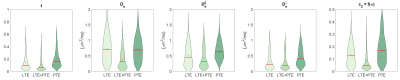 |
34 |
Double diffusion encoding enables unique parameter estimation of the Standard Model in diffusion MRI
Santiago Coelho, Jose Pozo, Sune Jespersen, Derek Jones, Alejandro Frangi
The widely adopted Standard Model (SM) for diffusion in white matter tissue has been shown to possess intrinsic degeneracies, making parameter estimation from single diffusion encoding (SDE) data ill-conditioned. We extend the SM to a multidimensional diffusion MRI acquisition and analyse the case where the fibre orientation distribution function (fODF) is a Watson distribution. The information contained in the kurtosis tensor, accessed by SDE, is insufficient to recover biophysical model parameters from the MR signal. We prove that Double Diffusion Encoding (DDE), letting us additionally access the full diffusion tensor covariance, makes parameter estimation well-posed.
|
|
3543.
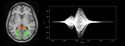 |
35 |
Estimation of Axon Conduction Delay, Conduction Speed, and Diameter from Information Flow using Diffusion MRI and MEG
Samuel Deslauriers-Gauthier, Rachid Deriche
Information transmission delays in the brain are generated by the physical properties of axons, including their length and diameter. These delays have a range of a few milliseconds and can thus be observed in bioelectric recordings such as electroencephalography and magnetoencephalography (MEG). In this work, we present a novel algorithm to estimate axon diameters from diffusion MRI and MEG. This approach identifies information flow between cortical regions using a model where transmission delays are parameters. The delays which maximize information transfer are identified and, using streamline length obtained through tractography, are then converted to axon diameters. We present results obtained on four subjects of the Human Connectome Project.
|
|
3544.
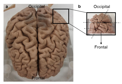 |
36 |
What perivascular spaces do to diffusion MRI?
Shi Jia Teo, Valerij Kiselev, Bibek Dhital, Marco Reisert
PVS is gaining importance in a many research fields. In using both in vivo and formalin fixed ex vivo methods, we sought to better understand what parameters in dMRI may be used to understood be as PVS in brain tissue.
|
|
3545.
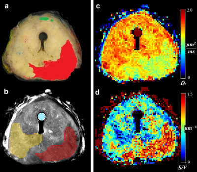 |
37 |
Surface-to-Volume ratio mapping using Oscillating Gradient Diffusion (OGSE) Spectroscopy for cancer characterization in the ex-vivo prostate
Jeevan Shah, Roger Bourne, Geoffrey Watson, Andre Bongers
This study investigates mapping of Surface-to-Volume (S/V) ratios from short diffusion time derived ADC maps to detect and characterize prostate cancer. ADC was mapped using a cos-OGSE DWI (fosc=50-200Hz) in one unfixed prostate (Gleason 4+4). Using a previously established model, S/V and free cytosol diffusivity (D0) maps were estimated by fitting the temporal behaviour of ADC. S/V and D0 were able to clearly and significantly separate the cancer and normal peripheral zone as indicated by a t-test and ROC curves. The geometrical nature of S/V may provide additional information to better characterize prostate cancer independent from diffusion related parameters.
|
|
3546.
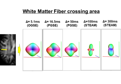 |
38 |
Diffusion Time Dependence in Crossing Fiber Area in in vivo Human White Matter
Masaaki Hori, Tomoko Maekawa, Kouhei Kamiya, Yasuhiko Tachibana, Koji Kamagata, Issei Fukunaga, Katsutoshi Murata, Thorsten Feiweier, Akifumi Hagiwara, Shohei Fujita, Ryusuke Irie, Christina Andica, Kanako Kumamaru, Akihiko Wada, Shigeki Aoki
We investigated the diffusion time dependency of diffusion metrics in the white matter crossing fiber areas with different diffusion times using OGSE, PGSE and STEAM-DTI in in vivo human white matter at 3T. Our results show that ADC and AD decreased with increasing diffusion time at Δless than 100ms; after that ADC increased with increasing diffusion time. RD decreased and FA increased with increasing diffusion time. Moreover, the changes of white matter fODF in the white matter crossing fiber area at different diffusion times are shown.
|
|
3547.
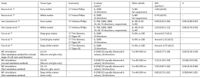 |
39 |
Random walk simulation of diffusion MRI in geometries constructed from two-photon microscopy of cortical gray matter
Nima Gilani, Sven Hildebrand, Anna Schueth, Alard Roebroeck
The purpose of this study was to more quantitatively characterize the cytoarchitecture of cortical tissue from microscopy in order to simulate diffusion signal in the cortex and to better understand its changes with density, size and permeability of cell bodies, axons and dendrites. Two random walk simulation methods are presented which show effects of cortical laminar microstructure on ADC and K. This could open possibilities of detecting cortical cytoarchitecture, i.e. cell body sizes and densities, and their pathologies with diffusion MRI.
|
|
3548.
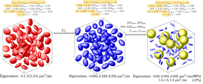 |
40 |
Probing tissue microstructure using filtered q-trajectory imaging
Lipeng Ning, Filip Szczepankiewicz, Markus Nilsson, Yogesh Rathi, Carl-Fredrik Westin
We introduce an approach to characterize tissue microstructure using an extension of the q-trajectory imaging (QTI) technique in diffusion MRI. Our approach applies multiplicative filters to the diffusion tensor distribution (DTD) functions of the underlying tissue to obtain more specific information of tissue microstructure using the statistical moments of the filtered DTD's.We propose several descriptive statistics to distinguish microstructures that cannot be separated using existing methods.These measures are demonstrated in simulations and in vivo data acquired from a human brain.
|
|
3549.
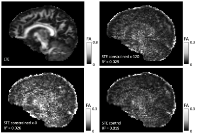 |
41 |
Time Dependence in B-Tensor Encoding
Noemi Gyori, Kiran Seunarine, Matt Hall, Christopher Clark, Daniel Alexander, Enrico Kaden
B-tensor encoding is becoming increasingly popular in microstructural imaging. In this technique microscopic tissue features are estimated by using different gradient waveforms that typically have different time regimes. This work studies for the first time two potential time-dependence issues that arise from these methods in-vivo in the human brain. We detect time-dependence effects in both cases, and even though these are small on a clinical system, they should not be overlooked.
|
|
3550.
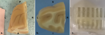 |
42 |
Comparing Fiber Orientation Estimates from CLARITY and Diffusion MRI in Macaque Visual Cortex
Muhamed Barakovic, Christoph Leuze, Ailey Crow, Qiyuan Tian, Alessandro Daducci, Jean-Philippe Thiran, Karl Deisseroth, Jennifer McNab
Tissue clearing techniques offer new possibilities for reconstructing precise 3D axons trajectories. Here, we cleared a 1.5cmx1.3cmx0.3cm cuboid of rhesus macaque visual cortex using CLARITY 3D histology with a neurofilament stain to highlight axonal projections and compared the CLARITY-based fiber orientation estimate to a diffusion tensor MRI atlas. We found better agreement in grey matter than in white matter, potentially reflecting difficulties estimating the structure tensor in highly saturated regions of the CLARITY sample. This work demonstrates one of the largest and highest quality CLARITY cuboids from a macaque brain and explores key steps in the co-registration and analysis required to make a robust comparison against DTI data.
|
|
3551.
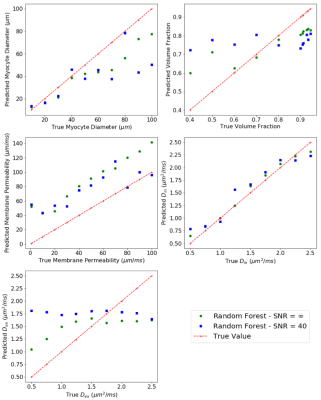 |
43 |
Microstructural Parameter Estimation of Skeletal Muscle Using Random Forest Model of dMRI
Noel Naughton, Nicolas Gallo, Aaron Anderson, John Georgiadis
Results are presented for a random forest model to estimate skeletal muscle microstructure parameters from dMRI data. The model exhibits the ability to estimate microstructural parameters from both numerically simulated and experimentally acquired dMRI data suggesting that random forests, and machine learning more generally, may be a useful tool in dMRI microstructure estimation of skeletal muscle.
|
|
3552.
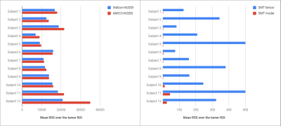 |
44 |
Reliability of microstructural quantification with diffusion MRI in brain tumors.
Umberto Villani, Erica Silvestri, Marco Castellaro, Maurizio Corbetta, Alessandra Bertoldo
Most existing biophysical models used to quantify diffusion microstructural information are designed for the healthy brain. When pathological processes occur, the diffusion signal of cancerous regions is altered, possibly biasing the fitting results of these models and making their parameter estimates unreliable. In this work, we investigate the precision and accuracy of estimates provided by several well known diffusion models, by evaluating their goodness-of-fit with residual sum of squares map and the parameter reliability with the bootstrap technique.
|
|
3553.
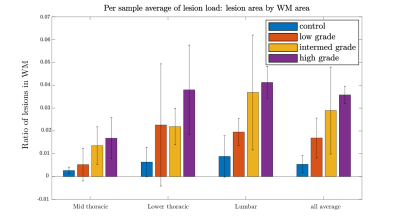 |
45 |
Diffusion Kurtosis Imaging (DKI) Biomarkers correlate with dysfunction in the EAE model of MS
Andrey Chuhutin, Brian Hansen, Agnieszka Wlodarczyk, Trevor Owens, Noam Shemesh, Sune Jespersen
To study the relationship between microstructure and disability, 18 ex-vivo spinal cords from a mouse model of MS (EAE) were investigated using DKI and a biophysical model of diffusion. Diffusion data were acquired together with T2* images to delineate lesions. Kurtosis tensors and microstructural parameters were used for statistical analysis using a LME model. The results show a strong relation between disability and kurtosis tensor parameters similar to observations in other hypomyelinating MS models and in patients. Conversely, changes in model parameters, such as extra-axonal axial diffusivity, are clearly different from previous studies using other animal models of MS.
|
|
3554.
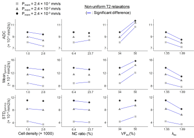 |
46 |
Effects of T2 relaxations on diffusion heterogeneity in the presence of tumor malignancy-related microstructural changes
Chu-Yu Lee, In-Young Choi, Phil Lee
Cell compartments can present non-uniform T2 relaxations, depending on tissue properties. The non-uniform T2 relaxation has been shown to affect measurements of apparent diffusion coefficients (ADC). In additional to ADC, diffusion heterogeneity measured by modeling non-monoexponential decay has been used to characterize water diffusion in tissue microstructure. The purpose of this study is to study the effects of non-uniform T2 relaxations on the diffusion heterogeneity using a Monte Carlo simulation. The results demonstrate that the diffusion heterogeneity may provide more information about water diffusion within microstructure with non-uniform T2 relaxations.
|
|
3555.
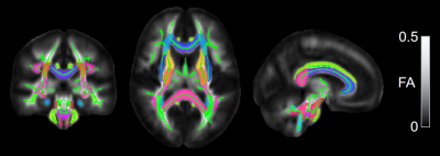 |
47 |
Delineation of white matter microstructural maturation of infant brain with DKI
Chenying Zhao, Minhui Ouyang, Michelle Slinger, Hao Huang
Diffusion kurtosis imaging (DKI) based on multi-shell diffusion MRI may offer extra information on WM microstructural complexity changes during infant development compared to conventional DTI. After acquiring DKI and DTI from 18 normal infants aged 0-1 year, we measured the mean kurtosis (MK) and fractional anisotropy (FA) of all major WM tracts by combining WM skeletonization process and WM tract labels. Significant age-related increases in both MK and FA were found in all tracts. Distinctive developmental trajectories of WM tracts with MK measurements compared to those with FA measurements were revealed, indicating heterogeneous increases of microstructural complexity among WM tracts.
|
|
3556.
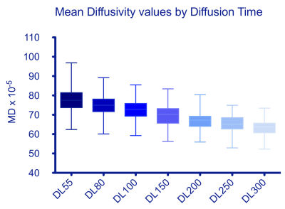 |
48 |
Time dependence and stability of diffusion tensor metrics in a hydrophilic, electrospun, water-perfused, hollow fiber phantom at 3T
Amy McDowell, Matt Hall, Fenglei Zhou, Thorsten Feiweier, Geoff Parker, Chris Clark
We assess the dependence of Diffusion Tensor Imaging metrics on diffusion time using a well-characterised hydrophilic phantom comprised of parallel hollow fibers with radii comparable to axons in healthy human white matter.
|
|
3557.
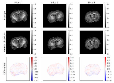 |
49 |
Experimental validation and SNR analysis of a clinical double diffusion encoding sequence
Leevi Kerkelä, Rafael Henriques, Matt Hall, Chris Clark, Noam Shemesh
Double diffusion encoding (DDE) allows the estimation of microscopic fractional anisotropy (μFA), which is a promising metric for studying microstructural properties of brain tissue independent of orientation dispersion. However, a large number of acquisitions is needed to obtain a rotationally invariant measurement of μFA, which poses a problem in clinical settings. A DDE protocol with a reduced number of acquisitions has recently been proposed as a solution. In this study, we assessed the accuracy of this approach and its potential for reporting μFA accurately. Our results show that a reduced number of acquisitions is sufficient for characterizing μFA.
|
|
3558.
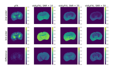 |
50 |
Precision of microscopic fractional anisotropy estimation using double diffusion encoding
Leevi Kerkelä, Rafael Henriques, Matt Hall, Noam Shemesh, Chris Clark
Microscopic fractional anisotropy (μFA) estimation using diffusion MRI is a promising new method for quantifying microstructure, diagnosing pathologies, and measuring brain development. In order for μFA to be a useful metric in clinical and neuroscientific research, its robustness to noise has to be properly understood. In this study, precision of μFA estimation was quantified for different noise levels using propagation of error calculations, simulations, and imaging experiments. We show that μFA is non-linearly sensitive to noise, being the most instable at low values of mean diffusivity (MD) and μFA, and that increasing accuracy by correcting for higher order effects results in decreased precision.
|
|
| Top |
Microstructure Modeling: 1
Digital Poster
Diffusion
Wednesday, 15 May 2019
| Exhibition Hall |
09:15 - 10:15 |
| |
|
Computer # |
|
3559.
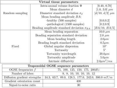 |
51 |
Machine learning based estimation of axonal properties in the presence of beading
Kévin GINSBURGER, Felix MATUSCHKE, Fabrice POUPON, Jean-François MANGIN, Markus AXER, Cyril POUPON
In this work, we investigate the potential of machine learning techniques to make one step forward by quantitatively estimating beading amplitude, a specific marker of pathological beading using frequency-dependent changes in diffusion measurements. Classification and regression are performed using Extremely Randomized Trees from OGSE signals corresponding to 6 distinct frequencies and synthesized from numerical simulations in realistic white matter phantoms depicting beaded axons.
|
|
3560.
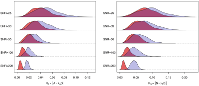 |
52 |
Tensorial formulation allowing to verify or falsify the microstructural standard model from multidimensional diffusion MRI
Jose Pozo, Santiago Coelho, Alejandro Frangi
The standard model (SM) has been proposed as an appropriate diffusion MRI microstructural model for brain white matter. Using the cumulant expansion up to 2nd order in the b-tensor, it has been recently shown that multidimensional diffusion encoding makes the model parameter estimation problem well-posed. However, the tensorial properties of the expansion and their relationship with the SM assumptions have not been exploited. We reformulate the solution of the SM in an elegant tensorial form and analyse the constraints the SM imposes, showing how they can potentially falsify the model. Some simulations show the test feasibility for high-quality signals (SNR≥50).
|
|
3561.
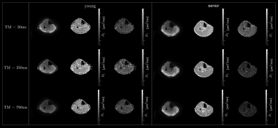 |
53 |
Permeable Barrier Modeling of Age Induced Changes in the Time Dependent Diffusion Eigenvalues
Vadim Malis, Sinha Shantanu, Edward Smitaman, Usha Sinha
The time dependence of the diffusion eigenvalues derived from diffusion tensor imaging allows one to probe tissue microstructure. A diffusion model is required to derive inferences about the microstructure from the time dependent eigenvalue data. We applied the Random Permeable Barrier Model1 to the time dependent diffusion data to infer age related changes in skeletal muscle microarchitecture. The tertiary eigenvalue at the largest diffusion time was significantly different between young and senior cohorts. Model derived myofiber size decreased and free diffusion coefficient increased with age, though neither parameter reached significance.
|
|
3562.
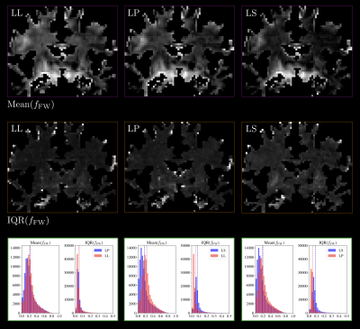 |
54 |
Estimating mixtures of 6D diffusion tensor distributions with "Magic DIAMOND": are b-plates better than b-spheres?
Alexis Reymbaut, Alex Valcourt Caron, Guillaume Gilbert, Simon Warfield, Maxime Descoteaux, Benoit Scherrer
Tensor-valued diffusion encoding enables disentangling isotropic and anisotropic diffusion components. However, its impact on estimating brain microstructural features has only been studied in a handful of parametric models. In this work, we evaluate the Magic DIAMOND model, that allows characterization of crossing fascicles and assessment of diffusivities for each, using combinations of linear, planar and spherical encodings in vivo. Building statistics through stratified bootstrap, we show that spherical encoding substantially increases the variance in estimated parameters and should be avoided. Planar encoding, on the other hand, did not offer clear improvement or worsening within our current acquisition scheme and setup.
|
|
3563.
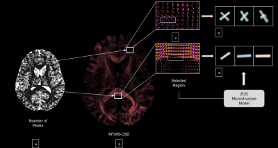 |
55 |
MicroLearn: Framework for machine learning, reconstruction, optimization and microstructure modeling
Shreyas Fadnavis, Marco Reisert, Hamza Farooq, Maryam Afzali, Cheng Hu, Bago Amirbekian, Eleftherios Garyfallidis
MicroLearn is a Machine Learning and Model Fitting framework that enables modular construction of multi-compartment microstructure models in crossings with fast and accurate parameter estimation.
|
|
3564.
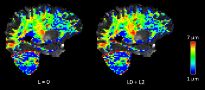 |
56 |
Investigating the Benefits of Incorporating Higher Order Spherical Harmonics in Axon Diameter Measurements
Qiuyun Fan, Aapo Nummenmaa, Qiyuan Tian, Ned Ohringer, Thomas Witzel, Lawrence Wald, Bruce Rosen, Susie Huang
Separating out the scalar and orientation-dependent components of the diffusion MRI signal offers the possibility of increasing sensitivity to microscopic tissue features unconfounded by the fiber orientation. Recent approaches to estimating apparent axon diameter in white matter have employed spherical averaging to avoid the confounding effects of fiber crossings and dispersion at the expense of losing sensitivity to effective compartment size. Here, we investigate the feasibility and benefits of incorporating higher-order spherical harmonic (SH) components into a rotationally invariant axon diameter estimation framework and demonstrate improved precision of axon diameter estimation in the in vivo human brain.
|
|
3565.
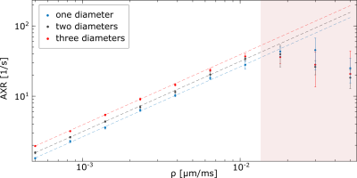 |
57 |
On the feasibility of apparent exchange rate mapping in non-periodic systems with multiple cell sizes
Dominik Ludwig, Frederik Laun, Peter Bachert, Tristan Kuder
Apparent exchange rate (AXR) mapping might provide an insight into the exchange of water between intra- and extracellular space by using a double-diffusion encoded sequence with varying mixing times between the two gradient pairs. The connection between AXR and permeability has already been validated for simplified two-compartment models. However, multi-compartment systems and non-periodic geometries have not been evaluated so far. In this study, we were able to show that it is also possible to reliably connect permeabilities to AXR-values for these geometries. Furthermore, the AXR was only dependent on average cell size and not on the number of compartments.
|
|
3566.
 |
58 |
Computational frameworks for multi-fascicle models: impact on microstructural descriptors
Benjamin Schloesing, Maxime Taquet, Jean-Philippe Thiran, Simon Warfield, Benoit Scherrer
When performing operations on multi-fascicle DCI models, the need to preserve microstructural descriptors such as fFA and fMD is important. In this work we compared different multi-fascicle computational frameworks by assessing their impact on microstructure properties. Specifically, we investigated the impact after geometrical transformation and averaging of multi-fascicle models, two key operations when carrying out population studies. We found that Euclidean and log-Euclidean frameworks resulted in a decrease of fFA and fMD. More surprisingly, the values of microstructural descriptors depended on the number of subjects. The quaternion framework, in contrast, was the best at preserving microstructural features.
|
|
3567.
 |
59 |
Non-parametric axon diameter distribution mapping with PGSE: reconstruction of uni- and multimodal distributions
David Romascano, Jonathan Rafael-Patino, Muhamed Barakovic, Alessandro Daducci, Jean-Philippe Thiran, Tim Dyrby
White matter diffusion MRI enables non-invasive estimation of the axon diameter distribution, which is of interest as it modulates communication speed and delays between brain regions, and changes during development and pathology. Distribution mapping is challenging: current methods simplify it by either estimating the mean diameter, imposing parametric distributions, or combining non-parametric approaches with Double Diffusion Encoding. We present a non-parametric framework based on a PGSE protocol. Simulations show robust reconstruction of unimodal and bimodal distributions. The method is sensitive to population specific changes within bimodal distributions, as long as the underlying populations are separated by a minimum distance.
|
|
3568.
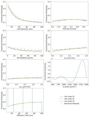 |
60 |
Polynomial Meta-Model of Bloch-Torrey Equation for Track-based Regularization of Microstructural Inversion
Noel Naughton, Arihant Jain, John Georgiadis
A meta-model approach is presented which fits a multi-dimensional polynomial to numerical solutions of the Bloch-Torrey equation after being parameterized by microstructural and diffusion encoding parameters. This meta-model allows analytical representation of the solution space enabling analytical analysis of the space as well as reduced computational cost to solutions that do not need precise results. Possible uses of such a model are outlined, such as its use in allowing muscle tractography results to regularized by neighboring voxels in its defined muscle tract.
|
|
3569.
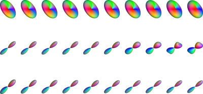 |
61 |
Improving Tractography in Baby Diffusion MRI via Asymmetric Spectrum Imaging
Ye Wu, Weili Lin, Dinggang Shen, Pew-Thian Yap, the UNC/UMN Baby Connectome Project Consortium Consortium
Fiber tractography in baby diffusion MRI is challenging due to the low and spatially-varying diffusion anisotropy. In this abstract, we will introduce a method called asymmetric spectrum imaging (ASI) to improve estimation of white matter pathways in the baby brain by (1) incorporating an asymmetric fiber orientation model to help resolve subvoxel fiber configurations such as fanning and bending, and (2) explicitly modeling the spectrum of diffusion typical in the developing brain.
|
|
3570.
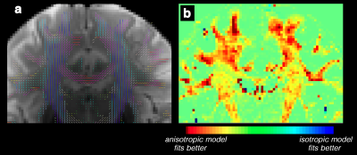 |
62 |
Diffusion of the perivascular space fluid is anisotropic in conventional DWI
Farshid Sepehrband, Ryan Cabeen, Jeiran Choupan, Giuseppe Barisano, Meng Law, Arthur Toga
Perivascular space (PVS), also known as Virchow-Robin space, is a pial-lined, fluid-filled structure that surrounds vessels in the cerebral cortex 1,2, and occupies a large portion of the cerebral tissue. PVS has an anisotropic morphology 3–5 and is mainly aligned with the white matter 6. However, its diffusion MRI profile has not been studied. Here we show experimental evidences that that PVS can be measured with diffusion MRI and the signature of this compartment is anisotropic.
|
|
3571.
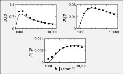 |
63 |
Dependence of harmonic power on b-value for fiber ball imaging: comparison of theory and experiment
Hunter Moss, Emilie McKinnon, Joseph Helpern, Jens Jensen
Fiber ball imaging (FBI) is a recently proposed diffusion MRI (dMRI) method for estimating fiber orientation density functions together with specific microstructural parameters in white matter. The theory underlying FBI predicts the b-value dependence for the dMRI harmonic power of any given degree as long as the b-value is sufficiently large. Good agreement between theory and experiment has been previously demonstrated for the zero-degree harmonic power. Here the predicted functional forms for higher degree harmonics are shown to also agree well with experimental measurements, providing additional support for the validity of FBI.
|
|
3572.
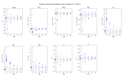 |
64 |
Diffusion time dependence in the mid-time regime: a simulation study using PGSE.
Annelinde Buikema, Arjan Dekker, Jan Sijbers
The purpose of this work is to study the effect of varying the diffusion time on the estimation of the parameters of the two-compartment diffusion tensor model in the mid-time regime. Simulation results show that the precision of the diffusion time-dependent compartmental parameter estimates increases when a variable echo time acquisition scheme is used. At low SNR, however, including diffusion time-dependence may lead to a high bias and variance compared to the more conventional non diffusion time-dependent model.
|
|
3573.
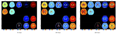 |
65 |
Biases of microstructure models in baby diffusion MRI
Khoi Huynh, Tiantian Xu, Ye Wu, Geng Chen, Weili Lin, Dinggang Shen, Pew-Thian Yap, the UNC/UMN Baby Connectome Project Consortium
In this abstract, we show that some commonly used microstructure models do not perform as aspected when dealing with baby diffusion MRI, resulting in biased measurements. We found that this is mainly due to the greater water content in the developing brain. We show that the recently introduced Spherical Mean Spectrum Imaging (SMSI) [4] gives stable measurements in the presence of free water, making it well-suited for baby diffusion MRI.
|
|
3574.
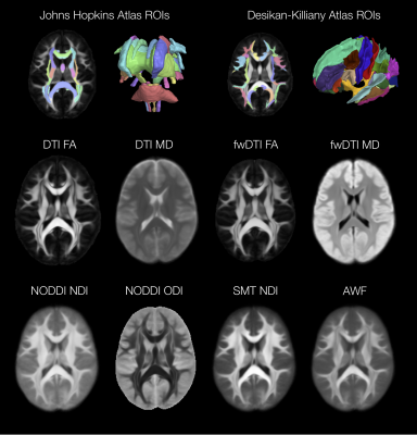 |
66 |
MicroQIT - A Computational Framework for Population Microstructure Imaging
Ryan Cabeen, Arthur Toga
Microstructure imaging provides a quantitative tool for characterizing neural tissue with diffusion MRI and expanding our understanding of how the brain changes in health and disease. However, robust computational tools are needed for population imaging studies, so we developed a computational framework (MicroQIT) using the Quantitative Imaging Toolkit to meet this need by providing regional summaries and spatially normalized microstructure parameter maps. It supports a variety of ways to extract microstructure information from multi-shell diffusion MRI, leverages grid computing environments, and is available for use by the research community for future studies.
|
|
3575.
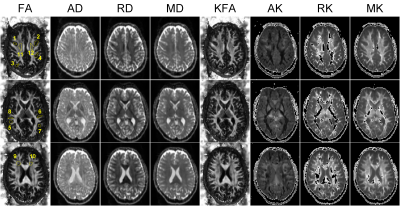 |
67 |
An in vivo investigation on quantitative metrics of diffusion kurtosis tensor: the effect of diffusion gradient parameters in the clinical setting
Kuan-Hung Cho, Richard Buschbeck, Shih-Yen Lin, Ezequiel Farrher, Ming-Jye Chen, Chia-Wen Chiang, N. Jon Shah, Chang-Hoon Choi, Li-Wei Kuo
Diffusion kurtosis imaging (DKI) is an emerging technique that provides additional information to delineate tissue microstructures by quantifying the non-Gaussian water molecular diffusion. Although the capability of DKI has been demonstrated, the effects of diffusion gradient parameters on its quantitative metrics, particularly in the clinical setting, have not been fully understood yet. This study aims to investigate the effect of diffusion gradient parameters on diffusion kurtosis tensor calculation and its quantitative metrics. In vivo results show that diffusion gradient duration has incremental influence on DKI quantitative metrics in the clinical setting. Further investigation with more subjects would help to statistically solidify our findings.
|
|
3576.
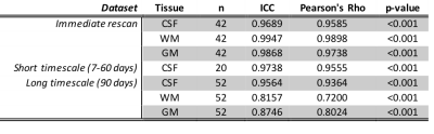 |
68 |
Test-retest reliability of 3-tissue constrained spherical deconvolution of diffusion MRI data by analysis of three separate cohorts
Benjamin Newman, Thijs Dhollander, Kristen Reynier, Matthew Panzer, Thomas Druzgal
It has previously been demonstrated that, by using 3-tissue constrained spherical deconvolution, separate compartments encompassing cerebrospinal fluid-like, white matter-like, and grey matter-like, signal fractions can be derived from diffusion MRI data. This study explores the reliability of these compartments in three test-retest cohorts with a variety of timescales and scanning parameters. Whole-brain average signal fractions show excellent reliability across all datasets, particularly in determining the CSF-like signal fraction. This suggests that variations in whole brain signal fraction measurements are likely to be attributable to experimental manipulation or pathology and not variation introduced by performing the analysis.
|
|
3577.
 |
69 |
Powering Up Microstructural Imaging: assessing cross-metric and cross-tract statistical power on an ultra-strong gradient MRI system
Kristin Koller, Suryanarayana Umesh Rudapandra, Maxime Chamberland, Erika Raven, Greg Parker, Chantal Tax, Mark Drakesmith, John Evans, Tobias Wood, Derek Jones
We present cross-metric and cross-tract assessment of test-retest repeatability for microstructure measures on an ultra strong gradient MRI scanner (CONNECTOM 3T) in the human brain. We show that several MRI metrics of tissue microstructure are reliable and present relative sample sizes required to provide sufficient statistical power across different white matter pathways and microstructure metrics.
|
|
3578.
 |
70 |
Improving reproducibility of diffusion connectome analysis using deep convolutional neural network model
Min-Hee Lee, Nolan O'Hara, Jeong-Won Jeong
Reproducibility of diffusion-weighted structural connectomes is highly dependent on acquisition and tractography model, limiting the interpretation of connectomes acquired in the clinical setting. This study proposes a novel deep convolutional neural network (DCNN) to improve the reproducibility of structural connectomes, by which highly reproducible streamlines can be identified via an end-to-end deep learning of reference streamline coordinates in Human Connectome Project diffusion data. Preliminary results demonstrate that the proposed DCNN prediction model can improve the reproducibility of clinical connectomes (31.29% of F-statistics in intraclass correlation coefficient) and effectively remove noisy streamlines based on based on their poor prediction probabilities.
|
|
3579.
 |
71 |
Resolving crossing fibers with the inclusion of intra-axonal diffusion modeling
Presentation Not Submitted
Chunyu Song, Peng Sun, Zezhong Ye, Sheng-Kwei (Victor) Song
A new diffusion histology imaging (DHI) model is proposed to model crossing fibers considering both intra- and extra-axonal water diffusion, along with extra-axonal isotropic diffusion within an image voxel. Both Monte-Carlo simulation and in vivo MRI data from one healthy volunteer brain were examined to assess whether DHI can resolve crossing fibers while quantifying axonal injury, demyelination, and inflammation.
|
|
3580.
 |
72 |
A diffusion MRI pipeline leveraging Nextflow & Singularity: Robust, Efficient, Reproducible in time!
Guillaume Theaud, Jean-Christophe Houde, Felix Morency, Maxime Descoteaux
How do we assure a diffusion MRI processing pipeline that is: i) deterministic, i.e. given two runs of the pipeline on the same data, the same output is returned, ii) reproducible in time, and iii) efficient? Diffusion MRI has several processing steps that may not be reproducible between multiple runs. This reproducibility varies because of the parameters, multi-threading and the versions of the tools used. Moreoever, processing time for a large database can take several hours when each step are ran sequentially. To solve these problems, we developed a reproducible and efficient diffusion MRI pipeline based on Nextflow and Singularity.
|
|
3581
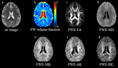 |
73 |
Diffusion Kurtosis Imaging of the Brain with Free Water Elimination
Video Permission Withheld
Teddy Salan, Sulaiman Sheriff, Varan Govind
In diffusion weighted imaging (DWI) of the brain, a single voxel may contain gray matter (GM), white matter (WM), as well as free water embedded in GM, WM, and cerebrospinal fluid (CSF), each with different diffusion profiles. Free water elimination (FWE) is a method used to separate the free water components from tissue water. In this work, we extend FWE by fitting with a diffusion kurtosis imaging (DKI) model as it is a better descriptor of the non-Gaussian diffusion that occurs in tissue water.
|
|
3582.
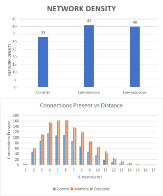 |
74 |
A Graph Theory and Spectral Graph Theory Approach to Correlating Tractography with Parkinson’s Disease.
Daniel DeYoung, Manish Amin, Catherine Price, Thomas Mareci
Diffusion imaging is a potentially powerful tool for analyzing the effect of Parkinson’s disease on the brain. DWI analysis may provide some insight into how Parkinson’s disease may affect low executive or low memory functioning in the brain on a structural level. This research aims to analyze the complexity of the brain connectivity using graph theory and spectral graph theory to correlate structural differences between the brains of Parkinson’s patients with low memory functioning or low executive functioning, and controls.
|
|
| Top |
Diffusion in Disease
Digital Poster
Diffusion
Wednesday, 15 May 2019
| Exhibition Hall |
09:15 - 10:15 |
| |
|
Computer # |
|
3583.
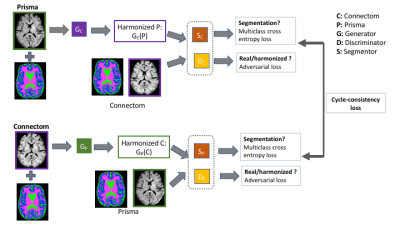 |
76 |
Multi-site diffusion MRI harmonization in the presence of gross pathology: How far can we push?
Suheyla Cetin Karayumak, Marek Kubicki, Yogesh Rathi
We present a multi-site diffusion MRI (dMRI) data harmonization method using CycleGAN network with segmentation loss (CycleGANS). This method aims to learn an efficient mapping of dMRI signal using rotation invariant spherical harmonics features from the same set of subjects across sites. At the same time, it has potential to learn the tumor pathology (if exists) during harmonization. We compare our CycleGANS network with the CycleGAN network. We show that our CycleGANS network has better multi-site diffusion MRI data harmonization accuracy. Moreover, our method shows up to 60% improvement on the prediction of tumor pathology.
|
|
3584.
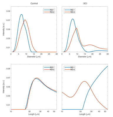 |
77 |
Joint distribution of axonal length and diameter quantifies beading in traumatic brain injury
Dan Benjamini, Michal Komlosh, Elizabeth Hutchinson, Peter Basser
We present a novel diffusion MRI approach to measure the three-dimensional axonal morphology alterations following traumatic brain injury that results in beading, by modeling axons with a two-dimensional joint distribution of diameters and lengths (DD-LL).
Here we study a segment of ferret spinal cord tissue with known and focal Wallerian degeneration of the corticospinal tract alongside an uninjured control. The results suggest that this approach can be used to specifically detect and quantify axonal beading.
|
|
3585.
 |
78 |
Quantitative Sodium Pump Activity, Cellularity, and Cytometry: In Vivo Analytical Pathology from DWI
Eric Baker, Brendan Moloney, Xin Li, Gregory Wilson, Thomas Barbara, Jeffrey Maki, Erin Gilbert, Charles Springer, Jr.
With cellular ensembles featuring stochastic geometries, Monte Carlo random walk simulated DWI b-space decays exhibit sensitivity to cell biology parameters measuring membrane Na+,K+-ATPase [NKA] activity, cell density, ρ, and voxel average cell volume, <V>. Furthermore, the simulation matching the experimental in vivohuman cerebral cortex b-space decay has parameters [cellular water efflux rate constant <kio> = 2 s-1, ρ = 98,000 cells/μL, and <V> = 8.2 pL] in near absolute agreement with the most pertinent literature. Inspecting the common, empirical early decay measure, ADC, of these simulations provides insights into acute and chronic tissue property changes in vivo.
|
|
3586.
 |
79 |
Investigation of relationships between systemic inflammation and brain structure in chronic obstructive pulmonary disease (COPD)
Mohani-Preet Bajaj, Sachelle Ruickbie, Daniel Burrage, Catherine Spilling, Abhiram Prasad, James Dodd, Paul Jones, Emma Baker, Thomas Barrick
COPD patients have increased systemic inflammation and increased structural brain damage. 23 COPD patients with coronary artery disease (CAD), and 24 people without COPD matched for smoking, and CAD severity (Gensini score) were studied. COPD patients had more white matter lesions and microstructural tract abnormalities compared to controls. Systemic inflammation (high sensitivity C-reactive protein and fibrinogen) was associated with microstructural brain damage in whole-group analyses. These results suggest a systemic inflammatory process in COPD, which may contribute to white matter abnormalities, consistent with those reported in small vessel disease.
|
|
3587.
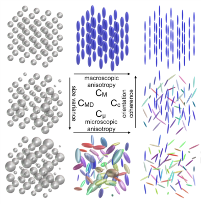 |
80 |
Q-Space Trajectory Imaging to Untangle Sources of Microstructural White Matter Changes: Comparison of Schizophrenia Subjects and Healthy Controls
Ofer Pasternak, Filip Szczepankiewicz, Zora Kikinis, Markus Nilsson, Tomáš Kašpárek, Carl-Fredrik Westin
The recently proposed q-space trajectory imaging (QTI) analysis for multidimensional dMRI acquisitions with free wave forms quantifies domains of variability (size, shape, orientation) that can explain the sources of microstructural changes in the brain. These previously inaccessible domains are used here to disentangle differences observed in the white matter of schizophrenia patients compared to healthy controls. In this population microscopic anisotropy (shape variability) explains changes in fractional anisotropy, in agreement with a hypothesis of white matter degeneration. This study provides a first proof of principal for the feasibility of applying free wave form based sequences on a clinical population.
|
|
3588.
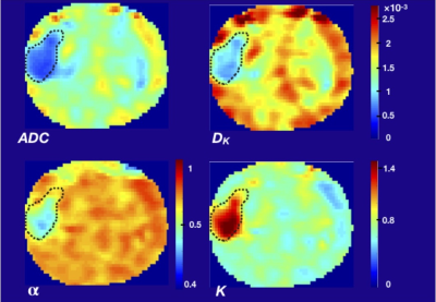 |
81 |
Prostate Cancer Classification by Using Mono Exponential, Stretched Exponential and Kurtosis Model Parameters of Diffusion Signal Decay
Meltem Uyanik, Rolf Rieter, Michael Abern, Winnie Mar, Virgilia Macias, Hari Vigneswaran, Richard Magin
Prostate cancer is the most common solid cancer occurring among men in the US. Diffusion-weighted MR imaging plays a complementary role to T2-weighted images in identifying regional changes in prostate tissue. Here, we fit the diffusion decay signal from patients using the stretched-exponential and the kurtosis models and compare the results with MR guided prostate biopsy histology. Our results showed that the kurtosis and stretched exponential models fit to multi-b values diffusion data have the potential to distinguish benign from malignant lesions. These model parameters identify tissue heterogeneity and structures that may be useful in the grading of prostate cancer.
|
|
3589
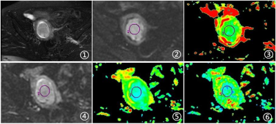 |
82 |
Application of DTI and DWI quantitative parameters in differential diagnosis of uterine sarcoma and degenerated hysteromyoma
Video Permission Withheld
Miao Niu, Ailian Liu, Lizhi Xie
To investigate the application of diffusion tensor imaging methods, including Diffusion Tensor Imaging(DTI) and Diffusion Weighted Imaging(DWI) in the differential disgnosis of uterine sarcoma and degenerated hysteromyoma.
|
|
3590.
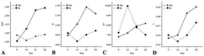 |
83 |
To Detect Metastatic Lymph Nodes Using Intravoxel Incoherent Motion Diffusion-weighted Imaging: A Longitudinal Study on Animal Model
Liuji Guo, Xiaodan Li, Xiaomin Liu, Zhiguang Si, Jie Qin, Yingjie Mei, Zhongping Zhang, Yikai Xu, Yuankui Wu
Lymph node (LN) involvement is a dynamic process which may be reflected by intravoxel incoherent motion diffusion-weighted imaging (IVIM-DWI). However, there is no such studies so far. Therefore, the potential influence on the diagnosis performance for metastatic LN needs to be investigated. In this longitudinal study on animal models, metastatic LNs evolved in a different pattern from inflammatory LNs pathologically, and IVIM parameters differed between the two groups at particular time points. This indicates that IVIM-DWI can reflect the dynamic pathological changes of LN diseases, and its ability to detect metastatic LN may be affected by the course of disease.
|
|
3591.
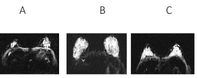 |
84 |
Apparent Diffusion Coefficient Using Diffusion Weighted MRI and Biochemical Correlates in Human Breast Cancer
Kavya Umachandran, Andres Saucedo, Stephanie Lee-Felker, Melissa Joines, Manoj Sarma, Sumit Kumar, Maggie DiNome, Nanette DeBruhl, Michael Thomas
Multiparametric MRI has been investigated in breast and prostate cancer, and other tumors. We evaluated diffusion weighted imaging in a pilot cohort of 20 malignant and 11 benign breast cancer patients, and 7 healthy women. MR spectra were recorded using an accelerated version of five dimensional echo-planar correlated spectroscopic imaging (5D EP-COSI). Significant decline in ADC values of malignant breast cancer compared to benign and healthy women. There was a negative correlation of choline with ADC values in malignant cancer patients. Our findings suggest that choline and lipids can be reliable biomarkers in addition to widely used ADC.
|
|
3592.
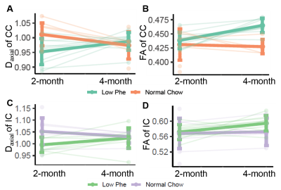 |
85 |
Diffusion Tensor Imaging of Phenylketonuria (PKU) Mouse Brain White Matter Integrity
Xia Ge, Zoe Hawks, Desiree White, Auro Ferreiro, Gautam Dantas, John Engelbach, Joshua Shimony, Joel Garbow
DTI was used to evaluate brain white-matter integrity and development in Pahenu2 mice, a model of phenylketonuria (PKU). At two months of age, the volume of the CC in Pahenu2 mice was smaller than that of C57Bl6 controls, and the FA of white matter is significantly lower. For PKU mice on a low-Phe diet, both Daxial and FA of CC and IC increased from two to four months, while these metrics decreased in Pahenu2 mice on a normal diet, suggesting a loss of axonal integrity. Our data suggest that Phe levels are critical to white-matter development in Pahenu2 mice.
|
|
3593.
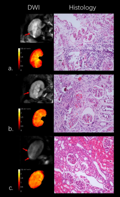 |
86 |
Apparent diffusion coefficient: Early Indicator of Atheroembolic Renal Disease
Presentation Not Submitted
Bihui Zhang, Min Yang, Guochen Niu, Ziguang Yan, Yinghua Zou, Xiaoqiang Tong, Xiaoying Wang
Atheroembolic Renal Disease (AERD) is part of a multisystemic disease and has a strong clinical impact on patient and renal survival. Its early diagnosis has a valuable prognostic meaning and huge impact on treatment planning. The tissue edema in early-stage AERD makes DWI a perfect tool for early diagnosis of AERD. In this study, we investigated the role of DWI to noninvasively detect AERD as well as the correlation between ADC values and disease severity identified by histology in an early stage.
|
|
3594.
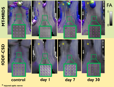 |
87 |
Longitudinal evaluation of bundle-wise water diffusion changes following axonal degeneration in a region of fiber crossing
Omar Narvaez, Ricardo Coronado-Leija, Gilberto Rojas-Vite, Marcos Aranda, Alonso Ramirez-Manzanares, Jose Marroquin, Jorge Larriva-Sahd, Luis Concha
Axonal degeneration is a hallmark of many neuropathologies, with a defined time course presenting distinct histological features. In single fiber regions, the tensor model provides reliable information in early and chronic phases of axonal damage. However, said model cannot accurately determine per-bundle characteristics in voxels occupied by axonal populations with different orientations. We evaluated two multiple-fiber methods in an animal model of axonal degeneration (unilateral retinal ischemia) to provide information about the sensitivity of these models related to microstructural changes occuring in the acute and early chronic stages of pathology in a crossing fiber region.
|
|
3595
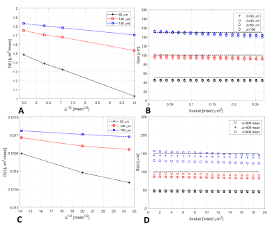 |
88 |
Towards MR-based interrogation of the hypoxia-driven insulin resistance mechanism: Adipocytes size estimation.
Video Permission Withheld
Darya Morozov, Alexander Sukstanskii, Scott C. Beeman
It has been suggested that adipocyte hypertrophy plays a key role in the pathogenesis of systemic insulin resistance and type 2 diabetes. A method for quantifying adipocyte size/hypertrophy in-vivo would be a major advance towards understanding the pathogenesis of type 2 diabetes. In-vivo quantification of adipocyte size might be achievable using short time regime diffusion MR, which carries information about size of the system. Herein, we explore the feasibility of measuring adipocyte size based on the diffusion of triglyceride within adipocytes and the principles of the short diffusion time regime.
|
|
3596.
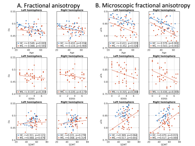 |
89 |
Decoupling effects of fiber dispersion and microscopic anisotropy fundamentally change interpretations of DTI in multiple sclerosis
Kasper Andersen, Samo Lasic, Henrik Lundell, Markus Nilsson, Daniel Topgaard, Filip Szczepankiewicz, Hartwig Siebner, Morten Blinkenberg, Tim Dyrby
Fractional anisotropy from diffusion tensor imaging (DTI-FA) has frequently been used to probe changes in white matter microstructure, but is also heavily affected by axonal fiber dispersion. μFA removes fiber dispersion effects and thereby estimates the microscopic anisotropy. Here, we found lower μFA in normal appearing white matter in multiple sclerosis patients as compared with healthy controls. In addition, μFA correlated significantly with age, disability and cognitive performance. These relations could not be established with DTI-FA. Our results indicate that μFA could be used as a powerful biomarker for diseases related to micro-structural changes in white matter as well as in studies of the healthy brain.
|
|
3597.
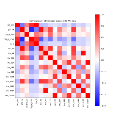 |
90 |
A Novel Characterization of Traumatic Brain Injury in White Matter with Diffusion MRI Spherical-Harmonics Rotation Invariants
Mauro Zucchelli, Drew Parker, Samuel Deslauriers-Gauthier, Junghoon Kim, Ragini Verma, Rachid Deriche
The current DTI-based markers of traumatic brain injury are able to capture affected white matter in the brain, but miss the areas of crossing fibers due to the simplicity of the model. In this work, we use a novel set of spherical-harmonics rotation invariants indices, recently proposed in the literature. We demonstrate that these 12 invariants capture all the information provided by DTI and, in addition, they capture differences in complex white matter beyond DTI measures. This, combined with the clinical feasibility of the method, paves the way for them to be used as better markers of brain injury.
|
|
3598.
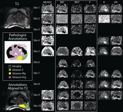 |
91 |
Multi-site concordance of diffusion weighted imaging derived metrics, a comparison of fitting algorithms for differentiating whole-mount, pathologically confirmed, prostate cancer of differing Gleason patterns
Peter LaViolette, Sean McGarry, John Bukowy, Allison Lowman, Anjishnu Banerjee, Dariya Malyarenko, Tom Chenevert, Yue Cao, Andrey Fedorov, Laura Bell, C. Quarles, Melissa Prah, Kathleen Schmainda, Stefanie Hectors, Bachir Taouli, Eve LoCastro, Yousef Mazaheri, Amita Shukla-Dave, Thomas Yankeelov, David Hormuth II, Ananth Madhuranthakam, Keith Hulsey, Kurt Li, Wei Huang, Mark Muzi, Kenneth Jacobsohn, Mark Hohenwalter, Petar Duvnjak, Michael Griffin, William See, Marja Nevalainen, Kenneth Iczkowski
We present a multi-site study measuring the ability of various software platforms to fit diffusion weighted imaging (DWI) models for differentiating prostate cancer (PCA) of different Gleason patterns. A dataset of DWI collected from 33 PCA patients was distributed to ten collaborating groups who fit the multi-b DWI models with onsite software and submitted the derived parametric maps to a central analysis site. Each map was aligned to the T2-weighted image and compared to pathologist annotations of whole-mount prostate samples. A statistical analysis was performed for similarity of the quantitative values, and the ability of each metric to differentiate PCA.
|
|
3599.
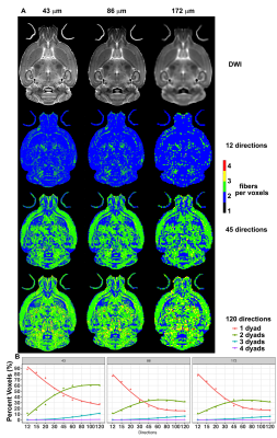 |
92 |
Optimizing Imaging Protocols for Structural Connectomics in Mouse Models of Neurological Conditions
Robert Anderson, Christopher Long, Evan Calabrese, Scott Robertson, Gary Cofer, G Johnson, Alexandra Badea
Network approaches provide sensitive biomarkers for neurological conditions such as Alzheimer’s disease. Mouse models provide tools to dissect vulnerable circuits at prodromal stages, and to assess the effects of interventions. We have simulated mouse brain structural connectomes, balancing angular, spatial resolution and scan time. Specifically, we evaluated protocols with 6, 12, 15, 20, 30, 45, 60 and 120 angles; and 3 voxel sizes at 43, 86 and 172 µm. Our results indicate schemes using 46 or 60 diffusion directions, acquired at 86 µm resolution achieve a good cost/performance balance relative to a high spatial, high angular resolution sampling scheme.
|
|
3600.
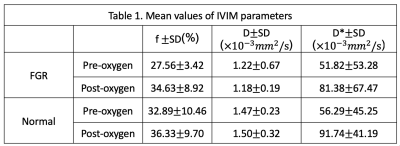 |
93 |
THE EFFECT OF MATERNAL OXYGENATION ON PLACENTAL PERFUSION IN NORMAL GROWTH AND FETUS GROWTH RESTRICTED FETUSES BY USING IVIM MRI
Ting Chen, Xuanyi Zhou, Yongming Dai
The aim of this study was to determine whether IVIM-MRI could evaluate oxygen inhalation therapy in placental perfusion during maternal hyperoxygenation in FGR and normal growth fetuses.
|
|
3601.
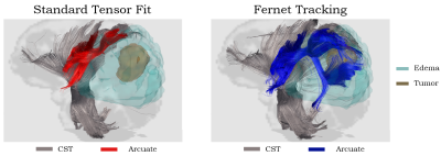 |
94 |
Free Water Characterization of Peritumoral Regions in Primary and Secondary Brain Neoplasms
Abdol Aziz Ould Ismail, Drew Parker, Simon Alexander, Steven Brem, Ragini Verma
There is a growing interest in characterizing the peritumoral regions in neoplasms, in order to distinguish primary and secondary neoplasms, improve tractography for surgical planning, and create radiomic markers of infiltration. Free water estimation is expected to reflect the differences in water content of the peritumoral region between these tumor types. In this work, we compare glioblastoma (primary) and metastatic (secondary) tumors based on their free water estimated using standard clinical diffusion MRI (dMRI). Results demonstrate significant difference in these tumor types based on the free water measures, fractional anisotropy post-correction and the coverage of the peritumoral region by tractography.
|
|
3602.
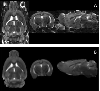 |
95 |
Diffusion Weighted MRI Characterization of Effects of Sex and APOE Genotype in a Mouse Model
Loi Do, Adam Bernstein, Aarti Mishra, Maunil Desai, Marc Lindley, Chidi Ugonna, Nan-kuei Chen, Roberta Brinton, Theodore Trouard
Structural and diffusion MRI (dMRI) analyses can be used to characterize neurodegeneration during Alzheimer’s disease progression. Male and female mice with a targeted replacement of mouse APOE gene with humanized APOEε3 or APOEε4, underwent behavioral, transcriptomic and imaging analyses. Postmortem MRI of fixed brains included high resolution T2-weighted and diffusion weighted imaging. Structural volume assessment revealed that APOE genotype and sex have a significant impact on regional brain volumes. dMRI quantified white matter microstructural differences between APOEε3 or APOEε4 females/males which maybe cross-validated with transcriptomic and behavioral findings. Further confirmation of microstructural assessment is pending by electron micrographs.
|
|
3603.
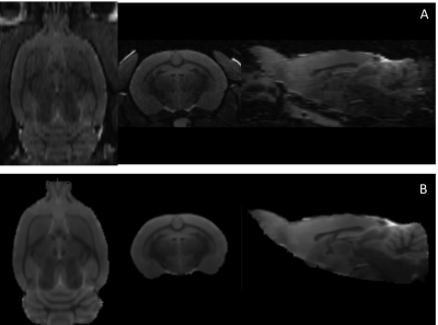 |
96 |
MRI Assessment of Sex Differences in APOEe4 Knock-in in Rat Brains
Loi Do, Aarti Mishra, Adam Bernstein, Marc Lindley, Chidi Ugonna, Nan-kuei Chen, Roberta Brinton, Theodore Trouard
Using a humanized APOEε4 gene knock-in ex-vivo rat brain model, the individual and combined impact of sex and APOEε4 genotype on white matter microstructure was measured using high T2-weighted and diffusion weighted MRI. Total brain volumes showed a significant sexual dimorphism in WT as well as APOEε4 animals, with the females having significantly lower volumes. Both volumetric and diffusion MRI measures were able to show trends of sexual dimorphism as well as genotype effect. These findings were supported with metabolomic data suggesting reduction in glucose utilization and possible shift to fatty acids derived from white matter catabolism as a fuel source.
|
|
3604.
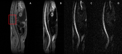 |
97 |
Multicomponent Analysis of High b-value Diffusion-Weighted MRI for Tracking Progression of Amyotrophic Lateral Sclerosis in A Mouse Model
Jin Gao, Mingchen Jiang, Richard Magin, Andrew Larson, Weiguo Li
Amyotrophic lateral sclerosis (ALS) is a progressive disease of motor neuron degeneration in brain and spinal cord with an unknown etiology. Diffusion MRI has potential to track the disease progression in ALS due to the technique’s intrinsic advantages in detecting structure changes and non-invasive nature. In this study, we investigated the feasibility of analyzing multiple high b-value diffusion-weighted images using a non-negative least squares method (requiring no prior assumptions about components) and a bi-compartment model with restricted and hindered diffusion components. Both methods were able to detect alterations of spinal cord in the G93A-SOD1 mouse model of ALS.
|
|
3605.
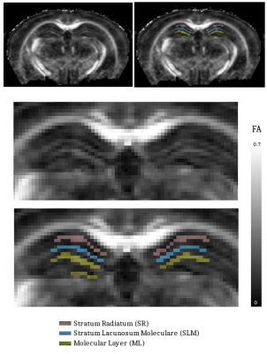 |
98 |
High B-value In-vivo DTI for Early Detection of Cerebral Microstructure Alteration: Application to Hippocampus Lesions in a Mouse Model of Multiple Sclerosis
Renaud NICOLAS, Amandine CROMBE, Nathalie RICHARD, Thomas TOURDIAS, Bassem HIBA
Few reports have highlighted the value of DTI achieved with a strong water-diffusion weighting to capture early and diffuse changes in cerebral microstructure of the white-matter. This study investigates the benefit of DTI with b-value=2700s/mm² to detect microstructure lesions of the hippocampus in a mouse model of multi-sclerosis. High b-value DTI allowed to detect microstructure changes invisible using DTI achieved with classical b-value=1000s/mm². In grey-matter tissues with complex architecture such as hippocampus, an increase of b-value appeared to be more relevant to monitor the microstructure changes compared to an increase of the number of diffusion directions.
|
|
3606.
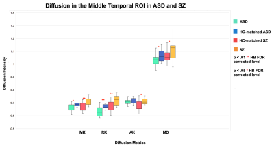 |
99 |
Diffusional Kurtosis Imaging of Temporal Lobe Gray Matter as a Biomarker of Neurological Disease: Schizophrenia and Autism Spectrum Disorder
Faye McKenna, Laura Miles, Jeffrey Donaldson, F. Xavier Castellanos, Donald Goff, Mariana Lazar
In this study, we employed diffusion kurtosis imaging (DKI) to test for differences in gray matter (GM) microstructure in schizophrenia (SZ) and autism spectrum disorder (ASD). Significantly increased metrics of DKI were found in SZ compared to HC participants, while significantly decreased metrics of DKI were found in ASD compared to HC in the temporal lobe and sub-lobar temporal regions of interest (ROIs). In vivo DKI metrics appear to be sensitive to GM microstructural pathology in SZ and ASD and may provide new information on the neural underpinnings of these disorders.
|
|
| Top |
Microstructure Modeling: 2
Digital Poster
Diffusion
Wednesday, 15 May 2019
| Exhibition Hall |
09:15 - 10:15 |
| |
|
Computer # |
|
3607.
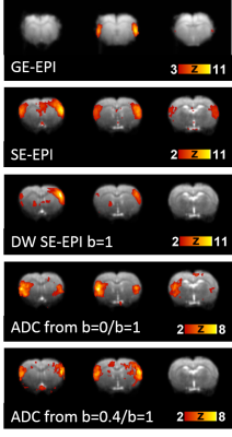 |
101 |
Resting-state diffusion fMRI bears strong resemblance and only subtle differences to BOLD fMRI
Ileana Jelescu
Diffusion fMRI (dfMRI) is a presumably non-BOLD technique sensitive to transient microstructural changes underlying neural activity. Previous task-fMRI studies have assessed the characteristics of the dfMRI signal and potential BOLD contamination, with conflicting results. Here we acquired resting-state fMRI data with five protocols with incrementally reduced BOLD contributions and analyzed the characteristics of resting-state networks and functional connectivity using model-free approaches. We report dfMRI data does not contain fundamentally different information to BOLD-fMRI, with the exception of a few regions that switch from anti- to positively-correlated. Future work will focus on removing any remaining BOLD contribution from the dfMRI acquisition.
|
|
3608.
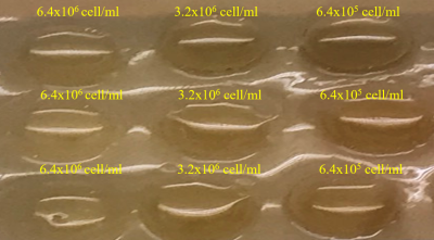 |
102 |
Diffusion Kurtosis Image in assessment of 3D Cell Culture
Jui-Heng Lin, Huei-Chun Hsiao, Shao-Chieh Lin, Yi-Jui Liu, Ruey-Hwang Chou, Ke-Sin Yan, Tan-Wei Liao, Chao-Chun Lin, Chia-Wei Lin, Wu-Chung Shen
A non-Gaussian kurtosis model has been shown to take into account tissue heterogeneity and two relative imaging biomarkers namely, the kurtosis coefficient and the corrected diffusion coefficient can be quantified. In this study, 3D cell culture with hydrogels ECM was used to investigate whether DKI may provide information on these microenvironmental parameters and the microenvironment-associated metastatic propensity of tumors. Our results demonstrated DKI-MRI may provide the potential biomarkers on the change of microenvironment in the application of 3D cell culture experiment.
|
|
3609.
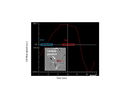 |
103 |
Does cerebrospinal fluid pulsation affect DWI thermometry?: healthy volunteer study
Koji Sakai, Jun Tazoe, Hiroyasu Ikeno, Kentaro Akazawa, Masashi Yasuike, Toshiaki Nakagawa, Bárbara Abecassis, Kei Yamada
Diffusion-weighted imaging (DWI) based thermometry has a potential to be a non-invasive method of temperature measurement for the deep inside of human brain. Nevertheless, the DWI at lateral ventricle in the brain might be influenced by the pulsation flow of cerebrospinal fluid (CSF), which is motivated by heartbeat. The purpose of this study was to investigate the influence of pulsation flow on brain DWI thermometry for healthy subjects. Comparisons were performed among ΔT at three CSF speed selections (slow vs. fast vs. random). There was no significant difference in ΔT among the CSF speed and volume on healthy subjects.
|
|
3610.
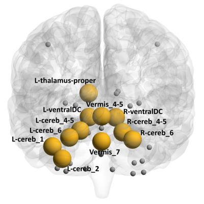 |
104 |
Abnormal cerebellar connectivity within the motor subnetwork in MSA with cerebellar dysfunction
Apurva Shah, Shweta Prasad, Santosh Dash, Jitender Saini, Pramod Kumar Pal, Madhura Ingalhalikar
Multiple system atrophy with cerebellar features (MSA-C) is a distinct subtype of MSA characterized by predominant cerebellar symptoms. Neuroimaging studies have demonstrated cerebellar abnormalities; however, abnormality of structural connectivity of the motor subnetwork has not been studied and this study aims to investigate this aspect. We observed impairment in the structural segregation, integration and network resilience with significantly reduced nodal strength and connectivity in several cerebellar as well as non-cerebellar regions that correlate with UMSARS scores. Our findings provide definitive evidence of abnormalities that may be causally implicated in the motor features of cerebellar dysfunction and Parkinsonism observed in MSA-C.
|
|
3611.
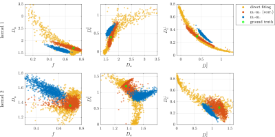 |
105 |
Propagation of Bias in Moment-Matching Based Standard Model Parameter Estimation
Jonas Olesen, Noam Shemesh, Sune Jespersen
Moment-matching is one proposed approach for estimating Standard Model parameters which partly overcomes the issues of the model’s notoriously shallow fitting landscape. The method achieves robustness by matching the model’s moments to the cumulants of the data determined by diffusion kurtosis imaging which is stable and clinically feasible. However, estimates of cumulants generally suffer from bias due to the use of finite b-values. Here, it is demonstrated that this bias propagates to the model-parameter estimates resulting in substantial inaccuracy even for small b-values.
|
|
3612.
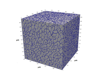 |
106 |
Random Walks in Stochastic Geometries
Eric Baker, Brendan Moloney, Xin Li, Erin Gilbert, Charles Springer
Monte Carlo random walk simulations of water molecule displacements in realistic cell ensembles are presented. Within an ensemble, the cells have stochastic distributions of sizes, shapes, and interstitial spacings. The probability of molecules permeating the cell membrane is varied. The irreducible, fundamental system parameters are the cell density, ρ, the mean cell volume, <V>, and the steady-state cellular water efflux rate constant, <kio>. Even though the self-diffusion coefficient is that of pure water, non-Gaussian displacements are observed.
|
|
3613.
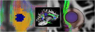 |
107 |
Characterising tissue heterogeneity in cerebral metastases using multi-shell multi-tissue constrained spherical deconvolution
Maxime Chamberland, Najmus Iqbal, Suryanarayana Rudrapatna, Greg Parker, Chantal Tax, John Staffurth, James Powell, Richard Wise, Derek Jones
Considerable attention has focused on characterizing brain tumours using diffusion tensor imaging, and only more recently using advanced modelling techniques. Building on the observation that metastatic tumors exhibit different signal intensities depending on their histological/cellular composition, we investigate how multi-shell multi-tissue constrained spherical deconvolution can characterise tissue heterogeneity within brain metastases.
|
|
3614.
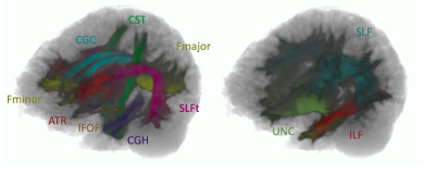 |
108 |
Reproducibility of tractography based on fibre orientation distribution and anatomical constraints applied on paediatric diffusion MRI.
Thanh Vân Phan, Thibo Billiet, Dirk Smeets, Maaike Vandermosten
Tractography is known to be sensitive to technical variations but might also be sensitive to issues related to paediatric data, such as head motion. We assessed the reproducibility in paediatric data of the probabilistic tractography algorithm based on fibre orientation distribution and anatomical constraints that enables to deal with crossing fibres and to reconstruct tracts with more anatomical accuracy. Our results showed that the reproducibility of this approach when it is applied on paediatric data is negatively affected by younger age and by head motion but can still achieve good reproducibility for selected tracts.
|
|
3615.
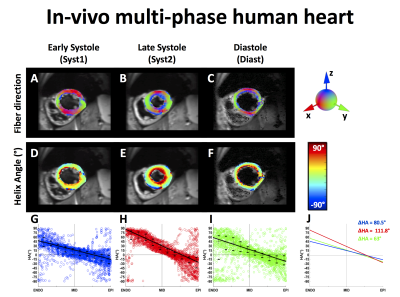 |
109 |
Cardiac Fiber Mobility During Contraction Using High Resolution Diffusion Tensor MRI
Kevin Moulin, Ilya Verzhbinsky, Tyler Cork, Nyasha Maforo, Luigi Perotti, Daniel Ennis
The main cardiomyocyte aggregate orientation, represented by the Helix Angle, remains poorly described in vivo during contractionas it is affected by the imaging resolution and thus by a change of wall thickening. This work evaluated the effect of the imaging resolution using a numerical phantom and ex vivoscans on porcine hearts. High-resolution cDTI was acquired in vivoto measure the transmural mobility of Helix Angle at three cardiac phases. A strong steepening of Helix Angle was observed as the resolution decreased in simulation and ex-vivo. A significant change in the Helix Angle distribution was observed during contraction.
|
|
3616.
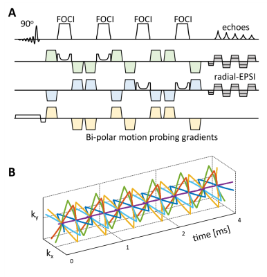 |
110 |
Metabolite diffusion weighted imaging with golden angle radial echo planar spectroscopic imaging
Vincent Boer, Itamar Ronen, Jan Pedersen, Esben Petersen, Henrik Lundell
Diffusion weighted spectroscopy (DWS) is a promising tool for investigating compartment specific microstructure in heterogeneous tissues. Unlike water abundant in all cellular spaces, the mobility of metabolites provides a window into the microstructure of specific cell types. Multi-shot sequences for diffusion spectroscopic imaging suffer from translation induced phase fluctuations. This has previously been addressed with additional phase navigators. In this work we propose self navigated metabolite diffusion weighted spectroscopic imaging using golden angle radial echo planar gradient readouts with semi-LASER voxel localization. Initial data shows good spatial localization and spectral quality.
|
|
3617.
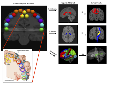 |
111 |
Needle in a Haystack: Finding connections of interest in the precentral gyrus from diffusion MRI based connectomics
Jasmeen Sidhu, Francois Rheault, Maxime Descoteaux
DMRI tractography and connectomics aim to create comprehensive maps of all neural circuitry in the brain, however, dMRI is a false-positive prone imaging modality. Yet, as it is the only non-invasive and in-vivo method to indirectly study the white matter connectivity of the brain it is heavily utilized. DMRI-Connectomic analyses often end with a “big data” problem, as there are many possible connections. Moreover, this data is likely populated by false-positive connections. By utilizing data-driven criteria to filter matrices this work attempts to narrow the “search window” by adding criteria on which we can discard “connections” or consider them potential candidates for further investigation. Such judgements may also lead to connections being evaluated as true or false-positives.
|
|
3618.
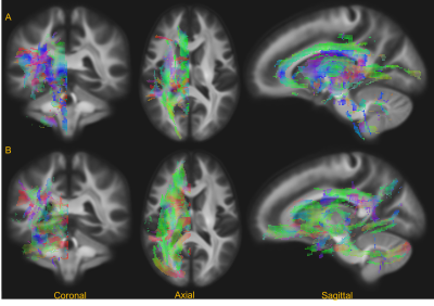 |
112 |
Characterization of white matter asymmetries in the healthy human brain using diffusion MRI fixel-based analysis
Arush Honnedevasthana Arun, Robert Smith, Alan Connelly, Fernando Calamante
The diffusion tensor model has been used extensively to study asymmetry in various regions of the human brain white matter. However, given the limitations of the tensor model, the nature of any underlying asymmetries remain uncertain, particularly in crossing fibre regions. Here, we applied fixel-based analysis (FBA) to state-of-the-art diffusion MRI data to assess white matter asymmetry of the healthy brain, demonstrating significant left/right asymmetries in various regions. The major advantage of this study is that FBA performs fibre-tract-specific modeling, and thus provides a method to assess the white matter asymmetry in a physically interpretable way.
|
|
3619.
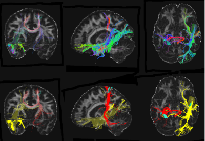 |
113 |
Toward prognostic sensitivity of DTI to cognitive outcomes following physical exercise interventions.
Anna Egbert, Ryan Falck, John Best, Teresa Liu-Ambrose
Diffusion Tensor Imaging (DTI) is widely implemented in clinical research, yet, its prognostic value in brain and cognitive health remains uncertain. Prospective estimation of the effectiveness of interventions, such as physical exercise, can increase cost-effectiveness of treatment, thus, maximizing the impact of accessible modifiable preventive factors in improving health outcomes in the general population. We examined DTI in relationship to cognitive outcomes of physical activity intervention. This is the first study to show that white matter integrity related to sleep efficiency can be an early predictor of the cognitive outcomes of physical exercise intervention.
|
|
3620.
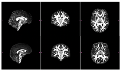 |
114 |
Assessment of Microstructural Changes Induced via Repeated Videogame Training as a Measure of Neuroplasticity in Normal Developing, College-age Brains
Austin Patrick, Douglas Dean, Thomas Gorman, C. Shawn Green, Andrew Alexander
In this study, diffusion tensor imaging (DTI) and neurite orientation dispersion and density imaging (NODDI) were used to assess brain changes in white matter regions as a result of repeated videogame training. In a cohort playing a simulated race car game, we observe evidence of microstructural changes in tracts associated with working and long-term memory. Subjects playing a guitar simulation game, experienced changes in regions associated with attention, visuomotor learning, and proprioception of the limbs. This study demonstrates that diffusion MRI is promising for characterizing subtle brain changes associated with learning.
|
|
3621.
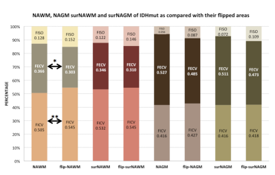 |
115 |
Finding the Surrogate Biomarkers of Glioma Infiltration in the Normal Appearing White Matter adjacent to Tumor using Neurite Orientation Dispersion and Density Imaging (NODDI)
Presentation Not Submitted
Yi-Cen Ting, Chou-Ming Cheng, Tzu-Chen Yeh
This work addresses the study in glioma, where Diffusion Tensor Imaging (DTI) provides incoherent information while Neurite orientation dispersion and density imaging (NODDI) may improve the microstructural characterization. Glioma recurrences are mostly located at the margin of the resection cavity as peritumoral area. NODDI, using a compartment-based biophysical model, would potentially provide specific surrogate biomarkers for the microscopic foci of glioma infiltration better than DTI. NODDI reveals that glioma cells can extend beyond the bulk tumor and peritumoral edema into normal appearing white matter (NAWM) adjacent to tumor or peritumoral edema by using Region of Interest (ROI)-based statistics.
|
|
3622.
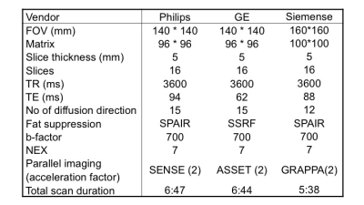 |
116 |
Assessing inter-vender reproducibility of diffusion tensor imaging of the spinal cord for multi-center imaging studies
Mitsuhiro Kitamura, Satoshi Maki, Takeo Furuya, Takuya Miyamoto, Yasuhiro Shiga, Kazuhide Inage, Sumihisa Orita, Koji Matsumoto, Shingo Terakado, Yoshitada Masuda, Seiji Ohtori
In this study, we assessed the inter-vendor precision of diffusion tensor imaging (DTI) metrics using consensus acquisition protocol across 3T scanners from 3 main vendor at the two sites. The data were acquired from the traveling human healthy volunteer. DTI data and anatomical images were also acquired and imaging data were processed using the Spinal Cord Toolbox (SCT). Relatively higher inter vender reproducibility of fractional anisotropy (FA) in and cross-sectional area were demonstrated and it is considered feasible to conduct multicenter DTI studies of the spinal cord using FA value as biomarker and SCT as postprocessing tool.
|
|
3623.
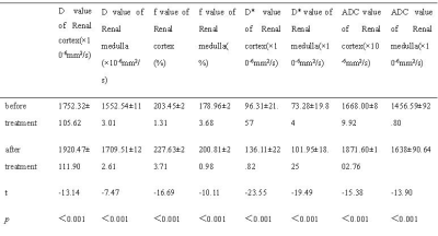 |
117 |
Performance of Intravoxel incoherent motion (IVIM) imaging in the curative effect evaluation of diabetic nephropathy - A preliminary study
Presentation Not Submitted
Jing Chen, Xirong Zhang, Nan Yu, Qi Yang, Yong Yu, Robert Grimm, Shaoyu Wang
Diabetes has nowadays become one of the major public health problems. Early detection and intervention is crucial to delay the progress of Diabetic kidney disease. In this study, IVIM imaging was used to evaluate the renal-cortex- and medulla-related parameter changes before and after the treatment of diabetic nephropathy. It was found that after treatment, D, f, D *and ADC values were increased, indicating the improved microvascular perfusion.
|
|
3624.
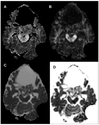 |
118 |
Diffusion MRI characterization of Proton-therapy-induced brain tissue changes: a case study
Lisa Novello, Nivedita Agarwal, Stefano Lorentini, Sabina Vennarini, Domenico Zacà, Ofer Pasternak, Jorge Jovicich
Radiotherapy-induced neurotoxicity may be life threatening and its characterization is crucial for cancer treatment management, especially in early treatment phases. Diffusion MRI allows to assess non-invasively microstructural changes occurring with radiation treatment, which are of interest in relation to local absorbed dose and cognitive changes. Proton therapy (PT) offers the promise of more focal tumor damage relative to conventional radiotherapy. However, few studies have evaluated how it affects brain microstructure. As part of an ongoing longitudinal study, we present the first available data of 5 acquisitions along PT course showing a possible treatment-related “necrosis-like” effect during treatment.
|
|
3625.
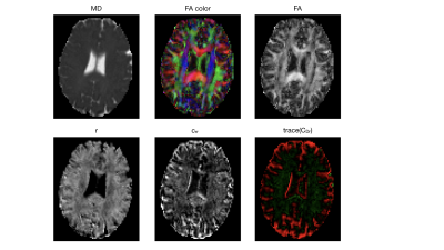 |
119 |
Diffusion-Relaxation Imaging with B-tensor Encoding using the Cumulant Expansion
Carl-Fredrik Westin, Filip Szczepankiewicz, Lipeng Ning, Yogesh Rathi, Markus Nilsson
Quantitative T2 and diffusion imaging provide important information about tissue microstructure. However, a joint knowledge of quantitative T2 and diffusion-derived measures can provide richer information about the microstructure that is not accessible when using these modalities independently. The standard approach for estimating the joint distributions of the T2-diffusion relies on the inverse Laplace transform. This transform is known to be unstable and difficult to invert. In this work, we introduce an alternative approach based on cumulant expansion, and extend the recently proposed multidimensional diffusion MRI framework ``Q-space Trajectory Imaging" (QTI) to include T2-relaxation modeling. The cumulants of the expansion include estimates of mean diffusion and T2 relaxation, as well as their variance and covariance. We demonstrate the feasibility of this approach in a healthy human brain.
|
|
3626
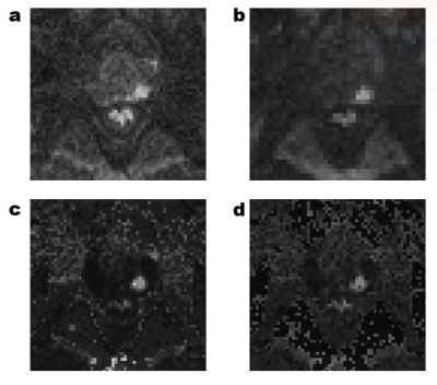 |
120 |
Qualitative Comparison of Calculated and Measured Ultra-High b-Value Diffusion-Weighted Images in the Assessment of Clinically Significant Prostate Cancer.
Video Permission Withheld
David Bonekamp, Christopher Edler, Jan Philipp Radtke, Frederik Laun, Markus Hohenfellner, Heinz-Peter Schlemmer, Tristan Kuder
To qualitatively compare measured and calculated ultra-high b-value (UHB, b-value up to 4000 s/mm2) DWI for detection of clinically significant prostate cancer. UHB-DWI was acquired in 55 patients at 3T in addition to standard DWI (S-DWI) extrapolated to UHB b-values. Two raters R1 and R2 independently assessed DWI and ADC in combination with T2w images. Lesion visibility was best on ultra-high b-value images monoexponentially extrapolated (ME-DWI) from S-DWI and equal on S- and UHB-ADC. The experienced rater was better able to adapt the improved lesion visibility on ME-DWI into the lesion detection task and utilized it for superior predictive performance.
|
|
3627.
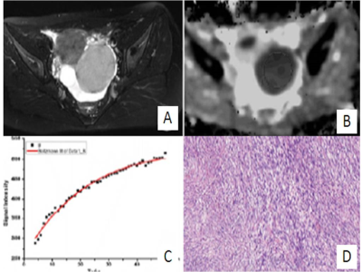 |
121 |
Qualitative Diagnosis of Complex Ovarian Tumors Using Diffusion Weighted Imaging and Dynamic Contrast-Enhanced Magnetic Resonance Imaging: Correlation with MR Features and Pathology
Shuping Weng, Ruqi Fang
Aims: To evaluate the effectiveness of diffusion weighted imaging and dynamic contrast enhanced magnetic resonance imaging in qualitative diagnosis of complex ovarian tumors. Methods: Semi-quantitative parameters of maximal slope of time-signal intensity curve and apparent diffusion coefficient measurements of diffusion-weighted imaging obtained from the tumor solid regions in 65 cases confirmed complex ovarian tumors were done.
Results: A cut-off value of 1.61 × 10⁻³mm2 /s for ADC was used in differentiating invasive from borderline tumors.Optimal threshold value for maximal slope was 4.58% per second identifying benign and malignant tumors.
Conclusion: Maximal slope value of time-signal intensity curve is the best index in distinguishing malignant from benign ovarian complex tumors,and ADC value of the solid component is the best index in differentiating invasive from borderline ovarian complex tumors.
|
|
3628.
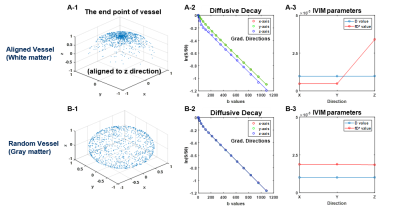 |
122 |
Directional Intravoxel Incoherent Motion (IVIM) MR in Rodent Brain
MinJung Jang, SeokHa Jin, MungSoo Kang, SoHyun Han, HyungJoon Cho
The measurement of white matter flow, which is directional along the neuronal fibers, is challenging due to inherent limitations of low sensitivity and signal-to-noise ratio with arterial spin labeling(ASL)- and dynamic susceptibility contrast(DSC)-MRI acquisitions mainly due to low blood volumes. Recently, flow sensitive intravoxel incoherent motion(IVIM)-diffusion MR has been recognized as being particularly sensitive to directional flows. However, the multi-variable fittings usually suffer from over-fitting and thresholding issues. This study investigated the feasibility of constrained non-negative matrix factorization(cNMF) implementation to IVIM-MR for the automatic segmentation of directional IVIM signals for the purpose of robust detection of directional white matter flow.
|
|
3629.
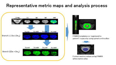 |
123 |
Application of Spinal Cord White Matter Tract Integrity Quantification with Atlas-based Analysis in Multiple Sclerosis and Neuromyelitis Optica Spectrum Disorder
Masaaki Hori, Akifumi Hagiwara, Kazumasa Yokoyama, Kouhei Kamiya, Issei Fukunaga, Tomoko Maekawa, Koji Kamagata, Katsutoshi Murata, Shohei Fujita, Ryusuke Irie, Christina Andica, Kanako Kumamaru, Akihiko Wada, Julien Cohen-Adad, Shigeki Aoki
We investigated both branches (Da ≤ De,? or Da > De,?) of Kurtosis-based white matter tract integrity to distinguish microstructural changes in the spinal cords of patients with MS and Neuromyelitis Optica. FA and AWF were significant higher and MD was significant lower in spinal cord white matter in MS (P=0.009, 0.024, 0.032, respectively). Di,axial, Di,radial and De,radial of spinal cord white matter in MS were significant lower (P=0.009, 0.024, 0.032, respectively,) in Branch 2(Da > De,?) in MS. There was no significant difference in Branch 1 ( Da ≤ De,?) metrics.
|
|
3630.
 |
124 |
Delineation of thalamic substructures in ultra-high b-value DWI-measurement with reasonable acquisition time
Nils Nuessle, Benjamin Bender, Uwe Klose
The purpose of this study was to evaluate the capability of optimized and faster ultra-high b-value DWI in separating and identifying intrathalamic substructures compared to a previously described protocol. 7 subjects underwent MR-imaging, including T1 MPRAGE and two DWI sequences at 0 and 5000 s/mm2. DWI was performed with 64 directions and 5 averages (17:56 min) and with 6 directions and 25 averages (8:23 min). Intrathalamic substructures were semi-automatically delineate 4mm above AC/PC line. Accordance between the original sequence and the new speeded-up measurement was high. Acquisition time was reduced by more than 50 % with comparable results.
|
|
3631.
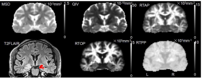 |
125 |
The value of Mean Apparent Propagator (MAP)-MRI in the diagnosis of hippocampal sclerosis
Presentation Not Submitted
Keran Ma, Jingliang Cheng, Xiaonan Zhang, Ankang Gao, Chengru Song, Shaoyu Wang, Xu Yan, Huiting Zhang
This study aimed to explore the value of mean apparent propogation (MAP)-MRI parameters in the diagnosis of hippocampal sclerosis. By comparing the MAP-MRI parameters of hippocampus from both the hippocampal sclerosis patients and the healthy controls, the study found that the MAP-MRI parameters showed high consistency with pathological results, particularly for MSD and QIV. It suggests that MAP-MRI may be used as a diagnostic method with high sensitivity and specificity besides magnetic resonance spectroscopy(MRS) in the future.
|
|
| Top |
Diffusion: Validation
Digital Poster
Diffusion
Wednesday, 15 May 2019
| Exhibition Hall |
09:15 - 10:15 |
| |
|
Computer # |
|
3632.
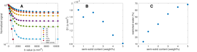 |
126 |
Tunable diffusion kurtosis in lamellar vesicle suspensions toward development of quantitative phantom surrogate of tumor microenvironment
Scott Swanson, Dariya Malyarenko, Thomas Chenevert
A set of materials based on nanostructured lamellar vesicles with restricted diffusion compartments is constructed to achieve tunable diffusion kurtosis behavior. The observed apparent diffusion (Dapp) and kurtosis (Kapp) model parameters span the range of values found in vivo. Effect of vesicle population, size, and porosity is studied for estimated diffusion parameters. These nanostructured systems provide an ideal platform for a diffusion kurtosis phantom used to validate quantitative imaging protocols and results.
|
|
3633
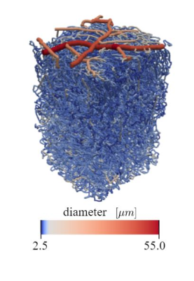 |
127 |
Simulation of Intravoxel Incoherent Perfusion Signal using a Realistic Capillary Network of a Mouse Brain
Video Permission Withheld
Valerie Phi van, Franca Schmidt, Linda Mathez, Georg Spinner, Sebastian Kozerke, Christian Federau
The microscopic origin of the IVIM perfusion signal is not well understood. We simulated blood motion in three realistic microvascular networks obtained by two-photon laser microscopy in mouse brain and computed the effect of this motion on the diffusion-weighted signal MR signal for b-values between 0 and 1000 s/mm2. We found pseudo-diffusion coefficients D* in the range of in vivo measurements. Further, the signal decay was strongly dependent on the specific type of vessel (capillary,arterioles/venules or pial vessels) and on the global vessel diameters of the network.
|
|
3634.
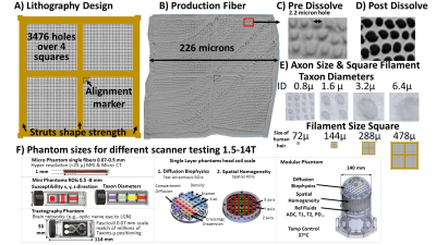 |
128 |
Taxon anisotropic phantom delivering human scale parametrically controlled diffusion compartments to advance cross laboratory research and calibration.
Walter Schneider, Sudhir Pathak, Yijen Wu, David Busch, John Dzikiy
Details a novel bi-component polymer Taxon (textile water filled tubes) anisotropic diffusion phantom providing 0.8 microndiameter tubes, packing density of 106 per mm2, water fraction of over 45% matched to human axon histology with parametric control of water compartment (intra/extra axon), size, density, angle dispersion that can be manufactured to supply hundreds of laboratories. Observed human tissue range FA (0.6-01.0), MD and RD in observed range. Providing ground truth measurement to advance and calibrate anisotropic diffusion measurement. Micro-CT and diffusion MRI indicate high water content in agreement with MRI and non MRI measurements.
|
|
3635.
 |
129 |
Phantoms for Diffusion Simulations: Multi-Objective Differential Evolution for Realistic Numerical (MODERN) Phantoms.
Presentation Not Submitted
Jonathan Rafael-Patino, Thomas Yu, Mariam Andersson, Hans Martin Kjer, Vedrana Andersen Dahl, Alexandra Pacureanu, Anders Bjorholm Dahl, Tim Dyrby, Jean-Philippe Thiran
The following work presents a novel, generally applicable framework (MODERN) which takes, as input, statistics from histology and automatically generates biologically plausible numerical phantoms whose morphological features are optimized to match the input statistics. As a proof of concept, MODERN is used to generate three-dimensional geometrical meshes representing a bundle configuration of axons which can be immediately integrated into Monte Carlo simulations for diffusion MRI acquisitions. The obtained features of the optimised phantoms are shown to match those from the input values. The statistics used were obtained from axonal segmentations from synchrotron imaging.
|
|
3636.
 |
130 |
Axon-mimicking hydrophilic fibre phantoms for diffusion MRI
Fenglei Zhou, Amy McDowell, Kiran Seunarine, Matt Hall, Damien McHugh, Zhanxiong Li, Chris Clark, Penny Hubbard Cristinacce, Geoff Parker
We report the development of an axon-mimicking phantom composed of hydrophilic hollow microfibres, and evaluate its potential for validating clinical diffusion MRI. Microfibers were fabricated by the co-electrospinning (co-ES) of polycaprolactone (PCL)-polysiloxane-based surfactant (PSi) mixture as shell and polyethylene oxide (PEO) as core, and characterized by scanning electron microscopy (SEM). Three material samples were constructed and included in the phantom within a water bath. SEM images reveal that PCL-PSi fibres in the samples were uniaxially aligned and hollow, with a similar distribution of pore sizes to axons in vivo. MR measurement shows similar anisotropic diffusion behaviour in each sample.
|
|
3637.
 |
131 |
Multisite evaluation of repeatability and stability for a novel quantitative diffusion kurtosis phantom
Dariya Malyarenko, Scott Swanson, Amaresha Konar, Eve LoCastro, Ramesh Paudyal, Michael Liu, Sachin Jambawalikar, Lawrence Schwartz, Amita Shukla-Dave, Thomas Chenevert
Multi-center clinical trials utilizing quantitative diffusion kurtosis imaging (DKI) protocols require accurate, precise, and stable phantoms for validation of derived imaging metrics. This study examines the precision and reproducibility of isotropic (i)DKI parameters obtained from a phantom based on nanostructured vesicles that restrict diffusion and mimic tissue cellularity. Ten test-retest iDKI studies were performed on four scanners at three imaging centers over a six-month period. The tested prototype phantoms exhibited physiologically-relevant and highly-repeatable apparent diffusion and kurtosis parameters. Achieved precision was sufficient to characterize thermal and temporal stability trends to guide robust quantitative iDKI phantom production.
|
|
3638.
 |
132 |
Comparison of dMRI Models for Skeletal Muscle Microstructure Estimation with Numerical Simulations and Porcine Phantom
Noel Naughton, Nicolas Gallo, Aaron Anderson, John Georgiadis
Four models used to estimate skeletal muscle microstructure from dMRI signal are applied to a ex-vivo porcine phantom and provide physiologically reasonable parameter estimates. These models are then compared to numerical simulations of dMRI over a simplified repeating elemental volume. When the estimated parameters are compared to the true underlying parameters of the numerical model, the estimated parameters are found to be substantially inaccurate, motivating the need for more sophisticated models of dMRI in muscle.
|
|
3639.
 |
133 |
Towards a more realistic and flexible white matter numerical phantom generator for diffusion MRI simulation
Ross Callaghan, Noam Shemesh, Daniel Alexander, Hui Zhang, Marco Palombo
We present a novel method for the generation of realistic white matter numerical phantoms with controllable morphology, and more realistic orientation-dispersion and packing-density. A shift of paradigm is proposed: rather than ‘packing fibres’, our algorithm ‘grows fibres’ contextually and efficiently, avoiding intersection between fibres. The potential of the method is demonstrated by reaching the highest orientation-dispersion and packing-density ever. The flexibility of the method is demonstrated with simulations of diffusion-weighted MR signal in three example substrates with differing orientation-dispersions, packing-densities and permeabilities. A proof-of-concept application is presented to investigate the impacts of undulation and beading on the intra-axonal axial diffusivity.
|
|
3640.
 |
134 |
Quantitative Comparison of 6 different Diffusion Weighted Imaging Approaches using NIST Diffusion Phantom
Sampada Bhave, S. Sivaram Kaushik, Robert Peters, Kevin Koch
The application of diffusion weighted imaging (DWI) is rapidly increasing in musculoskeletal system. DWI is useful in imaging diverse range of musculoskeletal pathologies like soft-tissue tumors, bone lesions, vertebral fractures pre and post treatment follow up. In this work, our goal is to quantitatively compare the accuracy of ADC estimation of EPI, and PROPELLER based techniques. We also optimize the imaging parameters for PROPELLER and MSI-PROPELLER techniques and provide a correction method to improve the accuracy of ADC estimation.
|
|
3641.
 |
135 |
Validation and Estimation of crossing angles of fiber bundle with different density using hollow textile-based phantom
Sudhir Pathak, Vinod Kumar, Walter Schneider
A textile-based hollow fiber (taxon) MRI phantom (TABIP) is used to assess the ability of diffusion MR imaging (dMRI) to accurately estimate taxon crossing angles. A parametric pattern of known taxon crossings and taxon bundle sizes was manufactured in the TABIP phantom, it was scanned on a 3T scanner and data was reconstructed with a constrained spherical deconvolution algorithm. 21 repeated scans were collected to enhance SNR. Taxon crossing angles were estimated, and it is shown that crossing angles of 45° and higher can be accurately recovered and that angle measurement reliability decreases with bundle size.
|
|
3642.
 |
136 |
Novel Atlas of Fiber Directions Built From Ex Vivo DT images of Porcine Hearts
Mia Mojica, Mihaela Pop, Maxime Sermesant, Mehran Ebrahimi
In this study we successfully created the first high-resolution cardiac Diffusion Tensor (DT) imaging-based fiber atlas from porcine hearts. Furthermore, we laid the foundation of a framework for building a statistical cardiac atlas by computing an average cardiac geometry from a small database (N=8) of explanted porcine hearts without the need for selecting landmarks, transforming the diffusion tensors of subjects, and obtaining the associated average DT field and fiber architecture. The fiber atlas can be used for personalized electro-mechanical simulations of cardiac function (in anisotropic models when DT data is not available), and aid in diagnosis or therapy guidance procedures.
|
|
3643.
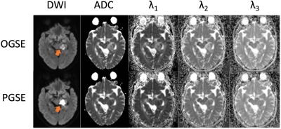 |
137 |
OGSE diffusion weighted imaging of epidermoid cysts: 3D Monte Carlo simulation
Saori Koshino, Akiyuki Sakakibara, Christina Andica, Akifumi Hagiwara, Masaaki Hori, Osamu Abe, Shigeki Aoki
Diffusion weighted imaging with shorter diffusion time using oscillating gradient spin echo (OGSE) may reveal microstructural features among brain disorders. Here we observed DTI eigenvalues in four patients with epidermoid cysts. The values measured by OGSE were higher than those measured by pulsed gradient spin echo (PGSE; used in conventional MRI), indicating restricted diffusion due to spatial restriction and/or viscosity. The results of our Monte Carlo simulation based on the pathological feature of epidermoid cysts suggest that spatial restriction of multiple ellipsoids formed by keratin filaments as well as viscosity and water exchange should influence the extent of restricted diffusion.
|
|
3644.
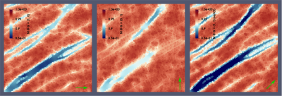 |
138 |
Studying the effect of membrane permeability with a GPU-based Bloch–Torrey simulator
Jan Rose, Lukasz Sliwinski, Sonia Nielles-Vallespin, Andrew Scott, Denis Doorly
To better understand diffusion MRI in biological tissues, numerical simulations are commonly used to model the MR signal. Realistic simulation substrates built directly from histology images help to reduce the model error, but intrinsic parameters other than the microstructure have an effect too. In this study, we investigate the relationship between diffusion tensor fractional anisotropy and membrane permeability. Using a GPU-accelerated Bloch–Torrey solver, we observe a significant difference from the impermeable case for long diffusion times on the order of 1s.
|
|
3645.
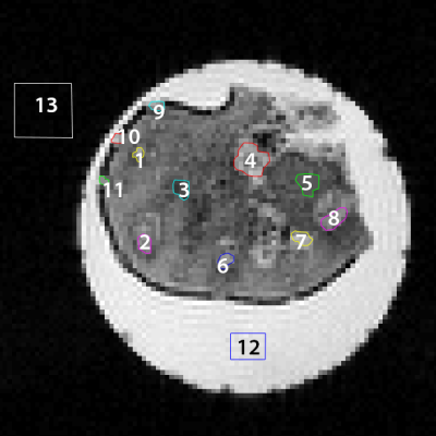 |
139 |
Comparison of Cylindrical and Spherical Geometric Models to Infer Cell Sizes in a Celery Sample
Sheryl Herrera, Morgan Mercredi, Henri Sanness Salmon, Guneet Uppal, Domenico Di Curzio, Melanie Martin
Temporal diffusion spectroscopy (TDS) can be used to infer sizes of cells in samples. It relies on a geometric model to relate the MRI signal to the cell sizes. Celery collenchyma tissue and vascular bundles have long cells while parenchyma cells are rounder. We compared a cylindrical and spherical geometric model in temporal diffusion spectroscopy to determine how important the geometrical model was for celery. The inferred diameters of cells in celery (14±6µm to 20±12µm) were not statistically different when using the two different geometric models. This is the first step toward understanding the importance of geometric models for TDS.
|
|
3646.
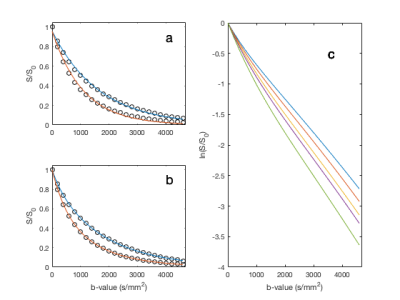 |
140 |
Kärger Model Difficulties for In Vivo DWI
Catherine Lian, Brendan Moloney, Eric Baker, Greg Wilson, Erin Gilbert, Thomas Barbara, Charles Springer, Xin Li
Although it is well known that diffusion-weighted imaging (DWI) is sensitive to in vivo trans-membrane water-exchange, quantitative interpretation of the diffusion b-space decay remains difficult. Using random-walk simulated DWI data, this study investigates the feasibility and reliability in studying the water exchange effects with a multi-exponential fitting approach on DWI data.
|
|
3647.
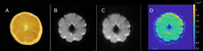 |
141 |
Non-destructive measurement and prediction of soluble solids of fruits using diffusion-weighted magnetic resonance imaging
Xinpei Wang, Jichang Zhang, Zhen Nan, Pengfei Xu, Richard Bowtell, Chengbo Wang
Diffusion-weighted magnetic resonance imaging (DW-MRI) was used to measure and predict total soluble solids (TSS, unit: °Brix) of fruits noninvasively. The results demonstrated that mean apparent diffusion coefficient (ADC) in fruits was strongly correlated with TSS of fruit juice (R ≥ 0.92 and p ≤ 1.51E-4). DW-MRI shows great potential for future commercial implementation of fruit quality evaluation.
|
|
3648.
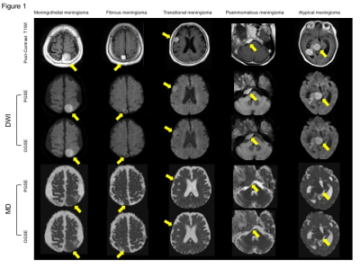 |
142 |
Diffusion Time Dependence of Diffusion Tensor Parameters in the Evaluation of Meningioma Subtype: a Preliminary Study
Tomoko Maekawa, Masaaki Hori, Katsutoshi Murata, Christina Andica, Yutaka Ikenouchi, Shohei Fujita, Ryusuke Irie, Akifumi Hagiwara, Kamiya Kouhei, Koji Kamagata, Akihiko Wada, Shigeki Aoki
We investigated the DTI eigenvalues and MD of five types of meningioma acquired with a shorter diffusion time using an OGSE sequence. Eleven meningiomas that consisted of 4 meningothelial meningiomas, 2 fibrous meningiomas, 2 transitional meningiomas, 1 psammomatous meningioma, and 2 atypical meningiomas were analyzed. Our results showed that the psammomatous and atypical meningiomas had a relatively strong diffusion time-dependence of diffusion tensor metrics. The use of shorter diffusion time with DTI provide additional information about the microstructure of each meningioma subtypes.
|
|
3649.
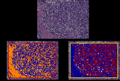 |
143 |
Whole-Tumor Histogram Analysis of Monoexponential and Advanced Diffusion-weighted Imaging for Sinonasal Malignant Tumors: Correlations with Histopathologic Features
Zebin Xiao, Zuohua Tang, Jing Zhang, Guang Yang, Wenjiao Zeng, Jianfeng Luo, Rong Wang, Linying Guo, Zhongshuai Zhang
This is the first study with a large sample size to systematically investigate the correlation of monoexponential diffusion-weighted imaging (DWI) and advanced DWI (intravoxel incoherent motion [IVIM] and diffusion kurtosis imaging [DKI]) parameters with histopathologic features of sinonasal malignant tumors using whole-tumor histogram analysis, which could improve the interpretation of DWI findings and promote the use of these diffusion methods in clinical practice. In comparison with monoexponential DWI and biexponential DWI (IVIM), histogram metrics derived from DKI may better reflect the microstructure of sinonasal malignant tumors, including the cellular, stromal and nuclear fractions.
|
|
3650.
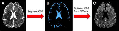 |
144 |
Examining Links Between Free Water and a TSPO-PET Marker of Neuroinflammation
Benjamin Reid, Maria Di Biase, Martin Pomper, Martha Shenton, Yong Du, Jennifer Coughlin, Ofer Pasternak
Free-water (FW) is a diffusion MRI marker of freely diffusing water in the extracellular space, which is expected to increase in the presence of neuroinflammation. Here, we test associations between FW and positron emission tomography (PET) imaging of the translocator protein (TSPO), which is a putative neuroinflammatory marker. We show that increased FW relates to higher TSPO binding in the hippocampi of healthy controls, but not of individuals with sports-related, repetitive traumatic brain injury. Thus, while FW relates to TSPO under healthy conditions, pathological variance in TSPO may complicate associations between FW and TSPO-indexed neuroinflammation.
|
|
3651.
 |
145 |
A Novel Metric Shows the Robustness of the Graph Communities to Brain-Tractography False-Positives.
Juan Villarreal-Haro, Alonso Ramirez-Manzanares, Juan Pichardo-Corpus
We study the impact of the brain tractography false positives in the brain connectivity graphs. The representative input database for the analysis is the set of tractograms from the participants on the ISMRM-2015 Tractography Challenge. We propose 2 novel metrics to rank the quality of a tractogram when it is compared with a known ground truth. The results of this study indicate that the the estimation of graph communities is robust to high levels of overestimation in the connectivity.
|
|
3652.
 |
146 |
The multi-play of diffusion tensor imaging and immunohistochemistry to unveil the longitudinal microstructural brain alterations in a closed head brain injury model.
Abdalla Mohamed, Lyndsey E. Collins-Praino, Frances Corrigan, Fatima Nasrallah
Traumatic brain injury (TBI) is a disease burden worldwide and it is associated with axonal injury and neuroinflammation. Investigating the underlying mechanisms and the progression in the acute and sub-acute stages following TBI non-invasively would aid for early diagnosis and intervention. In this study, we used diffusion tensor imaging (DTI) to investigate the microstructural changes following TBI. Furthermore, we used immunohistochemistry to investigate the axonal injury and microglial activity following TBI. Using DTI, we detected persistent microglial activity associated with TBI which might suggest a possible use of DTI for reflecting on the microstructural changes associated with TBI in humans.
|
|
3653.
 |
147 |
How reproducible are the results of probabilistic white matter tract estimation?
Irène Brumer, Enrico De Vita, Jonathan Ashmore, Jozef Jarosz, Marco Borri
Diffusion tractography and fMRI data is valuable for pre-surgical planning, but its analysis involves a number of user-dependent decisions. In particular the choice of activation threshold in fMRI maps and the definition of seed region in tractography may impact the results. This work evaluates both the intrinsic variability of probabilistic white matter tract estimation and the inter-user reproducibility of tractography analysis. The former was estimated from repeated identical processing, while for the latter tracts obtained by different users were compared. Achieving a good inter-user reproducibility (up to 85%) is possible, considering that the intrinsic reproducibility ranged between 72% and 89%.
|
|
3654.
 |
148 |
Consistency and Reproducibility of Probabilistic Tractography
Jian Lin, Mark Lowe, Robert Fox, Ken Sakaie
Deciding from among the many available tractography algorithms can be challenging. We demonstrate that track-based measures can be compared using standard statistical approaches to compare the performance of two probablistic tractography algoirthms to determine the conditions under which one algorithm can replace another.
|
|
3655.
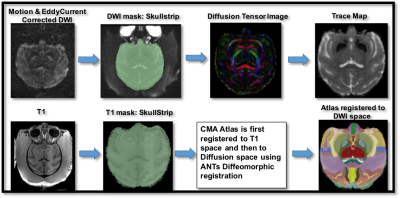 |
149 |
Validation of Diffusion Imaging Measures in Gray Matter using Histology from macaque monkey
Madhura Baxi, Suheyla Cetin Karayumak, Doug Richardson, George Papadimitriou, Sarina Karmacharya, Nikos Makris, Andre Kouwe, Bruce Jenkins, Thomas Witzel, Tara Moore, Douglas Rosene, Marek Kubicki, Yogesh Rathi
This study is the first attempt towards histological validation of diffusion imaging measure of Trace in the gray matter of macaque monkey. Heterogeneity of Trace (hTrace) computed for one monkey in 7 cytoarchitectonically different regions was compared with the variance in intracellular volume fraction for the same regions computed using digitized histological sections, stained for all cell bodies. hTrace was found to be significantly correlated with the variance in intracellular volume fraction. This shows that the diffusion measure of hTrace is sensitive to the underlying gray matter cytoarchitecture such as the cell size and cell packing density.
|
|
3656.
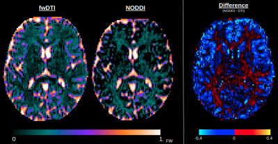 |
150 |
Free water mapping in diffusion MRI: How do two common approaches compare?
Jordan Chad, Ofer Pasternak, J. Chen
Free water eliminated diffusion tensor imaging (fwDTI) and neurite orientation dispersion and density imaging (NODDI) are two increasingly established techniques that measure free water (FW) in diffusion MRI. Yet, despite the utility of each approach, their corresponding FW estimates have yet to be compared. In this work, we find that FW measurements near cerebrospinal fluid are highly similar between the two approaches, but within tissue, NODDI tends to compute slightly higher FW values in the white matter and lower FW values in gray matter than fwDTI. Potential sources of this discrepancy are discussed.
|
|
Back to Program-at-a-Glance |  Back to Top Back to Top |
| The International Society for Magnetic Resonance in Medicine is accredited by the Accreditation Council for Continuing Medical Education to provide continuing medical education for physicians. |
