Digital Poster Session
Interventional MRI Back to Program-at-a-Glance Back to Program-at-a-Glance
|
Wednesday, 15 May 2019
Digital PosterInterventional MRI
3805 -3829 MR-Guided HIFU & MR Thermometry
3830 -3854 Non-Thermal Interventional MRI |
| |
MR-Guided HIFU & MR Thermometry
Digital Poster
Interventional MRI
Wednesday, 15 May 2019
| Exhibition Hall |
13:30 - 14:30 |
| |
|
Computer # |
|
3805.
 |
126 |
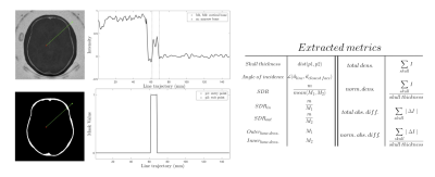
Zero TE based screening for transcranial MR guided focused ultrasound

Jaime Caballero, Rafael Rodríguez-Rojas, Raul Martínez-Fernández, Marta Del-Alamo, Pablo Garcia-Polo, Jose Pineda-Pardo
The high acoustic impedance of the skull limits the performance of tissue ablation applications with tcMRgFUS. Skull characterization is currently based on CT. Zero echo time (ZTE) images could represent a safer alternative in this matter. CT and ZTE images were acquired in sixteen essential tremor patients that underwent tcMRgFUS. Several skull measures were obtained for both images. ZTE and CT based metrics were strongly correlated. Furthermore skull thickness and the skull density ratio were able to predict the efficiency of the treatment. In conclusion, ZTE based measures are able to determine the suitability of a tcMRgFUS candidate.
|
|
3806.
 |
127 |
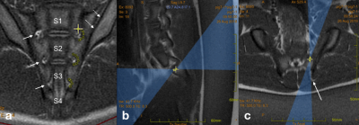
Sacroiliac joint ablation in a chronic swine model using MRgFUS
Viola Rieke, Ellen Liebenberg, Eugene Ozhinsky, Matthew Bucknor, Carol Stillson, Colin Yee, Teri Moore, Roland Krug
There is evidence that MRgFUS might be a very safe and effective minimally invasive technique to treat sacroiliac joint pain caused by arthritis and other degenerative changes. This study in a chronic swine model demonstrated safety and effectiveness of MRgFUS sacroiliac joint ablation and precise ablation of the posterior sacral nerve supply with MRgFUS.
|
|
3807.
 |
128 |
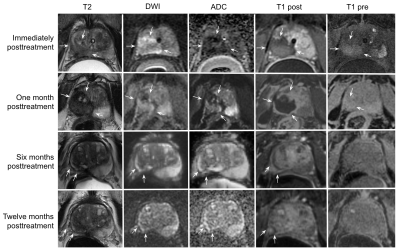
Temporal changes in multiparametric imaging features of the prostate gland following MRgFUS ablation
Signy Holmes, Ryan Brunsing, Rachelle Bitton, Bruce Daniel, Geoffrey Sonn, Pejman Ghanouni
Magnetic resonance guided high intensity focused ultrasound (MRgFUS) can be used for focal therapy of prostate cancer. After treatment, these men remain in active surveillance. Effective monitoring of these men requires an understanding of the expected MRI appearance of the prostate after MRgFUS. Twenty-three patients treated with MRgFUS for focal, intermediate risk MR imaging-visible prostate cancer were followed with serial MRI for 6-24 months. We describe temporal changes in the qualitative and quantitative multiparametric imaging features of the prostate gland post-MRgFUS ablation to aid radiologists to better interpret follow-up examinations to detect residual or new disease.
|
|
3808.
 |
129 |
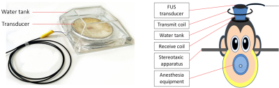
Longitudinal assessment of Focused ultrasound (FUS) induced Blood-Brain Barrier (BBB) opening in the non-human primate under 7T MRI
Kaiyue Wang, Chih-Hung Chai
Microbubble-mediated focused ultrasound can noninvasively induce reversible blood-brain barrier (BBB) opening in both rodents and non-human primates. However, it remains unclear whether FUS-induced BBB opening is accompanied by neuromodulation. Here we longitudinally characterized the duration and the functional effects of FUS-induced BBB opening by measuring changes in contrast-enhanced T1-weighted images (T1-WIs) and blood-oxygen-level dependent (BOLD) responses, respectively. The results show that BBB recovered at 6.5h post-FUS exposure. The blood-oxygen-level dependent (BOLD) changes during the visual stimulus pre- and post- FUS exposure didn't show similar change trend in both hemispheres. The results of fMRI-guided FUS may contribute to the development of FUS-induced BBB opening for clinical applications.
|
|
3809.
 |
130 |
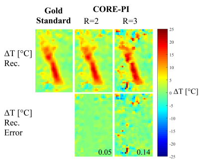
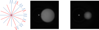
Rapid MR-guided-HIFU using Convolution-based Reconstruction and Parallel Imaging (CORE-PI)
Efrat Shimron, Haim Azhari
A novel reconstruction method for accelerated Magnetic Resonance guided High Intensity Focused Ultrasound (MRgHIFU) thermometry is presented. This method utilizes multi-coil acquisition, k-space undersampling and the recently introduced Convolution-based Reconstruction for Parallel Imaging (CORE-PI) technique. The proposed method utilizes data sparsity in the Stationary Wavelet Transform (SWT) domain. It is a parameter-free, non-iterative and calibrationless method. Retrospective experiments with in-vivo data from clinical human prostate ablation treatments show that the proposed method produces accurate temperature maps from two-fold and three-fold subsampled k-space data. The method is therefore suitable for real time application.
|
|
3810
 |
131 |
Enhancement of the HIFU thermal effect in ex-vivo kidneys using a new class of endovascular sono-sensitizers
Video Permission Withheld
Orane Lorton, Pauline Guillemin, Romain Breguet, Stéphane Desgranges, Laura Gui, François Lazeyras, Antonio Nastasi, Lindsey A. Crowe, Nicolas Taulier, Christiane Contino-Pépin, Rares Salomir
Magnetic Resonance guided High Intensity Focused Ultrasound (MRgHIFU) is a promising approach for the non-invasive ablation of localized tumors. Ablation of highly perfused tumors is challenging due to the heat sink effect. We developed a new concept of endovascular liquid core micro-droplets, used as sono-sensitizers for the enhanced absorption of the HIFU beam. We demonstrated the improvement of the HIFU thermal effect after adjunction of sono-activable micro-droplets in the perfusion fluid of freshly excised viable pig kidneys. Temperature maps were computed using the PRFS method to prove the enhancement of thermal contrast.
|
|
3811.
 |
132 |
Novel Acoustic Coupling Design to Improve MR Imaging Guidance for Focused Ultrasound Surgery
Steven Allen, Thomas Steeves, Eli Vlaisavljevich, Austin Fergusson, Richey Davis, Craig Meyer
We explore the feasibility of an acoustic coupling medium that is invisible to both T2-weighted anatomical scans and MR thermometry scans during MR-guided focused ultrasound surgery and yet still preserves the cooling and acoustic coupling functions normally provided by water.
|
|
3812.
 |
133 |
Detecting T1-based Signal Reduction in Focused Ultrasound Heating of Bone using a 3D Spiral Ultra-Short Echo Time Sequence
Yekaterina Gilbo, Helen Sporkin, Sam Fielden, John Mugler, Grady Miller, Steven Allen, Craig Meyer
MR-guided Focused Ultrasound (MRgFUS) is used transcranially to ablate brain tissue for the treatment of neurological diseases. Temperature monitoring of the skull is desired for increasing treatment safety and efficacy. Proton resonance frequency shift MR fails to detect heating in the cortical bone of the skull. T1-based MR thermometry uses T1 mapping to observe a linear increase in T1 with temperature but requires long acquisitions. We demonstrate a new thermometry method dependent on the linear relationship between signal magnitude from a T1-weighted 3D Spiral Ultra-short Echo Time sequence and test it across three scanners in multiple trials.
|
|
3813.
 |
134 |
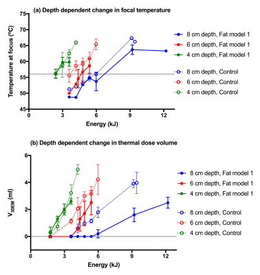
Assessing the effect of pre-focal fat on the feasibility of delivering MR guided high intensity focused ultrasound (MRgHIFU) treatment of intra-pelvic tumors
Sharon Giles, Ian Rivens, Gail ter Haar, Nandita deSouza
Models were used to simulate the pre-focal tissue distributions encountered when treating intra-pelvic tumors with magnetic resonance guided high intensity focused ultrasound (MRgHIFU). Focal peak temperature and thermal dose volumes were considerably affected by depth and fat thickness. Exposures of 300 W for ≥20 s (≥6 kJ) were required to generate measurable 30EM dose contours 8 cm deep (6 cm pre-focal fat). The relative distributions of fat and muscle layers had minimal effect on focal heating, but influenced location of potentially damaging pre-focal heating. MRgHIFU treatments to deep-seated intra-pelvic tumors require methods for improving dose delivery at depth.
|
|
3814.
 |
135 |
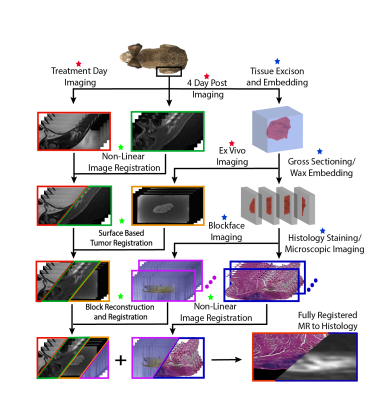
Registration of In Vivo MR images to Volumetric Histopathology
Blake Zimmerman, Sara Johnson, Jill Shea, Henrik Odeen, Elaine Hillas, Candace Winterton, Robb Merrill, Sarang Joshi, Allison Payne
As MR guided focused ultrasound (MRgFUS) treatments evolve to treat oncological diseases, the ability to accurately assess the efficacy of treatment is critical. Although there are several MR metrics proposed for assessing MRgFUS treatments, they have not been rigorously validated against gold standard histopathology treatment assessment. Current validation studies that register MR to histopathology do not comprehensively account for all deformations during histological processing. We present a rigorous MR to histopathology registration pipeline that estimates deformation at every step that can be used to accurately validate the efficacy of oncological MRgFUS treatment.
|
|
3815.
 |
136 |
Transcranial focused ultrasound localization in Money at 3T
Yangzi Qiao, Chao Zou, Jo Lee, Xiaojing Long, Teng Ma, Changjun Tie, Xin Liu, Hairong Zheng
Focused ultrasound has been a fast developing technology for non-invasive neuromodulation. Monkey is a good animal model for studying the mechanism of focused ultrasound on neural network. However, the ultrasound beam would be greatly attenuated and distorted by the monkey skull, making the precise focal localization a crucial step. MR-ARFI with little thermal effect was an attracting tool for focus localization. In this study, we investigated the transcranial MR-ARFI in monkey at 3T system. With high performance of the customer designed monkey head coil and the strong gradient system, reliable and repeatable results were obtained.
|
|
3816.
 |
137 |
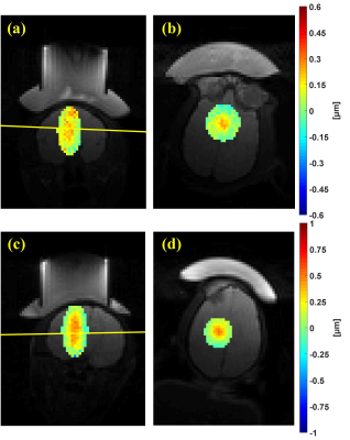
Longitudinal Registration for Voxel-wise Correlation of MRgFUS Treatment Assessment Metrics
Sara Johnson, Blake Zimmerman, Jill Shea, Elaine Hillas, Henrik Odéen, Sarang Joshi, Allison Payne
The acute assessment of MRgFUS therapies with metrics such as thermal dose and contrast-enhanced (CE) imaging have been shown to under- or over-estimate the final ablation lesion as assessed by delayed imaging or histological outcomes. The aim of this study is to longitudinally register MRgFUS treatment images to a final lesion outcome as determined by CE-T1w imaging three days post-treatment for voxel-wise analysis of these errors. MR thermometry (MRTI), cumulative thermal dose (CTD), and acute CE-T1w images are co-registered to Day 3 CE-T1w with millimeter accuracy and assessed for their predictive value.
|
|
3817.
 |
138 |
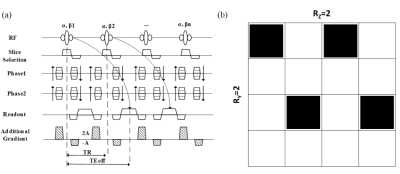
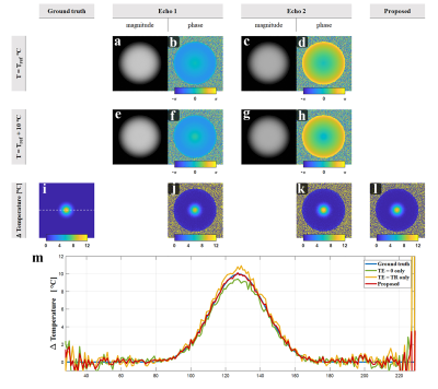
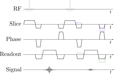
Fast 3D MR thermometry using echo-shifted sequence with parallel acquisition acceleration
Rui Jiang, Chao Zou, Yangzi Qiao, Changjun Tie, Xin Liu, Hairong Zheng
A high-precision, fast MR thermometry is always preferred for HIFU monitoring. However, the coverage of MRT is also important for the safety concern as the unwanted burn usually takes place in the tissue interface which might be far away from the focal area. In this work, we combined 3D echo-shifted GRE sequence with 2D parallel imaging acceleration to implement a fast 3D MRT.
|
|
3818.
 |
139 |
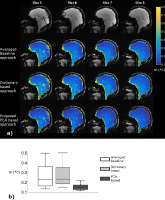
A Principal Component Analysis based Multi-baseline Phase Correction Method for PRF Thermometry
Waqas Majeed, Rainer Schneider, Sunil Patil, Florian Maier, Henrik Odéen, Dennis Parker, John Roberts, Adrienne Campbell, Himanshu Bhat
The use of Proton Resonance Frequency (PRF) based thermometry with thermal therapy procedures is indispensable. Variation in background phase due to motion related changes in B0 is a major source of inaccuracy in PRF thermometry. In this work we propose a novel Principal Component Analysis (PCA) based multi-baseline phase correction approach. We compare this approach with two existing methods using in-vivo human brain and heart data, and demonstrate significant reduction in bias as well as variance of temperature difference estimates. The proposed approach may increase the accuracy of PRF thermometry in or near moving organs, and hence result in improved clinical outcome.
|
|
3819.
 |
140 |
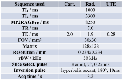
Development of 2D Cartesian, radial and UTE MP2RAGE sequences for fast T1 mapping: application to MR thermometry
Thibaut Faller, Alice Rousseau, Aurélien Trotier, Sylvain Miraux, Emeline Ribot
A surrogate strategy to the Proton Resonance Frequency technique that fails when short T2* tissue are present is the measurement of the longitudinal relaxation times T1. In order to monitor temperature in real-time, a 2D version of the Magnetization Prepared 2 Gradient Echo (MP2RAGE) sequence has been developed, and the cartesian encoding has been replaced by radial ones to obtain an ultra-short Echo Time (UTE). This enabled to measure T1 of short T2*-phantoms and to perform MR thermometry every 8s over a wide range of temperatures and consequently of T1.
|
|
3820.
 |
141 |
Active Tracking-based cardiac triggering of MR thermometry for MRI-guided cardiac ablation
Ronald Mooiweer, Rainer Schneider, Radhouene Neji, Thomas Pohl, Tom Lloyd, Rahul Mukherjee, Mark O'Neill, Reza Razavi, Sébastien Roujol
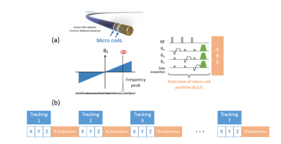
MR thermometry can be used for MRI-guided cardiac ablations, but ECG-triggering is not always effective. We propose to use the Active Tracking (AT) functionality included in the catheter to prospectively trigger the thermometry measurements. Modules measuring the position of the AT microcoil were interleaved with a thermometry sequence, which were repeated until a trigger condition is detected. Experiments showed successful triggering in a beating heart phantom, with a temperature stability of 1.12±0.36°C. For in-vivo interventional application, respiratory motion will need to be taken into account.
|
|
3821.
 |
142 |
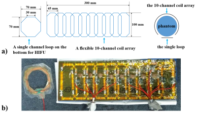
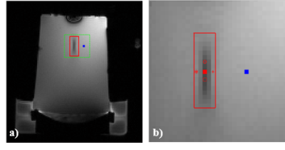
A flexible 11-channel coil array for MR-guided high-intensity focused ultrasound (HIFU) studies on rabbit leg muscle at 3 T
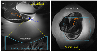
Qiaoyan Chen , Rui Jiang, Changjun Tie, Jianhong Wen, Xing Yang, Chao Zou, Xiaoliang Zhang, Xin Liu, Hairong Zheng, Ye Li
Signal-to-noise ratio (SNR) of the radio frequency (RF) receive coil array is a critical factor affecting the accuracy of temperature measurement in MR-guided high-intensity focused ultrasound (HIFU) for local heating. In this work, a flexible 11-channel coil array was designed, constructed and evaluated for MR-guided HIFU studies on rabbit leg muscle at a 3 T MRI scanner. Compared to a commercial available 4-channel flexible coil array, the dedicated 11-channel coil array provides improved performance in SNR, parallel imaging capability, and the accuracy of temperature measurement.
|
|
3822.
 |
143 |
Comparison of Cartesian MR thermometry approaches for focused ultrasound brain applications
Henrik Odéen, Viola Rieke, Sunil Patil, John Roberts, Himanshu Bhat, Bradley Bolster Jr., Dennis Parker
Magnetic resonance guided focused ultrasound using MR thermometry (MRT) for therapy guidance is a promising thermal therapy for a wide range of neurological disorders. The current MRT method acquires a single 2D slice with low readout bandwidth to increase SNR and MRT precision, compensating for the fact that the body coil is used to receive during treatments. In this work we investigate trade-offs between 2D and 3D GRE and EPI approaches of MRT for brain FUS applications. A comparison between using the body coil and two 4-channel flex coils is also performed.
|
|
3823.
 |
144 |
Improved MR thermometry using saturation bands to suppress water signal
Henrik Odéen, Allison Payne, Viola Rieke, Sunil Patil, Bradley Bolster Jr., Himanshu Bhat, Dennis Parker
MR guided focused ultrasound (MRgFUS) treatments are routinely monitored by MR thermometry (MRT). Image quality and MRT precision can be significantly degraded by water motion in the water bath used to couple the FUS transducer to the target tissue. This problem is especially challenging when the water bath surrounds the target tissue. In this work we investigate the use of saturation bands to suppress the unwanted signal in dedicated breast and brain MRgFUS systems. It is shown that using saturation bands to suppress signal from the water bath significantly reduces artifacts and improves MRT precision with potentially shorter scan times.
|
|
3824
 |
145 |
k-space MR Thermometry Using k-space Energy Spectrum Analysis
Video Permission Withheld
Shenyan Zong, Bruno Madore, Guofeng Shen, Chang-Sheng Mei
The goal of this work was to perform MR thermometry in the k-space domain, as opposed to the image domain, on the premise that a k-space approach might be better suited at capturing spatial trends. The method relies on the fact that spatial gradients in temperature cause k-space shifts in signals for heated materials. Traditional proton resonance frequency (PRF) thermometry was also performed, for validation purposes.
|
|
3825.
 |
146 |
A Dual-Echo bSSFP Imaging Method for Proton Resonance Frequency-Based Thermometry and Analysis of Phase Behavior in bSSFP
Seohee So, JaeJin Cho, Kinam Kwon, Byungjai Kim, HyunWook Park
Proton resonance frequency thermometry is useful to estimate temperature change that is proportional to the resonance frequency change. In this study, we propose a dual-echo bSSFP thermometry method that generates a high intensity signal and linear phase to the frequency shift. Off-centered acquisition in the balanced steady-state free precession creates an imperfect linear phase with respect to the frequency shift. The dual-echo acquisition method compensates for the phase nonlinearity and generates phase information that is linearly proportional to the frequency shift. This phase linearization makes it possible to accurately measure the proton resonance frequency shift caused by temperature change.
|
|
3826.
 |
147 |
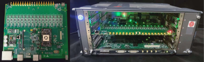
Design, evaluation and application of a 16-channel Frequency Synthesizer Module for Thermal Magnetic Resonance
Haopeng Han, Shuailin Wang, Thomas Eigentler, Lukas Winter, Eckhard Grass, Thoralf Niendorf
Thermal Magnetic Resonance makes use of the physics of the radio frequency fields applied at ultrahigh field magnetic resonance imaging. While UHF-MRI enables measuring temperature in vivo, highly localized power deposition can be achieved by interfering RF waveforms with a shortened wavelength. This constitutes a means for supervised in vivo temperature modulation. The number of RF signals and the signals’ properties greatly affect the heating performance. In this work, a 16-channel frequency synthesizer module was developed as an RF signal source for Thermal MR. Preliminary experiments were conducted to demonstrate that the proposed module is suitable for Thermal MR.
|
|
3827.
 |
148 |
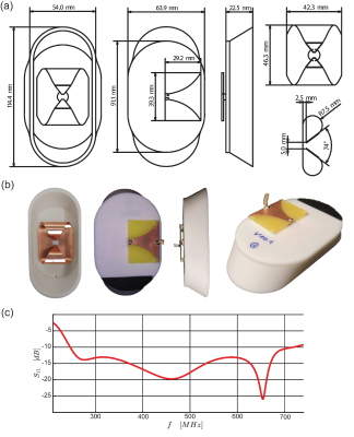
Ultra-Wideband Self-Grounded Bow-Tie Antenna Building Block for Thermal Intervention, Diagnostic MRI and MR Thermometry at 7.0 Tesla
Thomas Eigentler, Lukas Winter, Haopeng Han, Eva Oberacker, Andre Kuehne, Laura Boehmert, Hana Trefna, Thoralf Niendorf
A compact resonator antenna for broadband thermal intervention, diagnostic proton (1H) and fluorine (19F) imaging and MR-Thermometry was developed for operating at 7.0T MRI. The antenna is based on the concept of a self-grounded bow-tie (SGBT) antenna to enable thermal intervention frequencies ranging from 250MHz to 516MHz. The self-grounded bow-tie resonator antenna is smaller and lighter compared to previous dipole-antenna designs, enabling a high-density multi-channel array. The proposed antenna is demonstrated to be suitable for diagnostic imaging, thermal intervention, and MR thermometry.
|
|
3828.
 |
149 |
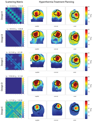
Power Considerations for Radiofrequency Applicator Concepts for Thermal Magnetic Resonance Interventions in the Brain at 297 MHz
Eva Oberacker, Andre Kuehne, Celal Oezerdem, Jason Millward, Cecilia Diesch, Thomas Eigentler, Jacek Nadobny, Sebastian Zschaeck, Pirus Ghadjar, Peter Wust, Lukas Winter, Thoralf Niendorf
There is a pressing need to implement Thermal MR therapies in the brain, particularly to sensitize treatment of aggressive cancers like glioblastoma multiforme. Given the high power transmission regime of Thermal MR therapies, it is crucial to understand the engineering constraints affecting RF power losses, since inaccurate estimates could compromise the efficiency and precision of the therapy. Here we conducted a thorough simulation of five RF applicator designs, using realistic loss estimates of material and electrical components, and considering antenna design, position and coupling. Results from simulated and patient-derived data underscore that clinical requirements must balance with practical engineering constraints.
|
|
3829.
 |
150 |
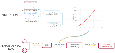
Flip angle optimization and in vitro demonstration of 2D DESPOT1-based fat thermometry
Beatrice Lena, Clemens Bos, Chrit Moonen, Max Viergever, Lambertus Bartels
During MR-HIFU ablations, fat MR thermometry (MRT) is needed for monitoring temperature in adipose tissues (both in target regions and in subcutaneous fat in the near field of the ultrasound beam). It can be based on T1 mapping calculated with 2D-DESPOT1 method and corrected for the FA variation over the slice profile. However, difficulties arise in the choice of 2 flip angles optimized for the whole expected range of T1s in fat MRT. We identify the optimal settings through simulation and demonstrate the feasibility of fat MR thermometry through 2D DESPOT1 sequences in phantom experiments.
|
|
| Top |
Non-Thermal Interventional MRI
Digital Poster
Interventional MRI
Wednesday, 15 May 2019
| Exhibition Hall |
13:30 - 14:30 |
| |
|
Computer # |
|
3830.
 |
151 |
Performance of an aerosol jet-deposited wireless resonant marker: in vitro temperature measurements and in vivo visualization
Caroline Jordan, Bradford Thorne, Arjun Wadhwa, Eugene Ozhinsky, Vincent Fratello, Sravani Kondapavulur, Aaron Losey, Teri Moore, Carol Stillson, Colin Yee, Ronald Watkins, Greig Scott, Alastair Martin, Xiaoliang Zhang, Mark Wilson, Steven Hetts
Endovascular catheter-based procedures under MRI can be challenging as standard fabrication methods for markers are rigid and bulky, and new microfabrication methods need more analysis on their tracking characteristics. We analyzed a wireless resonant circuit tracking marker that was printed using aerosol jet deposition on a polymer catheter. In a phantom, we acquired bSSFP sequences and a B1+ map, and measured temperature using probes and MR thermometry. In vivo, in the carotid arteries, we acquired GRE sequences and a B1+ map. The marker demonstrated good signal, with minimal temperature increases, suggesting that these markers have good tracking characteristics.
|
|
3831.
 |
152 |
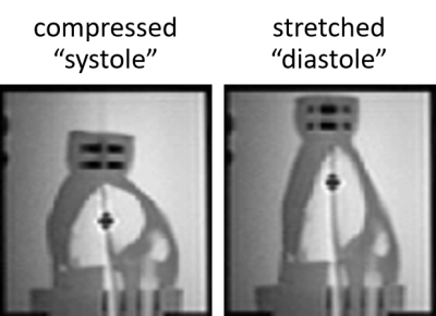
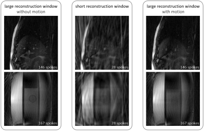
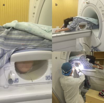
Motion-Adaptive Temporal Resolution for Radial Real-Time Imaging at a Low-Field MR-Linac
Florian Friedrich, Philipp Mann, C. Spindeldreier, Peter Bachert, Mark Ladd, Sebastian Klüter, Benjamin Knowles
Hybrid MRI linear accelerators (MR-linacs) enable real-time image guidance during radiotherapy. Under real-time MRI, a compromise must be found between spatio-temporal resolution and SNR, whereas to precisely track tumour position, both should be maximised. The presented method implements a motion-adaptive image reconstruction based on a golden-angle radial acquisition scheme. This allows producing SNR-optimised images under periods of small motion and images optimised for temporal resolution when motion is larger. The technique was implemented at a low-field MR-linac (0.35T) to image a free-breathing volunteer and a motion phantom.
|
|
3832.
 |
153 |
Interleaved White Marker Contrast with bSSFP Real-Time Imaging for Deep Learning based Needle Localization in MR-Guided Percutaneous Interventions
Jonathan Weine, Rainer Schneider, Urte Kägebein, Bennet Hensen, Frank Wacker, Florian Maier
Automatic localization of needles in real-time images can facilitate MR-guided percutaneous interventions. It enables automatic slice repositioning and targeting support and, thus, allows for faster workflows. The improvement of deep learning based passive needle tracking by using both, anatomical and positive contrast images as input was investigated. A prototype bSSFP sequence for interleaved acquisition of k-space lines for conventional and positive contrast with Cartesian readout was implemented and evaluated ex-vivo and in-vivo. The U-Net segmentation algorithm showed superior performance when using both contrasts. In conclusion, this method is a promising approach for robust needle localization in real-time interventional workflows.
|
|
3833.
 |
154 |
A simple freehand technique for MRI-guided needle localization of suspicious breast lesions
Presentation Not Submitted
Qing Zhang, Zhiguo Zhuang, Dandan Zhang, Jianrong Xu, Jia Hua
A total of 273 patients who received needle localization with a bare-hand technique for suspicious breast lesions by using 1.5 T MR-guided were included in our study. The puncture method was similar to CT-guided needle localization of lung nodule. The success rate of localization was 100% (273/273). Procedure time of all the cases ranged from 5 to 30 min (mean, 14.3 min). Our MRI-guided bare-hand needle localization of suspicious breast lesions is a handy, safe, rapid, and accurate interventional method. The lesions that are located at axillary region of breast,areola area, and the special area near the chest wall can all be precisely localized.
|
|
3834.
 |
155 |
Highly Undersampled Radial Passive Marker Tracking with Phase-only Cross-Correlation (POCC) for Real Time Image Guidance
Andreas Reichert, Simon Reiss, Michael Bock, Michael Vogele, Axel Krafft
Marker tracking with phase-only cross correlation (POCC) is an efficient technique to localize instruments during percutaneous MR-guided interventions in closed-bore MR systems. Unfortunately, POCC is time-consuming, as additional marker images need to be continuously acquired. Here, we present a drastically accelerated POCC sequence that uses radially undersampled images to detect the passive marker. With a modified POCC algorithm this sequence can detect marker movements within less than 108ms thus providing a very fast position feedback. The precision of the POCC sequence was evaluated in phantom measurements, and a possible in vivo targeting scenario was demonstrated in a volunteer.
|
|
3835.
 |
156 |
Radial Simultaneous Multi Slice Imaging for Marker Tracking
Andreas Reichert, Axel Krafft, Michael Bock
The phase-only cross correlation (POCC) algorithm efficiently and accurately detects passive MR markers used as needle guides. In a POCC sequence the marker is automatically detected in two cross-sectional images to continuously visualize the planned needle trajectory during motion. Image acquisition of the marker, however, is very time consuming which degrades the temporal resolution. Here, it is shown that two simultaneously acquired, highly undersampled radial images - together with the consideration of the point-spread-function in the POCC algorithm - can track the marker at substantially shorter acquisition times.
|
|
3836.
 |
157 |
MRI Driven Augmented Reality Image-Guidance Platform for Myocardial Cell Delivery
Mitchell Doughty, Jill Weyers, Xiuling Qi, Graham Wright, Michael Laflamme, Nilesh Ghugre
Myocardial infarction is a leading cause of heart failure, a condition with a 5-year mortality rate of ~50%. Current HF management techniques aim to slow disease progression rather than improve function. Cardiac regenerative medicine through cell-based approaches offers the potential to repopulate non-contractile scar tissue. However, regions of scar are not well delineated during the cell-delivery procedures, and accurate cell placement is vital to realize positive outcomes from therapy. We aim to develop a novel integrative image-guidance solution combining MRI for myocardial scar characterization with augmented reality via a head-mounted display to visualize critical MRI information intraoperatively and guide delivery.
|
|
3837.
 |
158 |
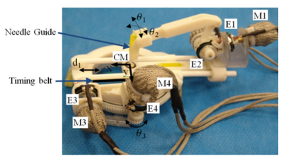
Phantom Study of Novel Biopsy Needle and Assisted Robotic System Designed for Prostate Biopsy Procedure under MRI
Davut Mahcicek, Dursun Yildirim, Gokce Kasaci, Ozgur Kocaturk
In this study, a novel MRI guided prostate biopsy device was designed, produced and tested under MRI. In this scope, a MRI compatible, remote controlled, transrectal biopsy delivery system, a MRI compatible biopsy gun, a MRI compatible, biopsy needle which can be tracked under MRI, and a computer based control unit were designed and a prototype was produced. The robotic prostate biopsy device prototype was tested under MRI with in-vitro experiments using a commercially available prostate phantom.
|
|
3838.
 |
159 |
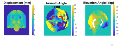
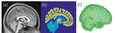
Ten Minutes for the Brain to Settle: an In-vivo Investigation of Positional Brain Shift
Stefano Zappala, Nicholas Bennion, Slawomir Kusmia, Jing Wu, Sam Evans, Derek Jones, David Marshall
Positional brain shift (BS), as the physiological sagging of the brain under the effect of gravity, has a magnitude that is comparable to the accuracy of neuronavigational systems. It is a complex interaction of gravity, anatomical boundaries and tissue mesostructure. A comprehensive investigation of such deformation is needed. This study aims to provide a rich set of volumetric measures to infer the pattern of deformation as well as the time evolution of positional BS. Results show that positional BS is a relatively fast process, stabilised in 10 minutes, with local variations strongly dependent on the variability of anatomical structures between different subjects.
|
|
3839
 |
160 |
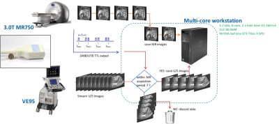
Motion tracking using simultaneous MR and 4D ultrasound acquisition for image guided radiation therapy
Video Permission Withheld
Thomas Foo, Bo Wang, Jhimli Mitra, David Mills, L Scott Smith, Heather Chan, Aqsa Patel, Shourya Sarcar, Eric Fiveland, Warren Lee, Sydney Jupitz, Alan McMillan, James Holmes, Wes Culberson, Michael Bassetti, Andrew Shepard, Bryan Bednarz
An MR-compatible 4D ultrasound probe allows hands-free, simultaneous MR and ultrasound image acquisition. This new imaging capability provides a path for tracking tumor target motion during radiation therapy, as an alternative to an integrated MR-LINAC system. To facilitate this, the ability to track the motion of fiducial markers as an indication of respiratory state is essential. In our approach, as the MR images are acquired outside of the radiation therapy procedure, motion tracking of endogenous ultrasound fiducials is proposed to determine respiratory states.
|
|
3840.
 |
161 |
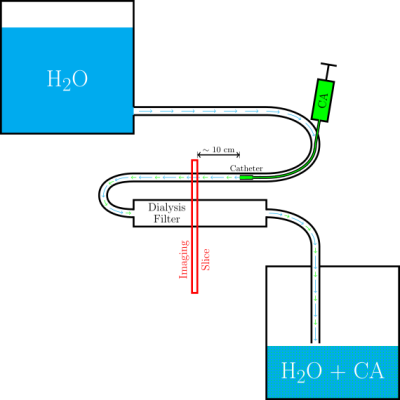
Catheter-based Arterial Input Function for Quantitative Perfusion Measurements with Intra-arterial Injection
Simon Stephan, Simon Reiss, Thomas Lottner, Ali Özen, Michael Bock
MR-guided vascular interventions allow for direct injection of contrast agent (CA) through a catheter, which can be used for perfusion measurements of the target tissue, for example, the myocardium in MR-guided coronary catheterizations. For a quantitative perfusion measurement an arterial input function (AIF) is required that describes the CA concentration-time curve in the artery. In this work we show initial results of two methods to determine the AIF during an intervention and demonstrate the feasibility of measuring the AIF in intra-arterial CA injections based on the signal of an active guiding catheter.
|
|
3841.
 |
162 |
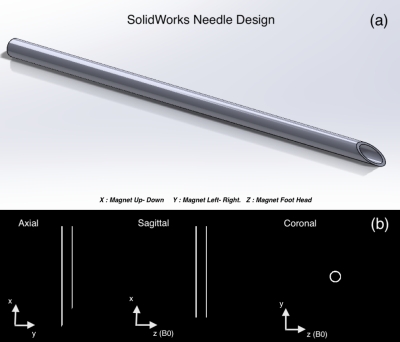
Modeling of Active Shimming of Metallic Needles for Interventional MRI
Saikat Sengupta
Needle artifacts caused by large magnetic susceptibility differences between metallic needles and stylets and the surrounding tissue are a persistent problem in interventional MRI. In this abstract, we present the concept, design and modeling results of a active shim system for needles inspired from degaussing coils used in naval vessels. Field disturbance induced by a Titanium needle at 3 Tesla is modeled and an active orthogonal shim coil insert design is presented to demonstrate shimming of the field variation around the needle. This work lays the foundation for designing full generalized active shim systems for Interventional MRI probes.
|
|
3842.
 |
163 |
A Machine Learning Based Biomechanical Model for Real-time MR-guided Neuro-intervention
Suhao Qiu, Alexa Singer, Changxin Lai, Blanca Zufiria, Danni Wang, Yao Li, Bomin Sun, Yiping Du, Zhi-Pei Liang, Yuan Feng
Magnetic resonance (MR) guided neuro interventions could be combined with robotic assisted manipulation to achieve optimal performance. Patient specific model constructed from MR images of the brain could have the best biophysical fidelity but suffers from high computational cost. For real-time applications, we proposed to construct an Artificial Neural Network (ANN) based on the training from computational outputs of Finite element Analysis (FEA). Results demonstrate the ability to achieve accurate predictions given by a mean square error (MSE) of 0.0338 mm2 within 10ms.
|
|
3843.
 |
164 |
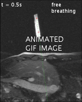
MRI artifact simulation for clinically relevant MRI sequences for guidance of prostate HDR brachytherapy
Ellis Beld, Marinus Moerland, Max Viergever, Jan Lagendijk, Peter Seevinck
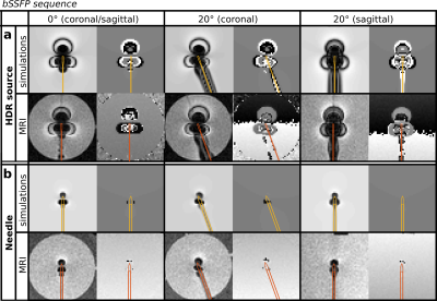
Object localization by MRI artifact simulation and template matching is valuable for MRI-guided HDR brachytherapy. Simulations of the artifacts induced by an HDR brachytherapy source and titanium needle were implemented for four types of MRI sequences: spoiled gradient echo, spin echo, bSSFP and bSSFP-SPAIR. The simulated artifacts were compared to MR images acquired in a phantom study and applied for object localization. High correspondences between the simulations and MR images were found as well as only slight variations between the obtained object positions for all applied sequences. This enables object localization for clinically relevant MRI sequences which allow anatomy visualization.
|
|
3844.
 |
165 |
Real-time Automatic Tip Tracking of Gadolinium-filled Balloon Wedge Catheter during MR-guided Cardiac Catheterization
Rohini Vidya Shankar, Radhouene Neji, Phuoc Duong, Bram Ruijsink, Kuberan Pushparajah, Reza Razavi, Sébastien Roujol
MRI-guided cardiac catheterisation procedures are commonly performed using balloon wedge catheters which are currently manually tracked. Furthermore, these procedures require frequent manual adjustment of the slice positioning during catheter navigation. In this study, we sought to develop a novel acquisition and reconstruction framework that enables automatic tracking of a gadolinium-filled balloon wedge catheter using (a) real-time partial saturation with increased spatial coverage, (b) automatic image-based estimation of the catheter balloon position, and (c) real-time slice tracking. The proposed framework is demonstrated during an MRI-guided cardiac catheterisation experiment in a 3D printed heart phantom.
|
|
3845.
 |
166 |
Ferromagnetic markers for interventional MRI devices at 0.55T
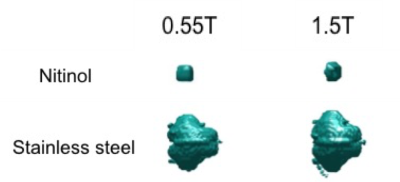
Burcu Basar, Merdim Sonmez, Ram Paul, Ozgur Kocaturk, Daniel Herzka, Robert Lederman, Adrienne Campbell-Washburn
Interventional cardiac MRI has been hampered for decades by the lack of safe and visible devices. Guidewires (long, elongated metals) risk RF-induced heating under MR, and often incorporate ferromagnetic markers (e.g.stainless steel) that can be used for image guidance. We propose MR-guided interventions at 0.55T to avoid RF-induced heating. Here we show that at 0.55T, ferromagnetic markers maintain visibility and create identical artifacts to those observed at 1.5T. We show that using a high-performance low field MRI system may enable progress in interventional MRI due to improved device safety with retained image quality.
|
|
3846.
 |
167 |
Clinical MRI-guided right heart catheterization with standard metallic devices using a high performance 0.55T system
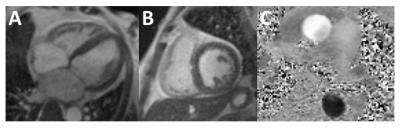
Adrienne Campbell-Washburn, Toby Rogers, Jaffar Khan, Burcu Basar, Rajiv Ramasawmy, Laurie Grant, William Schenke, Delaney McGuirt, Annette Stine, Daniel Herzka, Robert Lederman
MRI-guidance of invasive cardiovascular procedures has been limited by the unavailability of safe devices. Most cardiovascular procedures require long conductive devices (eg. guidewires and catheters) that are susceptible to RF-induced heating. Because heating is related quadratically to field strength, we propose to reduce the MRI field strength to 0.55T. Using a custom high-performance 0.55T MRI system, we performed MRI-guided catheterizations on patients with commercial metallic guidewires using standard high flip angle bSSSP real-time imaging to produce good image quality and contrast without RF-induced heating. Low field MRI combined with high-performance hardware will be fundamentally enabling for MRI guidance of invasive procedures.
|
|
3847.
 |
168 |
Proof-of-concept of retrospective gating for interventional cardiac MRI using catheter microcoils readings.
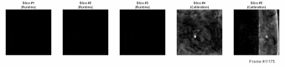
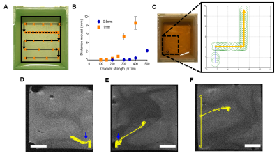
Marylène DELCEY, Pierre BOUR, Valery OZENNE, Wadie BENHASSEN, Bruno QUESSON
In the context of radiofrequency ablation of cardiac arrhythmia, a catheter is inserted into the heart for contact electrophysiology recording and/or ablating the arrhythmogenic substrate. In this context, we propose to exploit MR-compatible microcoils located near the catheter tip to estimate the local motion of the heart. For this, a tracking module was interleaved with segments of a radial acquisition in a FLASH sequence. k-space segments were sorted retrospectively according to tracking readings before image reconstruction. Using this approach, artifact free images of a pig in vivo could be reconstructed.
|
|
3848.
 |
169 |
3D CNN-based synthetic CT generation for head and neck radiotherapy using a clinically used turbo spin-echo T2-weighted sequence
Mateusz Florkow, Anna Dinkla, Frank Zijlstra, Matteo Maspero, Mark Savenije, Patricia Doornaert, Marijn van Stralen, Marielle Philippens, Cornelis van den Berg, Peter Seevinck
Deep learning-based synthetic CT (sCT) generation models are often based on T1-weighted gradient echo sequences. However, these sequences are generally not used for tumor/organs-at-risk delineation. In this study, we trained a U-Net type neural network using T2-weighted turbo spin-echo images from the clinical radiotherapy treatment planning protocol originally used for tumor/organs-at-risk contouring. The use of clinical images preserves scan times and facilitates soft tissue delineation on the source images of the sCTs, avoiding registrations. We showed that sCTs generated by the trained model provide accurate dosimetric results while limiting CT-induced streaking dental artefacts.
|
|
3849.
 |
170 |
MINIMA – Minimally invasive, image guided ablation using MRI
Presentation Not Submitted
Chris Payne, Rebecca Baker, Matin Mohseni, John Connell, Peter Patrick, Yichao Yu, Bernard Siow, Mark Lythgoe, Quentin Pankhurst
Minimally invasive, focal therapies aim to deliver more effective treatment to the patient while reducing off target effects. As such, we have developed MINIMA, a minimally invasive, image-guided ablation MRI technique, whereby a magnetic thermoseed can be manoeuvred through tissue, localised with real time imaging and then heated to cause cell death. We demonstrate submillimetre precision of movement through ex vivo brain tissue and that the seed can be heated via application of radiowaves to cause well defined regions of cell death. These results show that MINIMA is a promising new technique that could help transform MRI into a brand new theranostic device.
|
|
3850.
 |
171 |
Imaging latency of Golden Angle 2D MRI using real-time GPU-accelerated image reconstruction for MR-guided radiotherapy
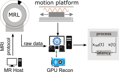
Pim Borman, Bas Raaymakers, Markus Glitzner
For tumor tracking in MRI-guided radiotherapy it is important to minimize the latency between the moment of anatomic change and its appearance on the MR image. By using a 2D golden angle sampling trajectory in combination with a sliding window reconstruction, the latency can be decoupled from the frame rate, yielding frame rates of up to one repetition time. We implemented a real-time GPU-accelerated reconstruction pipeline where k-space data is directly streamed to a reconstruction server during acquisition. Using this we investigated the influence of the sliding window width on the latency, reconstruction time, frame rate and image quality.
|
|
3851.
 |
172 |
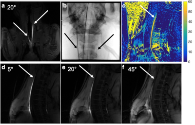
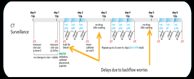
Feasibility and Demonstration of Controlled, Comprehensive Delivery of Clot-busting Drugs for MR Image-Guided Intracerebral Clot Evacuation
Robert Moskwa, Miles Olsen, Jen Meudt, Robby Weyker, Morgan McCue, Terry Oakes, Ethan Brodsky, Dhanansayan Shanmuganayagam, Azam Ahmed, Walter Block
A recent NIH Trial has shown success in minimizing the effects of intracerebral hemorrhage through insertion of a minimally invasive catheter into the clot through a craniotomy. The catheter then injects 1 ml of clot-busting drugs followed by an 8 hour period of draining the lysed blood. The conservative procedure is repeated for 3-5 days as only very small dosages can be administered due to CT’s inability to directly monitor the drug’s safe distribution. We demonstrate how pressurized infusions can create broader treatments that lyse the clot rapidly while MRI visualizes the drug distributions to maintain safety.
|
|
3852.
 |
173 |
EVALUATION OF FPI BASED CUSTOM FORCE SENSOR DESIGN FOR BIOPSY NEEDLES
Dogangun Uzun, Okan Ulgen, Ozgur Kocaturk
In this work, an MRI compatible Fabry-Pérot Interferometry (FPI) based force sensor is designed and fabricated using novel methods, and integrated into an 18-gauge nitinol biopsy needle. The resolution of the FPI based sensor is increased by coating optical fibers with magnesium and titanium thin film. The force sensing needle is tested by in vitro experiments using a prostate phantom and in vivo animal experiments under MRI.
|
|
3853.
 |
174 |
A Body-Mounted Robot with Integrated Single Loop Coil for MR-Guided Arthrography
Reza Monfaredi, Wolfgang Loew, Christopher Ireland, Viktoriya Beskin, Ronald Pratt, Randy Giaquinto, Charles (Chuck) Dumoulin, Kevin Cleary, Karun Sharma
The purpose of this project is to streamline the workflow for robotic-assisted MR-guided arthrography by integrating a single loop coil in the base / mounting adapter of the robot. This coil provides sufficient spatial coverage and sensitivity to localize anatomic points of interest and registration fiducials embedded in the robot. Integration of the coil with the robot places the imaging coil as close as possible to the patient and reduces the number of devices that need to be managed during an interventional procedure. Quantitative results for SNR and an end-to-end targeting study using a phantom are reported in the abstract.
|
|
3854.
 |
175 |
Automatic pelvic bone registration of low-field MRI to pre-operative 3T-MRA for vascular interventions
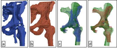
Derk Slotman, Jordy van Zandwijk, Frank Simonis, Ferdi van der Heijden, Robert Geelkerken, Bennie ten Haken
Intra-operative low-field magnetic resonance imaging (LF-MRI) is characterized by poor spatial resolution limiting its use in endovascular catheter tracking with fluoroscopy. Fusion of LF-MRI with 3T-MRI may overcome this issue. To fuse both volumes, we automatically segmented a pelvis from an LF-MRI scan, based on cortical bone detection with Laplacian edge detection. A similarity of 66% was reached between automatic and manual segmentation. Subsequently, the segmented volume was automatically registered to a 3T-MRA. Compared to a Procrustes based manual registration, the automatic registration resulted in a root-mean-square error of 3 mm.
|
|
Back to Program-at-a-Glance |  Back to Top Back to Top |
| The International Society for Magnetic Resonance in Medicine is accredited by the Accreditation Council for Continuing Medical Education to provide continuing medical education for physicians. |
